Embed Size (px)
Citation preview
Capillary electrophoresis in analysis of DNA variations in
rectal cancer.
A thesis for the doctor philosophiae degree
by
Annette Torgunrud Kristensen
Oslo, February 2007

© Annette Torgunrud Kristensen, 2007
Series of dissertations submitted to the Faculty of Medicine, University of Oslo No. 516
ISBN 978-82-8072-436-6
All rights reserved. No part of this publication may be reproduced or transmitted, in any form or by any means, without permission.
Cover: Inger Sandved Anfinsen. Printed in Norway: AiT e-dit AS, Oslo, 2007.
Produced in co-operation with Unipub AS. The thesis is produced by Unipub AS merely in connection with the thesis defence. Kindly direct all inquiries regarding the thesis to the copyright holder or the unit which grants the doctorate.
Unipub AS is owned by The University Foundation for Student Life (SiO)

Acknowledgment
This work has been carried out in the Department of Surgical Oncology at the Norwegian
Radium Hospital.
I would like to express gratitude to my scientific supervisor Per Olaf Ekstrøm for
introducing me to the field of cancer research and for invaluable and patient help with the
studies and manuscript preparation.
I highly appreciate the collaborations with all my co-authors. Special thanks are given to
Johan Wiig for supplying tumor material, lavage fluid, clinical data and comments on the
manuscripts. Jens Bjørheim for helping with various manuscripts, and sharing fruitful
discussions. Stein Larsen for supplying tumor material and lavage fluid.
I thank the head of the department Karl-Erik Giercksky for research ideas and providing the
research facilities and for being my link to the Faculty of Medicine.
I wish to thank present and former members of the Surgical research group David
Hinselwood (for comments on the manuscript), Karen-Marie Heintz (technical instrumental
support and comments on several manuscript), Torveig Weum Abrahamsen (technical
assistance and comments on the manuscript). Special thanks go to all the people in Gustav
Gaudernack’s research group for creating a great scientific and social environment and for
their encouragement and support.
Finally, I would like to thank my family for always being there for me.
This Thesis is dedicated to my husband and our two children.
February 2007
Annette Torgunrud Kristensen


5
Acknowledgment ............................................................................................................ 3
List of papers................................................................................................................... 6
Abbreviations .................................................................................................................. 7
General introduction...................................................................................................... 9
Colorectal cancer ......................................................................................................................................... 9Treatment and outcome ....................................................................................................................... 10
Genetic model for colorectal cancer........................................................................................................ 11Colorectal adenoma-carcinoma sequence model ............................................................................ 11Differences between the left- and the right side of colon ................................................................ 12Peritoneal cavity .................................................................................................................................... 14Histopatholgically stages...................................................................................................................... 15
Signal pathways and biological markers ................................................................................................ 17Ras-pathway with the MAPK-kinase pathway .................................................................................. 17The TP53-Bax pathway........................................................................................................................ 18Folate metabolism................................................................................................................................. 19Ataxia telangiectasia and the ATM gene ........................................................................................... 20
Methods to detect DNA variants .............................................................................................................. 21Denaturant gradient gel electrophoresis and related techniques................................................... 21Application of Denaturant capillary Electrophoresis (DCE) ............................................................ 24
Methodological considerations................................................................................................................. 32
Aims of this project...................................................................................................... 35
Results in brief .............................................................................................................. 36
Discussion...................................................................................................................... 39
Biological considerations .......................................................................................................................... 39
Instrumentation considerations ................................................................................................................ 43
Future Instrumentation perspectives....................................................................................................... 45
Conclusion ..................................................................................................................... 46
Further perspectives ................................................................................................... 46
Reference List ............................................................................................................... 47
6
List of papers
IKristensen AT, Bjørheim J, Minarik M, Giercksky K-E, Ekstrøm PO: Detection of
mutations in exon 8 of TP53 by temperature gradient 96-capillary array
electrophoresis. Biotechniques 2002, 33: 650-653.
IIBjørheim J, Abrahamsen TW, Kristensen AT, Gaudernack G, Ekstrøm PO: Approach to
analysis of single nucleotide polymorphisms by automated constant denaturant
capillary electrophoresis. Mutat Res 2003, 526: 75-83.
III
Kristensen AT, Bjørheim J, Wiig J, Giercksky KE, Ekstrøm PO: DNA variants in the
ATM gene are not associated with sporadic rectal cancer in a Norwegian population-
based study. Int J Colorectal Dis 2003.
IVKristensen A.T, Wiig JN, Larsen SG, Giercksky KE, Ekstrøm PO. Association study of
DNA variations in genes of the folate metabolism with rectal cancer in a Norwegian
population. Submitted January 2007.
V
Kristensen A.T, Wiig JN, Larsen SG, Giercksky KE, Ekstrøm PO. Molecular detection (k-
ras) of exfoliated tumour cells in the pelvis is a prognostic factor after resection of
rectal cancer? Submitted February 2007.

7
Abbreviations
5-FU 5- Flurouracil A AdenineACDCE Automated Constant Denaturant Capillary Electrophoresis AJCC American Joint Comitee on Cancer APC Adenomatosis Polyposis Coli AS-PCR Allele Specific Polymerase Chain Reaction AT Ataxia Telangiectasia ATM Ataxia Telangiectasia Mutated gene ATP Adenosine Triphosphate BAX BCL2-associated X protein BCL2 B-cell CLL/lymphoma 2 BP Base Pair BRAF v-raf murine sarcoma viral oncogene homolog B1 BRCA Breast cancer geneC CytosineCASP5 Caspase 5, Apoptosis-related Cysteine Peptidase CBS Cystathionine -synthaseCDCE Constant Denaturant Capillary Electrophoresis CDGE Constant Denaturant Gel Electrophoresis CE Capillary Electrophoresis CHIP Children's Hospital Informatics Program CIN Chromosome Instable CRC Colorectal Cancer CTCE Cycling Temperature Capillary Electrophoresis CTNNB1 Catenin, Beta-1 DCC Deleted in Colorectal Carcinoma DCE Denaturant Capillary Electrophoresis DGGE Denaturing Gradient Gel Electrophoresis DHFR Dihydrofolate Reduktase dHPLC denaturant High Performance Liquid Chromatography DNA Deoxyribonucleic acid DPD Dihydropyrimidine Dehydrogenase DSBs Double Stranded Breaks dsDNA double stranded DNA dTMP Deoxythymidine 5'-phosphate dUMP Deoxyuridine Monophosphate ERK Extracellular Signal–regulated Kinase FAP Familial Adenomatous Polyposis Fas/Apo1 Integral membrane protein with killer domain G GuanineGC-clamp Guanine and Cytosine rich domain GDP Guanosine Diphosphate GRB2 Growth factor receptor-bound protein 2 GTP Guanosine Triphosphate HNPCC Hereditary Non-Polyposis Colorectal CancerIn vivo (with)in the living organism

8
Killer/DR5 Death receptor 5, DR5, KILLER, KILLER/DR5 K-RAS Kirsten RAS gene, Ki-RAS LOH Loss of Heterozygozity MADH2, 4 MAD (mothers against decapentaplegic, Drosophila) homolog
2,4MAPK The Mitogen-Activated Protein Kinase MB MegaBaceMEK Mitogen Extracellular signal–regulated Kinase MEKK MAP Kinase Kinase Kinase MIN Microsatellite Instability MLH1 MutL, E. Coli, homolog of, 1 MS Methionine Synthase MSH 2,3,6 mutS homolog 2, 3 and 6 MTHFR 5,10-Methylenetetrahydrofolate Reductase (NADPH). MTRR Methionine Synthase ReductaseNCBI National Center for Biotechnology Information PCR Polymerase Chain Reaction RFLP Restriction Fragment Length Polymorphism PIK3CA Phosphatidylinositol 3-kinase, Catalytic, Alpha polypeptide RAF Serine/theronine Phosporylation Cascade RAS Rat Sarcoma virus RFLP Restriction Fragment Length Polymorphism SAM S-adensylmethionine SNP Single Nucleotide Polymorphism SHMT Serine Hydroxymethyltransferase SOS Son Of Sevenless SSCP Single Strand Confirmation Analysis ssDNA single stranded DNA T Thymidine TCF4 Transcription Factor 4 TGFBR2 Transforming Growth f\Factor, Beta receptor II TGGE Temperature Gradient Gel Electrophoresis THF Tetrahydrofolate TME Total Mesorectal Excision TNF beta Transforming growth factor, beta receptor I TNM Tumour, Node, Metastasis staging system TP53 Tumour protein p53 TS Thymidylate Synthase TTGE Temporal Temperature Gel Electrophoresis UV Ultraviolet

9
General introduction
Colorectal cancer
Cancer develops through accumulation of several different genetic changes in the cell. The
suggestion that cancer was a genetic disorder in somatic cells, was first presented in the end
of the last 19th century by Hansemann [1]. Some years later in 1914 Boveri [2] proposed
that chromosome abnormalities were fundamental to the development of cancer. During the
last decade, numerous studies have been published, showing that several oncogenes,
tumour-supressor and repair genes are important in colorectal cancer. These genes have an
important impact as prognostic factors in colorectal cancer, and may also be essential to
improved treatment and outcome for the patient.
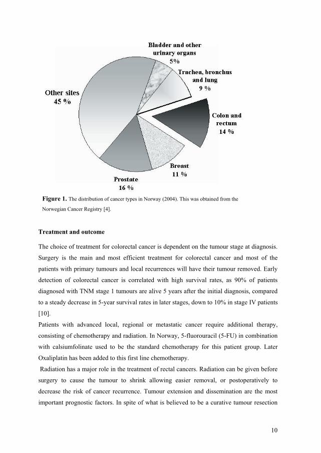
Colorectal cancer is one of the most common malignant diseases in the Western countries,
and increased incidence has been observed over the past decades [3]. In the Norwegian
population, the Cancer Registry of Norway Institute of Population-based Cancer Research
recorded 3482 new cases of colon (2345) and rectum (1137) cancer in 2004 [4]. Cancer in
the colon- and rectum compose 15% of all the new cancer cases, and is the most prevalent
cancer site within the Norwegian population. Rectal cancer has a male/female incidence
ratio of 1.5, while colon cancer has a male/female ratio incidence of 1.1 [4]. Cancer in the
colon and the rectum mostly affect the elderly and the age specific incidence of rectal cancer
increases sharply after the age of 50 years [5]. Some factors contribute to the increased risk
of colon and rectal cancer. Familial Adenomatous Polyposis (FAP) is one of the known
autosomal dominant inherited colorectal cancer (CRC) diseases, but accounts for less than
1% of all cases. This is a disease recognised by formation of polyposis at a young age [6].
Hereditary non-polyposis colorectal cancer (HNPCC), also known as Lynch syndrome is
another autosomal dominant cancer syndrome and is the most common form of hereditary
colorectal cancer accounting for as much as 5 % of all CRCs [7]. HNPCC typically
develops around the age of 45. Another factor causing increased risk of colon and rectum
cancer is chronic inflammatory diseases such as Ulcerative colitis and Crohns disease [8].
Patients that have received pelvic radiation in association with gynaecological cancers have
been observed to have an increased risk 2-3 times compared to that of a normal population
[9].
10
Treatment and outcome
The choice of treatment for colorectal cancer is dependent on the tumour stage at diagnosis.
Surgery is the main and most efficient treatment for colorectal cancer and most of the
patients with primary tumours and local recurrences will have their tumour removed. Early
detection of colorectal cancer is correlated with high survival rates, as 90% of patients
diagnosed with TNM stage 1 tumours are alive 5 years after the initial diagnosis, compared
to a steady decrease in 5-year survival rates in later stages, down to 10% in stage IV patients
[10].
Patients with advanced local, regional or metastatic cancer require additional therapy,
consisting of chemotherapy and radiation. In Norway, 5-fluorouracil (5-FU) in combination
with calsiumfolinate used to be the standard chemotherapy for this patient group. Later
Oxaliplatin has been added to this first line chemotherapy.
Radiation has a major role in the treatment of rectal cancers. Radiation can be given before
surgery to cause the tumour to shrink allowing easier removal, or postoperatively to
decrease the risk of cancer recurrence. Tumour extension and dissemination are the most
important prognostic factors. In spite of what is believed to be a curative tumour resection
Figure 1. The distribution of cancer types in Norway (2004). This was obtained from the
Norwegian Cancer Registry [4].

11
many patients later suffer from metastases or tumour recurrence. Possible causes of local
recurrences are believed to be; incomplete resection of the primary carcinoma [11],
insufficient removal of involved regional lymphatic vessels or nodes [12], implantation of a
secondary tumour near the suture line and exfoliated colorectal cancer cells released during
the surgical procedure [13].
Survival from rectal cancer has been continuously improved during the past decades. The
main reason for this seems to be the development of the surgical technique called total
mesorectal excision (TME) [14;15]. An additional local national contributing factor was the
establishment of The Rectum Cancer Registry at The Cancer Registry of Norway [4;16].
The main element in this initiative was an educational program to standardize and optimize
surgery for rectal cancer at a national level. At the same time pathologists were educated in
the optimal principles of handling and describing rectal cancer specimens. All surgical
departments treating rectal cancer were invited to transfer their clinical data to this registry.
Each department regularly receives its own results together with the national average for
comparison and quality control.
Genetic model for colorectal cancer
Colorectal adenoma-carcinoma sequence model
Development of colorectal cancer occurs through a number of genetic and epigenetic
changes, and was described as an adenoma–carcinoma sequence in 1975 by Muto et al. [17].
Almost twenty years later these stages were used in a genetic model put forward by Fearon
and Vogelstein in 1990 [18]. This model describes the stepwise progression from normal to
dysplastic epithelium to carcinoma associated with multiple genetic alterations. The authors
stressed that mutations in at least four to five regions, including Adenomatous Polyposis
Coli (APC), Kirsten RAS gene (k-ras), deleted in colorectal cancer (DCC) and Tumour
protein gene p53 (TP53) were required for formation of a malignant colorectal tumour.
Further studies have shown that the order of genetic events is essential [19;20]. This concept
not only provides an excellent model to study the genesis of invasive cancer, but also offers
a means of preventing colorectal cancer by endoscopic removal of precursor lesions.

12
Differences between the left- and the right side of colon
CRC’s that arise proximal (right) or distal (left) are different with respect to
epidemiological, clinicopathological, biochemical as well as genetic factors [21]. This lead
to the suggestion that proximal and distal tumours follow broadly different molecular
pathways in carcinogenesis. Two distinct pathways have been identified which result in
colorectal cancer. The tumour suppressor pathway, also termed the chromosomal instability
pathway (CIN), account for 85% of the colorectal carcinomas and most sporadic
carcinomas. The CIN pathway is characterized by loss and gains of chromosomes
(aneopleoidy) often 5q, 17p and 18q as well as loss of heterozygozity (LOH), which is a
loss of one of the parental alleles present in cells, and mutations in p53. The CIN pathway
most often appears in the distal part of the colon.
The second mutational pathway, the microsatelitte instability (MIN) pathway accounts for
the remaining 15% of colorectal carcinomas [22]. This pathway often has a family history of
colorectal cancer suggesting a genetic contribution or common environmental exposure
among family members, or a combination thereof. Existence of multiple alternative genetic
pathways have been suggested [23;24]. The MIN pathway is characterized by defects in the
mismatch repair systems, leading to downstream mutations in genes such as TGFBR2
(transforming growth factor- ) [25], BAX (BCL2-associated X protein) [26], k-ras (Kirsten
RAS gene), APC (adenomatosis polyposis coli) [27] and candidate target genes such as
WISP-3 [28]. Tumours with MIN have some clinicopathological features in common, like
proximal location in the colon [29]. MIN are also more likely to be present at an advanced
age [30] and to be associated with female gender [31].
13
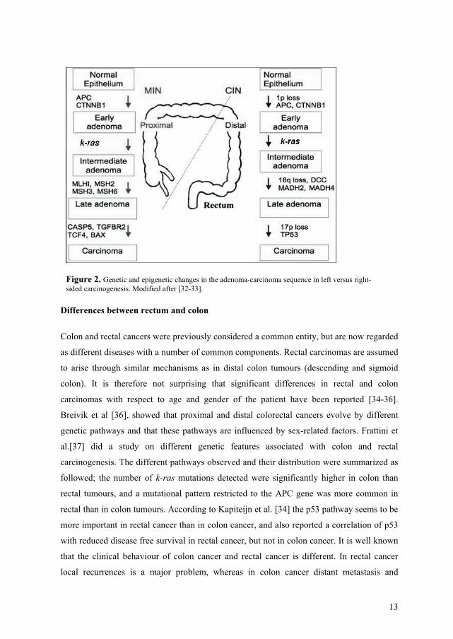
Differences between rectum and colon
Colon and rectal cancers were previously considered a common entity, but are now regarded
as different diseases with a number of common components. Rectal carcinomas are assumed
to arise through similar mechanisms as in distal colon tumours (descending and sigmoid
colon). It is therefore not surprising that significant differences in rectal and colon
carcinomas with respect to age and gender of the patient have been reported [34-36].
Breivik et al [36], showed that proximal and distal colorectal cancers evolve by different
genetic pathways and that these pathways are influenced by sex-related factors. Frattini et
al.[37] did a study on different genetic features associated with colon and rectal
carcinogenesis. The different pathways observed and their distribution were summarized as
followed; the number of k-ras mutations detected were significantly higher in colon than
rectal tumours, and a mutational pattern restricted to the APC gene was more common in
rectal than in colon tumours. According to Kapiteijn et al. [34] the p53 pathway seems to be
more important in rectal cancer than in colon cancer, and also reported a correlation of p53
with reduced disease free survival in rectal cancer, but not in colon cancer. It is well known
that the clinical behaviour of colon cancer and rectal cancer is different. In rectal cancer
local recurrences is a major problem, whereas in colon cancer distant metastasis and
Figure 2. Genetic and epigenetic changes in the adenoma-carcinoma sequence in left versus right-sided carcinogenesis. Modified after [32-33].

14
carsinomatosis are the most important problems. This again suggests that the cause factors
and the molecular basis may differ between colon and rectal cancer. When prognostics
markers are investigated in larger series, such differences in ethiology and biological
behaviour between colon and rectal cancer should be considered.
Peritoneal cavity
The peritoneum is a serous abdominal membrane, which lines both the abdominal wall and
the intra-abdominal viscera. Although the sheet of body tissue ultimately forms one
continuous sheet, two types or layers of peritoneum are usually referenced to as well as the
potential space between them: The outer layer, called the parietal peritoneum, is attached to
the abdominal wall. The inner layer, the visceral peritoneum, is more or less wrapped
around the internal organs located inside the abdominal cavity. The potential space between
these two layers is the peritoneal cavity [38]. The term mesentery is often used to refer to a
double layer of visceral peritoneum. Blood vessels, lymphnodes and nerves are located
between these layers. It should be noted that the space between these two mesenterial layers
is technically outside of the peritoneal sac, and not within the peritoneal cavity see Figure 3.
Figure 3. This picture show horizontal disposition of the peritoneum in the lower part of the abdomen.
http://education.yahoo.com/reference/gray/illustrations/figure?id=1038 (This figure has a GNU Free
Documentation License).

15
The initial dissemination of rectal cancer tumours cells occurs through three routes: lymphs,
portal blood, and peritoneal surfaces. Although lymphatic and hematogenous metastases
indicate an aggressive disease process, it is possible that dissemination to the peritoneal
surfaces may be the result of direct contamination with cancer cells of the parietal and
visceral peritoneum. This suggests that viable tumour cells with proliferative and perhaps
metastatic potential had been shed from the primary tumour site either before removal of the
tumour or during surgical resection [39]. Patients with overt peritoneal or local metastases
from colorectal cancer have a poor prognosis [40]. However, aggressive treatments by
surgery and infusion of intra-peritoneal chemotherapy have been tried and appear to benefit
selected patients [41].
Histopatholgically stages
The goal of staging is to determine the extent and location of the tumour in order to develop
appropriate treatment strategies. The staging of rectal cancer closely approximates the
staging of colon cancer. Dukes staging was introduced in 1932 [42] to characterise the
extent of tumour dissemination. This placed the cancer into one of three categories (Stages
A, B, C). Later the system was modified to include a fourth stage (Stage D) [43]. Many
other staging systems have been proposed trying to define a scheme which is more
predictive, e.g. The Gunderson-Sosin modification of the Astler-Coller system [44] and Jass
et al. [45]. More recently, the American Joint Committee on Cancer (AJCC) has introduced
the TNM staging system, which places the cancer into one of four stages (Stages I-IV) [46].
Listed below in Table 1 are the Duke and TNM staging systems.
Dukes’Stages
Diseasetype Stage
TNM staging
T N MA Local I T1, T2 N0 M0 B Local II T3-T4 N0 M0 C Regional III T1-T4 N1-N2 M0 D Distant IV T1-T4 N1-N2 M1 Table 1. This table show TNM staging. T1-4; the increasing spread of primary
tumour, N; Lymph node metastasis, M; distant metastasis.
16
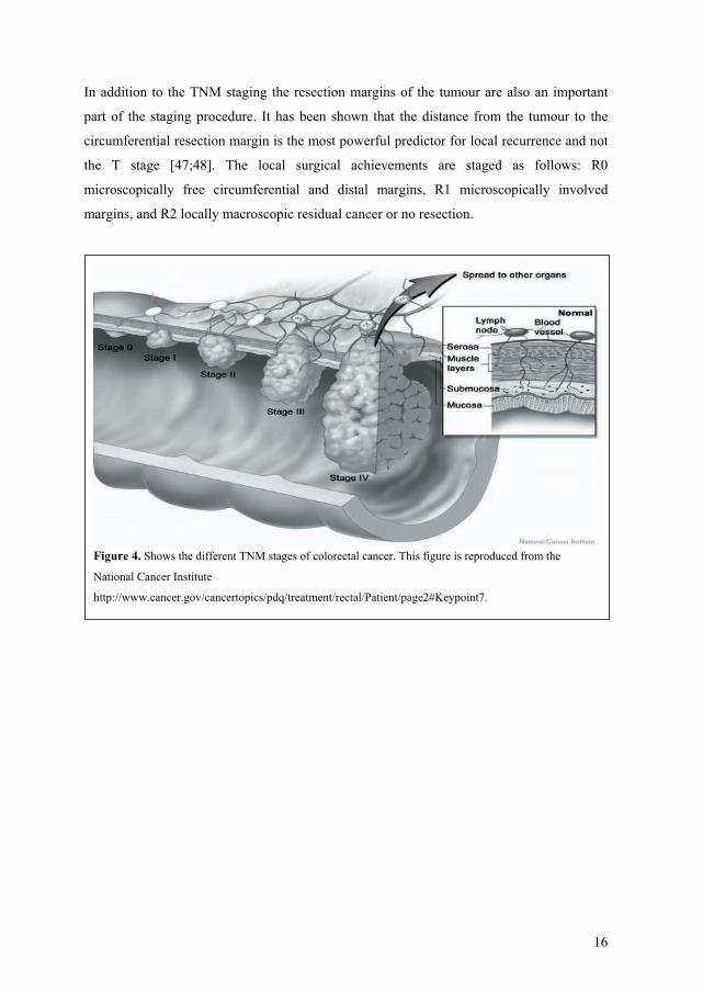
In addition to the TNM staging the resection margins of the tumour are also an important
part of the staging procedure. It has been shown that the distance from the tumour to the
circumferential resection margin is the most powerful predictor for local recurrence and not
the T stage [47;48]. The local surgical achievements are staged as follows: R0
microscopically free circumferential and distal margins, R1 microscopically involved
margins, and R2 locally macroscopic residual cancer or no resection.
Figure 4. Shows the different TNM stages of colorectal cancer. This figure is reproduced from the
National Cancer Institute
http://www.cancer.gov/cancertopics/pdq/treatment/rectal/Patient/page2#Keypoint7.

17
Signal pathways and biological markers
Selected genes from three signalling pathways have a central role in this thesis. The first one
is the Ras-pathway with the MAPK-kinase pathway. One major function of the ras protein
family is to couple growth factors to the Raf-mitogen-activated protein (MAP) kinase
kinase-MAP kinase signal transduction pathway, which leads to the nuclear expression of
early response genes. The mutated k-ras product maintains a prolonged state of activation
leading to cellular proliferation. Mutations in k-ras is described as an early event in the
process of colorectal carcinogenesis [18]. Another important pathway is the TP53-BAX
pathway, which is shown to be responsible for the progression of the colorectal tumour [19].
The last pathway described in this thesis is the folate metabolism. This metabolism is
thought to contribute to colorectal carcinogenesis by altering both DNA methylation and
nucleotide synthesis. Genes coding for metabolism enzymes, receptor proteins or protein
target of chemotherapy agents often present different genetic polymorphisms able to
influence drug sensitivity, toxicity and dosing [49].
Ras-pathway with the MAPK-kinase pathway
A large number of oncogenes have been identified in human tumours, including colon
cancer. The k-ras gene is the most commonly mutated RAS family member in colon cancer,
although N-RAS mutations are also observed in a small percentage of colon cancers [50].
The RAS family genes encode a highly conserved family of 21-kDa proteins involved in
signal transduction. Ras serves as a GDP/GTP related binary switch with a crucial role in
the transmission of growth regulating signals within the organism, and may be used as a
critical terminal through which signals are passed on to other signalling modules (see Figure
5). The best characterized of the signalling pathways regulated by Ras is the mitogen-
activated protein kinase (MAPK) pathway. This pathway is activated when Ras activates a
Serine/theronine Phosporylation Cascade (Raf). Raf thereafter phosphorylates and activates
a mitogen extracellular signal–regulated kinase (MEK), which phospyrates a mitogen-
activated protein kinase (MAPK). This in turn activates an extracellular signal–regulated
kinase (ERK) that plays pivotal roles in a wide variety of developmental cellular processes,
including growth, division, and differentiation [51].
The mutated k-ras product maintains a prolonged state of activation leading to cellular
proliferation. Novel activating mutations in sporadic CRC have recently been identified on
major kinase encoding genes such as BRAF [52] and PIK3CA [53]. The presence of these
18
activating point mutations, including the well-characterized k-ras oncogene mutations, is
represented in up to 75% of cases of CRC. These genes, which have been implicated in the
adenoma-carcinoma transition, cause deregulation and constitutive activation of the MAP
AKT/kinase pathways, rendering growth advantages to colon tumour cells [54;55]. Mutated
k-ras may also inhibit the TP53-BAX signalling pathway by phosporylating pro-caspase-9,
and thereby inhibiting cythochrome c induced apoptosis [19;51].
The TP53-Bax pathway
The TP53-Bax pathway is important in DNA damage, cell survival and proliferation. TP53
gene codes for a protein that acts as a transcription factor and serves as a key regulator of
the cell cycle, coupling stimuli that promote cell division to those that promote cell death
[57]. Mutations in the TP53 tumour suppressor gene are found at high frequency in a wide
range of human cancers [58]. TP53 is also in control of Bax, which is a pro-apoptopic
member of the BLC-2 family. It reduces cytochrome c released from mitochondria resulting
in apoptosis (see Figure 6).
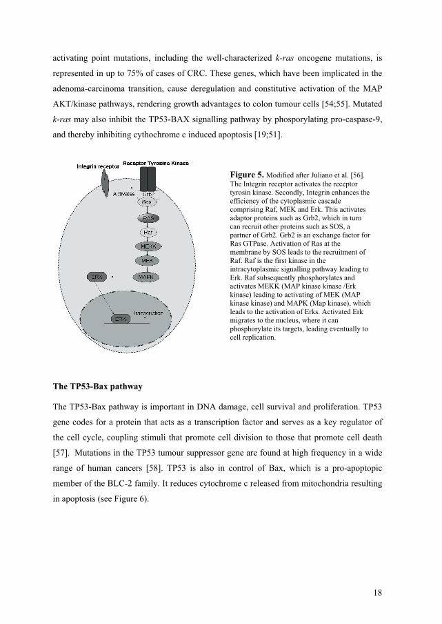
Figure 5. Modified after Juliano et al. [56]. The Integrin receptor activates the receptor tyrosin kinase. Secondly, Integrin enhances the efficiency of the cytoplasmic cascade comprising Raf, MEK and Erk. This activates adaptor proteins such as Grb2, which in turn can recruit other proteins such as SOS, a partner of Grb2. Grb2 is an exchange factor for Ras GTPase. Activation of Ras at the membrane by SOS leads to the recruitment of Raf. Raf is the first kinase in the intracytoplasmic signalling pathway leading to Erk. Raf subsequently phosphorylates and activates MEKK (MAP kinase kinase /Erk kinase) leading to activating of MEK (MAP kinase kinase) and MAPK (Map kinase), which leads to the activation of Erks. Activated Erk migrates to the nucleus, where it can phosphorylate its targets, leading eventually to cell replication.

19
Folate metabolism
Folate is a water-soluble B vitamin. The primary function of folate is as a coenzyme in
reactions that require transfer of a single carbon moiety in different oxidative states as a
methenyl, methylene, or methyl group. These reactions typically involve synthesis of
compounds such as thymidine, a pyrimidine base necessary for synthesis of DNA.
Consequently, folate status could potentially be perturbed by polymorphisms in genes
important in the folate metabolism (see Figure 7). Folate deficiency could also affect
malignancy by causing DNA hypomethylation and proto-oncogenic activation by inducing
uracil misincorporation during DNA synthesis, leading to catastrophic DNA repair, DNA
strand breakage and to chromosome damage [49;59]. A deficiency in folate plays an
important role in the pathogenesis of anemia [60], atherosclerotic cardiovascular diseases
[61], neural tube defects [62] neuropsychiatric and cognitive disorders [63] and cancer
[64;65]. Colon cells are subject to rapid turnover, and thus are the site of high rates of DNA
synthesis. Several studies have shown a link between low folate status and indicators for
DNA damage [59;66], and that dietary folate intake could reduce the risk of colon cancer
[67;68].
In cancer treatment it is well known that there are inter individual differences in tumour
response and normal tissue toxicity [69-72]. Many clinical variables with inter individual
differences have been associated with e.g. age, gender, diet and drug-drug interactions.
Figure 6 TP53-Bax pathway.
P53 transcritionally activates Bax,
and integral membrane proteins
Fas/APO1 and Killer/DR5. These
integral membrane proteins are
death receptors and activation of
these results in activation of the
caspase cascade. Cythocrome C
released by Bax also activates the
caspase cascade, which in turn
activates endonuclease and
thereafter DNA cleavage.
This Figure was modified from
[57].
20
However the major clinical variable is the DNA variation in genes of different populations.
Genes that code for metabolism enzymes, receptor proteins or protein targets of
chemotherapy agents often present different polymorphisms that can influence drug
sensitivity and optimal dosing.
Ataxia telangiectasia and the ATM gene
Appropriate cellular signalling responses to DNA damage and the ability to repair DNA are
fundamental processes required for an organism’s survival. Ataxia telangiectasia (AT) is a
rare neurodegenerative disease that results from defective DNA damage signalling [73].
This disease is characterized by neurological and immunological symptoms, sensitivity to
radiation, and cancer predisposition. AT patients have a higher incidence of breast cancer
and leukaemia than the general population and about one-third of patients develop a clinical
significant malignancy during their lifetime [74]. The incidence of AT heterozygous
individuals in the general population is estimated to be 0.5–1% [75-77]. Understanding the
molecular basis of AT has provided many critical insights into the cellular response to DNA
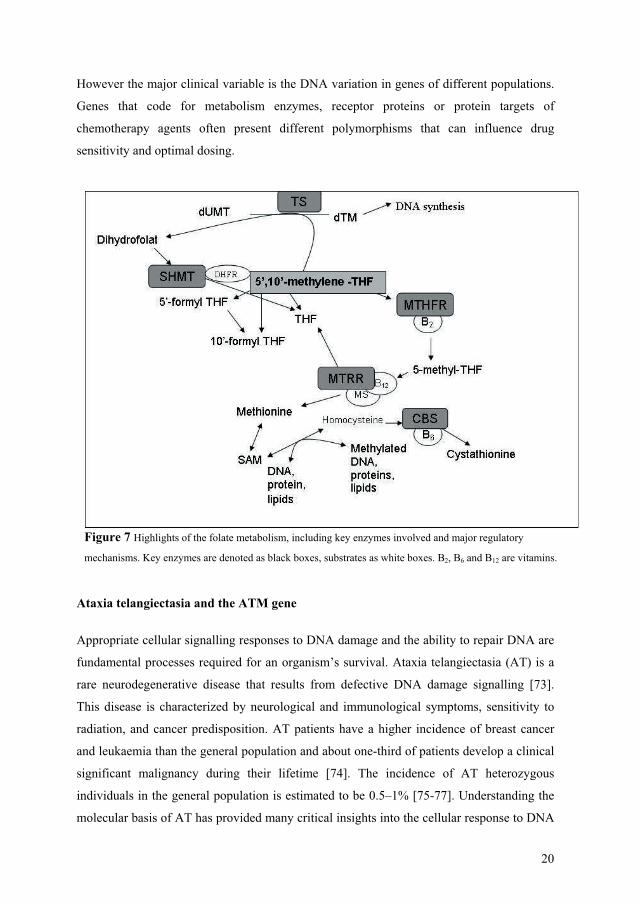
Figure 7 Highlights of the folate metabolism, including key enzymes involved and major regulatory
mechanisms. Key enzymes are denoted as black boxes, substrates as white boxes. B2, B6 and B12 are vitamins.

21
double-strand breaks (DSBs). DSBs are the major DNA lesion leading to chromosomal
aberrations and reliable repair is crucial for maintaining genomic stability. The gene
mutated in AT, the ATM gene, has a role as DSB detector. The ATM gene encodes a protein
kinase that is mainly distributed in the nucleus of proliferating cells. It appears to play a
central role in cell cycle regulation, DNA repair and apoptosis and control of cellular
responses to DNA damage [78]. The ATM gene has been fully elucidated [79] and the
complete genetic sequence is known [80]. However, it’s large size (146 kb) and complex
62-exon structure has greatly complicated the process of screening large sample sets for all
possible sequence variations. DNA variations in the ATM gene have been studied in
colorectal cancer [81-83]. These studies showed that expression of ATM could predict
survival in colorectal cancer, and increase radio sensitivity.
Methods to detect DNA variants
One of the major trends in post-genomic research is the exploration of genetic variation in
humans. The variation of a single base may lead to change in cellular behaviour, and this
change could serve as a diagnostic marker or a target for future therapeutic intervention
[84]. The completion of the Human Genome Project in 2003, and sequencing of the entire
genome in more than one individual, made it clear that there are major individual
differences in the genome, especially between different ethnic populations. Worldwide
efforts to collect DNA variations have lead to several public databases. Validation of DNA
variations and estimation of their allele frequencies are useful, because these markers can be
used in genetic studies [85]. A high-throughput capacity is crucial to complete the large
studies necessary for reports of such genetic studies.
Denaturant gradient gel electrophoresis and related techniques
Many techniques are now available for discovery and scoring of DNA variations. One group
of methods, the melting gel techniques have proven to be amenable and powerful tools for
analysing DNA variations. The reason for melting gel techniques popularity rests upon the
well-documented theoretical foundation of these methods, and the few laboratory steps that
are needed in the analysis in combination with high specificity and sensitivity. Denaturant
Gradient Gel Electrophoresis (DGGE) has been the most frequently used melting gel
electrophoresis technique and was first described by Fischer and Lerman [86]. DGGE
detects mutations (small deletions and insertions, point mutations) by separating PCR
22
amplified DNA fragments, which differ from wild-type DNA in their melting behaviour, on
a denaturing gradient gel. Fragments analyzed by DGGE must have appropriate melting
characteristics, namely a low temperature melting domain and a single contiguous high
temperature melting domain. A melting domain is defined as a region of DNA that melts
cooperatively at approximately the same temperature or concentration of denaturant. The
melting characteristics of a fragment can be predicted from its primary sequence, and
computer algorithms are available to perform this analysis [87]. To include a larger part of
the genome (those without a natural high temperature domains) applicable to this method
Myers et al. [88] has reported that a GC rich 40-basepair region attached to one of the
primers during initial PCR amplification increases the number of fragments that can be
analysed with melting gel techniques. Thus constructing a high melting domain adjacent to
the target sequence of the fragment. However there are some fragments with a single base
alteration where no thermodynamic difference between the wild-type homoduplex and
mutant homoduplex is achieved, even though a GC- rich content is attached [89]. These
fragments will not separate the homoduplexes. However heteroduplexes in the low
temperature domain of a fragment will resolve from the corresponding homoduplexes, so
that the mutation fraction can be seen. To determine the nature of the variant DNA
sequence, the heteroduplex peak can be collected and subsequently sequenced [90;91].
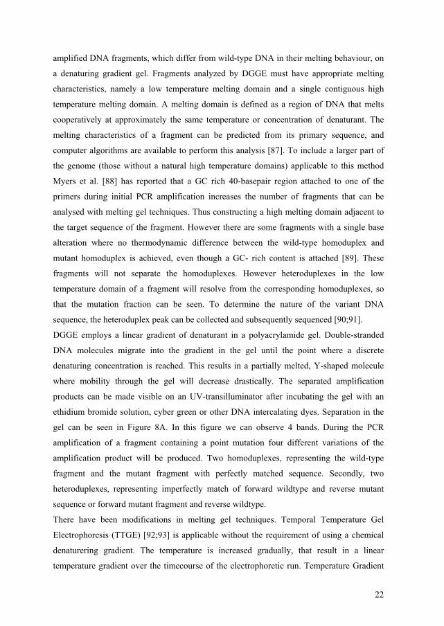
DGGE employs a linear gradient of denaturant in a polyacrylamide gel. Double-stranded
DNA molecules migrate into the gradient in the gel until the point where a discrete
denaturing concentration is reached. This results in a partially melted, Y-shaped molecule
where mobility through the gel will decrease drastically. The separated amplification
products can be made visible on an UV-transilluminator after incubating the gel with an
ethidium bromide solution, cyber green or other DNA intercalating dyes. Separation in the
gel can be seen in Figure 8A. In this figure we can observe 4 bands. During the PCR
amplification of a fragment containing a point mutation four different variations of the
amplification product will be produced. Two homoduplexes, representing the wild-type
fragment and the mutant fragment with perfectly matched sequence. Secondly, two
heteroduplexes, representing imperfectly match of forward wildtype and reverse mutant
sequence or forward mutant fragment and reverse wildtype.
There have been modifications in melting gel techniques. Temporal Temperature Gel
Electrophoresis (TTGE) [92;93] is applicable without the requirement of using a chemical
denaturering gradient. The temperature is increased gradually, that result in a linear
temperature gradient over the timecourse of the electrophoretic run. Temperature Gradient
23
Gel Electrophoresis (TGGE) [94] provides a temperature gradient instead of a chemical
gradient.
Constant Denaturant Gel Electrophoresis (CDGE) [95] is another modification where the
method has the advantage of enabling the fragments to migrate at a consistently different rate
through the whole gel. This allows greater separation between mutant and wild-type
fragments [95-97].
Modification of the CDGE to the capillary platform was first published by Khrapko in 1994
[98;99] (see Figure 8B). The application of Constant Denaturant Capillary Electrophoresis
(CDCE) was first developed on a laboratory-assembled instrument [100]. Benefits of
capillary-based electrophoresis (CE) include fast sample runs and data storage capability
and increased limit of detection; in addition, only minimal amounts of sample are required
with CE techniques. Elevating the temperature of a section of the capillary change the
denaturing conditions for CDCE. Various methods have been used to apply temperature
gradients to capillaries including the use of a voltage ramp [98], an external heating plate
[101], Peltier-based control of surrounding liquid [102] and heated air [103]. A further
improvement of CDCE allowing two-point detection and automated fraction collection has
been reported [104;105]. Conversion of CDCE into regular commercial capillary DNA
sequencing instruments has resulted in automated and standardized protocols [106].
Virtually any commercial single or multi capillary instrument may be used. The automation
allows for rapid analysis of a large number of samples for a short period of time, with no
interference by the operator. The method was first described with the use of an ABI 310
Genetic Analyzer, in which up to 48 samples could be analysed without need of the operator
[107]. This method has also been adapted to the Megabace 1000, allowing for analysis of
96 samples within 40 minutes [108;109], ABI 3100 genetic analyzer [110], a SCE2410 24-
capillary instrument from Spextrumedix [103;105], Beckman Coulter eight capillary
(unpublished data) and tested on MegaBACE 4000 with 384 capillaries (unpublished data).
The temperature control for the method has been improved by modifying the CE system
with the use of a temperature gradient [109].
Samples are analyzed with a temperature gradient starting above and declining beneath the
optimal separation temperature, controlled by the computer software. This modification
compensates for the temperature differences in the capillary chamber resulting in a robust
method. Additional improvement of the gradient is accomplished by cycling the gradient
several times around the optimal separation temperature [111;112].

24
Application of Denaturant capillary Electrophoresis (DCE)
Mutation analysis
It is widely accepted that human cancer is a genetic disorder caused by sequential
accumulation of mutations in oncogenes and tumour suppressor genes. These tumour-
specific mutations in cellular processes underlying tumorigenesis have proven to be useful
for diagnostic and therapeutic purposes. In the past, the selection of genes chosen for
mutational analyses in cancer has been guided by information of known functional attributes
of individual genes or gene families. With the determination of the human genome sequence
it is now in principle possible to examine the cancer cell genome in a comprehensive and
unbiased manner. Such an approach not only provides the means to discover other genes that
DCE
4
3
2
1
DGGE
1 2 3 4
Figure 8. This figure shows the
adaption of DGGE (A) to
Denaturant Capillary
Electrophoresis (DCE) (B), where
the picture of a gel with 4 bands
(nr 4 and 3 heteroduplexes 2;
mutant and 1; wild type)
corresponds to the
electropherogram obtained by
DCE.
A
B

25
contribute to tumorigenesis, but can also lead to mechanistic insights that are only evident
through a systems biological perspective. One consideration of mutation detection analysis
is the possibility of detecting mutations in a small fraction of samples. To study rare variants
in the human population or new somatic or germline mutations, a technique is required
which can detect and separate any and all of the variety of mutations in a target gene at
fractions from the wild-type DNA. This can be important for early detection of malignant
diseases, detection of remaining cells after surgery and possibly for prognosis and outcome
of the disease. Several different mutation analyses have been performed by DCE
[90;92;100;104;105;113-119]. A major improvement to the method was conversion to
commercially automated capillary instruments. The first report was on a single capillary
instrument; the ABI 310 Genetic Analyzer, where mutations of k-ras exon 1 [120] and exon
5-8 in TP53 gene were analyzed [107]. The same mutation analyses have also been
performed on the commercial MegaBACE 1000 instrument, [108;109;111;121;122], and a
SCE2410 from Spextrumedix [103;105].
Single Nucleotide Polymorphism (SNP)
Single nucleotide polymorphisms or SNPs are DNA sequence variations that occur when a
single nucleotide (A, T, C or G) in the genome sequence is altered. For a variation to be
considered a SNP, it must occur in at least 1% of the population. SNPs, which make up
about 90% of all human genetic variation, occur on average once every 87 bp along the 3-
billion-base human genome. 8 354 954 SNPs are currently registrated in the CHIP
Bioinformatic tool database [123;124], however only 60 % are validated, 11% of these
SNPs have frequency data and 40 % of the SNPs are located in genes. Two of every three
SNPs involve the replacement of cytosine (C) with thymine (T). SNPs can occur in both
coding and noncoding regions of the genome. Although more than 99% of the human DNA
sequence is identical in all human beings, variations in DNA sequence can have a major
impact on how humans respond to disease, drugs and other therapies. This makes SNPs of
great value in biomedical research and in developing pharmaceutical products or medical
diagnostics. SNPs are also evolutionarily stable. This makes SNPs a popular tool to
discriminate between alleles or haplotypes in population studies. The DCE method has
proven to be a robust method for genotyping alleles and also detection of new SNPs. Target
sequences require only PCR amplification followed by allele separation by capillary
electrophoresis. The genotypes of the individuals SNPs are scored based on co-migration to
26
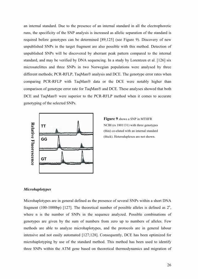
an internal standard. Due to the presence of an internal standard in all the electrophoretic
runs, the specificity of the SNP analysis is increased as allelic separation of the standard is
required before genotypes can be determined [89;125] (see Figure 9). Discovery of new
unpublished SNPs in the target fragment are also possible with this method. Detection of
unpublished SNPs will be discovered by aberrant peak pattern compared to the internal
standard, and may be verified by DNA sequencing. In a study by Lorentzen et al. [126] six
microsatelittes and three SNPs in two Norwegian populations were analysed by three
different methods; PCR-RFLP, TaqMan analysis and DCE. The genotype error rates when
comparing PCR-RFLP with TaqMan data or the DCE were notably higher than
comparison of genotype error rate for TaqMan and DCE. These analyses showed that both
DCE and TaqMan were superior to the PCR-RFLP method when it comes to accurate
genotyping of the selected SNPs.
Microhaplotypes
Microhaplotypes are in general defined as the presence of several SNPs within a short DNA
fragment (100-1000bp) [127]. The theoretical number of possible alleles is defined as 2n,
where n is the number of SNPs in the sequence analyzed. Possible combinations of
genotypes are given by the sum of numbers from zero up to numbers of alleles. Few
methods are able to analyze microhaplotypes, and the protocols are in general labour
intensive and not easily automated [127;128]. Consequently, DCE has been optimized for
microhaplotyping by use of the standard method. This method has been used to identify
three SNPs within the ATM gene based on theoretical thermodynamics and migration of
TT
GG
GTR
elative Fluorescence
Figure 9 shows a SNP in MTHFR
NCBI (rs 1801131) with three genotypes
(thin) co-eluted with an internal standard
(thick). Heteroduplexes are not shown.
27
various fragments [125]. Another study has also used this method to detect 3
microhaplotypes in the CTLA4 gene [126]. A review by Szantai et al. [129] summarizes the
recent examples of novel and emerging haplotype techniques by capillary electrophoresis
based on DNA fragment analysis, with DCE as one of the methods mentioned.
Gene copy number
Pooling designs are used in screening experiments in molecular biology. In some
applications, the property to be screened is defined on subsets of items, instead of on
individual items. Pooled genotyping is a powerful and efficient tool for high throughput
association analysis, both case-control and family based [130]. The use of a pooling design
may reduce the consumables and labour cost of a study compared with genotyping
individuals and counting alleles. Pooling allows a far smaller number of PCR reactions and
genotype assays than are used when genotyping individuals. The most important
consideration with DNA pooling is to ensure that the individuals that make up a pool
contribute equal amounts of DNA, from which robust PCR results can be obtained.
However random experimental errors in the constitution of DNA pools and in the
measurement of allele frequencies from pooled DNA should be taken into account in
statistical analysis [131]. Differential amplification occurs for many SNPs and this bias
should also be corrected for in the estimate of allele frequencies from pooled DNA. Several
reports have stated that multiple genotyping techniques are suitable for pooled genotyping
[132;133]. Pooled DNA has been used in studies of various diseases, such as head and neck
carcinogenesis [134], breast cancer [135], Liddle syndrome [136] and rheumatoid arthritis
[137]. The DCE method has also been shown to be an effective and cost- beneficial method
for single and pooled samples [110;119;138]. There is no need for any correction of the
signal of the separated alleles because both alleles are separated with the same flourophore
and because fragments of the same length with a difference of one base pair will pass the
detector with the same velocity [90]. By measuring the area under the peaks (alleles),
quantitative information about DNA copies entering the PCR reaction are obtained. Harbo
et al. [139] have recently published a study where four sets of 1000 blood donors were
pooled and analyzed for 41 SNPs involved in T cell signalling. The authors also concluded
that screening of SNPs in DNA pools proved to be efficient and cost-effective, because
many of the reported non-synonymous, polymorphic SNPs were in fact not polymorphic in
the large Norwegian cohort. Morgenthaler and Thilly [140] have recently described a

28
strategy to discover genes that carry multi-allelic or mono-allelic risk for common diseases
with a cohort allelic statistical sums test called CAST. The techniqual approach in this paper
is the DCE method. Based on genetics, technology and statistics, case cohort samples of
10,000 persons for each of 100 common diseases are proposed and evaluated.
Allelic imbalance
Most human cancers show genetic instabilities leading to allelic imbalances. Allelic
imbalance may be described by means of the two-hit model by Knudson in which one allele
is mutated and the other allele is lost through a number of possible mechanisms, resulting in
the loss of heterozygosity (LOH) at multiple loci [141]. Allelic imbalance has been detected
by allotyping using restriction fragment length or microsatelitte markers [142]. To
determine this allelic imbalance (allelic loss/reduction or gain) in tumour samples,
microsatelitte markers at gene loci of interest have been used. A disadvantage of using
microsatelittes is that they are rare compared to SNPs and generally located in non-coding
regions of the genome. To circumvent the use of microsatelittes we can apply regular SNP
analysis by DCE as means for determining allelic imbalance (see Figure 10).
Thermodynamics of double stranded DNA
In 1974 Poland proposed an algorithm able to calculate the melting probability of thousands
of nucleotides in dsDNA [143]. Based on nearest neighbour correlation in specific sequence
macromolecules and basic principles of DNA thermodynamics, the algorithm stated that
dsDNA melts to single stranded DNA (ssDNA) when exposed to sufficiently high
temperatures and/or chemical denaturants (i.e., formamide and urea). Specifically, the
length of the DNA fragment and the nucleotide sequence within the fragment defines the
27 29 31 33 Migration time, minutes
Rel
ativ
e flu
ores
cenc
e
Figure 10. Allelic imbalance
analyzed by DCE. Ratio of the two
alleles in the upper sample is 1:1 as
expected for a heterozygous sample.
In the lower electropherogram a clear
imbalance between alleles can be
seen.

29
melting temperature at which each bp of a DNA duplex is in perfect equilibrium between
the denatured and helical state [86]. Because GC pairs consist of three hydrogen bonds,
while AT pairs only have two, the temperature at which a particular DNA molecule
denaturates usually will increase with higher percentage of GC pairs. Importantly, DNA
variants differing by only one base will reveal different melting profiles based on the
sequence variation (see Figure 11).
Other scientists have subsequently proposed approximations and modifications of Poland’s
algorithm [87]. The need for approximations relied on the fact that an exact algorithm
required computer time proportional to N2, were N is number of bp in the target dsDNA.
Computer software programs such as SQHTX (Lerman and Silverstein), Melt87 [87],
WinMelt (Medprobe, Oslo, Norway) and the Poland internet web site [144] are important
tools to mimic the Tm melt-curves.
Mel
ting
tem
pera
ture
, Cel
sius
70
75
80
85
90
95
100
105
110
0 50 100 150Base pair
GA
Mel
ting
tem
pera
ture
, Cel
sius
70
75
80
85
90
95
100
105
110
0 50 100 150Base pair
GA
Figure 11. Thermodynamics of the DNA sequence from the gene MTHFR (NCBI rs 2274976) with use
of Poland’s algorithm [87].

30
Sensitivity
The quantitative sensitivity is here defined as the detection limit i.e. the lowest level an
aberrant fragment sequence can be detected in a background of wild-type fragment. On the
capillary platform this limit is reported to be 1% for the homoduplexes and 0.1% for the
heteroduplexes for a range of target sequences [104;109;145]. Furthermore by utilizing the
DCE method in combination with enrichment of mutants by fraction collection and high
fidelity PCR, mutant alleles in a ratio equal to or greater than 2 x 10⁻⁶ have been detected
[146].
Specificity
The specificity of melting gel techniques is defined as the ability to detect a wild type
sample as neither mutant nor aberrant. If a PCR is not optimal it can make artefact PCR
products, for example primer dimers or pseudogenes, which may form bands and peaks that
lower the specificity of the test. To increase the specificity several approaches can be taken.
The analysed sample with the corresponding homo and heteroduplexes can be reanalysed at
slightly different denaturing conditions. In the reanalysis target sequences will migrate
relative to each other due to altered denaturing conditions. Peaks that represent artifact
products will not.
Figure 12. A sample SNP analyzed with DCE
at three different cycling temperature conditions:
The peaks in the electropherograms numbered
from 1 to 4, represent the peaks of the sample.
Peak X represents an artifact PCR product. When
the temperature is increased, the average velocity
of the homo and hetero duplexes is reduced, thus
the amplified DNA will elute later. The artifact
peak does not migrate with a different velocity
when the temperature is changed. It either
represents a dsDNA molecule with a different
sequence or ssDNA. With permission from [147].

31
Another way to identify a “false positive” is to use a DCE unit with two detection points on
the same capillary. This permits the precise calculation of the fragment velocity after
separation in the heated zone because at room temperature all DNA fragments of the same
length have the same velocity. Also the two-point detection system allows rapid distinction
between double-stranded and single-stranded DNA fragments of the same length [90]. High
reproducibility is important and allows direct identification of known DNA variants based
on relative migration to an internal standard [89;125] see Figure 13. Notice that the
electropherograms are not corrected for differences in running conditions due to temperature
differences in the different capillaries.
Figure 13. This figure demonstrates
genotype identification of a SNP in MTHFR by
co-elution with an internal standard (thick line),
samples (thin line) were scored as TT, GG and
GT respectively. Heteroduplexes are not
shown. The figure shows representative
electropherograms from different arrays within
the MegaBACE instrument. The
electropherograms are not corrected for
differences in running conditions. The analysis
time for 96 samples was approximately 34 min.
32
Methodological considerations
Several methodical considerations have to be taken into account before adopting this
method into the lab. First of all it is important to choose genetic markers in the study design.
A selection of markers is often based on previous observation of gene mutations/alterations
in human cancers. Another consideration is proper primer design. When the DNA sequence
of the template is known it should be possible to select target specific primers based on
standard primers selection criteria’s [148]. Computer programs such as Primer 3 [149] can
be used to assist primer design. There is also important to make sure that primers do not
anneal to pseudogenes in the template. Pseudogene is a sequence of DNA that is very
similar to the normal gene but has been altered slightly so that it is not expressed. A nested
PCR protocol can circumvent amplification of a pseudogene.
When designing primers it is also important to be aware of duplicate gene nomenclature
given to the same gene product, distinct names given to splice variants of the same gene and
alterations of number of exons and introns in a gene. Furthermore it is important to evaluate
the target sequence with the allele differences with regard to the melting properties of the
DNA prior to any laboratory intervention. The simulation of the melting properties by a
computer program gives information if the fragments can be analysed by the DCE method.
All target sequences in this thesis were analyzed with the WinMelt (Medprobe, Oslo,
Norway) computer program, which revealed the theoretical melting profiles of the
fragments.
Once the primers are made the main attention is to make a reliable PCR product. The PCR
product is the key to this capillary method. Optimising the chemical conditions and PCR
programs for the target fragments is important. One challenge is the PCR specificity and the
accumulation of non-specific PCR products. We have observed labelled ss DNA as an
artifact peak, which can migrate similarly to the mutant peak and the heteroduplexs peaks,
and confuse the scoring of the alleles. Adding more of the unlabelled primer making an
unbalance in favour of the unmarked primer can solve this problem, because the amplifying
of this unlabelled ss DNA will not be visualized in the analysis. Another requirement is to
make a fragment specific internal standard for the genotyping of the analyzed fragments.
The internal standard is made from a heterozygous sample from the target sequence, by
reamplifying a diluted sample with a different fluorophore attached to a 20mer primer with

33
the same sequence as the 20 last base pair of the GC-clamp. Verification of the alleles in the
internal standards is done by DNA sequencing.
In this thesis we analysed tumour from rectal cancer patients. Solid tumour samples in
cancer research are invaluable, as they directly reflect the in vivo situation. However, as
soon as the tumour is removed from the body, the tumour environment is exposed to
possible degradation of the sample. For that reason the surgeons placed the tissue sample
directly in a tube with RNAlater®. RNAlater® (Ambion, United Kingdom) is an aqueous,
nontoxic, tissue storage reagent that rapidly permits most tissues to stabilize and protect
DNA and the more unstable RNA in fresh specimens.
Several considerations regarding the pelvic lavage fluid that has to be taken into account
before interpreting the data from the analysis. The first is the viability of the disseminated
cells in the lavage fluid. We concede that there are no causal connection between mutant
positive DNA and viable cells in the lavage. However, at the present there are dew methods,
to reliably test the viability of the tumour cells. Secondly, the surgical procedure involves a
washing step with sterile water that may compromise an unknown amount of the free
floating cells. However, it has previously been demonstrated that complete cell lysis with
sterile water requires considerably longer period of incubation than is currently practiced
[150].
Another important consideration is to secure that the detection limit for the mutation
fraction is good enough, especially for the lavage fluid. Minor or moderate bleeding in the
surgical area results in more white blood cells in the lavage samples, which can make
mutant fraction below the detection limit. This could have been the problem in the second of
the two samples because the haemostasis was not complete when lavage was carried out. It
was therefore important in the first study in this thesis to prove that the detection limit was
sufficient in automated standards sequencing instruments.
For many diseases such as rectal cancer, population-based studies of unrelated individuals
such as case-control and cohort studies serve as standard designs for genetic association
analysis and can be the most practical and powerful approach. However, extensive debate
has arisen about optimum study design, and concern has been expressed about the
population stratification of such approaches, which can lead to biased or false results.
Unfortunately, allele frequencies are known to vary widely within and between populations,
irrespective of disease status. This difference in frequencies arises because each population
has a unique genetic and social history. These differences are widespread throughout the
genome including many genes of known medical relevance. In effect, nearly all populations

34
are confounded by genetic admixture at some level. The challenge is not only to show that it
exists, but also to avoid making incorrect conclusions because of it.

35
Aims of this project
1. To adapt DCE methods for use with commercially available single-and multi-capillary
instruments.
2. To collect tissue and lavage fluid from rectal cancer patients operated at the Norwegian
Radium Hospital. The samples were prospectively registrated and anonymized before being
stored.
3. To apply the DCE method to detect selected DNA variations important in the adenoma-
carcinoma sequence and in pharmacogenomic aspects of rectal cancer patients.
4. To study the possibility of using the developed DCE method with a marker for the k-ras
gene to verify free tumour cells in the abdomen after surgery in rectal cancer patients.

36
Results in briefPaper I
In this paper we analysed PCR products from TP53 with temperature gradient 96-array
capillary electrophoresis to evaluate the sensitivity, robustness, and throughput of the
method. By gradually decreasing the temperature in combination with a chemical denaturant
in the gel, separation between homoduplexes and heteroduplexes in mutated TP53 exon 8
was achieved. PCR products from a mutated sample were analysed in all 96 capillaries
simultaneously. A sensitivity experiment was done, where mutants and wild-type PCR
products were mixed in various ratios. The results showed a loss of heteroduplex signal
below mutant fraction of 4 x 10⁻³. Another feature of this study was to show the robustness
of this method with respect to the separation of different mutations occurring in exon 8 of
the TP53 gene. The mutations displayed are sited within 48 bp of the conservative region of
the gene.
Paper II
The aim of this study was to demonstrate the use of automated constant denaturant capillary
electrophoresis (ACDCE) of various target sequences on an ABI 310. First the target
sequences were theoretically evaluated to see if they were suitable for the melting gel
analysis. Primers were ordered, PCR optimised and analysed with ACDCE. To determine
the best separating conditions, samples were reanalysed with increasing temperatures based
on the theoretical thermodynamics of the fragments. It was possible to adjust the
temperature so that the separation was achieved between both alleles for all amplified
fragments. For direct identification of SNPs, an internal standard with a known genotype
was added to each tube prior to ACDCE, and the sample was compared to the peak pattern
of the internal standard.
Paper III
In this paper three polymorphisms in the ATM gene IVS38-8T/C, 5557 G/A and 5558 A/T
were analysed in 3526 samples from different blood donors and 151 sporadic rectal cancer
37
patients by the method CTCE. The different alleles were resolved by melting of double
strand DNA. Partial melting of DNA fragments were detected as changes in mobility during
electrophoresis. All samples were run with an internal standard. Knowledge of melting
behaviour for different microhaplotypes relative to the wild type combination combined
with DNA sequencing allowed direct determination of microhaplotypes, and more than
7000 alleles were analysed this way. The ATM polymorphisms and microhaplotypes
examined did not significantly differ between sporadic rectal cancer patients and the normal
population.
Paper IV
In this study 333 patients diagnosed with rectal cancer and a control population of 384
anonymous blood donors were genotyped for nine SNPs in five different genes involved in
folate metabolism. SNPs in MTHFR c.1793C>A, MTHFR c.1298A>C, MTHFR c.677C>T,
MTRR c.66A>G, CBS c.699C>T, SHMT c.1420C>T, DPYD c.85G>A, DPYD c.1896G>A
and DPYD IVS14+1 G>A where genotyped by cycling temperature capillary
electrophoresis (CTCE). SNPs in the MTHFR gene (c.1298A>C and c.677C>T) and DPYD
c.1896 A>G showed evidence of being associated with rectal cancer with p value of 0.02,
0.03 and 0.01, respectively. However by applying the stringent multiple testing corrections
(Bonferroni adjustment), these p values were not considered significant in a multiple testing
settings.
Paper V
In this pilot study we found the frequency of k-ras mutations in rectal tumours to be 30% in
the Norwegian rectal cancer population and one-third of these showed k-ras mutations in the
lavage fluid. Of the eleven possible risk factors tested with regard to patients with positive
and negative k-ras markers in the lavage fluid only N- and R-stage were significantly
different between the two groups (p=0.03 and p= 0.002, respectively). This was mainly due
to a higher percentage of N0 and R0 stages in the k-ras negative group. Our results showed
that 12 of the 19 positive K-ras were R0, which was an unpredicted result. Of the 19 patients
that had a positive k-ras in the lavage fluid taken at the end of the surgical procedure, 13 had
a primary tumour and 6 had a recurrent tumour. For the patients negative for k-ras
mutations 178 had a primary tumour and 42 had a recurrent tumour. Survival rate was

38
estimated by Kaplan-meier’s plot and a log rank test. The observation time was a median of
22 months. Results showed that patients positive for the lavage marker has a mean survival
of 22 months compared to 46 months for where negative (p=0.006) patients. As a result,
differentiation between patients with or without free tumour cells in the peritoneal cavity
could indicate that an additional local or general treatment of patients with k-ras mutant
positive lavage was necessary, independent of the R stage or other pathological features,
which are the usual indicators of such treatment. This should be further studied in a large
multicenter study and if possible with more mutation markers to cover a wider number of
malignant tumors.

39
Discussion
CRC provides a good model both for the study of disease susceptibility and for the somatic
evolution of epithelial cancer, due to the fact that CRC of all stages are more readily
accessible than most other carcinomas. Furthermore, many of the genetic and molecular
alterations, which lead to CRC, have been identified. However, both clinical and laboratory
factors need to be considered when evaluating molecular tools of prognosis and prediction.
Biological considerations
In this thesis there have been two study populations. The case population were the rectal
cancer patients (paper I, III, IV and V) and the control population were blood donors at the
Blood Bank, Ullevål University Hospital (paper II, III and IV).
Tumour biopsies were taken with a chonchotome through a proctoscope before or in a few
cases after irradiation. In some patients a tumour sample was taken from the operative
specimen. One important consideration is to secure that the tissues really are tumour and not
normal surrounding tissue. Most tumours show genetic instability with loss of 50/50
heterozygous distribution. Hence detection of such allelic imbalance is a good indication
that a sample DNA is derived from tumour cells. In this thesis randomly selected tissue
samples were subjected to analysis of allelic imbalance to validate that the sample contained
tumour cells (data not shown).
We analysed rectal cancers, which have been proven in many aspects to be different from
colon cancers. This may explain some conflicting results in paper III-V compared to other
studies. It is well known that clinical behaviour is different between colon and rectal cancer,
and it is therefore reasonable to suggest that the cause factors and the molecular basis may
also differ. The colon cancer carcinomas that arise proximal (right) or distal (left) have
differences related to epidemiological, clinicopathological, biochemical as well as genetic
factors. Rectal carcinomas are assumed to arise through similar mechanisms as distal colon
tumours (descending and sigmoid colon). Other scientists [151] recommend not to
subdivide patients into colon and rectal cancer, but to see them as a homogenous
component. This may explain some of the different results obtained in our study compared
to others using a mixture of colon and rectal cancers. Another consideration in this thesis is
that The Norwegian Radium Hospital is a third line cancer center with a high-volume of
locally advanced or locally recurrent surgical rectal cancers (TNM stage II and higher).

40
Many markers are associated with rectal cancer, and in paper III we speculated that
ATM could be a candidate for a modifier gene. The ATM gene was investigated, due to a
target sequence with three polymorphisms (IVS 38-8 T/C in intron 38, 5557 G/A and 5558
A/T in exon 39) to demonstrate the ability of the method to detect 3 different
polymorphisms in a short target sequence. This resulted in eight possible microhaplotypes at
the DNA level. Furthermore, the two exonic SNPs are sited next to each other, allowing
four possible amino acids in the same codon. No association was found between the
polymorphisms and the haplotypes in the ATM gene with respect to the Norwegian sporadic
rectal cancer patients. A number of studies have been performed to determine if there is an
association between ATM mutations and cancer, but with conflicting results
[81;82;152;153]. However, it is common that studies of allele frequencies vary in different
populations. This can be explained by the fact that differences in population are influenced
by the contribution of several genetic polymorphisms and by various environmental factors.
In these cases any one of the genetic polymorphisms is neither necessary nor sufficient for
the development of a given phenotype. Thus replication of association studies in different
ethnic groups is of considerable importance, to better reveal the role of a given genetic
polymorphism.
Polymorphisms in genes involved in the metabolism of folate and methyl groups have been
implicated with risk of colorectal cancer. In paper IV we compared differences in DNA
variations in genes important in the folate metabolism between rectal cancer patients and the
general population. Our results indicate that there is a trend towards an association of DNA
variants in genes in the folate metabolic pathway and the Norwegian population of rectal
cancer patients. Different studies of these DNA variations report conflicting results [154].
Theses variations may rest upon several different factors; variant specificities of different
screening technologies used, the sample size of the studies and ethnical population
differences.
In the association studies (paper III and IV) we have used anonymous blood donors from the
Oslo blood bank as the normal population. Hence information on the age and gender
distributions was unknown. However two studies have reported on the age of the donors by
surveys of people donating blood to the Oslo blood bank in 1998 and 2000, the same years
these samples were collected [155;156]. They concluded that the donors ranged from 19-70
years (mean 40.5 years), and that donors were slightly overrepresented in the age 26-55. A
recent study of Hinselwood et al [110] on gender determination on the same anonymous
blood samples agreed with the results presented in the surveys. Rectal cancer is
41
predominantly a disease of the elderly. At the time of diagnosis more than 40% of rectal
cancer patients in Norway are older than 75 years [4]. The rectal patient population in this
thesis had a mean age of 65 years. We then have to assume that most of the blood donors
are in a different age group than the rectal patient group used in these studies, and some of
the people in the control group may obtain rectal cancer in a later period of their life.
Associations between genotype and outcome may be confused by unrecognized population
stratification, in this study the stratification is age. However the 25 years of age difference in
the two populations is not significant different due to probability of death at age [157]. For
this reason we can assume that no significant amounts of alleles important in the disease of
rectal cancer disappear between the age of 40 and 65. To control for hidden population
stratification in genetic-association studies, we are aware of proposed statistical methods
using marker genotype data to infer population [158-160]. However these methods are not
used in this study.
In paper IV we have corrected for multiple testing, by the Bonferroni method that allows
many comparison statements to be made or confidence intervals to be constructed while still
assuring that an overall confidence coefficient is maintained [161]. Results from different
association studies may differ as a result of Bonferroni correction or the absence of it.
In paper V, we use k-ras as a tumour-associated marker to determine the presence of
disseminated tumour cells in peritoneal lavage samples from patients undergoing surgery for
rectal cancer. The protooncogene k-ras was chosen because this gene is frequently mutated
in colorectal carcinoma [151;162] and located in a small hotspot region, which makes it
possible to detect 80-90% of k-ras gene alterations with a simple PCR followed by DCE.
Given the procedure and the sensitivity of the k-ras assay, minor or moderate bleeding in
the surgical area could result in a mutant fraction below the detection limit. E.g. 1 microliter
of blood contains on average 5000 white blood cells, hence if 10 millilitre of blood leaked
into the lavage area some 50 000 k-ras positive tumour cells would be required for
detection. Despite this stringent limitation, k-ras mutant positive cells were detected in
lavage fluids from the pelvic cavity. Mutated cells were found in the lavage fluid of 19
patients, which had a significant correlation to the survival. Microscopically and
macroscopically surgical margins are important indicators of recurrence and survival.
However, our results showed that 12 of the 19 patients positive for k-ras mutations were R0
stage, which was an unpredicted result. We would have expected R1 or R2 stage patients as
the biggest contributors to disseminated cells in the peritoneal cavity. Cutting into the
circumference of macroscopic (R2 resection) or microscopic tumour tissue (R1 resection) or
42
into the depth of the tumour can of course contribute to disseminated cells in the peritoneal
cavity as well as by tumour infiltration of serosa or peritoneal carcinomatosis. As there were
relatively more R0 stage resections in the k-ras negative group this should not explain our
finding. However, in this case one of the R0 patients had serosa infiltration and one
peritoneal carcinomatose. Lymph node metastases might contribute by a possible shedding
of cancer cells when seveing an infiltrated lymphatic vessel. Therefore detection of
exfoliated tumour cells may be a useful prognostic indicator based on these findings,
independently of the R and N stage or other pathological features. Several other studies have
performed detection of disseminated cells in the peritoneal cavity and prognostic value of
these methods. Most of them have used cytological and immunohistochemistry methods.
Low sensitivity and specificity might contribute to conflicting results using these methods in
detection of disseminated tumour cells [163-165]. With the introduction of PCR, smaller
quantities of DNA could be detected and the sensitivity and specificity be improved. The
robustness of the analysis has been improved by fluorescence-based allelotyping techniques
involving capillary electrophoresis [166;167].
In this pilot study we found the frequency of k-ras mutations in rectal cancer to be 30% and
one-third of these showed k-ras mutations in the lavage fluid. One limiting factor of this
study is that a marker is not present in about 70% of rectal cancer patients. Due to this other
gene mutations need to be investigated to approximate 100% sensitivity. Pelvic lavage
samples before and after tumour resection, tumour samples and blood samples from all the
patients would also have contributed to increased sensitivity and a more robust study. We
therefore underline that this study should merely act as a trigger for a larger multicenter
study using a number of genetic markers. Due to the above-mentioned reasons sufficient
number of cases allowing multiple regression analysis should allow a conclusion of
importance of preoperative intraoperative contamination of tumour material.
There are conflicting results when it comes to the prognostic significance of mutated k-ras
in the primary tumour. This is likely to arise from differences in study size, patient
selection, tumour sampling, in use of archival versus fresh/frozen material, and in laboratory
methods and data analyses. Mutant k-ras was in fact indicative of worse prognosis in CRC
in some published studies [168-174], but not in others [164;175-178]. Differences among
specific mutation classes were also observed independently in some studies
[169;173;174;179].

43
Instrumentation considerations
In this thesis we have demonstrated and used an automated DCE method on a single (paper
II), or a 96 capillary (paper I, III, IV, V) instrument.
We used the theoretical melting temperature corrected for chemical denaturant as a
guideline for an estimated run temperature and observed differences in temperature controls
in the two instruments used in this thesis. The irregularity may be explained by differences
in design and size of the capillary chamber. For the single capillary instrument the ABI 310,
the temperature control was satisfactory. The 310 Analyzer utilizes a ceramic heat plate to
control the temperature around parts of the capillary during the electrophoresis.
For the MegaBACE 1000 irregularity in temperature was observed. This instrument has a
rather large capillary chamber containing 6 arrays with 16 capillaries each. Within the
chamber, the airflow can give up to 1.5°C differences in temperature between capillaries
depending on the position in the chamber. To compensate for this temperature differences in
the chamber, gradually decreasing temperature was used during electrophoresis in paper I.
The temperature gradient makes the temperature control more robust, especially compared
to CDCE where the temperature has to be strictly controlled to achieve separation [92]. This
can be explained in that use of the gradient ensures that the capillary will pass through the
optimal temperature for the target fragment. However, Bjørheim et al. [180] demonstrated
later that neither constant temperature, nor temperature gradients allow optimal temperature
distribution across all capillaries in a 96-capillary format. Consequently, temperature
controls were improved by cycling the denaturing temperature around the theoretical
transition state temperature. Cycling of the temperature during electrophoresis leads to
almost equal conditions in all capillaries and between runs [112] (paper III, IV and V).
Minarik et al. [122] have also used cycling temperature for the DCE method for mutation
detection of several tumour suppressor genes. A total of 7 different somatic mutations were
identified among 32 k-ras mutant samples, 1 inherited mutation and 5 somatic mutations
were identified among 15 APC mutated samples. In paper II we used a commercial single
capillary instrument. The temperature control in the ABI is not able to make a temperature
gradient, and could only be adjusted with increments of 1°C. This may require more prerun
optimizing before 48 samples could be analyzed without any intervention. We used POP-
4TM and POP-6TM in the setup to make all parts of the protocol commercially available.
Migration time between homoduplexes was prolonged with POP-6TM as compared to POP-
4TM. This is most likely due to increased level of polyacrylamide in POP-6TM [181]. DCE is

44
a powerful tool for analysis of DNA variation. The development of a commercial multi-CE
instrument allows large-scale studies of DNA variation. However, the cost of consumables
like capillary arrays and sieving matrix might limit the use of DCE in such studies. Ekstrøm
and Bjørheim [182] has tested the ability of 72 different in-house formulated sieving
matrices to separate PCR-amplified alleles by CTCE. They demonstrated that allelic
separation is possible independently of matrix as long as the temperature, corrected for urea
concentration in the polymer, is cycled in the vicinity of the Tm of the target sequence. The
broad window of separation of alleles makes the method simple to apply to new fragments.
This will make CTCE more accessible to the general research community (not only
technology specialists), with the need only for theoretical estimation of melting
temperatures as the basis for selection of cycling conditions. This is in contrast to screening
methods like Single Strand Confirmation analysis (SSCP) and denaturant high performance
liquid chromography (dHPLC). SSCP has no theoretical foundation to predict the separation
condition, thus relying entirely upon empirical data for each fragment [183]. On the other
hand, dHPLC is based upon similar statistical mechanical calculations of the transition
temperature of DNA but has to accurately control the mobile phase and column temperature
[184].
The costs of fluorescently labelled primers for use in PCR are another major factor in large-
scale projects requiring mutation analysis in hundreds or thousands of samples.
We have therefore designed and developed a single, universal priming system for the highly
specific labelling of DNA amplification products. The procedure is based on a triplex-
primed PCR, employing two genome, or template, specific primers and a third labelled,
universal primer. The advantage of this design is the reduction in time required to obtain
labelling primer sets, the cost of primer sets, and the efficient use of labelled primers where
only a limited number of genotyping applications per locus may be required.
Benesova-Minarikova,L. et al. [185] have shown a simple approach to detection of
unlabeled DNA fragments through intercalation without a need for adding intercalator to the
separation polymer matrix on a MegaBACE 1000.
The sensitivity is an important factor in all molecular techniques used for detection of low
fraction mutations. The sensitivity of the DCE method was evaluated by mixing mutant and
normal alleles in different ratios (paper I). The areas under the peaks were used as a
quantitative measurement of the mutant fraction in the samples, as described earlier [90].
Loss of signal from the heteroduplex peaks was observed for mutant fractions below

45
4 x 10⁻³. The sensitivity found is in agreement with the sensitivity reported for automated
CDCE [107;120], and this sensitivity should be sufficient in many areas of mutation and
SNP detection/analysis. The practical confirmation of this method, detection of k-ras
mutations in tumours and lavage fluid was accomplished in paper V. In comparison
sequence analysis of tumour DNA is a widely applied method, and is considered a direct
and reliable approach. However, DNA sequencing has several disadvantages; the most
notable of these is the low detection rate (25%). Samples containing large fractions of
normal and fibrous tissues will interfere with accurate sequence analysis with respect to
mutation detection. Direct sequencing of such samples will not reveal the mutated sequence
since the detection limit is insufficient, and thus makes it difficult to identify the mutated
alleles. It is therefore important to develop a method capable of immediately identifying the
mutation without the need for sequencing. Bjørheim et al [111] have previously described a
method for direct identification of oncogenic mutants in K-ras exon 1 with the DCE
method.
Future Instrumentation perspectives
In this thesis the value of DCE method in terms of sensitivity, specificity, simplification and
throughput has explicitly been described. However several approaches can be used to
further increase the throughput capacity of this method. First, several fluorophores with
different wavelength can be used in the same electhrophoretic run. The capillary instruments
have four channels for registration of emitted light; only two of the channels were used in
the experiments reported in this thesis. Secondly this method can easily be adapted to a fully
automated setup with the Caddy™, a robotic sample plate manipulator used together with
the MegaBACE™ genetic analyzer [186]. And a throughput of more than 25 000 samples
weekly is accomplished with a MegaBACE 4000 (384 capillaries) [187]. Furthermore
development of a capillary instrument with 10 000 capillaries is under construction, and
comprehensive scanning of all (25 000) genes for DNA variation will be possible in
1 000000 people [140].
Discovery of mutants with allele frequency less than 0.1% in a multicapillary instrument
places great demands on sensitivity and dynamic range. With some improvements in optical
detection the commercial multicapillary DNA sequencer together with fraction collection
further facilitates the discovery of extremely low frequency alleles. Several groups have
previously reported methods for fraction collection both in single and multicapillary
46
systems. However commercial multicapillary DNA analyzers are not yet equipped with
automated fraction collection capabilities. This improvement of the automated capillary-
based fraction collection system could lead to high-throughput mutation detection by
heteroduplex analysis or constant denaturant capillary electrophoresis.
Conclusion
DCE methods were adapted for use with commercially available single-and multi-capillary
instruments. The adaption of the DCE method to commercially single and multicapillary
instruments has accomplished a high throughput method for detection of DNA variation.
This method has shown to be cost-efficient, with high sensitivity and specificity. In this
thesis we have shown that the method has several different applications, and that it is a
feasible method to detect different key mutations and polymorphisms, which can affect
outcome in cancer patients, as well as the pharmacogenetics of chemoprevention trials.
Further perspectives A large number of cellular oncogenes have been identified in human tumours, including
rectal cancer. To date there is no genetic marker that gives accurate information on the
prognostic impact for patients with rectal cancer. New analyses of other mutations or
combinations of genes that are specific for rectal carcinoma might lead to better candidates.
On the basis of recent changes in genomics and high-throughput genotyping platforms,
future genetic findings of association studies could have an impact on clinical care and
public health screening.
The variability in treatment responses and narrow therapeutic index of anticancer drugs are
some of the key challenges oncologists face. The identification of genetic variations that
predict for drug response is the first step towards the translation of pharmacogenetics into
clinical practice.

47
Reference List
1. von Hansemann D (1890) Über asymmetrische Zellteilung in Epithelkrebsen und deren biologische Bedeutung. Virchow's Arch.Path Anat., 119, 299-326
2. Boveri Theodor (1914) Zur Frage der Entstehung Maligner Tumoren. Jena Gustav Fischer Verlag,
3. Parkin,D.M., Bray,F., Ferlay,J., and Pisani,P. (2005) Global cancer statistics, 2002. CA Cancer J.Clin., 55, 74-108
4. (2006) The Cancer Registry of Norway. http://www.kreftregisteret.no/,
5. (2006) Ullevål Universitetssykehus ved Gastrokirurgisk avdeling. http://www.gastrokir-ullevaal.com/colorectal_cancer.htm,
6. Varesco,L. (2004) Familial adenomatous polyposis: genetics and epidemiology. Tech.Coloproctol., 8 Suppl 2, s305-s308
7. Bodmer,W.F. (2006) Cancer genetics: colorectal cancer as a model. J.Hum.Genet., 51, 391-396
8. Collins,P.D., Mpofu,C., Watson,A.J., and Rhodes,J.M. (2006) Strategies for detecting colon cancer and/or dysplasia in patients with inflammatory bowel disease. Cochrane.Database.Syst.Rev.,CD000279
9. Tamai,O., Nozato,E., Miyazato,H., Isa,T., Hiroyasu,S., Shiraishi,M., Kusano,T., Muto,Y., and Higashi,M. (1999) Radiation-associated rectal cancer: report of four cases. Dig.Surg., 16, 238-243
10. Ries LAG,H.D.K.M.e.al. (2006) SEER Cancer Statistics Review, 1975-2003, National Cancer Institute. Bethesda, MD. http://seer.cancer.gov/csr/1975_2003/,
11. Wiig,J.N., Carlsen,E., and Søreide,O. (1998) Mesorectal excision for rectal cancer: a view from Europe. Semin.Surg.Oncol., 15, 78-86
12. Herrera-Ornelas,L., Justiniano,J., Castillo,N., Petrelli,N.J., Stulc,J.P., and Mittelman,A. (1987) Metastases in small lymph nodes from colon cancer. Arch.Surg., 122, 1253-1256
13. Basha,G. and Penninckx,F. (1996) Exfoliated tumour cells and locally recurrent colorectal cancer. Acta Chir Belg., 96, 66-70
14. Chiappa,A., Biffi,R., Bertani,E., Zbar,A.P., Pace,U., Crotti,C., Biella,F., Viale,G., Orecchia,R., Pruneri,G., Poldi,D., and Andreoni,B. (2006) Surgical outcomes after total mesorectal excision for rectal cancer. J.Surg.Oncol., 94, 182-193
15. Heald,R.J., Husband,E.M., and Ryall,R.D. (1982) The mesorectum in rectal cancer surgery--the clue to pelvic recurrence? Br.J.Surg., 69, 613-616
16. Wibe,A., Carlsen,E., Dahl,O., Tveit,K.M., Weedon-Fekjaer,H., Hestvik,U.E., and Wiig,J.N.(2006) Nationwide quality assurance of rectal cancer treatment. Colorectal Dis., 8, 224-229
17. Muto,T., Bussey,H.J., and Morson,B.C. (1975) The evolution of cancer of the colon and rectum. Cancer., 36, 2251-2270
18. Fearon,E.R. and Vogelstein,B. (1990) A genetic model for colorectal tumorigenesis. Cell., 61, 759-767
19. Arends,J.W. (2000) Molecular interactions in the Vogelstein model of colorectal carcinoma. J.Pathol., 190, 412-416

48
20. Ilyas,M. and Tomlinson,I.P. (1996) Genetic pathways in colorectal cancer. Histopathology, 28, 389-399
21. Azzoni,C., Bottarelli,L., Campanini,N., Di Cola,G., Bader,G., Mazzeo,A., Salvemini,C., Morari,S., Di Mauro,D., Donadei,E., Roncoroni,L., Bordi,C., and Sarli,L. (2006) Distinct molecular patterns based on proximal and distal sporadic colorectal cancer: arguments for different mechanisms in the tumorigenesis. Int.J.Colorectal Dis.,
22. Lengauer,C., Kinzler,K.W., and Vogelstein,B. (1997) Genetic instability in colorectal cancers. Nature., 386, 623-627
23. Tang,R., Changchien,C.R., Wu,M.C., Fan,C.W., Liu,K.W., Chen,J.S., Chien,H.T., and Hsieh,L.L. (2004) Colorectal cancer without high microsatellite instability and chromosomal instability--an alternative genetic pathway to human colorectal cancer. Carcinogenesis., 25, 841-846
24. Smith,G., Carey,F.A., Beattie,J., Wilkie,M.J., Lightfoot,T.J., Coxhead,J., Garner,R.C., Steele,R.J., and Wolf,C.R. (2002) Mutations in APC, Kirsten-ras, and p53--alternative genetic pathways to colorectal cancer. Proc.Natl.Acad.Sci.U.S.A., 99, 9433-9438
25. Akiyama,Y., Iwanaga,R., Ishikawa,T., Sakamoto,K., Nishi,N., Nihei,Z., Iwama,T., Saitoh,K., and Yuasa,Y. (1996) Mutations of the transforming growth factor-beta type II receptor gene are strongly related to sporadic proximal colon carcinomas with microsatellite instability. Cancer, 78, 2478-2484
26. Rampino,N., Yamamoto,H., Ionov,Y., Li,Y., Sawai,H., Reed,J.C., and Perucho,M. (1997) Somatic frameshift mutations in the BAX gene in colon cancers of the microsatellite mutator phenotype. Science., 275, 967-969
27. Salahshor,S., Kressner,U., Pahlman,L., Glimelius,B., Lindmark,G., and Lindblom,A. (1999) Colorectal cancer with and without microsatellite instability involves different genes. Genes Chromosomes.Cancer., 26, 247-252
28. Thorstensen,L., Diep,C.B., Meling,G.I., Aagesen,T.H., Ahrens,C.H., Rognum,T.O., and Lothe,R.A. (2001) WNT1 inducible signaling pathway protein 3, WISP-3, a novel target gene in colorectal carcinomas with microsatellite instability. Gastroenterology., 121, 1275-1280
29. Watanabe,T., Kobunai,T., Toda,E., Yamamoto,Y., Kanazawa,T., Kazama,Y., Tanaka,J., Tanaka,T., Konishi,T., Okayama,Y., Sugimoto,Y., Oka,T., Sasaki,S., Muto,T., and Nagawa,H.(2006) Distal Colorectal Cancers with Microsatellite Instability (MSI) Display Distinct Gene Expression Profiles that Are Different from Proximal MSI Cancers. Cancer Res., 66, 9804-9808
30. Yiu,R., Qiu,H., Lee,S.H., and Garcia-Aguilar,J. (2005) Mechanisms of microsatellite instability in colorectal cancer patients in different age groups. Dis.Colon Rectum., 48, 2061-2069
31. Michalopoulos,N.V., Saetta,A.A., Lazaris,A.C., Gigelou,F., Koilakou,S., and Patsouris,E. (2005) Microsatellite instability in sporadic and inherited colon adenocarcinomas from Greek patients: correlation with several clinicopathological characteristics. Acta Gastroenterol.Belg., 68, 294-301
32. Gervaz,P., Bucher,P., and Morel,P. (2004) Two colons-two cancers: paradigm shift and clinical implications. J.Surg.Oncol., 88, 261-266
33. Bedenne,L., Faivre,J., Boutron,M.C., Piard,F., Cauvin,J.M., and Hillon,P. (1992) Adenoma--carcinoma sequence or "de novo" carcinogenesis? A study of adenomatous remnants in a population-based series of large bowel cancers. Cancer, 69, 883-888
34. Kapiteijn,E., Liefers,G.J., Los,L.C., Kranenbarg,E.K., Hermans,J., Tollenaar,R.A., Moriya,Y., van,d., V, and van Krieken,J.H. (2001) Mechanisms of oncogenesis in colon versus rectal cancer. J.Pathol., 195, 171-178

49
35. DeCosse,J.J., Ngoi,S.S., Jacobson,J.S., and Cennerazzo,W.J. (1993) Gender and colorectal cancer. Eur.J.Cancer Prev., 2, 105-115
36. Breivik,J., Lothe,R.A., Meling,G.I., Rognum,T.O., Børresen-Dale,A.L., and Gaudernack,G.(1997) Different genetic pathways to proximal and distal colorectal cancer influenced by sex-related factors. Int.J.Cancer, 74, 664-669
37. Frattini,M., Balestra,D., Suardi,S., Oggionni,M., Alberici,P., Radice,P., Costa,A., Daidone,M.G., Leo,E., Pilotti,S., Bertario,L., and Pierotti,M.A. (2004) Different genetic features associated with colon and rectal carcinogenesis. Clin.Cancer Res., 10, 4015-4021
38. Bengmark Stig (1989) The Periteneum and Peritoneal Access. Wright, Butterworth & Co. (Publishers) Ltd.
39. Koppe,M.J., Boerman,O.C., Oyen,W.J., and Bleichrodt,R.P. (2006) Peritoneal carcinomatosis of colorectal origin: incidence and current treatment strategies. Ann.Surg., 243, 212-222
40. Weitz,J., Koch,M., Debus,J., Hohler,T., Galle,P.R., and Buchler,M.W. (2005) Colorectal cancer. Lancet, 365, 153-165
41. Yan,T.D., Black,D., Savady,R., and Sugarbaker,P.H. (2006) Systematic review on the efficacy of cytoreductive surgery combined with perioperative intraperitoneal chemotherapy for peritoneal carcinomatosis from colorectal carcinoma. J.Clin.Oncol., 24, 4011-4019
42. Dukes CE and . (1932) The classification of the cancer of rectum. J.Pathol.Bacteriol,323-332
43. Dukes CE (1949) The surgical pathology of rectal cancer. J Clin Pathol,95
44. Gunderson,L.L. and Sosin,H. (1974) Areas of failure found at reoperation (second or symptomatic look) following "curative surgery" for adenocarcinoma of the rectum. Clinicopathologic correlation and implications for adjuvant therapy. Cancer, 34, 1278-1292
45. Jass,J.R., Love,S.B., and Northover,J.M. (1987) A new prognostic classification of rectal cancer. Lancet, 1, 1303-1306
46. (2006) American Joint Committee on Cancer. http://www.cancerstaging.org/,
47. Quirke,P. and Dixon,M.F. (1988) The prediction of local recurrence in rectal adenocarcinoma by histopathological examination. Int.J.Colorectal Dis., 3, 127-131
48. Wibe,A., Rendedal,P.R., Svensson,E., Norstein,J., Eide,T.J., Myrvold,H.E., and Soreide,O.(2002) Prognostic significance of the circumferential resection margin following total mesorectal excision for rectal cancer. Br.J.Surg., 89, 327-334
49. Novakovic,P., Stempak,J.M., Sohn,K.J., and Kim,Y.I. (2006) Effects of folate deficiency on gene expression in the apoptosis and cancer pathways in colon cancer cells. Carcinogenesis, 27, 916-924
50. Spandidos,D.A., Glarakis,I.S., Kotsinas,A., Ergazaki,M., and Kiaris,H. (1995) Ras oncogene activation in benign and malignant colorectal tumours. Tumori, 81, 7-11
51. Smakman,N., Borel,R., I, Voest,E.E., and Kranenburg,O. (2005) Control of colorectal metastasis formation by K-Ras. Biochim.Biophys.Acta., 1756, 103-114
52. Calistri,D., Rengucci,C., Seymour,I., Lattuneddu,A., Polifemo,A.M., Monti,F., Saragoni,L., and Amadori,D. (2005) Mutation analysis of p53, K-ras, and BRAF genes in colorectal cancer progression. J.Cell Physiol., 204, 484-488
53. Velho,S., Oliveira,C., Ferreira,A., Ferreira,A.C., Suriano,G., Schwartz S Jr, Duval,A., Carneiro,F., Machado,J.C., Hamelin,R., and Seruca,R. (2005) The prevalence of PIK3CA mutations in gastric and colon cancer. Eur.J.Cancer, 41, 1649-1654
50
54. Oikonomou,E. and Pintzas,A. (2006) Cancer genetics of sporadic colorectal cancer: BRAF and PI3KCA mutations, their impact on signaling and novel targeted therapies. Anticancer Res., 26, 1077-1084
55. Wan,P.T., Garnett,M.J., Roe,S.M., Lee,S., Niculescu-Duvaz,D., Good,V.M., Jones,C.M., Marshall,C.J., Springer,C.J., Barford,D., and Marais,R. (2004) Mechanism of activation of the RAF-ERK signaling pathway by oncogenic mutations of B-RAF. Cell, 116, 855-867
56. Juliano,R.L., Reddig,P., Alahari,S., Edin,M., Howe,A., and Aplin,A. (2004) Integrin regulation of cell signalling and motility. Biochem.Soc.Trans., 32, 443-446
57. Somasundaram,K. (2000) Tumor suppressor p53: regulation and function. Front Biosci., 5:D424-37., D424-D437
58. Hainaut,P., Hernandez,T., Robinson,A., Rodriguez-Tome,P., Flores,T., Hollstein,M., Harris,C.C., and Montesano,R. (1998) IARC Database of p53 gene mutations in human tumors and cell lines: updated compilation, revised formats and new visualisation tools. Nucleic Acids Res., 26, 205-213
59. Blount,B.C., Mack,M.M., Wehr,C.M., MacGregor,J.T., Hiatt,R.A., Wang,G., Wickramasinghe,S.N., Everson,R.B., and Ames,B.N. (1997) Folate deficiency causes uracil misincorporation into human DNA and chromosome breakage: implications for cancer and neuronal damage. Proc.Natl.Acad.Sci.U.S.A., 94, 3290-3295
60. Powers,H.J. (2003) Riboflavin (vitamin B-2) and health. Am.J.Clin.Nutr., 77, 1352-1360
61. Kruger,W.D., Evans,A.A., Wang,L., Malinow,M.R., Duell,P.B., Anderson,P.H., Block,P.C., Hess,D.L., Graf,E.E., and Upson,B. (2000) Polymorphisms in the CBS gene associated with decreased risk of coronary artery disease and increased responsiveness to total homocysteine lowering by folic acid. Mol.Genet.Metab., 70, 53-60
62. Heil,S.G., Van der Put,N.M., Waas,E.T., den Heijer,M., Trijbels,F.J., and Blom,H.J. (2001) Is mutated serine hydroxymethyltransferase (SHMT) involved in the etiology of neural tube defects? Mol.Genet.Metab., 73, 164-172
63. Coppen,A. and Bolander-Gouaille,C. (2005) Treatment of depression: time to consider folic acid and vitamin B12. J.Psychopharmacol., 19, 59-65
64. Jiang,Q., Chen,K., Ma,X., Li,Q., Yu,W., Shu,G., and Yao,K. (2005) Diets, polymorphisms of methylenetetrahydrofolate reductase, and the susceptibility of colon cancer and rectal cancer. Cancer Detect.Prev., 29, 146-154
65. Skibola,C.F., Smith,M.T., Kane,E., Roman,E., Rollinson,S., Cartwright,R.A., and Morgan,G.(1999) Polymorphisms in the methylenetetrahydrofolate reductase gene are associated with susceptibility to acute leukemia in adults. Proc.Natl.Acad.Sci.U.S.A., 96, 12810-12815
66. Ames,B.N. (1999) Micronutrient deficiencies. A major cause of DNA damage. Ann.N.Y.Acad.Sci.,889:87-106., 87-106
67. Le,M.L., Donlon,T., Hankin,J.H., Kolonel,L.N., Wilkens,L.R., and Seifried,A. (2002) B-vitamin intake, metabolic genes, and colorectal cancer risk (United States). Cancer Causes Control, 13, 239-248
68. Kono,S. and Chen,K. (2005) Genetic polymorphisms of methylenetetrahydrofolate reductase and colorectal cancer and adenoma. Cancer Sci., 96, 535-542
69. van Kuilenburg,A.B. (2004) Dihydropyrimidine dehydrogenase and the efficacy and toxicity of 5-fluorouracil. Eur.J.Cancer., 40, 939-950
51
70. Villafranca,E., Okruzhnov,Y., Dominguez,M.A., Garcia-Foncillas,J., Azinovic,I., Martinez,E., Illarramendi,J.J., Arias,F., Martinez,M.R., Salgado,E., Angeletti,S., and Brugarolas,A. (2001) Polymorphisms of the repeated sequences in the enhancer region of the thymidylate synthase gene promoter may predict downstaging after preoperative chemoradiation in rectal cancer. J.Clin.Oncol.,19, 1779-1786
71. Marcuello,E., Altes,A., del Rio,E., Cesar,A., Menoyo,A., and Baiget,M. (2004) Single nucleotide polymorphism in the 5' tandem repeat sequences of thymidylate synthase gene predicts for response to fluorouracil-based chemotherapy in advanced colorectal cancer patients. Int.J.Cancer., 112, 733-737
72. van Kuilenburg,A.B., Vreken,P., Abeling,N.G., Bakker,H.D., Meinsma,R., Van Lenthe,H., De Abreu,R.A., Smeitink,J.A., Kayserili,H., Apak,M.Y., Christensen,E., Holopainen,I., Pulkki,K., Riva,D., Botteon,G., Holme,E., Tulinius,M., Kleijer,W.J., Beemer,F.A., Duran,M., Niezen-Koning,K.E., Smit,G.P., Jakobs,C., Smit,L.M., Van Gennip,A.H., and . (1999) Genotype and phenotype in patients with dihydropyrimidine dehydrogenase deficiency. Hum.Genet., 104, 1-9
73. Frappart,P.O. and McKinnon,P.J. (2006) Ataxia-Telangiectasia and Related Diseases. Neuromolecular.Med., 8, 495-512
74. Becker-Catania,S.G. and Gatti,R.A. (2001) Ataxia-telangiectasia. Adv.Exp.Med.Biol., 495, 191-198
75. Maillet,P., Chappuis,P.O., Vaudan,G., Dobbie,Z., Müller,H., Hutter,P., and Sappino,A.-P.(2000) A polymorphism in the ATM gene modulates the penetrance of hereditary non-polyposis colorectal cancer. Int.J.Cancer, 88, 928-931
76. Rodriguez,C., Vallès,H., Causse,A., Johannsdottir,V., Eliaou,J.F., and Theillet,C. (2002) Involvement of ATM missense variants and mutations in a series of unselected breast cancer cases. Genes Chromosomes.Cancer, 33, 141-149
77. Sandoval,N., Platzer,M., Rosenthal,A., Dörk,T., Bendix,R., Skawran,B., Stuhrmann,M., Wegner,R.D., Sperling,K., Banin,S., Shiloh,Y., Baumer,A., Bernthaler,U., Sennefelder,H., Brohm,M., Weber,B.H., and Schindler,D. (1999) Characterization of ATM gene mutations in 66 ataxia telangiectasia families. Hum.Mol.Genet., 8, 69-79
78. Khanna,K.K., Lavin,M.F., Jackson,S.P., and Mulhern,T.D. (2001) ATM, a central controller of cellular responses to DNA damage. Cell Death.Differ., 8, 1052-1065
79. Uziel,T., Savitsky,K., Platzer,M., Ziv,Y., Helbitz,T., Nehls,M., Boehm,T., Rosenthal,A., Shiloh,Y., and Rotman,G. (1996) Genomic Organization of the ATM gene. Genomics, 33, 317-320
80. Platzer,M., Rotman,G., Bauer,D., Uziel,T., Savitsky,K., Bar-Shira,A., Gilad,S., Shiloh,Y., and Rosenthal,A. (1997) Ataxia-telangiectasia locus: sequence analysis of 184 kb of human genomic DNA containing the entire ATM gene. Genome Res., 7, 592-605
81. Arlehag,L., Adell,G., Knutsen,A., Thorstenson,S., and Sun,X.F. (2005) ATM expression in rectal cancers with or without preoperative radiotherapy. Oncol.Rep., 14, 313-317
82. Grabsch,H., Dattani,M., Barker,L., Maughan,N., Maude,K., Hansen,O., Gabbert,H.E., Quirke,P., and Mueller,W. (2006) Expression of DNA double-strand break repair proteins ATM and BRCA1 predicts survival in colorectal cancer. Clin.Cancer Res., 12, 1494-1500
83. Webb,E.L., Rudd,M.F., Sellick,G.S., El Galta,R., Bethke,L., Wood,W., Fletcher,O., Penegar,S., Withey,L., Qureshi,M., Johnson,N., Tomlinson,I., Gray,R., Peto,J., and Houlston,R.S. (2006) Search for low penetrance alleles for colorectal cancer through a scan of 1467 non-synonymous SNPs in 2575 cases and 2707 controls with validation by kin-cohort analysis of 14 704 first-degree relatives. Hum.Mol.Genet., 15, 3263-3271
84. Bernig,T. and Chanock,S.J. (2006) Challenges of SNP genotyping and genetic variation: its future role in diagnosis and treatment of cancer. Expert.Rev.Mol.Diagn., 6, 319-331

52
85. Risch,N.J. (2000) Searching for genetic determinants in the new millennium. Nature, 405, 847-856
86. Fischer,S.G. and Lerman,L.S. (1983) DNA fragments differing by single base-pair substitutions are separated in denaturing gradient gels: correspondence with melting theory. Proc.Natl.Acad.Sci.U.S.A., 80, 1579-1583
87. Lerman,L.S. and Silverstein,K. (1987) Computational simulation of DNA melting and its application to denaturing gradient gel electrophoresis. Methods Enzymol., 155, 482-501
88. Myers,R.M., Fischer,S.G., Maniatis,T., and Lerman,L.S. (1985) Modification of the melting properties of duplex DNA by attachment of a GC-rich DNA sequence as determined by denaturing gradient gel electrophoresis. Nucleic Acids Res., 13, 3111-3129
89. Bjørheim,J., Abrahamsen,T.W., Kristensen,A.T., Gaudernack,G., and Ekstrøm,P.O. (2003) Approach to analysis of single nucleotide polymorphisms by automated constant denaturant capillary electrophoresis. Mutat.Res., 526, 75-83
90. Ekstrøm,P.O., Wasserkort,R., Minarik,M., Foret,F., and Thilly,W.G. (2000) Two-point fluorescence detection and automated fraction collection applied to constant denaturant capillary electrophoresis. Biotechniques, 29, 582-589
91. Li-Sucholeiki,X.C., Tomita-Mitchell,A., Arnold,K., Glassner,B.J., Thompson,T., Murthy,J.V., Berk,L., Lange,C., Leong-Morgenthaler,P.M., MacDougall,D., Munro,J., Cannon,D., Mistry,T., Miller,A., Deka,C., Karger,B., Gillespie,K.M., Ekstrom,P.O., Todd,J.A., and Thilly,W.G. (2005) Detection and frequency estimation of rare variants in pools of genomic DNA from large populations using mutational spectrometry. Mutat.Res., 570, 267-280
92. Bjørheim,J., Lystad,S., Lindblom,A., Kressner,U., Westring,S., Wahlberg,S., Lindmark,G., Gaudernack,G., Ekstrøm,P., Roe,J., Thilly,W.G., and A.L. (1998) Mutation analyses of KRAS exon 1 comparing three different techniques: temporal temperature gradient electrophoresis, constant denaturant capillary electrophoresis and allele specific polymerase chain reaction. Mutat.Res., 403, 103-112
93. Sorlie,T., Johnsen,H., Vu,P., Lind,G.E., Lothe,R., and Borresen-Dale,A.L. (2005) Mutation screening of the TP53 gene by temporal temperature gradient gel electrophoresis. Methods Mol.Biol.,291, 207-216
94. Riesner,D., Steger,G., Zimmat,R., Owens,R.A., Wagenhofer,M., Hillen,W., Vollbach,S., and Henco,K. (1989) Temperature-gradient gel electrophoresis of nucleic acids: analysis of conformational transitions, sequence variations, and protein-nucleic acid interactions. Electrophoresis, 10, 377-389
95. Hovig,E., Smith-Sørensen,B., Brøgger,A., and Børresen,A.L. (1991) Constant denaturant gel electrophoresis, a modification of denaturing gradient gel electrophoresis, in mutation detection. Mutat.Res., 262, 63-71
96. Børresen,A.L., Hovig,E., Smith-Sørensen,B., Malkin,D., Lystad,S., Andersen,T.I., Nesland,J.M., Isselbacher,K.J., and Friend,S.H. (1991) Constant denaturant gel electrophoresis as a rapid screening technique for p53 mutations. Proc.Natl.Acad.Sci.U.S.A, 88, 8405-8409
97. Smith-Sørensen,B., Hovig,E., Andersson,B., and Børresen,A.L. (1992) Screening for mutations in human HPRT cDNA using the polymerase chain reaction (PCR) in combination with constant denaturant gel electrophoresis (CDGE). Mutat.Res., 269, 41-53
98. Gelfi,C., Righetti,P.G., Cremonesi,L., and Ferrari,M. (1994) Detection of point mutations by capillary electrophoresis in liquid polymers in temporal thermal gradients. Electrophoresis., 15, 1506-1511

53
99. Khrapko,K., Hanekamp,J.S., Thilly,W.G., Belenkii,A., Foret,F., and Karger,B.L. (1994) Constant denaturant capillary electrophoresis (CDCE): a high resolution approach to mutational analysis. Nucleic Acids Res., 22, 364-369
100. Khrapko,K., Hanekamp,J.S., Thilly,W.G., Belenkii,A., Foret,F., and Karger,B.L. (1994) Constant denaturant capillary electrophoresis (CDCE): a high resolution approach to mutational analysis. Nucleic Acids Res., 22, 364-369
101. Gao,Q. and Yeung,E.S. (2000) High-throughput detection of unknown mutations by using multiplexed capillary electrophoresis with poly(vinylpyrrolidone) solution. Anal.Chem., 72, 2499-2506
102. Schell,J., Wulfert,M., and Riesner,D. (1999) Detection of point mutations by capillary electrophoresis with temporal temperature gradients. Electrophoresis, 20, 2864-2869
103. Li,Q., Liu,Z., Monroe,H., and Culiat,C.T. (2002) Integrated platform for detection of DNA sequence variants using capillary array electrophoresis. Electrophoresis, 23, 1499-1511
104. Ekstrøm,P.O., Børresen-Dale,A.L., Qvist,H., Giercksky,K.E., and Thilly,W.G. (1999) Detection of low-frequency mutations in exon 8 of the TP53 gene by constant denaturant capillary electrophoresis (CDCE). Biotechniques, 27, 128-134
105. Li,Q., Deka,C., Glassner,B.J., Arnold,K., Li-Sucholeiki,X.C., Tomita-Mitchell,A., Thilly,W.G., and Karger,B.L. (2005) Design of an automated multicapillary instrument with fraction collection for DNA mutation discovery by constant denaturant capillary electrophoresis (CDCE). J.Sep.Sci., 28, 1375-1389
106. Bjørheim,J., Gaudernack,G., and Ekstrøm,P.O. (2002) Melting gel techniques in single nucleotide polymorphism and mutation detection: From theory to automation. Journal of Separation Science, 25, 637-647
107. Bjørheim,J., Gaudernack,G., and Ekstrøm,P.O. (2001) Mutation analysis of TP53 exons 5-8 by automated constant denaturant capillary electrophoresis. Tumour.Biol., 22, 323-327
108. Bjørheim,J., Minarik,M., Gaudernack,G., and Ekstrøm,P.O. (2002) Mutation detection in KRAS Exon 1 by constant denaturant capillary electrophoresis in 96 parallel capillaries. Anal.Biochem., 304, 200-205
109. Kristensen,A.T., Bjørheim,J., Minarik,M., Giercksky,K.-E., and Ekstrøm,P.O. (2002) Detection of mutations in exon 8 of TP53 by temperature gradient 96-capillary array electrophoresis. Biotechniques, 33, 650-653
110. Hinselwood,D.C., Warren,D.J., and Ekstrøm,P.O. (2005) High-throughput gender determination using automated denaturant gel capillary electrophoresis. Electrophoresis, 26, 2562-2566
111. Bjørheim,J., Gaudernack,G., Giercksky,K.E., and Ekstrøm,P.O. (2003) Direct identification of all oncogenic mutants in KRAS exon 1 by cycling temperature capillary electrophoresis. Electrophoresis, 24, 63-69
112. Minarik,M., Minarikova,L., Bjørheim,J., and Ekstrøm,P.O. (2003) Cycling gradient capillary electrophoresis: A low-cost tool for high-throughput analysis of genetic variations. Electrophoresis,24, 1716-1722
113. Kumar,R., Hanekamp,J.S., Louhelainen,J., Burvall,K., Onfelt,A., Hemminki,K., and Thilly,W.G. (1995) Separation of transforming amino acid-substituting mutations in codons 12, 13 and 61 the N-ras gene by constant denaturant capillary electrophoresis (CDCE). Carcinogenesis, 16, 2667-2673

54
114. Khrapko,K., Coller,H., and Thilly,W. (1996) Efficiency of separation of DNA mutations by constant denaturant capillary electrophoresis is controlled by the kinetics of DNA melting equilibrium. Electrophoresis, 17, 1867-1874
115. Khrapko,K., Coller,H.A., X.C., Andre,P.C., and Thilly,W.G. (2001) High resolution analysis of point mutations by constant denaturant capillary electrophoresis (CDCE). Methods Mol.Biol., 163, 57-72
116. Kim,A.S. and Thilly,W.G. (2003) Ligation of high-melting-temperature 'clamp' sequence extends the scanning range of rare point-mutational analysis by constant denaturant capillary electrophoresis (CDCE) to most of the human genome. Nucleic Acids Res., 31, e97
117. Li-Sucholeiki,X.C., Khrapko,K., Andre,P.C., Marcelino,L.A., Karger,B.L., and Thilly,W.G.(1999) Applications of constant denaturant capillary electrophoresis/high-fidelity polymerase chain reaction to human genetic analysis. Electrophoresis, 20, 1224-1232
118. Li-Sucholeiki,X.C. and Thilly,W.G. (2000) A sensitive scanning technology for low frequency nuclear point mutations in human genomic DNA. Nucleic Acids Res., 28, E44
119. Li-Sucholeiki,X.C., Hu,G., Perls,T., Tomita-Mitchell,A., and Thilly,W.G. (2005) Scanning the beta-globin gene for mutations in large populations by denaturing capillary and gel electrophoresis. Electrophoresis., 26, 2531-2538
120. Bjørheim,J., Ekstrøm,P.O., Fossberg,E., Børresen-Dale,A.L., and Gaudernack,G. (2001) Automated constant denaturant capillary electrophoresis applied for detection of KRAS exon 1 mutations. Biotechniques, 30, 972-975
121. Hinselwood,D.C., Abrahamsen,T.W., and Ekstrøm,P.O. (2005) BRAF mutation detection and identification by cycling temperature capillary electrophoresis. Electrophoresis, 26, 2553-2561
122. Minarik,M., Minarikova,L., Hrabikova,M., Minarikova,P., Hrabal,P., and Zavoral,M. (2004) Application of cycling gradient capillary electrophoresis to detection of APC, K-ras, and DCC point mutations in patients with sporadic colorectal tumors. Electrophoresis, 25, 1016-1021
123. (2006) CHIP Bioinformatics services. http://snpper.chip.org/,
124. Riva,A. and Kohane,I.S. (2004) A SNP-centric database for the investigation of the human genome. BMC.Bioinformatics., 5, 33
125. Kristensen,A.T., Bjørheim,J., Wiig,J., Giercksky,K.E., and Ekstrøm,P.O. (2003) DNA variants in the ATM gene are not associated with sporadic rectal cancer in a Norwegian population-based study. Int.J.Colorectal Dis.,
126. Lorentzen,A.R., Celius,E.G., Ekstrøm,P.O., Wiencke,K., Lie,B.A., Myhr,K.M., Ling,V., Thorsby,E., Vartdal,F., Spurkland,A., and Harbo,H.F. (2005) Lack of association with the CD28/CTLA4/ICOS gene region among Norwegian multiple sclerosis patients. J.Neuroimmunol.,166, 197-201
127. Eitan,Y. and Kashi,Y. (2002) Direct micro-haplotyping by multiple double PCR amplifications of specific alleles (MD-PASA). Nucleic Acids Res., 30, e62
128. Schmidt,P., Kuhn,C., Maillard,J.C., Pitra,C., Tiemann,U., Weikard,R., and Schwerin,M. (2002) A comprehensive survey for polymorphisms in the bovine IFN-gamma gene reveals a highly polymorphic intronic DNA sequence allowing improved genotyping of Bovinae. J.Interferon Cytokine Res., 22, 923-934
129. Szantai,E., Ronai,Z., Szilagyi,A., Sasvari-Szekely,M., and Guttman,A. (2005) Haplotyping by capillary electrophoresis. J.Chromatogr.A, 1079, 41-49

55
130. Risch,N. and Teng,J. (1998) The relative power of family-based and case-control designs for linkage disequilibrium studies of complex human diseases I. DNA pooling. Genome Res., 8, 1273-1288
131. Law,G.R., Rollinson,S., Feltbower,R., Allan,J.M., Morgan,G.J., and Roman,E. (2004) Application of DNA pooling to large studies of disease. Stat.Med., 23, 3841-3850
132. Rautanen,A., Zucchelli,M., Makela,S., and Kere,J. (2005) Gene mapping with pooled samples on three genotyping platforms. Mol.Cell Probes., 19, 408-416
133. Hofstra,R.M., Mulder,I.M., Vossen,R., de Koning-Gans,P.A., Kraak,M., Ginjaar,I.B., van der Hout,A.H., Bakker,E., Buys,C.H., van Ommen,G.J., van Essen,A.J., and den Dunnen,J.T.(2004) DGGE-based whole-gene mutation scanning of the dystrophin gene in Duchenne and Becker muscular dystrophy patients. Hum.Mutat., 23, 57-66
134. Huang,W.Y., Olshan,A.F., Schwartz,S.M., Berndt,S.I., Chen,C., Llaca,V., Chanock,S.J., Fraumeni,J.F., Jr., and Hayes,R.B. (2005) Selected genetic polymorphisms in MGMT, XRCC1, XPD, and XRCC3 and risk of head and neck cancer: a pooled analysis. Cancer Epidemiol.Biomarkers Prev., 14, 1747-1753
135. Spurdle,A.B., Hopper,J.L., Chen,X., Dite,G.S., Cui,J., McCredie,M.R., Giles,G.G., Ellis-Steinborner,S., Venter,D.J., Newman,B., Southey,M.C., and Chenevix-Trench,G. (2002) The BRCA2 372 HH genotype is associated with risk of breast cancer in Australian women under age 60 years. Cancer Epidemiol.Biomarkers Prev., 11, 413-416
136. Xue,M.Z., Bonny,O., Morgenthaler,S., Bochud,M., Mooser,V., Thilly,W.G., Schild,L., and Leong-Morgenthaler,P.M. (2002) Use of constant denaturant capillary electrophoresis of pooled blood samples to identify single-nucleotide polymorphisms in the genes (Scnn1a and Scnn1b) encoding the alpha and beta subunits of the epithelial sodium channel. Clin.Chem., 48, 718-728
137. Tamiya,G., Shinya,M., Imanishi,T., Ikuta,T., Makino,S., Okamoto,K., Furugaki,K., Matsumoto,T., Mano,S., Ando,S., Nozaki,Y., Yukawa,W., Nakashige,R., Yamaguchi,D., Ishibashi,H., Yonekura,M., Nakami,Y., Takayama,S., Endo,T., Saruwatari,T., Yagura,M., Yoshikawa,Y., Fujimoto,K., Oka,A., Chiku,S., Linsen,S.E., Giphart,M.J., Kulski,J.K., Fukazawa,T., Hashimoto,H., Kimura,M., Hoshina,Y., Suzuki,Y., Hotta,T., Mochida,J., Minezaki,T., Komai,K., Shiozawa,S., Taniguchi,A., Yamanaka,H., Kamatani,N., Gojobori,T., Bahram,S., and Inoko,H. (2005) Whole genome association study of rheumatoid arthritis using 27 039 microsatellites. Hum.Mol.Genet., 14, 2305-2321
138. Ekstrøm,P.O., Bjørheim,J., Gaudernack,G., and Giercksky,K.E. (2002) Population screening of single-nucleotide polymorphisms exemplified by analysis of 8000 alleles. Journal of Biomolecular Screening, 7, 501-506
139. Harbo,H.F., Ekstrøm,P.O., Lorentzen,A.R., Sundvold-Gjerstad,V., Celius,E.G., Sawcer,S., and Spurkland,A. (2006) Coding region polymorphisms in T cell signal transduction genes. Prevalence and association to development of multiple sclerosis. J.Neuroimmunol.,
140. Morgenthaler,S. and Thilly,W.G. (2007) A strategy to discover genes that carry multi-allelic or mono-allelic risk for common diseases: A cohort allelic sums test (CAST). Mutat.Res., 615, 28-56
141. Knudson,A.G. (2001) Two genetic hits (more or less) to cancer. Nat.Rev.Cancer, 1, 157-162
142. Zheng,H.T., Peng,Z.H., Li,S., and He,L. (2005) Loss of heterozygosity analyzed by single nucleotide polymorphism array in cancer. World J.Gastroenterol., 11, 6740-6744
143. Poland,D. (1974) Recursion relation generation of probability profiles for specific-sequence macromolecules with long-range correlations. Biopolymers, 13, 1859-1871
144. Heinrich-Heine-Universität Düsseldorf (2006) Poland service request form. http://www.biophys.uni-duesseldorf.de/local/POLAND/poland.html,

56
145. Bjørheim,J., Gaudernack,G., and Ekstrøm,P.O. (2001) Mutation analysis of TP53 exons 5-8 by automated constant denaturant capillary electrophoresis. Tumour.Biol., 22, 323-327
146. Khrapko,K., Coller,H., Andre,P., Li,X.C., Foret,F., Belenky,A., Karger,B.L., and Thilly,W.G.(1997) Mutational spectrometry without phenotypic selection: human mitochondrial DNA. Nucleic Acids Res., 25, 685-693
147. Bjørheim,J. and Ekstrøm,P.O. (2005) Review of denaturant capillary electrophoresis in DNA variation analysis. Electrophoresis, 26, 2520-2530
148. (2007) Primer 3. http://frodo.wi.mit.edu/primer3/input-help.htm#cautions,
149. (2000) Primer3 on the WWW for general users and for biologist programmers. Methods in Molecular Biology. In Krawetz S and Misener S (eds.) Bioinformatics Methods and Protocols.Humana Press, Totowa, NJ, pp 365-86.
150. Huguet,E.L. and Keeling,N.J. (2004) Distilled water peritoneal lavage after colorectal cancer surgery. Dis.Colon Rectum, 47, 2114-2119
151. Castagnola,P. and Giaretti,W. (2005) Mutant KRAS, chromosomal instability and prognosis in colorectal cancer. Biochim.Biophys.Acta, 1756, 115-125
152. Renwick,A., Thompson,D., Seal,S., Kelly,P., Chagtai,T., Ahmed,M., North,B., Jayatilake,H., Barfoot,R., Spanova,K., McGuffog,L., Evans,D.G., Eccles,D., Easton,D.F., Stratton,M.R., and Rahman,N. (2006) ATM mutations that cause ataxia-telangiectasia are breast cancer susceptibility alleles. Nat.Genet.,
153. Thompson,D., Duedal,S., Kirner,J., McGuffog,L., Last,J., Reiman,A., Byrd,P., Taylor,M., and Easton,D.F. (2005) Cancer risks and mortality in heterozygous ATM mutation carriers. J.Natl.Cancer Inst., 97, 813-822
154. Ulrich,C.M., Robien,K., and Sparks,R. (2002) Pharmacogenetics and folate metabolism -- a promising direction. Pharmacogenomics., 3, 299-313
155. Stigum,H., Bosnes,V., Magnus,P., and Orjasaeter,H. (2001) Risk behaviour among blood donors who give blood in order to be tested for the human immunodeficiency virus. Vox Sang., 80, 24-27
156. Misje,A.H., Bosnes,V., Gasdal,O., and Heier,H.E. (2005) Motivation, recruitment and retention of voluntary non-remunerated blood donors: a survey-based questionnaire study. Vox Sang., 89, 236-244
157. (2007) Statistics Norway. http://www.ssb.no/english/subjects/02/02/10/dode_en/tab-2006-04-27-05-en.html,
158. Hoggart,C.J., Parra,E.J., Shriver,M.D., Bonilla,C., Kittles,R.A., Clayton,D.G., and McKeigue,P.M. (2003) Control of confounding of genetic associations in stratified populations. Am.J.Hum.Genet., 72, 1492-1504
159. Pritchard,J.K. and Rosenberg,N.A. (1999) Use of unlinked genetic markers to detect population stratification in association studies. Am.J.Hum.Genet., 65, 220-228
160. Shmulewitz,D., Zhang,J., and Greenberg,D.A. (2004) Case-control association studies in mixed populations: correcting using genomic control. Hum.Hered., 58, 145-153
161. Bland,J.M. and Altman,D.G. (1995) Multiple significance tests: the Bonferroni method. BMJ., 310, 170
162. Barry,E.L., Baron,J.A., Grau,M.V., Wallace,K., and Haile,R.W. (2006) K-ras mutations in incident sporadic colorectal adenomas. Cancer., 106, 1036-1040

57
163. Vogel,P., Ruschoff,J., Kummel,S., Zirngibl,H., Hofstadter,F., Hohenberger,W., and Jauch,K.W.(2000) Prognostic value of microscopic peritoneal dissemination: comparison between colon and gastric cancer. Dis.Colon Rectum, 43, 92-100
164. Kanellos,I., Demetriades,H., Zintzaras,E., Mandrali,A., Mantzoros,I., and Betsis,D. (2003) Incidence and prognostic value of positive peritoneal cytology in colorectal cancer. Dis.Colon Rectum., 46, 535-539
165. Hase,K., Ueno,H., Kuranaga,N., Utsunomiya,K., Kanabe,S., and Mochizuki,H. (1998) Intraperitoneal exfoliated cancer cells in patients with colorectal cancer. Dis.Colon Rectum, 41, 1134-1140
166. Lloyd,J.M., McIver,C.M., Stephenson,S.A., Hewett,P.J., Rieger,N., and Hardingham,J.E. (2006) Identification of early-stage colorectal cancer patients at risk of relapse post-resection by immunobead reverse transcription-PCR analysis of peritoneal lavage fluid for malignant cells. Clin.Cancer Res., 12, 417-423
167. Wang,J.Y., Hsieh,J.S., Chang,M.Y., Huang,T.J., Chen,F.M., Cheng,T.L., Alexandersen,K., Huang,Y.S., Tzou,W.S., and Lin,S.R. (2004) Molecular detection of APC, K- ras, and p53 mutations in the serum of colorectal cancer patients as circulating biomarkers. World J.Surg., 28, 721-726
168. Benhattar,J., Losi,L., Chaubert,P., Givel,J.C., and Costa,J. (1993) Prognostic significance of K-ras mutations in colorectal carcinoma. Gastroenterology, 104, 1044-1048
169. Geido,E., Sciutto,A., Rubagotti,A., Oliani,C., Monaco,R., Risio,M., and Giaretti,W. (2002) Combined DNA flow cytometry and sorting with k-ras2 mutation spectrum analysis and the prognosis of human sporadic colorectal cancer. Cytometry, 50, 216-224
170. Moerkerk,P., Arends,J.W., van Driel,M., de Bruine,A., de Goeij,A., and ten Kate,J. (1994) Type and number of Ki-ras point mutations relate to stage of human colorectal cancer. Cancer Res., 54, 3376-3378
171. Dieterle,C.P., Conzelmann,M., Linnemann,U., and Berger,M.R. (2004) Detection of isolated tumor cells by polymerase chain reaction-restriction fragment length polymorphism for K-ras mutations in tissue samples of 199 colorectal cancer patients. Clin.Cancer Res., 10, 641-650
172. Tanaka,M., Omura,K., Watanabe,Y., Oda,Y., and Nakanishi,I. (1994) Prognostic factors of colorectal cancer: K-ras mutation, overexpression of the p53 protein, and cell proliferative activity. J.Surg.Oncol., 57, 57-64
173. Bishehsari,F., Mahdavinia,M., Malekzadeh,R., Verginelli,F., Catalano,T., Sotoudeh,M., Bazan,V., Agnese,V., Esposito,D.L., De Lellis,L., Semeraro,D., Colucci,G., Hormazdi,M., Rakhshani,N., Cama,A., Piantelli,M., Iacobelli,S., Russo,A., and Mariani-Costantini,R. (2006) Patterns of K-ras mutation in colorectal carcinomas from Iran and Italy (a Gruppo Oncologico dell'Italia Meridionale study): influence of microsatellite instability status and country of origin. Ann.Oncol., 17 Suppl 7, vii91-vii96
174. Bazan,V., Bruno,L., Augello,C., Agnese,V., Calo,V., Corsale,S., Gargano,G., Terrasi,M., Schiro,V., Di Fede,G., Adamo,V., Intrivici,C., Crosta,A., Rinaldi,G., Latteri,F., Dardanoni,G., Grassi,N., Valerio,M.R., Colucci,G., Macaluso,M., and Russo,A. (2006) Molecular detection of TP53, Ki-Ras and p16INK4A promoter methylation in plasma of patients with colorectal cancer and its association with prognosis. Results of a 3-year GOIM (Gruppo Oncologico dell'Italia Meridionale) prospective study. Ann.Oncol., 17 Suppl 7, vii84-vii90
175. Markowitz,S., Hines,J.D., Lutterbaugh,J., Myeroff,L., Mackay,W., Gordon,N., Rustum,Y., Luna,E., and Kleinerman,J. (1995) Mutant K-ras oncogenes in colon cancers Do not predict Patient's chemotherapy response or survival. Clin.Cancer Res., 1, 441-445

58
176. Andersen,S.N., Lovig,T., Breivik,J., Lund,E., Gaudernack,G., Meling,G.I., and Rognum,T.O.(1997) K-ras mutations and prognosis in large-bowel carcinomas. Scand.J.Gastroenterol., 32, 62-69
177. Rochlitz,C.F., Heide,I., de Kant,E., Bohmer,R., Peter,F.J., Neuhaus,P., Huhn,D., and Herrmann,R. (1993) Position specificity of Ki-ras oncogene mutations during the progression of colorectal carcinoma. Oncology, 50, 70-76
178. Kastrinakis,W.V., Ramchurren,N., Maggard,M., Steele,G., Jr., and Summerhayes,I.C. (1995) K-ras status does not predict successful hepatic resection of colorectal cancer metastasis. Arch.Surg.,130, 9-14
179. Russo,A., Bazan,V., Agnese,V., Rodolico,V., and Gebbia,N. (2005) Prognostic and predictive factors in colorectal cancer: Kirsten Ras in CRC (RASCAL) and TP53CRC collaborative studies. Ann.Oncol., 16 Suppl 4:iv44-iv49., iv44-iv49
180. Bjørheim,J., Minarik,M., Gaudernack,G., and Ekstrøm,P.O. (2003) Evalution of denaturering conditions in analysis of DNA variants applied to multi-capillary electrophoresis instruments. J.Sep.Sci., 26, 1163-1168
181. Rosenblum,B.B., Oaks,F., Menchen,S., and Johnson,B. (1997) Improved single-strand DNA sizing accuracy in capillary electrophoresis. Nucleic Acids Res., 25, 3925-3929
182. Ekstrom,P.O. and Bjorheim,J. (2006) Evaluation of sieving matrices used to separate alleles by cycling temperature capillary electrophoresis. Electrophoresis., 27, 1878-1885
183. Nataraj,A.J., Olivos-Glander,I., Kusukawa,N., and Highsmith,W.E., Jr. (1999) Single-strand conformation polymorphism and heteroduplex analysis for gel-based mutation detection. Electrophoresis, 20, 1177-1185
184. Hegde,M., Blazo,M., Chong,B., Prior,T., and Richards,C. (2005) Assay validation for identification of hereditary nonpolyposis colon cancer-causing mutations in mismatch repair genes MLH1, MSH2, and MSH6. J.Mol.Diagn., 7, 525-534
185. Benesova-Minarikova,L., Fantova,L., and Minarik,M. (2005) Multicapillary electrophoresis of unlabeled DNA fragments with high-sensitive laser-induced fluorescence detection by counter-current migration of intercalation dye. Electrophoresis, 26, 4064-4069
186. (2006) Watrex International, Inc. http://www.watrex.com/com/,
187. (2006) MegaBACE automation. http://www.megabace.net/megabace/,