Embed Size (px)
Citation preview
COMMENTARY Open Access
Cancer: tilting at windmills?Prakash Kulkarni1,2*, Takumi Shiraishi1 and Rahul V Kulkarni3
Abstract
One of the striking characteristics of cancer cells is their phenotypic diversity and ability to switch phenotypes inresponse to environmental fluctuations. Such phenotypic changes (e.g. from drug-sensitive to drug-resistant), whichare critical for survival and proliferation, are widely believed to arise due to mutations in the cancer cell’s genome.However, there is growing concern that such a deterministic view is not entirely consistent with multiple lines ofevidence which indicate that cancer can arise in the absence of mutations and can even be reversed to normalcydespite the mutations. In this Commentary, we wish to present an alternate view that highlights how stochasticityin protein interaction networks (PINs) may play a key role in cancer initiation and progression. We highlight thepotential role of intrinsically disordered proteins (IDPs) and submit that targeting IDPs can lead to new insights andtreatment protocols for cancer.
In light of the incessant deluge of data that often showmeager correlations to cancer cause, diagnosis or prog-nosis, one wonders whether cancer is indeed a geneticdisease and whether all cancers arise as a result ofchanges in the cancer cell’s genome as is commonly be-lieved [1,2]. Such a view implying that cancer is deter-ministic, perhaps raises more difficulties rather thanprovide a clearer understanding of the disease [3,4]. Inthe following, we focus on a single example which is il-lustrative of the kind of difficulties we are dealing with.Conventional wisdom suggests that cancer cells con-
tain so many mutations that their reversal to normalcy isunlikely. This belief has led to the development of treat-ments aimed at killing cancer cells – an ambitious aimthat has been difficult, if not impossible, to achieve formost cancers. However, in 2004, a landmark paper byFelsher and coworkers [5] startled the cancer world bydemonstrating that indeed, rogue tumor cells can be re-formed. By conditionally turning on the oncogene c-Mycin mice hepatocytes with the Tet system, the authors in-duced hepatocellular carcinoma in these transgenic ani-mals. They then turned off Myc expression in theseanimals and surprisingly observed that Myc inactivationresulted en masse in tumour cells differentiating into
hepatocytes and biliary cells forming bile duct structures.This was accompanied by a rapid loss of expression ofthe tumour marker α-fetoprotein and increase in expres-sion of liver cell markers such as cytokeratin 8 andcarcinoembryonic antigen demonstrating how oncogeneinactivation may reverse tumorigenesis in the most clin-ically difficult cancers. In other words, the cancerous he-patocytes had turned normal albeit, dormant. However,turning Myc on again resulted in the dormant cellsredeveloping cancer. Remarkably, the authors confirmedthat the genomic alterations that had occurred in thecancer cells overproducing Myc remained unchangedwhen the Myc-expressing cells cycled between the can-cerous and ‘normal’ states!As tantalizing as these observations were, they natur-
ally lead to the question: how does the cancer cell turn‘normal’ if mutations were driving tumorigenesis? Inlight of these, and numerous other similar observations[6-9] over the last 50 years, several groups have won-dered whether genetic alterations are merely a result ofcancer rather than the cause. In this brief Commentary,we would like to present an alternate view, emphasizinga stochastic rather than deterministic underpinning.It is now widely accepted that stochasticity or noise in
gene expression can give rise to phenotypic variationsamong clonal cells in homogeneous environments[10,11]. Thus, in response to the same stimulus, twogenetically identical cells can display very different pheno-types, and it has been argued that this inherent stochasticitycan serve as a key driving force for tumorigenesis [12].
* Correspondence: [email protected] of Urology, The Johns Hopkins University School of Medicine,600 N Wolfe St, 105B Marburg, 21287, Baltimore, MD, USA2Department of Oncology, James Buchanan Brady Urological Institute, JohnsHopkins University School of Medicine, Baltimore, MD, USAFull list of author information is available at the end of the article
© 2013 Kulkarni et al.; licensee BioMed Central Ltd. This is an Open Access article distributed under the terms of the CreativeCommons Attribution License (http://creativecommons.org/licenses/by/2.0), which permits unrestricted use, distribution, andreproduction in any medium, provided the original work is properly cited.
Kulkarni et al. Molecular Cancer 2013, 12:108http://www.molecular-cancer.com/content/12/1/108
However, in addition to noise in gene expression, emergingevidence indicates that the information transduced in cellu-lar signaling pathways is also significantly affected by noise[13]. It has been proposed that, noise in these pathwaysmay be generated by the interconnected and promiscuousnature of protein interactions that are necessary to trans-duce signals [13]. However, how this noise arises and whatconsequences it has on cell fate is poorly understood.A potential clue [14] can be obtained from a remark-
able study by Vavouri et al. [15] that demonstrated astrong correlation between the overexpression of intrin-sically disordered proteins (IDPs) and altered physio-logical states in model organisms. IDPs are proteins thatlack a rigid 3D structure at least in vitro [16]. However,a remarkable feature of most IDPs is their ability toundergo disorder-to-order transitions upon binding totheir biological target (coupled folding and binding) [17].Structural flexibility and plasticity are believed to repre-sent a major functional advantage for the IDPs enablingthem to interact with a broad range of binding partners[18]. Therefore, it was postulated that IDPs are proneto initiate promiscuous molecular interactions whenoverexpressed resulting in altered physiological states[15,19]. Consistent with this argument, several onco-genes [20], and other cancer-associated genes [21]that are overexpressed in cancer, encode IDPs. Thisidea that IDP-initiated promiscuity can result inpathological effects in the absence of any genetic al-terations may also help explain the remarkable results
of Shachaf et al. [5] that focus on Myc which is anIDP [20].We hypothesized [14] that noise in protein interaction
networks (PINs) contributed by the conformational dy-namics of IDPs may play a critical role in phenotypicswitching. More specifically, we posited that this con-formational noise due to the stochastic interactions initi-ated by the IDPs in response to a specific input, allowthe system to sample through the network interactionspace and drive transitions that generate phenotypic het-erogeneity. Thus, IDPs can rewire PINs and, by explor-ing the network interaction space, activate previouslymasked options potentially resulting in a transition fromone state (phenotype) to another.In this stochastic model, each cell has equal probability
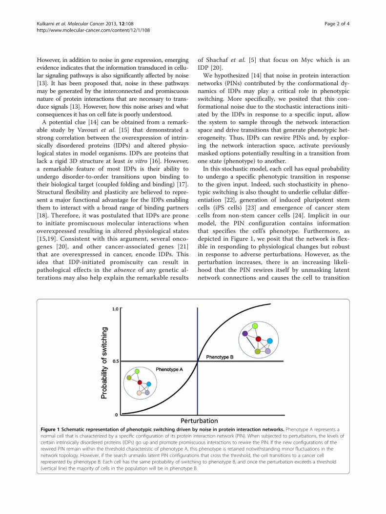
to undergo a specific phenotypic transition in responseto the given input. Indeed, such stochasticity in pheno-typic switching is also thought to underlie cellular differ-entiation [22], generation of induced pluripotent stemcells (iPS cells) [23] and emergence of cancer stemcells from non-stem cancer cells [24]. Implicit in ourmodel, the PIN configuration contains informationthat specifies the cell’s phenotype. Furthermore, asdepicted in Figure 1, we posit that the network is flex-ible in responding to physiological changes but robustin response to adverse perturbations. However, as theperturbation increases, there is an increasing likeli-hood that the PIN rewires itself by unmasking latentnetwork connections and causes the cell to transition
Figure 1 Schematic representation of phenotypic switching driven by noise in protein interaction networks. Phenotype A represents anormal cell that is characterized by a specific configuration of its protein interaction network (PIN). When subjected to perturbations, the levels ofcertain intrinsically disordered proteins (IDPs) go up and promote promiscuous interactions to rewire the PIN. If the new configurations of therewired PIN remain within the threshold characteristic of phenotype A, this phenotype is retained notwithstanding minor fluctuations in thenetwork topology. However, if the search unmasks latent PIN configurations that cross the threshold, the cell transitions to a cancer cellrepresented by phenotype B. Each cell has the same probability of switching to phenotype B, and once the perturbation exceeds a threshold(vertical line) the majority of cells in the population will be in phenotype B.
Kulkarni et al. Molecular Cancer 2013, 12:108 Page 2 of 4http://www.molecular-cancer.com/content/12/1/108
from one phenotype to the other; for example, fromnormal (non-dividing) to a malignant (proliferation)phenotype when Myc is overexpressed (Figure 1). Butit is important to note that, depending on the networktopology, lowering the perturbation (e.g. turning offMyc expression) can result in the PIN again rewiringitself to the normal (default) network configuration,thereby reversing the phenotypic switch (malignant tonormal). Interestingly, a similar situation is encounteredin stem cells where sustained Myc expression is critical tomaintain the pluripotent state; downregulating Mycpromotes differentiation of these cells [25]. Conversely,overexpression of Myc in dormant cells kicks theminto a proliferative mode.But how might information residing in PINs be
transmitted so that it can be stably inherited? It is nowwidely accepted that information that is transmittedtransgenerationally can be encoded epigenetically. Inter-estingly, several proteins that are involved in epigeneticallysculpting the chromatin are IDPs suggesting that rewiringof protein networks could result in heritable epigeneticchanges [14 and cfs therein]. Thus we conjectured [14]that, working together, these changes in the PIN institutedby the IDPs could account for stochastic phenotypicswitching.Cancer cells, like all other living (cells, organisms, and
ecosystems) and many non-living systems in the uni-verse (stars and galaxies), are self-organizing systemsthat exhibit nonlinear dynamics. However cancer cells,unlike their normal counterparts, exhibit traits typicallyassociated with primitive, single-celled organisms suchas bacteria that have an amazing adaptive tenacity[26,27]. As a result, it has been difficult to treat cancer.Thus, several groups [28], in particular, Huang and co-workers who have advanced the cancer attractor concept[4,29,30], have argued that there needs to be a paradigmshift from the prevailing view of cancer that is stronglyinfluenced by fundamentally deterministic approaches.By applying the tools of nonlinear dynamics, networktheory and stochastic modeling in combination with ex-periments to characterize cancer protein network con-nectivity and functionality we need to decipher howcancer cells self-organize to generate phenotypic hetero-geneity by different mechanisms. This knowledge canpotentially lead to a more fundamental understanding ofcancer and to the development of more effective thera-peutics. Perhaps, similar ideas may have already beendeployed by Nature. For example, a recent studyconcerning certain plant pathogens found that they se-lectively deploy independently evolved virulence proteinsthat interact with a limited set of highly connected cellu-lar hubs in order to take control of host cells [31]. Thelatter should serve as an inspiration, not just as a para-digm, to go after IDPs especially, those that are
aberrantly expressed only in cancer cells such as theCancer/Testis Antigens. Are we tilting at windmillsinstead?
AbbreviationsIDPs: Intrinsically disordered proteins; PIN: Protein interaction network;iPS: Induced pluripotent stem cells.
Competing interestsThe authors declare that they have no competing interests.
Authors’ contributionsPK conceived the idea. PK, TS and RVK wrote the manuscript. All authorsread and approved the final manuscript.
Authors’ informationPK, Assistant Professor. TS, Postdoctoral Fellow, and RVK is an AssociateProfessor.
AcknowledgementsPK wishes to thank the David Koch Fund for support. RVK would like toacknowledge funding support from the NSF through Award No. PHY-1307067 and from the NCI-funded U54 UMass Boston-Dana Farber/HarvardCancer Center Partnership Grant (5U54CA156734).
Author details1Department of Urology, The Johns Hopkins University School of Medicine,600 N Wolfe St, 105B Marburg, 21287, Baltimore, MD, USA. 2Department ofOncology, James Buchanan Brady Urological Institute, Johns HopkinsUniversity School of Medicine, Baltimore, MD, USA. 3Department of Physics,University of Massachusetts, Boston, MA, USA.
Received: 23 May 2013 Accepted: 19 September 2013Published: 24 September 2013
References1. Vogelstein B, Kinzler KW: Cancer genes and the pathways they control.
Nat Med 2004, 10(8):789–99.2. Stratton MR, Campbell PJ, Futreal PA: The cancer genome. Nature 2009,
458(7239):719–24.3. Soto AM, Sonnenschein C: The somatic mutation theory of cancer:
growing problems with the paradigm? Bioessays 2004, 26(10):1097–107.4. Huang S, Ingber DE: A non-genetic basis for cancer progression and
metastasis: self-organizing attractors in cell regulatory networks. BreastDis 2006, 26:27–54.
5. Shachaf CM, Kopelman AM, Arvanitis C, Karlsson A, Beer S, Mandl S,Bachmann MH, Borowsky AD, Ruebner B, Cardiff RD, Yang Q, Bishop JM,Contag CH, Felsher DW: MYC inactivation uncovers pluripotentdifferentiation and tumour dormancy in hepatocellular cancer. Nature2004, 431(7012):1112–7.
6. Mintz B, Illmensee K: Normal genetically mosaic mice produced frommalignant teratocarcinoma cells. Proc Natl Acad Sci U S A 1975,72(9):3585–9.
7. Hochedlinger K, Blelloch R, Brennan C, Yamada Y, Kim M, Chin L, Jaenisch R:Reprogramming of a melanoma genome by nuclear transplantation.Genes Dev 2004, 18(15):1875–85.
8. Sharma SV, Lee DY, Li B, Quinlan MP, Takahashi F, Maheswaran S,McDermott U, Azizian N, Zou L, Fischbach MA, Wong KK, Brandstetter K,Wittner B, Ramaswamy S, Classon M, Settleman J: A chromatin-mediatedreversible drug-tolerant state in cancer cell subpopulations. Cell 2010,141(1):69–80.
9. Allegrucci C, Rushton MD, Dixon JE, Sottile V, Shah M, Kumari R, Watson S,Alberio R, Johnson AD: Epigenetic reprogramming of breast cancer cellswith oocyte extracts. Mol Cancer 2011, 10(1):7.
10. Taniguchi Y, Choi PJ, Li GW, Chen H, Babu M, Hearn J, Emili A, Xie XS:Quantifying E. coli proteome and transcriptome with single-moleculesensitivity in single cells. Science 2010, 329:533–8.
11. Munsky B, Neuert G, van Oudenaarden A: Using gene expression noise tounderstand gene regulation. Science 2012, 336:183–7.
Kulkarni et al. Molecular Cancer 2013, 12:108 Page 3 of 4http://www.molecular-cancer.com/content/12/1/108
12. Capp JP: Stochastic gene expression, disruption of tissue averagingeffects and cancer as a disease of development. Bioessays 2005,27(12):1277–85.
13. Ladbury JE, Arold ST: Noise in cellular signaling pathways: causes andeffects. Trends Biochem Sci 2012, 37:173–178.
14. Mahmoudabadi G, Rajagopalan K, Getzenberg RH, Hannenhalli S,Rangarajan G, Kulkarni P: Intrinsically disordered proteins andconformational noise: implications in cancer. Cell Cycle 2013, 12(1):26–31.
15. Vavouri T, Semple JI, Garcia-Verdugo R, Lehner B: Intrinsic protein disorderand interaction promiscuity are widely associated with dosagesensitivity. Cell 2009, 138(1):198–208.
16. Uversky VN, Dunker AK: Understanding protein non-folding. BiochimBiophys Acta 1804, 2010:1231–64.
17. Tompa P, Csermely P: The role of structural disorder in the function ofRNA and protein chaperones. FASEB J 2004, 18:1169–75.
18. Cumberworth A, Lamour G, Babu MM, Gsponer J: Promiscuity as afunctional trait: intrinsically disordered regions as central players ofinteractomes. Biochem J 2013, 454(3):361–9.
19. Marcotte EM, Tsechansky M: Disorder, promiscuity, and toxic partnerships.Cell 2009, 138:16–18.
20. Iakoucheva LM, Brown CJ, Lawson JD, Obradović Z, Dunker AK: Intrinsicdisorder in cell-signaling and cancer-associated proteins. J Mol Biol 2002,323(3):573–84.
21. Rajagopalan K, Mooney SM, Parekh N, Getzenberg RH, Kulkarni P: A majorityof the cancer/testis antigens are intrinsically disordered proteins. J CellBiochem 2011, 112(11):3256–67.
22. Eldar A, Elowitz MB: Functional roles for noise in genetic circuits. Nature2010, 467(7312):167–73.
23. Yamanaka S: Elite and stochastic models for induced pluripotent stemcell generation. Nature 2009, 460:49–52.
24. Gupta PB, Fillmore CM, Jiang G, Shapira SD, Tao K, Kuperwasser C, LanderES: Stochastic state transitions give rise to phenotypic equilibrium inpopulations of cancer cells. Cell 2011, 146(4):633–44.
25. Kim J, Woo AJ, Chu J, Snow JW, Fujiwara Y, Kim CG, Cantor AB, Orkin SH:A Myc network accounts for similarities between embryonic stem andcancer cell transcription programs. Cell 2010, 143(2):313–24.
26. Lambert G, Estévez-Salmeron L, Oh S, Liao D, Emerson BM, Tlsty TD, AustinRH: An analogy between the evolution of drug resistance in bacterialcommunities and malignant tissues. Nat Rev Cancer 2011, 11(5):375–82.
27. Ben-Jacob E, Coffey DS, Levine H: Bacterial survival strategies suggestrethinking cancer cooperativity. Trends Microbiol 2012, 20(9):403–10.
28. Sonnenschein S, Soto AM, Rangarajan A, Kulkarni P: Competing views oncancer. J Biosciences 2013. in press.
29. Huang S, Ernberg I, Kauffman S: Cancer attractors: a systems view oftumors from a gene network dynamics and developmental perspective.Semin Cell Dev Biol 2009, 20(7):869–76.
30. Huang S: Tumor progression: chance and necessity in Darwinian andLamarckian somatic (mutationless) evolution. Prog Biophys Mol Biol 2012,110(1):69–86.
31. Mukhtar MS, Carvunis AR, Dreze M, Epple P, Steinbrenner J, Moore J, TasanM, Galli M, Hao T, Nishimura MT, Pevzner SJ, Donovan SE, Ghamsari L,Santhanam B, Romero V, Poulin MM, Gebreab F, Gutierrez BJ, Tam S,Monachello D, Boxem M, Harbort CJ, McDonald N, Gai L, Chen H, He Y,European Union Effectoromics Consortium, Vandenhaute J, Roth FP, Hill DE,et al: Independently evolved virulence effectors converge onto hubs in aplant immune system network. Science 2011, 333(6042):596–601.
doi:10.1186/1476-4598-12-108Cite this article as: Kulkarni et al.: Cancer: tilting at windmills? MolecularCancer 2013 12:108.
Submit your next manuscript to BioMed Centraland take full advantage of:
• Convenient online submission
• Thorough peer review
• No space constraints or color figure charges
• Immediate publication on acceptance
• Inclusion in PubMed, CAS, Scopus and Google Scholar
• Research which is freely available for redistribution
Submit your manuscript at www.biomedcentral.com/submit
Kulkarni et al. Molecular Cancer 2013, 12:108 Page 4 of 4http://www.molecular-cancer.com/content/12/1/108