Embed Size (px)
Citation preview
Cancer in the United States, 2009,
Jemal, A. et al. CA Cancer J Clin 2009;59:225-249
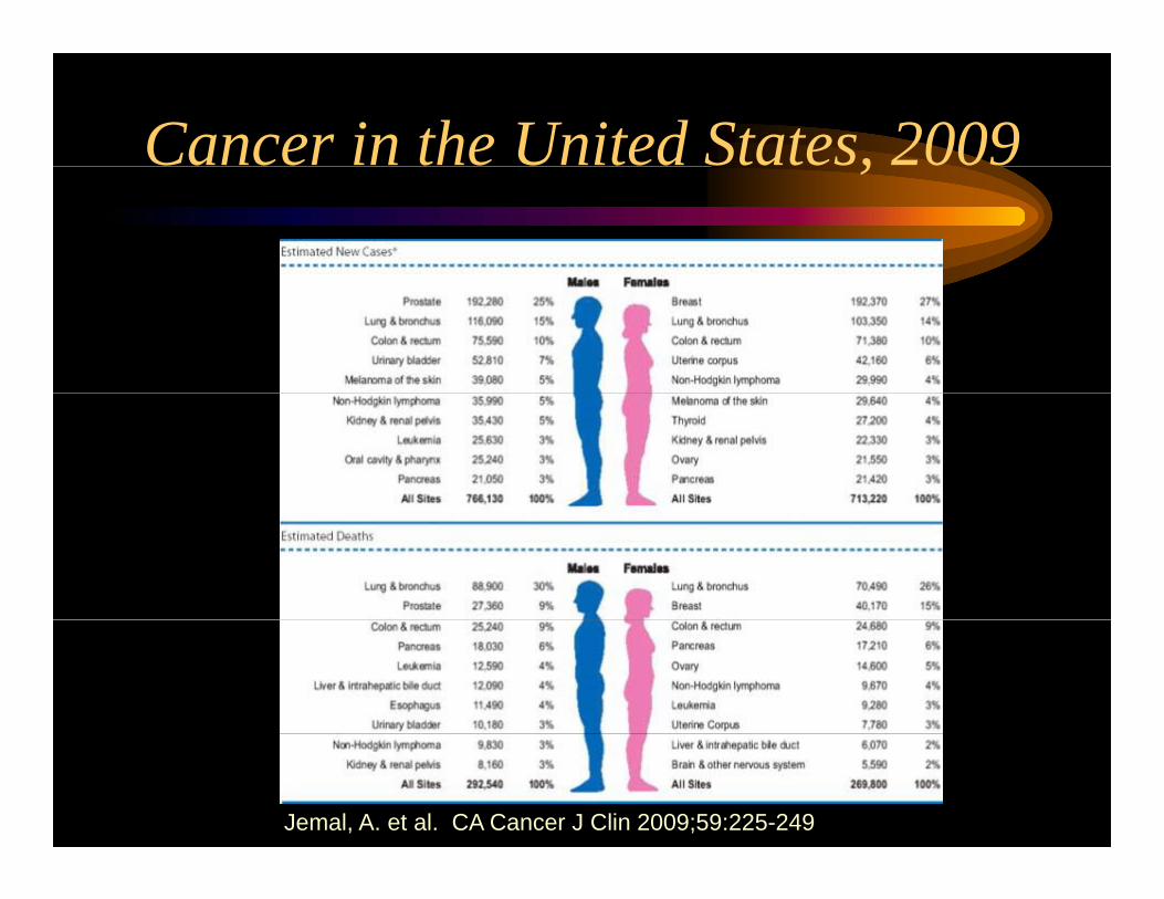
Lung Cancer in the United StatesLung Cancer in the United StatesAnnual Cancer Deaths
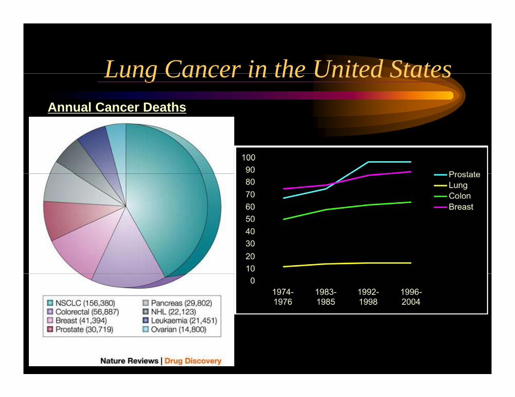
The Scheme: From Nicotine Addiction to Lung Cancer
Cigarette smoking Metabolic Activationeg. Cytochrome P450
Field Carcinogenesis
NICOTINE ADDICTIONNICOTINE ADDICTIONCARCINOGENS
Ba-P, NNKDNA ADDUCTS
MUTATIONS, etc
p53, k-ras, LOHLUNG CANCER
RepairMetabolic Detoxification Glutathione S-Transferase (alpha mu pi theta)(alpha, mu, pi, theta)
Excretion Normal DNA
ApoptosisDNA
Modified from Hecht JNCI; 1999
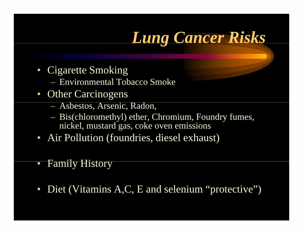
Lung Cancer Risksg
• Cigarette SmokingCigarette Smoking– Environmental Tobacco Smoke
• Other Carcinogens– Asbestos, Arsenic, Radon, – Bis(chloromethyl) ether, Chromium, Foundry fumes,
nickel, mustard gas, coke oven emissions• Air Pollution (foundries, diesel exhaust)
F il Hi t• Family History
• Diet (Vitamins A C E and selenium “protective”)Diet (Vitamins A,C, E and selenium protective )
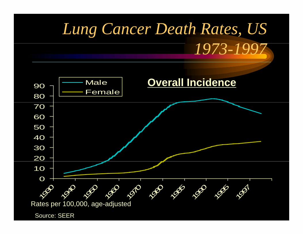
Lung Cancer Death Rates, US1973 19971973-1997
8090 Male
FemaleOverall Incidence
506070
203040
010
930
940
950
960
970
980
985
990
995
997
193
194
195
196
197
198
198
199
199
199
Source: SEER
Rates per 100,000, age-adjusted
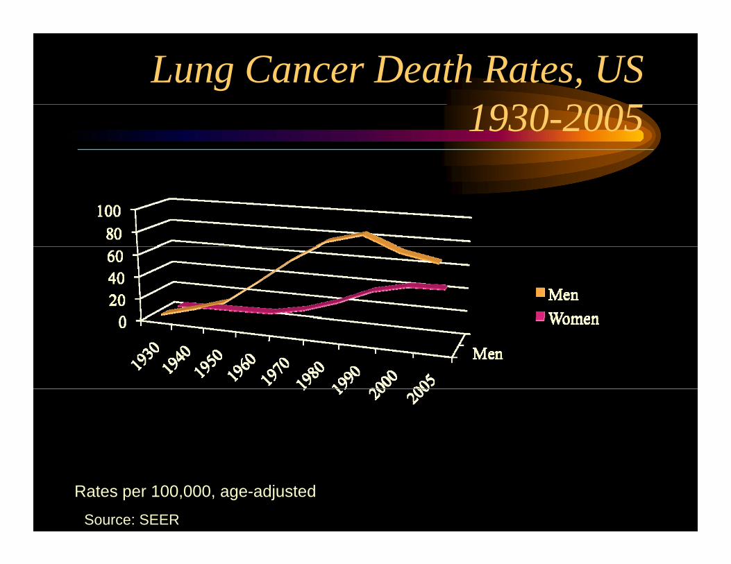
Lung Cancer Death Rates, US1930 20051930-2005
Source: SEER
Rates per 100,000, age-adjusted
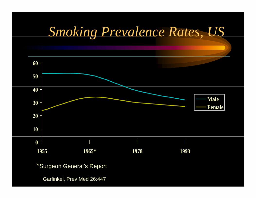
Smoking Prevalence Rates, USg ,
60
50
60
30
40MaleFemale
10
20
01955 1965* 1978 1993
*Surgeon General’s Report
Garfinkel, Prev Med 26:447
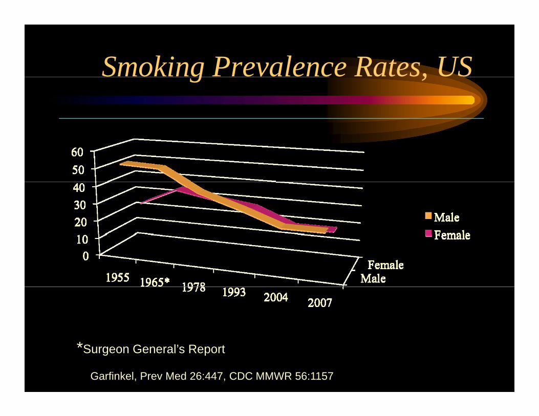
Smoking Prevalence Rates, USg ,
*Surgeon General’s Report
Garfinkel, Prev Med 26:447, CDC MMWR 56:1157

Percentage of High School Students Who Reported Current Cigarette SmokingReported Current Cigarette Smoking
34363840
2426283032
MaleFemale
202224
1991 1993 1995 1997 1999
Youth Behavior Survey, MMWR 2000; 49
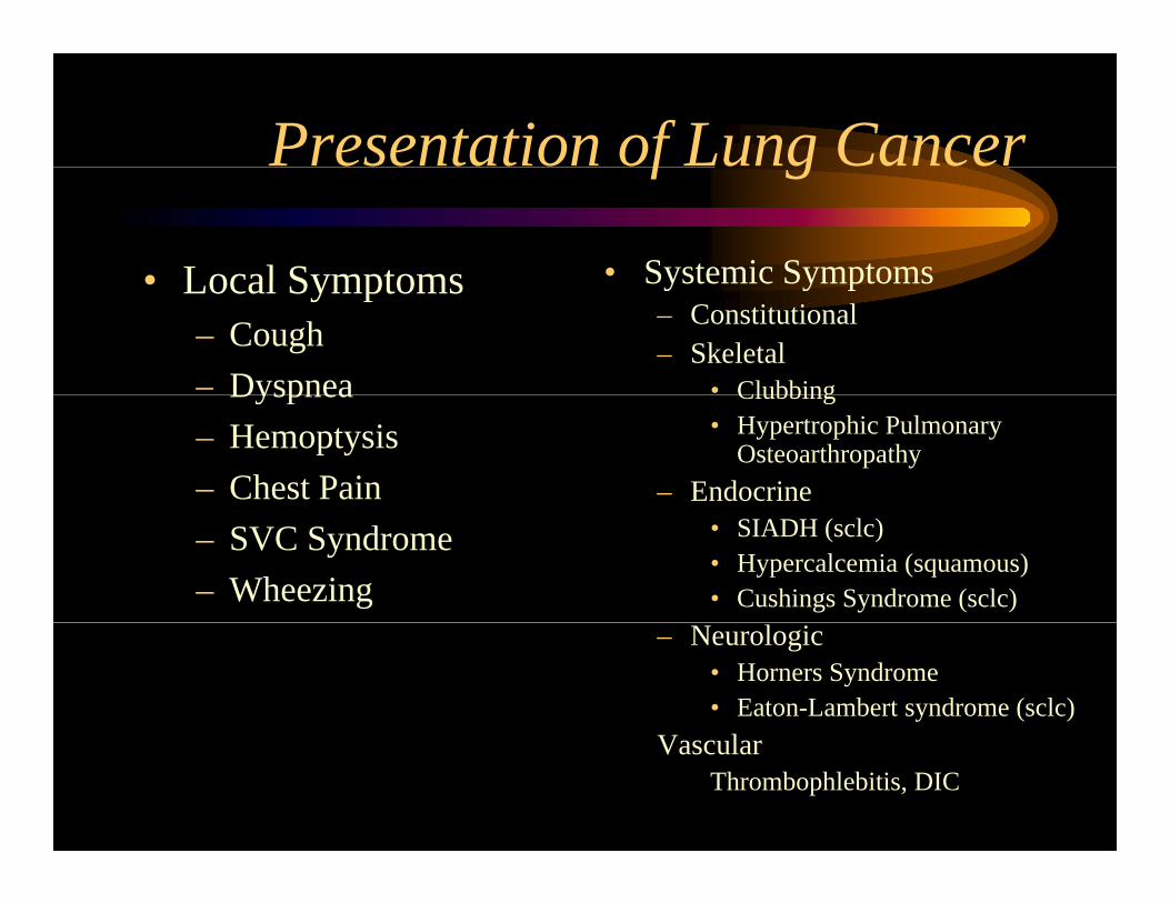
Presentation of Lung Cancerf g
• Local Symptoms • Systemic Symptoms• Local Symptoms– Cough– Dyspnea
Systemic Symptoms– Constitutional– Skeletal
• ClubbingDyspnea– Hemoptysis– Chest Pain
Clubbing• Hypertrophic Pulmonary
Osteoarthropathy– Endocrine
– SVC Syndrome– Wheezing
• SIADH (sclc)• Hypercalcemia (squamous)• Cushings Syndrome (sclc)
– Neurologic• Horners Syndrome• Eaton-Lambert syndrome (sclc)
V lVascularThrombophlebitis, DIC
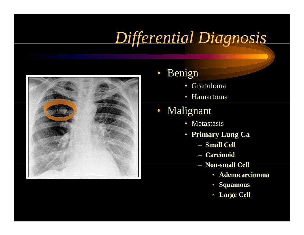
Differential Diagnosisff g
• Benign• Benign• Granuloma• Hamartoma
• Malignant• Metastasis
P i L C• Primary Lung Ca– Small Cell– Carcinoid– Non-small Cell
• Adenocarcinoma• Squamous• Large Cell
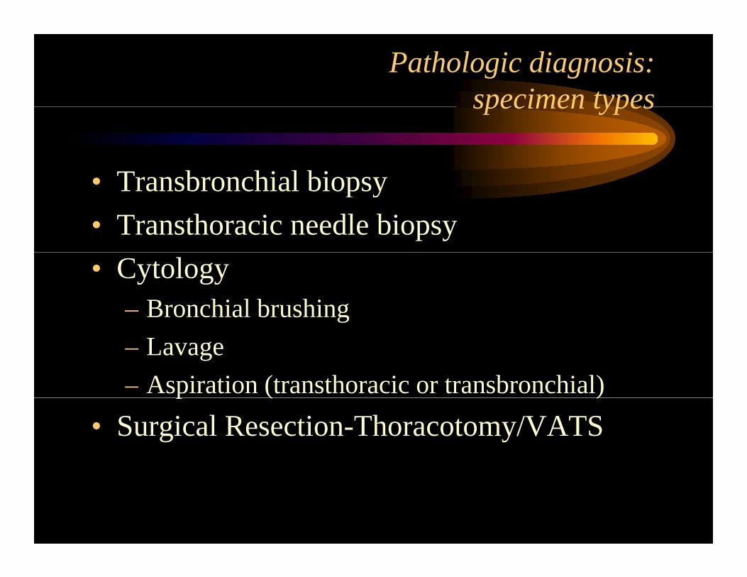
Pathologic diagnosis:specimen typesspecimen types
• Transbronchial biopsy• Transbronchial biopsy• Transthoracic needle biopsy• Cytology
– Bronchial brushing– Lavage– Aspiration (transthoracic or transbronchial)p ( )
• Surgical Resection-Thoracotomy/VATS
Lung tumors - Benigng g
• The majority of pulmonary neoplasms are• The majority of pulmonary neoplasms are malignant
• Benign tumors/lesionsBenign tumors/lesions– Hamartoma (most common)
Mesenchymal leiomyoma lipoma chondroma– Mesenchymal- leiomyoma, lipoma, chondroma (all unusual)
– Alveolar adenoma (rare)Alveolar adenoma (rare)
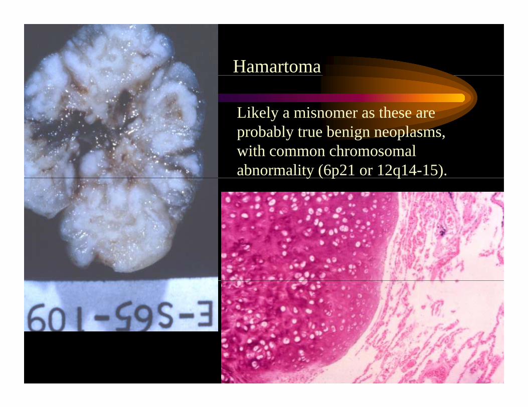
Hamartoma
Likely a misnomer as these are b bl b i lprobably true benign neoplasms,
with common chromosomal abnormality (6p21 or 12q14-15).y ( p q )
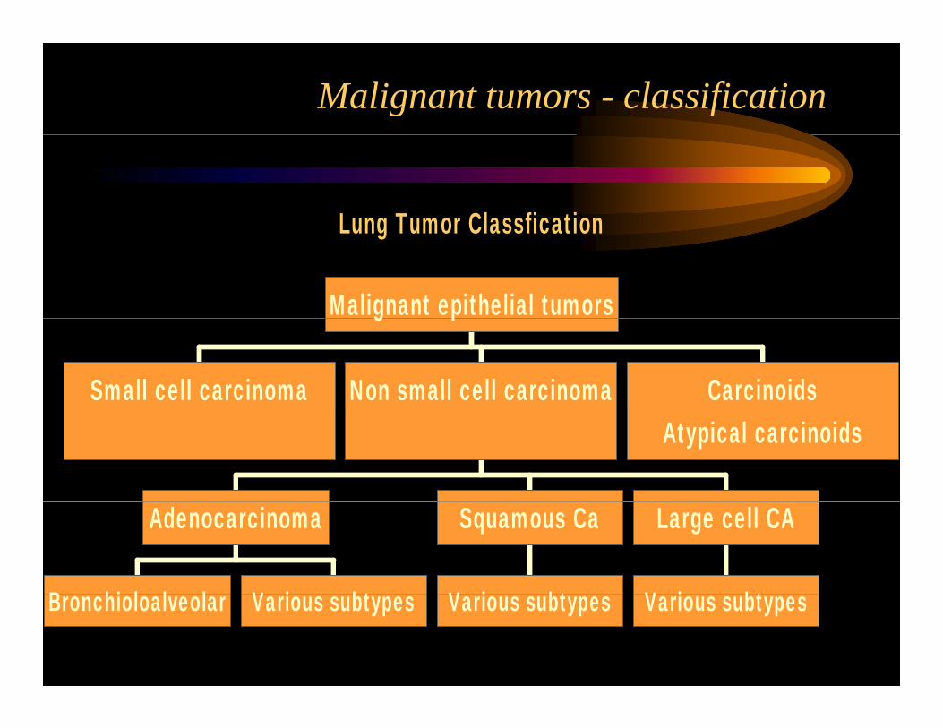
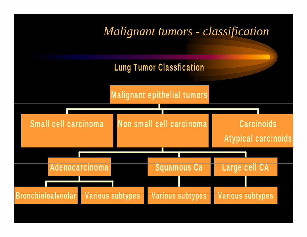
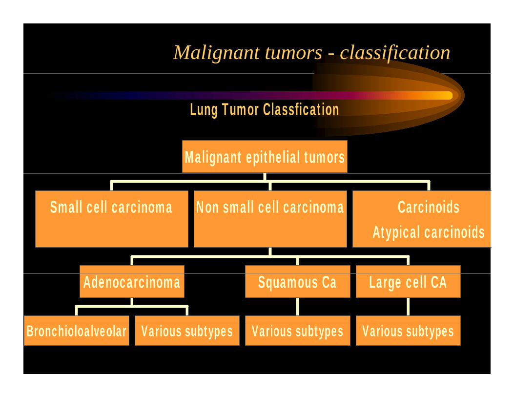
Malignant tumors - classification
Lung Tumor ClassficationLung Tumor Classfication
Malignant epithelial tumors
Small cell carcinoma Non small cell carcinoma Carcinoids
g p
Atypical carcinoids
B hi l l l V i bt
Adenocarcinoma
V i bt
Squamous Ca
V i bt
Large cell CA
Bronchioloalveolar Various subtypes Various subtypes Various subtypes
Small cell carcinoma
• Usually hilar/ central tumor• The majority have extrapulmonary spread at time
f iof presentation.• Only 5% present as early stage disease.• Critical divide between small cell and non-small
cell carcinomaS ll ll i t d diff tl t t d ith– Small cell carcinoma staged differently, treated with chemoradiation not surgery.
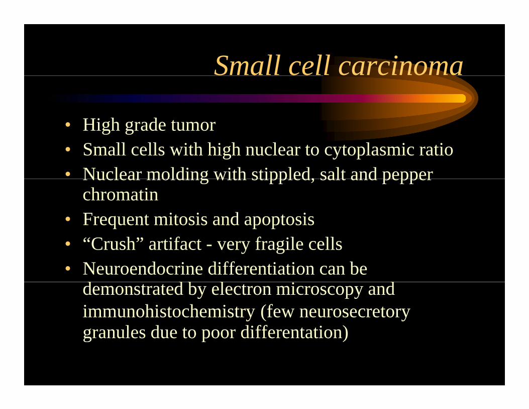
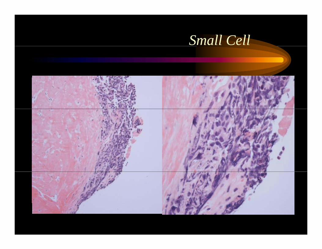
Small cell carcinoma
• High grade tumorHigh grade tumor• Small cells with high nuclear to cytoplasmic ratio• Nuclear molding with stippled, salt and pepperNuclear molding with stippled, salt and pepper
chromatin• Frequent mitosis and apoptosis• “Crush” artifact - very fragile cells• Neuroendocrine differentiation can be
d d b l i ddemonstrated by electron microscopy and immunohistochemistry (few neurosecretory granules due to poor differentation)granules due to poor differentation)
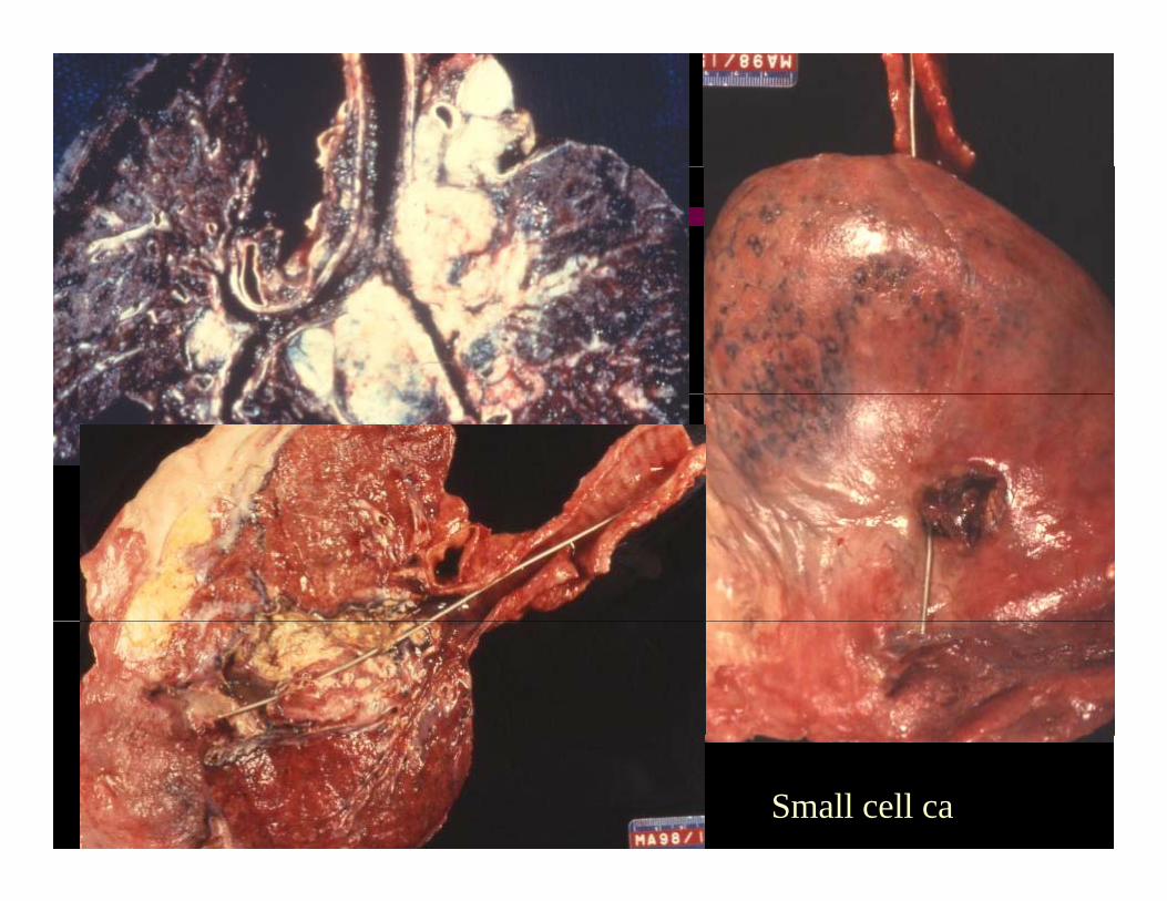
Small cell ca
Small Cell
Malignant tumors - classification
Lung Tumor Classfication
Malignant epithelial tumors
Small cell carcinoma Non small cell carcinoma Carcinoids
S C C
Atypical carcinoids
B hi l l l V i bt
Adenocarcinoma
V i bt
Squamous Ca
V i bt
Large cell CA
Bronchioloalveolar Various subtypes Various subtypes Various subtypes
Atypical adenomatous hyperplasia-adenocarcinoma precursoradenocarcinoma precursor
• Focal, 5.0 mm or less, with defined borders• Alveoli lined by cuboidal to low columnar cells
with variable atypia• Alveolar walls may be slightly thickened• Non-mucinous• Clinical significance unclear (?time to progression
to carcinoma)
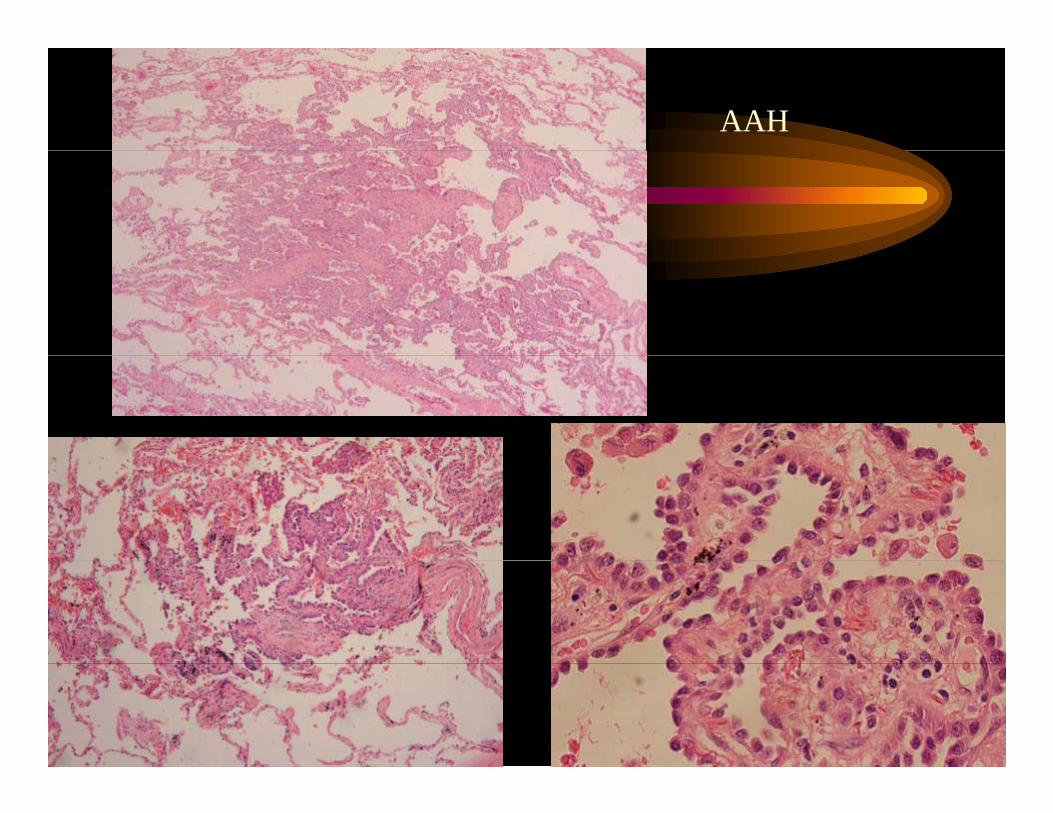
AAH
Adenocarcinoma
• Most often a peripheral tumorM l d l l k i• Many are near pleura and cause pleural puckering.
• Cut surface can be mucoid or firm, depending on degree of fibrosis and mucin productiondegree of fibrosis and mucin production
• Small tumors can be associated with lymph node and distant metastasisand distant metastasis.
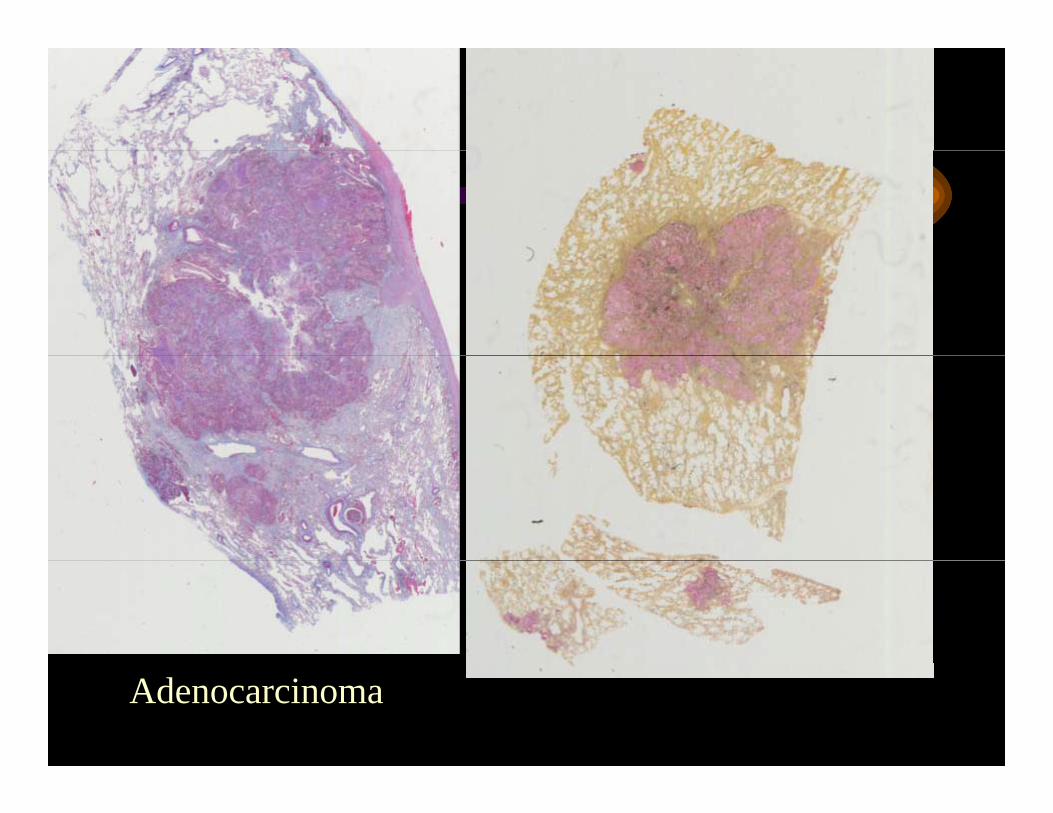
Adenocarcinoma
Adenocarcinoma
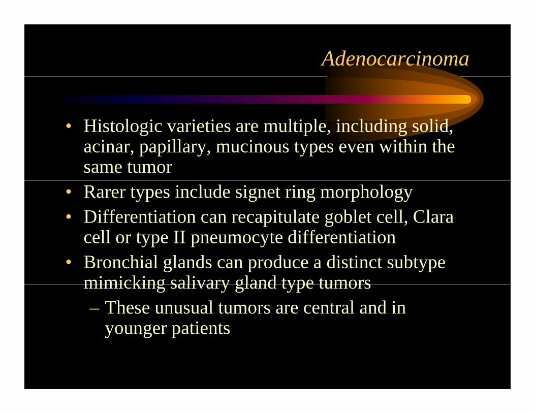
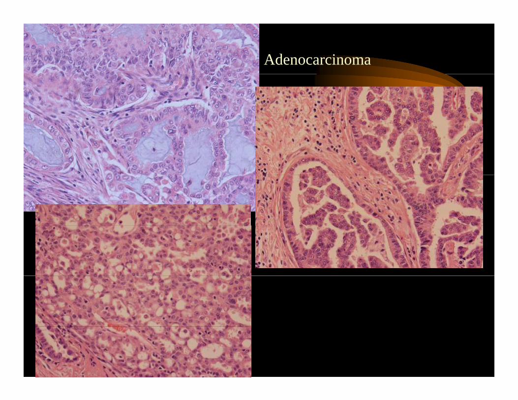
• Histologic varieties are multiple including solidHistologic varieties are multiple, including solid, acinar, papillary, mucinous types even within the same tumor
• Rarer types include signet ring morphology• Differentiation can recapitulate goblet cell, Clara
ll t II t diff ti ticell or type II pneumocyte differentiation• Bronchial glands can produce a distinct subtype
mimicking salivary gland type tumorsmimicking salivary gland type tumors– These unusual tumors are central and in
younger patients y g p
Adenocarcinoma
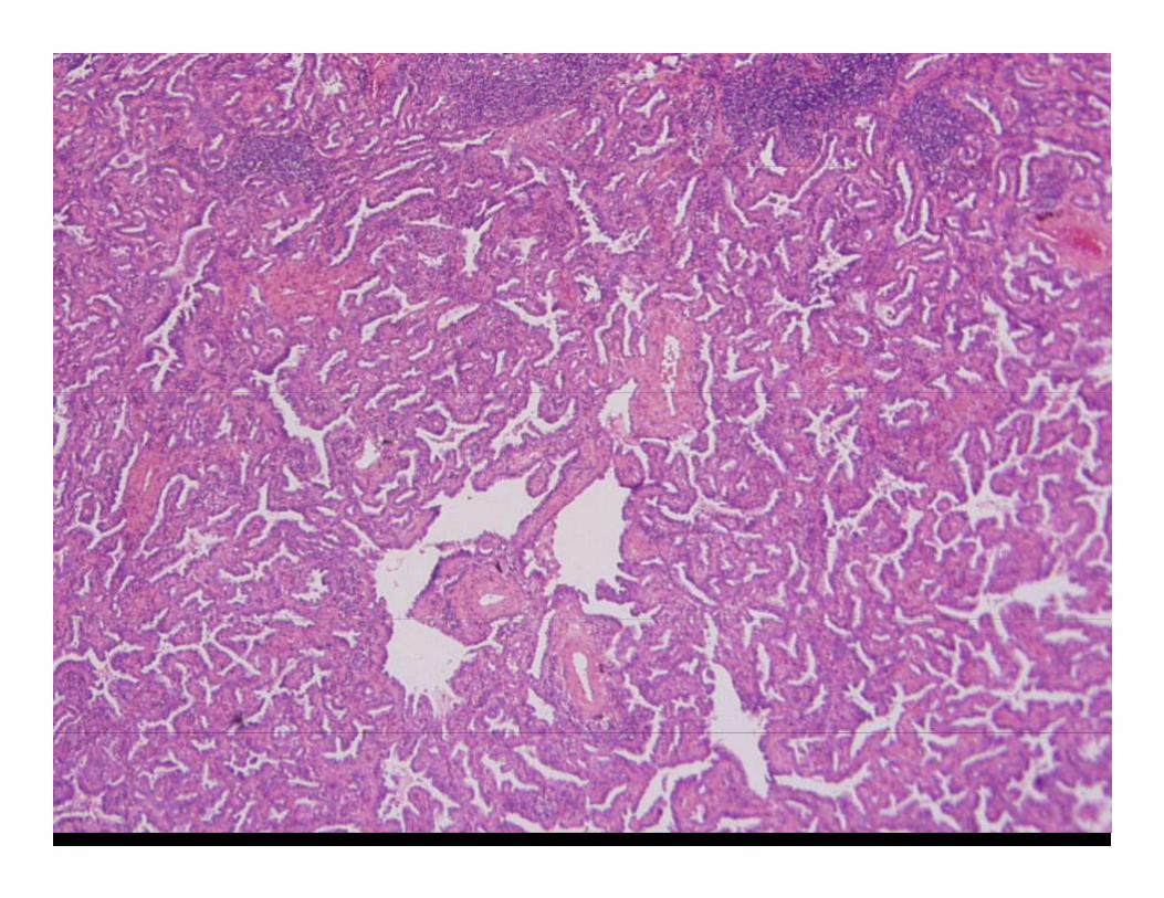
Adenocarcinoma - Bronchioloalveolar
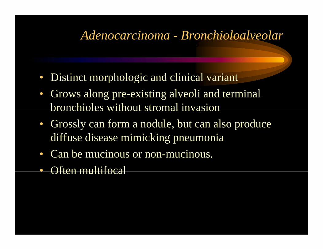
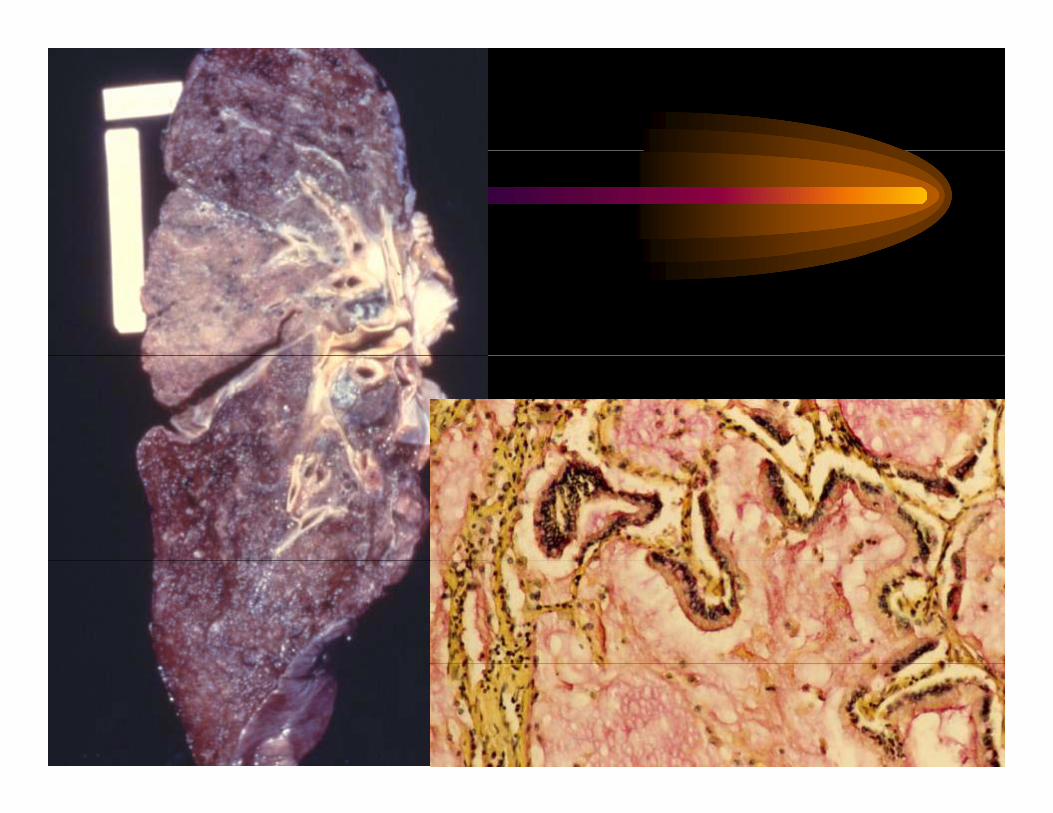
• Distinct morphologic and clinical variant• Distinct morphologic and clinical variant• Grows along pre-existing alveoli and terminal
bronchioles without stromal invasionbronchioles without stromal invasion• Grossly can form a nodule, but can also produce
diffuse disease mimicking pneumoniadiffuse disease mimicking pneumonia• Can be mucinous or non-mucinous.• Often multifocalOften multifocal
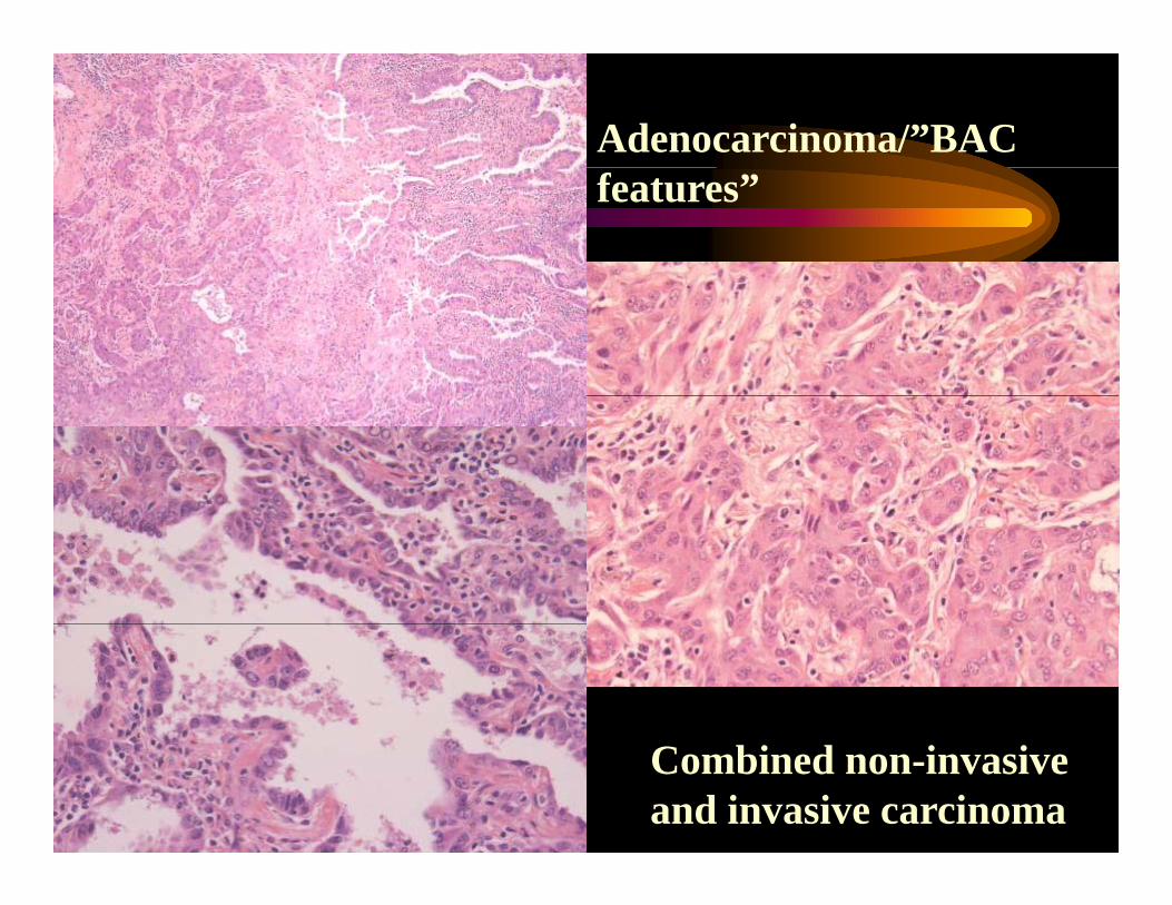
Adenocarcinoma/”BAC features”
Combined non-invasive and invasive carcinoma
Is there a meaning to the histologic di it f d i ?diversity of adenocarcinoma?
• Studies examining response to gefitinib• Studies examining response to gefitinib (EGFR targeting tyrosine kinase inhibitor) found activating EGFR mutations infound activating EGFR mutations in patients with favorable response.G fili di f d di i• Gene profiling studies found distinct subclasses of adenocarcinoma.
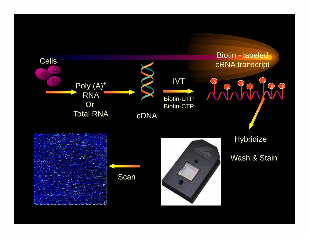
Biotin - labeled cRNA transcriptCells
Poly (A)+
RNAOr
IVT
Biotin-UTPBi ti CTP
B B B BB B
B
cDNAOr
Total RNABiotin-CTP
Wash & Stain
Hybridize
Scan
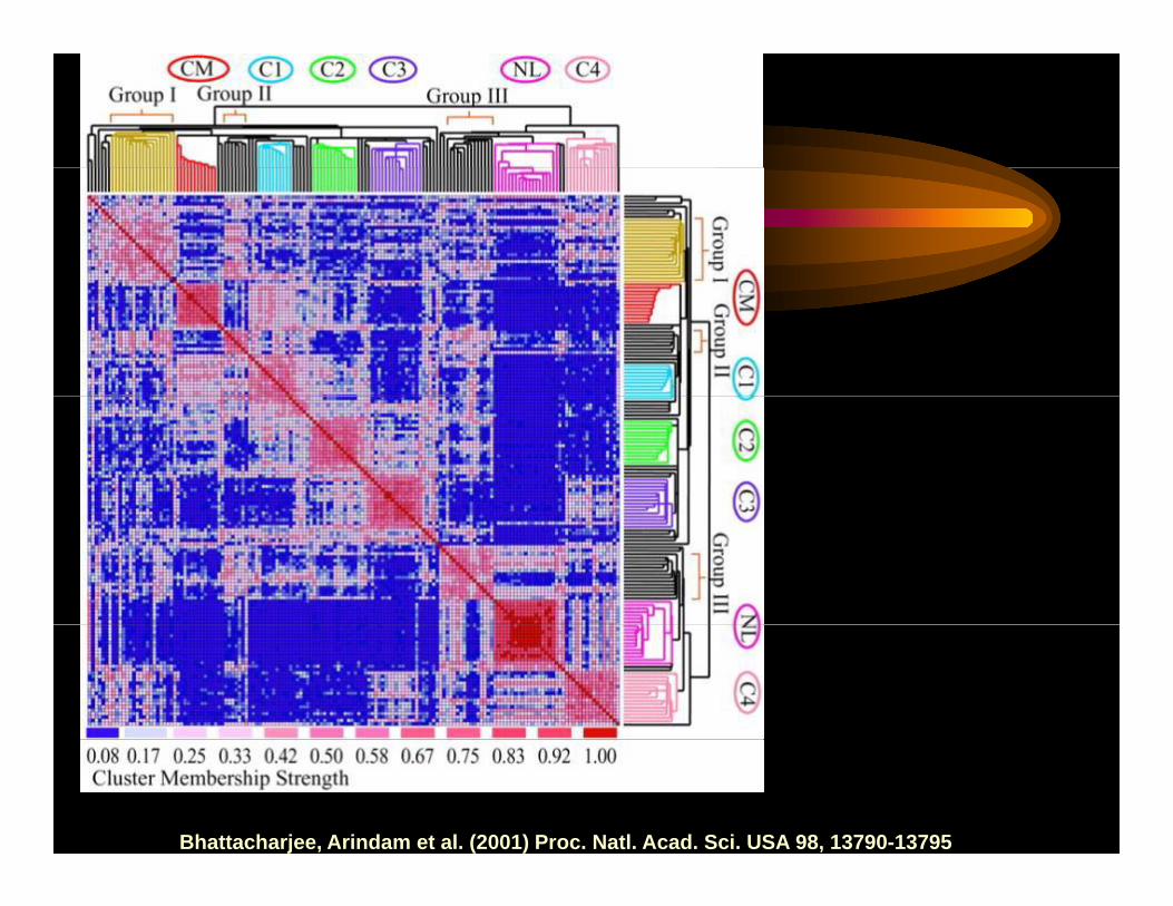
Bhattacharjee, Arindam et al. (2001) Proc. Natl. Acad. Sci. USA 98, 13790-13795
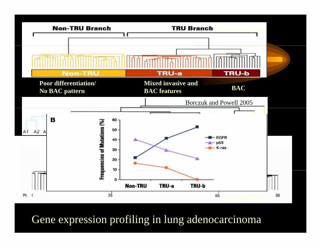
Takeuchi et al,2005
BACMixed invasive and BAC features
Poor differentiation/No BAC pattern
Borczuk and Powell 2005
Beer et al,2002
Gene expression profiling in lung adenocarcinoma
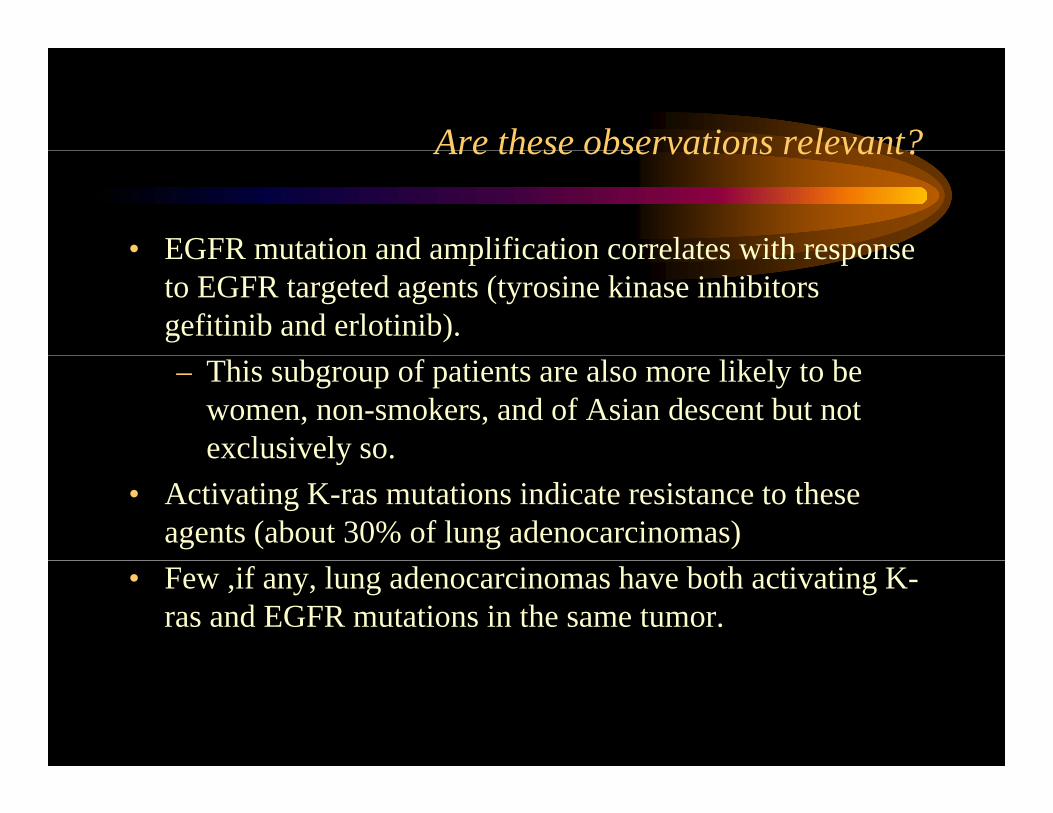
Are these observations relevant?Are these observations relevant?
• EGFR mutation and amplification correlates with responseEGFR mutation and amplification correlates with response to EGFR targeted agents (tyrosine kinase inhibitors gefitinib and erlotinib).– This subgroup of patients are also more likely to be
women, non-smokers, and of Asian descent but not exclusively so.exclusively so.
• Activating K-ras mutations indicate resistance to these agents (about 30% of lung adenocarcinomas)
• Few ,if any, lung adenocarcinomas have both activating K-ras and EGFR mutations in the same tumor.
Malignant tumors - classification
Lung Tumor Classfication
Malignant epithelial tumors
Small cell carcinoma Non small cell carcinoma CarcinoidsAtypical carcinoids
Adenocarcinoma Squamous Ca Large cell CA
Bronchioloalveolar Various subtypes Various subtypes Various subtypes
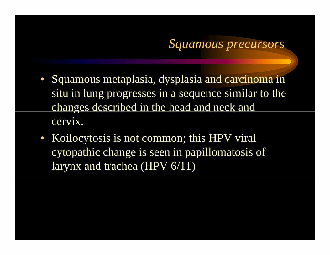
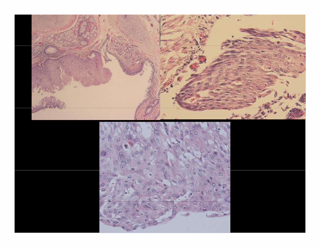
Squamous precursorsSquamous precursors
• Squamous metaplasia dysplasia and carcinoma in• Squamous metaplasia, dysplasia and carcinoma in situ in lung progresses in a sequence similar to the changes described in the head and neck and gcervix.
• Koilocytosis is not common; this HPV viral y ;cytopathic change is seen in papillomatosis of larynx and trachea (HPV 6/11)
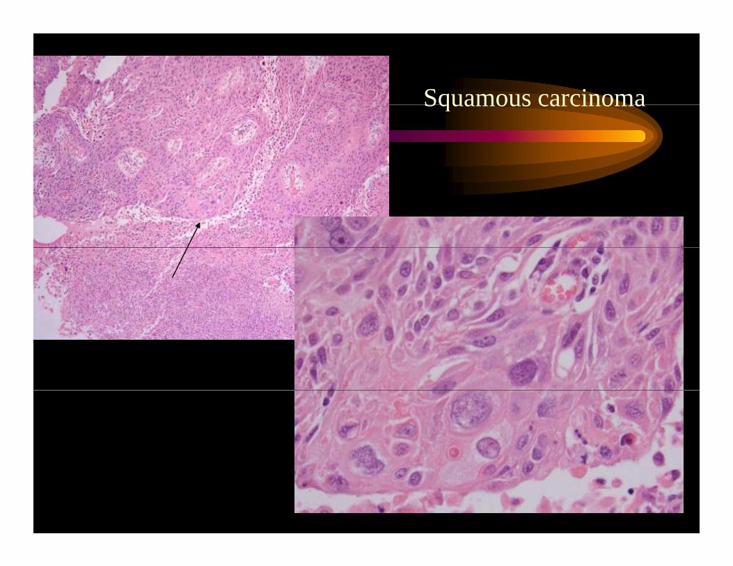
Squamous carcinoma
• Usually of bronchogenic origin; however• Usually of bronchogenic origin; however can also arise from peripheral areas of squamous metaplasiasquamous metaplasia
• Frequently have central necrosis• Faster doubling time than adenocarcinoma;
often larger at presentation• Metastasis in relation to tumor size may
occur later than adenocarcinoma
Squamous carcinomaSquamous carcinoma
Large cell carcinoma
• This subtype shows no differentiation towards• This subtype shows no differentiation towards either squamous or adenocarcinoma
• Aggressive tumors with poor prognosisAggressive tumors with poor prognosis• If subjected to ultrastructural examination, many
of these tumors show either glandular orof these tumors show either glandular or squamous differentiation.
• Nevertheless, these tumors are separated out , pbecause of their high grade and poor prognosis
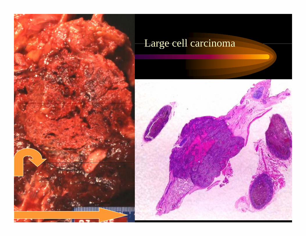
Large cell carcinomaLarge cell carcinoma
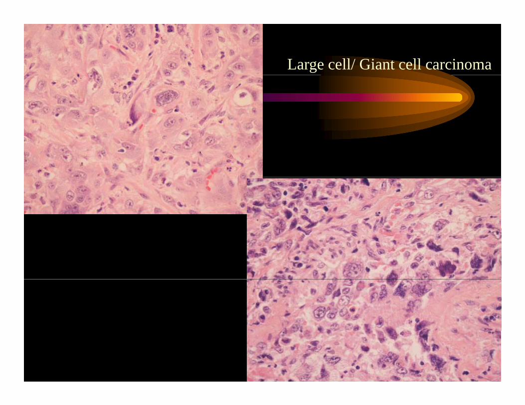
Large cell/ Giant cell carcinoma
CarcinoidsCarcinoids
• Malignant neoplasm of neuroendocrine cell originMalignant neoplasm of neuroendocrine cell origin• Can be central or peripheral; central lesions can
cause bronchial obstruction• Project into bronchial lumen but often have intact
mucosa above them (grow under the mucosa)• Typical carcinoids are low grade malignancies;
atypical carcinoids (mitoses and necrosis) are intermediate grade when compared to non-smallintermediate grade when compared to non-small cell carcinomas
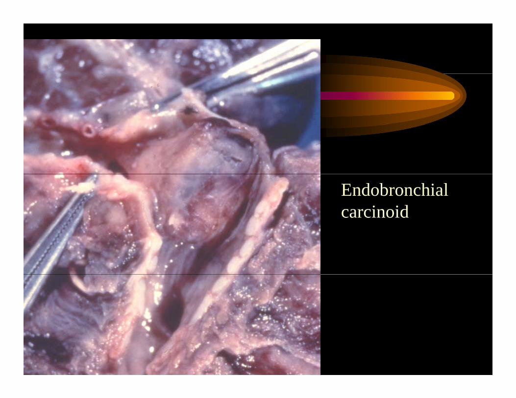
Endobronchial carcinoid
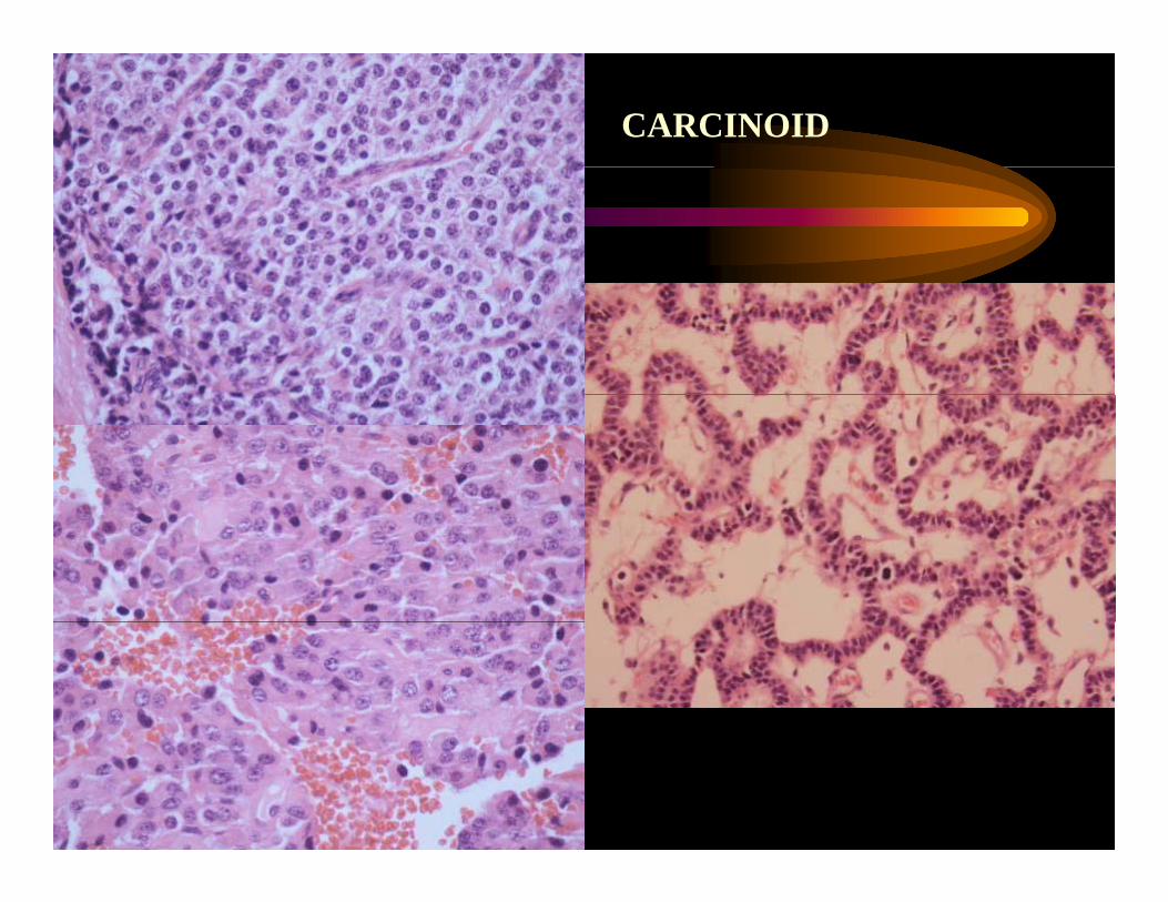
CarcinoidsCarcinoids
• Histologic features• Histologic features– Nests and cords surrounded by delicate stroma
Uniform cells with salt and pepper chromatin– Uniform cells with salt and pepper chromatin– Neurosecretory granules are abundant and
easily demonstrated by electron microscopy oreasily demonstrated by electron microscopy or immunohistochemistry (well differentiated tumors))
CARCINOID
Metastatic CarcinomaMetastatic Carcinoma
• The lung is a frequent site of metastatic tumor• The lung is a frequent site of metastatic tumor, both from extrapulmonary and intrapulmonary primaries. p
• In autopsy series, between 20 and 50% of patients that expire from extra-pulmonary primaries have p p y plung metastasis.
• Melanoma, sarcomas, renal cell carcinoma, germ cell tumors, breast carcinoma as well as carcinomas of bladder, larynx, thyroid and
t tprostate
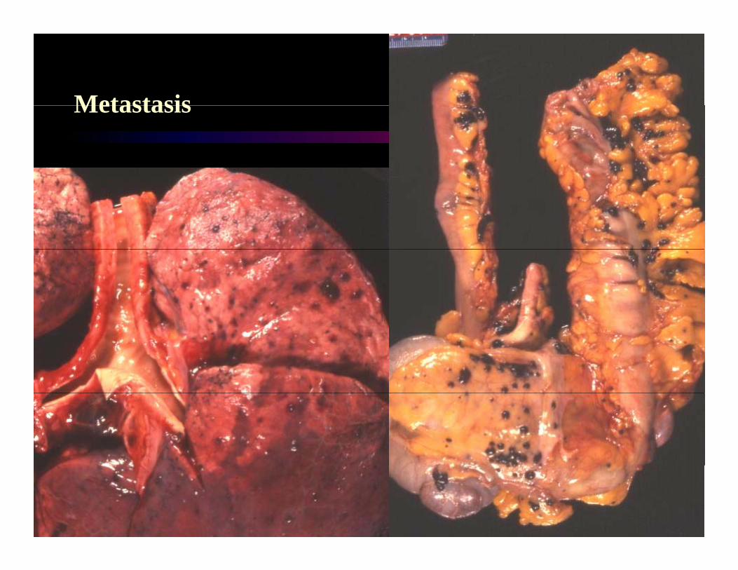
MetastasisMetastasis
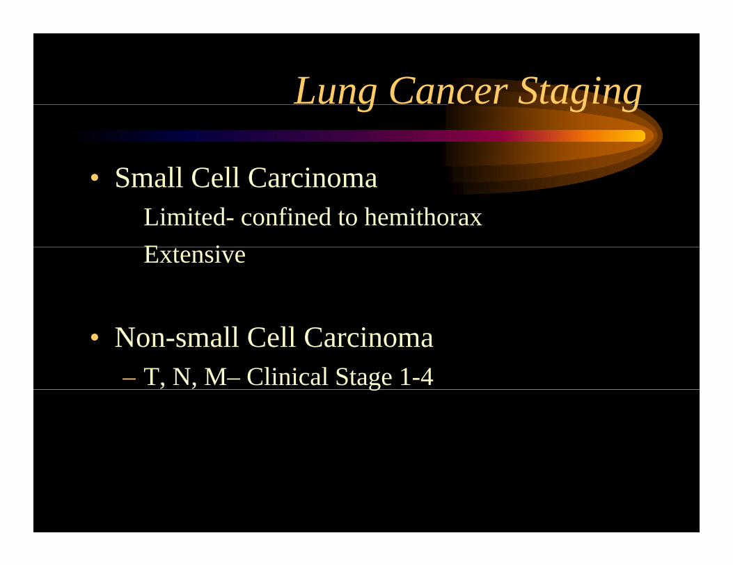
Lung Cancer Stagingg g g
• Small Cell Carcinoma• Small Cell CarcinomaLimited- confined to hemithoraxE iExtensive
• Non-small Cell Carcinoma– T, N, M– Clinical Stage 1-4g
Therapy- small cellpy
– LimitedCh th + R di ti• Chemotherapy + Radiation
– Extensive• Chemotherapy• Chemotherapy
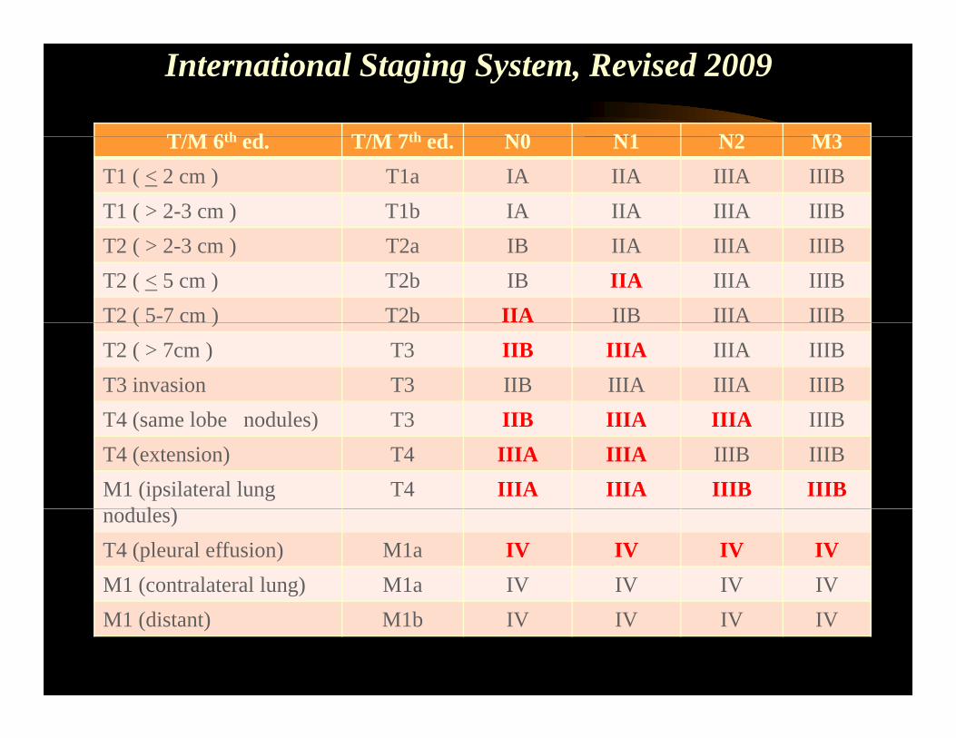
International Staging System, Revised 2009
T/M 6th d T/M 7th d N0 N1 N2 M3T/M 6th ed. T/M 7th ed. N0 N1 N2 M3T1 ( < 2 cm ) T1a IA IIA IIIA IIIBT1 ( > 2-3 cm ) T1b IA IIA IIIA IIIB
Chest 111:1710-17
T2 ( > 2-3 cm ) T2a IB IIA IIIA IIIBT2 ( < 5 cm ) T2b IB IIA IIIA IIIBT2 ( 5-7 cm ) T2b IIA IIB IIIA IIIB( )T2 ( > 7cm ) T3 IIB IIIA IIIA IIIBT3 invasion T3 IIB IIIA IIIA IIIBT4 ( l b d l ) T3 IIB IIIA IIIA IIIBT4 (same lobe nodules) T3 IIB IIIA IIIA IIIBT4 (extension) T4 IIIA IIIA IIIB IIIBM1 (ipsilateral lung
d l )T4 IIIA IIIA IIIB IIIB
nodules)T4 (pleural effusion) M1a IV IV IV IVM1 (contralateral lung) M1a IV IV IV IVM1 (distant) M1b IV IV IV IV
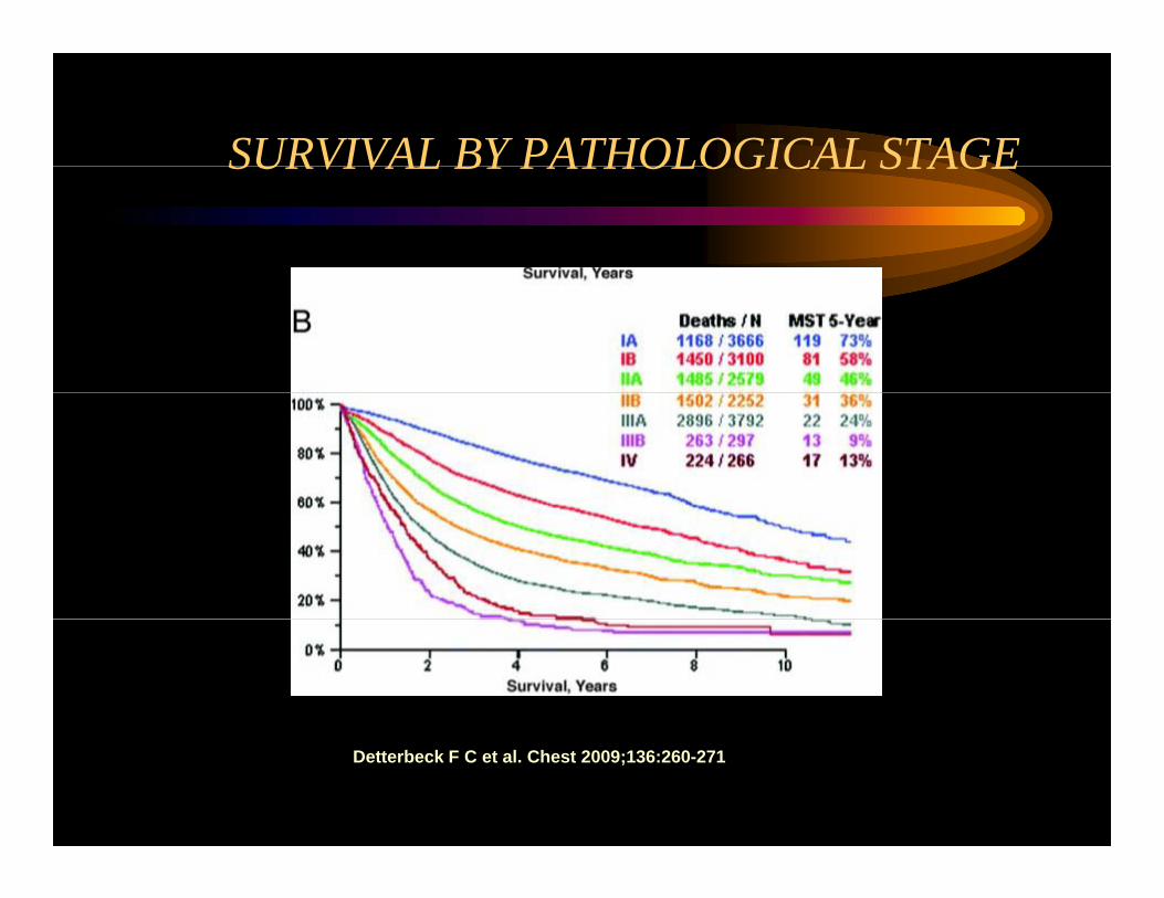
SURVIVAL BY PATHOLOGICAL STAGESURVIVAL BY PATHOLOGICAL STAGE
Detterbeck F C et al. Chest 2009;136:260-271
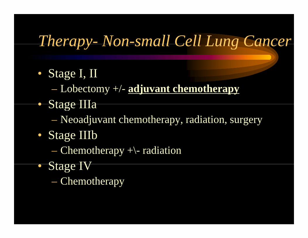
Therapy- Non-small Cell Lung CancerTherapy Non small Cell Lung Cancer
• Stage I II• Stage I, II– Lobectomy +/- adjuvant chemotherapy
• Stage IIIa• Stage IIIa– Neoadjuvant chemotherapy, radiation, surgery
• Stage IIIb• Stage IIIb– Chemotherapy +\- radiation
St IV• Stage IV– Chemotherapy
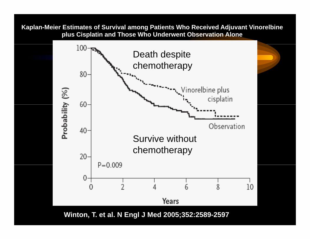
Kaplan-Meier Estimates of Survival among Patients Who Received Adjuvant Vinorelbine plus Cisplatin and Those Who Underwent Observation Alone
Death despite chemotherapy
Survive without chemotherapy
Winton, T. et al. N Engl J Med 2005;352:2589-2597
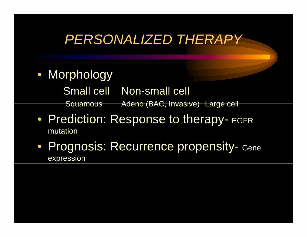
PERSONALIZED THERAPYPERSONALIZED THERAPY
• Morphology• MorphologySmall cell Non-small cellSquamous Adeno (BAC Invasive) Large cellSquamous Adeno (BAC, Invasive) Large cell
• Prediction: Response to therapy- EGFR mutationmutation
• Prognosis: Recurrence propensity- Gene expression
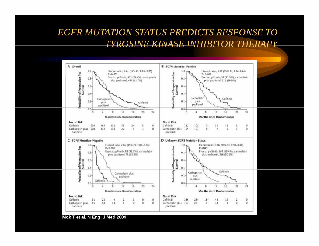
EGFR MUTATION STATUS PREDICTS RESPONSE TO TYROSINE KINASE INHIBITOR THERAPYTYROSINE KINASE INHIBITOR THERAPY
Mok T et al. N Engl J Med 2009
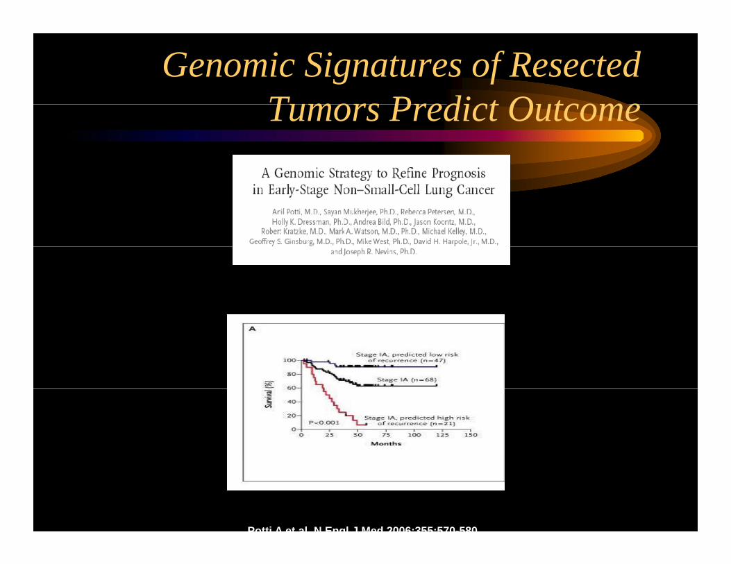
Genomic Signatures of Resected T P di t O tTumors Predict Outcome
Potti A et al N Engl J Med 2006;355:570-580