Embed Size (px)
Citation preview
cancers
Review
Cancer-Associated Fibroblasts in Breast Cancer TreatmentResponse and Metastasis
Patricia Fernández-Nogueira 1,2,*, Gemma Fuster 1,3,4, Álvaro Gutierrez-Uzquiza 5,6 , Pere Gascón 1,Neus Carbó 1 and Paloma Bragado 5,6,*
�����������������
Citation: Fernández-Nogueira, P.;
Fuster, G.; Gutierrez-Uzquiza, Á.;
Gascón, P.; Carbó, N.; Bragado, P.
Cancer-Associated Fibroblasts in
Breast Cancer Treatment Response
and Metastasis. Cancers 2021, 13, 3146.
https://doi.org/10.3390/cancers
13133146
Academic Editors: Stephan
Joel Reshkin and Rosa
Angela Cardone
Received: 30 April 2021
Accepted: 16 June 2021
Published: 23 June 2021
Publisher’s Note: MDPI stays neutral
with regard to jurisdictional claims in
published maps and institutional affil-
iations.
Copyright: © 2021 by the authors.
Licensee MDPI, Basel, Switzerland.
This article is an open access article
distributed under the terms and
conditions of the Creative Commons
Attribution (CC BY) license (https://
creativecommons.org/licenses/by/
4.0/).
1 Department of Biochemistry and Molecular Biomedicine, Institute of Biomedicine,University of Barcelona (IBUB), 08028 Barcelona, Spain; [email protected] (G.F.);[email protected] (P.G.); [email protected] (N.C.)
2 Department of Biomedicine, School of Medicine, University of Barcelona, 08028 Barcelona, Spain3 Department of Biochemistry & Physiology, School of Pharmacy and Food Sciences, University of Barcelona,
08028 Barcelona, Spain4 Department of Biosciences, Faculty of Sciences and Technology, University of Vic, 08500 Vic, Spain5 Department of Biochemistry and Molecular Biology, Faculty of Pharmacy, Complutense University of Madrid,
28040 Madrid, Spain; [email protected] Health Research Institute of the Hospital Clínico San Carlos, 28040 Madrid, Spain* Correspondence: [email protected] (P.F.-N.); [email protected] (P.B.);
Tel.: +34-934021871 (P.F.-N.); +34-913941853 (P.B.)
Simple Summary: Breast cancer is a major public health problem with a large impact on the lifeof patients and their families. It is a highly curable disease when detected early, and an inevitablymortal disease when discovered too late. Therapy resistance and metastases are the most criticalclinical issues faced by breast cancer oncologists nowadays. It has become evident that interactionsbetween carcinoma cells and tumor microenvironment are an essential part of tumor growth and pro-gression. Cells that support the function of epithelial cells, like cancer-associated fibroblasts (CAFs),contribute to therapy resistance and metastasis via the production of several secreted factors anddirect interaction with cancer cells. Here we review the role of CAFs in radiotherapy, chemotherapy,endocrine and targeted therapies resistance. We also highlight the role of CAFs and fibroblasts frommetastatic sites in metastasis progression. Finally, we discuss advances and potential therapeuticstrategies to target CAFs for overcoming resistance and preventing metastases.
Abstract: Breast cancer (BrCa) is the leading cause of death among women worldwide, with aboutone million new cases diagnosed each year. In spite of the improvements in diagnosis, early detectionand treatment, there is still a high incidence of mortality and failure to respond to current therapies.With the use of several well-established biomarkers, such as hormone receptors and human epidermalgrowth factor receptor-2 (HER2), as well as genetic analysis, BrCa patients can be categorized intomultiple subgroups: Luminal A, Luminal B, HER2-enriched, and Basal-like, with specific treatmentstrategies. Although chemotherapy and targeted therapies have greatly improved the survival ofpatients with BrCa, there is still a large number of patients who relapse or who fail to respond.The role of the tumor microenvironment in BrCa progression is becoming increasingly understood.Cancer-associated fibroblasts (CAFs) are the principal population of stromal cells in breast tumors. Inthis review, we discuss the current understanding of CAFs’ role in altering the tumor response totherapeutic agents as well as in fostering metastasis in BrCa. In addition, we also review the availableCAFs-directed molecular therapies and their potential implications for BrCa management.
Keywords: breast cancer; cancer-associated fibroblasts; therapy resistance; metastasis
1. Introduction
Breast cancer (BrCa) is the most commonly diagnosed cancer among females and thefirst leading cause of cancer death in women [1]. Despite the fact that the advancements in
Cancers 2021, 13, 3146. https://doi.org/10.3390/cancers13133146 https://www.mdpi.com/journal/cancers
Cancers 2021, 13, 3146 2 of 22
therapies and early detection have reduced the mortality rate for BrCa, advanced metastaticBrCa is still considered incurable. BrCa is a heterogeneous and complex disease. In 2000,Perou and Sorlie reported the molecular intrinsic classification of BrCa that distinguishedfour BrCa subtypes: Luminal A and Luminal B (both expressing the estrogen receptor (ER),Basal-like and HER2-enriched [2,3]. This classification completely altered BrCa clinicalmanagement. The main therapeutic approaches to manage BrCa are surgery, chemotherapy,radiotherapy, targeted therapy, and immunotherapy; however, all of them eventually fail,particularly in patients with advanced metastatic disease. Luminal BrCas are treated withendocrine therapy while HER2-enriched BrCas are treated with HER2-targeted therapiessuch as trastuzumab or pertuzumab. While chemotherapy is still the main treatmentfor TNBrCa patients, recently, two targeted therapies: olaparib and talazoparib, bothPoly(ADP-Ribose) polymerase 1 (PARP) inhibitors, have been approved by the FDA forthe management of TNBrCa with germline BRCA mutations [4,5].
Over the past few decades, it has become evident that the complexity of cancer is notonly dependent on the intrinsic characteristics of tumor cells but also mainly determined bythe crosstalk between tumor cells and various components of the tumor microenvironment(TME). The TME is an ecosystem composed of many cell types (e.g., cancer cells, fibrob-lasts, immune cells, endothelial cells) located in a complex physicochemical environment.Understanding how this complex ecosystem is organized is critical to further comprehendspecific cell population roles in cancer progression from carcinoma in situ to invasivetherapy responses and metastasis [6].
Among all components of the TME, fibroblasts are the most abundant cell type, play-ing an active role in the tumor mass. Fibroblasts are in constant communication with cancercells, either via direct cell–cell contact or through the secretion of soluble factors able toactivate multiple signaling pathways in tumor cells. Unlike normal fibroblasts, which dy-namically remodel the ECM, control normal tissue homeostasis and participate in woundhealing and senescence, cancer-associated fibroblasts (CAFs) can facilitate tumorigenesis.In particular, BrCa CAFs can represent up to 80% of the tumor mass and are active playersin breast tumor initiation and progression [7–10]. The absence of specific CAFs’ molecularmarkers has complicated their identification and data comparison between studies. Re-cently, the analysis of six fibroblast markers: fibroblast activated protein (FAP), integrinβ1/CD29, αsmooth muscle actin (αSMA), fibroblast surface protein (FSP1), platelet derivedgrowth factor receptor β (PDGFRβ), and caveolin 1 (CAV1) has allowed the identificationof four BrCa CAFs’ subsets that accumulate differentially in normal tissue and in BrCasubtypes, exerting different roles in tumor immunobiology and metastasis [11].
CAFs play multiple versatile functions in cancer, including extracellular matrix re-modeling, maintenance of stemness, blood vessel formation and promotion of cancer cellproliferation, migration and invasion, all of which leads to therapy resistance and metas-tasis formation [12,13]. In recent years, several reports have shown that CAFs play also acritical role in reprogramming the metabolic landscape of tumors [14]. Furthermore, CAFscan also regulate the neighboring immune cells contributing to immune escape of tumorsvia multiple mechanisms, including secretion of multiple cytokines and chemokines aswell as recruitment and modulation of tumor-infiltrating immune cells [15]. The crosstalkbetween CAFs and cancer cells is mediated by various intracellular and extracellular factorswhich could potentially be targeted for anticancer therapy.
Improving patient treatment requires a better understanding of the mechanismsleading to resistance to current therapies and metastasis, as well as a deeper comprehensionof the role played by the stroma-tumor cells’ crosstalk in these processes. In this review, wefocus specifically on the role of CAFs on BrCa progression from ductal carcinoma in situ(DCIS) to invasive carcinoma, therapy resistance and metastasis as well as the strategiesand challenges associated with therapeutic regimens targeting the stroma.
Cancers 2021, 13, 3146 3 of 22
2. Role of Fibroblasts on Ductal Carcinoma In Situ Progression
DCIS is the most common non-invasive, pre-malignant BrCa lesion and represents upto 25% of all newly diagnosed BrCa patients [16]. DCIS lesions are highly heterogeneous,and each DCIS has its own probability of progressing to invasive ductal carcinoma (IDC),and eventually undergo metastasis [17]. Deciphering the molecular and cellular processesthat are occurring in DCIS transition to IDC will shed light on DCIS biology and helpidentify tumor and stromal biomarkers, able to predict the IDC risk.
The DCIS TME shows a high variability in composition and collagen amounts andin the topological distribution of stromal cells, including fibroblasts [18–21]. In fact, highcollagen deposition and high mammographic density (HMD) are closely related to in-creased risk of BrCa [22]. The transcriptional profile of fibroblasts present in HMD samplesindicated that they are prone to over-activate stress response, inflammation, stemness andC-Jun N-terminal kinase 1(JNK1) pathway and expression patterns similar to those ofCAFs in BrCa [23]. 3D co-cultures of fibroblasts and MCF10DCIS.com, a mild aggressiveBrCa cell line with low invasive characteristics, have revealed that fibroblasts promoteDCIS invasiveness by increasing matrix metalloprotease 14 (MMP14) and MMP9 levels,changing collagen organization and rising BrCa cell proliferation rate [24–26]. Moreover,co-injection of MCF10DCIS.com with either normal fibroblasts, CAFs or rheumatoid arthri-tis fibroblasts resulted in an increased ability to invade the adjacent stroma, suggesting thatfibroblasts can facilitate tumor cell invasion, even in the presence of the tumor-suppressingmyoepithelial cells [24].
In Basal-like DCIS, the invasive potential seems to rely on the ability of fibroblaststo modify the orientation of collagen fibers [27], with an increased and perpendicularalignment of collagen near DCIS invasive lesions [28,29] compared to the normal collagenorganization, parallel to the duct perimeter. HER2-enriched DCIS has also been reportedto display perpendicular collagen orientation [28]. However, recurrence risk in DCIS wasnot associated to the collagen distribution itself, but to the fiber width and density [30].Interestingly, collagen radial distribution, typical of IDC, is also present in the pointsof invasiveness in the DCIS, along with collagen perpendicular disposition to the tumor.Therefore, it can be suggested that collagen organization can be informative of the prognosisof DCIS lesions.
Regarding soluble factors, interleukin 6 (IL6) production by CAFs promotes DCISinvasion through the induction of cathepsin B expression in tumor cells [26]. Moreover,it has been demonstrated that primary tumor CAFs enhance invasion of tumor cells, viaCAF chemokine (C-X-C motif) ligand 1 (CXCL1) secretion and interaction with C-X-Cmotif chemokine receptor 2 (CXCR2) in the tumor cells [31]. In DCIS human samples withinvasive component, the presence of myofibroblasts positive for urokinase/plaminogenactivator (uPA), plasminogen Activator/Urokinase Receptor (uPAR), MMP13, and/orprocoagulant factors, such as tissue factor (TF), thrombin and proteinase-activated receptor2 (PAR2), is related to progression to IDC [32–34]. Furthermore, the combination of highexpression of Fibroblast activated protein alpha (FAP-α) in fibroblasts and Golgi phospho-protein 3 (GOLPH3) in BrCa cells can be considered as predictive for DCIS recurrence andprogression to IDC [35]. Additionally, CAV1 loss of expression in DCIS fibroblasts has beensuggested to predict risk progression to IDC [36], although more studies are needed tovalidate these results. Therefore, CAFs can facilitate DCIS progression to IDC (Figure 1 andTable S1).
3. Role of CAFs in BrCa Therapy Resistance3.1. Radiotherapy Resistance and CAFs
Radiation therapy is recommended for most people who have lumpectomy to removeBrCa. The goal is to destroy any residual cancer cells that may have remained in thebreast after tumor removal and reduce the risk of recurrence [37]. However, radiotherapy(RT) usually results in fibrosis, which may lead to survival and RT resistance of cancercells [38,39] (Figure 1 and Table S3).
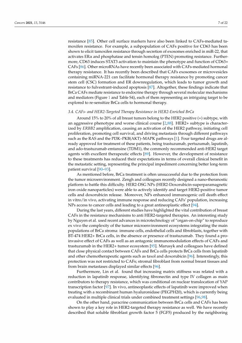
Cancers 2021, 13, 3146 4 of 22
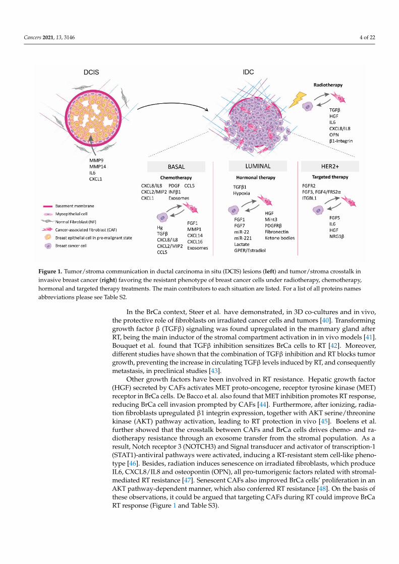
Figure 1. Tumor/stroma communication in ductal carcinoma in situ (DCIS) lesions (left) and tumor/stroma crosstalk ininvasive breast cancer (right) favoring the resistant phenotype of breast cancer cells under radiotherapy, chemotherapy,hormonal and targeted therapy treatments. The main contributors to each situation are listed. For a list of all proteins namesabbreviations please see Table S2.
In the BrCa context, Steer et al. have demonstrated, in 3D co-cultures and in vivo,the protective role of fibroblasts on irradiated cancer cells and tumors [40]. Transforminggrowth factor β (TGFβ) signaling was found upregulated in the mammary gland afterRT, being the main inductor of the stromal compartment activation in in vivo models [41].Bouquet et al. found that TGFβ inhibition sensitizes BrCa cells to RT [42]. Moreover,different studies have shown that the combination of TGFβ inhibition and RT blocks tumorgrowth, preventing the increase in circulating TGFβ levels induced by RT, and consequentlymetastasis, in preclinical studies [43].
Other growth factors have been involved in RT resistance. Hepatic growth factor(HGF) secreted by CAFs activates MET proto-oncogene, receptor tyrosine kinase (MET)receptor in BrCa cells. De Bacco et al. also found that MET inhibition promotes RT response,reducing BrCa cell invasion prompted by CAFs [44]. Furthermore, after ionizing, radia-tion fibroblasts upregulated β1 integrin expression, together with AKT serine/threoninekinase (AKT) pathway activation, leading to RT protection in vivo [45]. Boelens et al.further showed that the crosstalk between CAFs and BrCa cells drives chemo- and ra-diotherapy resistance through an exosome transfer from the stromal population. As aresult, Notch receptor 3 (NOTCH3) and Signal transducer and activator of transcription-1(STAT1)-antiviral pathways were activated, inducing a RT-resistant stem cell-like pheno-type [46]. Besides, radiation induces senescence on irradiated fibroblasts, which produceIL6, CXCL8/IL8 and osteopontin (OPN), all pro-tumorigenic factors related with stromal-mediated RT resistance [47]. Senescent CAFs also improved BrCa cells’ proliferation in anAKT pathway-dependent manner, which also conferred RT resistance [48]. On the basis ofthese observations, it could be argued that targeting CAFs during RT could improve BrCaRT response (Figure 1 and Table S3).
Cancers 2021, 13, 3146 5 of 22
3.2. CAFs and Chemotherapy Resistance in Triple Negative BrCa
TNBrCa or basal subtype represents around 15% of BrCa lesions diagnosed and ischaracterized by the expression of basal-related genes and the absence or low levels of ER,PR and, HER2 [2]. This BrCa subtype encompasses a wide and heterogeneous group oflesions and is generally associated with a poor prognosis due to the lack of appropriatetargeted therapies [49]. Therefore, the main clinical challenge in TNBrCa is to design newtherapies to reduce metastasis and improve patient’s quality of life and survival.
Concerning CAFs contribution to therapy-resistance, a 6-CAFs’ genes signature(CXCL2/MIP2 and its human homologous CXCL8/IL8, MMP1, Retinoic Acid Recep-tor Responder 1 (RARRES1), fibroblast growth factor 1 (FGF1), and CXCR7) has beenrelated to taxotere treatment resistance in an in vitro model using TNBrCa cells [50]. In ad-dition, EDALINE phase I clinical trial results suggest that blocking hedgehog (Hh) stimulion CAFs, by smoothened inhibitors (SMOi) treatment, sensitizes TNBrCa to docetaxel andleads to survival amelioration [51]. Moreover, inhibition of TGFβ production by CAFs withthe anti-fibrotic drug pirfenidone reduced BrCa tumor growth and also lung metastasiswhen combined with doxorubicin, acting in a synergistic way [52]. Combination of a hy-drogel assembled to losartan, an inhibitor of angiotensin-II that blocks the TGFβ pathway,sensitizes TNBrCa cell lines to doxorubicin-loaded liposomes facilitating the reduction ofchemotherapy resistance [53].
On the other hand, under chemotherapy pressure, interferon β1 (IFNβ1) secretionby cancer cells stimulates the transcription of pro-inflammatory cytokine genes in stromalfibroblasts, by which CAFs acquire an anti-viral state, essential for the recovery and re-sistance acquisition of BrCa cells after chemotherapy. Indeed, high expression of tumorIFNβ1 in TNBrCa patients correlated with an aggressiveness signature [54]. Another inter-esting paracrine relationship between cancer cells expressing platelet derived growth factor(PDGF) and CAFs positive for PDGF-receptors was described in basal-like BrCa patients.PDGF abrogation in the tumor produces a phenotype shift into a positive hormone-receptorsubtype, sensitive to endocrine therapy, leading to a more therapeutically affordable BrCasubtype. These results also suggest that BrCa subtype control and plasticity can be exertedby fibroblasts and cancer cells interactions [55].
Furthermore, the interaction between cancer cells, immune cells, mesenchymal stemcells (MSC), and CAFs led to the release of CXCL1, CXCL2/MIP (homologous to CXCL8/IL8in humans) (from TNBrCa cells), CXCL16 (from CAFs), and C-C motif chemokine ligand 5(CCL5) (from CAFs and MSCs), resulting in a more reactive stroma, inducing pro-tumoralactions, including chemotherapy resistance [56–61]. This pro-resistance context attractstumor-associated macrophages and stimulates the endothelial cell population throughthe release of CXCL8/IL8 and CCL5 by TNBrCa cells. All these results suggested thattherapeutically targeting CXCL8/IL8 and CCL5 could be useful in TNBrCa patient’s treat-ment [62,63]. Expression of CXCL14 or Programmed death-ligand 1 (PD-L1) by stromalCAFs has also been associated with shorter or better survival, respectively, in TNBrCapatients [64,65], and could be considered as independent prognosis markers in TNBrCapatients. Therefore, these data emphasize that there is a strong chemotherapy-resistancedependence of tumor-stromal interactions in TNBrCa, particularly evident between fibrob-lasts and cancer cells (Figure 1 and Table S4), highlighting the importance of targetingstroma and tumor cell interactions as a promising clinical therapeutic strategy in preventingdrug resistance in TNBrCa patients [66].
3.3. CAFs and Hormonal Therapy Resistance in Luminal BrCa
Luminal BrCa represents 70% of all BrCas and expresses hormonal receptors, hence itis considered sensitive to endocrine therapy, especially ER-targeted [67]. The prognosis forearly luminal BrCa patients is particularly good, however, up to 50% of patients relapse anddie from metastatic disease [67]. The treatment of patients with ER-positive BrCa is basedon ER modulators such as tamoxifen and/or aromatase inhibitors. Tamoxifen has becomethe drug of first choice in patients with luminal BrCa. Nevertheless, it is estimated that ap-
Cancers 2021, 13, 3146 6 of 22
proximately 45% of women do not respond to tamoxifen (de novo resistance) [68], whereasacquired resistance to the drug develops ultimately in all tamoxifen-receiving patients.Therefore, although tamoxifen was hailed as a major breakthrough in the management ofpatients with hormone-dependent BrCa, development of resistance to the treatment posesa serious clinical problem [69].
Endocrine-therapy resistance can likewise be linked to the biological action of luminalBrCa CAFs. They express a G protein-coupled estrogen receptor 1 (GPER), which suggestthat they are also sensitive to estrogens. CAFs expressing GPER can activate the epidermalgrowth factor receptor (EGFR) and extracellular signal-regulated kinase 2 (ERK) signalingwhich leads to (i) estradiol production by CAFs and endocrine therapy resistance [70] and(ii) cancer cell integrin β1 activation by increased CAF-secreted fibronectin [71], whichdrives EMT and the tamoxifen-resistant phenotype. In turn, CAFs can also become resistantto hormonal therapy. BrCa-resistant cells secrete TGFβ1 that induces ERK activation onCAFs and promotes their resistance [72]. In addition, activation of phosphatidylinositol-4,5-bisphosphate 3-kinase (PI3K)/AKT in BrCa cells increases CAFs’ GPER translocationto the membrane and induces the activation of PKA/cAMP responsive element-bindingprotein (CREB) signaling, which activates the Warburg effect in CAFs. This increasespyruvate and lactate levels, metabolites used by cancer cells to fuel their mitochondrialmetabolism and confer drug resistance to hormonal- and HER2-targeted therapies, aswell as chemotherapy [73]. CAFs-mediated metabolic reprograming of BrCa cells todecrease sensitivity to endocrine therapy has been proposed by others. For instance, CAFs’secretion of lactate and ketone bodies increases mitochondrial activity in BrCa cells andleads to tamoxifen resistance [74]. A recent report has shown that BrCa cells promoteneighboring CAFs’ hypoxia, which leads to autophagic degradation of CAFs CAV1 andactivation of the Warburg effect in them. These changes contribute to protect BrCa cellsfrom endocrine therapy-induced apoptosis and autophagy, by providing nutrients to fuelBrCa cell metabolism [75]. In fact, BrCa patients whose stroma is negative for CAV1expression develop resistance to tamoxifen and have worse prognosis [76].
Endocrine therapy resistance is also driven by CAFs’ secretion of growth factors. Forinstance, HGF secretion by CAFs leads to MET upregulation in ER-positive BrCa cell lines,which leads to increase migration, invasion, and resistance to fulvestrant through SRCproto-oncogene, non-receptor tyrosine kinase (SRC), AKT, and ERK1/2 activation [77].FGF1 has also been shown to induce hormonal therapy resistance through activation offibroblast growth factor receptor 3 (FGFR3) that causes PI3K/AKT and ERK1/2 inductionby phospholipase C γ (PLCγ) [78]. Additionally, FGF7 can block tamoxifen effect throughactivation of FGFR2, which enhances ER degradation by the proteasome. FGF7/FGFR2effect was mediated through activation of PI3K/AKT signaling and the upregulation ofB-cell lymphoma 2 (Bcl-2) expression in BrCa cells [79].
Several other reports have linked CAFs-elicited resistance to tamoxifen with activa-tion of PI3K/AKT and/or Ras/Raf/mitogen activated protein kinase kinase 1 (MEK1)/ERK1/2 [80–82]. Fibronectin, through interaction with Integrin β1, and soluble factorssecreted by CAFs, activate EGFR and MMPs which leads to induction of PI3K/AKT andERK1/2 in BrCa cells, protecting them from tamoxifen-induced cell death [83]. AKT andERK1/2 hyperactivation stimulates ER phosphorylation which promotes tamoxifen re-sistance [80]. Moreover, fibroblasts expressing amyloid beta precursor protein bindingfamily A, member 3 (MINT3) upregulate L1 cell adhesion molecule (L1CAM) which leadsto ERK1/2 activation in BrCa cells through integrin α5β1, promoting tumor growth andresistance [81]. Stromal cells have also been shown to elicit hormonal therapy resistancethrough inhibition of insulin-like growth factor binding protein 5 (IGFBP5) which inducesBcl-3 upregulation and activation of NF-κB, driving resistance to fulvestrant [84].
CD146 is a cell surface marker that characterizes two different subtypes of fibrob-lasts in BrCa tumors. CAFs negative for CD146 foster the upregulation and activation ofseveral tyrosine kinase receptors (TKRs), such as EGFR, HER2 and insulin-like growthfactor receptor (IGF1R), and the inhibition of ER in BrCa cells which leads to tamoxifen
Cancers 2021, 13, 3146 7 of 22
resistance [85]. Other cell surface markers have also been linked to CAFs-mediated ta-moxifen resistance. For example, a subpopulation of CAFs positive for CD63 has beenshown to elicit tamoxifen resistance through secretion of exosomes enriched in miR-22, thatactivates ERα and phosphatase and tensin homolog (PTEN) promoting resistance. Further-more, CD63 induces STAT3 activation to maintain the phenotype and function of CD63+CAFs [86]. Other microRNAs have recently been associated with CAFs-mediated hormonaltherapy resistance. It has recently been described that CAFs exosomes or microvesiclescontaining miRNA-221 can facilitate hormonal therapy resistance by promoting cancerstem cell (CSC) formation and ER downregulation, which leads to tumor growth andresistance to fulvestrant-induced apoptosis [87]. Altogether, these findings indicate thatBrCa CAFs mediate resistance to endocrine therapy through several molecular mechanismsand mediators (Figure 1 and Table S4), each of them representing an intriguing target to beexplored to re-sensitize BrCa cells to hormonal therapy.
3.4. CAFs- and HER2-Targeted Therapy Resistance in HER2-Enriched BrCa
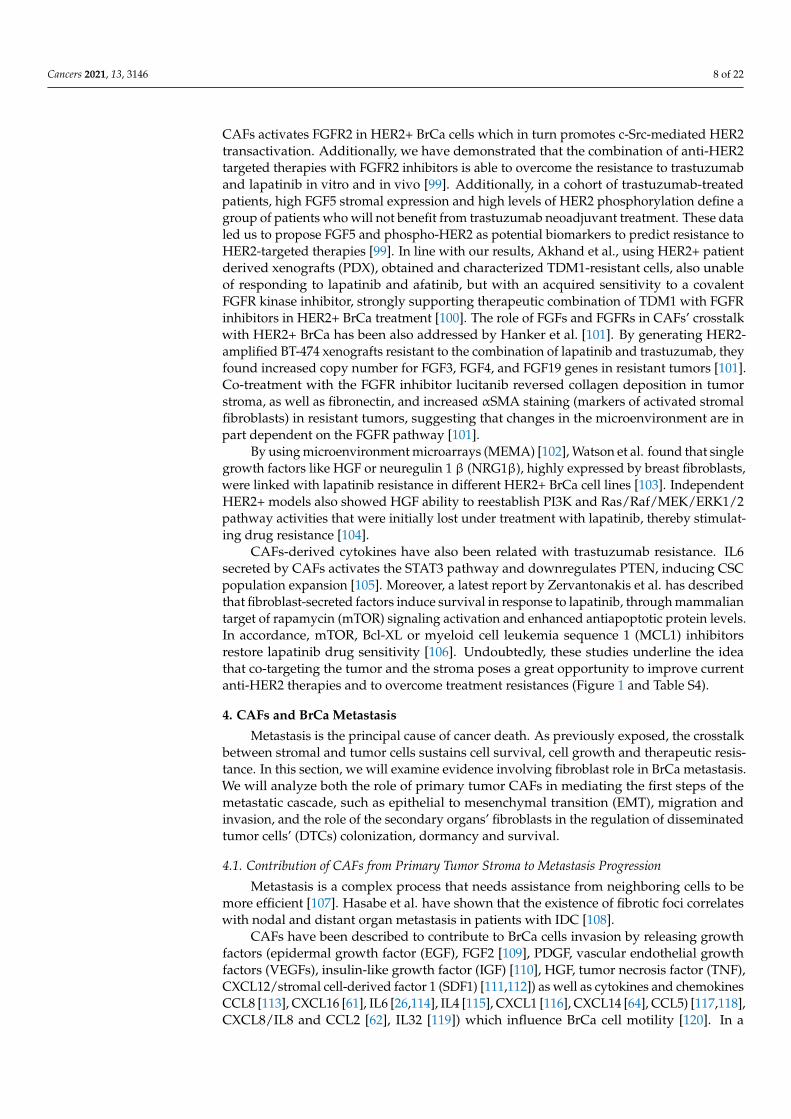
Around 15% to 20% of all breast tumors belong to the HER2 positive (+) subtype, withan aggressive phenotype and worse clinical course [2,88]. HER2+ subtype is character-ized by ERBB2 amplification, causing an activation of the HER2 pathway, initiating cellproliferation, promoting cell survival, and driving metastasis through different pathwayssuch as the RAS and the PI3K–PKB(AKT)–MAPK pathways [1]. Four targeted drugs are al-ready approved for treatment of these patients, being trastuzumab, pertuzumab, lapatinib,and ado-trastuzumab emtansine (TDM1), the commonly recommended anti-HER2 targetagents with excellent therapeutic effects [89]. However, the development of resistanceto these treatments has reduced their expectations in terms of overall clinical benefit inthe metastatic setting, representing the principal impediment concerning better long-termpatient survival [90–93].
As mentioned before, BrCa treatment is often unsuccessful due to the protection fromthe tumor microenvironment. Zengh and colleagues recently designed a nano-theranosticplatform to battle this difficulty. HER2-DSG NPs (HER2-Doxorubicin-superparamagneticiron oxide nanoparticles) were able to actively identify and target HER2-positive tumorcells and doxorubicin release. Moreover, NPs enhanced immunogenic cell death effectin vitro/in vivo, activating immune response and reducing CAFs’ population, increasingNPs access to cancer cells and leading to a great antineoplastic effect [94].
During the last years, different studies have highlighted the vital contribution of breastCAFs in the resistance mechanisms to anti HER2-targeted therapies. An interesting studyby Nguyen et al. used recent advances in microtechnology of “organ-on-chip” to reproduceex vivo the complexity of the tumor microenvironment ecosystems integrating the mainpopulations of BrCa stroma: immune cells, endothelial cells and fibroblasts, together withBT-474 HER2+ BrCa cells, in the absence or presence of trastuzumab. They found a pro-invasive effect of CAFs as well as an antagonic immunomodulation effects of CAFs andtrastuzumab in the HER2+ tumor ecosystem [95]. Marusyk and colleagues have definedthat close physical contact between CAFs and BrCa cells protects BrCa cells from lapatiniband other chemotherapeutic agents such as taxol and doxorubicin [96]. Interestingly, thisprotection was not restricted to CAFs; stromal fibroblast from normal breast tissues andfrom brain metastases displayed similar effects [96].
Furthermore, Lin et al. found that increasing matrix stiffness was related with areduction in lapatinib response, identifying fibronectin and type IV collagen as maincontributors to therapy resistance, which was conditional on nuclear translocation of YAPtranscription factor [97]. In vivo, antineoplastic effects of lapatinib were improved whentreating with a recombinant human hyaluronidase (PEGPH20), which is currently beingevaluated in multiple clinical trials under combined treatment settings [96,98].
On the other hand, paracrine communication between BrCa cells and CAFs has beenshown to play a key role in HER2-targeted therapy resistance as well. We have recentlydescribed that soluble fibroblast growth factor 5 (FGF5) produced by the neighboring
Cancers 2021, 13, 3146 8 of 22
CAFs activates FGFR2 in HER2+ BrCa cells which in turn promotes c-Src-mediated HER2transactivation. Additionally, we have demonstrated that the combination of anti-HER2targeted therapies with FGFR2 inhibitors is able to overcome the resistance to trastuzumaband lapatinib in vitro and in vivo [99]. Additionally, in a cohort of trastuzumab-treatedpatients, high FGF5 stromal expression and high levels of HER2 phosphorylation define agroup of patients who will not benefit from trastuzumab neoadjuvant treatment. These dataled us to propose FGF5 and phospho-HER2 as potential biomarkers to predict resistance toHER2-targeted therapies [99]. In line with our results, Akhand et al., using HER2+ patientderived xenografts (PDX), obtained and characterized TDM1-resistant cells, also unableof responding to lapatinib and afatinib, but with an acquired sensitivity to a covalentFGFR kinase inhibitor, strongly supporting therapeutic combination of TDM1 with FGFRinhibitors in HER2+ BrCa treatment [100]. The role of FGFs and FGFRs in CAFs’ crosstalkwith HER2+ BrCa has been also addressed by Hanker et al. [101]. By generating HER2-amplified BT-474 xenografts resistant to the combination of lapatinib and trastuzumab, theyfound increased copy number for FGF3, FGF4, and FGF19 genes in resistant tumors [101].Co-treatment with the FGFR inhibitor lucitanib reversed collagen deposition in tumorstroma, as well as fibronectin, and increased αSMA staining (markers of activated stromalfibroblasts) in resistant tumors, suggesting that changes in the microenvironment are inpart dependent on the FGFR pathway [101].
By using microenvironment microarrays (MEMA) [102], Watson et al. found that singlegrowth factors like HGF or neuregulin 1 β (NRG1β), highly expressed by breast fibroblasts,were linked with lapatinib resistance in different HER2+ BrCa cell lines [103]. IndependentHER2+ models also showed HGF ability to reestablish PI3K and Ras/Raf/MEK/ERK1/2pathway activities that were initially lost under treatment with lapatinib, thereby stimulat-ing drug resistance [104].
CAFs-derived cytokines have also been related with trastuzumab resistance. IL6secreted by CAFs activates the STAT3 pathway and downregulates PTEN, inducing CSCpopulation expansion [105]. Moreover, a latest report by Zervantonakis et al. has describedthat fibroblast-secreted factors induce survival in response to lapatinib, through mammaliantarget of rapamycin (mTOR) signaling activation and enhanced antiapoptotic protein levels.In accordance, mTOR, Bcl-XL or myeloid cell leukemia sequence 1 (MCL1) inhibitorsrestore lapatinib drug sensitivity [106]. Undoubtedly, these studies underline the ideathat co-targeting the tumor and the stroma poses a great opportunity to improve currentanti-HER2 therapies and to overcome treatment resistances (Figure 1 and Table S4).
4. CAFs and BrCa Metastasis
Metastasis is the principal cause of cancer death. As previously exposed, the crosstalkbetween stromal and tumor cells sustains cell survival, cell growth and therapeutic resis-tance. In this section, we will examine evidence involving fibroblast role in BrCa metastasis.We will analyze both the role of primary tumor CAFs in mediating the first steps of themetastatic cascade, such as epithelial to mesenchymal transition (EMT), migration andinvasion, and the role of the secondary organs’ fibroblasts in the regulation of disseminatedtumor cells’ (DTCs) colonization, dormancy and survival.
4.1. Contribution of CAFs from Primary Tumor Stroma to Metastasis Progression
Metastasis is a complex process that needs assistance from neighboring cells to bemore efficient [107]. Hasabe et al. have shown that the existence of fibrotic foci correlateswith nodal and distant organ metastasis in patients with IDC [108].
CAFs have been described to contribute to BrCa cells invasion by releasing growthfactors (epidermal growth factor (EGF), FGF2 [109], PDGF, vascular endothelial growthfactors (VEGFs), insulin-like growth factor (IGF) [110], HGF, tumor necrosis factor (TNF),CXCL12/stromal cell-derived factor 1 (SDF1) [111,112]) as well as cytokines and chemokinesCCL8 [113], CXCL16 [61], IL6 [26,114], IL4 [115], CXCL1 [116], CXCL14 [64], CCL5) [117,118],CXCL8/IL8 and CCL2 [62], IL32 [119]) which influence BrCa cell motility [120]. In a
Cancers 2021, 13, 3146 9 of 22
recent study Suh et al. evidenced that CAFs secretion of FGF2 was sufficient to enhanceMDA-MB-231 cells growth, migration and invasion via FGFR1 signaling [109]. Dvoraket al. showed that CXCL12/SDF1 secretion induces mammalian Diaphanous-relatedformin-2 (MDia2) deregulation, leading to breakdown of the F-actin cytoskeleton and asubsequent increase of BrCa cell motility [112]. Moreover, αSMA+ fibroblasts producehigh amounts of CXCL12/SDF1 and IGF1, which select cancer cells with high Src activityprone to colonize CXCL12/SDF1-rich bone marrow microenvironment, further suggestinga potential role of primary tumor CAFs in educating BrCa cells to metastasize to the bonemicroenvironment [121]. In addition, fibroblasts at tumor margins secrete higher levels ofCCL8, generating a gradient between the epithelium, the stroma and the periphery thatattracts BrCa cells to migrate and disseminate [113]. Besides, high stromal expression ofCXCL14 is significantly associated with shorter recurrence periods [64]. A tumor necrosisfactor alfa (TNFα) and IL1β enriched environment promotes metastasis in TNBrCa cellsthrough the shift to a metastatic phenotype in both stromal and cancer cells, leading toan increased expression of pro-metastatic and immune-evasive chemokines, CXCL8/IL8,CCL2 and CCL5, among others, induction of angiogenesis and enhanced invasive andmigration abilities of TNBrCa cells [62,122–127].
Several reports have also defined the importance of the release by CAFs of adipokinessuch as leptin in promoting the malignant aggressiveness of BrCa cells [128]. Giordano et al.described leptin as a regulator of the crosstalk between the stromal and BrCa cells. Leptinproduction by CAFs induce mammosphere formation, suggesting leptin can promote BrCacells stemness [129]. Moreover, the inhibition of leptin with a synthetic farnesoid X receptor(FXR) agonist GW4064, reduces the pro-tumorigenic effect of CAFs in BrCa [130]. Othergroups have also described an important role of leptin in potentiating BrCa cells migrationand angiogenesis, being also involved in EMT [131]
Luga et al. also reported that CAFs produce exosomes that boost the motility andmetastatic potential of BrCa cells by activating Wintless-INT family (Wnt) signaling [132].Otherwise, BrCa exosomes such as miR-146a regulate the activation of Wnt/β-catenin sig-naling pathway on fibroblasts, which become CAFs contributing to invasive and metastaticabilities of BrCa cells [133]. Conversely, CAFs-derived exosomes induce proliferationand metastasis through the miR-500a-5p transference to luminal and basal-like BrCa cells.miR-500a-5p interacts with and blocks the effects of USP28, considered by the authors as apossible BrCa tumor suppressor [134]. Another work demonstrates that CAF exosomes canrelease miR-21, -143, and -378 that enhance important aggressive cancer hallmarks suchas stemness, EMT and the ability to grow in an anchorage-independent way in TNBrCacells [135].
One of the key steps in the metastatic process is cancer cell EMT and CAFs have beendescribed to secrete growth factors such as transforming growth factor beta 1 (TGFβ1),EGF, PDGF and HGF that will promote EMT in primary tumor cells [107,120,136,137].Likewise, the increased OPN secretion by TIAM rac1 associated GEF 1 (TIAM-1)-defectiveCAFs stimulates BrCa EMT, stemness and invasion [138]. Recently, Wen et al. havedescribed that CAFs secrete IL32 that binds integrin β3 on BrCa cells resulting in p38MAPK pathway activation, further enhancing the expressions of fibronectin, N-cadherinand vimentin [119]. In addition, it seems that activation of autophagy and expression ofFAP-α in CAFs can activate EMT in cancer cells via Wnt pathway, resulting in an increasein cell migration, invasion and proliferation [139,140]. An interesting study describesthat CAFs expression of phosphatidylinositol-4,5-bisphosphate 3-kinase catalytic subunitdelta (PIK3Cδ) fosters TNBrCa cell metastasis, controlling mainly invasion through theinhibition of tumor suppressor genes such as Nuclear Receptor Subfamily 4 Group AMember 1 (NR4A1) [141]. Recently, Matsumura and colleagues have established thatCAFs-secreted factors control the epithelial/mesenchymal plasticity of distinct metastatictumor clusters, which comprise two different cell populations, one with a more epithelialphenotype and another with a mixed epithelial-mesenchymal phenotype. Both populationscollectively contribute to enhanced tumor cell cluster formation and metastatic seeding in
Cancers 2021, 13, 3146 10 of 22
secondary organs [142]. Choi et al. have demonstrated, using three-dimensional in vitromodels, that CAFs can facilitate transmigration of BrCa cells through the blood–brainbarrier by increasing their expression of α5β1 and αvβ1 integrin and MET [143].
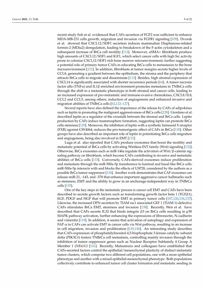
It is accepted that fibroblasts contribute to the remodeling of the surrounding stroma,which is a prerequisite for cancer cells’ invasion and metastasis. In vitro and in vivo studieshave indicated that CAFs promote BrCa cell invasion and metastasis by secreting MMPs,such as MMP1, -2, -3, -7, -9, -13, and -14 [136,144,145]. Moreover, different research groupshave observed that CAFs may generate tracks for tumor cells to go forward. For instance,Syndecan-1 expressing fibroblasts modify the collagen fibers in the extracellular matrix toform parallel structures that can be utilized by BrCa cells as a guide [146]. Ahirwar andcolleagues showed that CAFs secreted CXCL12/SDF1, generating endothelial instabilityand hyper-permeable vasculature, facilitating the escape of tumor cells from the primarytumor to distant organs [147]. Other studies have additionally indicated that CAFs adopt adesmoplastic program with altered cell adhesion properties which pushes the progressionof metastasis through induction of mechanical pressure on cancer tissue [148]. Thesestudies highlight the important role of CAFs in mediating BrCa cell migration, EMT andinvasion (Figure 2 and Table S5), hence suggesting stroma-targeted therapies might be aninteresting approach to address metastatic disease.
Figure 2. Pro-metastatic CAFs-secreted factors at the primary tumor promote tumor cell migration, invasion, and dissemi-nation. For a list of all proteins names abbreviations please see Table S2.
Cancers 2021, 13, 3146 11 of 22
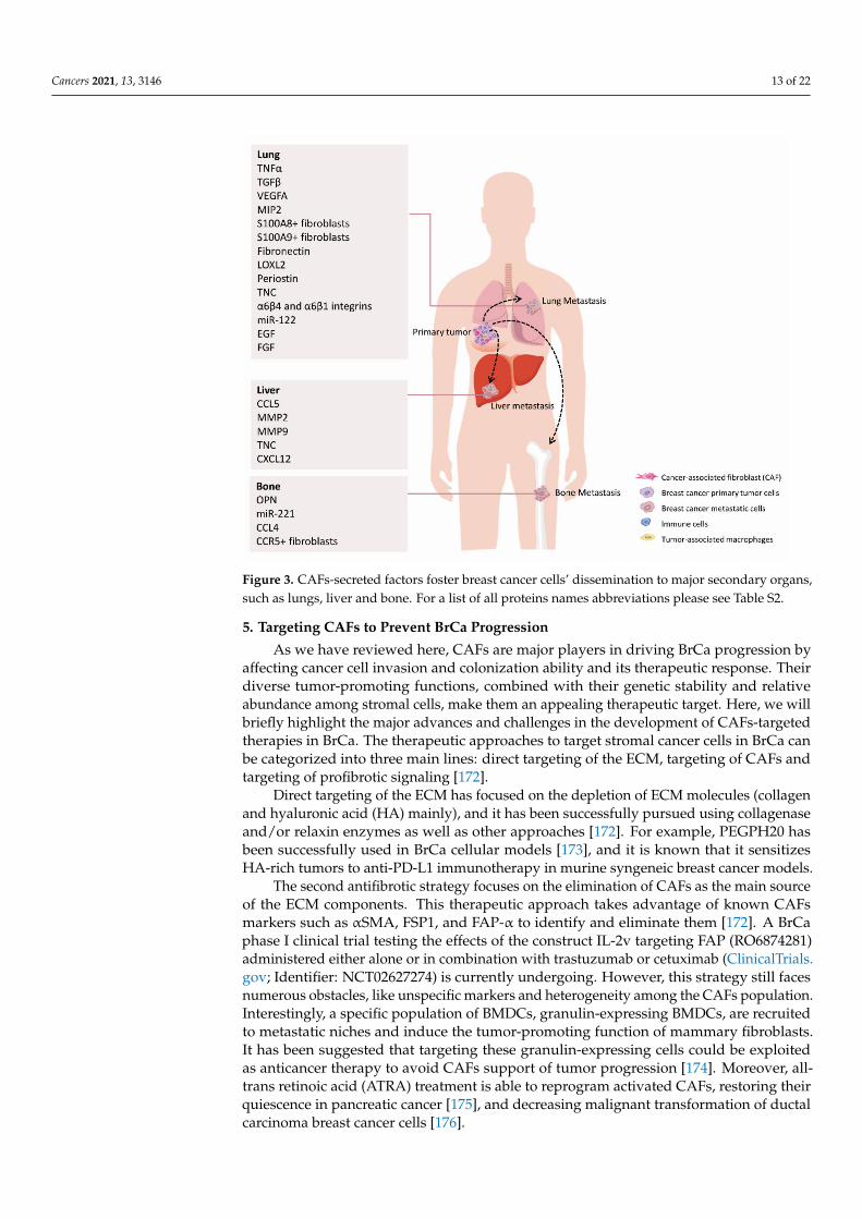
4.2. Contribution of Fibroblasts from Metastatic Sites to Metastasis Formation
Once disseminated, tumor cells arrive to metastatic target organs where they willstart interacting with stromal cells of the target organ. This new dialogue between DTCsand stromal cells will determine DTCs survival, activation of dormancy or the shift to aproliferative phenotype, hence regulating metastasis progression [149]. Gui et al. recentlypublished a comparative study with BrCa fibroblasts from primary site (pCAFs), metastaticorgans (mCAFs) and normal fibroblasts. They showed that mCAFs display more markedpro-tumorigenic effects, also conferring greater protection to cytotoxic drugs and improvingmetastatic capacity. All these differential capabilities of mCAFs were associated in partwith their higher secretion of IFNβ and IGF2, which in turn activates IGF1R signaling [150].BrCa DTCs usually colonize lungs, bone marrow and brain [151]. It has been describedthat communication between primary tumors and their potential metastatic sites startseven prior to the arrival of DTCs to the target organ. In the next paragraphs, we will focuson the accommodation of the stroma of the target organs and its role on DTCs fate.
During the early dissemination phase to the lungs, different primary tumor-derivedmediators, such as growth factors, cytokines, extracellular matrix (ECM)-remodelingenzymes or extracellular vesicles and exosomes, have the ability to modify the pulmonaryparenchyma creating a welcoming microenvironment for the tumor cells to seed, proliferateand survive [152]. Secretion of TNFα and TGFβ, along with vascular endothelial growthfactor A (VEGFA), induce the expression of S100 calcium binding protein A8 (S100A8)and S100 calcium binding protein A9 (S100A9) in lung fibroblasts to prepare premetastaticniches [153]. S100A8 and S100A9 stimulate the recruitment of Mac-1+ myeloid cellsto the lung, resulting in the secretion of factors able to stimulate migration like TNFα,CXCL2/MIP2 and TGFβ, as well as ECM remodeling [154]. A study by Raz et al. furtherdemonstrated that bone marrow-derived stromal cells (BM-MSCs) are the precursors of asignificant fraction of CAFs that are present in both primary breast and metastatic lunglesions [155], which are able to trigger a distinctive inflammatory profile, inducing pro-metastatic characteristics like angiogenesis and supporting metastasis to the lungs [155].
Exosomes derived from PT cancer cells play a critical role in transforming the lungmicroenvironment. A thorough study by Hoshino and colleagues revealed an organspecificity of exosome biodistribution that matched the organotrophic distribution of thecell line of origin [156]. Interestingly, they also found that lung-tropic exosomes, expressingα6β4 and α6β1 integrins, predominantly co-localized with S100 calcium binding proteinA4 (S100A4)-positive fibroblasts and surfactant protein C-positive epithelial cells in thelungs, enhancing lung metastases [156]. Accordingly, integrin α6β4 targeting reduced lungmetastasis [156]. Interestingly, Fong et al. have shown that exosomal miR-122 is able toreprogram lung niche cells to a lower glucose demand, by down-regulating pyruvate kinaseactivity. This metabolic reprogramming results in an increased DTCs glucose availabilityto effectively drive their proliferation [157].
Additionally, the contribution of BrCa-secreted exosomal long non-coding RNAs(LncRNAs) on lung pre-metastatic niche education has been described. These LncRNAsstimulated lung fibroblast proliferation and migration by targeting different genes andsignaling pathways, including TGFβ, pentose phosphate, Hh, metabolic, and complementand coagulation cascade pathways [158]. Medeiros et al. found that extracellular vesicles(EVs) from TNBrCa cells were able to stimulate the expression of pre-metastatic ECMmarkers in lung fibroblasts [159].
During early metastatic colonization, lung DTCs reprogram resident lung fibroblaststo create a more favorable environment for proliferation and survival. For instance, DTCsdecrease miR-30 family members’ expression in lung fibroblasts, which inhibit MMP9expression, increasing vascular permeability and consequently favoring metastasis [160].BrCa DTCs also induce the expression of αSMA in resident fibroblasts in the lung. Acti-vated fibroblasts direct extracellular matrix remodeling by the secretion of fibronectin andLOXL2, creating a more tolerant microenvironment for DTCs to survive and outgrow [161].Moreover, cancer cells are able to educate fibroblasts to produce periostin and Tenascin C
Cancers 2021, 13, 3146 12 of 22
(TNC), enhancing metastatic colonization of the lung [162]. Periostin expression in lungfibroblasts may be modulated by cancer cells through the secretion of transforming GrowthFactor Beta 3 (TGFβ3). Fibroblasts in turn recruit Wnt ligands to present to cancer cells andpromote DTCs stemness by inducing Wnt signaling, enhancing lung colonization [152].Periostin also fosters the lung recruitment of MDSCs at the metastatic site, especially inER-negative tumors, essential for achieving an immunosuppressive premetastatic nicheenvironment. In addition, periostin can bind to TNC, anchoring it to ECM componentssuch as fibronectin and type I collagen [163]. DTCs-derived TNC supports early metastasisoutgrowth, until the lung stroma takes over as the main source of TNC. TNC can alsoinduce the secretion of growth factors, like EGF or FGF, and interact with fibronectin,heparin-sulfate proteoglycans, fibrinogen, integrins, MMPs, and EGFR [164]. Addition-ally, high thrombospondin 2 (THBS2)-expressing cancer cells are also more efficient onactivating lung fibroblasts, which subsequently induce the transition of tumor cells into amore epithelial phenotype [165]. In addition, Shu et al. have described a new role of thecomplement system C3a-C3aR on metastasis promotion by modulating CAFs activation,enhancing pro-metastatic cytokine secretion via the activation of PI3K-AKT signaling [166].
Bone is the main target organ for HER2+ BrCa metastasis, representing the majorcause of pain and severe complications for these patients, including fracture, spinal cordcompression or hypercalcemia [167]. Kang et al. have described a 6-gene bone metastasissignature in BrCa cell lines using mouse models. Interestingly, one of the genes wasFGF5 that our group has recently described to be implicated in HER2+ targeted therapyresponse [99,168]. It has also been reported that BrCa cells in the bone marrow displayhigher expression of nanog homeobox (NANOG) OCT4 and SOX2 in the presence ofMSC-derived CAFs and OPN [169], enhancing BrCa cells’ stemness. In turn, vimentin-expressing FAP+ fibroblasts produce miR-221 containing exosomes that further contributeto maintain the stem phenotype in favor of metastasis [87]. Furthermore, DTCs in thebone cavity produce abundant C-C motif chemokine ligand 4 (CCL4) as well, to attracttype I collagen-expressing α-SMA+ fibroblasts expressing C-C motif chemokine receptor 5(CCR5). Accumulated fibroblasts support cancer cells’ growth by enhancing connectivetissue growth factor signals [170].
Regarding BrCa liver metastasis and the potential role of fibroblasts aiding the process,it has been defined that OPN promotes tumor progression via the transformation of MSCinto CAFs in the liver as well. OPN was responsible for MSC CCL5 production. Moreover,the MSCs recovered from liver metastatic sites present CAF markers’ expression (α-SMA,TNC, CXCL12/SDF1, and fibroblast-specific protein-1) as well as MMP-2 and MMP-9 [171].
All these studies underline the essential role of resident fibroblasts in reprogrammingthe metastatic niche to favor DTCs seeding and survival at secondary organs (Figure 3and Table S5). These insights could help design therapies targeting the cancer cell-stromalcrosstalk of primary tumors and metastases, likely improving the clinical management andoutcome of the advanced BrCa patients.
Cancers 2021, 13, 3146 13 of 22
Figure 3. CAFs-secreted factors foster breast cancer cells’ dissemination to major secondary organs,such as lungs, liver and bone. For a list of all proteins names abbreviations please see Table S2.
5. Targeting CAFs to Prevent BrCa Progression
As we have reviewed here, CAFs are major players in driving BrCa progression byaffecting cancer cell invasion and colonization ability and its therapeutic response. Theirdiverse tumor-promoting functions, combined with their genetic stability and relativeabundance among stromal cells, make them an appealing therapeutic target. Here, we willbriefly highlight the major advances and challenges in the development of CAFs-targetedtherapies in BrCa. The therapeutic approaches to target stromal cancer cells in BrCa canbe categorized into three main lines: direct targeting of the ECM, targeting of CAFs andtargeting of profibrotic signaling [172].
Direct targeting of the ECM has focused on the depletion of ECM molecules (collagenand hyaluronic acid (HA) mainly), and it has been successfully pursued using collagenaseand/or relaxin enzymes as well as other approaches [172]. For example, PEGPH20 hasbeen successfully used in BrCa cellular models [173], and it is known that it sensitizesHA-rich tumors to anti-PD-L1 immunotherapy in murine syngeneic breast cancer models.
The second antifibrotic strategy focuses on the elimination of CAFs as the main sourceof the ECM components. This therapeutic approach takes advantage of known CAFsmarkers such as αSMA, FSP1, and FAP-α to identify and eliminate them [172]. A BrCaphase I clinical trial testing the effects of the construct IL-2v targeting FAP (RO6874281)administered either alone or in combination with trastuzumab or cetuximab (ClinicalTrials.gov; Identifier: NCT02627274) is currently undergoing. However, this strategy still facesnumerous obstacles, like unspecific markers and heterogeneity among the CAFs population.Interestingly, a specific population of BMDCs, granulin-expressing BMDCs, are recruitedto metastatic niches and induce the tumor-promoting function of mammary fibroblasts.It has been suggested that targeting these granulin-expressing cells could be exploitedas anticancer therapy to avoid CAFs support of tumor progression [174]. Moreover, all-trans retinoic acid (ATRA) treatment is able to reprogram activated CAFs, restoring theirquiescence in pancreatic cancer [175], and decreasing malignant transformation of ductalcarcinoma breast cancer cells [176].
Cancers 2021, 13, 3146 14 of 22
Interfering with the TGFβ or Hh signaling pathways have also been successfullytested in BrCa. TGFβ signaling blocking, by either soluble TGFβ type II receptor or TGFβneutralizing antibody, significantly decreased tumor growth and metastasis in BrCa [177].Remarkably, TGFβ signaling can also be blocked through angiotensin receptor inhibitors(ARB), such as losartan, which enhanced delivery of chemotherapy and improved over-all survival in BrCa models [178]. Pirfenidone (PFD), an anti-fibrotic agent as well as aTGFβ antagonist, has also been effectively examined in TNBrCa patient-derived xenograftmodels [66]. Several Hh pathway inhibitors have also been tested, being IPI-926, the onewith the best results [179]. Furthermore, focal adhesion kinase (FAK) signaling has alsobeen related with the formation of fibrotic tumor microenvironment [172] and appears asa druggable target, not only in tumor cells but also in the tumor microenvironment [180].The stromal targets list in BrCa is continually growing, as do newer therapies againststromal components [181,182]. For example, in vitro and in vivo approaches againstthe phosphodiesterase PDE5 (Sildenafil) or the nuclear receptors FXR (GW4064) haveshown promising results due to their relevant role in CAFs biology and pro-tumoraleffect [130,181,183,184].
These therapeutic strategies targeting CAF/stroma-specific pathways are alreadybeing evaluated in several clinical trials on BrCa, or other solid tumors. Most of themcombine classical strategies with CAFs-targeted therapies compounds to successfullyimprove cancer therapy. The main targets are the following: TGFβ-pathway (fresoli-mumab (NCT01401062) or galunisertib (NCT02304419)), Hh (vismodegib (NCT02694224) orLDE225 (NCT02027376, NCT02694224)), Notch (MK0752 (NCT00645333)), FGFR-FGFR lig-and (Dovitinib (NCT01548924), AZD4547 (NCT01791985), FAP (RO6874813 (NCT02558140)),Hyaluronic acid in pancreatic cancer (PEGPH20 (NCT01453153)) and Multitargets ((Dasa-tinib (NCT00546104), lucitanib (NCT02053636), Famitinib (NCT04733417, NCT01653574,NCT04129996), Motesanib (NCT01349088).
6. Conclusions
In this review, we have summarized the most recent and significant findings on the roleof CAFs in BrCa therapy resistance and metastasis. The data revised here emphasizes therole of CAFs as novel, promising a therapeutic target in BrCa suggesting that combinationstrategies aimed at targeting both BrCa cells and the surrounding microenvironment mayenable more efficient therapeutic responses. Furthermore, the identification of novel andspecific biomarkers of CAFs will be priceless for accelerating the translation into the clinicof CAFs-targeted therapies.
Supplementary Materials: The following are available online at https://www.mdpi.com/article/10.3390/cancers13133146/s1, Table S1: CAFs secreted molecules and function in DCIS, Table S2: Proteinnames abbreviations, Table S3: CAFs secreted molecules and function in radiotherapy. Table S4:CAFs secreted molecules and function in BrCa subtypes therapy resistance, Table S5: CAFs secretedmolecules function in metastasis,
Author Contributions: Conceptualization, P.F.-N. and P.B.; writing—original draft preparation,P.F.-N., G.F., Á.G.-U., and P.B.; writing—review and editing, P.F.-N., G.F., Á.G.-U., P.G., N.C., P.B.;funding acquisition, P.G., N.C., P.B. All authors have read and agreed to the published version of themanuscript.
Funding: This work was supported by grants from the Spanish Ministry of Economy and Com-petitiveness (PID2019-104991RB-I00 to PB). All funding was cosponsored by the European FEDERProgram. Á.G.-U. is supported by the Madrid Community Program for Talent Attraction (MRF2017-T1/BMD-5468). P.F.-N. is supported by a Juan de la Cierva contract from the Spanish Ministry ofscience, innovation and universities. P.F.-N. and G.F. are supported by Cellex Foundation, and P.F.-N.,G.F., N.C., and P.G. are funded by La Marató TV3 (201915-30-31), and by Secretaria d’Universitats iRecerca del Departament d’Economia i Coneixement (2017_SGR_1305).
Institutional Review Board Statement: Not applicable.
Informed Consent Statement: Not applicable.
Cancers 2021, 13, 3146 15 of 22
Data Availability Statement: Not applicable.
Acknowledgments: The authors thank Darya Kulyk for her help preparing the schemes that areshown in the figures and the graphical abstract.
Conflicts of Interest: The authors declare no conflict of interest.
References1. Harbeck, N.; Penault-Llorca, F.; Cortes, J.; Gnant, M.; Houssami, N.; Poortmans, P.; Ruddy, K.; Tsang, J.; Cardoso, F. Breast cancer.
Nat. Rev. Dis. Prim. 2019, 5, 1–31. [CrossRef] [PubMed]2. Perou, C.M.; Sørlie, T.; Eisen, M.B.; van de Rijn, M.; Jeffrey, S.S.; Rees, C.A.; Pollack, J.R.; Ross, D.T.; Johnsen, H.; Akslen, L.A.;
et al. Molecular portraits of human breast tumours. Nature 2000, 406, 747–752. [CrossRef] [PubMed]3. Sorlie, T.; Perou, C.M.; Tibshirani, R.; Aas, T.; Geisler, S.; Johnsen, H.; Hastie, T.; Eisen, M.B.; van de Rijn, M.; Jeffrey, S.S.; et al.
Gene expression patterns of breast carcinomas distinguish tumor subclasses with clinical implications. Proc. Natl. Acad. Sci. USA2001, 98, 10869–10874. [CrossRef]
4. Robson, M.; Im, S.-A.; Senkus, E.; Xu, B.; Domchek, S.M.; Masuda, N.; Delaloge, S.; Li, W.; Tung, N.; Armstrong, A.; et al. Olaparibfor metastatic breast cancer in patients with a germline BRCA mutation. N. Engl. J. Med. 2017, 377, 523–533. [CrossRef]
5. Robson, M.; Goessl, C.; Domchek, S. Olaparib for metastatic germline BRCA-mutated breast cancer. N. Engl. J. Med. 2017, 377,1792–1793. [CrossRef] [PubMed]
6. Klemm, F.; Joyce, J.A. Microenvironmental regulation of therapeutic response in cancer. Trends Cell Biol. 2015, 25, 198–213. [CrossRef]7. Ruocco, M.R.; Avagliano, A.; Granato, G.; Imparato, V.; Masone, S.; Masullo, M.; Nasso, R.; Montagnani, S.; Arcucci, A.
Involvement of breast cancer-associated fibroblasts in tumor development, therapy resistance and evaluation of potentialtherapeutic strategies. Curr. Med. Chem. 2018, 25, 3414–3434. [CrossRef] [PubMed]
8. Arcucci, A.; Ruocco, M.R.; Granato, G.; Sacco, A.M.; Montagnani, S. Cancer: An oxidative crosstalk between solid tumor cells andcancer associated fibroblasts. BioMed Res. Int. 2016, 2016, 1–7. [CrossRef] [PubMed]
9. Buchsbaum, R.J.; Oh, S.Y. Breast cancer-associated fibroblasts: Where we are and where we need to go. Cancers 2016, 8, 19.[CrossRef] [PubMed]
10. Ao, Z.; Shah, S.H.; Machlin, L.M.; Parajuli, R.; Miller, P.C.; Rawal, S.; Williams, A.J.; Cote, R.J.; Lippman, M.E.; Datar, R.H.; et al.Identification of cancer-associated fibroblasts in circulating blood from patients with metastatic breast cancer. Cancer Res. 2015,75, 4681–4687. [CrossRef]
11. Costa, A.; Kieffer, Y.; Scholer-Dahirel, A.; Pelon, F.; Bourachot, B.; Cardon, M.; Sirven, P.; Magagna, I.; Fuhrmann, L.; Bernard, C.; et al.Fibroblast heterogeneity and immunosuppressive environment in human breast cancer. Cancer Cell 2018, 33, 463–479. [CrossRef]
12. Wu, F.; Yang, J.; Liu, J.; Wang, Y.; Mu, J.; Zeng, Q.; Deng, S.; Zhou, H. Signaling pathways in cancer-associated fibroblasts andtargeted therapy for cancer. Signal Transduct. Target Ther. 2021, 6, 218. [CrossRef] [PubMed]
13. Ganguly, D.; Chandra, R.; Karalis, J.; Teke, M.; Aguilera, T.; Maddipati, R.; Wachsmann, M.B.; Ghersi, D.; Siravegna, G.; Iii, H.;et al. Cancer-associated fibroblasts: Versatile players in the tumor microenvironment. Cancers 2020, 12, 2652. [CrossRef] [PubMed]
14. Avagliano, A.; Granato, G.; Ruocco, M.R.; Romano, V.; Belviso, I.; Carfora, A.; Montagnani, S.; Arcucci, A. Metabolic repro-gramming of cancer associated fibroblasts: The slavery of stromal fibroblasts. BioMed Res. Int. 2018, 2018, 1–12. [CrossRef][PubMed]
15. Monteran, L.; Erez, N. The dark side of fibroblasts: Cancer-associated fibroblasts as mediators of immunosuppression in thetumor microenvironment. Front. Immunol. 2019, 10, 1835. [CrossRef]
16. American Cancer Society. Cancer Facts & Figures 2020; American Cancer Society: Atlanta, GA, USA, 2020.17. Pareja, F.; Brown, D.N.; Lee, J.Y.; Paula, A.D.C.; Selenica, P.; Bi, R.; Geyer, F.C.; Gazzo, A.; da Silva, E.M.; Vahdatinia, M.; et al.
Whole-exome sequencing analysis of the progression from non–low-grade ductal carcinoma in situto invasive ductal carcinoma.Clin. Cancer Res. 2020, 26, 3682–3693. [CrossRef] [PubMed]
18. Hanahan, D.; Weinberg, R.A. Hallmarks of cancer: The next generation. Cell 2011, 144, 646–674. [CrossRef]19. Nelson, A.C.; Machado, H.L.; Schwertfeger, K.L. Breaking through to the other side: Microenvironment contributions to DCIS
initiation and progression. J. Mammary Gland. Biol. Neoplasia 2018, 23, 207–221. [CrossRef]20. Ma, X.-J.; Dahiya, S.; Richardson, E.; Erlander, M.; Sgroi, D.C. Gene expression profiling of the tumor microenvironment during
breast cancer progression. Breast Cancer Res. 2009, 11, R7. [CrossRef]21. Sharma, M.; Beck, A.H.; Webster, J.A.; Espinosa, I.; Montgomery, K.; Varma, S.; van de Rijn, M.; Jensen, K.C.; West, R.B. Analysis
of stromal signatures in the tumor microenvironment of ductal carcinoma in situ. Breast Cancer Res. Treat. 2010, 123, 397–404.[CrossRef] [PubMed]
22. Fernández-Nogueira, P.; Mancino, M.; Fuster, G.; Bragado, P.; de Puig, M.P.; Gascón, P.; Casado, F.J.; Carbó, N. Breast mammo-graphic density: Stromal implications on breast cancer detection and therapy. J. Clin. Med. 2020, 9, 776. [CrossRef]
23. Lisanti, M.P.; Reeves, K.; Peiris-Pagès, M.; Chadwick, A.L.; Sanchez-Alvarez, R.; Howell, A.; Martinez-Outschoorn, U.E.; Sotgia, F.;Tsirigos, A.; Pavlides, S.; et al. JNK1 stress signaling is hyper-activated in high breast density and the tumor stroma: Connectingfibrosis, inflammation, and stemness for cancer prevention. Cell Cycle 2013, 13, 580–599. [CrossRef] [PubMed]
24. Hu, M.; Peluffo, G.; Chen, H.; Gelman, R.; Schnitt, S.; Polyak, K. Role of COX-2 in epithelial-stromal cell interactions andprogression of ductal carcinoma in situ of the breast. Proc. Natl. Acad. Sci. USA 2009, 106, 3372–3377. [CrossRef] [PubMed]
Cancers 2021, 13, 3146 16 of 22
25. Sung, K.E.; Yang, N.; Pehlke, C.; Keely, P.J.; Eliceiri, K.W.; Friedl, A.; Beebe, D.J. Transition to invasion in breast cancer: Amicrofluidic in vitro model enables examination of spatial and temporal effects. Integr. Biol. 2011, 3, 439–450. [CrossRef][PubMed]
26. Osuala, K.O.; Sameni, M.; Shah, S.; Aggarwal, N.; Simonait, M.L.; Franco, O.E.; Hong, Y.; Hayward, S.W.; Behbod, F.; Mattingly,R.R.; et al. Il-6 signaling between ductal carcinoma in situ cells and carcinoma-associated fibroblasts mediates tumor cell growthand migration. BMC Cancer 2015, 15, 1–15. [CrossRef] [PubMed]
27. Dang, T.T.; Prechtl, A.M.; Pearson, G.W. Breast cancer subtype-specific interactions with the microenvironment dictate mechanismsof invasion. Cancer Res. 2011, 71, 6857–6866. [CrossRef]
28. Conklin, M.W.; Gangnon, R.E.; Sprague, B.L.; van Germert, L.; Hampton, J.M.; Eliceiri, K.W.; Bredfeldt, J.S.; Liu, Y.; Surachaicharn,N.; Newcomb, P.A.; et al. Collagen alignment as a predictor of recurrence after ductal carcinoma in situ. Cancer Epidemiol. Biomark.Prev. 2017, 27, 138–145. [CrossRef]
29. Provenzano, P.P.; Eliceiri, K.W.; Campbell, J.M.; Inman, D.R.; White, J.G.; Keely, P.J. Collagen reorganization at the tumor-stromalinterface facilitates local invasion. BMC Med. 2006, 4, 38. [CrossRef]
30. Sprague, B.L.; Vacek, P.M.; Mulrow, S.E.; Evans, M.F.; Trentham-Dietz, A.; Herschorn, S.D.; James, T.A.; Surachaicharn, N.;Keikhosravi, A.; Eliceiri, K.W.; et al. Collagen organization in relation to ductal carcinoma in situ pathology and outcomes. CancerEpidemiol. Biomark. Prev. 2021, 30, 80–88. [CrossRef]
31. Bernard, S.; Myers, M.; Bin Fang, W.; Zinda, B.; Smart, C.; Lambert, D.; Zou, A.; Fan, F.; Cheng, N. CXCL1 derived from mammaryfibroblasts promotes progression of mammary lesions to invasive carcinoma through CXCR2 dependent mechanisms. J. MammaryGland. Biol. Neoplasia 2018, 23, 249–267. [CrossRef]
32. Shaker, H.; Bundred, N.; Albadry, H.; Nicholson, S.; Castle, J.; Lumsden, L.; Pritchard, S.; Landberg, G.; Kirwan, C. PO-21—Stromalfibroblasts in preinvasive breast cancer (ductal carcinoma in situ, DCIS) demonstrate a cancer-like procoagulant phenotypicswitch that may facilitate invasion. Thromb. Res. 2016, 140, S184. [CrossRef]
33. Sameni, M.; Cavallo-Medved, D.; Franco, O.E.; Chalasani, A.; Ji, K.; Aggarwal, N.; Anbalagan, A.; Chen, X.; Mattingly, R.R.;Hayward, S.W.; et al. Pathomimetic avatars reveal divergent roles of microenvironment in invasive transition of ductal carcinomain situ. Breast Cancer Res. 2017, 19, 56. [CrossRef] [PubMed]
34. Shaker, H.; Bundred, N.J.; Landberg, G.; Pritchard, S.A.; Albadry, H.; Nicholson, S.L.; Harries, L.J.; Heah, J.Y.E.; Castle, J.; Kirwan,C.C. Breast cancer stromal clotting activation (Tissue Factor and thrombin): A pre-invasive phenomena that is prognostic ininvasion. Cancer Med. 2020, 9, 1768–1778. [CrossRef] [PubMed]
35. Yu, L.-N.; Liu, Z.; Tian, Y.; Zhao, P.-P.; Hua, X. FAP-a and GOLPH3 are hallmarks of DCIS progression to invasive breast cancer.Front. Oncol. 2019, 9, 1424. [CrossRef]
36. Witkiewicz, A.K.; Dasgupta, A.; Nguyen, K.H.; Liu, C.; Kovatich, A.J.; Schwartz, G.F.; Pestell, R.G.; Sotgia, F.; Rui, H.; Lisanti,M.P. Stromal caveolin-1 levels predict early DCIS progression to invasive breast cancer. Cancer Biol. Ther. 2009, 8, 1071–1079.[CrossRef]
37. Krisnawan, V.E.; Stanley, J.A.; Schwarz, J.K.; DeNardo, D.G. Tumor microenvironment as a regulator of radiation therapy: Newinsights into stromal-mediated radioresistance. Cancers 2020, 12, 2916. [CrossRef] [PubMed]
38. Wang, Z.; Tang, Y.; Tan, Y.; Wei, Q.; Yu, W. Cancer-associated fibroblasts in radiotherapy: Challenges and new opportunities. CellCommun. Signal. 2019, 17, 47. [CrossRef]
39. Barker, H.E.; Paget, J.T.E.; Khan, A.; Harrington, K. The tumour microenvironment after radiotherapy: Mechanisms of resistanceand recurrence. Nat. Rev. Cancer 2015, 15, 409–425. [CrossRef]
40. Steer, A.; Cordes, N.; Jendrossek, V.; Klein, D. Impact of cancer-associated fibroblast on the radiation-response of solid xenografttumors. Front. Mol. Biosci. 2019, 6, 70. [CrossRef]
41. Barcellos-Hoff, M.H.; Derynck, R.; Tsang, M.L.; A Weatherbee, J. Transforming growth factor-beta activation in irradiated murinemammary gland. J. Clin. Investig. 1994, 93, 892–899. [CrossRef] [PubMed]
42. Bouquet, F.; Pal, A.; Pilones, K.A.; Demaria, S.; Hann, B.; Akhurst, R.J.; Babb, J.; Lonning, S.M.; Dewyngaert, J.K.; Formenti, S.C.;et al. TGFβ1 inhibition increases the radiosensitivity of breast cancer cells in vitro and promotes tumor control by radiationin vivo. Clin. Cancer Res. 2011, 17, 6754–6765. [CrossRef]
43. Biswas, S.; Guix, M.; Rinehart, C.; Dugger, T.C.; Chytil, A.; Moses, H.L.; Freeman, M.L.; Arteaga, C.L. Inhibition of TGF-β withneutralizing antibodies prevents radiation-induced acceleration of metastatic cancer progression. J. Clin. Investig. 2007, 117,1305–1313. [CrossRef] [PubMed]
44. De Bacco, F.; Luraghi, P.; Medico, E.; Reato, G.; Girolami, F.; Perera, T.; Gabriele, P.; Comoglio, P.; Boccaccio, C. Induction ofMET by ionizing radiation and its role in radioresistance and invasive growth of cancer. J. Natl. Cancer Inst. 2011, 103, 645–661.[CrossRef]
45. Park, C.C.; Zhang, H.J.; Yao, E.S.; Park, C.J.; Bissell, M.J. β1 integrin inhibition dramatically enhances radiotherapy efficacy inhuman breast cancer xenografts. Cancer Res. 2008, 68, 4398–4405. [CrossRef] [PubMed]
46. Boelens, M.C.; Wu, T.J.; Nabet, B.Y.; Xu, B.; Qiu, Y.; Yoon, T.; Azzam, D.J.; Victor, C.T.-S.; Wiemann, B.Z.; Ishwaran, H.; et al.Exosome transfer from stromal to breast cancer cells regulates therapy resistance pathways. Cell 2014, 159, 499–513. [CrossRef][PubMed]
Cancers 2021, 13, 3146 17 of 22
47. Pazolli, E.; Alspach, E.; Milczarek, A.; Prior, J.; Piwnica-Worms, D.; Stewart, S.A. Chromatin remodeling underlies the senescence-associated secretory phenotype of tumor stromal fibroblasts that supports cancer progression. Cancer Res. 2012, 72, 2251–2261.[CrossRef]
48. Tsai, K.K.C.; Stuart, J.; Chuang, Y.-Y.E.; Little, J.B.; Yuan, Z.-M. Low-dose radiation-induced senescent stromal fibroblasts rendernearby breast cancer cells radioresistant. Radiat. Res. 2009, 172, 306–313. [CrossRef]
49. Pareja, F.; Geyer, F.C.; Marchiò, C.; Burke, K.A.; Weigelt, B.; Reis-Filho, J.S. Triple-negative breast cancer: The importance ofmolecular and histologic subtyping, and recognition of low-grade variants. NPJ Breast Cancer 2016, 2, 16036. [CrossRef]
50. Rong, G.; Kang, H.; Wang, Y.; Hai, T.; Sun, H. Candidate markers that associate with chemotherapy resistance in breast cancerthrough the study on taxotere-induced damage to tumor microenvironment and gene expression profiling of Carcinoma-Associated Fibroblasts (CAFs). PLoS ONE 2013, 8, e70960. [CrossRef]
51. Cazet, A.S.; Hui, M.N.; Elsworth, B.; Wu, S.Z.; Roden, D.; Chan, C.-L.; Skhinas, J.N.; Collot, R.; Yang, J.; Harvey, K.; et al. Targetingstromal remodeling and cancer stem cell plasticity overcomes chemoresistance in triple negative breast cancer. Nat. Commun.2018, 9, 1–18. [CrossRef]
52. Li, Q.; Li, M.; Zheng, K.; Tang, S.; Ma, S. Expression pattern analysis and drug differential sensitivity of cancer-associatedfibroblasts in triple-negative breast cancer. Transl. Oncol. 2021, 14, 100891. [CrossRef] [PubMed]
53. Hu, C.; Liu, X.; Ran, W.; Meng, J.; Zhai, Y.; Zhang, P.; Yin, Q.; Yu, H.; Zhang, Z.; Li, Y. Regulating cancer associated fibroblastswith losartan-loaded injectable peptide hydrogel to potentiate chemotherapy in inhibiting growth and lung metastasis of triplenegative breast cancer. Biomaterials 2017, 144, 60–67. [CrossRef]
54. Maia, A.; Gu, Z.; Koch, A.; Berdiel-Acer, M.; Will, R.; Schlesner, M.; Wiemann, S. IFNβ1 secreted by breast cancer cells undergoingchemotherapy reprograms stromal fibroblasts to support tumour growth after treatment. Mol. Oncol. 2021, 15, 1308–1329.[CrossRef]
55. Roswall, P.; Bocci, M.; Bartoschek, M.; Li, H.; Kristiansen, G.; Jansson, S.; Lehn, S.; Sjolund, J.; Reid, S.; Larsson, C.; et al.Microenvironmental control of breast cancer subtype elicited through paracrine platelet-derived growth factor-CC signaling. Nat.Med. 2018, 24, 463–473. [CrossRef]
56. Wee, Z.N.; Yatim, S.M.J.M.; Kohlbauer, V.K.; Feng, M.; Goh, J.Y.; Bao, Y.; Lee, P.L.; Zhang, S.; Wang, P.P.; Lim, E.; et al. IRAK1is a therapeutic target that drives breast cancer metastasis and resistance to paclitaxel. Nat. Commun. 2015, 6, 8746. [CrossRef][PubMed]
57. Karagoz, K.; Sinha, R.; Arga, K.Y. Triple negative breast cancer: A multi-omics network discovery strategy for candidate targetsand driving pathways. OMICS: A J. Integr. Biol. 2015, 19, 115–130. [CrossRef]
58. Hamaguchi, T.; Wakabayashi, H.; Matsumine, A.; Sudo, A.; Uchida, A. TNF inhibitor suppresses bone metastasis in a breastcancer cell line. Biochem. Biophys. Res. Commun. 2011, 407, 525–530. [CrossRef]
59. Jotzu, C.; Alt, E.; Welte, G.; Li, J.; Hennessy, B.T.; Devarajan, E.; Krishnappa, S.; Pinilla, S.; Droll, L.; Song, Y.-H. Adiposetissue-derived stem cells differentiate into carcinoma-associated fibroblast-like cells under the influence of tumor-derived factors.Anal. Cell. Pathol. 2010, 33, 61–79. [CrossRef]
60. Pinilla, S.; Alt, E.; Khalek, F.A.; Jotzu, C.; Muehlberg, F.; Beckmann, C.; Song, Y.-H. Tissue resident stem cells produce CCL5 underthe influence of cancer cells and thereby promote breast cancer cell invasion. Cancer Lett. 2009, 284, 80–85. [CrossRef] [PubMed]
61. Allaoui, R.; Bergenfelz, C.; Mohlin, S.; Hagerling, C.; Salari, K.; Werb, Z.; Anderson, R.; Ethier, S.P.; Jirström, K.; Påhlman, S.; et al.Cancer-associated fibroblast-secreted CXCL16 attracts monocytes to promote stroma activation in triple-negative breast cancers.Nat. Commun. 2016, 7, 13050. [CrossRef]
62. Liubomirski, Y.; Lerrer, S.; Meshel, T.; Morein, D.; Rubinstein-Achiasaf, L.; Sprinzak, D.; Wiemann, S.; Körner, C.; Ehrlich, M.;Ben-Baruch, A. Notch-mediated tumor-stroma-inflammation networks promote invasive properties and CXCL8 expression intriple-negative breast cancer. Front. Immunol. 2019, 10, 804. [CrossRef] [PubMed]
63. Liubomirski, Y.; Lerrer, S.; Meshel, T.; Rubinstein-Achiasaf, L.; Morein, D.; Wiemann, S.; Körner, C.; Ben-Baruch, A. Tumor-stroma-inflammation networks promote pro-metastatic chemokines and aggressiveness characteristics in triple-negative breast cancer.Front. Immunol. 2019, 10, 757. [CrossRef]
64. Sjöberg, E.; Augsten, M.; Bergh, J.; Jirström, K.; Östman, A. Expression of the chemokine CXCL14 in the tumour stroma is anindependent marker of survival in breast cancer. Br. J. Cancer 2016, 114, 1117–1124. [CrossRef]
65. Yoshikawa, K.; Ishida, M.; Yanai, H.; Tsuta, K.; Sekimoto, M.; Sugie, T. Prognostic significance of PD-L1-positive cancer-associatedfibroblasts in patients with triple-negative breast cancer. BMC Cancer 2021, 21, 1–10. [CrossRef]
66. Takai, K.; Le, A.; Weaver, V.M.; Werb, Z. Targeting the cancer-associated fibroblasts as a treatment in triple-negative breast cancer.Oncotarget 2016, 7, 82889–82901. [CrossRef]
67. Coates, A.S.; Winer, E.P.; Goldhirsch, A.; Gelber, R.D.; Gnant, M.; Piccart-Gebhart, M.; Thürlimann, B.; Senn, H.-J.; Andre, F.;Baselga, J.; et al. Tailoring therapies—improving the management of early breast cancer: St Gallen International Expert Consensuson the Primary Therapy of Early Breast Cancer 2015. Ann. Oncol. 2015, 26, 1533–1546. [CrossRef] [PubMed]
68. Clarke, R.; Liu, M.C.; Bouker, K.B.; Gu, Z.; Lee, R.Y.; Zhu, Y.; Skaar, T.C.; Gomez, B.; O’Brien, K.; Wang, Y.; et al. Antiestrogenresistance in breast cancer and the role of estrogen receptor signaling. Oncogene 2003, 22, 7316–7339. [CrossRef] [PubMed]
69. Early Breast Cancer Trialists’ Collaborative Group (EBCTCG); Davies, C.; Godwin, J.; Gray, R.; Clarke, M.; Cutter, D.; Darby, S.;McGale, P.; Pan, H.C.; Taylor, C.; et al. Relevance of breast cancer hormone receptors and other factors to the efficacy of adjuvanttamoxifen: Patient-level meta-analysis of randomised trials. Lancet 2011, 378, 771–784. [CrossRef]
Cancers 2021, 13, 3146 18 of 22
70. Luo, H.; Yang, G.; Yu, T.; Luo, S.; Wu, C.; Sun, Y.; Liu, M.; Tu, G. GPER-mediated proliferation and estradiol production in breastcancer-associated fibroblasts. Endocr.-Relat. Cancer 2014, 21, 355–369. [CrossRef]
71. Yuan, J.; Liu, M.; Yang, L.; Tu, G.; Zhu, Q.; Chen, M.; Cheng, H.; Luo, H.; Fu, W.; Li, Z.; et al. Acquisition of epithelial-mesenchymaltransition phenotype in the tamoxifen-resistant breast cancer cell: A new role for G protein-coupled estrogen receptor in mediatingtamoxifen resistance through cancer-associated fibroblast-derived fibronectin and β1-integrin signaling pathway in tumor cells.Breast Cancer Res. 2015, 17, 1–18.
72. Jena, B.C.; Das, C.K.; Banerjee, I.; Das, S.; Bharadwaj, D.; Majumder, R.; Mandal, M. Paracrine TGF-β1 from breast cancercontributes to chemoresistance in cancer associated fibroblasts via upregulation of the p44/42 MAPK signaling pathway. Biochem.Pharmacol. 2021, 186, 114474. [CrossRef]
73. Yu, T.; Yang, G.; Hou, Y.; Tang, X.; Wu, C.; Wu, X.-A.; Guo, L.; Zhu, Q.; Luo, H.; Du, Y.-E.; et al. Cytoplasmic GPER translocationin cancer-associated fibroblasts mediates cAMP/PKA/CREB/glycolytic axis to confer tumor cells with multidrug resistance.Oncogene 2017, 36, 2131–2145. [CrossRef] [PubMed]
74. Martinez-Outschoorn, U.E.; Goldberg, A.F.; Lin, Z.; Ko, Y.-H.; Flomenberg, N.; Wang, C.; Pavlides, S.; Pestell, R.G.; Howell, A.;Sotgia, F.; et al. Anti-estrogen resistance in breast cancer is induced by the tumor microenvironment and can be overcome byinhibiting mitochondrial function in epithelial cancer cells. Cancer Biol. Ther. 2011, 12, 924–938. [CrossRef] [PubMed]
75. Martinez-Outschoorn, U.E.; Trimmer, C.; Lin, Z.; Whitaker-Menezes, D.; Chiavarina, B.; Zhou, J.; Wang, C.; Pavlides, S.; Cantarin,M.P.M.; Capozza, F.; et al. Autophagy in cancer associated fibroblasts promotes tumor cell survival. Cell Cycle 2010, 9, 3515–3533.[CrossRef] [PubMed]
76. Witkiewicz, A.K.; Dasgupta, A.; Sotgia, F.; Mercier, I.; Pestell, R.G.; Sabel, M.; Kleer, C.G.; Brody, J.R.; Lisanti, M.P. An absence ofStromal Caveolin-1 expression predicts early tumor recurrence and poor clinical outcome in human breast cancers. Am. J. Pathol.2009, 174, 2023–2034. [CrossRef]
77. Hiscox, S.; Jordan, N.J.; Jiang, W.; Harper, M.; McClelland, R.; Smith, C.; I Nicholson, R. Chronic exposure to fulvestrant promotesoverexpression of the c-Met receptor in breast cancer cells: Implications for tumour–stroma interactions. Endocrine-Related Cancer2006, 13, 1085–1099. [CrossRef] [PubMed]
78. Tomlinson, D.; Knowles, M.; Speirs, V. Mechanisms of FGFR3 actions in endocrine resistant breast cancer. Int. J. Cancer 2011, 130,2857–2866. [CrossRef] [PubMed]
79. Turczyk, L.; Kitowska, K.; Mieszkowska, M.; Mieczkowski, K.; Czaplinska, D.; Piasecka, D.; Kordek, R.; Skladanowski, A.C.;Potemski, P.; Romanska, H.M.; et al. FGFR2-driven signaling counteracts tamoxifen effect on ERα-positive breast cancer cells.Neoplasia 2017, 19, 791–804. [CrossRef] [PubMed]
80. Shekhar, M.P.; Santner, S.; Carolin, K.A.; Tait, L. Direct involvement of breast tumor fibroblasts in the modulation of tamoxifensensitivity. Am. J. Pathol. 2007, 170, 1546–1560. [CrossRef]
81. Nakaoka, H.J.; Tanei, Z.; Hara, T.; Weng, J.S.; Kanamori, A.; Hayashi, T.; Sato, H.; Orimo, A.; Otsuji, K.; Tada, K.; et al. Mint3-mediated L1CAM expression in fibroblasts promotes cancer cell proliferation via integrin α5β1 and tumour growth. Oncogenesis2017, 6, e334. [CrossRef]
82. Busch, S.; Rydén, L.; Stål, O.; Jirstrom, K.; Landberg, G. Low ERK phosphorylation in cancer-associated fibroblasts is associatedwith tamoxifen resistance in pre-menopausal breast cancer. PLoS ONE 2012, 7, e45669. [CrossRef]
83. Pontiggia, O.; Sampayo, R.; Raffo, D.; Motter, A.; Xu, R.; Bissell, M.J.; Joffé, E.B.D.K.; Simian, M. The tumor microenvironmentmodulates tamoxifen resistance in breast cancer: A role for soluble stromal factors and fibronectin through β1 integrin. BreastCancer Res. Treat. 2011, 133, 459–471. [CrossRef] [PubMed]
84. Leyh, B.; Dittmer, A.; Lange, T.; Martens, J.W.M.; Dittmer, J. Stromal cells promote anti-estrogen resistance of breast cancer cellsthrough an insulin-like growth factor binding protein 5 (IGFBP5)/B-cell leukemia/lymphoma 3 (Bcl-3) axis. Oncotarget 2015, 6,39307–39328. [CrossRef]
85. Brechbuhl, H.M.; Finlay-Schultz, J.; Yamamoto, T.M.; Gillen, A.; Cittelly, D.M.; Tan, A.C.; Sams, S.B.; Pillai, M.M.; Elias, A.D.;Robinson, W.A.; et al. Fibroblast subtypes regulate responsiveness of luminal breast cancer to estrogen. Clin. Cancer Res. 2017, 23,1710–1721. [CrossRef] [PubMed]
86. Gao, Y.; Li, X.; Zeng, C.; Liu, C.; Hao, Q.; Li, W.; Zhang, K.; Zhang, W.; Wang, S.; Zhao, H.; et al. CD63 + Cancer-AssociatedFibroblasts Confer Tamoxifen Resistance to Breast Cancer Cells through Exosomal miR-22. Adv. Sci. 2020, 7, 2002518. [CrossRef]
87. Sansone, P.; Berishaj, M.; Rajasekhar, V.K.; Ceccarelli, C.; Chang, Q.; Strillacci, A.; Savini, C.; Shapiro, L.; Bowman, R.L.; Mastroleo,C.; et al. Evolution of cancer stem-like cells in endocrine-resistant metastatic breast cancers is mediated by stromal microvesicles.Cancer Res. 2017, 77, 1927–1941. [CrossRef]
88. Carr, J.A.; Havstad, S.; Zarbo, R.J.; Divine, G.; Mackowiak, P.; Velanovich, V. The association of HER-2/neu amplification withbreast cancer recurrence. Arch. Surg. 2000, 135, 1469–1474. [CrossRef]
89. Wang, J.; Xu, B. Targeted therapeutic options and future perspectives for HER2-positive breast cancer. Signal Transduct. Target.Ther. 2019, 4, 1–22. [CrossRef]
90. Baselga, J.; Swain, S.M. Novel anticancer targets: Revisiting ERBB2 and discovering ERBB3. Nat. Rev. Cancer 2009, 9, 463–475.[CrossRef]
91. Campone, M.; Berton-Rigaud, D.; Bourbouloux, E.; Sophie, S.; Zanetti, A.; Frenel, J.-S. Her2 positive breast cancer: Practices. Bull.Cancer 2011, 98, 154–163. [CrossRef] [PubMed]
Cancers 2021, 13, 3146 19 of 22
92. Gomez, H.L.; Doval, D.C.; Chavez, M.A.; Ang, P.C.-S.; Aziz, Z.; Nag, S.; Ng, C.; Franco, S.X.; Chow, L.W.; Arbushites, M.C.; et al.Efficacy and safety of lapatinib as first-line therapy for ErbB2-amplified locally advanced or metastatic breast cancer. J. Clin.Oncol. 2008, 26, 2999–3005. [CrossRef] [PubMed]
93. Pinto, A.C.; Ades, F.; de Azambuja, E.; Piccart-Gebhart, M. Trastuzumab for patients with HER2 positive breast cancer: Delivery,duration and combination therapies. Breast 2013, 22, S152–S155. [CrossRef] [PubMed]
94. Zheng, D.; Wan, C.; Yang, H.; Xu, L.; Dong, Q.; Du, C.; Du, J.; Li, F. Her2-targeted multifunctional nano-theranostic platformmediates tumor microenvironment remodeling and immune activation for breast cancer treatment. Int. J. Nanomed. 2020, 15,10007–10028. [CrossRef]
95. Nguyen, M.; de Ninno, A.; Mencattini, A.; Mermet-Meillon, F.; Fornabaio, G.; Evans, S.S.; Cossutta, M.; Khira, Y.; Han, W.; Sirven,P.; et al. Dissecting effects of anti-cancer drugs and cancer-associated fibroblasts by on-chip reconstitution of immunocompetenttumor microenvironments. Cell Rep. 2018, 25, 3884–3893.e3. [CrossRef]
96. Marusyk, A.; Tabassum, D.P.; Janiszewska, M.; Place, A.E.; Trinh, A.; Rozhok, A.I.; Pyne, S.; Guerriero, J.L.; Shu, S.; Ekram, M.;et al. Spatial proximity to fibroblasts impacts molecular features and therapeutic sensitivity of breast cancer cells influencingclinical outcomes. Cancer Res. 2016, 76, 6495–6506. [CrossRef]
97. Lin, C.-H.; Pelissier, F.A.; Zhang, H.; Lakins, J.; Weaver, V.M.; Park, C.; LaBarge, M.A. Microenvironment rigidity modulatesresponses to the HER2 receptor tyrosine kinase inhibitor lapatinib via YAP and TAZ transcription factors. Mol. Biol. Cell 2015, 26,3946–3953. [CrossRef] [PubMed]
98. Infante, J.R.; Korn, R.L.; Rosen, L.S.; Lorusso, P.; Dychter, S.S.; Zhu, J.; Maneval, D.C.; Jiang, P.; Shepard, H.M.; Frost, G.; et al.Phase 1 trials of PEGylated recombinant human hyaluronidase PH20 in patients with advanced solid tumours. Br. J. Cancer 2018,118, 153–161. [CrossRef] [PubMed]
99. Fernandez-Nogueira, P.; Mancino, M.; Fuster, G.; Lopez-Plana, A.; Jauregui, P.; Almendro, V.; Enreig, E.; Menéndez, S.; Rojo, F.;Noguera-Castells, A.; et al. Tumor-associated fibroblasts promote HER2-targeted therapy resistance through FGFR2 Activation.Clin. Cancer Res. 2019, 26, 1432–1448. [CrossRef]
100. Akhand, S.S.; Chen, H.; Purdy, S.C.; Liu, Z.; Anderson, J.C.; Willey, C.D.; Wendt, M.K. Fibroblast growth factor receptor facilitatesrecurrence of minimal residual disease following trastuzumab emtansine therapy. NPJ Breast Cancer 2021, 7, 1–11. [CrossRef]
101. Hanker, A.; Garrett, J.J.; Estrada, M.V.; Moore, P.P.; Ericsson, P.G.P.; Koch, J.J.; Langley, E.E.; Singh, S.S.; Kim, P.P.; Frampton, G.G.;et al. HER2-overexpressing breast cancers amplify FGFR signaling upon acquisition of resistance to dual therapeutic blockade ofHER2. Clin. Cancer Res. 2017, 23, 4323–4334. [CrossRef]
102. Lin, C.-H.; Lee, J.K.; LaBarge, M.A. Fabrication and Use of MicroEnvironment microArrays (MEArrays). J. Vis. Exp. 2012, 2012,e4152. [CrossRef] [PubMed]
103. Watson, S.S.; Dane, M.; Chin, K.; Tatarova, Z.; Liu, M.; Liby, T.; Thompson, W.; Smith, R.; Nederlof, M.; Bucher, E.; et al.Microenvironment-mediated mechanisms of resistance to HER2 inhibitors differ between HER2+ breast cancer subtypes. CellSyst. 2018, 6, 329–342. [CrossRef] [PubMed]
104. Wilson, T.R.; Fridlyand, J.; Yan, Y.; Penuel, E.; Burton, L.; Chan, E.; Peng, J.; Lin, E.; Wang, Y.; Sosman, J.; et al. Widespreadpotential for growth-factor-driven resistance to anticancer kinase inhibitors. Nat. Cell Biol. 2012, 487, 505–509. [CrossRef][PubMed]
105. Mao, Y.; Zhang, Y.; Zhao, M.; Shen, K.; Qu, Q.; Lou, Y.; Liu, J.; Huang, O.; Chen, X.; Wu, J. Cancer-associated fibroblasts inducetrastuzumab resistance in HER2 positive breast cancer cells. Mol. BioSyst. 2015, 11, 1029–1040. [CrossRef]
106. Zervantonakis, I.K.; Poskus, M.D.; Scott, A.L.; Selfors, L.M.; Lin, J.-R.; Dillon, D.A.; Pathania, S.; Sorger, P.K.; Mills, G.B.; Brugge,J.S. Fibroblast–tumor cell signaling limits HER2 kinase therapy response via activation of MTOR and antiapoptotic pathways.Proc. Natl. Acad. Sci. USA 2020, 117, 16500–16508. [CrossRef]
107. Qiao, A.; Gu, F.; Guo, X.; Zhang, X.; Fu, L. Breast cancer-associated fibroblasts: Their roles in tumor initiation, progression andclinical applications. Front. Med. 2016, 10, 33–40. [CrossRef]
108. Hasebe, T. Tumor–stromal interactions in breast tumor progression—Significance of histological heterogeneity of tumor—Stromalfibroblasts. Expert Opin. Ther. Targets 2013, 17, 449–460. [CrossRef]
109. Suh, J.; Kim, D.; Lee, Y.; Jang, J.; Surh, Y. Fibroblast growth factor-2, derived from cancer-associated fibroblasts, stimulates growthand progression of human breast cancer cells via FGFR1 signaling. Mol. Carcinog. 2020, 59, 1028–1040. [CrossRef]
110. Matà, R.; Palladino, C.; Nicolosi, M.L.; Presti, A.R.L.; Malaguarnera, R.; Ragusa, M.; Sciortino, D.; Morrione, A.; Maggiolini, M.;Vella, V.; et al. IGF-I induces upregulation of DDR1 collagen receptor in breast cancer cells by suppressing MIR-199a-5p throughthe PI3K/AKT pathway. Oncotarget 2015, 7, 7683–7700. [CrossRef] [PubMed]
111. Orimo, A.; Gupta, P.B.; Sgroi, D.C.; Arenzana-Seisdedos, F.; Delaunay, T.; Naeem, R.; Carey, V.J.; Richardson, A.L.; Weinberg, R.A.Stromal fibroblasts present in invasive human breast carcinomas promote tumor growth and angiogenesis through elevatedSDF-1/CXCL12 secretion. Cell 2005, 121, 335–348. [CrossRef] [PubMed]
112. Dvorak, K.M.; Pettee, K.M.; Rubinic-Minotti, K.; Su, R.; Nestor-Kalinoski, A.; Eisenmann, K.M. Carcinoma associated fibroblasts(CAFs) promote breast cancer motility by suppressing mammalian Diaphanous-related formin-2 (mDia2). PLoS ONE 2018, 13,e0195278. [CrossRef] [PubMed]
113. Farmaki, E.; Chatzistamou, I.; Kaza, V.; Kiaris, H. A CCL8 gradient drives breast cancer cell dissemination. Oncogene 2016, 35,6309–6318. [CrossRef]
Cancers 2021, 13, 3146 20 of 22
114. Erez, N.; Glanz, S.; Raz, Y.; Avivi, C.; Barshack, I. Cancer Associated Fibroblasts express pro-inflammatory factors in humanbreast and ovarian tumors. Biochem. Biophys. Res. Commun. 2013, 437, 397–402. [CrossRef] [PubMed]
115. Liao, D.; Luo, Y.; Markowitz, D.; Xiang, R.; Reisfeld, R.A. Cancer associated fibroblasts promote tumor growth and metastasis bymodulating the tumor immune microenvironment in a 4T1 murine breast cancer model. PLoS ONE 2009, 4, e7965. [CrossRef][PubMed]
116. Acharyya, S.; Oskarsson, T.; Vanharanta, S.; Malladi, S.; Kim, J.; Morris, P.G.; Manova-Todorova, K.; Leversha, M.; Hogg, N.;Seshan, V.E.; et al. A CXCL1 paracrine network links cancer chemoresistance and metastasis. Cell 2012, 150, 165–178. [CrossRef]
117. Öhlund, D.; Elyada, E.; Tuveson, D. Fibroblast heterogeneity in the cancer wound. J. Exp. Med. 2014, 211, 1503–1523. [CrossRef]118. Lee, K.W.; Cho, J.A.; Park, H.; Lim, E.H. Exosomes from breast cancer cells can convert adipose tissue-derived mesenchymal stem
cells into myofibroblast-like cells. Int. J. Oncol. 2011, 40, 130–138. [CrossRef]119. Wen, S.; Hou, Y.; Fu, L.; Xi, L.; Yang, D.; Zhao, M.; Qin, Y.; Sun, K.; Teng, Y.; Liu, M. Cancer-associated fibroblast (CAF)-derived
IL32 promotes breast cancer cell invasion and metastasis via integrin β3–p38 MAPK signalling. Cancer Lett. 2019, 442, 320–333.[CrossRef]
120. Giorello, M.B.; Borzone, F.R.; Labovsky, V.; Piccioni, F.V.; Chasseing, N.A. Cancer-associated fibroblasts in the breast tumormicroenvironment. J. Mammary Gland. Biol. Neoplasia 2021, 1–21. [CrossRef]
121. Zhang, X.H.-F.; Jin, X.; Malladi, S.; Zou, Y.; Wen, Y.H.; Brogi, E.; Smid, M.; Foekens, J.A.; Massagué, J. Selection of bone metastasisseeds by mesenchymal signals in the primary tumor stroma. Cell 2013, 154, 1060–1073. [CrossRef]
122. Fernando, R.I.; Castillo, M.D.; Litzinger, M.; Hamilton, D.H.; Palena, C. IL-8 signaling plays a critical role in the epithelial–mesenchymal transition of human carcinoma cells. Cancer Res. 2011, 71, 5296–5306. [CrossRef] [PubMed]
123. Nam, J.-S.; Kang, M.-J.; Suchar, A.M.; Shimamura, T.; Kohn, E.A.; Michalowska, A.M.; Jordan, V.C.; Hirohashi, S.; Wakefield, L.M.Chemokine (C-C Motif) Ligand 2 mediates the prometastatic effect of dysadherin in human breast cancer cells. Cancer Res. 2006,66, 7176–7184. [CrossRef] [PubMed]
124. Yasuhara, R.; Irié, T.; Suzuki, K.; Sawada, T.; Miwa, N.; Sasaki, A.; Tsunoda, Y.; Nakamura, S.; Mishima, K. The β-catenin signalingpathway induces aggressive potential in breast cancer by up-regulating the chemokine CCL5. Exp. Cell Res. 2015, 338, 22–31.[CrossRef]
125. Chen, J.-Q.; Russo, J. ERα-negative and triple negative breast cancer: Molecular features and potential therapeutic approaches.Biochim. Biophys. Acta Bioenerg. 2009, 1796, 162–175. [CrossRef]
126. Hartman, Z.C.; Poage, G.M.; Den Hollander, P.; Tsimelzon, A.; Hill, J.; Panupinthu, N.; Zhang, Y.; Mazumdar, A.; Hilsenbeck, S.G.;Mills, G.B.; et al. Growth of triple-negative breast cancer cells relies upon coordinate autocrine expression of the proinflammatoryCytokines IL-6 and IL-8. Cancer Res. 2013, 73, 3470–3480. [CrossRef]
127. Lv, D.; Zhang, Y.; Kim, H.-J.; Zhang, L.; Ma, X. CCL5 as a potential immunotherapeutic target in triple-negative breast cancer. Cell.Mol. Immunol. 2013, 10, 303–310. [CrossRef] [PubMed]
128. Andò, S.; Catalano, S. The multifactorial role of leptin in driving the breast cancer microenvironment. Nat. Rev. Endocrinol. 2011,8, 263–275. [CrossRef]
129. Giordano, C.; Chemi, F.; Panza, S.; Barone, I.; Bonofiglio, D.; Lanzino, M.; Cordella, A.; Campana, A.; Hashim, A.; Rizza, P.;et al. Leptin as a mediator of tumor-stromal interactions promotes breast cancer stem cell activity. Oncotarget 2015, 7, 1262–1275.[CrossRef] [PubMed]
130. Giordano, C.; Barone, I.; Vircillo, V.; Panza, S.; Malivindi, R.; Gelsomino, L.; Pellegrino, M.; Rago, V.; Mauro, L.; Lanzino, M.;et al. Activated FXR inhibits leptin signaling and counteracts tumor-promoting activities of cancer-associated fibroblasts in breastmalignancy. Sci. Rep. 2016, 6, 21782. [CrossRef] [PubMed]
131. Wei, L.; Li, K.; Pang, X.; Guo, B.; Su, M.; Huang, Y.; Wang, N.; Ji, F.; Zhong, C.; Yang, J.; et al. Leptin promotes epithelial-mesenchymal transition of breast cancer via the upregulation of pyruvate kinase M2. J. Exp. Clin. Cancer Res. 2016, 35, 1–10.[CrossRef] [PubMed]
132. Luga, V.; Wrana, J.L. Tumor–stroma interaction: Revealing fibroblast-secreted exosomes as potent regulators of wnt-planar cellpolarity signaling in cancer metastasis. Cancer Res. 2013, 73, 6843–6847. [CrossRef]
133. Yang, S.-S.; Ma, S.; Dou, H.; Liu, F.; Zhang, S.-Y.; Jiang, C.; Xiao, M.; Huang, Y.-X. Breast cancer-derived exosomes regulate cellinvasion and metastasis in breast cancer via miR-146a to activate cancer associated fibroblasts in tumor microenvironment. Exp.Cell Res. 2020, 391, 111983. [CrossRef]
134. Chen, B.; Sang, Y.; Song, X.; Zhang, D.; Wang, L.; Zhao, W.; Liang, Y.; Zhang, N.; Yang, Q. Exosomal miR-500a-5p derived fromcancer-associated fibroblasts promotes breast cancer cell proliferation and metastasis through targeting USP28. Theranostics 2021,11, 3932–3947. [CrossRef] [PubMed]
135. Donnarumma, E.; Fiore, D.; Nappa, M.; Roscigno, G.; Adamo, A.; Iaboni, M.; Russo, V.; Affinito, A.; Puoti, I.; Quintavalle, C.; et al.Cancer-associated fibroblasts release exosomal microRNAs that dictate an aggressive phenotype in breast cancer. Oncotarget 2017,8, 19592–19608. [CrossRef]
136. Dumont, N.; Liu, B.; DeFilippis, R.A.; Chang, H.; Rabban, J.T.; Karnezis, A.N.; Tjoe, J.A.; Marx, J.; Parvin, B.; Tlsty, T.D. Breastfibroblasts modulate early dissemination, tumorigenesis, and metastasis through alteration of extracellular matrix characteristics.Neoplasia 2013, 15, 249–262. [CrossRef] [PubMed]
Cancers 2021, 13, 3146 21 of 22
137. Ren, Y.; Jia, H.-H.; Xu, Y.-Q.; Zhou, X.; Zhao, X.-H.; Wang, Y.; Song, X.; Zhu, Z.-Y.; Sun, T.; Dou, Y.; et al. Paracrine and epigeneticcontrol of CAF-induced metastasis: The role of HOTAIR stimulated by TGF-ß1 secretion. Mol. Cancer 2018, 17, 1–14. [CrossRef][PubMed]
138. Xuejun, T.; Tian, X.; Mohammad, M.; Movassaghi, M.; Naber, S.P.; Kuperwasser, C.; Buchsbaum, R.J. The fibroblast Tiam1-osteopontin pathway modulates breast cancer invasion and metastasis. Breast Cancer Res. 2016, 18, 1–15.
139. Wang, M.; Zhang, J.; Huang, Y.; Ji, S.; Shao, G.; Feng, S.; Chen, D.; Zhao, K.; Wang, Z.; Wu, A. Cancer-associated fibroblastsautophagy enhances progression of triple-negative breast cancer cells. Med Sci. Monit. 2017, 23, 3904–3912. [CrossRef]
140. Huang, M.; Fu, M.; Wang, J.; Xia, C.; Zhang, H.; Xiong, Y.; He, J.; Liu, J.; Liu, B.; Pan, S.; et al. TGF-β1-activated cancer-associatedfibroblasts promote breast cancer invasion, metastasis and epithelial-mesenchymal transition by autophagy or overexpression ofFAP-α. Biochem. Pharmacol. 2021, 188, 114527. [CrossRef]
141. Gagliano, T.; Shah, K.; Gargani, S.; Lao, L.; Alsaleem, M.; Chen, J.; Ntafis, V.; Huang, P.; Ditsiou, A.; Vella, V.; et al. PIK3Cδ
expression by fibroblasts promotes triple-negative breast cancer progression. J. Clin. Investig. 2020, 130, 3188–3204. [CrossRef]142. Matsumura, Y.; Ito, Y.; Mezawa, Y.; Sulidan, K.; Daigo, Y.; Hiraga, T.; Mogushi, K.; Wali, N.; Suzuki, H.; Itoh, T.; et al. Stromal
fibroblasts induce metastatic tumor cell clusters via epithelial–mesenchymal plasticity. Life Sci. Alliance 2019, 2, e201900425.[CrossRef]
143. Choi, Y.P.; Lee, J.H.; Gao, M.-Q.; Gil Kim, B.; Kang, S.; Kim, S.H.; Cho, N.H. Cancer-associated fibroblast promote transmigrationthrough endothelial brain cells in three-dimensionalin vitromodels. Int. J. Cancer 2014, 135, 2024–2033. [CrossRef] [PubMed]
144. Kessenbrock, K.; Plaks, V.; Werb, Z. Matrix metalloproteinases: Regulators of the tumor microenvironment. Cell 2010, 141, 52–67.[CrossRef] [PubMed]
145. Del Casar, J.M.; Alvarez, E.; Junquera, S.; Marin, L.; Bongera, M.; Vazquez, J.; Vizoso, F.J.; González, L.O.; González, L.Comparative analysis and clinical value of the expression of metalloproteases and their inhibitors by intratumor stromalfibroblasts and those at the invasive front of breast carcinomas. Breast Cancer Res. Treat. 2009, 116, 39–52. [CrossRef] [PubMed]
146. Yang, N.; Mosher, R.; Seo, S.; Beebe, D.; Friedl, A. Syndecan-1 in breast cancer stroma fibroblasts regulates extracellular matrixfiber organization and carcinoma cell motility. Am. J. Pathol. 2011, 178, 325–335. [CrossRef] [PubMed]
147. Ahirwar, D.K.; Nasser, M.W.; Ouseph, M.M.; Elbaz, M.; Cuitiño, M.C.; Kladney, R.D.; Varikuti, S.; Kaul, K.; Satoskar, A.R.;Ramaswamy, B.; et al. Fibroblast-derived CXCL12 promotes breast cancer metastasis by facilitating tumor cell intravasation.Oncogene 2018, 37, 4428–4442. [CrossRef] [PubMed]
148. Karagiannis, G.S.; Poutahidis, T.; Erdman, S.E.; Kirsch, R.; Riddell, R.H.; Diamandis, E.P. Cancer-associated fibroblasts drivethe progression of metastasis through both paracrine and mechanical pressure on cancer tissue. Mol. Cancer Res. MCR 2012, 10,1403–1418. [CrossRef]
149. Sosa, M.S.; Bragado, P.; Aguirre-Ghiso, J.A. Mechanisms of disseminated cancer cell dormancy: An awakening field. Nat. Rev.Cancer 2014, 14, 611–622. [CrossRef]
150. Gui, Y.; Aguilar-Mahecha, A.; Krzemien, U.; Hosein, A.; Buchanan, M.; LaFleur, J.; Pollak, M.N.; Ferrario, C.; Basik, M. Metastaticbreast carcinoma–Associated fibroblasts have enhanced protumorigenic properties related to increased IGF2 Expression. Clin.Cancer Res. 2019, 25, 7229–7242. [CrossRef]
151. Weigelt, B.; Peterse, J.L.; van ’t Veer, L.J. Breast cancer metastasis: Markers and models. Nat. Rev. Cancer 2005, 5, 591–602.[CrossRef]
152. Malanchi, I.; Santamaria-Martínez, A.; Susanto, E.; Peng, H.; Lehr, H.-A.; Delaloye, J.-F.; Huelsken, J. Interactions between cancerstem cells and their niche govern metastatic colonization. Nat. Cell Biol. 2011, 481, 85–89. [CrossRef] [PubMed]
153. Peinado, H.; Zhang, H.; Matei, I.R.; Costa-Silva, B.; Hoshino, A.; Rodrigues, G.; Psaila, B.; Kaplan, R.N.; Bromberg, J.F.; Kang, Y.;et al. Pre-metastatic niches: Organ-specific homes for metastases. Nat. Rev. Cancer 2017, 17, 302–317. [CrossRef] [PubMed]
154. Hiratsuka, S.; Watanabe, A.; Aburatani, H.; Maru, Y. Tumour-mediated upregulation of chemoattractants and recruitment ofmyeloid cells predetermines lung metastasis. Nat. Cell Biol. 2006, 8, 1369–1375. [CrossRef]
155. Raz, Y.; Cohen, N.; Shani, O.; Bell, R.E.; Novitskiy, S.V.; Abramovitz, L.; Levy, C.; Milyavsky, M.; Leider-Trejo, L.; Moses, H.L.;et al. Bone marrow–derived fibroblasts are a functionally distinct stromal cell population in breast cancer. J. Exp. Med. 2018, 215,3075–3093. [CrossRef] [PubMed]
156. Hoshino, A.; Costa-Silva, B.; Shen, T.-L.; Rodrigues, G.; Hashimoto, A.; Mark, M.T.; Molina, H.; Kohsaka, S.; Di Giannatale, A.;Ceder, S.; et al. Tumour exosome integrins determine organotropic metastasis. Nature 2015, 527, 329–335. [CrossRef]
157. Fong, M.Y.; Zhou, W.; Liu, L.; Alontaga, A.Y.; Chandra, M.; Ashby, J.; Chow, A.; O’Connor, S.T.F.; Li, S.; Chin, A.R.; et al.Breast-cancer-secreted miR-122 reprograms glucose metabolism in premetastatic niche to promote metastasis. Nat. Cell Biol. 2015,17, 183–194. [CrossRef] [PubMed]
158. Feng, T.; Zhang, P.; Sun, Y.; Wang, Y.; Tong, J.; Dai, H.; Hua, Z. High throughput sequencing identifies breast cancer-secretedexosomal LncRNAs initiating pulmonary pre-metastatic niche formation. Gene 2019, 710, 258–264. [CrossRef] [PubMed]
159. Medeiros, B.; Allan, A.L. Molecular mechanisms of breast cancer metastasis to the lung: Clinical and experimental perspectives.Int. J. Mol. Sci. 2019, 20, 2272. [CrossRef]
160. Qi, F.; He, T.; Jia, L.; Song, N.; Guo, L.; Ma, X.; Wang, C.; Xuhui, M.; Fu, Y.; Lin, J.; et al. The miR-30 family inhibits pulmonaryvascular hyperpermeability in the premetastatic phase by direct targeting of Skp2. Clin. Cancer Res. 2015, 21, 3071–3080.[CrossRef]
Cancers 2021, 13, 3146 22 of 22
161. Canesin, G.; Cuevas, E.P.; Santos, V.; López-Menéndez, C.; Moreno-Bueno, G.; Huang, Y.; Csiszar, K.; Portillo, F.; Peinado, H.;Lyden, D.; et al. Lysyl oxidase-like 2 (LOXL2) and E47 EMT factor: Novel partners in E-cadherin repression and early metastasiscolonization. Oncogene 2014, 34, 951–964. [CrossRef]
162. Oskarsson, T.; Acharyya, S.; Zhang, X.H.-F.; Vanharanta, S.; Tavazoie, S.F.; Morris, P.G.; Downey, R.J.; Manova-Todorova, K.;Brogi, E.; Massague, J. Breast cancer cells produce tenascin C as a metastatic niche component to colonize the lungs. Nat. Med.2011, 17, 867–874. [CrossRef]
163. Wang, Z.; Xiong, S.; Mao, Y.; Chen, M.; Ma, X.; Zhou, X.; Ma, Z.; Liu, F.; Huang, Z.; Luo, Q.; et al. Periostin promotesimmunosuppressive premetastatic niche formation to facilitate breast tumour metastasis. J. Pathol. 2016, 239, 484–495. [CrossRef]
164. Bonnans, C.; Chou, J.; Werb, Z. Remodelling the extracellular matrix in development and disease. Nat. Rev. Mol. Cell Biol. 2014,15, 786–801. [CrossRef] [PubMed]
165. Martin, Y.D.P.; Park, D.; Ramachandran, A.; Ombrato, L.; Calvo, F.; Chakravarty, P.; Spencer-Dene, B.; Derzsi, S.; Hill, C.; Sahai, E.;et al. Mesenchymal cancer cell-stroma crosstalk promotes niche activation, epithelial reversion, and metastatic colonization. CellRep. 2015, 13, 2456–2469. [CrossRef] [PubMed]
166. Shu, C.; Zha, H.; Long, H.; Wang, X.; Yang, F.; Gao, J.; Hu, C.; Zhou, L.; Guo, B.; Zhu, B. C3a-C3aR signaling promotes breast cancerlung metastasis via modulating carcinoma associated fibroblasts. J. Exp. Clin. Cancer Res. 2020, 39, 1–14. [CrossRef] [PubMed]
167. Chen, F.; Han, Y.; Kang, Y. Bone marrow niches in the regulation of bone metastasis. Br. J. Cancer 2021, 124, 1912–1920. [CrossRef]168. Kang, Y.; Siegel, P.M.; Shu, W.; Drobnjak, M.; Kakonen, S.M.; Cordón-Cardo, C.; Guise, T.A.; Massagué, J. A multigenic program
mediating breast cancer metastasis to bone. Cancer Cell 2003, 3, 537–549. [CrossRef]169. Kuo, M.C.; Kothari, A.N.; Kuo, P.C.; Mi, Z. Cancer stemness in bone marrow micrometastases of human breast cancer. Surgery
2018, 163, 330–335. [CrossRef]170. Sasaki, S.; Baba, T.; Nishimura, T.; Hayakawa, Y.; Hashimoto, S.-I.; Gotoh, N.; Mukaida, N. Essential roles of the interaction
between cancer cell-derived chemokine, CCL4, and intra-bone CCR5-expressing fibroblasts in breast cancer bone metastasis.Cancer Lett. 2016, 378, 23–32. [CrossRef]
171. Mi, Z.; Bhattacharya, S.D.; Kim, V.M.; Guo, H.; Talbot, L.J.; Kuo, P.C. Osteopontin promotes CCL5-mesenchymal stromalcell-mediated breast cancer metastasis. Carcinogenesis 2011, 32, 477–487. [CrossRef]
172. Hauge, A.; Rofstad, E.K. Antifibrotic therapy to normalize the tumor microenvironment. J. Transl. Med. 2020, 18, 1–11. [CrossRef][PubMed]
173. Singha, N.C.; Nekoroski, T.; Zhao, C.; Symons, R.; Jiang, P.; Frost, G.I.; Huang, Z.; Shepard, H.M.; Goglia, A.G.; Delsite, R.; et al.Tumor-associated hyaluronan limits efficacy of monoclonal antibody therapy. Mol. Cancer Ther. 2015, 14, 523–532. [CrossRef][PubMed]
174. Elkabets, M.; Gifford, A.M.; Scheel, C.; Nilsson, B.; Reinhardt, F.; Bray, M.-A.; Carpenter, A.; Jirström, K.; Magnusson, K.; Ebert,B.L.; et al. Human tumors instigate granulin-expressing hematopoietic cells that promote malignancy by activating stromalfibroblasts in mice. J. Clin. Investig. 2011, 121, 784–799. [CrossRef] [PubMed]
175. Chronopoulos, A.; Robinson, B.; Sarper, M.; Cortes, E.; Auernheimer, V.; Lachowski, D.; Attwood, S.; García, R.; Ghassemi, S.;Fabry, B.; et al. ATRA mechanically reprograms pancreatic stellate cells to suppress matrix remodelling and inhibit cancer cellinvasion. Nat. Commun. 2016, 7, 12630. [CrossRef] [PubMed]
176. Al-Qassab, Y.; Grassilli, S.; Brugnoli, F.; Vezzali, F.; Capitani, S.; Bertagnolo, V. Protective role of all-trans retinoic acid (ATRA)against hypoxia-induced malignant potential of non-invasive breast tumor derived cells. BMC Cancer 2018, 18, 1194. [CrossRef][PubMed]
177. Liu, J.; Liao, S.; Diop-Frimpong, B.; Chen, W.; Goel, S.; Naxerova, K.; Ancukiewicz, M.; Boucher, Y.; Jain, R.K.; Xu, L. TGF-blockade improves the distribution and efficacy of therapeutics in breast carcinoma by normalizing the tumor stroma. Proc. Natl.Acad. Sci. USA 2012, 109, 16618–16623. [CrossRef] [PubMed]
178. Chauhan, V.P.; Martin, J.D.; Liu, H.; Lacorre, D.A.; Jain, S.R.; Kozin, S.V.; Stylianopoulos, T.; Mousa, A.S.; Han, X.; Adsta-mongkonkul, P.; et al. Angiotensin inhibition enhances drug delivery and potentiates chemotherapy by decompressing tumourblood vessels. Nat. Commun. 2013, 4, 2516. [CrossRef]
179. Olive, K.P.; Jacobetz, M.A.; Davidson, C.J.; Gopinathan, A.; McIntyre, D.; Honess, D.; Madhu, B.; Goldgraben, M.A.; Caldwell,M.E.; Allard, D.; et al. Inhibition of hedgehog signaling enhances delivery of chemotherapy in a mouse model of pancreaticcancer. Science 2009, 324, 1457–1461. [CrossRef]
180. Wendt, M.K.; Schiemann, W.P. Therapeutic targeting of the focal adhesion complex prevents oncogenic TGF-β signaling andmetastasis. Breast Cancer Res. 2009, 11, R68. [CrossRef]
181. Van der Spek, Y.M.; Kroep, J.R.; Tollenaar, R.A.E.M.; Mesker, W.E. Chemotherapy resistance and stromal targets in breast cancertreatment: A review. Mol. Biol. Rep. 2020, 47, 8169–8177. [CrossRef]
182. Gascard, P.; Tlsty, T.D. Carcinoma-associated fibroblasts: Orchestrating the composition of malignancy. Genes Dev. 2016, 30,1002–1019. [CrossRef] [PubMed]
183. Catalano, S.; Panza, S.; Augimeri, G.; Giordano, F.; Malivindi, R.; Gelsomino, L.; Marsico, S.; Gyorffy, B.; Bonofiglio, D.; Andò, S.;et al. Phosphodiesterase 5 (PDE5) is Highly Expressed in Cancer-Associated Fibroblasts and Enhances Breast Tumor Progression.Cancers 2019, 11, 1740. [CrossRef] [PubMed]
184. Pantziarka, P.; Sukhatme, V.; Crispino, S.; Bouche, G.; Meheus, L.; Sukhatme, V.P. Repurposing drugs in oncology (ReDO)—Selective PDE5 inhibitors as. Ecancermedicalscience 2018, 12, 824. [CrossRef] [PubMed]


![Multiple Changes Induced by Fibroblasts on Breast Cancer Cells Tissue Res 2010.pdf · Colli 312, 90146 Palermo, Italy. E-mail: idapucci@unipa.it cancer [1]. This genetic model of](https://img.pdfslide.us/doc/110x75/5cf676d088c99346318b67bb/multiple-changes-induced-by-fibroblasts-on-breast-cancer-cells-tissue-res-2010pdf.jpg)