Embed Size (px)
Citation preview
3253
Abstract. – OBJECTIVE: Procalcitonin (PCT) is a useful biomarker for systemic bacterial in-fection, and many studies have described the correlation between high serum PCT level and Gram-negative bloodstream infection (BSI), whereas the diagnostic accuracy of PCT for this kind of episode has not been summarized. This study aimed to estimate the overall accuracy of serum PCT for diagnosing Gram-negative BSI through a meta-analysis.
MATERIALS AND METHODS: We searched PubMed, EMBASE, Web of Science, and Scopus database for studies those met the inclusion cri-teria. The pooled sensitivity, specificity, posi-tive/negative likelihood ratio (PLR/NLR), and di-agnostic odds ratio (DOR) were calculated us-ing bivariate random-effects models. Summary receiver operating characteristic (SROC) curve and area under the curve (AUC) were used to summarize overall diagnostic accuracy.
RESULTS: Our meta-analysis included 13 studies involving 4,513 subjects. Summary esti-mates for PCT in diagnosing Gram-negative BSI were as follows: sensitivity, 0.73 (95% CI 0.68 to 0.78); specificity, 0.74 (95% CI 0.64 to 0.81); PLR, 2.77 (95% CI 2.07 to 3.70); NLR, 0.37 (95% CI 0.31 to 0.42); DOR, 7.59 (95% CI 5.31 to 10.85); AUC, 0.79 (95% CI 0.75 to 0.82). The correspond-ing summary performance estimates for using PCT in differentiating Gram-negative BSI from gram-positive BSI were as follows: sensitivity, 0.73 (95% CI 0.66 to 0.78); specificity, 0.70 (95% CI 0.59 to 0.78); PLR, 2.40 (95% CI, 1.83 to 3.15); NLR, 0.39 (95% CI 0.33 to 0.46); DOR, 6.15 (95% CI 4.40 to 8.60); AUC, 0.77 (95% CI 0.73 to 0.81).
CONCLUSIONS: PCT may have a limited diag-nostic value for Gram-negative BSI.
Key Words:Procalcitonin, Bloodstream infection, Gram-nega-
tive bacteria, Diagnosis, Meta-analysis.
Introduction
Bloodstream infection (BSI) is a life-threaten-ing situation resulting from the presence of organ-isms in the blood. Gram-negative bacteria have emerged as the prevalent pathogens causing BSI1-
4. Gram-negative BSI is associated with longer length of hospital stay and high mortality (36.0%-47.9%)5-8. Moreover, ineffective initial antimicrobial therapy for such kind of episode was associated with poor outcome7,9,10. In practice, it is hard to diag-nose BSI alone based on clinical manifestations. So, a reliable laboratory diagnostic method, which can guide early and accurate diagnosis of Gram-nega-tive BSI, is crucial for patients.
Blood culture is regarded as the gold standard for laboratory diagnosis of bacterial BSI. Usually, 1-2 day is required to obtain Gram-stain result by direct smear from positive blood culture bottles. Sometimes, the result of direct smear method is not available because of its low sensitivity and specificity, and another 1-2 day is needed for obtaining the stain results from bacterial col-ony on the culture plate. Moreover, sensitivity of blood culture method for diagnosing BSI is relatively low11 and sample contamination issue is also a challenge12. So biomarkers, which can help to early and accurately diagnose Gram-negative BSI, are useful for appropriate initial antibiotic therapy of the patients.
Procalcitonin (PCT) is a 116-amino-acid pep-tide synthesized by the C cells in the thyroid gland. Elevated serum levels of PCT are strongly associated with systemic bacterial infections13. Recently, higher serum PCT level was found to be associated with Gram-negative BSI, which
European Review for Medical and Pharmacological Sciences 2017; 21: 3253-3261
C. HE1, B. WANG2, Y.-F. WANG1, Y.-C. SHEN3
1Department of Laboratory Medicine, West China Hospital of Sichuan University, Chengdu, Sichuan Province, China2Intensive Care Unit, West China Hospital of Sichuan University, Chengdu, Sichuan Province, China3Department of Respiratory and Critical Care Medicine, West China Hospital of Sichuan University and Division of Pulmonary Diseases, State Key Laboratory of Biotherapy of China, Chengdu, Sichuan Province, China
Chao He and Bo Wang contributed equally to this work
Corresponding Author: Yongchun Shen, MD; e-mail: [email protected]
Can procalcitonin be used to diagnose Gram-negative bloodstream infection? Evidence based on a meta-analysis
C. He, B. Wang, Y.-F. Wang, Y.-C. Shen
3254
suggested PCT may be a promising biomarker for diagnosing Gram-negative BSI14-17. Therefore, we conducted a meta-analysis to evaluate the overall diagnostic accuracy of serum PCT for Gram-neg-ative BSI.
Materials and Methods
Study SelectionTwo investigators (YCS and CH) conducted
an independent literature search to identify rel-evant studies among the articles published up to January 2017 in PubMed, EMBASE, Web of Science, and Scopus database. The following search syntax was used as Medical Headings and/or text words: ‘‘procalcitonin or PCT’’ and ‘‘bacteremia or bloodstream infection or sep-sis” and “sensitivity or specificity or accuracy”. Reference lists of the included studies or related review articles were also checked to identify potentially eligible studies. The following inclu-sion criteria were applied: (1) studies were orig-inal research articles and published in English; (2) studies evaluated the accuracy of serum PCT level for diagnosing Gram-negative BSI in adults (>18 years old); (3) studies reported sufficient data for calculating the value of true positive (TP), false positive (FP), false negative (FN) and true negative (TN). Conference pro-ceedings and studies published only as abstracts and studies involving fewer than 20 patients were excluded. Discrepancies between these two investigators were resolved by consultation with a third researcher (BW).
Data Extraction and Quality Assessment of the Studies
Two investigators (BW and YFW) inde-pendently extracted data from the eligible studies and conducted 2 × 2 tables for calculating TP, FP, FN and TN values. The following data were also extracted: name of first author, publication year, country, study setting, study design, PCT assay method, and cut-off value. The quality of these studies was assessed using Quality Assessment of Diagnostic Accuracy Studies (QUADAS)18.
Statistical Analysis Using bivariate regression model, we calcu-
lated pooled estimates of sensitivity and speci-ficity, positive likelihood ratios (PLR), negative likelihood ratios (NLR), diagnostic odds ratios (DOR) and constructed summary receiver op-
erating characteristic (SROC) curves18. The area under the curve (AUC) was calculated to assess the overall diagnostic performance. Heterogene-ity was assessed using the I2 inconsistency test. Possible causes of heterogeneity among studies were explored through subgroup analyses: study site (European vs. Asian), sample size (< 100 sub-jects vs. ≥ 100 subjects), study population/setting (multi-departments vs. other), study design (pro-spective vs. other), assay method (immunofluo-rescent assay vs. other), serum PCT cut-off value (< 1 ng/mL vs. ≥ 1 ng/mL), and QUADAS score (< 10 vs. ≥ 10). Deeks’s funnel plot was used to detect publication bias19.
Statistical Analysis Statistical analysis was conducted by software
STATA 12.0 (Stata Corporation, Lakeway Drive College Station, TX, USA) and Meta-Disc XI for Windows (Cochrane Colloquium, Barcelona, Spain). All statistical tests were two-sided, with p < 0.05 as the threshold for statistical significance.
Results
Characteristics of Included StudiesIn our present meta-analysis, we included 13
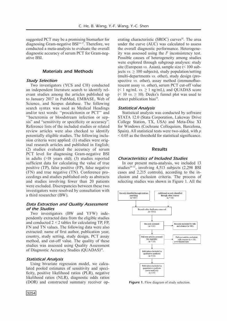
studies20-32, involving 4,513 subjects (2,298 BSI cases and 2,215 controls), according to the in-clusion and exclusion criteria. The process of selecting studies was shown in Figure 1. All the
Figure 1. Flow diagram of study selection.
Can procalcitonin be used to diagnose Gram-negative bloodstream infection?
3255
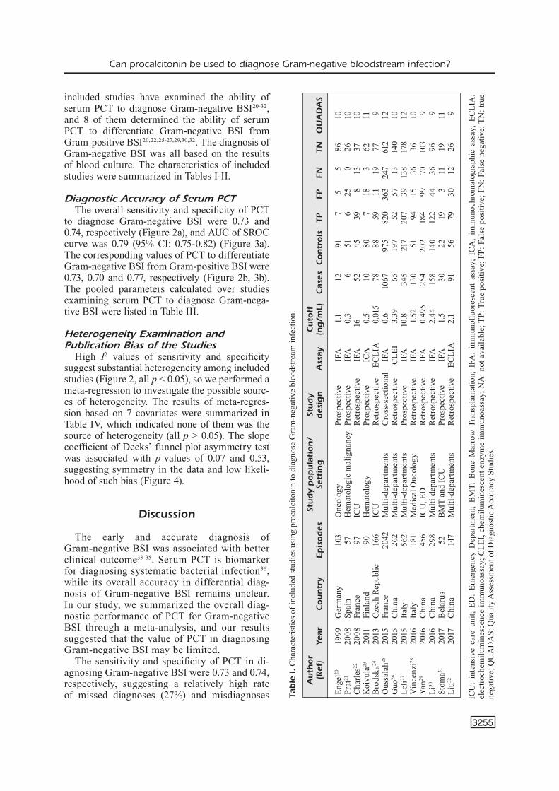
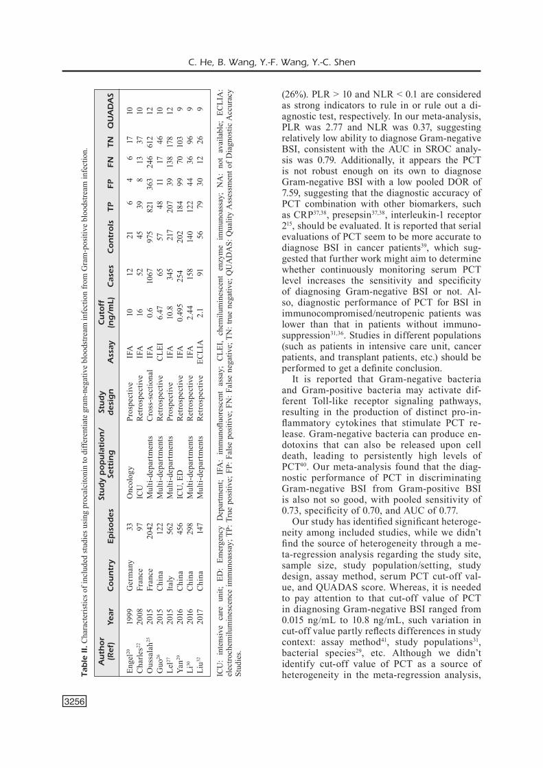
included studies have examined the ability of serum PCT to diagnose Gram-negative BSI20-32, and 8 of them determined the ability of serum PCT to differentiate Gram-negative BSI from Gram-positive BSI20,22,25-27,29,30,32. The diagnosis of Gram-negative BSI was all based on the results of blood culture. The characteristics of included studies were summarized in Tables I-II.
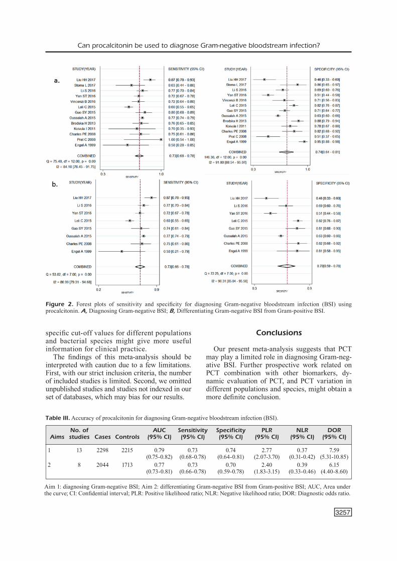
Diagnostic Accuracy of Serum PCT The overall sensitivity and specificity of PCT
to diagnose Gram-negative BSI were 0.73 and 0.74, respectively (Figure 2a), and AUC of SROC curve was 0.79 (95% CI: 0.75-0.82) (Figure 3a). The corresponding values of PCT to differentiate Gram-negative BSI from Gram-positive BSI were 0.73, 0.70 and 0.77, respectively (Figure 2b, 3b). The pooled parameters calculated over studies examining serum PCT to diagnose Gram-nega-tive BSI were listed in Table III.
Heterogeneity Examination and Publication Bias of the Studies
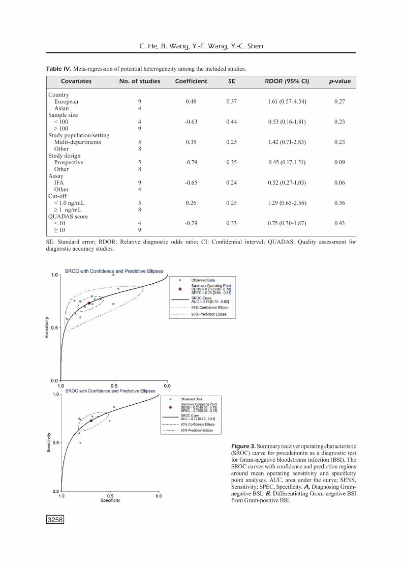
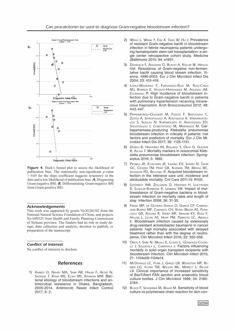
High I2 values of sensitivity and specificity suggest substantial heterogeneity among included studies (Figure 2, all p < 0.05), so we performed a meta-regression to investigate the possible sourc-es of heterogeneity. The results of meta-regres-sion based on 7 covariates were summarized in Table IV, which indicated none of them was the source of heterogeneity (all p > 0.05). The slope coefficient of Deeks’ funnel plot asymmetry test was associated with p-values of 0.07 and 0.53, suggesting symmetry in the data and low likeli-hood of such bias (Figure 4).
Discussion
The early and accurate diagnosis of Gram-negative BSI was associated with better clinical outcome33-35. Serum PCT is biomarker for diagnosing systematic bacterial infection36, while its overall accuracy in differential diag-nosis of Gram-negative BSI remains unclear. In our study, we summarized the overall diag-nostic performance of PCT for Gram-negative BSI through a meta-analysis, and our results suggested that the value of PCT in diagnosing Gram-negative BSI may be limited.
The sensitivity and specificity of PCT in di-agnosing Gram-negative BSI were 0.73 and 0.74, respectively, suggesting a relatively high rate of missed diagnoses (27%) and misdiagnoses Ta
ble
I. C
hara
cter
istic
s of i
nclu
ded
stud
ies u
sing
pro
calc
itoni
n to
dia
gnos
e G
ram
-neg
ativ
e bl
oods
tream
infe
ctio
n.
A
uth
or
St
ud
y p
op
ula
tion
/ St
ud
y
Cu
toff
(Ref
) Ye
ar
Cou
ntr
y Ep
isod
es
Sett
ing
d
esig
n
Ass
ay
(ng
/mL)
C
ases
C
on
trols
TP
FP
FN
TN
Q
UA
DA
S
Enge
l20
1999
G
erm
any
103
Onc
olog
y
Pros
pect
ive
IFA
1.
1 12
91
7 5
5 86
10
Prat
21
2008
Sp
ain
57
Hem
atol
ogic
mal
igna
ncy
Pros
pect
ive
IFA
0.
3 6
51
6
25
0
26
10C
harle
s22
2008
Fr
ance
97
IC
U
Ret
rosp
ectiv
e IF
A
16
52
45
39
8
13
37
10K
oivu
la23
20
11
Finl
and
90
Hem
atol
ogy
Pros
pect
ive
ICA
0.
5 10
80
7 1
8
3 62
11
Bro
dska
24
2013
C
zech
Rep
ublic
16
6 IC
U
Ret
rosp
ectiv
e EC
LIA
0.
015
78
88
59
11
19
77
9O
ussa
lah25
20
15
Fran
ce
2042
M
ulti-
depa
rtm
ents
C
ross
-sec
tiona
l IF
A
0.6
1067
97
5 82
0 36
3 24
7 61
2 12
Guo
26
2015
C
hina
26
2 M
ulti-
depa
rtm
ents
R
etro
spec
tive
CLE
I 3.
39
65
197
52
57
13
140
10Le
li27
2015
Ita
ly
562
Mul
ti-de
part
men
ts
Pros
pect
ive
IFA
10
.8
345
217
207
39
138
178
12V
ince
nzi28
20
16
Italy
18
1 M
edic
al O
ncol
ogy
R
etro
spec
tive
IFA
1.
52
130
51
94
15
36
36
10Ya
n29
2016
C
hina
45
6 IC
U, E
D
Ret
rosp
ectiv
e IF
A
0.49
5 25
4 20
2 18
4 9
9 7
0 10
3 9
Li30
20
16
Chi
na
298
Mul
ti-de
part
men
ts
Ret
rosp
ectiv
e IF
A
2.44
15
8 14
0 12
2 4
4 3
6 96
9
Stom
a31
2017
B
elar
us
52
BMT
and
ICU
Pr
ospe
ctiv
e IF
A
1.5
30
22
19
3
11
19
11Li
u32
2017
C
hina
14
7 M
ulti-
depa
rtm
ents
R
etro
spec
tive
ECLI
A
2.1
91
56
79
30
12
26
9
ICU
: in
tens
ive
care
uni
t; ED
: Em
erge
ncy
Dep
artm
ent;
BM
T: B
one
Mar
row
Tra
nspl
anta
tion;
IFA
: im
mun
ofluo
resc
ent
assa
y; I
CA
, im
mun
ochr
omat
ogra
phic
ass
ay;
ECLI
A:
elec
troch
emilu
min
esce
nce
imm
unoa
ssay
; CLE
I, ch
emilu
min
esce
nt e
nzym
e im
mun
oass
ay; N
A: n
ot a
vaila
ble;
TP:
Tru
e po
sitiv
e; F
P: F
alse
pos
itive
; FN
: Fal
se n
egat
ive;
TN
: tru
e ne
gativ
e; Q
UA
DA
S: Q
ualit
y A
sses
smen
t of D
iagn
ostic
Acc
urac
y St
udie
s.
C. He, B. Wang, Y.-F. Wang, Y.-C. Shen
3256
(26%). PLR > 10 and NLR < 0.1 are considered as strong indicators to rule in or rule out a di-agnostic test, respectively. In our meta-analysis, PLR was 2.77 and NLR was 0.37, suggesting relatively low ability to diagnose Gram-negative BSI, consistent with the AUC in SROC analy-sis was 0.79. Additionally, it appears the PCT is not robust enough on its own to diagnose Gram-negative BSI with a low pooled DOR of 7.59, suggesting that the diagnostic accuracy of PCT combination with other biomarkers, such as CRP37,38, presepsin37,38, interleukin-1 receptor 215, should be evaluated. It is reported that serial evaluations of PCT seem to be more accurate to diagnose BSI in cancer patients39, which sug-gested that further work might aim to determine whether continuously monitoring serum PCT level increases the sensitivity and specificity of diagnosing Gram-negative BSI or not. Al-so, diagnostic performance of PCT for BSI in immunocompromised/neutropenic patients was lower than that in patients without immuno-suppression31,36. Studies in different populations (such as patients in intensive care unit, cancer patients, and transplant patients, etc.) should be performed to get a definite conclusion.
It is reported that Gram-negative bacteria and Gram-positive bacteria may activate dif-ferent Toll-like receptor signaling pathways, resulting in the production of distinct pro-in-flammatory cytokines that stimulate PCT re-lease. Gram-negative bacteria can produce en-dotoxins that can also be released upon cell death, leading to persistently high levels of PCT40. Our meta-analysis found that the diag-nostic performance of PCT in discriminating Gram-negative BSI from Gram-positive BSI is also not so good, with pooled sensitivity of 0.73, specificity of 0.70, and AUC of 0.77.
Our study has identified significant heteroge-neity among included studies, while we didn’t find the source of heterogeneity through a me-ta-regression analysis regarding the study site, sample size, study population/setting, study design, assay method, serum PCT cut-off val-ue, and QUADAS score. Whereas, it is needed to pay attention to that cut-off value of PCT in diagnosing Gram-negative BSI ranged from 0.015 ng/mL to 10.8 ng/mL, such variation in cut-off value partly reflects differences in study context: assay method41, study populations31, bacterial species29, etc. Although we didn’t identify cut-off value of PCT as a source of heterogeneity in the meta-regression analysis, Ta
ble
II.
Cha
ract
eris
tics o
f inc
lude
d st
udie
s usi
ng p
roca
lcito
nin
to d
iffer
entia
te g
ram
-neg
ativ
e bl
oods
tream
infe
ctio
n fr
om G
ram
-pos
itive
blo
odst
ream
infe
ctio
n.
A
uth
or
St
ud
y p
op
ula
tion
/ St
ud
y
Cu
toff
(Ref
) Ye
ar
Cou
ntr
y Ep
isod
es
Sett
ing
d
esig
n
Ass
ay
(ng
/mL)
C
ases
C
on
trols
TP
FP
FN
TN
Q
UA
DA
S
Enge
l20
1999
G
erm
any
33
Onc
olog
y
Pros
pect
ive
IFA
10
12
21
6
4
6
17
10C
harle
s22
2008
Fr
ance
97
IC
U
Ret
rosp
ectiv
e IF
A
16
52
4
5 3
9
8 1
3 3
7 10
Ous
sala
h25
2015
Fr
ance
20
42
Mul
ti-de
part
men
ts
Cro
ss-s
ectio
nal
IFA
0.
6 10
67
975
821
363
246
612
12G
uo26
20
15
Chi
na
122
Mul
ti-de
part
men
ts
Ret
rosp
ectiv
e C
LEI
6.47
65
57
48
11
17
46
10Le
l27
2015
Ita
ly
562
Mul
ti-de
part
men
ts
Pros
pect
ive
IFA
10
.8
345
2
17
207
39
138
178
12Ya
n29
2016
C
hina
45
6 IC
U, E
D
Ret
rosp
ectiv
e IF
A
0.49
5 25
4 20
2 18
4 9
9 7
0 10
3 9
Li30
20
16
Chi
na
298
Mul
ti-de
part
men
ts
Ret
rosp
ectiv
e IF
A
2.44
1
58
140
122
44
36
96
9Li
u32
2017
C
hina
14
7 M
ulti-
depa
rtm
ents
R
etro
spec
tive
ECLI
A
2.1
91
5
6 7
9 3
0 1
2 2
6 9
ICU
: in
tens
ive
care
uni
t; ED
: Em
erge
ncy
Dep
artm
ent;
IFA
: im
mun
ofluo
resc
ent
assa
y; C
LEI,
chem
ilum
ines
cent
enz
yme
imm
unoa
ssay
; N
A:
not
avai
labl
e; E
CLI
A:
elec
troch
emilu
min
esce
nce
imm
unoa
ssay
; TP:
Tru
e po
sitiv
e; F
P: F
alse
pos
itive
; FN
: Fal
se n
egat
ive;
TN
: tru
e ne
gativ
e; Q
UA
DA
S: Q
ualit
y A
sses
smen
t of
Dia
gnos
tic A
ccur
acy
Stud
ies.
Can procalcitonin be used to diagnose Gram-negative bloodstream infection?
3257
specific cut-off values for different populations and bacterial species might give more useful information for clinical practice.
The findings of this meta-analysis should be interpreted with caution due to a few limitations. First, with our strict inclusion criteria, the number of included studies is limited. Second, we omitted unpublished studies and studies not indexed in our set of databases, which may bias for our results.
Conclusions
Our present meta-analysis suggests that PCT may play a limited role in diagnosing Gram-neg-ative BSI. Further prospective work related on PCT combination with other biomarkers, dy-namic evaluation of PCT, and PCT variation in different populations and species, might obtain a more definite conclusion.
Figure 2. Forest plots of sensitivity and specificity for diagnosing Gram-negative bloodstream infection (BSI) using procalcitonin. A, Diagnosing Gram-negative BSI; B, Differentiating Gram-negative BSI from Gram-positive BSI.
Aim 1: diagnosing Gram-negative BSI; Aim 2: differentiating Gram-negative BSI from Gram-positive BSI; AUC, Area under the curve; CI: Confidential interval; PLR: Positive likelihood ratio; NLR: Negative likelihood ratio; DOR: Diagnostic odds ratio.
Table III. Accuracy of procalcitonin for diagnosing Gram-negative bloodstream infection (BSI).
No. of AUC Sensitivity Specificity PLR NLR DOR Aims studies Cases Controls (95% CI) (95% CI) (95% CI) (95% CI) (95% CI) (95% CI)
1 13 2298 2215 0.79 0.73 0.74 2.77 0.37 7.59 (0.75-0.82) (0.68-0.78) (0.64-0.81) (2.07-3.70) (0.31-0.42) (5.31-10.85)2 8 2044 1713 0.77 0.73 0.70 2.40 0.39 6.15 (0.73-0.81) (0.66-0.78) (0.59-0.78) (1.83-3.15) (0.33-0.46) (4.40-8.60)
C. He, B. Wang, Y.-F. Wang, Y.-C. Shen
3258
SE: Standard error; RDOR: Relative diagnostic odds ratio; CI: Confidential interval; QUADAS: Quality assessment for diagnostic accuracy studies.
Table IV. Meta-regression of potential heterogeneity among the included studies.
Covariates No. of studies Coefficient SE RDOR (95% CI) p-value
Country European 9 0.48 0.37 1.61 (0.57-4.54) 0.27 Asian 4 Sample size < 100 4 -0.63 0.44 0.53 (0.16-1.81) 0.23 ≥ 100 9 Study population/setting Multi-departments 5 0.35 0.25 1.42 (0.71-2.83) 0.23 Other 8 Study design Prospective 5 -0.79 0.35 0.45 (0.17-1.21) 0.09 Other 8 Assay IFA 9 -0.65 0.24 0.52 (0.27-1.03) 0.06 Other 4 Cut-off < 1.0 ng/mL 5 0.26 0.25 1.29 (0.65-2.56) 0.36 ≥ 1 ng/mL 8 QUADAS score < 10 4 -0.29 0.33 0.75 (0.30-1.87) 0.43 ≥ 10 9
Figure 3. Summary receiver operating characteristic (SROC) curve for procalcitonin as a diagnostic test for Gram-negative bloodstream infection (BSI). The SROC curves with confidence and prediction regions around mean operating sensitivity and specificity point analyses. AUC, area under the curve; SENS, Sensitivity; SPEC, Specificity. A, Diagnosing Gram-negative BSI; B, Differentiating Gram-negative BSI from Gram-positive BSI.
Can procalcitonin be used to diagnose Gram-negative bloodstream infection?
3259
AcknowledgementsThis work was supported by grants No.81201342 from the National Natural Science Foundation of China, and projects No.16PJ325 from Health and Family Planning Commission of Sichuan province. The funders had no role in study de-sign, data collection and analysis, decision to publish, or preparation of the manuscript.
Conflict of InterestNo conflict of interests to disclose.
References
1) Ahmed d, NAhid mA, SAmi AB, hAlim F, Akter N, SAdique t, rANA mS, elAhi mS, rAhmAN mm. Bac-terial etiology of bloodstream infections and an-timicrobial resistance in Dhaka, Bangladesh, 2005-2014. Antimicrob Resist Infect Control 2017; 6: 2.
2) WANg l, WANg Y, FAN X, tANg W, hu J. Prevalence of resistant Gram-negative bacilli in bloodstream infection in febrile neutropenia patients undergo-ing hematopoietic stem cell transplantation: a sin-gle center retrospective cohort study. Medicine (Baltimore) 2015; 94: e1931.
3) dAXBoeck F, ASSAdiAN o, BlAckY A, koller W, hirSchl Am. Resistance of Gram-negative non-fermen-tative bacilli causing blood stream infection, Vi-enna, 1996-2003. Eur J Clin Microbiol Infect Dis 2004; 23: 415-416.
4) lopez-medrANo F, FerNANdez-ruiz m, ruiz-cANo mJ, BArrioS e, ViceNte-herNANdez m, AguAdo Jm, eScriBANo P. High incidence of bloodstream in-fection due to Gram-negative bacilli in patients with pulmonary hypertension receiving intrave-nous treprostinil. Arch Bronconeumol 2012; 48: 443-447.
5) pApAdimitriou-oliVgeriS m, Fligou F, BArtzAVAli c, zotou A, SpYropoulou A, koutSileou k, VAmVAkopou-lou S, SioulAS N, kArAmouzoS V, ANAStASSiou ed, Spiliopoulou i, chriStoFidou m, mArANgoS m. Car-bapenemase-producing Klebsiella pneumoniae bloodstream infection in critically ill patients: risk factors and predictors of mortality. Eur J Clin Mi-crobiol Infect Dis 2017; 36: 1125-1131.
6) durdu B, hAkYemez iN, Bolukcu S, okAY g, gultepe B, ASlAN t. Mortality markers in nosocomial Kleb-siella pneumoniae bloodstream infection. Spring-erplus 2016; 5: 1892.
7) proWle Jr, echeVerri Je, ligABo eV, SherrY N, tAori gc, crozier tm, hArt gk, kormAN tm, mAYAll Bc, JohNSoN pd, Bellomo r. Acquired bloodstream in-fection in the intensive care unit: incidence and attributable mortality. Crit Care 2011;15: R100.
8) SoStArich Am, zolldANN d, hAeFNer h, luettickeN r, Schulze-roeBecke r, lemmeN SW. Impact of mul-tiresistance of Gram-negative bacteria in blood-stream infection on mortality rates and length of stay. Infection 2008; 36: 31-35.
9) Freire mp, de oliVeirA gArciA d, gArciA cp, cAmpAg-NAri BueNo mF, cAmArgo ch, koNo mAgri AS, FrAN-ciSco gr, reghiNi r, VieirA mF, iBrAhim kY, roSSi F, hAJJAr l, leViN AS, hoFF pm, pierrotti lc, ABdAlA e. Bloodstream infection caused by extensively drug-resistant acinetobacter baumannii in cancer patients: high mortality associated with delayed treatment rather than with the degree of neutro-penia. Clin Microbiol Infect 2016; 22: 352-358.
10) oriol i, SABe N, melilli e, llAdo l, goNzAlez-coStel-lo J, SoldeVilA l, cArrAtAlA J. Factors influencing mortality in solid organ transplant recipients with bloodstream infection. Clin Microbiol Infect 2015; 21: 1104e09-1104e14.
11) mcdoNAld lc, FuNe J, gAido lB, WeiNSteiN mp, re-imer lg, FlYNN tm, WilSoN ml, mirrett S, reller lB. Clinical importance of increased sensitivity of BacT/Alert FAN aerobic and anaerobic blood culture bottles. J Clin Microbiol 1996; 34: 2180-2184.
12) BlooS F, SeNderrek m, BAuer m. Sensitivity of blood culture vs polymerase chain reaction for skin con-
Figure 4. Deek’s funnel plot to assess the likelihood of publication bias. The statistically non-significant p-value > 0.05 for the slope coefficient suggests symmetry in the data and a low likelihood of publication bias. A, Diagnosing Gram-negative BSI; B, Differentiating Gram-negative BSI from Gram-positive BSI.
C. He, B. Wang, Y.-F. Wang, Y.-C. Shen
3260
taminants in specimen retrieved via the distal lu-men of Seldinger-guided central venous cathe-ters. Chest 2014; 145: 430-431.
13) chriSt-crAiN m, muller B. Procalcitonin in bacterial infections--hype, hope, more or less? Swiss Med Wkly 2005; 135: 451-460.
14) NAkAJimA A, YAzAWA J, Sugiki d, mizuguchi m, SAgArA h, FuJiSiro m, ShiBAzAki m, hitANi A, to m, hAruki k. Clinical utility of procalcitonin as a marker of sep-sis: a potential predictor of causative pathogens. Intern Med 2014; 53: 1497-1503.
15) lANg Y, JiANg Y, gAo m, WANg W, WANg N, WANg k, zhANg h, cheN g, liu k, liu m, YANg m, XiAo X. In-terleukin-1 receptor 2: a new biomarker for sepsis diagnosis and Gram-negative/Gram-positive bac-terial differentiation. Shock 2017; 47: 119-124.
16) ArAi t, ohtA S, tSurukiri J, kumASAkA k, NAgAtA k, okitA t, oomurA t, hoShiAi A, koYAmA m, YukiokA t. Procalcitonin levels predict to identify bacterial strains in blood cultures of septic patients. Am J Emerg Med 2016; 34: 2150-2153.
17) WAtANABe Y, oikAWA N, hAriu m, Fuke r, Seki m. Abil-ity of procalcitonin to diagnose bacterial infection and bacteria types compared with blood culture findings. Int J Gen Med 2016; 9: 325-331.
18) reitSmA JB, glAS AS, rutJeS AW, ScholteN rJ, BoSSuYt pm, zWiNdermAN Ah. Bivariate analysis of sensitiv-ity and specificity produces informative summary measures in diagnostic reviews. J Clin Epidemiol 2005; 58: 982-990.
19) deekS JJ, mAcASkill p, irWig l. The performance of tests of publication bias and other sample size ef-fects in systematic reviews of diagnostic test ac-curacy was assessed. J Clin Epidemiol 2005; 58: 882-893.
20) eNgel A, SteiNBAch g, kerN p, kerN WV. Diagnostic value of procalcitonin serum levels in neutropenic patients with fever: comparison with interleukin-8. Scand J Infect Dis 1999; 31: 185-189.
21) prAt c, SANcho Jm, domiNguez J, XicoY B, gimeNez m, FerrA c, BlANco S, lAcomA A, riBerA Jm, AuSiNA V. Evaluation of procalcitonin, neopterin, C-reac-tive protein, IL-6 and IL-8 as a diagnostic mark-er of infection in patients with febrile neutropenia. Leuk Lymphoma 2008; 49: 1752-1761.
22) chArleS pe, lAdoire S, Aho S, queNot Jp, doiSe Jm, priN S, olSSoN No, BletterY B. Serum procalci-tonin elevation in critically ill patients at the onset of bacteremia caused by either Gram negative or Gram positive bacteria. BMC Infect Dis 2008; 8: 38.
23) koiVulA i, hAmAlAiNeN S, JANtuNeN e, pulkki k, kuitti-NeN t, NouSiAiNeN t, JuutilAiNeN A. Elevated procal-citonin predicts Gram-negative sepsis in haema-tological patients with febrile neutropenia. Scand J Infect Dis 2011; 43: 471-478.
24) BrodSkA h, mAlickoVA k, AdAmkoVA V, BeNAkoVA h, StAStNA mm, zimA t. Significantly higher procal-citonin levels could differentiate Gram-negative sepsis from Gram-positive and fungal sepsis. Clin Exp Med 2013; 13: 165-170.
25) ouSSAlAh A, FerrANd J, FilhiNe-treSArrieu p, AiSSA N, AimoNe-gAStiN i, NAmour F, gArciA m, lozNieWSki A, gueANt Jl. Diagnostic accuracy of procalcitonin for predicting blood culture results in patients with suspected bloodstream infection: an obser-vational study of 35,343 consecutive patients (a STROBE-compliant article). Medicine (Baltimore) 2015; 94: e1774.
26) guo SY, zhou Y, hu qF, YAo J, WANg h. Procalci-tonin is a marker of Gram-negative bacteremia in patients with sepsis. Am J Med Sci 2015; 349: 499-504.
27) leli c, FerrANti m, moretti A, Al dhAhAB zS, ceNci e, meNcAcci A. Procalcitonin levels in Gram-posi-tive, Gram-negative, and fungal bloodstream in-fections. Dis Markers 2015; 2015: 701480.
28) ViNceNzi B, FioroNi i, pANtANo F, ANgeletti S, Dicuon-zo G, Zoccoli A, Santini D, Tonini G. Procalcitonin as diagnostic marker of infection in solid tumors patients with fever. Sci Rep 2016; 6: 28090.
29) YAN St, SuN lc, JiA hB, gAo W, YANg Jp, zhANg gq. Procalcitonin levels in bloodstream infec-tions caused by different sources and species of bacteria. Am J Emerg Med 2017; 35: 579-583.
30) li S, roNg h, guo q, cheN Y, zhANg g, YANg J. Se-rum procalcitonin levels distinguish Gram-nega-tive bacterial sepsis from Gram-positive bacterial and fungal sepsis. J Res Med Sci 2016; 21: 39.
31) StomA i, kArpoV i, uSS A, rummo o, milANoVich N, iSkroV i. Diagnostic value of sepsis biomarkers in hematopoietic stem cell transplant recipients in a condition of high prevalence of Gram-negative pathogens. Hematol Oncol Stem Cell Ther 2017; 10: 15-21.
32) liu hh, zhANg mW, guo JB, li J, Su l. Procal-citonin and C-reactive protein in early diagno-sis of sepsis caused by either Gram-negative or Gram-positive bacteria. Ir J Med Sci 2017; 186: 207-212.
33) micek St, Welch ec, khAN J, perVez m, dohertY JA, reichleY rm, hoppe-BAuer J, duNNe Wm, kolleF mh. Resistance to empiric antimicrobial treat-ment predicts outcome in severe sepsis associat-ed with Gram-negative bacteremia. J Hosp Med 2011; 6: 405-410.
34) michAlopouloS A, FAlAgAS me, kArAtzA dc, AleXAN-dropoulou p, pApAdAkiS e, gregorAkoS l, chAleVelAkiS g, pAppAS g. Epidemiologic, clinical characteris-tics, and risk factors for adverse outcome in mul-tiresistant Gram-negative primary bacteremia of critically ill patients. Am J Infect Control 2011; 39: 396-400.
35) Blot S, VANdeWoude k, de BAcquer d, colArdYN F. Nosocomial bacteremia caused by antibiotic-re-sistant Gram-negative bacteria in critically ill pa-tients: clinical outcome and length of hospitaliza-tion. Clin Infect Dis 2002; 34: 1600-1606.
36) hoeBoer Sh, VAN der geeSt pJ, NieBoer d, groeNeVeld AB. The diagnostic accuracy of procalcitonin for bacteraemia: a systematic review and meta-anal-ysis. Clin Microbiol Infect 2015; 21: 474-481.

Can procalcitonin be used to diagnose Gram-negative bloodstream infection?
3261
37) eNguiX-ArmAdA A, eScoBAr-coNeSA r, gArciA-de lA torre A, de lA torre-prAdoS mV. Usefulness of several biomarkers in the management of septic patients: C-reactive protein, procalcitonin, prese-psin and mid-regional pro-adrenomedullin. Clin Chem Lab Med 2016; 54: 163-168.
38) pleSko m, SuVAdA J, mAkohuSoVA m, WAczulikoVA i, BehuloVA d, VASileNkoVA A, VArgoVA m, StecoVA A, kAiSeroVA e, koleNoVA A. The role of CRP, PCT, IL-6 and presepsin in early diagnosis of bacte-rial infectious complications in paediatric hae-mato-oncological patients. Neoplasma 2016; 63: 752-760.
39) SBrANA A, torchio m, comolli g, ANtoNuzzo A, dANoVA m, itAliAN NetWork For SupportiVe cAre iN o. Use of procalcitonin in clinical oncology: a litera-ture review. New Microbiol 2016; 39: 174-180.
40) kumAr S, iNgle h, prASAd dV, kumAr h. Recognition of bacterial infection by innate immune sensors. Crit Rev Microbiol 2013; 39: 229-246.
41) dupuY Am, Ne m, BArgNouX AS, BAdiou S, criStol Jp. Analytical evaluation of Lumipulse(R) BRAHMS PCT CLEIA assay and clinical performances in an unselected population as compared with cen-tral lab PCT assay. Clin Biochem 2017; 50: 248-250.