Embed Size (px)
Citation preview

DISCOVERY AND CHARACTERIZATION OF SMALL MOLECULE INHIBITORS
OF THE ALDEHYDE DEHYDROGENASE 1/2 FAMILY
Cameron D. Buchman
Submitted to the faculty of the University Graduate School
in partial fulfillment of the requirements
for the degree
Doctor of Philosophy
in the Department of Biochemistry and Molecular Biology,
Indiana University
December 2016
ii
Accepted by the Graduate Faculty, Indiana University, in partial
fulfillment of the requirements for the degree of Doctor of Philosophy
_____________________________________
Thomas D. Hurley, Ph.D., Chairman
_____________________________________
Jeffrey S. Elmendorf, Ph.D.
Doctoral Committee
_____________________________________
Quyen Q. Hoang, Ph.D.
September 1, 2016
_____________________________________
Ronald C. Wek, Ph.D.

iii
Dedication
To my mother, who has always supported my dreams and encouraged me to fulfill them,
and to my sister, who has always kept me level-headed while pursuing them.

iv
Acknowledgements
I would like to start by thanking my graduate advisor, Dr. Thomas Hurley, for being an
outstanding mentor and friend. I am grateful to him for allowing me to complete my
research thesis with him and for providing me with multiple learning opportunities.
Despite my limited prior experience, he allowed me to pursue this project independently,
while always being there when I needed help. Above all, I thank him for his patience and
wisdom. I firmly believe that he is the best mentor I could ever have had for my graduate
studies. I also thank the other members of my committee; Dr. Jeffrey Elmendorf, Dr.
Quyen Hoang, and Dr. Ronald Wek; for their guidance, suggestions, and constructive
criticism, and challenging me to think beyond the test tube, or this case, the cuvette.
Science is a highly collaborative endeavor, and as such multiple individuals and
laboratories helped me complete this project. I thank the Chemical Genomics Core
facility, especially Dr. Lan Chen, for providing the use of their facilities and compound
libraries and assisting with the high-throughput screens. I thank Dr. Maureen Harrington
and Dr. Danielle Matei for the use of their cell culture facilities, and Dr. Donghui Zhou
and Dr. Salvadore Condello for teaching me cell culture techniques. I thank Dr. Clark
Wells for providing the MDA-MB-468 cells. I also thank Dr. John Turchi for the use of
his fluorimeter.
My colleagues in Dr. Hurley’s laboratory have also aided me in this project. I thank Dr.
Bibek Parajuli for aiding me in my rotation and helping me get started in the lab; Dr. May
v
Khanna, Dr. Vimbai Chikwana, Dr. Ann Kimble Hill, and Dr. Cindy Morgan for their
answers to my many questions; Lanmin Zhai for assistance with multiple protein
purifications, and Krishna Kishore Mahalinghan for completing Screen 1 of the high-
throughput screen. I also thank all of them, and Mikhail Chtcherbinine, for their
friendship, support, and providing the necessary distractions from an often hectic
schedule.
I would also like to thank members of the Department of Biochemistry and Molecular
office, Sandy, Melissa, Jack, Patty, Darlene, and Sheila, for their assistance and working
behind-the-scenes to make my graduate studies run smoothly.
This research was supported by the U.S. National Institutes of Health (grants
RO1AA018123 and R21CA198409 to T.D.H.). Results shown in this report are derived
from work performed at Argonne National Laboratory, Structural Biology Center at the
Advanced Photon Source. Argonne is operated by UChicago Argonne, LLC, for the U.S.
Department of Energy, Office of Biological and Environmental Research under contract
DE-AC02-06CH11357. This research used resources of the Advanced Photon Source, a
U.S. Department of Energy (DOE) Office of Science User Facility operated for the DOE
Office of Science by Argonne National Laboratory under Contract No. DE-AC02-
06CH11357.

vi
Cameron D. Buchman
DISCOVERY AND CHARACTERIZATION OF SMALL MOLECULE INHIBITORS
OF THE ALDEHYDE DEHYDROGENASE 1/2 FAMILY
The human aldehyde dehydrogenase (ALDH) superfamily consists of 19 isoenzymes that
are critical for normal physiology as well as the removal of toxic aldehydes. Members of
the ALDH1/2 family have vital roles in cell signaling during early development, ethanol
metabolism, and the removal of aldehydes derived from oxidative stress. We sought to
develop selective compounds toward ALDH2 to help determine its individual
contribution to biological function, as many of the ALDH1/2 family possess overlapping
substrate preferences. A high-throughput screen of over 100,000 compounds uncovered a
class of aromatic lactones which inhibit the ALDH1/2 enzyme family. The lactones were
then characterized using a combination of enzyme kinetics, X-ray crystallography, and
cell culture experiments. We found that many of the lactones are over ten times more
potent toward ALDH2 than daidzin, a previously described ALDH2 inhibitor. Our ability
to produce many more ALDH isoenzymes allowed us to determine that daidzin is not as
selective as previously believed, inhibiting ALDH2, ALDH1B1, and ALDH1A2 with
equal potency. This inhibition pattern was seen with several of the aromatic lactones as
well. Structural studies show that many of the lactones bind between key aromatic
residues in the ALDH1/2 enzyme substrate-binding sites. One lactone in particular
mimics the position of an aldehyde substrate and alters the position of the catalytic

vii
cysteine to interfere with the productive binding of NAD+ for enzyme catalysis. Further
characterization of related compounds led to the realization that the mechanism of
inhibition, potency, and selectivity differs amongst the lactones based off the substituents
on the aromatic scaffold and its precise binding location. Two of these compounds were
found to be selective for one of the ALDH1/2 family members, BUC22, selective for
ALDH1A1, and BUC27, selective for ALDH2. BUC22 demonstrates ten-fold selectivity
for ALDH1A1 over ALDH1A2 and does not inhibit the remaining ALDH1/2 enzymes.
Additionally, treatment with BUC22 led to decreased growth of triple-negative breast
cancer cells in culture. BUC27 inhibits ALDH2 with the same potency as daidzin. Both
BUC22 and BUC27 could be further developed to use as chemical tools to better
understand the functional roles of ALDH1A1 and ALDH2 in biological systems.
Thomas D. Hurley, Ph.D., Chairman

viii
Table of Contents
List of Tables ................................................................................................................. xi
List of Figures ................................................................................................................ xii
I. Introduction ................................................................................................................ 1
A. Aldehydes............................................................................................................ 1
B. Aldehyde Dehydrogenase Superfamily ............................................................... 5
C. Aldehyde Dehydrogenase 1/2 Family ................................................................. 13
D. ALDH1A1, Cancer, and Cancer Stem Cells ....................................................... 19
E. ALDH2, ALDH2*2, and Alcohol-Induced Pathophysiology ............................. 22
F. ALDH2 and Cardiovascular Disease ................................................................... 25
G. ALDH1A1, ALDH2, and Neurological Disease ................................................ 27
H. ALDH1/2 Activators and Inhibitors ................................................................... 29
I. Hypothesis and Approach ..................................................................................... 31
II. Materials and Methods .............................................................................................. 34
A. Materials.............................................................................................................. 34
B. Methods ............................................................................................................... 34
1. Production and Purification of ALDH Isoenzymes ....................................... 34

ix
2. Esterase High-Throughput Screen ................................................................. 37
3. Aldehyde Oxidation Activity Assays for ALDH Isoenzymes ....................... 39
4. Steady-state Kinetic Characterization with ALDH1A1 and ALDH2 ............ 41
5. Characterization of 2BS4’s Inhibition of ALDH2 ......................................... 43
6. Crystallization of ALDH1A1 and ALDH2 Complexes ................................. 44
7. Cellular Effect of the Coumarin Derivative BUC22...................................... 47
III. Results ...................................................................................................................... 48
A. Esterase High-Throughput Screen for ALDH2 Modulators ............................... 48
1. Z’-factor Calculation ...................................................................................... 48
2. High-Throughput Screen Results ................................................................... 50
B. Hit Compound Validation and Initial Determination of Selectivity ................... 52
C. EC50 Determination of Hit Compounds .............................................................. 57
1. Alda-1 Mimics ............................................................................................... 57
2. Coumarin and Psoralen Derivatives, in Comparison to Daidzin ................... 59
D. Characterization of Interaction of Aromatic Lactones with ALDH2 ................. 62
1. Kinetic Characterization of 2P4 ..................................................................... 62
2. 2P3-ALDH2 Crystal Structure ....................................................................... 64
x
3. Kinetic Characterization of 2BS4 .................................................................. 66
E. Characterization of Additional Psoralen and Coumarin Analogs ....................... 69
1. Single Concentration Selectivity for Structure-Activity Relationships ......... 69
2. EC50 Determination ........................................................................................ 76
3. Characterization of BUC11’s Interaction with ALDH1A1 and ALDH2 ...... 78
4. Characterization of BUC25’s Interaction with ALDH1A1 ........................... 81
5. Characterization of BUC22’s Interaction with ALDH1A1 ........................... 83
6. Characterization of BUC27’s Interaction with ALDH2 ................................ 85
F. Effect of BUC22 on Viability of Breast Cancer Cells in 3D Culture .................. 86
IV. Discussion ................................................................................................................ 88
V. Conclusion and Future Directions............................................................................. 108
References ...................................................................................................................... 113
Curriculum vitae

xi
List of Tables
Table 1: Common sources of aldehydes ........................................................................ 2
Table 2: The 19 human aldehyde dehydrogenase isoenzymes ...................................... 7
Table 3: AC50 determination of Alda-1 mimics with ALDH2 ...................................... 59
Table 4: IC50 values for four aromatic lactones with nine ALDH isoenzymes ............. 61
Table 5: Collection and refinement statistics for the 2P3-ALDH2 complex ................. 65
Table 6: Structure activity relationships for psoralen derivatives ................................. 71
Table 7: Structure activity relationships for coumarin derivatives ................................ 72
Table 8: IC50 values for four analogs with ALDH1A1, ALDH2, and ALDH3A1 ........ 76
Table 9: EC50 values for seven analogs with the ALDH1/2 family and ALDH3A1 ..... 78
Table 10: Collection and refinement statistics for the BUC11-ALDH1A1 complex .... 79
Table 11: Collection and refinement statistics for the BUC25-ALDH1A1 complex .... 82
Table 12: Collection and refinement statistics for the BUC22-ALDH1A1 complex .... 83
xii
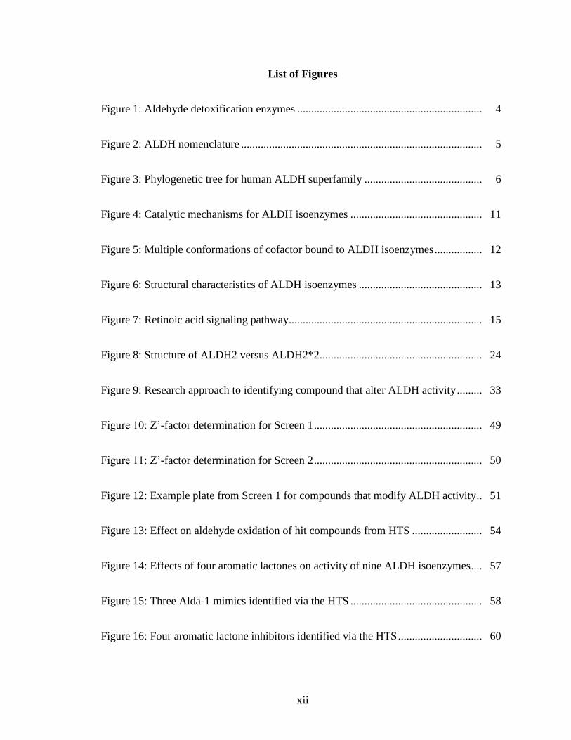
List of Figures
Figure 1: Aldehyde detoxification enzymes .................................................................. 4
Figure 2: ALDH nomenclature ...................................................................................... 5
Figure 3: Phylogenetic tree for human ALDH superfamily .......................................... 6
Figure 4: Catalytic mechanisms for ALDH isoenzymes ............................................... 11
Figure 5: Multiple conformations of cofactor bound to ALDH isoenzymes ................. 12
Figure 6: Structural characteristics of ALDH isoenzymes ............................................ 13
Figure 7: Retinoic acid signaling pathway..................................................................... 15
Figure 8: Structure of ALDH2 versus ALDH2*2.......................................................... 24
Figure 9: Research approach to identifying compound that alter ALDH activity ......... 33
Figure 10: Z’-factor determination for Screen 1 ............................................................ 49
Figure 11: Z’-factor determination for Screen 2 ............................................................ 50
Figure 12: Example plate from Screen 1 for compounds that modify ALDH activity .. 51
Figure 13: Effect on aldehyde oxidation of hit compounds from HTS ......................... 54
Figure 14: Effects of four aromatic lactones on activity of nine ALDH isoenzymes .... 57
Figure 15: Three Alda-1 mimics identified via the HTS ............................................... 58
Figure 16: Four aromatic lactone inhibitors identified via the HTS .............................. 60

xiii
Figure 17: Standard curve for fluorescence of NADH .................................................. 63
Figure 18: Substrate Km determination using fluorescence assay ................................. 63
Figure 19: Steady-state kinetic characterization of inhibition of ALDH2 by 2P4 ........ 64
Figure 20: Crystal structure of 2P3-ALDH2 complex ................................................... 66
Figure 21: Steady-state kinetic characterization of inhibition of ALDH2 by 2BS4 ...... 67
Figure 22: Incubation of ALDH2 with 2BS4 ................................................................ 68
Figure 23: IC50 values for BUC22 with ALDH2 after different incubation periods ..... 69
Figure 24: Compound scaffolds of psoralen- and coumarin-derived analogs ............... 70
Figure 25: Crystal structure of BUC11-ALDH1A1 complex ........................................ 80
Figure 26: Steady-state kinetic characterization of BUC11 with ALDH enzymes ....... 81
Figure 27: Crystal structure of BUC25-ALDH1A1 complex ........................................ 82
Figure 28: Crystal structure of BUC22-ALDH1A1 complex ........................................ 84
Figure 29: Steady-state kinetic characterization of inhibition by BUC22 and BUC20 . 85
Figure 30: Steady-state kinetic characterization of inhibition of ALDH2 by BUC27 .. 86
Figure 31: Effect of BUC22 treatment on proliferation of breast cancer cells .............. 87
Figure 32: Effect of different compounds on esterase and dehydrogenase assays ........ 90
Figure 33: Orientation of 2P3 in catalytic tunnel of ALDH2 ........................................ 92

xiv
Figure 34: Comparison of NAD+ and 2P3 binding to ALDH2 ..................................... 93
Figure 35: Schematic of 2P3/4 inhibition of ALDH2 .................................................... 95
Figure 36: Comparison of BUC22 binding to ALDH1A1 versus ALDH2 ................... 100
Figure 37: Docking of BUC27 in substrate binding site of ALDH2 ............................. 102
Figure 38: Alignment of three enzyme-inhibitor complexes ......................................... 104

1
I. Introduction
A. Aldehydes
Aldehydes are organic compounds which contain a terminal carbonyl carbon (RCHO)
where the R group can range from a hydrogen atom to complex organic structures.
Aldehydes are found in a variety of endogenous and exogenous sources (Table 1).
Amino acid catabolism produces aldehyde intermediates such as glutamic γ-
semialdehyde. The catabolism of various neurotransmitters including gamma-
aminobutyric acid (GABA), dopamine, adrenaline, and serotonin also produces several
aldehyde intermediates.1 Many open chain forms of carbohydrates contain an aldehyde
group, or contain a ketone which can tautomerize in solution to form an aldehyde group.
The metabolism of ethanol and other foods can generate acetaldehyde as well as other
aldehyde products. Aldehydes such as citral and benzaldeyde are also added to foods to
provide flavor and odor.2 Several aldehydes play important roles in normal physiological
processes. Retinaldehydes are an example that has multiple functions; as transcription
factors critical for cellular growth and differentiation pathways, or as the ligand for
rhodopsin, which covalently binds 11-cis-retinal and enables vision in low-light
conditions.3, 4
Despite being critical for many biological functions, many aldehydes are carcinogenic
and cytotoxic. Oxidative degradation of membrane lipids, or lipid peroxidation, can
produce over 200 aldehyde species, including 4-hydroxynonenal (4-HNE) and
malondialdehyde (MDA).5 Environmental exposure to smog, cigarette smoke, and motor
vehicle exhaust are potential sources for formaldehyde, acetaldehyde, and acrolein,

2
Table 1 Common sources of aldehydes.
Endogenous Source Aldehyde
Lipid peroxidation 4-hydroxynonenal, malondialdehyde, hexanal
Dopamine catabolism 3-4-Dihydroxyphenylacetaldehyde
GABA metabolism succinic semialdehyde
Serotonin metabolism 4-hydroxyindoleacetaldehyde
Putrescine catabolism γ-amino butyraldehyde
Vitamin A metabolism retinaldehyde
Carbohydrate metabolism glyceralehyde, glycolaldehyde, glyoxal
Exogenous Source Aldehyde
Ethanol acetaldehyde
Foods citral, benzaldehyde, crotonaldehyde
Combustion formaldehyde, acetaldehyde, acrolein
Cigarettes γ-3-pyridyl-γ-methylbutyraldehyde
amongst other aldehydes.6 Aldehydes are also used or produced in various industrial
applications including the production of resins, polyurethane, and polyester plastics. Each
of these sources can contribute to the aldehydic load within the body. Highly reactive and
electrophilic carbonyl groups in aldehydes can form adducts with various
macromolecules including proteins, nucleic acids, and glutathione. Unlike reactive
oxygen species and free radicals, aldehydes have relatively long lifespans and once
formed, can react with both nearby cellular components and targets some distance away
via diffusion or molecular transport.5 Aldehydes can form a covalent adduct with proteins
either by formation of a Schiff base via the ε-amino of lysine or by a Michael addition to
cysteine, histidine, or lysine residues in a process called protein carbonylation.7 These
reactions will typically result in a loss of function due to the importance of cysteine,
histidine, and lysine in many enzyme catalytic mechanisms. Although moderate amounts

3
of carbonylation can be accommodated by cells, increasing amounts of carbonylation will
eventually lead to protein dysfunction, cell death, and ultimately disease progression. It is
important to note that protein carbonylation is elevated during oxidative stress and
correspondingly a high proportion of carbonylated proteins are found in the
mitochondria.8 Aldehydes have also been shown to inactivate antioxidant enzymes such
as glutathione reductase and glutathione peroxidase by protein carbonylation, which as a
consequence perpetuates oxidative stress in a positive feedback loop.9, 10
Aldehydes can
also form adducts with DNA creating DNA-DNA and DNA-protein crosslinks,
chromosomal aberrations, and other DNA damage.11, 12
Cells have several mechanisms to alleviate aldehyde stress. Aldehydes can be reduced to
an alcohol, oxidized to a carboxylic acid, or eliminated via interaction with glutathione.
The main enzyme classes that reduce aldehydes to alcohols are alcohol dehydrogenases
and aldo-keto reductases, and the main enzyme classes that oxidize aldehyde to
carboxylic acids are aldehyde/xanthine oxidases and aldehyde dehydrogenases (Figure
1). Alcohol dehydrogenases can catalyze the reversible reduction of aldehydes and
ketones to an alcohol. The direction of this reaction is heavily dependent on the
NAD+/NADH ratio. Oxidation of an alcohol to an aldehyde is the predominant reaction
due to the normal 500:1 ratio of NAD+ to NADH found within cells.
13 The aldo-keto
reductase superfamily consists of 13 human enzymes and catalyzes the reversible
reduction of a variety of aldehydes and ketones to their corresponding alcohols.14
Aldehyde/xanthine oxidases catalyze the conversion of aromatic and heterocyclic
aldehydes to their corresponding carboxylic acids.15
Aldehyde dehydrogenases catalyze

4
the NAD(P)+-dependent oxidation of an aldehyde to its corresponding carboxylic acid or
ester CoA.16
Short-chain dehydrogenases/reductases compose a large subset of all
dehydrogenases and can catalyze the NAD(P)H-dependent oxidation or reduction of
aldehydes depending on the individual enzyme.17
Lipid peroxidation products can be
eliminated via conjugation to glutathione via glutathione S-transferase.18
Although all of
these enzymes contribute to the alleviation of the aldehydic load, aldehyde
dehydrogenases are capable of oxidizing a variety of aromatic and aliphatic aldehydes
and thus contribute to a wide range of physiological functions and generation of
biological products.
Figure 1 Aldehyde detoxification enzymes.

5
B. Aldehyde Dehydrogenase Superfamily
The aldehyde dehydrogenase (ALDH) superfamily exists in all three taxonomic domains,
Archaea, Eubacteria, and Eukarya, highlighting the important role that these enzymes
have had throughout evolution.19
A nomenclature system based on sequence alignments
and evolutionary relationships was established in 1999 and is used primarily in Eukarya
(Figure 2).20
By convention members of different ALDH families have less than 40%
protein sequence identity, while members of the same subfamily have more than 60%
protein sequence identity. Due to the extensive
amount of research done on ALDH2 in the field of
ethanol metabolism prior to 1999, its name was
grandfathered into the system, despite its amino
acid sequence placing it in the ALDH1B
subfamily.
Currently there are 24 recognized ALDH families in eukaryotes; 11 of these families are
represented in the human genome totaling 19 isoenzymes (Figure 3).21
ALDH
isoenzymes differ in tissue location, subcellular location, substrate specificity, and
structure. Many ALDH isoenzymes possess leader sequences that direct their
translocation to specific subcellular locations.22
ALDHs have a variety of functions.
ALDHs contribute to the synthesis of numerous important biological molecules including
GABA, retinal, and tetrahydrofolate, and the removal of toxic aldehydes through their
aldehyde oxidation activity.23
Physiological roles for ALDHs are not limited to enzymatic
function. ALDHs in the eye function as structural proteins called crystallins which absorb

6
harmful UV light.24
ALDH16A1 may also serve as a structural protein as it lacks a
functional nucleophile in its active site.25
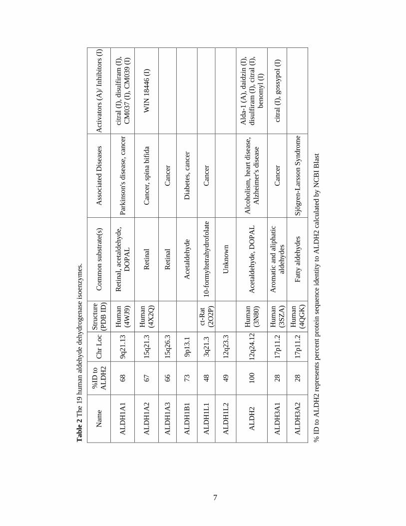
A summary of the 19 ALDH isoenzymes is
shown in Table 2.
Figure 3 Phylogenetic tree for the human ALDH superfamily.
Dendrogram of the 19 human ALDH members. Branch lengths depict degree of
evolutionary divergence between isoenzymes.26
7
Ta
ble
2 T
he
19
hum
an a
ldeh
yde
deh
ydro
gen
ase
isoen
zym
es.
Act
ivat
ors
(A
)/ I
nhib
itors
(I)
citr
al (
I), d
isu
lfir
am (
I),
CM
03
7 (
I),
CM
03
9 (
I)
WIN
18
446
(I)
Ald
a-1 (
A),
dai
dzi
n (
I),
dis
ulf
iram
(I)
, ci
tral
(I)
,
ben
om
yl
(I)
citr
al (
I), g
oss
yp
ol
(I)
% I
D t
o A
LD
H2
rep
rese
nts
per
cent
pro
tein
seq
uen
ce i
den
tity
to A
LD
H2 c
alcu
late
d b
y N
CB
I B
last
Ass
oci
ated
Dis
ease
s
Par
kin
son's
dis
ease
, ca
nce
r
Can
cer,
spin
a bif
ida
Can
cer
Dia
bet
es, ca
nce
r
Can
cer
Alc
oholi
sm, h
eart
dis
ease
,
Alz
hei
mer
's d
isea
se
Can
cer
Sjö
gre
n-L
arss
on
Sy
nd
rom
e
Com
mon s
ubst
rate
(s)
Ret
inal
, ac
etal
deh
yde,
DO
PA
L
Ret
inal
Ret
inal
Ace
tald
ehyde
10
-form
ylt
etra
hydro
fola
te
Unknow
n
Ace
tald
ehyde,
DO
PA
L
Aro
mat
ic a
nd a
liphat
ic
aldeh
ydes
Fat
ty a
ldeh
ydes
Str
uct
ure
(PD
B I
D)
Hum
an
(4W
J9)
Hum
an
(4X
2Q
)
ct-R
at
(2O
2P
)
Hum
an
(3N
80)
Hum
an
(3S
ZA
)
Hum
an
(4Q
GK
)
Chr
Loc
9q21.1
3
15q21.3
15q26.3
9p13.1
3q21.3
12q23.3
12q24.1
2
17p11.2
17p11.2
%ID
to
AL
DH
2
68
67
66
73
48
49
10
0
28
28
Nam
e
AL
DH
1A
1
AL
DH
1A
2
AL
DH
1A
3
AL
DH
1B
1
AL
DH
1L
1
AL
DH
1L
2
AL
DH
2
AL
DH
3A
1
AL
DH
3A
2

8
Tab
le 2
, co
nti
nu
ed T
he
19 h
um
an a
ldeh
yde
deh
ydro
gen
ase
isoen
zym
es.
Act
ivat
ors
(A
)/ I
nhib
itors
(I)
% I
D t
o A
LD
H2
rep
rese
nts
per
cent
pro
tein
seq
uen
ce i
den
tity
to A
LD
H2 c
alcu
late
d b
y N
CB
I B
last
Ass
oci
ated
Dis
ease
s
Sch
izop
hre
nia
Ty
pe
II
hyp
erpro
linem
ia
γ-hydro
xy-b
utr
yic
acid
uri
a
Met
abo
lic
abn
orm
alit
ies
pyri
do
xin
e-dep
end
ent
epil
epsy
Go
ut
Met
abo
lic
and
neu
rolo
gic
abn
orm
alit
ies
Com
mon s
ubst
rate
(s)
Med
ium
and l
ong c
hai
ned
alip
hat
ic a
ldeh
ydes
Unknow
n
Glu
tam
ate
γ-se
mia
ldeh
yde
Succ
inat
e se
mia
ldeh
yde
Mal
onat
e se
mia
ldeh
yde,
met
hylm
alonat
e
sem
iald
ehyde
α-a
min
oad
ipic
sem
iald
ehyde
2-a
min
o-m
uci
nat
e
sem
iald
ehyde?
γ-am
ino
-butr
yal
deh
yde
Unknow
n
Glu
tam
ic A
cid
Str
uct
ure
(PD
B I
D)
Hum
an
(4O
E5)
Hum
an
(2W
8R
)
Bac
teri
a
(4E
4G
)
Hum
an
(4Z
UK
)
Cod
(1A
4S
)
Hum
an
(2H
5G
)
Chr
Loc
11q13.2
11q13.2
1p36.1
3
6p22.3
14q24.3
5q23.2
6q23.3
1q24.1
19q13.3
3
10q24.1
%ID
to
AL
DH
2
25
26
27
35
30
30
39
43
29
30
Nam
e
AL
DH
3B
1
AL
DH
3B
2
AL
DH
4A
1
AL
DH
5A
1
AL
DH
6A
1
AL
DH
7A
1
AL
DH
8A
1
AL
DH
9A
1
AL
DH
16
A1
AL
DH
18
A1

9
Human ALDHs are associated with a number of diseases due to their involvement in a
variety of physiological processes (Table 2). Mutations in multiple ALDHs (ALDH1A1,
ALDH1A2, ALDH1A3, ALDH1B1, ALDH1L1, ALDH2, and ALDH3A1) are associated
with cancer, and overexpression of ALDH1A1 and ALDH3A1 in cancer is linked to poor
prognosis and decreased efficacy of certain chemotherapeutics, including
cyclophosphamide.23, 27
Aldh1a1-/-
and Aldh3a1-/-
single knockout mice as well as
Aldh1a1-/-
Aldh3a1-/-
double knockout mice develop cataracts prematurely as ALDH1A1
and ALDH3A1 both protect ocular tissue from UV radiation.28
The ALDH1A subfamily
is significantly involved in retinoic acid signaling and mutations in these genes are often
detrimental to embryogenesis and can lead to spina bifida.23, 29
ALDH1A1 and ALDH2
have been linked to neurodegenerative diseases due to their involvement in dopamine
metabolism.30
ALDH1B1 has been shown to be important in beta cell development;
mutations may contribute to the eventual development of diabetes.31
ALDH3A2 metabolizes the oxidization of fatty aldehydes and mutations are associated
with Sjögren-Larssson Syndrome, an autosomal recessive disorder that leads to skin,
cognitive, and neurological defects.32
A single nucleotide polymorphism in ALDH3B1
has been linked to the development of paranoid schizophrenia.33
ALDH4A1 is involved
in proline degradation and mutations in this gene lead to Type II hyperprolinemia
characterized by seizures and mental retardation.34
ALDH5A1 is involved in the
metabolism of the neurotransmitter GABA; mutations lead to γ-hydroxybutyric acidura
characterized by neurological and cognitive defects.35
ALDH6A1 is the only known
CoA-dependent human ALDH and this enzyme catalyzes the conversion of malonate
10
semialdehyde and methymalonate semialdehyde to acetyl-CoA and propionyl-CoA,
respectively, in valine and pyrimidine catabolism.36
Mutations in the ALDH6A1 gene
lead to a variety of metabolic abnormalities, which are typically accompanied by
psychomotor delay.37
ALDH7A1 catalyzes the oxidation of α-aminoadipic semialdehyde
during lysine metabolism; mutations are linked to pyridoxine-dependent epilepsy which
causes severe seizures during infancy and early childhood.38
ALDH16A1 is unique
amongst the human ALDH enzymes as the active site lacks key catalytic residues.
Although the enzyme does not have aldehyde oxidation activity, ALDH16A1 may act as
a binding partner to other proteins and has been linked to gout.25
ALDH18A1 catalyzes
the reduction of glutamate to ∆-pyrroline-5-carboxylate during the de novo synthesis of
the amino acids proline and arginine.39
Mutations in the ALDH18A1 gene result in
various metabolic and neurologic abnormalities characterized by cataract formation,
neurodegeneration, and connective tissue disorders.40
The primary function for ALDH enzymes is aldehyde oxidation. Sequence alignment of
145 ALDHs indicated that there is a small number of residues conserved in catalytically
active enzymes, which include the catalytic cysteine and residues involved in cofactor
binding.41
The reaction mechanism for aldehyde oxidation by ALDH isoenzymes is
shown in Figure 4A. The process begins with binding of the cofactor NAD(P)+ in the
Rossmann fold of the enzyme.42, 43
NAD(P)+ can adopt multiple conformations when
bound to ALDHs (Figure 5).44, 45
Cys302 is the active site nucleophile and Glu268 acts
as a general base (numbering based on mature ALDH2 sequence).46, 47
Cys302 is
activated either directly by Glu268 or by a hydroxyl ion generated by Glu268 to perform

11
a nucleophilic attack on the carbonyl carbon of the aldehyde which yields the formation
of a thioacyl-enzyme intermediate and hydride transfer to NAD(P)+. Glu268 then
activates a water molecule that performs a nucleophilic attack on the carbonyl carbon of
the thioacyl-enzyme intermediate which, after rearrangement, yields the reduced cofactor
NAD(P)H and carboxylic acid product. The reduced cofactor is thought to be released
after the carboxylic acid.48
Due to structural differences, the rate limiting step differs
between enzymes. For example, the rate limiting step is cofactor dissociation for the
ALDH1A subfamily, deacylation for ALDH2, and hydride transfer for ALDH3A1.49-51
Many ALDHs, including ALDH2, also have a NAD(P)+-independent esterase activity
which uses the same active site residues and is the same reaction that hydrolyzes the
thioacyl-enyzme intermediate to regenerate the free enzyme (Figure 4B).52, 53
Figure 4 Catalytic mechanisms for ALDH isoenzymes.
Aldehyde oxidation (A) requires cofactor NAD+, while ester hydrolysis (B) does not.

12
Figure 5 Multiple conformations of cofactor bound to ALDH isoenzymes.
NAD+ (black) and NADH (cyan) bound to human ALDH2 (purple) and NADP
+ (yellow)
bound to glyceraldehyde 3-phosphate dehydrogenase (E250A) from Streptococcus
mutans (green). PDB codes 1O00, 1O02, 1O04, 2QE0.
However, the presence of cofactor can stimulate esterase activity by either enhancing the
nucleophilicity of Cys302 or increasing the frequency of productive encounters with
Cys302.54, 55
Cofactor’s effect on esterase activity is due in part to the shape of the active
site. Mammalian ALDHs function as either dimers or tetramers, with each monomer
containing a catalytic domain, a cofactor binding domain (Rossman fold), and an
oligomerization domain (Figure 6A & B).56
The active site of ALDH enzymes contains a
tunnel where the cofactor binding site is located on one end, with the substrate site at the
other end of the tunnel and the catalytic cysteine situated in the middle (Figure 6C). The
ALDHs have evolved to recognize different aldehyde substrates due to differences in the
size and shape of their respective binding site.56, 57

13
Figure 6 Structural characteristics of ALDH isoenzymes.
(A) Ribbon diagram of the ALDH2 monomer. (B) Ribbon diagram of ALDH2
homotetramer with each monomer colored individually. (C) Surface representation of
tunnel connecting cofactor binding site and catalytic site of ALDH1A1. Region of
catalytic cysteine colored in yellow. NADH (black) bound to cofactor binding site. (PDB
Code 4WB9).
C. Aldehyde Dehydrogenase 1/2 Family
The aldehyde dehydrogenase 1/2 family in humans consists of seven isoenzymes:
ALDH1A1, ALDH1A2, ALDH1A3, ALDH1B1, ALDH2, ALDH1L1, and ALDH1L2.
These enzymes are present among different tissues, and each can have variable
expression patterns, along with oxidizing a variety of aldehydes. Thus the ALDHs have a
diverse set of physiological functions.

14
The ALDH1A subfamily consists of the cytosolic ALDH1A1, ALDH1A2, and
ALDH1A3 that share ~70% sequence identity. Each of the three isoenzymes can
facilitate oxidation of retinal to retinoic acid (RA), though the substrate specificity for
each of these enzymes differs. ALDH1A1 catalyzes the oxidation of all-trans-,9-cis-, and
13-cis-retinal with Km values ranging from 2 – 6 µM.58
ALDH1A2 and ALDH1A3 prefer
the all-trans isomer, but also can utilize the cis-isomers as substrates.23, 59
The RA
products can then act on other cells in an endocrine or paracrine manner.60
Once in the
nucleus of the target cell, the RA products then bind to retinoic acid (RAR) and retinoic
X receptors (RXR) (Figure 7). Both RAR and RXR are type II nuclear receptors and as
such are retained in the nucleus regardless of the presence or absence of ligand. RA
products will bind to heterodimers of RAR and RXR, which interact with the retinoic
acid response element (RARE), a specific DNA sequence in the promoter region of the
target gene to regulate gene expression and cellular phenotype. RXRs also can form
heterodimers with other nuclear receptors such as thyroid hormone receptors and
peroxisomal proliferator activated receptors. Retinoid metabolism and signaling are
involved in embryogenesis, cell cycle control, cell growth, and differentiation.61, 62
Dysregulation of these functions has been linked to obesity, diabetes, cardiovascular
disease, cancer, and fetal/birth defects.61, 62
Whereas Aldh1a1 knockout mice are viable,
both Aldh1a2 and Aldh1a3 knockout mice do not produce viable animals, suggesting that
ALDH1A2 and ALDH1A3 are more involved in embryogenesis and tissue patterning.63
Aldh1a2-/-
knockout mice are characterized by defects in heart development and death at
midgestation, while Aldh1a3-/-
knockout mice are characterized by nasal and ocular

15
Figure 7 Retinoic acid signaling pathway.
Retinol (vitamin A) is oxided by retinal dehydrogenases (RDH) to produce retinal, which
is further oxidized by ALDH1A enzymes to produce retinoic acid (RA). RA then diffuses
into the nucleus and initiates transcription of target genes which contain RARE elements
in their target genes via activation of heterodimers of RAR and RXR. Depending on the
cellular context, the increased transcription can lead to differentiation, apoptososis, or cell
cycle arrest. Adapted from Marcato et al.64
defects, similar to RAR mutants, which lead to respiratory failure shortly after birth. 65, 66
Although Aldh1a1-/-
knockout mice are viable, the absence of Aldh1a1 activity results in
reduced retinoic acid synthesis later in life.67
Mouse Aldh1a7, an ALDH1A1 paralog not
found in humans, may partially rescue the Aldh1a1-/-
knockout mouse phenotype.68
Aldh1a1-/-
knockout mice are also resistant to diet-induced obesity and insulin
resistance.69
Based on in vitro studies, the major enzyme expressed during adipogenesis
is ALDH1A1, compared to other vitamin-A metabolizing enzymes.70
The increased
levels of retinaldehyde in the Aldh1a1-/-
knockout mice could have several beneficial
effects in regards to adipogenesis. Retinaldehyde decreased fat levels and increased

16
insulin sensitivity in an obese mouse model.69
Retinaldehyde is also an inhibitor of
peroxisomal proliferator activated receptors gamma (PPARγ), a transcription factor
heavily involved in adipogenesis, and in activation of RXRs.69
There is a fourth possible
retinal dehydrogenase in humans, ALDH8A1, though it shares only 40% sequence
identity with ALDH1A1 and is most similar to bacterial tryptophan metabolism
enzymes.71
The function of ALDH1A1 is not limited to retinoid signaling. ALDH1A1 can also
catalyze the oxidation of 3-4-dihydroxyphenylacetaldehyde (DOPAL) to 3-4-
dihydroxyphenylacetic acid (DOPAC) and the conversion of acetaldehyde to acetic acid
during ethanol metabolism; both of these processes will be discussed further in later
sections concerning neurodegenerative diseases and alcohol abuse.30, 72
ALDH1A1, as
well as ALDH1A2 and ALDH1A3, contribute to the oxidation of lipid peroxidation
products such as 4-hydroxynonenal (4-HNE), malondialdehyde (MDA), and hexanal.23
ALDH1A1 will act as corneal and lens crystallins in eye tissue alongside other ALDHs.73
In this role, ALDH1A1 protects the tissue from ultraviolet radiation damage and
contributes to transparent and refractive properties of the eye tissue.74
ALDH1A1 can
also bind to androgens, thyroid hormone, and cholesterol and interact with drugs like
quinolone and flavopiridol.21, 75, 76
ALDH2 is a mitochondrial homotetramer with 67% sequence identity to the ALDH1A
subfamily. ALDH2 is ubiquitously expressed, though the expression levels are variable
with the heart, liver, kidney, lung, and brain having high levels.23
ALDH2 is well-known
17
for being the primary enzyme responsible for the catalysis of acetaldehyde to acetic acid
during ethanol metabolism.77
ALDH2 is involved in the metabolism of lipid peroxidation
derived aldehydes, including 4-HNE and MDA.16
ALDH2 also can oxidize DOPAL to
DOPAC and bioactivate nitroglycerin.30, 78
The various functions of ALDH2 and how
each contribute to various disease states will be discussed further in later sections.
ALDH1B1 is a mitochondrial homotetramer that has 72% sequence identity to ALDH2.
Although the physiological function of ALDH1B1 is still unknown, ALDH1B1 was
reported to oxidize acetaldehyde with a low KM (55 µM) suggesting that this enzyme has
a role in ethanol metabolism, especially when ALDH2 activity is low.79
ALDH1B1 can
also catalyze the oxidation of 4-HNE and MDA, like ALDH2 and members of the
ALDH1A subfamily.79
Aldh1b1-/-
knockout mice displayed increased blood acetaldehyde
levels post ethanol consumption and higher fasted blood glucose levels.80
ALDH1B1 also
influences the transition from the pancreas endocrine progenitor to the mature beta cell.31
Expression of ALDH1B1 was found to be increased in pancreatic adenocarcinoma,
suggesting that this enzyme contributes to cancer proliferation.81
Furthermore,
ALDH1B1 has been proposed to be a biomarker for colon cancer.82
ALDH1B1 may
contribute to colon cancer growth by helping regulate the Wnt/β-catenin, Notch, and
phosphoinositide 3-kinase (PI3K)/Akt signaling pathways.83
ALDH1L1 and ALDH1L2 are two enzymes that are the fusion of two or more distinct
activities. Both are included in the aldehyde dehydrogenase 1/2 family due to the
sequence similarity in their aldehyde dehydrogenase domains. ALDH1L1 is a multi-
18
domain protein with two distinct catalytic domains, an amino-terminal formyl transferase
domain and a carboxy-terminal aldehyde dehydrogenase domain.84
Unlike the
aforementioned members of the ALDH1/2 family, ALDH1L1 prefers NADP+ as a
substrate.85
ALDH1L1, also known as 10-formyltetrahydrofoltate (10-FTHF)
dehydrogenase, catalyzes the conversion of 10-FTHF to tetrahydrofolate (THF).86
THF,
the major metabolite of dietary folate, is important in one-carbon metabolism and 10-
FTHF is involved in purine synthesis and may have an impact on DNA replication and
repair.87
ALDH1L1 is suggested to have a role in cancer development. Overexpression of
ALDH1L1 in multiple cancer cell lines suppressed cellular proliferation and increased
cytotoxicity.88
In keeping with the idea that ALDH1L1 may suppress cancer progression,
ALDH1L1 was reported to be downregulated in human liver, lung, prostrate, pancreas,
and ovarian cancers.88
ALDH1L1 also contributes to alleviating methanol toxicity.
Methanol is metabolized in the liver by two steps to form formate, with formaldehyde as
the intermediate product.89
Accumulation of formate is believed to cause the majority of
the deleterious effects associated with methanol poisoning.89
Formate is oxidized to
carbon dioxide by first being condensed with THF to form 10-FTHF by
methylenetetrahydrofolate dehydrogenase. 10-FTHF is then converted to THF by
ALDH1L1 to regenerate THF while releasing carbon dioxide as a product.23
ALDH1L2 shares 72% sequence identity with ALDH1L1 and has a mitochondrial leader
sequence. ALD1L2 is also a multi-domain protein containing an amino-terminal formyl-
trans-N-formyl transferase domain, a formyltransferase domain in the middle, and a
carboxy-terminal aldehyde dehydrogenase domain.26
The physiological function of

19
ALDH1L2 is still unknown, though it likely relates to folate metabolism in the
mitochondria.
D. ALDH1A1, Cancer, and Cancer Stem Cells
According to the National Cancer Institute, approximately 40% of all men and women
will be diagnosed with cancer at some point in their lifetime and national expenditures for
cancer care totaled nearly $125 billion in 2010 and could reach $150 billion by 2020. The
longstanding model for development of solid tumors stated that these tissues are
heterogeneous and most, if not all, cells are capable of metastasis. The cancer stem cell
model states that a small subset of cells, called cancer stem cells, have the ability to
proliferate extensively while the vast majority of cancer cells are differentiated and have
a limited capacity for tumor formation.90
Cancer stem cells, also known as tumor-
initiating cells, are defined by enhanced tumorigenicity and the capacity for self-
renewal/differentiation.64
Some cancer stem cells may arise from dedifferentiation of
differentiated cancer cells through the right combination of transcription factors.91
Though the idea that cancer may originate from a small population of cells with stem
cell-like properties was first proposed in the 19th
century, cancer stem cells were first
identified in1994 in acute myeloid leukemia.92, 93
Cancer stem cells were later found in
solid tumors as well.94
Signal transduction pathways involved in oncogenesis, including
those featuring Notch, Sonic hedgehog, and Wnt, are also important for stem cell self-
renewal.95
Many current cancer treatments utilize a stochastic approach, targeting all cells
to minimize tumor size. However, if cancer stem cells are inherently resistant to
chemotherapeutics and represent a minority of the total tumor cell population, then

20
cancer stem cells may still be present in the smaller tumor and be able to regenerate the
cancer. This may explain why tumor regression does not correlate with increases in
patient survival in clinical trials for advanced cancers.96
Specifically targeting cancer
stem cells, while sparing normal stem cells, is one way to treat cancer by eliminating the
tumor-initiating cell population.
ALDHs are widely used to identify cancer stem cells and ALDH1A1 is considered a
cancer stem cell marker.97
ALDH1A1 is overexpressed in many cancers, including lung,
ovarian, stomach, and breast, and is associated with poor outcomes in many cases.98
The
manner in which ALDH1A1 contributes to stem cell biology is not well understood as
Aldh1a1 deficient mice have no noticeable disruption in stem cell function, though
ALDH1A1’s function in retinoid metabolism is worth noting as retinoids effect cellular
differentiation.99
Retinoic signaling has been shown to play a major role in embryonic
stem cells and cancer cells.100, 101
Breast cancer stem cells treated with the non-selective
ALDH inhibitor diethylaminobenzaldehyde showed enrichment with genes involved with
cell self-renewal while cancer stem cells treated with all-trans retinoic acid had similar
gene expression profiles as differentiated cancer cells.102
ALDH enzymes also protect
cancer stem cells from reactive oxygen species generated under oxidative stress
conditions.103
Tumor cells can undergo hypoxia and a loss of oxygen tension, giving rise
to reactive oxygen species and oxidative stress.104
ALDH1A1, alongside ALDH3A1, also
helps provide cellular protection from cytotoxic drugs; for example, both ALDH1A1 and
ALDH3A1 can convert cyclophosphamide, an alkylating agent, to the inactive product
carboxyphosphamide.105, 106

21
Mammary cancer cells form spheroids in 3D culture that are rich in progenitor cells and
have metastatic capacity.107
These spheroids are rich in ALDH1A1 enzymatic activity
and have been associated with poorer clinical outcome in breast cancer cases.108
Other
cancers, including prostrate and ovarian, have also been shown to form spheroids.109, 110
The ability of ovarian cancer cells to form spheroids may facilitate metastases and enable
cancers to survive chemotherapy.111
Spheroids grown in 3D culture in vitro represent a
better approximation of tumor cells in vivo compared to cancer cells grown in monolayer
culture.111
Ovarian cancer spheroids have elevated ALDH1A1 enzyme activity, but not
elevated transcription of other ALDHs linked to cancer, such as ALDH1A2, ALDH1A3,
ALDH2, and ALDH3A1.111
Although the exact role of ALDH1A1 in cancer stem cell
viability is unclear, it is not related to its role in retinoic acid signaling as these cells
remain dedifferentiated. The elevation in ALDH1A1 activity may be in response to
increased oxidative stress due to decreased oxygen tension in the interior of the
spheroids.
The role of ALDH1A1 in cancer stem cells may also be related to various signaling
pathways. Inhibition of the β-catenin/Wnt pathway prevented multicellular aggregation in
cancer spheroids. Given that ALDH1A1 is important for spheroid formation, and
ALDH1A1 is a direct target of the transcription factor β-catenin, it is likely that this
ALDH enzyme functions downstream of the β-catenin/Wnt pathway in cancer
progression.111
Likewise activation of β-catenin and dysregulation of the Wnt pathway is
common in cancer.112
ALDH1A1 expression and activity have also been connected to the
Hippo pathway. Inhibition of ALDH1A1 led to growth attenuation of MDA-MB-468

22
cells in 3D culture as well as a decrease in the activity of YAP, a transcription factor in
the Hippo pathway.113
Overexpression of YAP is also correlated with an increase in
ALDH1A1 expression in MDA-MB-468 cells. YAP binds to TEAD elements in
promoters of target genes and the ALDH1A1 promoter region contains a TEAD
element.113
Compounds which selectively inhibit ALDH1A1 could be used as chemical
probes to better understand the role ALDH1A1 plays in cancer stem cell function and
tumor metastasis.
E. ALDH2, ALDH2*2, and Alcohol-Induced Pathophysiology
Aldehyde dehydrogenase 2 (ALDH2) is a mitochondrial enzyme that adopts a
homotetramer structure. The enzyme functions more accurately as a dimer of dimers as
only two of the catalytic sites seem to be active at any given time.114
An N-terminal
mitochondrial targeting sequence (MTS) directs the protein to the mitochondria, where
upon entrance of the enzyme into this organelle, the sequence is cleaved to form the
mature protein.22
ALDH2 is the primary ALDH isoenzyme responsible for the oxidation
of acetaldehyde (Km < 1 µM) during ethanol metabolism.115
Other isoenzymes, such as
ALDH1A1 and ALDH1B1, can also contribute to the oxidation of acetaldehyde,
especially in the case of the ALDH2*2 variant.72, 116
The ALDH2*2 allele is among the
most prevalent human enzyme deficiencies, affecting ~540 million people worldwide.117
Found in ~40% of individuals of East Asian descent, the ALDH2*2 allele can be traced
back to the Han Chinese population over 2000 years ago.118, 119
Individuals with the
ALDH2*2 allele will develop nausea, dizziness, headaches, and vasodilation (facial
flushing), also known as alcohol flush reaction, after ethanol consumption due to

23
decreased ALDH2 enzymatic activity.117
The lower activity is caused by a single
nucleotide change (G→A) that results in an E487K substitution (E504K with the MTS
included), which causes a 10-fold reduction in kcat and increases the Km for coenzyme
(NAD+) above physiological levels.
120 Glu487 appears to maintain a stable structural
scaffold and facilitate catalysis by connecting the cofactor binding domain and substrate
binding domains in ALDH2 through interactions with other residues in the enzyme.121
The crystal structure of ALDH2*2 lacks density for the helix αG of the Rossman fold and
a loop in the active site (Figure 8A).122
The ALDH2*2 allele is dominant as ALDH2*2
homozygotes have little to no ALDH2 activity and ALDH2 activity in heterozygotes is
severely reduced.123
If one ALDH2*2 monomer is present in the tetrameric unit the entire
tetrameric unit is enzymatically compromised due to Glu487 being located along the
interface between monomers (Figure 8B).121
The ALDH2*2 allele is associated with a
lower risk of alcoholism due in part to the adverse physiological response upon alcohol
consumption.124
However, societal pressure can overcome this reduced risk.125, 126
ALDH2*2 has also been associated with alcoholic liver disease and cirrhosis.127

24
Figure 8 Structure of ALDH2 versus ALDH2*2.
(A) Monomer of ALDH2 (orange) vs ALDH2*2 (cyan). The E487K substitution is
located in the oligomerization domain. (B) E487K substitution is located at the dimer
interface. The E487K residue (yellow/mauve) faces the active-site loop of the
neighboring monomer (orange/cyan). PDB Code 1O02 and 1ZUM.
Approximately 16 million people in the United States have an Alcohol Use Disorder,
which encompasses both alcohol abuse and alcohol dependence, and the misuse of
alcohol costs the United States over $200 billion annually.128, 129
Ethanol was classified as
a group 1 carcinogen by the International Agency for Research on Cancer in 2007;
however, acetaldehyde is most likely the chemical species that leads to malignancy.130
Blood acetaldehyde levels are significantly higher after moderate ethanol consumption in
ALDH2-deficient individuals.131-133
Acetaldehyde can form DNA-DNA and DNA-
protein crosslinks.134, 135
Multiple DNA adducts have been seen in human cells and in
mice exposed to acetaldehyde.136, 137
DNA damage contributing to the development of
upper aerodigestive track (UADT) cancers is most likely caused by acetaldehydes.138, 139
ALDH2*2 carriers have an increased risk of developing UADT cancers and have
increased levels of acetaldehyde-induced DNA damage.140, 141
Acetaldehyde-induced
DNA damage may also contribute to the diseases phenotypes seen in Fetal Alcohol

25
Syndrome and Fanconi Anemia.142, 143
Fanconi Anemia (FA) is caused by a recessive
mutation to one of many identified FA genes, including FANCD2, which function to
repair DNA cross-links.144
A maternal functional copy of ALDH2 was necessary to
counteract aldehyde toxicity in the development of Aldh2-/-
Fancd2-/-
double knockout
mouse embryos. Aldh2-/-
Fancd2-/-
mouse embryos were also sensitive to developmental
deformities in the uterus when the mothers were exposed to ethanol.143
Interestingly, the
most common cancers in adult FA patients are UADT or head and neck cancers.145
Accumulation of acetaldehyde can also lead to cardiotoxicity. Acetaldehyde can cause
cardiac hypertrophy and contractile dysfunction.146, 147
Adult mice overexpressing
ALDH2 are more resistant to acute ethanol cardiotoxicity.148
ALDH2 confers cardiac
protection partly through the regulation of Akt and AMPK signaling and mTOR and
thereby autophagic flux.149
Pharmacological activation of ALDH2 reversed ethanol-
induced cardiac defects and apoptosis, as did the AMPK inhibitor compound C.150
Although an excess amount of alcohol will lead to cardiotoxicity, a moderate amount of
ethanol will activate ALDH2 through phosphorylation by PKCε to alleviate cardiac
stress.151
F. ALDH2 and Cardiovascular Disease
Cardiovascular diseases are a major cause of morbidity and mortality worldwide. The
role of ALDH2 in cardiac health is not limited to acetaldehyde-induced cardiotoxicity.
Other cardiotoxic aldehydes originating from reactive oxygen species-induced stress can
also cause myocardial dysfunction.152, 153
Reactive oxygen species generate lipid

26
peroxidation products, such as 4-hydroxynonenal (4-HNE) and malondialdehyde, which
will form adducts with various proteins, which can culminate in cardiac dysfunction.154
4-
HNE modified proteins are elevated in patients with hypertrophic and dilated
cardiomyopathy, hypertension, and peripheral artery disease.155
Mice overexpressing
ALDH2*2 exhibited impaired mitochondrial bioenergetics and elevated levels of 4-HNE
protein adducts.156
Overexpression of ALDH2 prevented 4-HNE protein adduct
formation and alleviated myocardial damage from ischemia reperfusion.157
However, 4-
HNE can form adducts with ALDH2 itself and inactivate the enzyme.158
Pharmacological
activation of ALDH2 offers cardioprotection from lipid peroxidation aldehydes, and can
prevent the inactivation of the enzyme by 4-HNE.151
Specifically, Alda-1, an ALDH2
activator, decreased ischemic damage after a myocardial infarction.151
The ALDH2*2
allele is also a risk factor for acute coronary syndrome and hypertension.159-161
Nitroglycerin (GTN) is often used to treat angina and heart failure.162
GTN works by
increasing blood flow to the heart through the vasodilator nitric oxide (NO).163
ALDH2 is
the key enzyme that catalyzes the bioactivation of GTN to 1,2-glyceryl dinitrite (1,2-
GDN).164
The proposed mechanism involves the formation a thionitrate on the catalytic
cysteine (Cys302) and release of 1,2-GDN. A disulfide bond is then formed between
Cys302 and one of the two adjacent cysteine residues and nitrite is released.165
Sustained
GTN treatment leads to tolerance and is associated with pro-oxidant effects and
endothelial dysfunction.166, 167
Tolerance is mainly mediated by the inability of ALDH2
to catalyze the conversion of GTN to 1,2-GDN and nitrite, which is then further reduced
to NO.168
Acetaldehyde, acting as a competitive substrate, and the ALDH2 inhibitor

27
daidzin both suppress the bioactivation of GTN by ALDH2.169, 170
Individuals with the
ALDH2*2 mutation have a reduced vasodilatory response to GTN and have higher
incidences of GTN tolerance.171, 172
Pharmacological activation of ALDH2 or ALDH2*2
by Alda-1 did not increase the denitration and bioactivation of GTN, though Alda-1 does
help prevent the development of GTN tolerance by preventing inactivation by produced
NO, similar to the protection from 4-HNE adduct formation by Alda-1.173, 174
G. ALDH1A1, ALDH2, and Neurological Disease
Multiple neurological diseases are highlighted by the accumulation of aldehyde products
and mitochondrial dysfunction.175, 176
Two of these diseases are Parkinson’s disease (PD)
and Alzheimer’s disease. PD is a neurodegenerative disorder that is found in 1% of the
population over 65 years old and is characterized by the intracellular aggregation of α-
synuclein in so-called Lewy bodies and the loss of dopaminergic neurons in the
substantia nigra.177, 178
3,4-dihydroxyphenylacetaldehyde (DOPAL) has been identified as
a neurotoxin that will induce Parkinsonism.179
DOPAL is a metabolic product of the
neurotransmitter dopamine formed via monoamine oxidase. Most DOPAL is then
converted by an ALDH to the carboxylic acid 3,4-dihydrophenylacetic acid (DOPAC),
though reduction to the alcohol 3,4-dihydrophenylethanol (DOPET) also occurs. One of
the most prevalent treatments for PD is the dopamine precursor levodopa, which serves to
increase the levels of dopamine in the brain, but does not halt the progression of the
disease.180
Accumulation of the lipid peroxidation product 4-HNE can also contribute to
the symptoms of PD.181
Both ALDH1A1 and ALDH2 have been linked to the
progression of PD. Although DOPAL is a substrate, DOPAL can also inhibit ALDH1A1

28
and ALDH2 decreasing its own catabolism.30, 182, 183
4-HNE also will inhibit the
conversion of DOPAL to DOPAC, presumably by covalently binding to ALDH2.183
Exposure to benomyl, a fungicide and potent inhibitor of ALDH2, led to increased
development of PD in factory workers.184
ALDH1A1 mRNA and protein have been
found to be downregulated in PD as well.185, 186
Double knockout mice of ALDH1A1 and
ALDH2 have elevated levels of DOPAL and 4-HNE, loss of dopaminergic neurons in the
substantia nigra, and age-dependent deficits in motor performance.187
As neither single
knockout develop substantial neurodegeneration, it remains unclear which enzyme,
ALDH1A1 or ALDH2, is the major contributor to the metabolism of dopamine with
regards to PD.188, 189
Alzheimer’s disease (AD) is a progressive neurodegenerative disease characterized by a
loss of cortical neurons that results in the onset of dementia.190
By 2050 it is estimated
that 13.2 million Americans will have the disease.191
4-HNE accumulates in the
hippocampus of patients with early AD.192
4-HNE adduct formation is believed to
contribute to amyloid plague formation seen in the later stages of AD.193
Individuals with
the ALDH2*2 allele have a higher incidence of AD.194
195
Likely this is due to the
decreased ability of the ALDH2*2 allele to oxidize 4-HNE and prevent adduct
formation.196
ALDH2*2 transgenic mice developed age-related neuronal loss associated
with memory loss, premature aging, and shortened life span.197
Pharmacological
activation of ALDH2 prevented amyloid β peptide-induced impairment of angiogenesis
in cultured endothelial cells.198

29
Dopamine metabolism, and thereby ALDH2, is also linked to the development of
addiction. Addictive drugs lead to increased dopamine release in the nucleus
accumbens.199
Inhibition of ALDH2 led to a decrease in cocaine seeking behavior in rats
through the formation of tetrahydropapaveroline (THP).200
Inhibition of ALDH2 causes
an increase in DOPAL concentration leading to the condensation of DOPAL and
dopamine to form THP. THP selectively inhibits tyrosine hydroxylase to reduce
dopamine signaling via a negative-feedback loop.200
Inhibition of ALDH2 has also been
found to prevent alcohol-induced dopamine release.201
H. ALDH1/2 Activators and Inhibitors
Activating and/or inhibiting the function of various ALDHs are potential
pharmacological approaches for the treatment of human pathologies. Development of
inhibitors/activators has so far been primarily limited to three isoenzymes: ALDH1A1,
ALDH2, and ALDH3A1.56
However, selectivity amongst the ALDH isoenzymes has
remained a challenge. One of the first clinical applications for the inhibition of ALDHs
was the inhibition of ALDH2 as an antidipsotropic therapy.202
ALDH2 inhibitors, like
daidzin and disulfiram, cause a buildup in acetaldehyde during consumption of alcohol,
which yields symptoms similar to the alcohol flush reaction seen with ALDH2*2. As a
consequence the drug decreases the consumption of alcohol. Daidzin is an isoflavone
derived from the kudzu plant and is a 100x more potent inhibitor of ALDH2 compared to
ALDH1A1.203, 204
Analogs of daidzin including CVT-10216 are being developed to be
more selective for ALDH2.201
However, the effects of daidzin and its analogs on the
activity of ALDH1A2, ALDH1A3, and ALDH1B1 are understudied. Disulfiram, itself,

30
inhibits ALDH1A1 more strongly than ALDH2, although its in vivo metabolites are more
potent towards ALDH2.205, 206
Disulfiram will also inhibit ALDH1B1.79
The sulfur atoms
in disulfiram also make it a copper chelator and disulfiram will inhibit tyrosine β-
hydroxylase and other copper-dependent enzymes.207
Disulfiram has been used to treat
cocaine addiction by decreasing norepinephrine release and as an anti-cancer agent
utilizing some of these non-ALDH effects.208, 209
Several ALDH inhibitors act as slow
substrates for the target enzyme(s). Citral is a volatile α,β-unsaturated aldehyde found
naturally in herbs and citrus fruits. Citral can inhibit multiple ALDH isoenzymes by
acting as a slow substrate thereby inhibiting the oxidation of faster substrates.56
4-
(Diethylamino)benzaldehyde, the control inhibitor for the Aldeflour Assay for cancer
stem cell detection, also acts as a slow substrate for many of the ALDH isoenzymes and
as a mechanism-based inhibitor for others.210
Gosspyl is also an aldehyde, though its
inhibition does not seem to be derived from being a slow substrate. Gosspyl is more
selective for ALDH3A1 compared to the ALDH1/2 family, and also inhibits other
NAD(P)+-dependent enzymes. Gosspyl most likely interacts with the cofactor binding
sites of these enzymes.211
Many biocides, including benomyl, also inhibit ALDH
isoenzymes.212
WIN 18446, a potent inhibitor of spermatogenesis, has been found to
inhibit the activity of ALDH1A2; the activity of WIN 18446 on other ALDH isoenzymes
remains unknown.213
Although achieving selectivity for one of the ALDHs is challenging, a few compounds
have recently been discovered via high throughput screening (HTS). HTS has been used
to develop more selective ALDH3A1 inhibitors in use for sensitizing ALDH3A1-positive

31
cancer cells to oxazaphosphorine treatment.214, 215
HTS has also been used to discover
ALDH1A1 inhibitors such as CM037. CM037, a benzothienopyrimidinone with an ester
side chain, is a selective inhibitor of ALDH1A1 by competitively interfering with
propionaldehyde binding to the enzyme. CM037 takes advantage of Gly458 in the active
site of ALDH1A1, which is a much larger residue in the other catalytically active ALDH
isoenzymes.216
However, as an ester, CM037 will most likely be hydrolyzed quickly in
vivo.
Activators can also serve as chemical probes to elucidate the function of the various
ALDH isoenzymes. There are only a few small molecular activators of ALDHs compared
to the large number of ALDH inhibitors. Tamoxifen has been found to induce the activity
of ALDH1A1 by 2-fold.217
Alda-1 also activates ALDH2 by 2-fold and can prevent
inactivation by toxic aldehyde substrates, such as 4-HNE, increasing protection from
oxidative stress.151
Alda-1 can also act as a chemical chaperone and restore ALDH2*2 to
near-normal function.55
Alda-1 may also activate ALDH1A1 activity after DOPAL-
induced inactivation.218
Alda-1 and its analogs are being further developed as ALDH2-
selective activators; however, this compound series has solubility issues which most
likely will preclude its use in a clinical setting.
I. Hypothesis and Approach
Even though ALDH2 has been targeted for the treatment of alcoholism since 1948, there
remains a lack of selective ALDH2 inhibitors.219
Additionally, the selective ALDH2
activator Alda-1 has solubility issues which limit its use. We hypothesized that the unique
32
topologies of the substrate sites in ALDH isoenzymes will permit the discovery and
optimization of inhibitors that can specifically probe the function of selected ALDHs, in
this case ALDH2. These selective compounds could then be used to better elucidate the
individual function of ALDH2 in both health and disease.
As selective compounds for ALDH1A1 and ALDH3A1 have been previously found
through in vitro high-throughput screening (HTS), we used an in vitro HTS to identify
potential selective inhibitors for ALDH2.214, 220
Normally ALDH activity is measured by
oxidation of aldehyde substrate by measuring the production of NADH at 340 nm on a
spectrophotomer (ε = 6220 M-1
cm-1
). However, this assay is not ideal for high-
throughput screening of chemical libraries as many compounds have similar absorption
properties as NADH and provide interference. Although measuring the fluorescence of
NADH is one alternative option, we utilized the sterase activity of ALDH2 to perform a
high-throughput screen. In this reaction para-nitrophenylacetate is hydrolyzed to para-
nitrophenol, the production of which is measured spectrophotometrically at 405 nm. Both
the dehydrogenase and esterase reactions utilize the same critical residues, with Glu268
acting as a general base to activate Cys302, thus any compound that modulates esterase
activity will also likely modulate aldehyde oxidation activity.56
. The esterase reaction
also has the advantage of being NAD+-independent, meaning compounds which bind to
the coenzyme-binding site are less likely to be identified.
Once compounds were identified via the HTS, several additional approaches were used to
characterize and determine compound selectivity: (1) steady state competition assays to
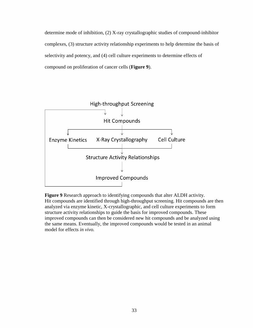
33
determine mode of inhibition, (2) X-ray crystallographic studies of compound-inhibitor
complexes, (3) structure activity relationship experiments to help determine the basis of
selectivity and potency, and (4) cell culture experiments to determine effects of
compound on proliferation of cancer cells (Figure 9).
Figure 9 Research approach to identifying compounds that alter ALDH activity.
Hit compounds are identified through high-throughput screening. Hit compounds are then
analyzed via enzyme kinetic, X-crystallographic, and cell culture experiments to form
structure activity relationships to guide the basis for improved compounds. These
improved compounds can then be considered new hit compounds and be analyzed using
the same means. Eventually, the improved compounds would be tested in an animal
model for effects in vivo.

34
II. Materials and Methods
A. Materials
Chemicals and reagents used for protein expression and purification, enzyme kinetics, X-
ray crystallography, and cell culture were purchased from Sigma-Aldrich (St. Louis, MO)
unless otherwise noted. Compounds were purchased from ChemDiv Corporation (San
Diego, CA), ChemBridge Corporation (San Diego, CA), InterBioScreen Limited
(Moscow, Russia), Life Chemicals (Burlington, ON), and Princeton BioMolecular
Research Inc. (Princeton, NJ). Dithiothreitol (DTT) was purchased from GoldBio (St.
Louis, MO). Sitting drop plates for crystallography were purchased from Charles Supper
Co (Natick, MA).
B. Methods
1. Production and Purification of ALDH Isoenzymes
The full-length cDNA for human ALDH2 was generously provided by Dr. Henry
Weiner, Purdue University, in a pT7-7 vector and used to transform E. coli BL21 cells for
subsequent expression and purification of the enzyme.221
The general protocol for the
production and purification for ALDH1A1, ALDH1A2, and ALDH2 has been published
previously.214, 221
Specifically, a single transformed BL21 colony was transferred to 10
mL 2xTY broth containing 100 µg/mL ampicillin and grown overnight in culture at 37°C
while shaking. A 5 mL aliquot of the overnight culture was used to inoculate 100 mL
2xTY containing 100 µg/mL ampicillin the next morning and was grown at 37°C while
shaking. After 1-2 hours 10 mL of the 100 mL was added to 8 x 1L 2xTY broth
containing 100 µg/mL ampicillin and incubated at 37°C with shaking at 200 rpm. Once

35
the culture reached an optical density of 0.6 to 0.8 at 600 nm, isopropyl-β-D-
thiogalactopyranoside was added to a final concentration of 100 µM to induce the
synthesis of the enzyme. Cultures were incubated overnight at 16°C while shaking at 200
rpm and then collected via centrifugation, flash frozen in liquid nitrogen and then stored
at -80°C. Cell pellets were thawed at room temperature and then resuspended in lysis
buffer (10 mM sodium phosphate (NaPi) pH 7.0, 2 mM EDTA, 1 mM benzamidine, 1
mM DTT). Cells were lysed via three passages through a microfluidizer (DivTech
Equipment). Cell lysate was clarified by centrifugation at 35,000 rpm for 25 min at 4°C
(Beckman-Coulter Optima L-90K Ultracentrifuge, Ti-45 Rotor). The clarified lysate was
dialyzed against 2 x 4 L changes of lysis buffer at 4°C each for at least 5 hours. The
lysate was then loaded onto a DEAE-sepharose column and eluted using gradient from 0-
250 mM NaCl in lysis buffer. The eluted fractions from the DEAE-sepharose column
were analyzed by SDS gel and an activity assay to confirm the presence of active
enzyme. In the activity assay 20 µL of the eluted fraction was added to saturating
amounts of NAD+ (75 mM) and propionaldehyde (10 mM) in 100 mM sodium
pyrophosphate buffer, pH 9.5 and activity determined by measuring the change in
absorbance at λ=340 nm due to NADH production. Fractions containing protein were
pooled and dialyzed into 4L 4-hydroxyacetophenone (4-HAP) column buffer (20 mM
NaPi pH 7.5, 1 mM EDTA, 50 mM NaCl, 1 mM DTT) overnight. The dialyzed enzyme
was then loaded onto the 4-HAP column and eluted in a single step with approximately
75 mL of 4-HAP buffer containing 10 mM 4-hydroxyacetophenone. The protein
preparations were then dialyzed exhaustively against 10 mM ACES, pH 6.6 with 1 mM
DTT at 4°C. Enzyme was concentrated to 5-10 mg/mL using Amicon Ultra Centrifugal

36
Devices (Millipore Corp, Bedford, MA). Prior to storage, the protein concentration was
measured using the BioRad Protein Assay (BioRad Laboratories, Hercules, CA) and its
aldehyde oxidation activity measured using saturating amounts of NAD+ and
propionaldehyde to determine specific activity. An 8 L preparation generated ~150 mg
ALDH1A1, ~60 mg ALDH2, or ~35 mg ALDH1A2. Aliquots were flash frozen in liquid
nitrogen and stored at -80°C. Portions of the ALDH1A1 and ALDH2 preparation were
stored at -20°C in a 50% (v/v) solution with glycerol for future crystallization. Prior to
crystallization set-up the enzyme was dialyzed against 3 X 4L changes of 10 mM sodium
ACES, pH 6.6 with 1 mM DTT at 4°C for at least 5 hours each. All solutions were
sparged with helium to remove dissolved oxygen before use to help prevent oxidation of
cysteine residues in addition to the DTT already present.
ALDH1B1 and ALDH3A1 were produced and purified as previously described.214, 222
ALDH3A1 was purified and provided by Dr. Bibek Parajuli and a portion of ALDH1B1
used was purified and provided by Lanmin Zhai. The full length cDNA for human
ALDH4A1 and ALDH5A1 was generously provided by Dr. Daria Mochly-Rosen and the
carboxyl terminus of rat ALDH1L1 was generously provided by Dr. Sergey Krupenko in
the pRSET expression plasmid. ALDH1L1 was expressed and purified as previously
described for ALDH3A1 with the following modifications: 1) the medium contained 100
µg/mL ampicillin, 2) cells were lysed via passage through a microfluidizer, 3) the second
passage through a Q-sepharose column was not included. The purification of ALDH4A1
and ALDH5A1 followed the same protocol as ALDH1L1.210
The purification of
ALDH1A3 followed one of two protocols. The first is consistent with that of ALDH1A1,

37
ALDH1A2, and ALDH2 and produced very small amounts of ALDH1A3.214
Dr. Jaume
Ferres (Barcelona, Spain) generously provided a His-tagged construct for full length
ALDH1A3. The His-tagged ALDH1A3 was produced and purified by a single passage
through a nickel-NTA column similar to the purification of ALDH4A1, ALDH5A1, and
ALDH1L1 and yielded ~120 mg per 8 L culture for enzymatic study.
2. Esterase High-Throughput Screen
The high-throughput screen (HTS) was performed using a reaction where para-
nitrophenyl acetate (pNPA) is hydrolyzed to form para-nitrophenol and acetic acid. The
esterase assay was used for screening as it is cofactor independent and the spectral
properties of para-nitrophenol do not overlap with the absorbance characteristics of the
majority of compounds within the tested libraries. The substrate was prepared by adding
0.0725 g of pNPA to 6 mL of 100% DMSO. This mixture was added slowly to 94 mL of
water with continuous stirring for a final concentration of 4 mM pNPA in 6% (v/v)
DMSO. Screens were completed with two different libraries at the Indiana University
Chemical Genomics Core Facility in 384-well, clear-bottomed plates. Screen 1 consisted
of 63,000 compounds from ChemDiv Corporation (San Diego, CA) and utilized 75 nM
ALDH2, 0.8 mM pNPA, 10 µM library compound, 2% DMSO, and 25 mM HEPES
buffer, pH 7.5 for a final reaction volume of 50 µL. Screen 2 consisted of 50,000
compounds from ChemBridge Corporation (San Diego, CA) and utilized 75 nM ALDH2,
1.6 mM pNPA, 10 µM library compound, 3.2% DMSO, and 25 mM HEPES buffer, pH
7.5 also in a final volume of 50 µL. The concentration of library compound is an
approximation based upon the average mass of the compounds in the entire library. The

38
production of para-nitrophenol was monitored by measuring the increase in absorbance at
λ=405 nm (molar extinction coefficient of 18,000 M-1
cm-1
) on a Spectromax Plus 384
plate reader for 10 minutes. Additional wells on each plate either had an inhibitor control
or no compound present to determine the normal rate. The inhibitor control for Screen 1
was 100 µM daidzin. Screen 2 used 1 µM 2,3,5-trimethyl-6-propyl-7H-furo[3,2-
g]chromen-7-one (2P3), an inhibitor discovered in the Screen 1. A multichannel pipet
was used to add either 20µL of 2% DMSO solution, 20µL of 250 µM daidzin in 2%
DMSO, or 20µL of 2.5 µM 2P3 in 2% DMSO to their respective control column. The
Genesis (Tecan) Workstation TeMO with a 96-channel pipetting head was used to make
all subsequent reagent additions. The rate of reaction was used to determine a ratio to
control for each compound with control being the average of the rates for the positive
control (wells with no compound) on that compound’s plate (n=16):
Equation 1: 𝑅𝑎𝑡𝑖𝑜 𝑡𝑜 𝐶𝑜𝑛𝑡𝑟𝑜𝑙 =𝑅𝑎𝑡𝑒 𝑜𝑓 𝑟𝑒𝑎𝑐𝑡𝑖𝑜𝑛 𝑤𝑖𝑡ℎ 𝑐𝑜𝑚𝑝𝑜𝑢𝑛𝑑
𝐴𝑣𝑒𝑟𝑎𝑔𝑒 𝑟𝑎𝑡𝑒 𝑜𝑓 𝑟𝑒𝑎𝑐𝑡𝑖𝑜𝑛 𝑜𝑓 𝑐𝑜𝑛𝑡𝑟𝑜𝑙
Compounds which had ratios to control less than 0.60 (>40% inhibition) were considered
inhibitors and those with a value greater than 1.25 (1.25-fold activation) were considered
activators in Screen 1. Compounds which met these selection criteria were rescreened for
validation using the same protocol. Selection criteria for Screen 2 was greater than 40%
inhibition or 2-fold activation in both the primary and secondary screens.
A Z’-factor was calculated for the screening conditions prior to completion of each HTS
by comparing the values of ALDH2 plus/minus the control inhibitor (each n=384) under
the conditions of that particular HTS using the following formula:
Equation 2: 𝑍′-𝑓𝑎𝑐𝑡𝑜𝑟 = 1 −3(𝛿𝑝+𝛿𝑛)
|𝜇𝑝−𝜇𝑛|
39
where µp and δp are the mean and standard deviation of the control reaction and µn and δn
are the mean and standard deviation of the inhibition reaction. A Z’-factor was also
calculated using 1 mM NAD+ as an esterase activator during Screen 2. A Z’-factor
between 0.5 and 1.0 indicates a good assay that is statistically strong enough to detect
inhibitors and/or activators. Confirmed chemical hits that were commercially available
were purchased from ChemDiv Corporation (San Diego, CA), ChemBridge Corporation
(San Diego, CA), Interbioscreen Limited (Moscow, Russia), Life Chemicals (Burlington,
ON) or Princeton BioMolecular Research Inc. (Princeton, NJ) to determine their effect on
aldehyde oxidation.
3. Aldehyde Oxidation Activity Assays for ALDH Isoenzymes
The effect on the aldehyde oxidation activity of ALDH isoenzymes by the hit compounds
from the screens was determined by measuring the formation of NAD(P)H
spectrophotometrically at λ=340 nm (molar extinction coefficient of 6220 M-1
cm-1
) on a
Beckman DU-640 spectrophotometer using purified recombinant enzyme. Compounds
were initially tested for their effect against ALDH1A1, ALDH2, and ALDH3A1. For
ALDH1A1 and ALDH2, the reaction contained 100-200 nM enzyme, 200 µM NAD+,
100 µM propionaldehyde, and 1% DMSO in 50 mM BES, pH 7.5 at room temperature.
For ALDH3A1, the reaction contained 20-25 nM enzyme, 300 µM NADP+, 300 µM
benzaldehyde, and 1% DMSO in 100 mM NaPi buffer, pH 7.5. The reaction was initiated
by addition of aldehyde substrate after a 2 min pre-incubation of enzyme, coenzyme, and
compound. Compounds were initially screened at a concentration of 10 µM.
Measurements at higher concentrations of compound were occasionally used to aid in

40
interpretation of data. Compounds that were potentially selective for ALDH1A1 and/or
ALDH2 were then tested with other ALDH isoenzymes. For ALDH1A2 and ALDH1A3,
the reaction contained 100-400 nM enzyme, 200 µM NAD+, 100 µM propionaldehyde,
and 1% DMSO. The assay for ALDH1B1 used 500 µM NAD+ and 200 µM
propionaldehyde. For ALDH4A1 the assay included 500 µM NAD+ and 20 mM
propionaldehyde. For ALDH5A1 the assay included 200 µM NAD+ and 2 mM
propionaldehyde. For rat ALDH1L1 the assay included 500 µM NADP+ and 4 mM
propionaldehyde. The assay components for the selectivity assays were designed to
provide the maximal stringency toward ALDH2, such that the substrate concentration
utilized was >500-fold above KM for ALDH2 while keeping below 15-fold over Km for
the other isoenzymes. All reactions were initiated with the addition of aldehyde substrate
after a 2 min pre-incubation period. EC50 values for aldehyde oxidation were calculated
by varying the concentration of compound from 0 - 100 µM for compounds which
showed greater than 50% activation/inhibition at 10 µM concentration of compound.
Assay parameters for EC50 calculation were the same as for the initial selectivity screen,
with the use of 500 µM acetaldehyde as an additional aldehyde substrate for AC50
calculations with ALDH2. Data were fit to the four parameter EC50 equation using
SigmaPlot (v12):
Equation 3: 𝑦 = 𝑚𝑖𝑛 +(𝑚𝑎𝑥−𝑚𝑖𝑛)
1+(𝑥
𝐸𝐶50)
−𝐻𝑖𝑙𝑙𝑆𝑙𝑜𝑝𝑒
where y is the rate of activity and x is the concentration of compound. The values
represent the mean/SEM of three independent experiments, with each experiment n = 3.

41
Thirty-four additional psoralen and coumarin derivatives were ordered from ChemDiv
Corporation (San Diego, CA) and ChemBridge Corporation (San Diego, CA) to build
structure-activity relationships for the hit compounds. Analogs were selected as a
representative sample of the 600+ commercially available compounds which had >95%
structure similarity. EC50 values for aldehyde oxidation for these compounds were
calculated using the same protocol as the initial hit compounds. Data were fit to the four
parameter EC50 equation (Equation 3) using SigmaPlot (v12) . The values represent the
mean/SEM of three independent experiments, with each experiment n=3.
4. Steady-State Kinetic Characterization with ALDH1A1 and ALDH2
The mode of inhibition of the compounds was characterized by steady state kinetics
through covariance of compound and either aldehyde substrate or coenzyme
concentration. The secondary substrate was kept at a saturating amount. The mode of
inhibition towards NAD+ binding with ALDH1A1 was determined by covarying inhibitor
and coenzyme concentrations at a fixed aldehyde substrate concentration. Dehydrogenase
activity was measured spectrophotometrically by measuring the formation of NADH at
340 nm (molar extinction coefficient of 6200 M-1
cm-1
) on a Beckman DU-640
spectrophotomer for 3 min. The assay included 100-200 nM ALDH1A1, 20-200 µM
NAD+, 1 mM propionaldehyde, and 1% DMSO in 25 mM BES buffer, pH 7.5. The
reaction was initiated by the addition of propionaldehyde after a 2 minute pre-incubation
with enzyme, NAD+, and compound. The mode of inhibition towards NAD
+ binding with
ALDH2 was determined in a similar fashion. The activity of ALDH2 was determined in a
reaction containing 100-200 nM ALDH2, 15-400 µM NAD+, 1 mM propionaldehyde,

42
and 1% DMSO in 25 mM BES buffer, pH 7.5. All data were fit to the tight binding or
single substrate-single inhibitor non-linear velocity expressions for competitive, non-
competitive, mixed type non-competitive, and uncompetitive inhibiton using SigmaPlot
(v12, Enzyme Kinetics Module). Lineweaver-Burk plots were generated using SigmaPlot
(v12) to better visualize the model fit. The appropriate model was selected through
analysis of goodness-of fit. Values shown represent the mean/SEM of three independent
experiments (each n=3).
The mode of inhibition towards aldehyde substrate binding for ALDH2 was measured via
fluorescence spectroscopy. The KM of ALDH2 for propionaldehyde (<1 µM) is so low
that measurement of activity at near KM concentrations is nearly impossible by
measurement of absorbance.223
Utilizing the fluorescent characteristics of NADH allowed
for the measurement of these activities. A standard curve of the fluorescence of NADH
from 10 nM to 3000 nM was first measured for use in converting the arbitrary
fluorescence units into the amount of NADH produced during the experiment. The KM
values for propionaldehyde and cofactor NAD+ for ALDH2 were then determined for
ALDH2 using the fluorescence assay as these values may differ from those obtained
using the absorbance assay. Dehydrogenase activity was measured by monitored the
fluorescence of NADH at λ = 470 nm after excitation at λ = 320 nm for 2 min measuring
every five seconds. Reactions for KM calculations contained 5 nM ALDH2 in 100 mM
NaPi, pH 7.0 in a reaction volume of 3 mL. The KM for NAD+ was determined by
plotting increasing concentrations of NAD+ (10-400 µM) versus the velocity of the
reaction with a saturating amount of propionaldehyde (1 µM). The KM for

43
propionaldehyde was determined by plotting increasing concentrations of
propionaldehyde (50-750 nM) versus the velocity of the reaction with a saturating
amount of NAD+ (400 µM). Data were plotted using the Simple Ligand Binding Toolbox
in SigmaPlot (v12) and the values represent the mean/SEM of a single experiment (n=3).
Steady-state kinetics were used to measure the mode of inhibition towards
propionaldehyde binding in ALDH2. Assay solutions included 5 nM ALDH2, 400 µM
NAD+, 75-600 nM propionaldehyde, 0.2% DMSO, and 0-80 nM inhibitor in 100 mM
NaPi, pH 7.0. Due to low concentrations of substrate used in the assay, all reagents were
filtered to remove additional aldehyde contaminants. All data were fit to the tight binding
non-linear velocity expressions for competitive, non-competitive, mixed-type non-
competitive and uncompetitive inhibition using SigmaPlot (v12, Enzyme Kinetics
Module) to evaluate goodness of fit. Lineweaver-Burk plots were generate using
SigmaPlot (v12) to better visualize the mode of inhibition. All data represent the
mean/SEM of the four independent experiments (n=4) of duplicate assays at each
concentration.
5. Characterization of 2BS4’s Inhibition of ALDH2
IC50 curves were measured for the inhibition of ALDH2 by 2BS4 utilizing different
incubation times. The rate of aldehyde oxidation was determined by measuring the
formation of NADH spectrophotometrically at λ=340 nm (molar extinction coefficient of
6220 M-1
cm-1
) on a Beckman DU-640 spectrophotometer using purified recombinant
enzyme. The assay solutions included 150 nM ALDH2, 200 µM NAD+, 100 µM
propionaldehyde, 0 – 50 µM 2BS4, and 1% DMSO in 25 mM BES buffer, pH 7.5. The

44
reaction was initiated by addition of enzyme to coenzyme, compound, and substrate (0
min incubation) or by addition of aldehyde substrate after either a 2 or 10 min pre-
incubation of enzyme, coenzyme, and compound. Data were fit to the four parameter
EC50 equation (Equation 3) using SigmaPlot (v12). Values represent the mean/SEM of
one experiment with three replicates for each data point (n=3).
The interaction of the compound 2BS4 with ALDH2 was characterized further by
incubating 2BS4 with ALDH2 before the addition of NAD+ and aldehyde substrate.
Dehydrogenase activity was measured spectrophotometrically by determining the
formation of NADH at 340 nm (molar extinction coefficient of 6200 M-1
cm-1
) using a
Beckman DU-640 spectrophotomer for 3 min. The assay included 200 nM ALDH2, 0.3
µM or 1.5 µM 2BS4, 200 µM NAD+, 1 mM propionaldehyde, and 1% DMSO in 25 mM
BES buffer, pH 7.5. ALDH2 was incubated with either 3 µM or 15 µM 2BS4 in 10%
DMSO for various time periods before subsequent 10-fold dilution into the assay. After
dilution of the ALDH2/2BS4 solution, the reaction was initiated by the addition of
propionaldehyde after 2 minute incubation with ALDH2/2BS4 and NAD+. The
measurement at zero hour(s) incubation was taken by diluting the enzyme-compound
mixture immediately after addition of compound. Each data point represents the average
of two measurements. (n=2)
6. Crystallization of ALDH1A1 and ALDH2 Complexes
The general crystallization conditions for both ALDH1A1 and ALDH2 have been
reported previously.44, 220
Crystals of ALDH1A1 were grown by equilibrating 3 mg/mL

45
ALDH1A1 with 100 mM sodium BisTris, pH 6.2 – 6.6, 9 – 11% PEG 3350, 200 mM
NaCl, and 5 mM YbCl3 using the sitting drop method with 500 µL mother liquor. Drop
composition consisted of 2 – 4 µL protein mixed with 2 – 4 µL crystallization solution.
The drop was then mixed by agitation. Crystallization was allowed to proceed at 22°C in
an incubation chamber and crystals grew in 1 – 2 weeks. BUC11 and BUC22 complexes
were prepared by growing ALDH1A1 crystals in the presence of 200 µM compound and
2% (v/v) DMSO (co-crystallization). Compound was first added to the protein solution
before mixing with the crystallization solution. The BUC25 complex was prepared by
soaking an apo-ALDH1A1 crystal overnight with crystallization solution containing 500
µM BUC25 with 2% (v/v) DMSO. All ALDH1A1 crystals were cryoprotected for flash-
freezing by adding 20% (v/v) ethylene glycol to the final crystallization solution. Crystals
were frozen in a liquid nitrogen cryostream and were tested on the home source Bruker
D8 system for diffraction quality. Crystals with good diffraction were stored in liquid
nitrogen until data collection at a synchrotron source.
Crystals of the 2P3-ALDH2 complex were grown by equilibrating 8 mg/mL ALDH2 and
200 µM 2P3 in 2% DMSO with 100 mM sodium ACES, pH 6.4, 100 mM guanidine-
HCl, 10 mM MgCl2, 4 mM dithiothreitol, and 18% PEG 6000 using the sitting drop
method with 500 µL mother liquor. Crystal plates were set up in a glove box then
transferred to an incubation chamber at 22°C for crystal growth. Crystals typically grew
in 1 – 2 weeks. Crystals were cryoprotected for flash-freezing by adding 18% (v/v)
ethylene glycol to the final crystallization solution. Crystals were frozen in a liquid
nitrogen cryostream and were tested on the home source Bruker D8 system for diffraction
46
quality. Crystals with good diffraction were stored in liquid nitrogen until data collection
at a synchrotron source.
Diffraction data was collected at Beamline 19-ID operated by the Structural Biology
Consortium at the Advanced Photon Source, Argonne National Laboratory, Chicago, IL.
Diffraction data was indexed, integrated, and scaled using the HKL2000 or HKL3000
program suites.224
The CCP4 program suite was used for molecular replacement and
refinement.225
The human apo-ALDH2 structure (PDB Code 3N80) was used as a model
for ALDH2 and the human apo-ALDH1A1 structure (PDB Code 4WJ9) was used as a
model for ALDH1A1. Molecular replacement was necessary as both the ALDH1A1-
BUC22 and ALDH2-2P3 complexes were obtained in orientations and/or space groups
not seen previously using the same conditions. The ALDH2-2P3 complex was obtained
in the previously seen P212121 crystal lattice but with different unit cell dimensions. The
ALDH1A1-BUC22 complex was obtained in a previously unseen P1 crystal lattice that
contained two tetramers in the asymmetric unit. Previous ALDH1A1 structures, and the
ALDH1A1-BUC25 and ALDH1A1-BUC11 structures, were obtained in a P422 space
group with a single monomer in the asymmetric unit. The molecular graphics application
Coot was used for model building.226
Ligand maps were generated using Sketcher
(CCP4) to use for refinement. The TLSMD (Translation/Libration/Screw Motion
Determination) server was used to determine dynamic properties of ALDH1A1.227, 228

47
7. Cellular Effect of the Coumarin Derivative BUC22
MDA-MB-468 (ER-/PR-/HER-) breast cancer cells were generously provided by Dr.
Clark Wells (IUSM). MDA-MB-468 cells were cultured in 1X DMEM (Cellgro,
Mediatech Inc, Manassa, VA) supplemented with 10% fetal bovine serum (FBS) (Cell
Applications Inc, San Diego, CA) and 100 Units/mL penicillin and 100 µg/mL
streptomycin (P/S)(Lona, Walkersville, MD). Cells were passaged after every 80-90%
confluence (normally every 3-4 days).
The 3-(4,5-dimethylthiazol-2-yl)-2-5-diphenyltetrazolium bromide (MTT) assay was
used to measure the proliferative potential of cancer cells after treatment with BUC22.
Cells were plated at 5000 cells/well density in 96-well clear bottom cell culture plates on
40 µL ~7mg/mL Matrigel Matrix Basement Membrane (Corning, Bedford, MA). Cells
were treated starting on the day of plating (Day 1) by adding compound in 0.5% (v/v)
DMSO to 200 µL DMEM supplemented with FBS and P/S. One set of wells contained
Matrigel alone and one set contained cells on Matrigel, both in compound-free media.
Media was changed on Day 3 and Day 5. On Day 6 media was aspirated and 0.1 mL of
MTT (0.5 mg/mL in PBS) was added to each well followed by incubation for 4 hours at
37°C to allow for reduction of MTT to its formazan. After reduction 100 µL 0.05N HCl
in 10% SDS was added to each well followed by incubation for 4 hours at 37°C to lyse
the cells. After the second incubation the absorbance of each well was measured at λ=595
nm. Each experiment had three duplicate wells of each condition. Values were
normalized by subtracting absorbance values of wells which contained only Matrigel.
Data represent the mean/SEM of three independent experiments, each n=3.
48
III. Results
A. Esterase High-Throughput Screen for ALDH2 Modulators
Two high-throughput screens (HTS) were performed to identify activators and inhibitors
of ALDH2 as measured by its in vitro esterase activity. Screen 1 consisted of 63,000
compounds from ChemDiv Corporation (San Diego, CA) and Screen 2 consisted of
50,000 compounds from ChemBridge Corporation (San Diego, CA). Both the esterase
hydrolysis and aldehyde oxidation reactions use the same catalytic residues. The use of
the esterase reaction for screening purposes has two advantages over the aldehyde
oxidation reaction. First, interference from compounds with overlapping UV absorbance
characteristics with NADH is minimized. Secondly, the esterase reaction does not require
NAD+ and thus the esterase reaction limits identification of compounds that interfere with
cofactor binding. The cofactor binding site is highly conserved among ALDH
isoenzymes, thus compounds that bind in the cofactor binding site are less likely to be
selective and options to achieve selectivity are limited.
1. Z’-factor Calculation
A Z’-factor score was calculated prior to the start of each HTS due to the use of different
control inhibitors for each screen. A Z’-factor between 0.5 and 1.0 indicates an excellent
assay to distinguish differences in activity from control reactions, i.e. determine
activators and inhibitors. The Z’-factor for Screen 1 comparing ALDH2 activity
with/without inhibitor (100 µM Daidzin) was 0.60 (Figure 10). During the course of
Screen 1 a more potent ALDH2 inhibitor was discovered and used as the control inhibitor
for Screen 2. The Z’-factor for Screen 2 comparing ALDH2 activity with/without

49
inhibitor (1 µM 2P3) was 0.50 (Figure 11). The better inhibitor did not improve the Z’-
factor. For Screen 2 a Z’-factor comparing ALDH2 activity with/without activator (1 mM
NAD) was also calculated and found to be 0.46 (Figure 11). Based off the Z’-factor both
screens were excellent at discovering inhibitors of ALDH2 esterase activity. The Z’-
factor for Screen 2 for determination of activation was nearly excellent as well. Although
increasing the amount of enzyme could have made it easier to distinguish activators, it
also would have made it more likely that any activator would not follow a linear rate for
the duration of the screen. The lower Z’-factor for Screen 2 may be due to higher
background due to the use of twice as much substrate.
Figure 10 Z’-factor determination for Screen 1.
Each point represents the rate of change in absorbance at 405 nm of a reaction. The x-
axis is the column (1-24) on the 384-well plate. The blue data points correspond to the
enzyme + substrate (ES) control (mean=4.02); the red is enzyme + substrate + inhibitor
(ESI) (mean=1.54); the black is the no enzyme blank control (mean=0.380). The lines
represent 3x standard deviation from ES mean (blue), ESI mean (red), and blank (black).
Each condition performed on a separate plate with n=384.

50
Figure 11 Z’-factor determination for Screen 2.
Each point represents the rate of change in absorbance at 405 nm of a reaction. The x-
axis is the column (1-24) on the 384-well plate. The blue data points represent the
enzyme + substrate (ES) control (mean=3.50); the red is enzyme + substrate + inhibitor
(ESI) (mean=1.23); the green in enzyme + substrate + activator (ESA); the black in no
enzyme blank control (mean=0.990). The lines represent 3x standard deviation from ES
mean (blue), ESI mean (red), ESA mean (green) and blank (black). Z’-factor for ESA vs
ES shown in green and for ESI vs ES shown in red. Each condition performed on a
separate plate with n=384.
2. High-Throughput Screen Results
The two libraries totaling over 110,000 compounds from ChemDiv and ChemBridge
were screened using the esterase assay. Plates were acquired from the Indiana University
Chemical Genomics Core with 20 µL of 25 µM of each compound, which was already
present in the first 22 columns of each 384-well plate. Each plate also contained a control
column with enzyme and substrate (ES control). The average of intra-plate ES control
served as the basis to determine whether a compound modulated esterase activity. Each
plate also contained an additional control column with enzyme and substrate and inhibitor
to ensure significant inhibition could be measured on each plate. A representative plate is

51
shown in Figure 12. Selection criteria were initially set at either greater than 2-fold
activation or less than 60% activity. Due to the lack of activators obtained from Screen 1,
the selection threshold was lessened to 1.25-fold activation for only Screen 1. The
primary screen of Screen 1 yielded a total of 1395 compounds and that of Screen 2
yielded 536 compounds that were activators or inhibitors of ALDH2’s esterase activity.
These 1931 compounds were then rescreened using the same assay parameters. Only 53
compounds from Screen 1 and 14 compounds from Screen 2 met the threshold criteria in
the secondary screen, meaning that over 95% of compounds did not meet the selection
criteria a second time. Of the 67 compounds identified through the high-throughput
screens, 59 were commercially available and purchased for further study.
Figure 12 Example plate from Screen 1 for compounds that modify ALDH activity.
Each point represent one well, with the x axis the column (1-24) on the plate and the y
axis the rate of change in absorbance measured at λ=405 nm. Column 23 is the ESI
control with a mean value of 1.78 (n=16). Column is the ES control with a mean value of
4.93 (n=16). On this plate we identified one activator and fifteen inhibitors.

52
B. Hit Compound Validation and Initial Determination of Selectivity
We were able to obtain 59 of the 67 compounds identified through the high-throughput
screen. At this point we switched from monitoring the esterase function of aldehyde
dehydrogenase to the aldehyde oxidation function in part to better evaluate how the
compounds will modulate the complete catalytic mechanism of ALDHs. The effect of the
hit compounds on the activity of ALDH1A1, ALDH2, and ALDH3A1 was tested as an
initial determination of selectivity and potency. Of the 59 compounds examined at 10
µM concentration, 45 did not modulate the activity of ALDH2 more than 20% in either
direction relative to control. Several of the 45 compounds which did not modulate
ALDH2 activity influenced the activity of one or both of the ALDH1A1 and ALDH3A1
enzymes by more than 20% (2CD12, 2BS2, 2BS6, 2P5, 2P7, 2P9, 2P11, 2P12, 2LC1,
2F3, 2F8, 2F10, 2F11, and 2F12). Of the 14 compounds that did modulate ALDH2
activity, several were determined to have low potency and/or selectivity based off the
measurements at 10 µM concentration. 2C1 activated each enzyme ~1.2 fold at 10 µM
concentration. 2CB4 inhibited all three isoenzymes greater than 80% and 2F13 inhibited
all three around 50%. 2C2 inhibited ALDH1A1 activity much greater than ALDH2
activity. Although 2CD4 inhibited the activity of ALDH2, the compound showed
activation of ALDH1A1. 2CD5 showed some selectivity towards ALDH2, though the
potency was low compared to other hit compounds. 2CB5, 2P2, and 2P4 were selected
for further analysis due to their high potency towards ALDH2 (≥80% inhibition) and
relative selectivity against ALDH1A1 and ALDH3A1 (Figure 13). However, additional
batches of 2P2 ordered to continue enzymatic studies no longer inhibited ALDH2 at this
concentration. The first batch of 2P2 may have had an additional contaminant leftover
53
from synthesis of the compound which enhanced ALDH2 inhibition; however, this could
not be verified due to the exhausted supply of the initial stock. As a consequence, 2CB5
and 2P4 were the two most promising remaining hit compounds. Both 2BS4 and 2LC2
showed selectivity towards ALDH2 but contain a terminal methyl ester group; these
potential esterase substrates were initially avoided, as inhibition could be lost or
diminished after ester hydrolysis. 2BS4 was eventually included again due to its
structural similarity to 2P4 and 2CB5. 2P3, which did not show the same level of
selectivity towards ALDH2 as 2P4 and 2CB5 but had the highest potency, was also
included due to the structural similarity.
.

54
Figure 13 Effect on aldehyde oxidation of hit compounds from HTS.
The activity of 59 compounds (10 µM) identified via HTS was tested on three ALDH
isoenzymes. Average of at least two (n≥2) independent trials.

55
Figure 13 Continued Effect on aldehyde oxidation of hit compounds from HTS.
The activity of 59 compounds (10 µM) identified via HTS was tested on three ALDH
isoenzymes. Average of at least two (n≥2) independent trials.

56
Figure 13 Continued Effect on aldehyde oxidation of hit compounds from HTS.
The activity of 59 compounds (10 µM) identified via HTS was tested on three ALDH
isoenzymes. Average of at least two (n≥2) independent trials.
The effect of the four compounds (2BS4, 2P3, 2P4, and 2CB5) on other ALDH
isoenzymes was tested to generate a broader view of the compounds’ selectivity towards
ALDH2 (Figure 14). The effect of 10 µM of each compound on the dehydrogenase
activity of the other three ALDH1A/2 family members (ALDH1A2, ALDH1A3, and
ALDH1B1) was determined, in parallel with assays of ALDH4A1, ALDH5A1, and
ALDH1L1 (rat). Inhibition of the isoenzymes by the four compounds was limited to the
ALDH1/2 family as the compounds did not modulate the activity of ALDH3A1,
ALDH4A1, ALDH5A1, or ALDH1L1 (rat). Each compound showed the greatest amount
of inhibition towards ALDH2 of the isoenzymes tested. For any particular ALDH1/2

57
enzyme, 2P3 inhibited the most, followed by 2CB5, then 2P4 at 10 µM. The inhibition
profile for the four compounds was very similar between ALDH1A2, ALDH1A3, and
ALDH1B1. 2BS4 only showed inhibition towards ALDH2 at this single concentration of
compound.
Figure 14 Effects of four aromatic lactones on activity of nine ALDH isoenzymes.
Percent dehydrogenase activity for nine ALDH isoenzymes in the presence of 10 µM
compound compared to control reaction with no compound present. Value is the average
of at least three independent trials (n≥3) with standard error, except for the measurement
of ALDH1B1 activity with 2P4 and ALDH1L1 activity with each compound (n=2).
C. EC50 Determination of Hit Compounds
1. Alda-1 Mimics
Although no compounds from the HTS activated ALDH2’s oxidation of propionaldehyde
by more than 20% at 10 µM concentration, three compounds (2CD11, 2CB8, and 2CB9)
identified in the HTS were structurally similar to Alda-1 and thus were examined further
(Figure 15). Each share the benzodioxol group that in Alda-1 is bound within the
substrate tunnel oriented towards Cys302. The three compounds were found to activate
the oxidation of propionaldehyde by ALDH2 while having no effect on the activity of
ALDH1B1, ALDH1A1, ALDH1A2, ALDH1A3, or ALDH3A1. Each of the compounds

58
had an AC50 equal to approximately 50-60 µM and activated the oxidation of
propionaldehyde by at most 2-fold (Table 3). Each compound became insoluble in the
assay solution between 200 and 500 µM; therefore solubility limits prevented a more
accurate measurement of the AC50 for propionaldehyde oxidation by ALDH2. For 2CB8,
full dose-response analysis could not be completed. The AC50 of ~60 µM is based off the
assumption that the maximum activity would be near 2-fold if the curve could be
completed.
Figure 15 Three Alda-1 mimics identified via the HTS.
Compounds share a benzodioxol group linked to a carboxamide through an isoxazole
linker (shown in red).
Since Alda-1 activated the oxidation of smaller substrates more effectively, we also
tested the effect the three activators had on the oxidation of acetaldehyde by ALDH2.
The three compounds had varying effects on acetaldehyde oxidation. 2CB9 was the most
potent (AC50 = 9.6 ± 2.2 µM) but increased the maximum acetaldehyde oxidation activity
the least (160 ± 4%). 2CD11 was the least potent (AC50 = 28 ± 5 µM) but increased the
maximum acetaldehyde oxidation activity of ALDH2 the most (210 ± 9%) (Table 3).

59
The AC50 values for propionaldehyde and acetaldehyde oxidation with each compound
are higher than those seen with Alda-1.55
For this reason, along with the solubility issues,
these compounds were not further studied.
Table 3 AC50 determination of Alda-1 mimics with ALDH2.
Values are the mean of a single experiment of three independent measurements (n=3).
Values for 2CB8 for propionaldehyde oxidation are approximated based off an
incomplete curve using the assumption that binding behavior is consistent with the other
compounds.
Compound
Aldehyde Substrate
Acetaldehyde Propionaldehyde
Max Activity AC50 (µM) Max Activity AC50 (µM)
2CD11 210 ± 9% 28 ± 5 180 ± 11% 50 ± 16
2CB8 200 ± 10% 14 ± 4 ~200% ~60
2CB9 160 ± 4% 9.6 ± 2.2 200 ± 19% 61 ± 29
2. Coumarin and Psoralen Derivatives, in Comparison to Daidzin
The four compounds (2BS4, 2P3, 2P4, and 2CB5) are aromatic lactones (Figure 16).
2P3, 2P4 and 2CB5 are psoralen derivatives and 2BS4 is a coumarin derivative. The
differences between 2P3, 2P4, and 2CB5 lie in the presence and length of the various
alkyl substituents attached to the aromatic substructure. 2BS4 contains an alkyl chain
with a terminal methyl ester group bound to the psoralen substructure via an ether
linkage. The ether oxygen is in the same relative position as the furan oxygen of 2P3,
2P4, and 2CB5.

60
Figure 16 Four aromatic lactone inhibitors identified via the HTS.
Compounds each share a coumarin aromatic structure with an oxygen atom bound to the
benzyl ring. For 2CB5, 2P3, and 2P4; this takes the form of a psoralen aromatic structure.
The IC50 values for daidzin for the ALDH1/2 family were measured to compare our
results to compounds (including CVT-10216) already described in the literature as
ALDH2-selective inhibitors.201, 204
Consistent with prior work, daidzin was found to be
about 100-fold more potent toward ALDH2 than ALDH1A1 (Table 4). However our
IC50 values are 10-fold higher than prior values, likely due to differing substrate
concentrations in the assays. In regards to the other ALDH1/2 enzymes, we found daidzin
to inhibit ALDH1B1 (IC50 = 5.1 ± 0.5 µM) and ALDH1A2 (IC50 = 4.5 ± 0.6 µM) with
similar potencies to ALDH2 (IC50 = 3.5 ± 0.1 µM), while being less potent against
ALDH1A1 and ALDH1A3. Inhibition of ALDH1A1 and ALDH1A2 by daidzin was
observed, but compound solubility issues in the assay prevented full dose-response
analyses.
The IC50 values of the four aromatic lactones were then determined for nine ALDH
isoenzymes (Table 4) in order to better characterize the selectivity of the compounds

61
towards individual members of the ALDH1/2 family of isoenzymes and against other
ALDH isoenzymes. None of the four compounds inhibited the activity of ALDH1L1
(rat), ALDH3A1, ALDH4A1, or ALDH5A1 when present in the reaction mixtures up to
100 µM. 2BS4 showed selectivity towards ALDH2 versus the other eight isoenzymes
tested with an IC50 value of 1.5 ± 0.3 µM. The other three compounds inhibited ALDH2
more strongly, though they also inhibited the majority of the other ALDH1/2 isoenzymes
at sub-micromolar concentrations. The lowest IC50 measured was that of 2P3 with ALDH
(0.11 ± 0.02 µM), 30 times as potent as daidzin. 2P3 inhibited each of the five ALDH1/2
isoenzymes the strongest and was the only compound which completely inhibited each of
the five isoenzymes.
Table 4 IC50 values for four aromatic lactones with nine ALDH isoenzymes.
Values are the mean/SEM of three independent experiments, each n=3.
IC50 (µM)
Inhibitor ALDH2 ALDH1B1 ALDH1A1 ALDH1A2 ALDH1A3
Daidzin 3.5 ± 0.1 5.1 ± 0.5 >200 4.5 ± 0.6 >200
2CB5 0.34 ± 0.05 0.88 ± 0.28 0.34 ± 0.05# 0.76 ± 0.07 1.1 ± 0.1
2P3 0.11 ± 0.02 0.36 ± 0.12 0.22 ± 0.05 0.30 ± 0.04 0.40 ± 0.06
2P4 0.19 ± 0.01 0.31 ± 0.07# NI 0.33 ± 0.04
# 0.27 ± 0.05
2BS4 1.5 ± 0.3 NI NI NI NI
IC50 (µM)
Inhibitor
ALDH1L1
(rat) ALDH3A1 ALDH4A1 ALDH5A1
Daidzin NI NI NI NI
2CB5 NI NI NI NI
2P3 NI NI NI NI
2P4 NI NI NI NI
2BS4 NI NI NI NI
#Compounds had ~60% maximum inhibition NI=No Inhibition

62
2P4 has a similar IC50 value for ALDH2 (0.19 ± 0.01 µM) and ALDH1A3 as 2P3, but
only partially inhibits ALDH1B1 and ALDH1A2 and does not inhibit ALDH1A1. 2CB5
has the highest IC50 values for the five ALDH1A/2 isoenzymes and only partially
inhibited ALDH1A1. The number and length of alkyl substitutions on the aromatic
lactone appears to correlate with the potency of inhibition towards the isoenzymes. This
correlation is best seen in terms of ALDH1A1 inhibition. 2P3 and 2CB5 have a propyl
chain proximal to the lactone carbonyl and inhibit ALDH1A1, whereas 2P4 has a methyl
chain in the same position and does not inhibit ALDH1A1. Additionally, 2CB5 lacks the
methyl chain proximal to the furan oxygen seen in 2P3 and does not inhibit ALDH1A1 as
strongly as 2P3.
D. Characterization of Interaction of Aromatic Lactones with ALDH2
1. Kinetic Characterization of 2P4 in ALDH2 Assays
Covariation experiments were completed with 2P4 to better understand the mechanisms
by which this compound inhibits ALDH2. 2P4 was chosen as a representative of the
psoralen derivatives as 2P4 showed the most selectivity for inhibition of this enzyme.
Due to the low Km of ALDH2 for propionaldehyde (<1 µM), the production of NADH by
ALDH2 was measured via fluorescence when varying aldehyde substrate.223
A standard
curve for the fluorescence of NADH was generated to convert the arbitrary fluorescence
units to nanomoles of NADH produced (Figure 17). The Km for propionaldehyde was 49
± 9 nM and the Km for NAD+ was 31 ± 4 µM using the fluorescence assay (Figure 18).
2P4 was found to non-competitively inhibit (mixed-type) the binding of aldehyde
substrate to ALDH2 with a Ki = 31 ± 3 nM (Figure 19A). Covariation experiments

63
varying the amount of NAD+ were able to be completed using the absorbance assay. 2P4
was found to competitively inhibit the binding of NAD+ to ALDH2 with a Ki =87 ± 8 nM
(Figure 19B).
Figure 17 Standard curve for fluorescence of NADH.
Each point represents the mean/SEM of two separate experiments of three replicates each
(n=6).
Figure 18 Substrate Km determination using fluorescence assay.
The Km for (A) NAD and (B) propionaldehyde using fluorescence assay was measured.
Each point represents the mean/SEM of one experiment consisting of three replicates
(n=3).

64
Figure 19 Steady-state kinetic characterization of inhibition of ALDH2 by 2P4.
(A) Lineweaver-Burk representation of (mixed-type) non-competitive inhibition of 2P4
versus varied propionaldehyde with ALDH at fixed concentration of NAD+ (400 µM)
measured via fluorescence. Values are the mean/SEM of four independent experiments,
each n=2. (B) Lineweaver-Burk representation of competitive inhibition of 2P4 versus
varied NAD+ with ALDH2 at fixed concentration of propionaldehyde (1 mM) measured
via absorbance. Values are the mean/SEM of three independent experiments, each n=3.
2. 2P3-ALDH2 Crystal Structure
We wished to solve the structure of an enzyme-compound complex to understand where
the aromatic lactone binds to the enzyme and what residues and interactions lead to
selectivity. 2P3 was used for crystallographic studies as it was the most potent for
ALDH2 among the compounds. The structure of the 2P3-ALDH2 complex was solved to
a resolution of 2.40Å (Table 5).

65
Table 5 Collection and refinement statistics for the 2P3-ALDH2 complex.
PDB Code 5L13
Collection
Space Group P212121
Cell Dimensions
a,b,c (Å) 99,127,295
α,β,γ (deg) 90,90,90
Resolution (Å) 50.0-2.40
Rmerge 0.093 (0.280)*
I/σ 16.2 (5.0)
Completeness (%) 94.2 (85.7)
Redundancy 5.0 (4.8)
Refinement
No. of reflections 131537
Rwork/Rfree 0.16/0.21
RMSD
Bond Length (Å) 0.009
Bond Angle (deg) 1.324
*Values in parentheses are for data in the highest resolution shell (2.44 to 2.40 Å)
2P3 was found to bind in the substrate binding site of ALDH2 (Figure 20A). The binding
site is formed in part by the side chains of four phenylalanine residues (170, 296, 459,
and 465). The side chain of the catalytic Cys302 rotates toward the NAD+ binding site to
accommodate the binding of 2P3. The propyl chain proximal to the lactone extends into a
pocket formed in part by Glu268, Glu476, Trp177, and Thr244, while the alkyl chains on
the furan ring are oriented towards the solvent exposed surface. The carbonyl of 2P3
mimics the position of a potential aldehyde substrate engaging the oxyanion hole during
aldehyde oxidation and hydrogen bonds with the peptide nitrogen of Cys302 (Figure
20B). The hydrophobic interactions and this single hydrogen bond orient the compound
in the substrate binding site.
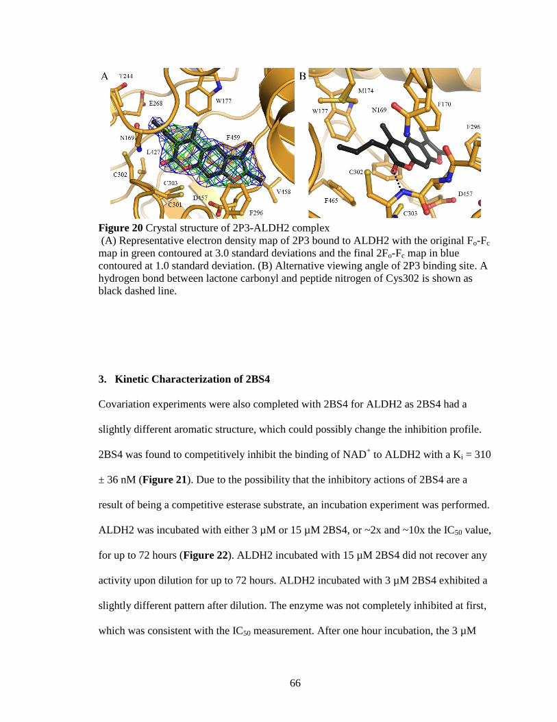
66
Figure 20 Crystal structure of 2P3-ALDH2 complex
(A) Representative electron density map of 2P3 bound to ALDH2 with the original Fo-Fc
map in green contoured at 3.0 standard deviations and the final 2Fo-Fc map in blue
contoured at 1.0 standard deviation. (B) Alternative viewing angle of 2P3 binding site. A
hydrogen bond between lactone carbonyl and peptide nitrogen of Cys302 is shown as
black dashed line.
3. Kinetic Characterization of 2BS4
Covariation experiments were also completed with 2BS4 for ALDH2 as 2BS4 had a
slightly different aromatic structure, which could possibly change the inhibition profile.
2BS4 was found to competitively inhibit the binding of NAD+ to ALDH2 with a Ki = 310
± 36 nM (Figure 21). Due to the possibility that the inhibitory actions of 2BS4 are a
result of being a competitive esterase substrate, an incubation experiment was performed.
ALDH2 was incubated with either 3 µM or 15 µM 2BS4, or ~2x and ~10x the IC50 value,
for up to 72 hours (Figure 22). ALDH2 incubated with 15 µM 2BS4 did not recover any
activity upon dilution for up to 72 hours. ALDH2 incubated with 3 µM 2BS4 exhibited a
slightly different pattern after dilution. The enzyme was not completely inhibited at first,
which was consistent with the IC50 measurement. After one hour incubation, the 3 µM

67
2BS4/ALDH2 mixture was completely inhibited after dilution. However, the 3 µM
2BS4/ALDH2 mixture slowly regained activity starting after one day. This would suggest
that 2BS4 is losing the ability to inhibit ALDH2 over time, possibly through hydrolysis
of the ester.
Figure 21 Steady-state kinetic characterization of inhibition of ALDH2 by 2BS4.
Lineweaver-Burk representation of competitive inhibition for 2BS4 versus varied NAD+
with ALDH2 at fixed concentration of propionaldehyde (1 mM). Values are the
mean/SEM of four independent experiments (n=4).

68
Figure 22 Incubation of ALDH2 with 2BS4.
Dehydrogenase activity of ALDH2 after incubation with either 0 µM, 3 µM, or 15 µM
2BS4. Each data point represents the mean of duplicate assays (n=2).
The complete inhibition of ALDH2 by 2BS4 after dilution could be consistent with a
covalent mechanism of inhibition. For this reason, IC50 curves for 2BS4 with ALDH2
were measured using different incubation times (Figure 23). No time dependence was
seen for the inhibition phase. 2BS4 exhibited approximately the same IC50 value after
zero, two, or ten minutes of pre-incubation of compound and enzyme.
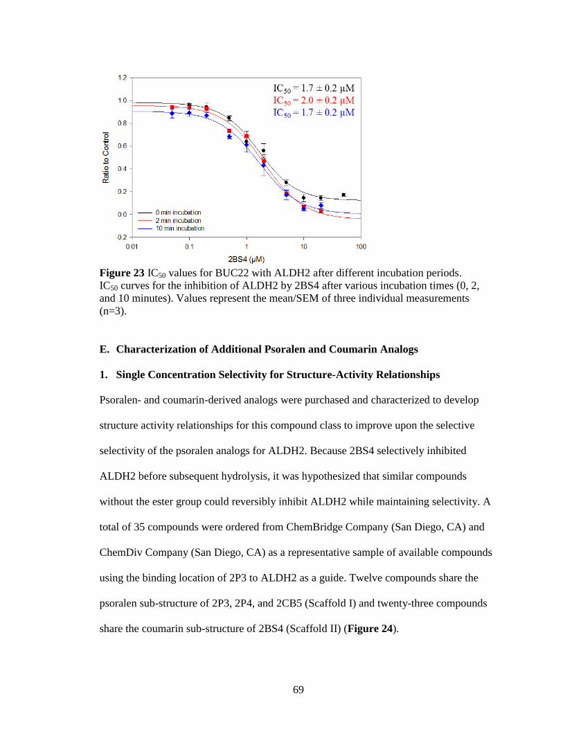
69
Figure 23 IC50 values for BUC22 with ALDH2 after different incubation periods.
IC50 curves for the inhibition of ALDH2 by 2BS4 after various incubation times (0, 2,
and 10 minutes). Values represent the mean/SEM of three individual measurements
(n=3).
E. Characterization of Additional Psoralen and Coumarin Analogs
1. Single Concentration Selectivity for Structure-Activity Relationships
Psoralen- and coumarin-derived analogs were purchased and characterized to develop
structure activity relationships for this compound class to improve upon the selective
selectivity of the psoralen analogs for ALDH2. Because 2BS4 selectively inhibited
ALDH2 before subsequent hydrolysis, it was hypothesized that similar compounds
without the ester group could reversibly inhibit ALDH2 while maintaining selectivity. A
total of 35 compounds were ordered from ChemBridge Company (San Diego, CA) and
ChemDiv Company (San Diego, CA) as a representative sample of available compounds
using the binding location of 2P3 to ALDH2 as a guide. Twelve compounds share the
psoralen sub-structure of 2P3, 2P4, and 2CB5 (Scaffold I) and twenty-three compounds
share the coumarin sub-structure of 2BS4 (Scaffold II) (Figure 24).
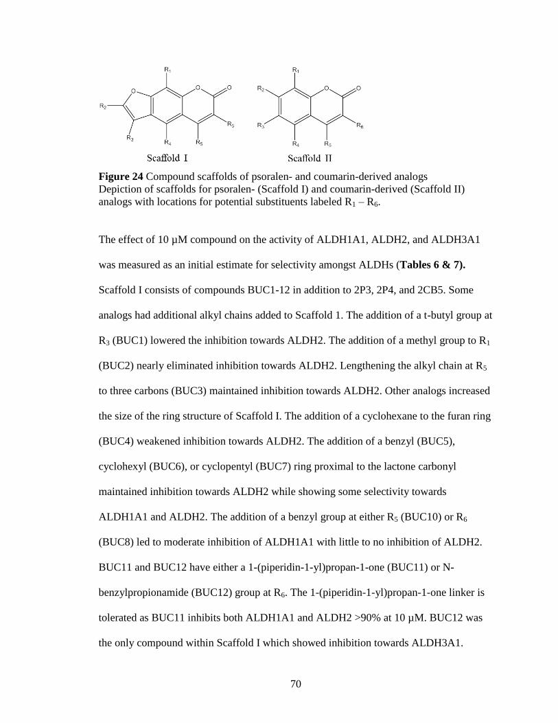
70
Figure 24 Compound scaffolds of psoralen- and coumarin-derived analogs
Depiction of scaffolds for psoralen- (Scaffold I) and coumarin-derived (Scaffold II)
analogs with locations for potential substituents labeled R1 – R6.
The effect of 10 µM compound on the activity of ALDH1A1, ALDH2, and ALDH3A1
was measured as an initial estimate for selectivity amongst ALDHs (Tables 6 & 7).
Scaffold I consists of compounds BUC1-12 in addition to 2P3, 2P4, and 2CB5. Some
analogs had additional alkyl chains added to Scaffold 1. The addition of a t-butyl group at
R3 (BUC1) lowered the inhibition towards ALDH2. The addition of a methyl group to R1
(BUC2) nearly eliminated inhibition towards ALDH2. Lengthening the alkyl chain at R5
to three carbons (BUC3) maintained inhibition towards ALDH2. Other analogs increased
the size of the ring structure of Scaffold I. The addition of a cyclohexane to the furan ring
(BUC4) weakened inhibition towards ALDH2. The addition of a benzyl (BUC5),
cyclohexyl (BUC6), or cyclopentyl (BUC7) ring proximal to the lactone carbonyl
maintained inhibition towards ALDH2 while showing some selectivity towards
ALDH1A1 and ALDH2. The addition of a benzyl group at either R5 (BUC10) or R6
(BUC8) led to moderate inhibition of ALDH1A1 with little to no inhibition of ALDH2.
BUC11 and BUC12 have either a 1-(piperidin-1-yl)propan-1-one (BUC11) or N-
benzylpropionamide (BUC12) group at R6. The 1-(piperidin-1-yl)propan-1-one linker is
tolerated as BUC11 inhibits both ALDH1A1 and ALDH2 >90% at 10 µM. BUC12 was
the only compound within Scaffold I which showed inhibition towards ALDH3A1.

71
Table 6 Structure activity relationships for psoralen derivatives.
Percent activity compared to control was measured at 10 µM compound with ALDH1A1,
ALDH2, and ALDH3A1. Compounds picked for further study highlighted in gray. Mean
value from at least three trials (n≥3).
Cmpd
Scaffold I Position
LogP
% Activity
R1 R2 R3 R4 R5 R6 ALDH
1A1
ALDH
2
ALDH
3A1
2CB5 H H Me H Me propyl 4.47 48.6 6.25 96.1
2P3 H Me Me H Me propyl 4.92 36.3 1.61 86.3
2P4 H Me Me H Me Me 4.39 90.5 19.5 95.3
BUC1 H H t-Bu H Me Me 3.94 94.5 69.7 87.0
BUC2 Me Me Me H Me Me 4.36 78.4 87.2 102
BUC3 H Me Me H propyl H 4.47 74.1 23.8 80.4
BUC4 H
H Me Me 4.48 64.3 61.3 96.1
BUC5 H Me Me H
3.89 82.4 42.2 93.4
BUC6 H Me Me H
4.27 56.3 16.3 94.1
BUC7 H Me Me H
4.26 50.8 8.25 87.1
BUC8 H H Me H Me benzyl 4.98 58.5 83.6 90.3
BUC9 H H phenyl H Me Me 4.80 87.4 92.8 88.9
BUC10 H Me Me H benzyl H 4.80 57.0 73.7 81.6
BUC11 H Me Me H Me
4.06 8.69 3.80 105
BUC12 H Me Me H Me
4.52 93.6 69.8 68.3

72
Table 7 Structure activity relationships for coumarin derivatives.
Percent activity compared to control was measured at 10 µM compound with ALDH1A1,
ALDH2, and ALDH3A1. Compounds picked for further study highlighted in gray. Mean
value from at least three trials (n≥3).
Cmpd
Scaffold II Position
LogP
% Activity
R1 R2 R3 R4 R5 R6 ALDH
1A1
ALDH
2
ALDH
3A1
BUC13 H Me
Me Me 3.92 71.5 46.4 98.1
BUC14
H H H H 1.77 61.0 95.2 28.3
BUC15 Me
H Me Me 2.10 67.4 80.2 79.2
BUC16 H
H Me H 2.11 65.2 25.5 53.6
BUC17 H
H H Me H 3.01 35.5 94.6 9.46
BUC18 H
H H Me H 3.94 37.3 93.3 23.7
BUC19 H
H H Me H 3.19 29.0 97.8 67.1
BUC20 H
H H Me H 1.22 35.7 41.3 20.4
BUC21 H
H H Me H 2.85 49.6 95.0 95.5
BUC22 H
H H Me H 3.63 14.3 71.7 109
BUC23 H
H H Me H 3.92 8.80 101 41.5
BUC24 H
H H
2.64 58.7 64.4 103
BUC25 H
H H Me benzyl 3.44 24.6 99.7 86.9

73
Table 7, continued Structure activity relationships for coumarin derivatives.
Percent activity compared to control was measured at 10 µM compound with ALDH1A1,
ALDH2, and ALDH3A1. Compounds picked for further study highlighted in gray. Mean
value from at least three trials (n≥3).
Cmpd
Scafford II Position
LogP
% Activity
R1 R2 R3 R4 R5 R6 ALDH
1A1
ALDH
2
ALDH
3A1
BUC26 H OH H H Me
1.82 96.6 101 105
BUC27 H H Br H H
2.10 96.0 48.2 91.7
BUC28 H Me
H Me H 2.05 59.8 96.3 105
2BS4 H
H H Me H 1.90 84.8 5.72 94.5
2BS4A H
H H Me H 1.90 68.7 94.4 101
2BS4B H
H H Me H 1.72 52.2 99.1 64.1
2BS4C H
H H Me H 1.37 91.4 93.3 106
2BS4D H
H H Me H 1.71 28.5 89.5 67.1
2P14 H
H H Me H 2.46 80.2 94.8 36.4
2P15 H
H H Me H 1.63 97.5 99.3 93.9

74
Scaffold II compounds are variations of 2BS4, but all lack the ester group of 2BS4. Three
different methyl ketones (2BS4A-C) similar to 2BS4 did not inhibit ALDH2. 2BS4A and
2BS4B also inhibited ALDH1A1 and/or ALDH3A1 while 2BS4C did not inhibit any of
the three isoenzymes. An amide (2BS4D) similar to 2BS4 only inhibited ALDH1A1
activity. Shorting the side chain of 2BS4 to the methyl ether (2P14) led to inhibition of
ALDH3A1; while the free acid form of 2BS4 (2P15) did not inhibit any of the three
isoenzymes.
More diverse additions to Scaffold II were also examined (BUC13-28). The addition of
oxygen-containing rings had varied results. A 3-methylfuran ring at R3 and R4 (BUC13)
led to partial inhibition of ALDH2 while addition of a furan ring at R1 and R2 (BUC14)
led to strong inhibition of ALDH3A1 with no inhibition of ALDH2. The addition of 4H-
pyran-4-one at R2 and R3 (BUC15) led to slight inhibition of ALDH1A1. The addition of
δ-valerolactone at R2 and R3 (BUC16) led to inhibition of ALDH2 with lesser inhibition
of ALDH1A1 and ALDH3A1. Compounds BUC17-BUC23 changed the methyl 2-
methoxypropanoate group at R2 in 2BS4 to more diverse structural groups. BUC17 and
BUC18 contain unsaturated alkyl chains at R2 and inhibit ALDH1A1 and ALDH3A1.
BUC19 has a 2-oxo-2-phenylethoxy group and inhibits ALDH1A1 with moderate
inhibition of ALDH3A1. BUC20 has a terminal nitrile and inhibits ALDH1A1, ALDH2,
and ALDH3A1 at similar levels. BUC21 and BUC22 both appear to selectively inhibit
ALDH1A1, though BUC21 only inhibits 50% at 10 µM mostly due to the large N-
phenylacetamdie group at R2. BUC22 contains a diethylamine in the same position and
inhibits ALDH1A1 ~85% at 10 µM. BUC23, which substituted a 1,4-

75
bis(methoxymethyl)benzene at R2, strongly inhibited ALDH1A1 with partial inhibition
towards ALDH3A1. Compounds BUC24-28 have changes at multiple positions
compared to 2BS4. BUC24 has a (3-oxobutan-2-yl)oxy group at R2 and benzyl ring
attached at R5 and R6 which led to moderate inhibition of ALDH1A1 and ALDH2.
BUC26 has a hydroxyl group at R2 and propionic acid group at R6 and did not inhibit any
of the three isoenzymes. The other three compounds showed some level of selectivity for
either ALDH1A1 or ALDH2. BUC25, which added a methane sulfonyl group at R2 and a
benzyl group at R6, and BUC28, which added isobutyramide at R3, selectively inhibited
ALDH1A1 and BUC27, which has only hydrogen at R2 and added a bromine at R3 and
ketoxime at R6, selectively inhibited ALDH2. Of the 35 additional analogs tested, seven
showed potential selectivity for ALDH2 based off activity at 10 µM compound (BUC3,
BUC5-BUC7, BUC13, BUC16, and BUC27). Three compounds showed potential
selectivity towards ALDH1A1 (BUC22, BUC23, and BUC25). These ten compounds and
BUC11, which inhibited both ALDH1A1 and ALDH2 >90% at 10 µM compound, were
selected for further analysis.
Due to the hydrophobic nature of the substrate binding site within the free enzyme
species of ALDH2, the logP values for the 35 analogs in addition to the initial four hits
were examined (Table 4). No direct correlation between logP values and inhibition of
ALDH2 could be developed. For example, BUC3 and BUC16 both inhibit ALDH2 with
the same potency at 10 µM compound and have vastly different logP values. Although
BUC3 has a logP value of 4.47 and BUC16 has a logP value of 2.11, both compounds
inhibit ALDH2 ~75% at 10 µM compound.
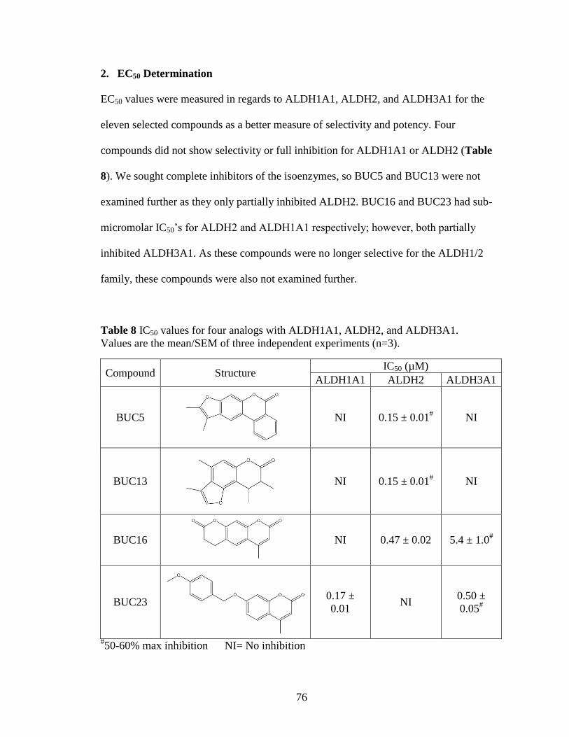
76
2. EC50 Determination
EC50 values were measured in regards to ALDH1A1, ALDH2, and ALDH3A1 for the
eleven selected compounds as a better measure of selectivity and potency. Four
compounds did not show selectivity or full inhibition for ALDH1A1 or ALDH2 (Table
8). We sought complete inhibitors of the isoenzymes, so BUC5 and BUC13 were not
examined further as they only partially inhibited ALDH2. BUC16 and BUC23 had sub-
micromolar IC50’s for ALDH2 and ALDH1A1 respectively; however, both partially
inhibited ALDH3A1. As these compounds were no longer selective for the ALDH1/2
family, these compounds were also not examined further.
Table 8 IC50 values for four analogs with ALDH1A1, ALDH2, and ALDH3A1.
Values are the mean/SEM of three independent experiments (n=3).
Compound Structure IC50 (µM)
ALDH1A1 ALDH2 ALDH3A1
BUC5
NI 0.15 ± 0.01# NI
BUC13
NI 0.15 ± 0.01# NI
BUC16
NI 0.47 ± 0.02 5.4 ± 1.0#
BUC23
0.17 ±
0.01 NI
0.50 ±
0.05#
#50-60% max inhibition NI= No inhibition

77
The other seven compounds did not inhibit ALDH3A1 and fully inhibited ALDH1A1
and/or ALDH2. EC50 values for ALDH1B1, ALDH1A2, and ALDH1A3 were also
measured in addition to those for ALDH1A1, ALDH2, and ALDH3A1 for these seven
compounds (Table 9). Compounds BUC3, BUC6, and BUC7 inhibit the majority of the
ALDH1/2 family with sub-micromolar IC50 values but exhibit little selectivity within the
family. Each one of the three compounds fully inhibits ALDH2, ALDH1B1, and
ALDH1A2 and does not inhibit ALDH3A1. The three compounds have differing effects
towards ALDH1A1 and ALDH1A3. BUC3 does not inhibit ALDH1A1 and activates
ALDH1A3 ~2-fold with an AC50 value of 0.87 ± 0.09 µM. BUC6 partially inhibits
ALDH1A1 and does not inhibit ALDH1A3. BUC7 partially inhibits both ALDH1A1 and
ALDH1A3. BUC11 also does not show selectivity within the ALDH1/2 family, though it
does show ~10-fold preference towards inhibiting ALDH1A1 (IC50 = 0.13 ± 0.01 µM)
versus the other four ALDH1/2 isoenzymes. BUC22 was found to selectively inhibit
ALDH1A1 (IC50 = 0.76 ± 0.07 µM) with only minor effects on ALDH1A2. BUC25
showed a preference towards ALDH1A1 (IC50 = 2.8 ± 0.1 µM) while not inhibiting
ALDH2. BUC27 selectively inhibited ALDH2 (IC50 = 4.6 ± 0.5 µM) while also
exhibiting two-fold activation of ALDH1A3 (AC50 = 35 ± 3 µM).

78
Table 9 EC50 values for seven analogs with the ALDH1/2 family and ALDH3A1.
The coumarin derivatives are more potent, while the psoralen derivatives are more
selective. AC50 curves had a maximum activity of ~200% control. Values are the
mean/SEM of three independent experiments (n=3).
Compound Structure
IC50 (µM)
ALDH
2
ALDH
1B1
ALDH
1A1
ALDH
1A2
ALDH
1A3
ALDH
3A1
BUC3
0.36 ±
0.03
0.086
±
0.002
NI
0.069
±
0.009
AC50
0.87 ±
0.09
NI
BUC6
0.067
±
0.006
0.095
±
0.015
0.13 ±
0.01#
0.065
±
0.004
NI NI
BUC7
0.067
±
0.003
0.16 ±
0.01
0.17 ±
0.02#
0.13 ±
0.01
0.17 ±
0.02#
NI
BUC11
2.0 ±
0.2
2.6 ±
0.3
0.13 ±
0.01
1.6 ±
0.1 14 ± 1 NI
BUC22
NI NI 0.76 ±
0.07 >10 NI NI
BUC25
NI 13 ± 1 2.8 ±
0.1 11 ± 1 10 ± 1 NI
BUC27
4.6 ±
0.6 NI NI NI
AC50
35 ± 3 NI
#50-60% max inhibition NI= No Inhibition
3. Characterization of BUC11’s Interaction with ALDH1A1 and ALDH2
The interaction of BUC11 with ALDH1A1 and ALDH2 was investigated further through
the use of X-ray crystallography and kinetic studies. The structure of BUC11 bound to
ALDH1A1 in the absence of coenzyme was solved to a resolution of 1.70Å (Table 10).

79
Table 10 Collection and refinement statistics for the BUC11-ALDH1A1 complex.
PDB Code 5L2M
Collection
Space Group P422
Cell Dimensions
a,b,c (Å) 109,109,83
α,β,γ (deg) 90,90,90
Resolution (Å) 50.0-1.70
Rmerge 0.119 (0.526)*
I/σ 17.2 (5.5)
Completeness
(%) 98.4 (97.6)
Redundancy 12.6 (12.2)
Refinement
No. of reflections 52110
Rwork/Rfree 0.23/0.26
RMSD
Bond Length (Å) 0.011
Bond Angle
(deg) 1.500
* Values in parentheses are for data in the highest resolution shell (1.73 to 1.70Å)
BUC11 was found to bind within the substrate binding site of ALDH1A1 (Figure 25A)
with the psoralen backbone oriented between Tyr297, Gly458, His293, and Phe290. The
piperidine ring of BUC11is oriented towards the catalytic cysteine rather the carbonyl of
the lactone as seen with 2P3 bound to ALDH2. The piperidine ring in this position does
not approach the NAD(H) binding site (Figure 25B). Covariation experiments were used
to evaluate if the change in binding site changed the enzymatic mechanism of inhibition.
BUC11 was found to exhibit uncompetitive inhibition (Ki = 170 ± 13 nM) versus varied
NAD+ for ALDH1A1 (Figure 26A). However, BUC11 was found to exhibit competitive
inhibition (Ki = 1.1 ± 0.1 µM) versus varied NAD+ for ALDH2 (Figure 26B).

80
Figure 25 Crystal structure of BUC11-ALDH1A1 complex.
(A) Electron density map of BUC11 bound to ALDH2 with the original Fo-Fc map in
green contoured at 2.5 standard deviations and the final 2Fo-Fc map in blue contoured at
1.0 standard deviation. (B) Comparison of binding modes of BUC11 (black) bound to
ALDH1A1 (orange) and 2P3 (yellow), NADH (green), and NAD+ (purple) bound to
ALDH2. NAD+/NADH binding positions obtained from PDB1O02 and 1O04.

81
Figure 26 Steady state kinetic characterization of BUC11 with ALDH enzymes.
(A) Lineweaver-Burk representation of uncompetitive inhibition for BUC11 versus
varied NAD+ with ALDH1A1 at fixed concentration of propionaldehyde (1 mM). (B)
Lineweaver-Burk representation of competitive inhibition for BUC11 versus varied
NAD+ with ALDH2 at fixed concentration of propionaldehyde (1 mm). Vales are
mean/SEM of three independent experiments (n=3).
4. Characterization of BUC25’s Interaction with ALDH1A1
The interaction of BUC25 with ALDH1A1 was investigated further by solving the crystal
structure of BUC25 bound to ALDH1A1 to a resolution of 1.70 Å (Table 10). BUC25
binds in the substrate binding site (Figure 27A) and the lactone carbonyl is oriented
towards the catalytic cysteine though not as close as 2P3 bound to ALDH2 (Figure 27B).
The additional benzyl group would appear to prevent the compound from approaching
the catalytic cysteine. Although the electron density for the benzyl group of BUC25 was
not as strong as the remainder of the compound, its position and movement of Trp178
was reminiscent of the interaction between CM037 and ALDH1A1 in the same region
which has similar weak density near Trp178.216

82
Table 11 Collection and refinement statistics for the BUC25-ALDH1A1 complex.
PDB Code 5L2N
Collection
Space Group P422
Cell Dimensions
a,b,c (Å) 109,109,83
α,β,γ (deg) 90,90,90
Resolution (Å) 50.0-1.70
Rmerge 0.094 (0.639)*
I/σ 16.9 (3.3)
Completeness
(%) 100 (100)
Redundancy 8.4 (8.2)
Refinement
No. of reflections 52587
Rwork/Rfree 0.23/0.26
RMSD
Bond Length (Å) 0.012
Bond Angle
(deg) 1.538
*Values in parentheses are for data in the highest resolution shell (1.73 to 1.70Å)
Figure 27 Crystal structure of BUC25-ALDH1A1 complex.
(A) Electron density map of BUC25 bound to ALDH1A1 with the original F0-Fc map in
green contoured at 2.5 standard deviations and the final 2Fo-Fc map in blue contoured at
1.0 standard deviation. (B) Overlay of BUC25 (black) bound to ALDH1A1 (orange) and
2P3 (yellow) bound to ALDH2 (cyan).
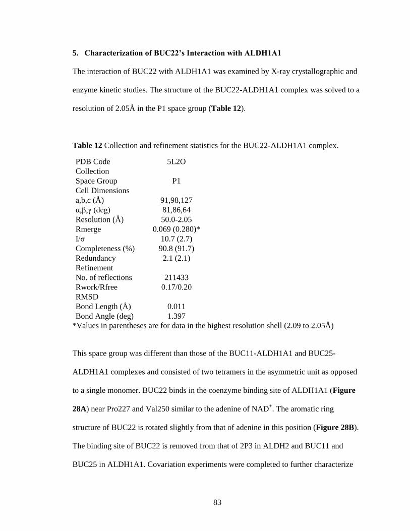
83
5. Characterization of BUC22’s Interaction with ALDH1A1
The interaction of BUC22 with ALDH1A1 was examined by X-ray crystallographic and
enzyme kinetic studies. The structure of the BUC22-ALDH1A1 complex was solved to a
resolution of 2.05Å in the P1 space group (Table 12).
Table 12 Collection and refinement statistics for the BUC22-ALDH1A1 complex.
PDB Code 5L2O
Collection
Space Group P1
Cell Dimensions
a,b,c (Å) 91,98,127
α,β,γ (deg) 81,86,64
Resolution (Å) 50.0-2.05
Rmerge 0.069 (0.280)*
I/σ 10.7 (2.7)
Completeness (%) 90.8 (91.7)
Redundancy 2.1 (2.1)
Refinement
No. of reflections 211433
Rwork/Rfree 0.17/0.20
RMSD
Bond Length (Å) 0.011
Bond Angle (deg) 1.397
*Values in parentheses are for data in the highest resolution shell (2.09 to 2.05Å)
This space group was different than those of the BUC11-ALDH1A1 and BUC25-
ALDH1A1 complexes and consisted of two tetramers in the asymmetric unit as opposed
to a single monomer. BUC22 binds in the coenzyme binding site of ALDH1A1 (Figure
28A) near Pro227 and Val250 similar to the adenine of NAD+. The aromatic ring
structure of BUC22 is rotated slightly from that of adenine in this position (Figure 28B).
The binding site of BUC22 is removed from that of 2P3 in ALDH2 and BUC11 and
BUC25 in ALDH1A1. Covariation experiments were completed to further characterize
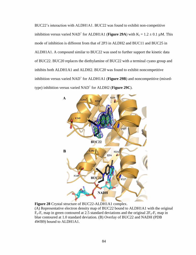
84
BUC22’s interaction with ALDH1A1. BUC22 was found to exhibit non-competitive
inhibition versus varied NAD+ for ALDH1A1 (Figure 29A) with Ki = 1.2 ± 0.1 µM. This
mode of inhibition is different from that of 2P3 in ALDH2 and BUC11 and BUC25 in
ALDH1A1. A compound similar to BUC22 was used to further support the kinetic data
of BUC22. BUC20 replaces the diethylamine of BUC22 with a terminal cyano group and
inhibits both ALDH1A1 and ALDH2. BUC20 was found to exhibit noncompetitive
inhibition versus varied NAD+ for ALDH1A1 (Figure 29B) and noncompetitive (mixed-
type) inhibition versus varied NAD+ for ALDH2 (Figure 29C).
Figure 28 Crystal structure of BUC22-ALDH1A1 complex.
(A) Representative electron density map of BUC22 bound to ALDH1A1 with the original
Fo-Fc map in green contoured at 2.5 standard deviations and the original 2Fo-Fc map in
blue contoured at 1.0 standard deviation. (B) Overlay of BUC22 and NADH (PDB
4WB9) bound to ALDH1A1.
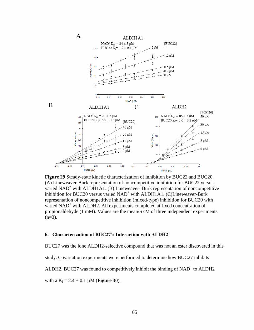
85
Figure 29 Steady-state kinetic characterization of inhibition by BUC22 and BUC20.
(A) Lineweaver-Burk representation of noncompetitive inhibition for BUC22 versus
varied NAD+ with ALDH1A1. (B) Lineweaver- Burk representation of noncompetitive
inhibition for BUC20 versus varied NAD+ with ALDH1A1. (C)Lineweaver-Burk
representation of noncompetitive inhibition (mixed-type) inhibition for BUC20 with
varied NAD+ with ALDH2. All experiments completed at fixed concentration of
propionaldehyde (1 mM). Values are the mean/SEM of three independent experiments
(n=3).
6. Characterization of BUC27’s Interaction with ALDH2
BUC27 was the lone ALDH2-selective compound that was not an ester discovered in this
study. Covariation experiments were performed to determine how BUC27 inhibits
ALDH2. BUC27 was found to competitively inhibit the binding of NAD+ to ALDH2
with a Ki = 2.4 ± 0.1 µM (Figure 30).
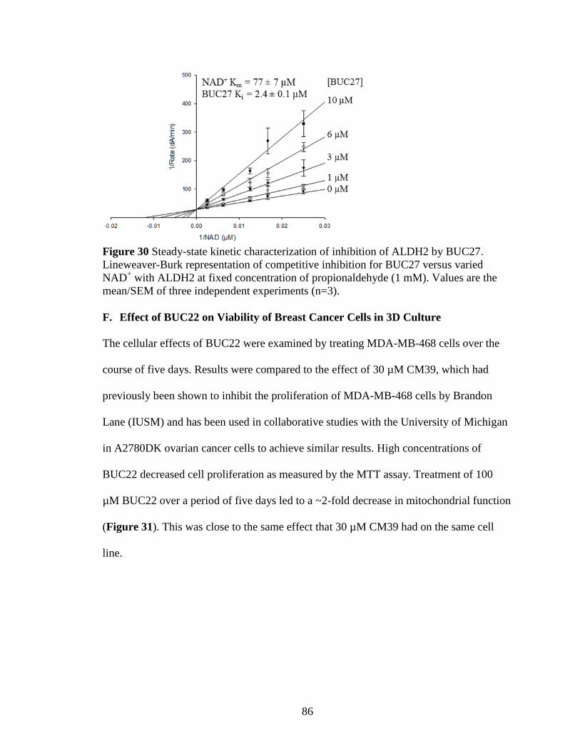
86
Figure 30 Steady-state kinetic characterization of inhibition of ALDH2 by BUC27.
Lineweaver-Burk representation of competitive inhibition for BUC27 versus varied
NAD+ with ALDH2 at fixed concentration of propionaldehyde (1 mM). Values are the
mean/SEM of three independent experiments (n=3).
F. Effect of BUC22 on Viability of Breast Cancer Cells in 3D Culture
The cellular effects of BUC22 were examined by treating MDA-MB-468 cells over the
course of five days. Results were compared to the effect of 30 µM CM39, which had
previously been shown to inhibit the proliferation of MDA-MB-468 cells by Brandon
Lane (IUSM) and has been used in collaborative studies with the University of Michigan
in A2780DK ovarian cancer cells to achieve similar results. High concentrations of
BUC22 decreased cell proliferation as measured by the MTT assay. Treatment of 100
µM BUC22 over a period of five days led to a ~2-fold decrease in mitochondrial function
(Figure 31). This was close to the same effect that 30 µM CM39 had on the same cell
line.

87
Figure 31 Effect of BUC22 treatment on proliferation of breast cancer cells
MTT assay was performed on MDA-MB-468 cells after treatment with BUC22 for five
days. CM39 was included as a positive control for inhibition. Values are the mean/SEM
of three independent experiments relative to vehicle control, each n=3. *p<0.01 versus
vehicle
88
IV. Discussion
The ALDH1/2 family plays a major role in many physiological processes and
pathologies. Five members of the family share approximately 70% protein sequence
identity and demonstrate significant overlap in their substrate specificity. Consequently,
selective inhibitors of these enzymes would help better delineate their individual
function. Although the cofactor binding site is highly conserved amongst ALDH
isoenzymes, there are distinct differences within the substrate binding sites that support
the discovery of compounds which could selectively target individual isoenzymes. For
example, the substrate binding site of ALDH2 is the narrowest and most hydrophobic.
Although inhibition of ALDH2 has been utilized as an approach for the treatment of
alcohol abuse for over 60 years, selective inhibitors of ALDH2 remain elusive.219
Based
on prior success in finding selective modulators for ALDH1A1 and ALDH3A1 by high-
throughput screening, we performed a high-throughput screen to identify potential
inhibitors and activators of ALDH2.214, 220
Our high-throughput screen encompassed over 110,000 compounds. The primary screens
identified 1931 compounds that met our selection criteria; of these, 67 compounds (<5%)
rose to the same selection criteria during retest of the hits. HTS are inherently noisy and
produce many false positives. For example, outliers were found in the Z’-factor
determination (Figures 10 & 11) from identical reactions run simultaneously on the same
plate. False positives can occur due to spectral interference from compounds, robot
pipetting errors, debris or bubbles in the well, misalignment of the plate during plate
reading, or inaccuracies in compound concentration. For instance, during Screen 2 the
89
robot failed to pipet substrate to a subset of wells on several plates; these compounds
were included in the secondary screen for evaluation to ensure complete library coverage.
The secondary screen was designed to remove false positives from further consideration,
saving time and resources.
The effect of 59 of the 67 compounds identified via the esterase HTS on the
dehydrogenase activity of ALDH1A1, ALDH2, and ALDH3A1 was examined as an
initial determination of selectivity. Only 9 of the 59 compounds modulated the
dehydrogenase activity of ALDH2 by more than 20% at 10 µM concentration, the same
concentration tested in the HTS. Switching to the dehydrogenase assay eliminates more
false positives as the assay is not dependent on the same readout. Additionally,
compound concentration in the HTS is determined by the average mass of the compounds
in the entire library. After purchase of compounds, the concentrations tested in the
dehydrogenase assay are most likely not identical. This may eliminate compounds which
were near the selection threshold, but whose concentration was underestimated with the
assumptions made in the HTS.

90
Figure 32 Effect of different compounds on esterase and dehydrogenase assays.
In the absence of NAD+ (a), only ester hydrolysis will occur. NAD
+ (b) or compounds
1(c) or 2 (d), which block one end of the tunnel, will activate esterase activity. Compound
1 will not inhibit dehydrogenase activity unless it can outcompete NAD+. Compound 2
could also activate oxidation provided the substrate can fit between the compound and
the catalytic cysteine. Compound 3 (e), which blocks access to the catalytic cysteine,
would inhibit both reactions. Compound 4 (f) would prevent NAD+ binding and inhibit
aldehyde oxidation, but would have minimal effects on esterase activity.

91
The esterase screen may also identify compounds which activate the esterase reaction in a
similar fashion to NAD+ by binding in the coenzyme binding site closing one end of the
catalytic tunnel resulting in more productive encounters between the ester substrate and
the catalytic cysteine (Figure 32-c).55
These compounds may not be able to bind to the
enzyme in the presence of NAD+, minimizing their effect on dehydrogenase activity.
Other compounds which bind in the coenzyme binding site but do not alter access to the
tunnel would not be identified through the HTS (Figure 32-f). This limits the
identification of compounds that bind in the NAD+-binding site, as many of these
compounds would not be selective due to the high conservation of the site among ALDH
enzymes.
Although initial characterization of the compounds interaction with ALDH2 did not
identify any substantial activators of ALDH dehydrogenase activity, three compounds
(2CD11, 2CB8, and 2CB9) shared the benzodioxol of Alda-1, the ALDH2 activator, and
were examined further. Alda-1 binds to ALDH2 similar to compound 2 in Figure 32-d
with the benzodioxol group in the substrate binding site oriented towards the catalytic
cysteine. Alda-1 displays substrate-dependent effects due to the limited space between
the compound and the catalytic cysteine in the substrate binding site. Like Alda-1, the
three compounds also exhibit substrate-dependent activation of ALDH2. The three
compounds most likely bind in a similar position in ALDH2 as the activator Alda-1,
where smaller substrates can still access the active cysteine. However, the three
compounds are less potent activators than Alda-1.55
These compounds and Alda-1 have

92
similar solubility issues, and the additional decreased potency of the compounds made it
difficult to complete full dose-response curves.
The HTS identified a class of four aromatic lactones which selectively inhibit the
ALDH1/2 family. The four compounds can be further divided by kinetic data and
structure similarity into three psoralen derivatives and one coumarin derivative. The three
psoralen derivatives inhibit most or all of the ALDH1/2 family with IC50 values between
0.1 – 1.0 µM. The differences in alkyl substituents to the psoralen substructure have a
measurable impact on the inhibition of the ALDH1/2 family. The additional methyl
groups at positions R2 and R3 on the furan ring increase inhibition towards the ALDH1/2
family, most likely due to increased hydrophobic contacts (Figure 33).
Figure 33 Orientation of 2P3 in catalytic tunnel of ALDH2.
2P3 is shown in its binding location in the catalytic site of ALDH2. Substituent positions
are labeled.
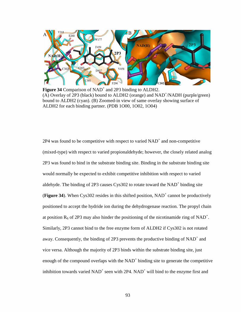
93
Figure 34 Comparison of NAD+ and 2P3 binding to ALDH2.
(A) Overlay of 2P3 (black) bound to ALDH2 (orange) and NAD+/NADH (purple/green)
bound to ALDH2 (cyan). (B) Zoomed-in view of same overlay showing surface of
ALDH2 for each binding partner. (PDB 1O00, 1O02, 1O04)
2P4 was found to be competitive with respect to varied NAD+ and non-competitive
(mixed-type) with respect to varied propionaldehyde; however, the closely related analog
2P3 was found to bind in the substrate binding site. Binding in the substrate binding site
would normally be expected to exhibit competitive inhibition with respect to varied
aldehyde. The binding of 2P3 causes Cys302 to rotate toward the NAD+ binding site
(Figure 34). When Cys302 resides in this shifted position, NAD+ cannot be productively
positioned to accept the hydride ion during the dehydrogenase reaction. The propyl chain
at position R6 of 2P3 may also hinder the positioning of the nicotinamide ring of NAD+.
Similarly, 2P3 cannot bind to the free enzyme form of ALDH2 if Cys302 is not rotated
away. Consequently, the binding of 2P3 prevents the productive binding of NAD+ and
vice versa. Although the majority of 2P3 binds within the substrate binding site, just
enough of the compound overlaps with the NAD+ binding site to generate the competitive
inhibition towards varied NAD+ seen with 2P4. NAD
+ will bind to the enzyme first and

94
can adopt multiple conformations (Figure 5) prior to assuming the productive
conformation required for catalysis.43, 48
2P3 could bind to one of the non-productively
positioned complexes with NAD+. These off-pathway complexes would be consistent
with the non-competitive (mixed-type) inhibition towards aldehyde substrate. Although
NAD+ and 2P3/4 could be bound to ALDH2 simultaneously, 2P3/4 must dissociate
before NAD+ can isomerize into a productive conformation for hydride transfer (Figure
35). Once NAD+ is in the correct position, substrate aldehyde will bind to the enzyme and
the reaction proceeds. ALDH2 most likely follows an ordered bi-bi mechanism where
NADH is released last.43, 48
2P3 cannot bind to the enzyme alongside NADH (Figure 34),
so NADH must be released before 2P3/4 can bind to the enzyme again to inhibit the
reaction.

95
Figure 35 Schematic of 2P3/4 inhibition of ALDH2
2P4 exhibits competitive inhibition with respect to varied NAD+ and non-competitive
(mixed-type) with respect to varied propionaldehyde. 2P3 can bind to the free enzyme,
and it is possible and likely that 2P3 can also bind simultaneously with NAD+ in a non-
productive conformation. Isomerization of NAD+, and subsequent aldehyde oxidation,
can only occur once 2P3/4 dissociates from the enzyme.
Of the four initially identified lactones, 2BS4 has the most unique inhibition profile.
2BS4 is less potent than 2P3/4 but is selective for ALDH2 against all other tested ALDH
isoenzymes. Although 2BS4 contains the aromatic lactone moiety which helps orient 2P3
in the substrate binding site, 2BS4 also contains a terminal ester group and could be a
potential substrate for ALDH2. Either the lactone carbonyl or the terminal ester could be
oriented towards the catalytic cysteine of ALDH2 based off the binding position of 2P3.
Data shows that the ester of 2BS4 is slowly hydrolyzed over time, possibly through a
96
combination of ALDH2-catalyzed and non-catalyzed hydrolysis in solution. The relative
contribution of each is unclear due to the timeframe (2-3 days). 2BS4 exhibits a time-
dependence for inhibition, but the time-dependence was not observed for the inhibition
phase, but is related to how long the enzyme remains inhibited after complex formation.
2BS4 behaves like a “slow off” inhibitor of ALDH2. The hydrolysis limits the use of
2BS4 as an ALDH2 inhibitor for in vivo studies, since the free acid form is not inhibitory
(see below).
A total of 35 aromatic lactone analogs were purchased in an attempt to improve the
selectivity of the three psoralen compounds for ALDH2, and to achieve the selectivity
seen with the coumarin 2BS4, minus the ester group. Although the analogs were picked
based off the binding position of 2P3 to ALDH2, many of the analogs do not inhibit
ALDH2 activity. The narrow catalytic tunnel of ALDH2 limits the size of substituents
that can be added to the structure of the scaffold (Figure 33). Several compounds were
also ordered to confirm the binding mode of 2P3 since additions at certain positions to
the compound should eliminate ALDH2 inhibition. For instance, BUC2 has a methyl
group at position R1 which no longer fits within the narrow binding tunnel and does not
inhibit ALDH2. There are many other compounds that have larger additions at position
R1 that were not ordered; these compounds would also not inhibit ALDH2. Bulky side
chains such as t-butyl and phenyl groups at position R3 or benzyl groups at positions R5
or R6 also cause the compound to no longer fit as well in the narrow tunnel. The best
additions amongst the psoralen compounds (Scaffold I) were increasing the size of the
alkyl substitutions at positions R5 and R6 adjacent to the lactone carbonyl carbons,
97
including the addition of a fourth ring. The coumarin analogs which most closely
resembled 2BS4 did not inhibit ALDH2, supporting the idea that 2BS4 inhibits the
enzyme differently than the other compounds in the series. Carboxylates are generally
poor inhibitors of the ALDH1/2 family, as in the case for the free acid of 2BS4 (2P15)
and the inhibition of 2BS4 appears to be derived from the terminal ester. Several of the
coumarin analogs inhibit the activity of ALDH3A1, suggesting the additional furan ring
of the psoralen compounds helps select against ALDH3A1, though is not essential. After
initial testing with ALDH1A1, ALDH2, and ALDH3A1, 11 of the additional analogs
warranted further dose-response analysis.
The eleven compounds showed the same similar pattern at the four initial aromatic
lactones; that is, the psoralen derivatives are more potent, but less selective, than the
coumarin derivatives. BUC5, BUC6, and BUC7 add an additional ring structure at
positions R5 and R6 proximal to the aromatic lactone (Figure 33). The additional
cyclohexyl and cyclopentyl rings in BUC6 and BUC7 slightly increase the potency
towards ALDH2 while lessening the total amount of inhibition towards ALDH1A1 and
ALDH1A3. BUC3, which opens the cyclopentyl ring of BUC7 to form a propyl chain at
position R5, also does not inhibit ALDH1A1, though BUC3 does activate ALDH1A3
through a-yet-unknown mechanism. BUC5, which increases the size of the aromatic ring
structure by adding a benzyl ring, only partially inhibits ALDH2 while selecting against
ALDH1A1 inhibition. The amount of inhibition towards ALDH1A1 and ALDH2 by
these compounds with larger rings is most likely related to how well the larger compound
fits in the active site. The ring additions at R5 and R6 must fit between Trp177 and

98
Met174 in ALDH2, which are both conserved throughout the ALDH1/2 family (Figure
33). The benzyl ring of BUC5 may not fit as well due to the constraints of maintaining
aromaticity whereas the cyclopentyl and cyclohexyl rings do not have these restraints.
BUC11 represents a large number of available analogs which use a 1-acylpiperidine
linker to connect the psoralen ring structure to a variety of other chemical moieties. These
compounds also have variations in the amount of alkyl substituents on the psoralen
scaffold, similar to 2P3, 2P4, and 2CB5. BUC11 exhibits different modes of inhibition
towards varied NAD+ for ALDH1A1 and ALDH2, suggesting that BUC11 inhibits these
two enzymes through different mechanisms. BUC11 exhibits uncompetitive inhibition
towards varied NAD+ for ALDH1A1, which is consistent with the distance between the
NAD+ and BUC11 binding sites in ALDH1A1 (Figure 25B) and is expected for
compounds that displace aldehyde substrates for ordered Bi Bi systems. The competitive
inhibition towards varied NAD+ for ALDH2 is similar to the mechanism exhibited by
2P4. This would suggest that BUC11 binds to ALDH2 in a similar position as 2P3 with
the 1-acylpiperidine extending past Cys302 and posed toward the NAD+ site, consistent
with its competitive mode of inhibition. However, for ALDH1A1 the presence of Gly458
and its larger substrate pocket promotes a shift in binding mode and mechanism of
inhibition for BUC11. The corresponding residue to Gly458 in the other four ALDH1/2
isoenzymes is either an asparagine or aspartate, which precludes the binding mode
observed in ALDH1A1. The 1-acylpiperide may have more difficulty extending into the
NAD+ site for ALDH1A3, as the IC50 value is 10x larger than those for ALDH2,
ALDH1B1, and ALDH1A2.
99
The coumarin derivative BUC25 also binds in the substrate binding site. The distance
between the carbonyl lactone and catalytic cysteine suggests that binding of BUC25 does
not prevent the productive binding of NAD+ to ALDH1A1, as the additional benzyl
group of BUC25 prevents the compound from binding closer to the catalytic cysteine.
BUC25 does not inhibit ALDH2 and the mode of binding provides insight into this
selectivity. The methane sulfonyl group of BUC25 binds in the pocket formed by Gly458
in ALDH1A1. The corresponding aspartate/asparagine residues in the other ALDH1/2
isoenzymes would force the coumarin scaffold to adopt a position akin to 2P4 in ALDH2.
However, unlike the more flexible linker in BUC11, the large benzyl ring cannot adopt a
conformation to productively bind within the NAD+-binding cleft and the benzyl ring of
BUC25 would encounter more steric hindrance from the conserved tryptophan (Trp177
in ALDH2) since the residues surrounding Trp177 are bulkier in ALDH2. The tryptophan
may be able to slightly move in ALDH1B1, ALDH1A2, and ALDH1A3, though not as
much as in ALDH1A1. This is evidenced by the compound being ~5x less potent for
these three isoenzymes compared to ALDH1A1.
Surprisingly, unlike 2P3, BUC11, or BUC25, BUC22 was found to bind in the in
coenzyme binding site of ALDH1A1. The difference in binding location, and the crystal
forming in a unique space group (P1), raised the possibility that the binding site might be
influenced by the crystal environment. However, there is a structural basis for the
selectivity between ALDH2 and ALDH1A1 in this binding location which suggests that
the binding within the coenzyme site may be more than an artifact of this particular
crystal form (Figure 36). Ile249 in ALDH2 is replaced by Val250 in ALDH1A1 and

100
although it is the addition of a single methyl group, the substitution narrows this side of
the site enough to impede binding of the diethylamino substituent in ALDH2.
Additionally, Val252 and Ala233 in ALDH2 versus Leu253 and Ser234 in ALDH1A1
may loosen the contacts between the enzyme and BUC22 enough that the binding
energetics cannot overcome the steric clash with Ile249 through slightly shifting to the
other side of the blinding cleft.
Figure 36 Comparison of BUC22 binding to ALDH1A1 versus ALDH2.
BUC22 (black) binds to ALDH1A1 (purple) in the NAD+-binding site. Three amino
acids in the vicinity of the binding location are not conserved between ALDH1A1 and
ALDH2 (green) and may explain the selectivity seen between the two enzymes.
The kinetic data of BUC22, however, is not completely consistent with this binding
location. Compounds which bind in the adenine binding site should exhibit competitive
inhibition towards varied NAD+, but BUC22 exhibits noncompetitive inhibition. Though
101
inconsistent with binding in the adenine binding site, it is also a different mode of
inhibition from compounds determined to bind in the substrate binding site, 2P4
(competitive) and BUC11 (uncompetitive), which suggests BUC22 binds differently than
these other analogs. BUC20, a related analog, also exhibits a noncompetitive mode of
inhibition towards varied NAD+ for ALDH1A1 and ALDH2. Due to its linear cyano
group, BUC20 is not predicted to clash with Ile249 in ALDH2. Consequently BUC20
inhibits both ALDH1A1 and ALDH2. The reason behind the discrepancy between the
kinetic data and structural data is unclear, though there is a possibility that BUC22 could
bind to multiple sites and enzyme species. When NAD+ is absent, the compound prefers
to bind in the NAD+ binding site (as in the case of the solved structure), but when NAD
+
is present, the compound may bind in another location giving rise to the observed kinetic
pattern. This additional site could be the substrate binding site, such as where BUC25
binds.
BUC27 was the only compound that selectively inhibited ALDH2 in this study, other
than the ester 2BS4. Both BUC27 and 2P4 exhibit competitive inhibition towards varied
NAD+, suggesting that BUC27 likely alters the conformation of the catalytic cysteine to
interfere with the productive binding of NAD+ for catalysis, similar to 2P3. If BUC27
were to adopt a similar conformation to that of 2P3 bound to ALDH2, the ketoxime
would occupy the same pocket as the propyl chain. The bromine would be oriented
towards Met124 would could lead to favorable interaction between the halogen
substituent and the sulfur side chain atom (Figure 37).
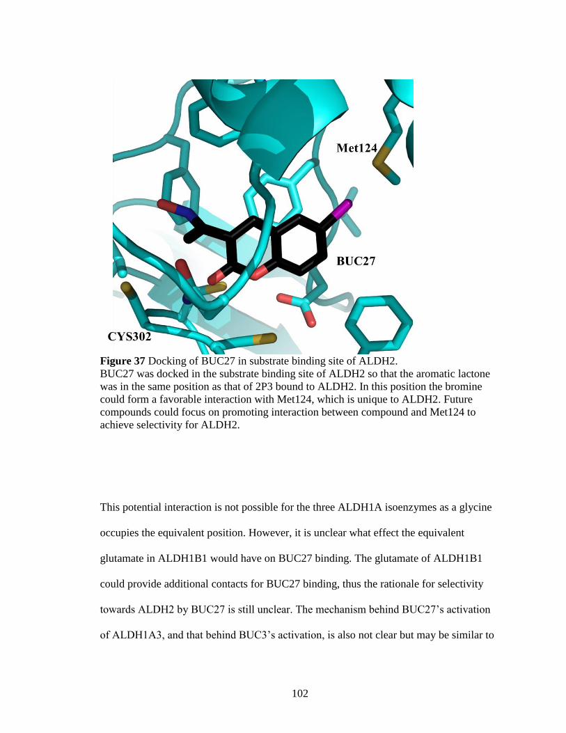
102
Figure 37 Docking of BUC27 in substrate binding site of ALDH2.
BUC27 was docked in the substrate binding site of ALDH2 so that the aromatic lactone
was in the same position as that of 2P3 bound to ALDH2. In this position the bromine
could form a favorable interaction with Met124, which is unique to ALDH2. Future
compounds could focus on promoting interaction between compound and Met124 to
achieve selectivity for ALDH2.
This potential interaction is not possible for the three ALDH1A isoenzymes as a glycine
occupies the equivalent position. However, it is unclear what effect the equivalent
glutamate in ALDH1B1 would have on BUC27 binding. The glutamate of ALDH1B1
could provide additional contacts for BUC27 binding, thus the rationale for selectivity
towards ALDH2 by BUC27 is still unclear. The mechanism behind BUC27’s activation
of ALDH1A3, and that behind BUC3’s activation, is also not clear but may be similar to

103
that of Alda-1, where binding of Alda-1 in the outer portion of the substrate binding site
increases the amount of productive encounters with the catalytic cysteine for smaller
aldehydes.
The inhibition pattern of these compounds was compared to that of daidzin, and we found
that these compounds are more potent than daidzin under our assay parameters.
Additionally, daidzin was discovered be less selective than previously reported. Daidzin
inhibited the activity of ALDH2, ALDH1A2, and ALDH1B1 with equal potency, while
being 100-fold less potent against ALDH1A1 and ALDH1A3. This same pattern of
inhibition was also found in the psoralen derivatives, where many compounds inhibit
ALDH2, ALDH1B1 and ALDH1A2 with similar potency and are often less active
against ALDH1A1 and ALDH1A3 at some level. Although IC50 values may be similar,
many of the psoralen derivatives only partially inhibited ALDH1A1 and ALDH1A3, or
did not inhibit at all.
In order to correlate the different binding modes and inhibition patterns to underlying
structural differences, we examined the binding sites and interactions in more detail.
Structural comparison of three compound-enzyme complexes revealed the presence of
two distinct aromatic binding “boxes” or “zones” (Figure 38). In ALDH2 the aromatic
binding region is located between four phenylalanine residues (170,296,459,465) near the
catalytic cysteine which surround 2P3 when bound. The activator Alda-1 and the
isoflavone of the ALDH2 inhibitor daidzin bind in this same aromatic site. 55, 204
The
aromatic binding region of ALDH1A1 is wider, extends farther from the catalytic

104
cysteine and is more surface exposed. In ALDH1A1, Phe459 is replaced by a valine and
Phe296 is replaced by tyrosine. The aromatic binding region of ALDH1A1 has additional
aromatic residues at His293 and Phe290. Although the histidine residue is a
phenylalanine in ALDH2, the side chain of Asp457 in ALDH2 blocks any connection of
the two regions and gives rise to the smaller site in ALDH2 compared to ALDH1A1,
which has a glycine in an equivalent position.
Figure 38 Alignment of three enzyme-inhibitor complexes.
Alignment of 2P3 (orange) bound to ALDH2 and BUC11 (purple) and BUC25 (cyan)
bound to ALDH1A1 shows an overlap of separate aromatic binding “boxes” or “zones”.
D457 in ALDH2 limits the size of the zone in ALDH2, whereas G458 in ALDH1A1
provide a larger zone for the binding of aromatic compounds.

105
The aromatic lactones are able to shift positions to maximize contacts due to the
hydrophobicity of the substrate binding site of ALDHs. Most enzymes rely on hydrogen
bonding and/or ion interactions to align substrates in the substrate binding site; in
contrast, the substrate binding site of ALDHs is highly hydrophobic with limited
potential for hydrogen bonding. When substituents are added or removed from an
activator/inhibitor that result in the compound no longer filling the available space, the
compound can either not bind at all, or find another site where binding is also possible.
The hydrophobicity of the tunnel, and lack of dependence on hydrogen bonding and ion
interactions, provides more potential sites for compound binding in ALDHs.
The ability for the compounds to shift in the hydrophobic substrate binding site may help
explain the difference in inhibition of ALDH1A1 between 2P3 and 2P4. The shortening
of the alkyl chain at position R5 from a propyl to a methyl group results in 2P4 not
inhibiting ALDH1A1. Loss of contact between the scaffold and Val460 in ALDH1A1 (as
compared to Phe459 in ALDH2) may require the longer propyl chain to “anchor” the
compound near the catalytic cysteine. The methyl group may not create enough
additional contacts which will cause the scaffold of 2P4 to shift to the location of BUC11
when bound to ALDH1A1 to increase the amount of contacts, which is possible due to
the presence of Gly458. If bound in this location, 2P4 would not inhibit the enzyme as the
compound would not impede access to the catalytic tunnel. 2P3, even with the longer
propyl chain, would also not inhibit ALDH1A1 if bound similar to BUC11, which
supports the idea that the longer alkyl chain enables 2P3 to bind closer to the catalytic
cysteine to inhibit the enzyme. The additional contacts from the propyl chain may not be

106
needed with ALDH1A2, ALDH1A3, and ALDH1B1 (all of which replace Phe459 in
ALDH2 with smaller aliphatic residues) as these three enzymes have an asparagine at the
equivalent position of Gly458 and are each inhibited by 2P4. The additional side chain
may provide enough binding surface that the smaller compound remains bound to the
enzyme.
A second binding pocket exists in ALDH isoenzymes that can accommodate aromatic
ring structures—the adenine binding cleft within the coenzyme binding site. When NAD+
is bound, the adenine ring is positioned between a conserved proline and a beta-branched
amino acid (Pro226 and Ile249 in ALDH1A1). The crystal structure of BUC22 shows
that at least one of the compounds can bind to the adenine site, though kinetic data
supports the binding of BUC22 at an additional site, most likely the aromatic binding
region in the substrate binding site.
The multiple aromatic binding sites within ALDH isoenzymes can help explain how the
ALDH1A1-selective inhibitor BUC22 was discovered from a series of compounds
identified through a screen targeting ALDH2. BUC22 was not one of the compounds
found in the high-throughput screen, but a structural analog. Changing the substituents of
the aromatic scaffold likely caused BUC22 to shift binding locations to the adenine
binding site, which then provided selectivity against ALDH2. The alteration of
substituents has been seen to change the selectivity of indole-2,3-diones for a particular
ALDH isoenzyme.229
In that particular case, the compounds had different orientations

107
within the same binding site, while the coumarin and psoralen derivatives show a greater
amount of movement with multiple binding sites.
An important factor for the use of any compound as a chemical probe is the ability to
affect the activity of the target enzyme in a cellular context. Coumarins and psoralens can
either be cell permeable or cell impermeable depending on the exact structure. Some
psoralens are known to become trapped in cell membranes, with the most hydrophobic
compounds predicted to be the most permeable.230
BUC22 was found to have a
physiological effect on MDA-MB-468 breast cancer cells after five days of treatment. A
significant two-fold decrease in the proliferative ability of the breast cancer cells was
seen when treated with 100 µM BUC22, 100-fold higher than the in vitro Ki (~1 µM), as
measured by the MTT assay. The loss of potency could arise from a portion of BUC22
becoming trapped in the cell membrane, by the conversion of the compound into other,
less potent derivatives upon entrance into the cell, or by export from the cell before
inhibition occurs. As BUC22 is more hydrophobic than other permeable psoralens, a
large portion of BUC22 should be able to enter the cell by predicted models.
Additionally, this concentration is high enough that inhibition of ALDH1A2 by BUC22
may also contribute to the cell phenotype. Regardless, BUC22 is capable of causing a
physiologic effect in cancer cells in 3D culture.

108
V. Conclusion and Future Directions
Members of the ALDH1/2 family are involved in retinoic acid signaling, the metabolism
of dietary aldehydes, and the removal of toxic aldehydes from oxidative stress. ALDH1/2
enzymes have also been linked to the development of cancer, obesity, and neurological
disease. Small molecule probes are needed to study the role of individual enzymes in
both normal and disease states. The development of selective modulators has been
difficult due to the structural similarity and overlapping substrate preferences within the
ALDH1/2 family. At present, there are no commercially available compounds that
selectively modulate ALDH2 activity compared to other ALDH isoenzymes.
Inhibitors targeting ALDH2, such as daidzin and its derivatives, have been used
previously to study the function of ALDH2; however, daidzin is not as selective as
previously reported. We found that daidzin inhibits ALDH2, ALDH1B1, and ALDH1A2
with equivalent potency. In light of these findings, the selectivity of daidzin derivatives
including CVT-10216 should be examined further. Many of the aromatic lactones found
during the course of this study have similar or better selectivity and/or potency than
daidzin in vitro, suggesting the aromatic lactones, with further development, may be a
better option for an ALDH2-selective inhibitor.
The aromatic lactones show the ability to bind throughout the aromatic “zones” in the
catalytic site of ALDH1/2 enzymes. The aromatic “zone” in ALDH2 is smaller due to the
presence of Asp457, limiting the potential orientations of the lactone within the catalytic
site. The lactone can only be positioned with its carbonyl oriented directly toward the

109
catalytic cysteine (or rotated 180°). This allows for an easier interpretation of SAR data
in regards to activity against ALDH2; for example, bulky substituents cause a loss of
inhibition toward ALDH2 as the compound no longer fits in the narrow binding tunnel
and any addition to R6 next to the lactone carbonyl must likely fit into the coenzyme
binding site to maintain binding and subsequent inhibition.
The interpretation for SAR data in regards to ALDH1A1 with these compounds is more
challenging as multiple binding orientations are possible in the larger substrate binding
site. The lactones can shift within the hydrophobic pocket to increase the amount of
contacts while decreasing the amount of clashing based upon the substituents added to
the scaffold. If a large substituent is placed at position R6, the larger hydrophobic space
provides an opportunity for the scaffold to shift away from the catalytic cysteine to
provide room, as in the case for BUC11. Compounds are also likely to shift due to
decreased contacts with the non-aromatic Val460. Smaller compounds, like 2P3, could
bind in multiple locations within the catalytic site depending on the specific substituents
on the aromatic scaffold. Although a crystal structure of 2P3 bound to ALDH1A1 would
definitely answer where 2P3 binds, it may not help in determining the binding position
and rationale behind the SAR of other smaller analogs with regards to ALDH1A1, as
different substituents could shift the scaffold to a different location. If the compound is
small enough the binding location could shift to the adenosine binding site, as in the case
for BUC22 bound to ALDH1A1.

110
The structure of 2P3 bound to ALDH2 can reliably be used to predict how structural
modifications will affect the activity of the lactones for ALDH2; however, care must be
taken when making future modifications in regards to activity versus ALDH1A1 and the
rest of the ALDH1/2 family. The addition of a substituent to the scaffold to increase
interaction with ALDH2, such as an interaction between a halogen and Met124, which is
not a conserved residue, could have unintended effects on the activity versus other
isoenzymes through shifting of the aromatic scaffold.
Although the complete rationale for selectivity remains unknown, two selective
compounds were discovered and characterized—the ALDH1A1-selective BUC22 and the
ALDH2-selective BUC27. Initial experiments with cells in 3D culture suggest that
BUC22 can enter cancer cells and impact cell proliferation through inhibition of
ALDH1A1, though off-target effects are still possible. Previous studies with CM037, an
ALDH1A1 inhibitor, have found that both ovarian cancer and MDA-MB-468 breast
cancer cells are more sensitive to ALDH1A1 inhibition in 3D culture as opposed to
monolayers. This is due in part to an increase in ALDH1A1 expression in cells grown in
3D culture.111, 113
Based on this data, BUC22 should also have differing effects on MDA-
MB-468 cells in 3D vs 2D culture and should be confirmed. BUC22 or future derivatives
could be used to examine the mechanism by which ALDH1A1 inhibition leads to
decreased mitochondrial function and proliferative potential. Pharmacodynamic and
pharmacokinetic experiments could also be completed in mice to determine BUC22’s
future prospective for in vivo studies. Unlike CM037 and other ALDH1A1-selective
inhibitors, BUC22 does not rely on Gly458, which is not conserved in rodents, for
111
inhibition or selectivity. BUC22 should be able to inhibit murine ALDH1A1 by binding
in the more conserved NAD+-binding site. There are potential limitations that should be
addressed before BUC22, or future derivatives, are used in animal models. Mainly, the
residues Ile249 and Val242 that helped provide selectivity against human ALDH2 are
substituted by a valine and isoleucine in murine ALDH2, identical to the residues in
human ALDH1A1. Selectivity against ALDH2 in murine systems is potentially lost,
though interactions at the second proposed binding site leave this uncertain. Likewise,
BUC22 may inhibit other NAD+-dependent enzymes which would limit its use. The
variability of the ALDH1/2 family between rodents and humans complicates the animal
studies that must precede any attempts at human trials, since there are no direct models
leading from rodents to humans that maintain the human ALDH1/2 structure-activity
relationship. Possible solutions to this conundrum include the creation of a humanized
mouse/rat line where the human target of interest is integrated in place of the rodent
enzyme.
Concerning the ALDH2-selective BUC27, a crystal structure of BUC27 bound to
ALDH2 would potentially provide a rationale for the selectivity against the other
ALDH1/2 members. The structure may confirm the proposed interaction between the
bromine and Met124 and drive development of more potent inhibitors. It would also be
beneficial to examine if BUC27 is capable of inhibiting ALDH2 within cells by
measuring potential effects on alcohol metabolism. Unfortunately, to this point there is no
known liver cell line or primary cell which maintains expression of ALDH2 in culture,
limiting future studies. For this reason prior development of ALDH2-selective

112
compounds have gone from in vitro studies straight to animal models. BUC27, or
subsequent derivatives, could be examined for their ability to affect alcohol drinking of
selectively bred rats to determine bioavailability and potency in vivo. Both BUC27, and
the ALDH1A1-selective BUC22, should be further developed for use as chemical probes
to study the individual functions of ALDH2 and/or ALDH1A1.

113
References
[1] Vasiliou, V., Pappa, A., and Estey, T. (2004) Role of human aldehyde
dehydrogenases in endobiotic and xenobiotic metabolism, Drug metabolism reviews
36, 279-299.
[2] Lindahl, R. (1992) Aldehyde dehydrogenases and their role in carcinogenesis, Crit
Rev Biochem Mol Biol 27, 283-335.
[3] Rhinn, M., and Dolle, P. (2012) Retinoic acid signalling during development,
Development 139, 843-858.
[4] Zhou, X. E., Melcher, K., and Xu, H. E. (2012) Structure and activation of rhodopsin,
Acta Pharmacol Sin 33, 291-299.
[5] Esterbauer, H., Schaur, R. J., and Zollner, H. (1991) Chemistry and biochemistry of
4-hydroxynonenal, malonaldehyde and related aldehydes, Free radical biology &
medicine 11, 81-128.
[6] O'Brien, P. J., Siraki, A. G., and Shangari, N. (2005) Aldehyde sources, metabolism,
molecular toxicity mechanisms, and possible effects on human health, Critical
reviews in toxicology 35, 609-662.
[7] Curtis, J. M., Hahn, W. S., Long, E. K., Burrill, J. S., Arriaga, E. A., and Bernlohr, D.
A. (2012) Protein carbonylation and metabolic control systems, Trends Endocrinol
Metab 23, 399-406.
[8] Hauck, A. K., and Bernlohr, D. A. (2016) Oxidative stress and lipotoxicity, J Lipid
Res.
[9] Vander Jagt, D. L., Hunsaker, L. A., Vander Jagt, T. J., Gomez, M. S., Gonzales, D.
M., Deck, L. M., and Royer, R. E. (1997) Inactivation of glutathione reductase by 4-
hydroxynonenal and other endogenous aldehydes, Biochemical pharmacology 53,
1133-1140.
[10] Kinter, M., and Roberts, R. J. (1996) Glutathione consumption and glutathione
peroxidase inactivation in fibroblast cell lines by 4-hydroxy-2-nonenal, Free radical
biology & medicine 21, 457-462.
[11] Brooks, P. J., and Theruvathu, J. A. (2005) DNA adducts from acetaldehyde:
implications for alcohol-related carcinogenesis, Alcohol 35, 187-193.
[12] Cheng, G., Shi, Y., Sturla, S. J., Jalas, J. R., McIntee, E. J., Villalta, P. W., Wang,
M., and Hecht, S. S. (2003) Reactions of formaldehyde plus acetaldehyde with
deoxyguanosine and DNA: formation of cyclic deoxyguanosine adducts and
formaldehyde cross-links, Chemical research in toxicology 16, 145-152.

114
[13] McMahon, R. E. (1982) Chapter 5: Alcohols, aldehyde, and ketones, In Metabolic
basis of detoxication: metabolism of functional groups (Jakoby, W. B., Bend, J. R.,
and Coldwell, J., Eds.), Academic Press, NY.
[14] Penning, T. M. (2015) The aldo-keto reductases (AKRs): Overview, Chemico-
biological interactions 234, 236-246.
[15] Beedham, C., Peet, C. F., Panoutsopoulos, G. I., Carter, H., and Smith, J. A. (1995)
Role of aldehyde oxidase in biogenic amine metabolism, Prog Brain Res 106, 345-
353.
[16] Vasiliou, V., Pappa, A., and Petersen, D. R. (2000) Role of aldehyde
dehydrogenases in endogenous and xenobiotic metabolism, Chemico-biological
interactions 129, 1-19.
[17] Kavanagh, K. L., Jornvall, H., Persson, B., and Oppermann, U. (2008) Medium- and
short-chain dehydrogenase/reductase gene and protein families : the SDR
superfamily: functional and structural diversity within a family of metabolic and
regulatory enzymes, Cell Mol Life Sci 65, 3895-3906.
[18] Berhane, K., Widersten, M., Engstrom, A., Kozarich, J. W., and Mannervik, B.
(1994) Detoxication of base propenals and other alpha, beta-unsaturated aldehyde
products of radical reactions and lipid peroxidation by human glutathione
transferases, Proceedings of the National Academy of Sciences of the United States of
America 91, 1480-1484.
[19] Jackson, B., Brocker, C., Thompson, D. C., Black, W., Vasiliou, K., Nebert, D. W.,
and Vasiliou, V. (2011) Update on the aldehyde dehydrogenase gene (ALDH)
superfamily, Human genomics 5, 283-303.
[20] Vasiliou, V., Bairoch, A., Tipton, K. F., and Nebert, D. W. (1999) Eukaryotic
aldehyde dehydrogenase (ALDH) genes: human polymorphisms, and recommended
nomenclature based on divergent evolution and chromosomal mapping,
Pharmacogenetics 9, 421-434.
[21] Vasiliou, V., Thompson, D. C., Smith, C., Fujita, M., and Chen, Y. (2013) Aldehyde
dehydrogenases: From eye crystallins to metabolic disease and cancer stem cells,
Chemico-biological interactions 202, 2-10.
[22] Braun, T., Bober, E., Singh, S., Agarwal, D. P., and Goedde, H. W. (1987) Evidence
for a signal peptide at the amino-terminal end of human mitochondrial aldehyde
dehydrogenase, FEBS letters 215, 233-236.
[23] Marchitti, S. A., Brocker, C., Stagos, D., and Vasiliou, V. (2008) Non-P450
aldehyde oxidizing enzymes: the aldehyde dehydrogenase superfamily, Expert
opinion on drug metabolism & toxicology 4, 697-720.

115
[24] Estey, T., Cantore, M., Weston, P. A., Carpenter, J. F., Petrash, J. M., and Vasiliou,
V. (2007) Mechanisms involved in the protection of UV-induced protein inactivation
by the corneal crystallin ALDH3A1, The Journal of biological chemistry 282, 4382-
4392.
[25] Vasiliou, V., Sandoval, M., Backos, D. S., Jackson, B. C., Chen, Y., Reigan, P.,
Lanaspa, M. A., Johnson, R. J., Koppaka, V., and Thompson, D. C. (2013)
ALDH16A1 is a novel non-catalytic enzyme that may be involved in the etiology of
gout via protein-protein interactions with HPRT1, Chemico-biological interactions
202, 22-31.
[26] Vasiliou, V., and Nebert, D. W. (2005) Analysis and update of the human aldehyde
dehydrogenase (ALDH) gene family, Human genomics 2, 138-143.
[27] Emadi, A., Jones, R. J., and Brodsky, R. A. (2009) Cyclophosphamide and cancer:
golden anniversary, Nat Rev Clin Oncol 6, 638-647.
[28] Lassen, N., Bateman, J. B., Estey, T., Kuszak, J. R., Nees, D. W., Piatigorsky, J.,
Duester, G., Day, B. J., Huang, J., Hines, L. M., and Vasiliou, V. (2007) Multiple and
additive functions of ALDH3A1 and ALDH1A1: cataract phenotype and ocular
oxidative damage in Aldh3a1(-/-)/Aldh1a1(-/-) knock-out mice, The Journal of
biological chemistry 282, 25668-25676.
[29] Deak, K. L., Dickerson, M. E., Linney, E., Enterline, D. S., George, T. M., Melvin,
E. C., Graham, F. L., Siegel, D. G., Hammock, P., Mehltretter, L., Bassuk, A. G.,
Kessler, J. A., Gilbert, J. R., Speer, M. C., and Group, N. T. D. C. (2005) Analysis of
ALDH1A2, CYP26A1, CYP26B1, CRABP1, and CRABP2 in human neural tube
defects suggests a possible association with alleles in ALDH1A2, Birth Defects Res A
Clin Mol Teratol 73, 868-875.
[30] Marchitti, S. A., Deitrich, R. A., and Vasiliou, V. (2007) Neurotoxicity and
metabolism of the catecholamine-derived 3,4-dihydroxyphenylacetaldehyde and 3,4-
dihydroxyphenylglycolaldehyde: the role of aldehyde dehydrogenase,
Pharmacological reviews 59, 125-150.
[31] Anastasiou, V., Ninou, E., Alexopoulou, D., Stertmann, J., Muller, A., Dahl, A.,
Solimena, M., Speier, S., Serafimidis, I., and Gavalas, A. (2016) Aldehyde
dehydrogenase activity is necessary for beta cell development and functionality in
mice, Diabetologia 59, 139-150.
[32] Rizzo, W. B., and Carney, G. (2005) Sjogren-Larsson syndrome: diversity of
mutations and polymorphisms in the fatty aldehyde dehydrogenase gene
(ALDH3A2), Human mutation 26, 1-10.

116
[33] Sun, X., Jia, Y., Zhang, X., Xu, Q., Shen, Y., and Li, Y. (2005) Multi-locus
association study of schizophrenia susceptibility genes with a posterior probability
method, Sci China C Life Sci 48, 263-269.
[34] Geraghty, M. T., Vaughn, D., Nicholson, A. J., Lin, W. W., Jimenez-Sanchez, G.,
Obie, C., Flynn, M. P., Valle, D., and Hu, C. A. (1998) Mutations in the Delta1-
pyrroline 5-carboxylate dehydrogenase gene cause type II hyperprolinemia, Human
molecular genetics 7, 1411-1415.
[35] Akaboshi, S., Hogema, B. M., Novelletto, A., Malaspina, P., Salomons, G. S.,
Maropoulos, G. D., Jakobs, C., Grompe, M., and Gibson, K. M. (2003) Mutational
spectrum of the succinate semialdehyde dehydrogenase (ALDH5A1) gene and
functional analysis of 27 novel disease-causing mutations in patients with SSADH
deficiency, Human mutation 22, 442-450.
[36] Goodwin, G. W., Rougraff, P. M., Davis, E. J., and Harris, R. A. (1989) Purification
and characterization of methylmalonate-semialdehyde dehydrogenase from rat liver.
Identity to malonate-semialdehyde dehydrogenase, The Journal of biological
chemistry 264, 14965-14971.
[37] Chambliss, K. L., Gray, R. G., Rylance, G., Pollitt, R. J., and Gibson, K. M. (2000)
Molecular characterization of methylmalonate semialdehyde dehydrogenase
deficiency, J Inherit Metab Dis 23, 497-504.
[38] Mills, P. B., Struys, E., Jakobs, C., Plecko, B., Baxter, P., Baumgartner, M.,
Willemsen, M. A., Omran, H., Tacke, U., Uhlenberg, B., Weschke, B., and Clayton,
P. T. (2006) Mutations in antiquitin in individuals with pyridoxine-dependent
seizures, Nature medicine 12, 307-309.
[39] Hu, C. A., Lin, W. W., Obie, C., and Valle, D. (1999) Molecular enzymology of
mammalian Delta1-pyrroline-5-carboxylate synthase. Alternative splice donor
utilization generates isoforms with different sensitivity to ornithine inhibition, The
Journal of biological chemistry 274, 6754-6762.
[40] Baumgartner, M. R., Rabier, D., Nassogne, M. C., Dufier, J. L., Padovani, J. P.,
Kamoun, P., Valle, D., and Saudubray, J. M. (2005) Delta1-pyrroline-5-carboxylate
synthase deficiency: neurodegeneration, cataracts and connective tissue
manifestations combined with hyperammonaemia and reduced ornithine, citrulline,
arginine and proline, Eur J Pediatr 164, 31-36.
[41] Perozich, J., Nicholas, H., Wang, B. C., Lindahl, R., and Hempel, J. (1999)
Relationships within the aldehyde dehydrogenase extended family, Protein Sci 8,
137-146.

117
[42] Hammen, P. K., Allali-Hassani, A., Hallenga, K., Hurley, T. D., and Weiner, H.
(2002) Multiple conformations of NAD and NADH when bound to human cytosolic
and mitochondrial aldehyde dehydrogenase, Biochemistry 41, 7156-7168.
[43] MacGibbon, A. K., Blackwell, L. F., and Buckley, P. D. (1977) Kinetics of sheep-
liver cytoplasmic aldehyde dehydrogenase, European journal of biochemistry / FEBS
77, 93-100.
[44] Perez-Miller, S. J., and Hurley, T. D. (2003) Coenzyme isomerization is integral to
catalysis in aldehyde dehydrogenase, Biochemistry 42, 7100-7109.
[45] D'Ambrosio, K., Pailot, A., Talfournier, F., Didierjean, C., Benedetti, E., Aubry, A.,
Branlant, G., and Corbier, C. (2006) The first crystal structure of a thioacylenzyme
intermediate in the ALDH family: new coenzyme conformation and relevance to
catalysis, Biochemistry 45, 2978-2986.
[46] Farres, J., Wang, T. T., Cunningham, S. J., and Weiner, H. (1995) Investigation of
the active site cysteine residue of rat liver mitochondrial aldehyde dehydrogenase by
site-directed mutagenesis, Biochemistry 34, 2592-2598.
[47] Wang, X., and Weiner, H. (1995) Involvement of glutamate 268 in the active site of
human liver mitochondrial (class 2) aldehyde dehydrogenase as probed by site-
directed mutagenesis, Biochemistry 34, 237-243.
[48] MacGibbon, A. K., Blackwell, L. F., and Buckley, P. D. (1977) Pre-steady-state
kinetic studies on cytoplasmic sheep liver aldehyde dehydrogenase, The Biochemical
journal 167, 469-477.
[49] Blackwell, L. F., Motion, R. L., MacGibbon, A. K., Hardman, M. J., and Buckley, P.
D. (1987) Evidence that the slow conformation change controlling NADH release
from the enzyme is rate-limiting during the oxidation of propionaldehyde by aldehyde
dehydrogenase, The Biochemical journal 242, 803-808.
[50] Weiner, H., Hu, J. H., and Sanny, C. G. (1976) Rate-limiting steps for the esterase
and dehydrogenase reaction catalyzed by horse liver aldehyde dehydrogenase, The
Journal of biological chemistry 251, 3853-3855.
[51] Mann, C. J., and Weiner, H. (1999) Differences in the roles of conserved glutamic
acid residues in the active site of human class 3 and class 2 aldehyde dehydrogenases,
Protein Sci 8, 1922-1929.
[52] Feldman, R. I., and Weiner, H. (1972) Horse liver aldehyde dehydrogenase. II.
Kinetics and mechanistic implications of the dehydrogenase and esterase activity, The
Journal of biological chemistry 247, 267-272.

118
[53] Sidhu, R. S., and Blair, A. H. (1975) Human liver aldehyde dehydrogenase. Esterase
activity, The Journal of biological chemistry 250, 7894-7898.
[54] Takahashi, K., Weiner, H., and Filmer, D. L. (1981) Effects of pH on horse liver
aldehyde dehydrogenase: alterations in metal ion activation, number of functioning
active sites, and hydrolysis of the acyl intermediate, Biochemistry 20, 6225-6230.
[55] Perez-Miller, S., Younus, H., Vanam, R., Chen, C. H., Mochly-Rosen, D., and
Hurley, T. D. (2010) Alda-1 is an agonist and chemical chaperone for the common
human aldehyde dehydrogenase 2 variant, Nature structural & molecular biology 17,
159-164.
[56] Koppaka, V., Thompson, D. C., Chen, Y., Ellermann, M., Nicolaou, K. C., Juvonen,
R. O., Petersen, D., Deitrich, R. A., Hurley, T. D., and Vasiliou, V. (2012) Aldehyde
dehydrogenase inhibitors: a comprehensive review of the pharmacology, mechanism
of action, substrate specificity, and clinical application, Pharmacological reviews 64,
520-539.
[57] Wang, M. F., Han, C. L., and Yin, S. J. (2009) Substrate specificity of human and
yeast aldehyde dehydrogenases, Chemico-biological interactions 178, 36-39.
[58] Bhat, P. V., and Samaha, H. (1999) Kinetic properties of the human liver cytosolic
aldehyde dehydrogenase for retinal isomers, Biochemical pharmacology 57, 195-197.
[59] Wang, X., Penzes, P., and Napoli, J. L. (1996) Cloning of a cDNA encoding an
aldehyde dehydrogenase and its expression in Escherichia coli. Recognition of retinal
as substrate, The Journal of biological chemistry 271, 16288-16293.
[60] Duester, G. (2008) Retinoic acid synthesis and signaling during early organogenesis,
Cell 134, 921-931.
[61] Germain, P., Chambon, P., Eichele, G., Evans, R. M., Lazar, M. A., Leid, M., De
Lera, A. R., Lotan, R., Mangelsdorf, D. J., and Gronemeyer, H. (2006) International
Union of Pharmacology. LX. Retinoic acid receptors, Pharmacological reviews 58,
712-725.
[62] Germain, P., Chambon, P., Eichele, G., Evans, R. M., Lazar, M. A., Leid, M., De
Lera, A. R., Lotan, R., Mangelsdorf, D. J., and Gronemeyer, H. (2006) International
Union of Pharmacology. LXIII. Retinoid X receptors, Pharmacological reviews 58,
760-772.
[63] Kumar, S., Sandell, L. L., Trainor, P. A., Koentgen, F., and Duester, G. (2012)
Alcohol and aldehyde dehydrogenases: retinoid metabolic effects in mouse knockout
models, Biochimica et biophysica acta 1821, 198-205.

119
[64] Marcato, P., Dean, C. A., Giacomantonio, C. A., and Lee, P. W. (2011) Aldehyde
dehydrogenase: its role as a cancer stem cell marker comes down to the specific
isoform, Cell Cycle 10, 1378-1384
[65] Niederreither, K., Subbarayan, V., Dolle, P., and Chambon, P. (1999) Embryonic
retinoic acid synthesis is essential for early mouse post-implantation development,
Nature genetics 21, 444-448.
[66] Dupe, V., Matt, N., Garnier, J. M., Chambon, P., Mark, M., and Ghyselinck, N. B.
(2003) A newborn lethal defect due to inactivation of retinaldehyde dehydrogenase
type 3 is prevented by maternal retinoic acid treatment, Proceedings of the National
Academy of Sciences of the United States of America 100, 14036-14041.
[67] Molotkov, A., and Duester, G. (2003) Genetic evidence that retinaldehyde
dehydrogenase Raldh1 (Aldh1a1) functions downstream of alcohol dehydrogenase
Adh1 in metabolism of retinol to retinoic acid, The Journal of biological chemistry
278, 36085-36090.
[68] Canestro, C., Catchen, J. M., Rodriguez-Mari, A., Yokoi, H., and Postlethwait, J. H.
(2009) Consequences of lineage-specific gene loss on functional evolution of
surviving paralogs: ALDH1A and retinoic acid signaling in vertebrate genomes,
PLoS Genet 5, e1000496.
[69] Ziouzenkova, O., Orasanu, G., Sharlach, M., Akiyama, T. E., Berger, J. P., Viereck,
J., Hamilton, J. A., Tang, G., Dolnikowski, G. G., Vogel, S., Duester, G., and Plutzky,
J. (2007) Retinaldehyde represses adipogenesis and diet-induced obesity, Nature
medicine 13, 695-702.
[70] Reichert, B., Yasmeen, R., Jeyakumar, S. M., Yang, F., Thomou, T., Alder, H.,
Duester, G., Maiseyeu, A., Mihai, G., Harrison, E. H., Rajagopalan, S., Kirkland, J.
L., and Ziouzenkova, O. (2011) Concerted action of aldehyde dehydrogenases
influences depot-specific fat formation, Mol Endocrinol 25, 799-809.
[71] Lin, M., and Napoli, J. L. (2000) cDNA cloning and expression of a human aldehyde
dehydrogenase (ALDH) active with 9-cis-retinal and identification of a rat ortholog,
ALDH12, The Journal of biological chemistry 275, 40106-40112.
[72] Ueshima, Y., Matsuda, Y., Tsutsumi, M., and Takada, A. (1993) Role of the
aldehyde dehydrogenase-1 isozyme in the metabolism of acetaldehyde, Alcohol and
alcoholism 1B, 15-19.
[73] King, G., and Holmes, R. (1997) Human corneal and lens aldehyde dehydrogenases.
Purification and properties of human lens ALDH1 and differential expression as
major soluble proteins in human lens (ALDH1) and cornea (ALDH3), Advances in
experimental medicine and biology 414, 19-27.

120
[74] Lassen, N., Black, W. J., Estey, T., and Vasiliou, V. (2008) The role of corneal
crystallins in the cellular defense mechanisms against oxidative stress, Semin Cell
Dev Biol 19, 100-112.
[75] Zhou, J., and Weiner, H. (1997) Binding of thyroxine analogs to human liver
aldehyde dehydrogenases, European journal of biochemistry / FEBS 245, 123-128.
[76] Schnier, J. B., Kaur, G., Kaiser, A., Stinson, S. F., Sausville, E. A., Gardner, J.,
Nishi, K., Bradbury, E. M., and Senderowicz, A. M. (1999) Identification of cytosolic
aldehyde dehydrogenase 1 from non-small cell lung carcinomas as a flavopiridol-
binding protein, FEBS letters 454, 100-104.
[77] Klyosov, A. A., Rashkovetsky, L. G., Tahir, M. K., and Keung, W. M. (1996)
Possible role of liver cytosolic and mitochondrial aldehyde dehydrogenases in
acetaldehyde metabolism, Biochemistry 35, 4445-4456.
[78] Chen, Z., Foster, M. W., Zhang, J., Mao, L., Rockman, H. A., Kawamoto, T.,
Kitagawa, K., Nakayama, K. I., Hess, D. T., and Stamler, J. S. (2005) An essential
role for mitochondrial aldehyde dehydrogenase in nitroglycerin bioactivation,
Proceedings of the National Academy of Sciences of the United States of America
102, 12159-12164.
[79] Stagos, D., Chen, Y., Brocker, C., Donald, E., Jackson, B. C., Orlicky, D. J.,
Thompson, D. C., and Vasiliou, V. (2010) Aldehyde dehydrogenase 1B1: molecular
cloning and characterization of a novel mitochondrial acetaldehyde-metabolizing
enzyme, Drug metabolism and disposition: the biological fate of chemicals 38, 1679-
1687.
[80] Singh, S., Chen, Y., Matsumoto, A., Orlicky, D. J., Dong, H., Thompson, D. C., and
Vasiliou, V. (2015) ALDH1B1 links alcohol consumption and diabetes, Biochemical
and biophysical research communications 463, 768-773.
[81] Singh, S., Arcaroli, J. J., Orlicky, D. J., Chen, Y., Messersmith, W. A., Bagby, S.,
Purkey, A., Quackenbush, K. S., Thompson, D. C., and Vasiliou, V. (2016) Aldehyde
Dehydrogenase 1B1 as a Modulator of Pancreatic Adenocarcinoma, Pancreas 45,
117-122.
[82] Chen, Y., Orlicky, D. J., Matsumoto, A., Singh, S., Thompson, D. C., and Vasiliou,
V. (2011) Aldehyde dehydrogenase 1B1 (ALDH1B1) is a potential biomarker for
human colon cancer, Biochemical and biophysical research communications 405,
173-179.
[83] Singh, S., Arcaroli, J., Chen, Y., Thompson, D. C., Messersmith, W., Jimeno, A.,
and Vasiliou, V. (2015) ALDH1B1 Is Crucial for Colon Tumorigenesis by
Modulating Wnt/beta-Catenin, Notch and PI3K/Akt Signaling Pathways, PloS one
10, e0121648.

121
[84] Krupenko, S. A., Horstman, D. A., Wagner, C., and Cook, R. J. (1995) Baculovirus
expression and purification of rat 10-formyltetrahydrofolate dehydrogenase, Protein
Expr Purif 6, 457-464.
[85] Tsybovsky, Y., Donato, H., Krupenko, N. I., Davies, C., and Krupenko, S. A. (2007)
Crystal structures of the carboxyl terminal domain of rat 10-formyltetrahydrofolate
dehydrogenase: implications for the catalytic mechanism of aldehyde
dehydrogenases, Biochemistry 46, 2917-2929.
[86] Krupenko, S. A. (2009) FDH: an aldehyde dehydrogenase fusion enzyme in folate
metabolism, Chemico-biological interactions 178, 84-93.
[87] Wagner, C. (2001) Biochemical role of folate in cellular metabolism (Reprinted
from Folate and Health Disease, pgs 23-42, 1995), Clin Res Regul Aff 18, 161-180.
[88] Krupenko, S. A., and Oleinik, N. V. (2002) 10-formyltetrahydrofolate
dehydrogenase, one of the major folate enzymes, is down-regulated in tumor tissues
and possesses suppressor effects on cancer cells, Cell Growth Differ 13, 227-236.
[89] Skrzydlewska, E. (2003) Toxicological and metabolic consequences of methanol
poisoning, Toxicol Mech Methods 13, 277-293.
[90] Reya, T., Morrison, S. J., Clarke, M. F., and Weissman, I. L. (2001) Stem cells,
cancer, and cancer stem cells, Nature 414, 105-111.
[91] Friedmann-Morvinski, D., and Verma, I. M. (2014) Dedifferentiation and
reprogramming: origins of cancer stem cells, EMBO Rep 15, 244-253.
[92] Sell, S. (2004) Stem cell origin of cancer and differentiation therapy, Crit Rev Oncol
Hematol 51, 1-28.
[93] Lapidot, T., Sirard, C., Vormoor, J., Murdoch, B., Hoang, T., Caceres-Cortes, J.,
Minden, M., Paterson, B., Caligiuri, M. A., and Dick, J. E. (1994) A cell initiating
human acute myeloid leukaemia after transplantation into SCID mice, Nature 367,
645-648.
[94] Al-Hajj, M., Wicha, M. S., Benito-Hernandez, A., Morrison, S. J., and Clarke, M. F.
(2003) Prospective identification of tumorigenic breast cancer cells, Proceedings of
the National Academy of Sciences of the United States of America 100, 3983-3988.
[95] Taipale, J., and Beachy, P. A. (2001) The Hedgehog and Wnt signalling pathways in
cancer, Nature 411, 349-354.
[96] Wicha, M. S., Liu, S., and Dontu, G. (2006) Cancer stem cells: an old idea--a
paradigm shift, Cancer research 66, 1883-1890; discussion 1895-1886.

122
[97] Douville, J., Beaulieu, R., and Balicki, D. (2009) ALDH1 as a functional marker of
cancer stem and progenitor cells, Stem Cells Dev 18, 17-25.
[98] Tomita, H., Tanaka, K., Tanaka, T., and Hara, A. (2016) Aldehyde dehydrogenase
1A1 in stem cells and cancer, Oncotarget 7, 11018-11032.
[99] Levi, B. P., Yilmaz, O. H., Duester, G., and Morrison, S. J. (2009) Aldehyde
dehydrogenase 1a1 is dispensable for stem cell function in the mouse hematopoietic
and nervous systems, Blood 113, 1670-1680.
[100] Chanda, B., Ditadi, A., Iscove, N. N., and Keller, G. (2013) Retinoic acid signaling
is essential for embryonic hematopoietic stem cell development, Cell 155, 215-227.
[101] Qiu, J. J., Zeisig, B. B., Li, S., Liu, W., Chu, H., Song, Y., Giordano, A., Schwaller,
J., Gronemeyer, H., Dong, S., and So, C. W. (2015) Critical role of retinoid/rexinoid
signaling in mediating transformation and therapeutic response of NUP98-RARG
leukemia, Leukemia 29, 1153-1162.
[102] Ginestier, C., Wicinski, J., Cervera, N., Monville, F., Finetti, P., Bertucci, F.,
Wicha, M. S., Birnbaum, D., and Charafe-Jauffret, E. (2009) Retinoid signaling
regulates breast cancer stem cell differentiation, Cell Cycle 8, 3297-3302.
[103] Singh, S., Brocker, C., Koppaka, V., Chen, Y., Jackson, B. C., Matsumoto, A.,
Thompson, D. C., and Vasiliou, V. (2013) Aldehyde dehydrogenases in cellular
responses to oxidative/electrophilic stress, Free radical biology & medicine 56, 89-
101.
[104] Miyata, T., Takizawa, S., and van Ypersele de Strihou, C. (2011) Hypoxia. 1.
Intracellular sensors for oxygen and oxidative stress: novel therapeutic targets, Am J
Physiol Cell Physiol 300, C226-231.
[105] Moreb, J., Zucali, J. R., Zhang, Y., Colvin, M. O., and Gross, M. A. (1992) Role of
aldehyde dehydrogenase in the protection of hematopoietic progenitor cells from 4-
hydroperoxycyclophosphamide by interleukin 1 beta and tumor necrosis factor,
Cancer research 52, 1770-1774.
[106] Kastan, M. B., Schlaffer, E., Russo, J. E., Colvin, O. M., Civin, C. I., and Hilton, J.
(1990) Direct demonstration of elevated aldehyde dehydrogenase in human
hematopoietic progenitor cells, Blood 75, 1947-1950.
[107] Saw, Y. T., Yang, J., Ng, S. K., Liu, S., Singh, S., Singh, M., Welch, W. R., Tsuda,
H., Fong, W. P., Thompson, D., Vasiliou, V., Berkowitz, R. S., and Ng, S. W. (2012)
Characterization of aldehyde dehydrogenase isozymes in ovarian cancer tissues and
sphere cultures, BMC Cancer 12, 329.

123
[108] Charafe-Jauffret, E., Ginestier, C., Iovino, F., Tarpin, C., Diebel, M., Esterni, B.,
Houvenaeghel, G., Extra, J. M., Bertucci, F., Jacquemier, J., Xerri, L., Dontu, G.,
Stassi, G., Xiao, Y., Barsky, S. H., Birnbaum, D., Viens, P., and Wicha, M. S. (2010)
Aldehyde dehydrogenase 1-positive cancer stem cells mediate metastasis and poor
clinical outcome in inflammatory breast cancer, Clin Cancer Res 16, 45-55.
[109] Lukacs, R. U., Lawson, D. A., Xin, L., Zong, Y., Garraway, I., Goldstein, A. S.,
Memarzadeh, S., and Witte, O. N. (2008) Epithelial stem cells of the prostate and
their role in cancer progression, Cold Spring Harb Symp Quant Biol 73, 491-502.
[110] Sodek, K. L., Ringuette, M. J., and Brown, T. J. (2009) Compact spheroid
formation by ovarian cancer cells is associated with contractile behavior and an
invasive phenotype, Int J Cancer 124, 2060-2070.
[111] Condello, S., Morgan, C. A., Nagdas, S., Cao, L., Turek, J., Hurley, T. D., and
Matei, D. (2015) beta-Catenin-regulated ALDH1A1 is a target in ovarian cancer
spheroids, Oncogene 34, 2297-2308.
[112] Polakis, P. (2012) Wnt signaling in cancer, Cold Spring Harb Perspect Biol 4.
[113] Martien, M. F. (2015) Bidirectional Regulation of YAP and ALDH1A1, In
Biochemistry and Molecular Biology, Indiana University.
[114] Rodriguez-Zavala, J. S., and Weiner, H. (2002) Structural aspects of aldehyde
dehydrogenase that influence dimer-tetramer formation, Biochemistry 41, 8229-8237.
[115] Eriksson, C. J. (1977) Acetaldehyde metabolism in vivo during ethanol oxidation,
Advances in experimental medicine and biology 85A, 319-341.
[116] Stewart, M. J., Malek, K., Xiao, Q., Dipple, K. M., and Crabb, D. W. (1995) The
novel aldehyde dehydrogenase gene, ALDH5, encodes an active aldehyde
dehydrogenase enzyme, Biochemical and biophysical research communications 211,
144-151.
[117] Brooks, P. J., Enoch, M. A., Goldman, D., Li, T. K., and Yokoyama, A. (2009) The
alcohol flushing response: an unrecognized risk factor for esophageal cancer from
alcohol consumption, PLoS medicine 6, e50.
[118] Luo, H. R., Wu, G. S., Pakstis, A. J., Tong, L., Oota, H., Kidd, K. K., and Zhang,
Y. P. (2009) Origin and dispersal of atypical aldehyde dehydrogenase
ALDH2487Lys, Gene 435, 96-103.
[119] Goedde, H. W., Agarwal, D. P., Fritze, G., Meier-Tackmann, D., Singh, S.,
Beckmann, G., Bhatia, K., Chen, L. Z., Fang, B., Lisker, R., and et al. (1992)
Distribution of ADH2 and ALDH2 genotypes in different populations, Hum Genet
88, 344-346.

124
[120] Farres, J., Wang, X., Takahashi, K., Cunningham, S. J., Wang, T. T., and Weiner,
H. (1994) Effects of changing glutamate 487 to lysine in rat and human liver
mitochondrial aldehyde dehydrogenase. A model to study human (Oriental type) class
2 aldehyde dehydrogenase, The Journal of biological chemistry 269, 13854-13860.
[121] Larson, H. N., Zhou, J., Chen, Z., Stamler, J. S., Weiner, H., and Hurley, T. D.
(2007) Structural and functional consequences of coenzyme binding to the inactive
asian variant of mitochondrial aldehyde dehydrogenase: roles of residues 475 and
487, The Journal of biological chemistry 282, 12940-12950.
[122] Larson, H. N., Weiner, H., and Hurley, T. D. (2005) Disruption of the coenzyme
binding site and dimer interface revealed in the crystal structure of mitochondrial
aldehyde dehydrogenase "Asian" variant, The Journal of biological chemistry 280,
30550-30556.
[123] Chao, Y. C., Liou, S. R., Tsai, S. F., and Yin, S. J. (1993) Dominance of the mutant
ALDH2(2) allele in the expression of human stomach aldehyde dehydrogenase-2
activity, Proceedings of the National Science Council, Republic of China. Part B, Life
sciences 17, 98-102.
[124] Higuchi, S., Matsushita, S., Murayama, M., Takagi, S., and Hayashida, M. (1995)
Alcohol and aldehyde dehydrogenase polymorphisms and the risk for alcoholism,
Am J Psychiatry 152, 1219-1221.
[125] Higuchi, S., Matsushita, S., Imazeki, H., Kinoshita, T., Takagi, S., and Kono, H.
(1994) Aldehyde dehydrogenase genotypes in Japanese alcoholics, Lancet 343, 741-
742.
[126] Asakage, T., Yokoyama, A., Haneda, T., Yamazaki, M., Muto, M., Yokoyama, T.,
Kato, H., Igaki, H., Tsujinaka, T., Kumagai, Y., Yokoyama, M., Omori, T., and
Watanabe, H. (2007) Genetic polymorphisms of alcohol and aldehyde
dehydrogenases, and drinking, smoking and diet in Japanese men with oral and
pharyngeal squamous cell carcinoma, Carcinogenesis 28, 865-874.
[127] Enomoto, N., Takase, S., Takada, N., and Takada, A. (1991) Alcoholic liver
disease in heterozygotes of mutant and normal aldehyde dehydrogenase-2 genes,
Hepatology 13, 1071-1075.
[128] (2014) National Survey on Drug Use and Health, Substance Abuse and Mental
Health Services Administration.
[129] Sacks, J. J., Gonzales, K. R., Bouchery, E. E., Tomedi, L. E., and Brewer, R. D.
(2015) 2010 National and State Costs of Excessive Alcohol Consumption, Am J Prev
Med 49, e73-79.

125
[130] Baan, R., Straif, K., Grosse, Y., Secretan, B., El Ghissassi, F., Bouvard, V., Altieri,
A., Cogliano, V., and Group, W. H. O. I. A. f. R. o. C. M. W. (2007) Carcinogenicity
of alcoholic beverages, Lancet Oncol 8, 292-293.
[131] Chen, Y. C., Peng, G. S., Tsao, T. P., Wang, M. F., Lu, R. B., and Yin, S. J. (2009)
Pharmacokinetic and pharmacodynamic basis for overcoming acetaldehyde-induced
adverse reaction in Asian alcoholics, heterozygous for the variant ALDH2*2 gene
allele, Pharmacogenetics and genomics 19, 588-599.
[132] Vakevainen, S., Tillonen, J., Agarwal, D. P., Srivastava, N., and Salaspuro, M.
(2000) High salivary acetaldehyde after a moderate dose of alcohol in ALDH2-
deficient subjects: strong evidence for the local carcinogenic action of acetaldehyde,
Alcoholism, clinical and experimental research 24, 873-877.
[133] Yokoyama, A., Tsutsumi, E., Imazeki, H., Suwa, Y., Nakamura, C., Mizukami, T.,
and Yokoyama, T. (2008) Salivary acetaldehyde concentration according to alcoholic
beverage consumed and aldehyde dehydrogenase-2 genotype, Alcoholism, clinical
and experimental research 32, 1607-1614.
[134] Lorenti Garcia, C., Mechilli, M., Proietti De Santis, L., Schinoppi, A., Kobos, K.,
and Palitti, F. (2009) Relationship between DNA lesions, DNA repair and
chromosomal damage induced by acetaldehyde, Mutation research 662, 3-9.
[135] Cho, Y. J., Wang, H., Kozekov, I. D., Kurtz, A. J., Jacob, J., Voehler, M., Smith, J.,
Harris, T. M., Lloyd, R. S., Rizzo, C. J., and Stone, M. P. (2006) Stereospecific
formation of interstrand carbinolamine DNA cross-links by crotonaldehyde- and
acetaldehyde-derived alpha-CH3-gamma-OH-1,N2-propano-2'-deoxyguanosine
adducts in the 5'-CpG-3' sequence, Chemical research in toxicology 19, 195-208.
[136] Stein, S., Lao, Y., Yang, I. Y., Hecht, S. S., and Moriya, M. (2006) Genotoxicity of
acetaldehyde- and crotonaldehyde-induced 1,N2-propanodeoxyguanosine DNA
adducts in human cells, Mutation research 608, 1-7.
[137] Nagayoshi, H., Matsumoto, A., Nishi, R., Kawamoto, T., Ichiba, M., and Matsuda,
T. (2009) Increased formation of gastric N(2)-ethylidene-2'-deoxyguanosine DNA
adducts in aldehyde dehydrogenase-2 knockout mice treated with ethanol, Mutation
research 673, 74-77.
[138] Boffetta, P., and Hashibe, M. (2006) Alcohol and cancer, Lancet Oncol 7, 149-156.
[139] Seitz, H. K., and Meier, P. (2007) The role of acetaldehyde in upper digestive tract
cancer in alcoholics, Transl Res 149, 293-297.
[140] Yokoyama, A., Muramatsu, T., Ohmori, T., Higuchi, S., Hayashida, M., and Ishii,
H. (1996) Esophageal cancer and aldehyde dehydrogenase-2 genotypes in Japanese
males, Cancer Epidemiol Biomarkers Prev 5, 99-102.

126
[141] Matsuda, T., Yabushita, H., Kanaly, R. A., Shibutani, S., and Yokoyama, A. (2006)
Increased DNA damage in ALDH2-deficient alcoholics, Chemical research in
toxicology 19, 1374-1378.
[142] O'Shea, K. S., and Kaufman, M. H. (1979) The teratogenic effect of acetaldehyde:
implications for the study of the fetal alcohol syndrome, J Anat 128, 65-76.
[143] Langevin, F., Crossan, G. P., Rosado, I. V., Arends, M. J., and Patel, K. J. (2011)
Fancd2 counteracts the toxic effects of naturally produced aldehydes in mice, Nature
475, 53-58.
[144] Moldovan, G. L., and D'Andrea, A. D. (2009) How the fanconi anemia pathway
guards the genome, Annu Rev Genet 43, 223-249.
[145] Kutler, D. I., Auerbach, A. D., Satagopan, J., Giampietro, P. F., Batish, S. D.,
Huvos, A. G., Goberdhan, A., Shah, J. P., and Singh, B. (2003) High incidence of
head and neck squamous cell carcinoma in patients with Fanconi anemia, Arch
Otolaryngol Head Neck Surg 129, 106-112.
[146] Duan, J., McFadden, G. E., Borgerding, A. J., Norby, F. L., Ren, B. H., Ye, G.,
Epstein, P. N., and Ren, J. (2002) Overexpression of alcohol dehydrogenase
exacerbates ethanol-induced contractile defect in cardiac myocytes, American journal
of physiology. Heart and circulatory physiology 282, H1216-1222.
[147] Hintz, K. K., Relling, D. P., Saari, J. T., Borgerding, A. J., Duan, J., Ren, B. H.,
Kato, K., Epstein, P. N., and Ren, J. (2003) Cardiac overexpression of alcohol
dehydrogenase exacerbates cardiac contractile dysfunction, lipid peroxidation, and
protein damage after chronic ethanol ingestion, Alcoholism, clinical and experimental
research 27, 1090-1098.
[148] Ma, H., Li, J., Gao, F., and Ren, J. (2009) Aldehyde dehydrogenase 2 ameliorates
acute cardiac toxicity of ethanol: role of protein phosphatase and forkhead
transcription factor, Journal of the American College of Cardiology 54, 2187-2196.
[149] Zhang, Y., and Ren, J. (2011) ALDH2 in alcoholic heart diseases: molecular
mechanism and clinical implications, Pharmacology & therapeutics 132, 86-95.
[150] Ge, W., Guo, R., and Ren, J. (2011) AMP-dependent kinase and autophagic flux
are involved in aldehyde dehydrogenase-2-induced protection against cardiac toxicity
of ethanol, Free radical biology & medicine 51, 1736-1748.
[151] Chen, C. H., Budas, G. R., Churchill, E. N., Disatnik, M. H., Hurley, T. D., and
Mochly-Rosen, D. (2008) Activation of aldehyde dehydrogenase-2 reduces ischemic
damage to the heart, Science 321, 1493-1495.

127
[152] Chen, C. H., Sun, L., and Mochly-Rosen, D. (2010) Mitochondrial aldehyde
dehydrogenase and cardiac diseases, Cardiovascular research 88, 51-57.
[153] Radovanovic, S., Savic-Radojevic, A., Pljesa-Ercegovac, M., Djukic, T., Suvakov,
S., Krotin, M., Simic, D. V., Matic, M., Radojicic, Z., Pekmezovic, T., and Simic, T.
(2012) Markers of oxidative damage and antioxidant enzyme activities as predictors
of morbidity and mortality in patients with chronic heart failure, J Card Fail 18, 493-
501.
[154] Roede, J. R., and Jones, D. P. (2010) Reactive species and mitochondrial
dysfunction: mechanistic significance of 4-hydroxynonenal, Environmental and
molecular mutagenesis 51, 380-390.
[155] Chen, C. H., Ferreira, J. C., Gross, E. R., and Mochly-Rosen, D. (2014) Targeting
aldehyde dehydrogenase 2: new therapeutic opportunities, Physiological reviews 94,
1-34.
[156] Endo, J., Sano, M., Katayama, T., Hishiki, T., Shinmura, K., Morizane, S.,
Matsuhashi, T., Katsumata, Y., Zhang, Y., Ito, H., Nagahata, Y., Marchitti, S.,
Nishimaki, K., Wolf, A. M., Nakanishi, H., Hattori, F., Vasiliou, V., Adachi, T.,
Ohsawa, I., Taguchi, R., Hirabayashi, Y., Ohta, S., Suematsu, M., Ogawa, S., and
Fukuda, K. (2009) Metabolic remodeling induced by mitochondrial aldehyde stress
stimulates tolerance to oxidative stress in the heart, Circ Res 105, 1118-1127.
[157] Ma, H., Guo, R., Yu, L., Zhang, Y., and Ren, J. (2011) Aldehyde dehydrogenase 2
(ALDH2) rescues myocardial ischaemia/reperfusion injury: role of autophagy
paradox and toxic aldehyde, Eur Heart J 32, 1025-1038.
[158] Doorn, J. A., Hurley, T. D., and Petersen, D. R. (2006) Inhibition of human
mitochondrial aldehyde dehydrogenase by 4-hydroxynon-2-enal and 4-oxonon-2-
enal, Chemical research in toxicology 19, 102-110.
[159] Takagi, S., Iwai, N., Yamauchi, R., Kojima, S., Yasuno, S., Baba, T., Terashima,
M., Tsutsumi, Y., Suzuki, S., Morii, I., Hanai, S., Ono, K., Baba, S., Tomoike, H.,
Kawamura, A., Miyazaki, S., Nonogi, H., and Goto, Y. (2002) Aldehyde
dehydrogenase 2 gene is a risk factor for myocardial infarction in Japanese men,
Hypertension research : official journal of the Japanese Society of Hypertension 25,
677-681.
[160] Xu, F., Chen, Y. G., Xue, L., Li, R. J., Zhang, H., Bian, Y., Zhang, C., Lv, R. J.,
Feng, J. B., and Zhang, Y. (2011) Role of aldehyde dehydrogenase 2 Glu504lys
polymorphism in acute coronary syndrome, J Cell Mol Med 15, 1955-1962.
128
[161] Kato, N., Takeuchi, F., Tabara, Y., Kelly, T. N., Go, M. J., Sim, X., Tay, W. T.,
Chen, C. H., Zhang, Y., Yamamoto, K., Katsuya, T., Yokota, M., Kim, Y. J., Ong, R.
T., Nabika, T., Gu, D., Chang, L. C., Kokubo, Y., Huang, W., Ohnaka, K., Yamori,
Y., Nakashima, E., Jaquish, C. E., Lee, J. Y., Seielstad, M., Isono, M., Hixson, J. E.,
Chen, Y. T., Miki, T., Zhou, X., Sugiyama, T., Jeon, J. P., Liu, J. J., Takayanagi, R.,
Kim, S. S., Aung, T., Sung, Y. J., Zhang, X., Wong, T. Y., Han, B. G., Kobayashi, S.,
Ogihara, T., Zhu, D., Iwai, N., Wu, J. Y., Teo, Y. Y., Tai, E. S., Cho, Y. S., and He, J.
(2011) Meta-analysis of genome-wide association studies identifies common variants
associated with blood pressure variation in east Asians, Nature genetics 43, 531-538.
[162] Marsh, N., and Marsh, A. (2000) A short history of nitroglycerine and nitric oxide
in pharmacology and physiology, Clin Exp Pharmacol Physiol 27, 313-319.
[163] Abrams, J. (1985) Hemodynamic effects of nitroglycerin and long-acting nitrates,
American heart journal 110, 216-224.
[164] Chen, Z., Zhang, J., and Stamler, J. S. (2002) Identification of the enzymatic
mechanism of nitroglycerin bioactivation, Proceedings of the National Academy of
Sciences of the United States of America 99, 8306-8311.
[165] Lang, B. S., Gorren, A. C., Oberdorfer, G., Wenzl, M. V., Furdui, C. M., Poole, L.
B., Mayer, B., and Gruber, K. (2012) Vascular bioactivation of nitroglycerin by
aldehyde dehydrogenase-2: reaction intermediates revealed by crystallography and
mass spectrometry, The Journal of biological chemistry 287, 38124-38134.
[166] Kosugi, M., Nakagomi, A., Shibui, T., Kato, K., Kusama, Y., Atarashi, H., and
Mizuno, K. (2011) Effect of long-term nitrate treatment on cardiac events in patients
with vasospastic angina, Circulation journal : official journal of the Japanese
Circulation Society 75, 2196-2205.
[167] Munzel, T., Daiber, A., and Mulsch, A. (2005) Explaining the phenomenon of
nitrate tolerance, Circ Res 97, 618-628.
[168] Chen, Z., and Stamler, J. S. (2006) Bioactivation of nitroglycerin by the
mitochondrial aldehyde dehydrogenase, Trends Cardiovasc Med 16, 259-265.
[169] Kollau, A., Hofer, A., Russwurm, M., Koesling, D., Keung, W. M., Schmidt, K.,
Brunner, F., and Mayer, B. (2005) Contribution of aldehyde dehydrogenase to
mitochondrial bioactivation of nitroglycerin: evidence for the activation of purified
soluble guanylate cyclase through direct formation of nitric oxide, The Biochemical
journal 385, 769-777.

129
[170] Sydow, K., Daiber, A., Oelze, M., Chen, Z., August, M., Wendt, M., Ullrich, V.,
Mulsch, A., Schulz, E., Keaney, J. F., Jr., Stamler, J. S., and Munzel, T. (2004)
Central role of mitochondrial aldehyde dehydrogenase and reactive oxygen species in
nitroglycerin tolerance and cross-tolerance, The Journal of clinical investigation 113,
482-489.
[171] Li, Y., Zhang, D., Jin, W., Shao, C., Yan, P., Xu, C., Sheng, H., Liu, Y., Yu, J.,
Xie, Y., Zhao, Y., Lu, D., Nebert, D. W., Harrison, D. C., Huang, W., and Jin, L.
(2006) Mitochondrial aldehyde dehydrogenase-2 (ALDH2) Glu504Lys
polymorphism contributes to the variation in efficacy of sublingual nitroglycerin, The
Journal of clinical investigation 116, 506-511.
[172] Mackenzie, I. S., Maki-Petaja, K. M., McEniery, C. M., Bao, Y. P., Wallace, S. M.,
Cheriyan, J., Monteith, S., Brown, M. J., and Wilkinson, I. B. (2005) Aldehyde
dehydrogenase 2 plays a role in the bioactivation of nitroglycerin in humans,
Arterioscler Thromb Vasc Biol 25, 1891-1895.
[173] Beretta, M., Gorren, A. C., Wenzl, M. V., Weis, R., Russwurm, M., Koesling, D.,
Schmidt, K., and Mayer, B. (2010) Characterization of the East Asian variant of
aldehyde dehydrogenase-2: bioactivation of nitroglycerin and effects of Alda-1, The
Journal of biological chemistry 285, 943-952.
[174] Sun, L., Ferreira, J. C., and Mochly-Rosen, D. (2011) ALDH2 activator inhibits
increased myocardial infarction injury by nitroglycerin tolerance, Science
translational medicine 3, 107ra111.
[175] Reed, T. T. (2011) Lipid peroxidation and neurodegenerative disease, Free radical
biology & medicine 51, 1302-1319.
[176] Zhu, X., Smith, M. A., Perry, G., and Aliev, G. (2004) Mitochondrial failures in
Alzheimer's disease, Am J Alzheimers Dis Other Demen 19, 345-352.
[177] Farrer, M. J. (2006) Genetics of Parkinson disease: paradigm shifts and future
prospects, Nat Rev Genet 7, 306-318.
[178] Michotte, A. (2003) Recent developments in the neuropathological diagnosis of
Parkinson's disease and parkinsonism, Acta Neurol Belg 103, 155-158.
[179] Panneton, W. M., Kumar, V. B., Gan, Q., Burke, W. J., and Galvin, J. E. (2010)
The neurotoxicity of DOPAL: behavioral and stereological evidence for its role in
Parkinson disease pathogenesis, PloS one 5, e15251.
[180] Tarazi, F. I., Sahli, Z. T., Wolny, M., and Mousa, S. A. (2014) Emerging therapies
for Parkinson's disease: from bench to bedside, Pharmacology & therapeutics 144,
123-133.

130
[181] Yoritaka, A., Hattori, N., Uchida, K., Tanaka, M., Stadtman, E. R., and Mizuno, Y.
(1996) Immunohistochemical detection of 4-hydroxynonenal protein adducts in
Parkinson disease, Proceedings of the National Academy of Sciences of the United
States of America 93, 2696-2701.
[182] MacKerell, A. D., Jr., and Pietruszko, R. (1987) Chemical modification of human
aldehyde dehydrogenase by physiological substrate, Biochimica et biophysica acta
911, 306-317.
[183] Florang, V. R., Rees, J. N., Brogden, N. K., Anderson, D. G., Hurley, T. D., and
Doorn, J. A. (2007) Inhibition of the oxidative metabolism of 3,4-
dihydroxyphenylacetaldehyde, a reactive intermediate of dopamine metabolism, by 4-
hydroxy-2-nonenal, Neurotoxicology 28, 76-82.
[184] Fitzmaurice, A. G., Rhodes, S. L., Lulla, A., Murphy, N. P., Lam, H. A.,
O'Donnell, K. C., Barnhill, L., Casida, J. E., Cockburn, M., Sagasti, A., Stahl, M. C.,
Maidment, N. T., Ritz, B., and Bronstein, J. M. (2013) Aldehyde dehydrogenase
inhibition as a pathogenic mechanism in Parkinson disease, Proceedings of the
National Academy of Sciences of the United States of America 110, 636-641.
[185] Galter, D., Buervenich, S., Carmine, A., Anvret, M., and Olson, L. (2003) ALDH1
mRNA: presence in human dopamine neurons and decreases in substantia nigra in
Parkinson's disease and in the ventral tegmental area in schizophrenia, Neurobiol Dis
14, 637-647.
[186] Werner, C. J., Heyny-von Haussen, R., Mall, G., and Wolf, S. (2008) Proteome
analysis of human substantia nigra in Parkinson's disease, Proteome Sci 6, 8.
[187] Wey, M. C., Fernandez, E., Martinez, P. A., Sullivan, P., Goldstein, D. S., and
Strong, R. (2012) Neurodegeneration and motor dysfunction in mice lacking cytosolic
and mitochondrial aldehyde dehydrogenases: implications for Parkinson's disease,
PloS one 7, e31522.
[188] Yu, H. S., Oyama, T., Isse, T., Kitakawa, K., Ogawa, M., Pham, T. T., and
Kawamoto, T. (2009) Characteristics of aldehyde dehydrogenase 2 (Aldh2) knockout
mice, Toxicol Mech Methods 19, 535-540.
[189] Anderson, D. W., Schray, R. C., Duester, G., and Schneider, J. S. (2011) Functional
significance of aldehyde dehydrogenase ALDH1A1 to the nigrostriatal dopamine
system, Brain Res 1408, 81-87.
[190] Norfray, J. F., and Provenzale, J. M. (2004) Alzheimer's disease: neuropathologic
findings and recent advances in imaging, AJR Am J Roentgenol 182, 3-13.
[191] Brookmeyer, R., Johnson, E., Ziegler-Graham, K., and Arrighi, H. M. (2007)
Forecasting the global burden of Alzheimer's disease, Alzheimers Dement 3, 186-191.

131
[192] Williams, T. I., Lynn, B. C., Markesbery, W. R., and Lovell, M. A. (2006)
Increased levels of 4-hydroxynonenal and acrolein, neurotoxic markers of lipid
peroxidation, in the brain in Mild Cognitive Impairment and early Alzheimer's
disease, Neurobiol Aging 27, 1094-1099.
[193] McGrath, L. T., McGleenon, B. M., Brennan, S., McColl, D., Mc, I. S., and
Passmore, A. P. (2001) Increased oxidative stress in Alzheimer's disease as assessed
with 4-hydroxynonenal but not malondialdehyde, QJM 94, 485-490.
[194] Hao, P. P., Chen, Y. G., Wang, J. L., Wang, X. L., and Zhang, Y. (2011) Meta-
analysis of aldehyde dehydrogenase 2 gene polymorphism and Alzheimer's disease in
East Asians, Can J Neurol Sci 38, 500-506.
[195] Kamino, K., Nagasaka, K., Imagawa, M., Yamamoto, H., Yoneda, H., Ueki, A.,
Kitamura, S., Namekata, K., Miki, T., and Ohta, S. (2000) Deficiency in
mitochondrial aldehyde dehydrogenase increases the risk for late-onset Alzheimer's
disease in the Japanese population, Biochemical and biophysical research
communications 273, 192-196.
[196] Ohsawa, I., Nishimaki, K., Yasuda, C., Kamino, K., and Ohta, S. (2003) Deficiency
in a mitochondrial aldehyde dehydrogenase increases vulnerability to oxidative stress
in PC12 cells, Journal of neurochemistry 84, 1110-1117.
[197] Ohsawa, I., Nishimaki, K., Murakami, Y., Suzuki, Y., Ishikawa, M., and Ohta, S.
(2008) Age-dependent neurodegeneration accompanying memory loss in transgenic
mice defective in mitochondrial aldehyde dehydrogenase 2 activity, The Journal of
neuroscience : the official journal of the Society for Neuroscience 28, 6239-6249.
[198] Solito, R., Corti, F., Chen, C. H., Mochly-Rosen, D., Giachetti, A., Ziche, M., and
Donnini, S. (2013) Mitochondrial aldehyde dehydrogenase-2 activation prevents beta-
amyloid-induced endothelial cell dysfunction and restores angiogenesis, Journal of
cell science 126, 1952-1961.
[199] Berke, J. D., and Hyman, S. E. (2000) Addiction, dopamine, and the molecular
mechanisms of memory, Neuron 25, 515-532.
[200] Yao, L., Fan, P., Arolfo, M., Jiang, Z., Olive, M. F., Zablocki, J., Sun, H. L., Chu,
N., Lee, J., Kim, H. Y., Leung, K., Shryock, J., Blackburn, B., and Diamond, I.
(2010) Inhibition of aldehyde dehydrogenase-2 suppresses cocaine seeking by
generating THP, a cocaine use-dependent inhibitor of dopamine synthesis, Nature
medicine 16, 1024-1028.

132
[201] Arolfo, M. P., Overstreet, D. H., Yao, L., Fan, P., Lawrence, A. J., Tao, G., Keung,
W. M., Vallee, B. L., Olive, M. F., Gass, J. T., Rubin, E., Anni, H., Hodge, C. W.,
Besheer, J., Zablocki, J., Leung, K., Blackburn, B. K., Lange, L. G., and Diamond, I.
(2009) Suppression of heavy drinking and alcohol seeking by a selective ALDH-2
inhibitor, Alcoholism, clinical and experimental research 33, 1935-1944.
[202] Soyka, M., and Rosner, S. (2010) Emerging drugs to treat alcoholism, Expert Opin
Emerg Drugs 15, 695-711.
[203] Keung, W. M., and Vallee, B. L. (1993) Daidzin: a potent, selective inhibitor of
human mitochondrial aldehyde dehydrogenase, Proceedings of the National Academy
of Sciences of the United States of America 90, 1247-1251.
[204] Lowe, E. D., Gao, G. Y., Johnson, L. N., and Keung, W. M. (2008) Structure of
daidzin, a naturally occurring anti-alcohol-addiction agent, in complex with human
mitochondrial aldehyde dehydrogenase, Journal of medicinal chemistry 51, 4482-
4487.
[205] Lam, J. P., Mays, D. C., and Lipsky, J. J. (1997) Inhibition of recombinant human
mitochondrial and cytosolic aldehyde dehydrogenases by two candidates for the
active metabolites of disulfiram, Biochemistry 36, 13748-13754.
[206] Mays, D. C., Ortiz-Bermudez, P., Lam, J. P., Tong, I. H., Fauq, A. H., and Lipsky,
J. J. (1998) Inhibition of recombinant human mitochondrial aldehyde dehydrogenase
by two intermediate metabolites of disulfiram, Biochemical pharmacology 55, 1099-
1103.
[207] Barth, K. S., and Malcolm, R. J. (2010) Disulfiram: an old therapeutic with new
applications, CNS Neurol Disord Drug Targets 9, 5-12.
[208] Schroeder, J. P., Cooper, D. A., Schank, J. R., Lyle, M. A., Gaval-Cruz, M.,
Ogbonmwan, Y. E., Pozdeyev, N., Freeman, K. G., Iuvone, P. M., Edwards, G. L.,
Holmes, P. V., and Weinshenker, D. (2010) Disulfiram attenuates drug-primed
reinstatement of cocaine seeking via inhibition of dopamine beta-hydroxylase,
Neuropsychopharmacology 35, 2440-2449.
[209] Lin, L. Z., and Lin, J. (2011) Antabuse (disulfiram) as an affordable and promising
anticancer drug, Int J Cancer 129, 1285-1286; author reply 1286-1287.
[210] Morgan, C. A., Parajuli, B., Buchman, C. D., Dria, K., and Hurley, T. D. (2015)
N,N-diethylaminobenzaldehyde (DEAB) as a substrate and mechanism-based
inhibitor for human ALDH isoenzymes, Chemico-biological interactions 234, 18-28.

133
[211] Rekha, G. K., and Sladek, N. E. (1997) Inhibition of human class 3 aldehyde
dehydrogenase, and sensitization of tumor cells that express significant amounts of
this enzyme to oxazaphosphorines, by the naturally occurring compound gossypol,
Advances in experimental medicine and biology 414, 133-146.
[212] Staub, R. E., Quistad, G. B., and Casida, J. E. (1998) Mechanism for benomyl
action as a mitochondrial aldehyde dehydrogenase inhibitor in mice, Chemical
research in toxicology 11, 535-543.
[213] Amory, J. K., Muller, C. H., Shimshoni, J. A., Isoherranen, N., Paik, J., Moreb, J.
S., Amory, D. W., Sr., Evanoff, R., Goldstein, A. S., and Griswold, M. D. (2011)
Suppression of spermatogenesis by bisdichloroacetyldiamines is mediated by
inhibition of testicular retinoic acid biosynthesis, J Androl 32, 111-119.
[214] Parajuli, B., Georgiadis, T. M., Fishel, M. L., and Hurley, T. D. (2014)
Development of selective inhibitors for human aldehyde dehydrogenase 3A1
(ALDH3A1) for the enhancement of cyclophosphamide cytotoxicity, Chembiochem :
a European journal of chemical biology 15, 701-712.
[215] Parajuli, B., Fishel, M. L., and Hurley, T. D. (2014) Selective ALDH3A1 inhibition
by benzimidazole analogues increase mafosfamide sensitivity in cancer cells, Journal
of medicinal chemistry 57, 449-461.
[216] Morgan, C. A., and Hurley, T. D. (2015) Characterization of two distinct structural
classes of selective aldehyde dehydrogenase 1A1 inhibitors, Journal of medicinal
chemistry 58, 1964-1975.
[217] Belmont-Diaz, J. A., Calleja-Castaneda, L. F., Yoval-Sanchez, B., and Rodriguez-
Zavala, J. S. (2015) Tamoxifen, an anticancer drug, is an activator of human aldehyde
dehydrogenase 1A1, Proteins 83, 105-116.
[218] Kotraiah, V., Pallares, D., Toema, D., Kong, D., and Beausoleil, E. (2012)
Identification of aldehyde dehydrogenase 1A1 modulators using virtual screening,
Journal of enzyme inhibition and medicinal chemistry.
[219] Bell, R. G., and Smith, H. W. (1949) Preliminary report on clinical trials of
antabuse, Canadian Medical Association journal 60, 286-288.
[220] Morgan, C. A., and Hurley, T. D. (2015) Development of a high-throughput in
vitro assay to identify selective inhibitors for human ALDH1A1, Chemico-biological
interactions 234, 29-37.
[221] Zheng, C. F., Wang, T. T., and Weiner, H. (1993) Cloning and expression of the
full-length cDNAS encoding human liver class 1 and class 2 aldehyde
dehydrogenase, Alcoholism, clinical and experimental research 17, 828-831.
134
[222] Parajuli, B., Kimble-Hill, A. C., Khanna, M., Ivanova, Y., Meroueh, S., and
Hurley, T. D. (2011) Discovery of novel regulators of aldehyde dehydrogenase
isoenzymes, Chemico-biological interactions 191, 153-158.
[223] Klyosov, A. A. (1996) Kinetics and specificity of human liver aldehyde
dehydrogenases toward aliphatic, aromatic, and fused polycyclic aldehydes,
Biochemistry 35, 4457-4467.
[224] Otwinowski, Z., and Minor, W. (1997) Processing of X-ray diffraction data
collected in oscillation mode, Method Enzymol 276, 307-326.
[225] Bailey, S. (1994) The Ccp4 Suite - Programs for Protein Crystallography, Acta
Crystallogr D 50, 760-763.
[226] Emsley, P., and Cowtan, K. (2004) Coot: model-building tools for molecular
graphics, Acta Crystallogr D 60, 2126-2132.
[227] Painter, J., and Merritt, E. A. (2006) Optimal description of a protein structure in
terms of multiple groups undergoing TLS motion, Acta Crystallogr D 62, 439-450.
[228] Painter, J., and Merritt, E. A. (2005) A molecular viewer for the analysis of TLS
rigid-body motion in macromolecules, Acta Crystallogr D 61, 465-471.
[229] Kimble-Hill, A. C., Parajuli, B., Chen, C. H., Mochly-Rosen, D., and Hurley, T. D.
(2014) Development of selective inhibitors for aldehyde dehydrogenases based on
substituted indole-2,3-diones, Journal of medicinal chemistry 57, 714-722.
[230] dos Santos, D. J., and Eriksson, L. A. (2006) Permeability of psoralen derivatives
in lipid membranes, Biophys J 91, 2464-2474.
Curriculum vitae
Cameron D. Buchman
Education
Ph.D., Biochemistry and Molecular Biology 2011-2016
Indiana University, Indianapolis, IN
Thesis Title: Discovery and characterization of small molecule inhibitors of the aldehyde
dehydrogenase 1/2 family
B.S., Biochemistry (summa cum laude) 2007-2011
Marshall University, Huntington, WV
Capstone Title: Understanding how photosynthesis functions at a molecular level:
assembly of the water-splitting catalyst
Research Experience
Graduate Student Research Scientist 2011-Present
Department of Biochemistry and Molecular Biology, Indiana University School of
Medicine
Research Advisor: Dr. Thomas D. Hurley
Performed in vitro high-throughput screen to identify novel small molecule
modulators of ALDH2.
Characterized compounds further using steady-state kinetics to determined mode
of inhibition, protein X-ray crystallography to determine molecular bases for
selectivity and potency, and mammalian cell culture to determine compound
bioavailablity. Determined structure activity relationships with several
isoenzymes using enzyme kinetics and protein expression and purification.
Undergraduate Student Research Scientist 2010-2011
Department of Chemistry, Marshall University
Research Advisor: Dr. Derrick R.J. Kolling
Investigated relationship between manganese concentration and the rate of
photoassembly in photosynthesis organisms. Used 3,3’,5,5’-tetramethylbenzidine
to measure the manganese concentration of Chlaymdomonas reinhardtii.
Learned skills include UV spectroscopy, molecular biology, and bacterial cell
culture.
Teaching Experience
Teaching Assistant
Principles of Chemistry Lab, Marshall University 2009-2010
Graded lab reports and helped with in-lab training of students
Biochemistry Lab, Marshall University 2011
Prepared the reagents used in each week’s experiment
Helped develop new experiments to be used within the class

Awards and Grants
Trainee Award, 17th
International Workshop of the Enzymology and
Molecular Biology of Carbonyl Metabolism 2014
NASA WV Space Grant Recipient 2010-2011
Completion of Marshall University Honors Program 2011
Marshall University Outstanding Graduating Chemist 2011
Marshall University Outstanding Freshman Chemist 2008
Publications
Buchman CD, Hurley TD. Inhibition of the Aldehyde Dehydrogenase 1/2 Family
by Psoralen and Coumarin Derivatives. Submitted to Journal of Medicinal
Chemistry
Buchman CD, Mahalingan KK, Hurley TD. (2015) Discovery of a series of
aromatic lactones as ALDH1/2-directed inhibitors. Chem. Biol. Interact. 234:38-
44
Morgan CA, Parajuli B, Buchman CD, Dria K, Hurley TD. (2015) N,N-
dietheylaminobenzaldehyde(DEAB) as a substrate and mechanism-based inhibitor
for human ALDH isoenzymes. Chem. Biol. Interact. 234:18-28
Wang B, Buchman CD, Li L, Hurley TD, Meroueh SO. (2014) Enrichment of
chemical libraries docked to protein conformational ensembles and application to
aldehyde dehydrogenase 2. J. Chem. Inf. Model. 54(7):2105-16
Abstracts, Posters, Presentations
Buchman CD, Mahalingan KK, Hurley TD. “Inhibition of Aldehyde
Dehydrogenase 2 by a Class of Aromatic Lactones”, Department of Biochemistry
and Molecular Biology, Indiana University School of Medicine, February 2015
Buchman CD, Mahalingan KK, Hurley TD. “Inhibition of Aldehyde
Dehydrogenase 2 by a Class of Aromatic Lactones”, Indiana University School of
Medicine Biochemistry Research Day, November 2014
Buchman CD, Mahalingan KK, Hurley TD. “Inhibition of Aldehyde
Dehydrogenase 2 by a Class of Aromatic Lactones” 17th
International Workshop
on the Enzymology and Molecular Biology of Carbonyl Metabolism, Poconos,
PA, July 2014
Buchman CD, Zeid Y, Kolling DRJ. “Understanding How Photosynthesis
Functions at a Molecular Level: Assembly of the Water-Splitting Catalyst” 8th
Annual Undergraduate Research Day at the Capitol, Charleston, WV, January
2011


![Leeds School of Business | University of Colorado Boulderleeds-faculty.colorado.edu/buchman/gates-rules.pdf · 2004. 2. 11. · Author: buchman [ BUS9901407 ] Created Date: 2/11/2004](https://img.pdfslide.us/doc/110x75/60aad71806830c72a67e73c0/leeds-school-of-business-university-of-colorado-boulderleeds-2004-2-11-author.jpg)