Embed Size (px)
Citation preview
Research ArticleCamel Mastitis: Prevalence, Risk Factors, and Isolation of MajorBacterial Pathogens in Gomole District of Borena Zone,Southern Ethiopia
Minda Asfaw Geresu ,1 Shubisa Abera Leliso ,2 and Galma Wako Liben3
1Department of Veterinary Science, College of Agriculture and Environmental Science, Arsi University, Asella, Ethiopia2National Animal Health Diagnostics and Investigation Center (NAHDIC), Sebeta, Ethiopia3Gomole District Pastoral Office, Borena Zone, Ethiopia
Correspondence should be addressed to Minda Asfaw Geresu; [email protected]
Received 22 March 2021; Revised 19 July 2021; Accepted 16 August 2021; Published 31 August 2021
Academic Editor: Antonio Ortega-Pacheco
Copyright © 2021 Minda Asfaw Geresu et al. *is is an open access article distributed under the Creative Commons AttributionLicense, which permits unrestricted use, distribution, and reproduction in any medium, provided the original work isproperly cited.
As of other dairy animals, dromedary camel could be affected by mastitis, a complex disease occurring worldwide among dairyanimals, with heavy economic losses largely due to clinical and subclinical mastitis. Yet, little is known about the occurrence andpotential risk factors exposing to lactating camel mastitis in Ethiopia. Consequently, a cross-sectional study was carried out fromNovember 2018 to April 2019 so as to determine the prevalence, associated risk factors, and major bacterial pathogens causingmastitis in traditionally managed lactating camels in Gomole district of Borena Zone. Consequently, 348 lactating camels wereexamined for clinical and subclinical mastitis, using CaliforniaMastitis Test (CMT).*e overall prevalence of mastitis was 22.4% (78/348), including clinical 4.3% (15/348) and subclinical 18.1% (63/348) cases, respectively, whereas the quarter level prevalence ofmastitis was 16.6% (232/1,392). Of the total 1,392 examined teats, the right hind (RHQ) (4.3%, 60/1392) and left hind quarters (LHQ)(4.3%, 60/1392) were the most frequently infected quarter, whereas the left front quarter (LFQ) (3.9%, 55/1392) was the least infectedquarter. Age, body condition score, and lactation stages were significantly associated (p< 0.05) with lactating camel mastitisprevalence among the putative risk factors. Among 312 quarters milk samples subjected to bacteriological examination, 69.9% (218/312) yielded mastitis causing pathogens, both Gram-positive and -negative bacterial isolates, while no growth was observed in 30.1%(94/312) of quarters sampled. Of the bacterial isolates obtained by culturing, Streptococcus spp. excluding Streptococcus agalactiae(S. agalactiae) (26.1%; 57/218) and Coagulase negative Staphylococci (22.9%, 50/218) were the dominant isolates identified, whereasS. agalactiae (3.2%, 7/218) was the least isolates obtained. *e prevalence of camel mastitis in the study area was found to beconsiderably high. Hence, implementation of integrated approaches has great importance in the study setting for the prevention andcontrol of mastitis so as to improve quality of camel milk, minimize economic loss, and prevent significant public health risks.
1. Introduction
Of more than 35 million camels in the world [1], Ethiopiahas 4.5 million camels and 89% are one-humped (Camelusdromedarius) camels [2, 3]. *e camel is a multipurposeanimal that has outstanding performance in the arid andsemiarid environments where browse and water are limited,and it makes an important contribution to human survivaland utilization of these dry and arid lands [3, 4]. In Ethiopia,camels are mostly kept by pastoralists of Borana, Kereyu,
Afar, Somali, Beja, and Rashaida, which cover more than50% of the pastoralist area in the country [2, 5]. With itsunique biophysiological characteristics, the dromedary hasbecome an icon of adaptation to challenging ways of living inthese arid and semiarid regions [6].
Camel plays a significant role as a source of milk, meat,and draft power in addition to being financial reserve andsocial security. Camel milk is a key food in arid and semiaridareas of the African and Asian countries where camelpastoralists prefer camel milk to other types of milk due to
HindawiVeterinary Medicine InternationalVolume 2021, Article ID 9993571, 11 pageshttps://doi.org/10.1155/2021/9993571
the fact that it is nutritious, is thirst quenching, is easilydigestible, and can be preserved much longer [2, 4, 7].Camel’s milk is rich in protein, fat, minerals, and vitamins,especially in vitamin C. *e high vitamin C content hassignificant importance to human diet particularly in dryareas where green vegetables and fruit are not readilyavailable [4].
Additionally, the phosphorus contents are also higher incamel milk than that of other livestock. It is, therefore,evident that camel milk is superior to the milk of otherdomestic animals in many aspects [8]. Moreover, camel milkhas been reported to possess medicinal value against variousailments such as dropsy, jaundice, spleen ailments, tuber-culosis, asthma, anemia, piles, and food allergies [9, 10].
*ough its milk has an ample of significance, like otherdairy animals, dromedary camel could be affected bymastitis, a complex disease occurring worldwide amongdairy animals, with heavy economic losses largely due toclinical and subclinical mastitis, the latter requiring indirectmeans of diagnosis [11]. Evidence indicates that subclinicalmastitis causes suffering of the animal, reduces milk yield,alters milk properties, impairs preservation and processing,and is a public health concern for consumers of camel milk[12, 13].
Recently, however, occurrence of mastitis amonglactating camel has been reported from different camelrearing countries like Somalia [14], Sudan [15], Kenya[16], Israel [17], and different parts of Ethiopia [4, 5,18–24]. However, there is paucity of information on theprevalence of camel mastitis and its risk factors in Gomoledistrict of Borena zone, Southern Ethiopia. To designappropriate control and prevention program in she-cameldairy herd, up to date information on the nature ofmastitis and major bacterial pathogens associated with itsoccurrence need to be identified. *erefore, this study wasconducted to determine the prevalence of camel mastitisto identify the major bacterial pathogens contributing tomastitis and risk factors associated with mastitis occur-rence in traditionally managed lactating camels in Gomoledistrict of Borena Zone.
2. Materials and Methods
2.1. Study Area. *e study was conducted from November2018 to April 2019 in Gomole district of Borana Zone,Oromia Regional state, Southern Ethiopia. Generally, Bor-ena area represents a vast lowland area in Southern Ethiopiacovering about 95,000 km2.*e area is bordering with Kenyato the South, Somali region to the East, Guji zone to theNorth and Southern People, Nation and Nationalities Re-gion to the West. Gomole district is located at altitude of1857 meters above sea level, 4°52ʹN 38°5ʹE latitude, and4.884°N 38.082°E longitudes, in the southern part of Ethiopiaat about 530 km away from Addis Ababa in southern di-rection. Borena plateau gently slopes from high mountainmassifs, 1650 masl in the North to 1000 masl in the Southbordering Kenya, with slight variation due to centralmountain ranges and scattered volcanic cones andcraters [25].
*e climate is generally semiarid with annual averagerainfalls ranging from 300mm in the south to >700mm inthe north. *e rain pattern is of a bimodal type with themain rainy season called Ganna extending from mid-March to May and the small rainy season (Hagayya) frommid-September to mid-November. *e other two seasonsare the cool dry season (Adolessa) extending from June toAugust and the major dry season (Bonna) extending fromDecember to February, whereas annual mean dailytemperature varies from 19 to 24°C with moderate sea-sonal variation [26].
Animal husbandry in the Zone is characterized by ex-tensive pastoral productions system and seasonal mobility.Cattle are the dominant animal species followed by goats,camels, and sheep.
According to Borana Zone Department of Planning andEconomic Development Bureau, the total camel populationof Borena zone was estimated to be about 450,570 of whichabout 30,113 camel population were found in the Gomoledistrict [27]. Seasons affect herding strategies due to its effecton forage and water resource availability. As aridity in-creases, the principal stock shifts gradually from cattlecombined with small stock to camels combined with smallstock, with a relative degree of the social and cultural valuesaccounting for differences. Camel herd movement maymove the whole herd to water points and to relatively betterareas where green fodder is available, or by herd splittingwhere lactating and young animals are kept aroundhomesteads and moving the rest to distantly located forageareas [28, 29].
2.2. Study Population. *e study animals consisted of in-digenous breeds of Camelus dromedarius reared underpastoral management system which allows free grazing,usually mixed with livestock from other villages, theanimals move from feed shortage area to feed abundantareas especially during drought season. *e populationconsisted of lactating camels residing in Gomole districtthat were managed under pastoral production systems.*e study animals were selected from the population atsatellite livestock camps (“Fora”) and base livestockcamps (“Warra”).
2.3. Study Design. A cross-sectional study supported byquestionnaire survey was conducted to determine theprevalence of camel mastitis, to identify the major bacterialpathogens contributing to mastitis and its associated riskfactors in Gomole district of Borena zone.
Questionnaire survey was conducted to assess themanagement aspects and possible risk factors contributingto mastitis occurrence and milk handling method with eachselected lactating camel owners/herders. Data were collectedby four (4) animal health extension workers through face-to-face exit interview. Structured and pretested questionnairewas used which is prepared in English and then translated tolocal language, “Afaan oromoo,” by the third (3rd) authorwho knows the accent of the local community. One-daytraining was given for the 4 animal health extension workers
2 Veterinary Medicine International
by emphasizing on the purpose of the study, significance,and appropriate meanings of each question, as well as the artof interviewing the participants. Individuals owned lactatingcamels from each “Kebele” (the smallest administrative unitin Ethiopia). livestock satellite and base camps were selectedby the data collectors. *en, the selected lactating camelowners were interviewed, and their responses were recordedby the interviewer. *e potential risk factors such as age,body condition score, lactation stage, and “Kebeles” (origin)were given due attention while interviewing camel owners/herders. *e age of the camels was estimated using rostraldentition [30] and then categorized as young (<5 years) andadult (≥5 years of age), and body condition score of thecamels was assessed according to Faye et al. [31] and thengrouped as poor (score 1), medium (score 2 and 3), and good(score 4), whereas lactation stage was categorized into threecategories as early (1-2 months), middle (3–9 months), andlate (10–18 months) to see if there is any significant dif-ference in the occurrence of mastitis during these stages [23]for ease of data analysis.
2.4. Sample Size Determination and Sampling Strategy.*e number of animals sampled was calculated according to*rusfield [32] considering a minimum expected prevalenceof 50%, the desired absolute precision level of 5%, and aconfidence level of 95%. A previous study conducted byWubishet et al. [24] revealed an overall prevalence of 37.4%camel mastitis in Borena zone. Hence, the sample size for thestudy animals in the study area was determined using thestandard formula indicated below by considering 37.4% asexpected prevalence:
n �1.962Pexp(1− Pexp)
d2 , (1)
where n� the required sample size, 1.962 � the value of Z atconfidence level, Pexp� expected prevalence (50%) andd� the desired absolute precision level at 95% confidenceinterval (0.05). Accordingly, 348 lactating camels in thestudy site were considered in this study.
Gomole district was purposely selected for the studyby considering its largest camel population, camel milkmarketing, and accessibility of infrastructures. In thedistrict, there were 14 “Kebeles,” of these four, namely,Dase-Gora, Buya, Bildim, and Kela-Kufa “Kebeles” wereselected purposely by their proximity to roads, accessi-bility of infrastructure and camel holdings of each“Kebele.”
Prior to commencement of the study, lists of households(HH) of those Kebeles (sampling frame) was obtained fromthe district Agricultural and Rural development office, andthen the HHs were randomly selected with lottery system.From each HH proportional number of lactating camels wasselected by simple random sampling for collection of milksample and physical examination. If the selected HH had nolactating camels, the next HH in the list was included till 348camels were obtained. *e sample size of HHs was deter-mined using the formula recommended by Arsham [33] forsurvey studies:
N �0.25
(SE)2, (2)
where N is sample size and SE is standard error of the pro-portion. Assuming the standard error of 7.9% at a precisionlevel of 5%, and the confidence interval of 95%, 40 HHsowning lactating camel were selected by a simple randomsampling technique for interview. *e numbers of HHsselected per Kebeles were fixed based on the proportion ofHHs owning lactating camel in each Kebele.
2.5. Clinical Examination of the Udder and Milk SampleCollection. Animals were individually identified, and clin-ical examination of udder was performed by visualizationand palpation. During examination, palpation of udder andvisual observation of udder lesion, clinical mastitis, uddersymmetry, and size, as well as observation of milk consis-tency, color changes, and presence of grossly visible sub-stances, were performed. Clinical mastitis was defined as anudder quarter with visible abnormal inflammatory changesin the mammary gland tissue such as redness, swelling, pain,or increased heat and/or visible inflammatory changes in themilk such as a change in color (watery, bloody, blood-tinged,serum-like, etc.) or a change in consistency (clots or flakes,or stringy or viscous) [4, 34, 35]. Moreover, blind teats werealso considered as clinical cases as the blockage of the teat isthe chronic stage of mastitis, which can be clinically diag-nosed. Contrarily, subclinical mastitis was characterized byapparently normal milk and increased leukocyte counts. Itcauses cost loss because the quantity production decreasesthrough somatic cells count (SCC) increase. *e presence ofSCC affects milk production reversibly, so when it increasesthe milk yield decrease and vice versa. To detect subclinicalmastitis, milk let-down was initiated by allowing the calf tosuckle for a short time, prior to milking, and then quartermilk was screened for inflammation using the CaliforniaMastitis Test (CMT) [36].
*e milk samples were collected according to the sterilemilk sampling protocol explained by Kirk [37]. First, steriletube was labeled, and the udder was cleaned and dried usingcotton. *en, the end of each teat was sanitized with 70%alcohol starting from the teat that is farthest away to thenearest one and 1–2 streams of milk from each teat wereremoved. Finally, 75% of the sterile sample tube was filledwith the milk samples, which are first taken from the nearestone. It was then transported to laboratory using icebox andplaced in a refrigerator at 4°C for less than 72 hours beforefurther processing.
2.6. California Mastitis Test. California Mastitis Test wasperformed before taking milk samples for bacteriologicalculturing. *is test was conducted after discarding the firststreaks of milk; following this, about 10mL of milk perquarter was milked into the CMT paddle, and then visualassessment of the milk was performed, with respect toconsistency, color, and clots. *e milk was then mixed withan equal amount of 3% CMT fluid and blended using acircular motion. Scores represented four categories: 0,
Veterinary Medicine International 3
negative (-) or trace (±); 1, positive (+); 2, positive (++) and3, positive (+++). Negative (-) and trace (±) reactions wereconsidered as “negatives” and different intensities of positivereactions (+, ++, +++) were considered as “positives” [22].
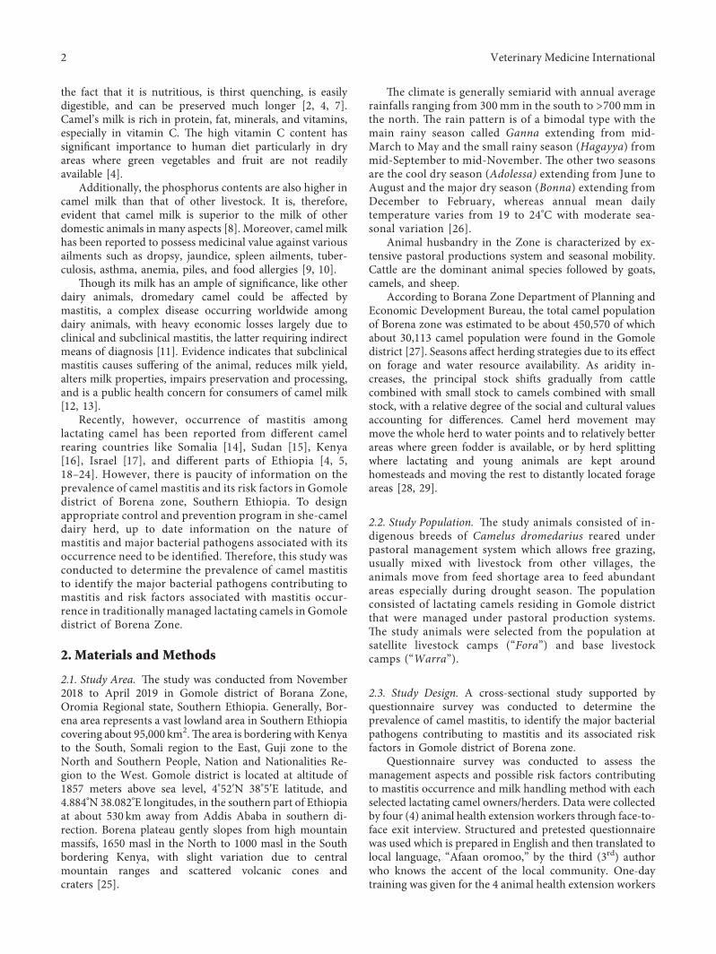
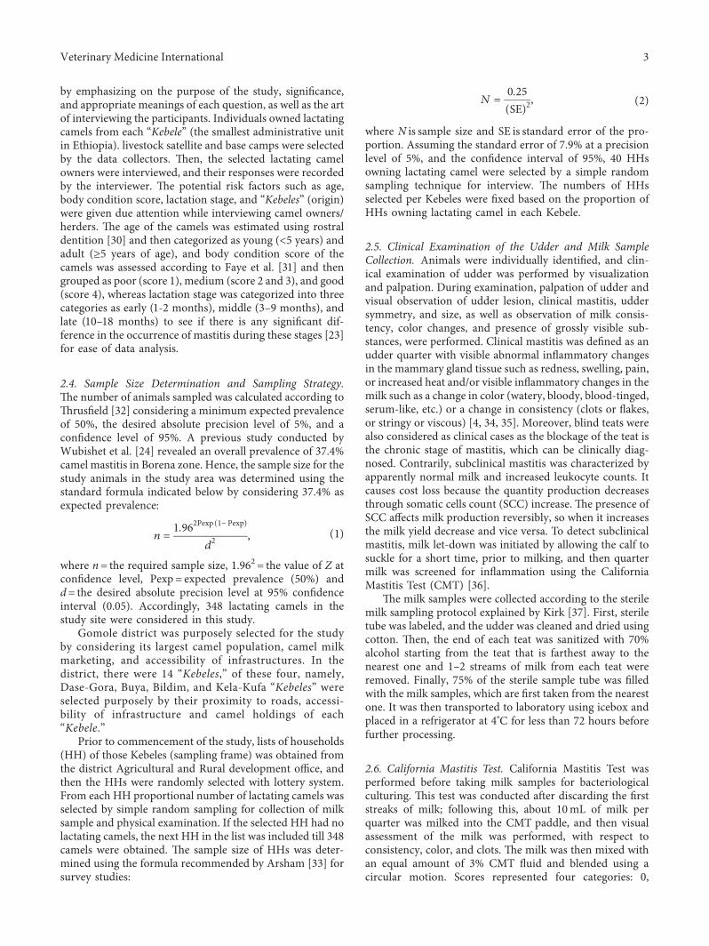
2.7. Bacterial Isolation and Identification. *e milk samplesthat were positive for CMT were kept in an icebox andtransported immediately to Yabello Regional VeterinaryLaboratory (Figure 1), and then 10 μL of milk from eachsample was cultured on sheep blood agar and MacConkeyagar (Oxoid Ltd., Cambridge, UK) for bacteriologicalanalysis. Inoculated plates were incubated aerobically at37°C and evaluated for the growth of bacteria at 24 and 48 hof incubation. *en, presumptive identification of bacterialisolates was carried out based on colony morphologicalfeatures, Gram-staining reactions, hemolytic reactions,catalase test, potassium hydroxide (KOH) test, and otherbiochemical tests [38]. Pure culture of ≥5 colony formingunit (CFU) was recorded as significant with the exception ofS. agalactiae and S. aureus which were classified as signif-icant if≥ 1CFU was present.
*en, bacterial isolates were transferred to their re-spective selective media for further characterizations andspecies identifications. Gram-positive cocci were tested forcatalase, and catalase-positive isolates were further tested forcoagulase production. Briefly, Staphylococci spp. wereidentified based on their growth characteristics on mannitolsalt agar, coagulase, catalase, and oxidase tests. S. aureus wasdifferentiated from other Staphylococcus species by coagu-lase test and maltose fermentation test. Streptococci isolateswere evaluated based on CAMP reaction, hydrolysis ofesculin and sodium hippurate, catalase production, andsugar fermentation tests. Specifically, S. agalactiae wasdifferentiated from other mastitis-causing streptococci byusing CAMP test, esculin hydrolysis on Edwards medium,and growth on MacConkey agar. Gram-negative isolateswere further tested using triple sugar iron (TSI), IMViC,motility, urea, and oxidase test.
2.8. Data Storage and Analysis. Data generated from ques-tionnaire survey and laboratory investigations were recor-ded and coded using Microsoft Excel spreadsheet (MicrosoftCorporation) and analyzed using SPSS version 24.0. Anoverall prevalence of mastitis was calculated as the numberof clinical and subclinical mastitis cases divided by the totalnumber of samples tested. Association of prevalence withthe potential risk factors (age, body condition score, origin(“Kebele’s), and lactation stage) were computed by Chi-square (χ2) test.*en, logistic regression was conducted so asto detect the strength of association of the exposing riskfactors towards the prevalence of lactating camel mastitis.Finally, associations were reported as being statisticallysignificant whenever the p value was <0.05.
3. Results
3.1. Animal and Kebele Level Prevalence of Clinical andSubclinical Mastitis in Lactating Camels. Out of 348
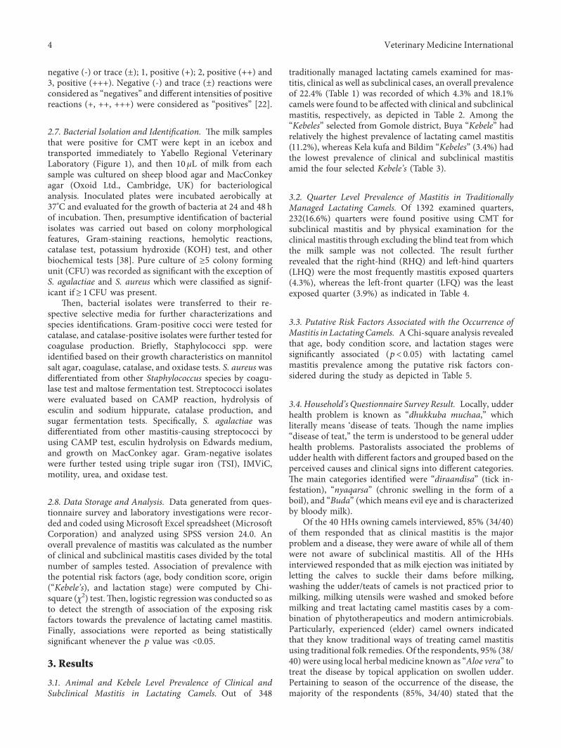
traditionally managed lactating camels examined for mas-titis, clinical as well as subclinical cases, an overall prevalenceof 22.4% (Table 1) was recorded of which 4.3% and 18.1%camels were found to be affected with clinical and subclinicalmastitis, respectively, as depicted in Table 2. Among the“Kebeles” selected from Gomole district, Buya “Kebele” hadrelatively the highest prevalence of lactating camel mastitis(11.2%), whereas Kela kufa and Bildim “Kebeles” (3.4%) hadthe lowest prevalence of clinical and subclinical mastitisamid the four selected Kebele’s (Table 3).
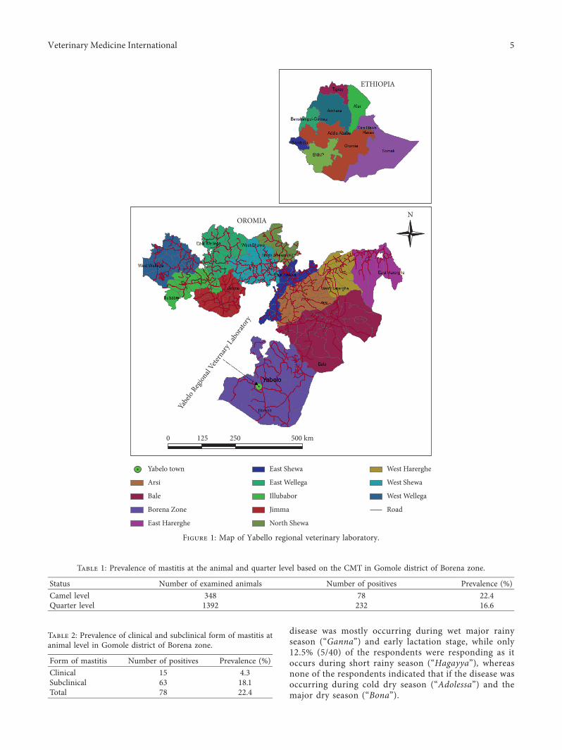
3.2. Quarter Level Prevalence of Mastitis in TraditionallyManaged Lactating Camels. Of 1392 examined quarters,232(16.6%) quarters were found positive using CMT forsubclinical mastitis and by physical examination for theclinical mastitis through excluding the blind teat from whichthe milk sample was not collected. *e result furtherrevealed that the right-hind (RHQ) and left-hind quarters(LHQ) were the most frequently mastitis exposed quarters(4.3%), whereas the left-front quarter (LFQ) was the leastexposed quarter (3.9%) as indicated in Table 4.
3.3. Putative Risk Factors Associated with the Occurrence ofMastitis in LactatingCamels. A Chi-square analysis revealedthat age, body condition score, and lactation stages weresignificantly associated (p< 0.05) with lactating camelmastitis prevalence among the putative risk factors con-sidered during the study as depicted in Table 5.
3.4. Household’s Questionnaire Survey Result. Locally, udderhealth problem is known as “dhukkuba muchaa,” whichliterally means ‘disease of teats. *ough the name implies“disease of teat,” the term is understood to be general udderhealth problems. Pastoralists associated the problems ofudder health with different factors and grouped based on theperceived causes and clinical signs into different categories.*e main categories identified were “diraandisa” (tick in-festation), “nyaqarsa” (chronic swelling in the form of aboil), and “Buda” (which means evil eye and is characterizedby bloody milk).
Of the 40 HHs owning camels interviewed, 85% (34/40)of them responded that as clinical mastitis is the majorproblem and a disease, they were aware of while all of themwere not aware of subclinical mastitis. All of the HHsinterviewed responded that as milk ejection was initiated byletting the calves to suckle their dams before milking,washing the udder/teats of camels is not practiced prior tomilking, milking utensils were washed and smoked beforemilking and treat lactating camel mastitis cases by a com-bination of phytotherapeutics and modern antimicrobials.Particularly, experienced (elder) camel owners indicatedthat they know traditional ways of treating camel mastitisusing traditional folk remedies. Of the respondents, 95% (38/40) were using local herbal medicine known as “Aloe vera” totreat the disease by topical application on swollen udder.Pertaining to season of the occurrence of the disease, themajority of the respondents (85%, 34/40) stated that the
4 Veterinary Medicine International
disease was mostly occurring during wet major rainyseason (“Ganna”) and early lactation stage, while only12.5% (5/40) of the respondents were responding as itoccurs during short rainy season (“Hagayya”), whereasnone of the respondents indicated that if the disease wasoccurring during cold dry season (“Adolessa”) and themajor dry season (“Bona”).
ETHIOPIA
OROMIAN
Yabe
lo Reg
ional
Vetern
ary La
borat
ory
Yabelo town
Arsi
Bale
Borena Zone
East Harerghe
East Shewa
East Wellega
Illubabor
Jimma
North Shewa
500 km2501250
West Shewa
West Wellega
Road
West Harerghe
Figure 1: Map of Yabello regional veterinary laboratory.
Table 1: Prevalence of mastitis at the animal and quarter level based on the CMT in Gomole district of Borena zone.
Status Number of examined animals Number of positives Prevalence (%)Camel level 348 78 22.4Quarter level 1392 232 16.6
Table 2: Prevalence of clinical and subclinical form of mastitis atanimal level in Gomole district of Borena zone.
Form of mastitis Number of positives Prevalence (%)Clinical 15 4.3Subclinical 63 18.1Total 78 22.4
Veterinary Medicine International 5
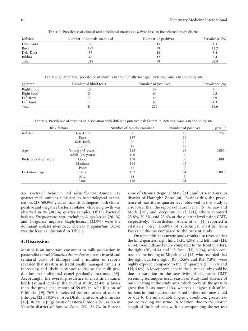
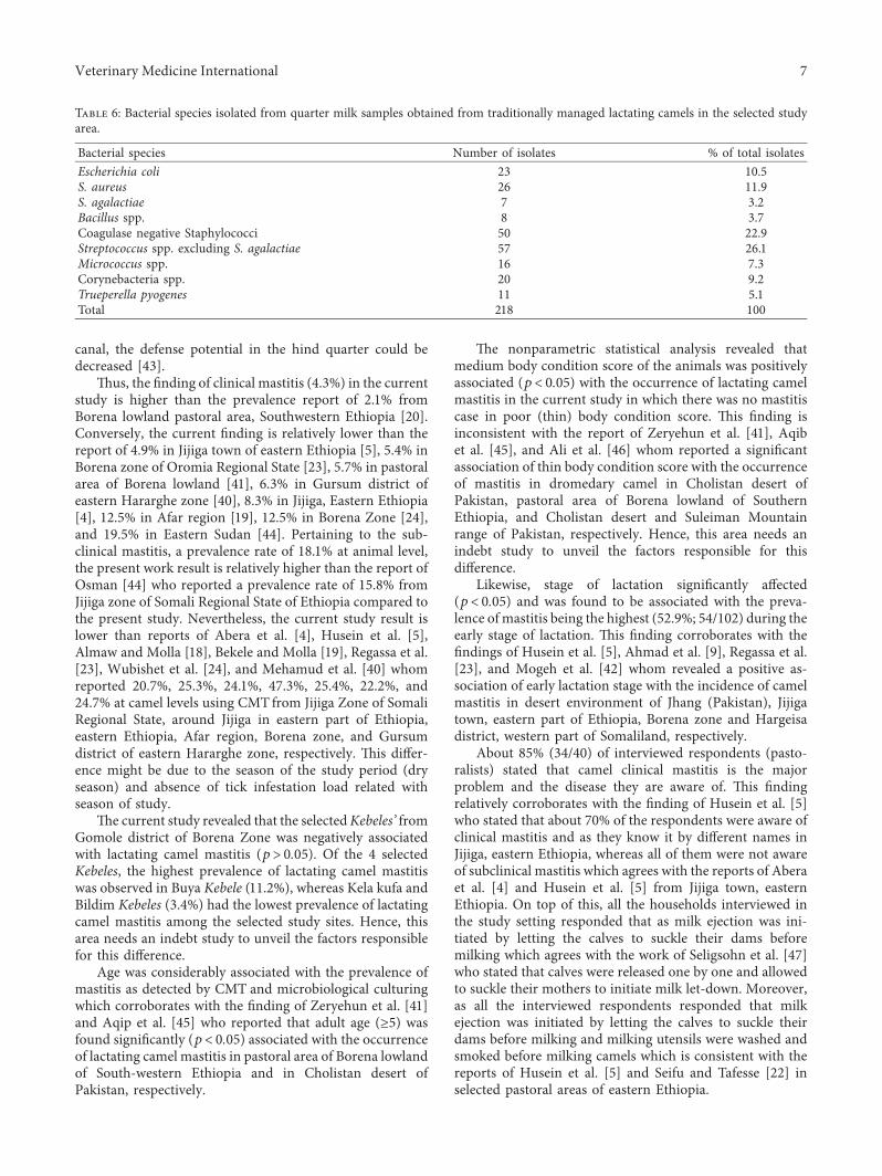
3.5. Bacterial Isolation and Identification. Among 312quarter milk samples subjected to bacteriological exami-nation, 218 (69.9%) yielded mastitis pathogens, both Gram-positive and -negative bacteria isolates, while no growth wasobserved in 94 (30.1%) quarter samples. Of the bacterialisolates, Streptococcus spp. excluding S. agalactiae (26.1%)and Coagulase negative Staphylococci (22.9%) were thedominant isolates identified, whereas S. agalactiae (3.2%)was the least as illustrated in Table 6.
4. Discussion
Mastitis is an important constraint to milk production inpastoralist camel (Camelus dromedarius) herds in arid andsemiarid parts of Ethiopia and a number of reportsrevealed that mastitis in traditionally managed camels isincreasing and likely continues to rise as the milk pro-duction per individual camel gradually increases [39].Accordingly, the overall prevalence of mastitis in camelherds (animal level) in the current study, 22.4%, is lowerthan the prevalence report of 59.8% in Afar Region ofEthiopia [19], 76% in selected pastoral areas of easternEthiopia [22], 18.5% in Abu Dhabi, United Arab Emirates[40], 30.2% in Jijiga town of eastern Ethiopia [5], 44.8% inYabello district of Borena Zone [23], 34.7% in Borena
zone of Oromia Regional State [24], and 31% in Gursumdistrict of Hararghe Zone [40]. Besides this, the preva-lence of mastitis at quarter level observed in this study isalso lower than the reports of Husein et al. [5], Almaw andMolla [18], and Zeryehun et al. [41], whom reported25.8%, 20.5%, and 25.6% at the quarter level using CMT,respectively. Nevertheless, Abera et al. [4] reported arelatively lower (15.8%) of subclinical mastitis fromEastern Ethiopia compared to the present study.
On top of this, the current study results also revealed thatthe hind quarters, right hind (RH, 4.3%) and left hind (LH,4.3%), were inflamed more compared to the front quarters,the right (RF, 41%) and left front (LF, 3.9%), which con-tradicts the finding of Mogeh et al. [42] who recorded thatthe right quarters, right (RF, 15.6% and RH, 7.8%), werehighly exposed compared to the left quarters (LF, 5.2% andLH, 4.6%). A lower prevalence in the current study could bedue to variation in the sensitivity of diagnostic CMTscreening techniques used, season of study, and absence ofbush clearing in the study area, which prevents the grass togrow that hosts more ticks, whereas a higher risk of in-fections in hind quarters compared to the front ones couldbe due to the unfavorable hygienic condition, greater ex-posure to dung and urine. In addition, due to the shorterlength of the hind teats with a corresponding shorter teat
Table 3: Prevalence of clinical and subclinical mastitis at Kebele level in the selected study district.
Kebele’s Number of animals examined Number of positives Prevalence (%)Dase-Gora 56 15 4.3Buya 187 39 11.2Kela-Kufa 57 12 3.4Bildim 48 12 3.4Total 348 78 22.4
Table 4: Quarter level prevalence of mastitis in traditionally managed lactating camels in the study site.
Quarter Number of blind teats Number of positives Prevalence (%)Right front 15 57 4.1Right hind 8 60 4.3Left front 7 55 3.9Left hind 11 60 4.3Total 41 232 16.6
Table 5: Prevalence of mastitis in association with different putative risk factors in lactating camels in the study site.
Risk factors Number of camels examined Number of positives p valueKebeles Dase-Gora 56 15 0.772
Buya 187 39Kela-Kufa 57 12Bildim 48 12
Age Young (<5 years) 248 69 0.000Adult (≥5 years) 100 9
Body condition score Good 138 33 0.001Medium 168 45Poor 42 0
Lactation stage Early 102 54 0.000Mid 98 3Late 148 21
6 Veterinary Medicine International
canal, the defense potential in the hind quarter could bedecreased [43].
*us, the finding of clinical mastitis (4.3%) in the currentstudy is higher than the prevalence report of 2.1% fromBorena lowland pastoral area, Southwestern Ethiopia [20].Conversely, the current finding is relatively lower than thereport of 4.9% in Jijiga town of eastern Ethiopia [5], 5.4% inBorena zone of Oromia Regional State [23], 5.7% in pastoralarea of Borena lowland [41], 6.3% in Gursum district ofeastern Hararghe zone [40], 8.3% in Jijiga, Eastern Ethiopia[4], 12.5% in Afar region [19], 12.5% in Borena Zone [24],and 19.5% in Eastern Sudan [44]. Pertaining to the sub-clinical mastitis, a prevalence rate of 18.1% at animal level,the present work result is relatively higher than the report ofOsman [44] who reported a prevalence rate of 15.8% fromJijiga zone of Somali Regional State of Ethiopia compared tothe present study. Nevertheless, the current study result islower than reports of Abera et al. [4], Husein et al. [5],Almaw and Molla [18], Bekele and Molla [19], Regassa et al.[23], Wubishet et al. [24], and Mehamud et al. [40] whomreported 20.7%, 25.3%, 24.1%, 47.3%, 25.4%, 22.2%, and24.7% at camel levels using CMT from Jijiga Zone of SomaliRegional State, around Jijiga in eastern part of Ethiopia,eastern Ethiopia, Afar region, Borena zone, and Gursumdistrict of eastern Hararghe zone, respectively. *is differ-ence might be due to the season of the study period (dryseason) and absence of tick infestation load related withseason of study.
*e current study revealed that the selectedKebeles’ fromGomole district of Borena Zone was negatively associatedwith lactating camel mastitis (p> 0.05). Of the 4 selectedKebeles, the highest prevalence of lactating camel mastitiswas observed in Buya Kebele (11.2%), whereas Kela kufa andBildim Kebeles (3.4%) had the lowest prevalence of lactatingcamel mastitis among the selected study sites. Hence, thisarea needs an indebt study to unveil the factors responsiblefor this difference.
Age was considerably associated with the prevalence ofmastitis as detected by CMT and microbiological culturingwhich corroborates with the finding of Zeryehun et al. [41]and Aqip et al. [45] who reported that adult age (≥5) wasfound significantly (p< 0.05) associated with the occurrenceof lactating camel mastitis in pastoral area of Borena lowlandof South-western Ethiopia and in Cholistan desert ofPakistan, respectively.
*e nonparametric statistical analysis revealed thatmedium body condition score of the animals was positivelyassociated (p< 0.05) with the occurrence of lactating camelmastitis in the current study in which there was no mastitiscase in poor (thin) body condition score. *is finding isinconsistent with the report of Zeryehun et al. [41], Aqibet al. [45], and Ali et al. [46] whom reported a significantassociation of thin body condition score with the occurrenceof mastitis in dromedary camel in Cholistan desert ofPakistan, pastoral area of Borena lowland of SouthernEthiopia, and Cholistan desert and Suleiman Mountainrange of Pakistan, respectively. Hence, this area needs anindebt study to unveil the factors responsible for thisdifference.
Likewise, stage of lactation significantly affected(p< 0.05) and was found to be associated with the preva-lence of mastitis being the highest (52.9%; 54/102) during theearly stage of lactation. *is finding corroborates with thefindings of Husein et al. [5], Ahmad et al. [9], Regassa et al.[23], and Mogeh et al. [42] whom revealed a positive as-sociation of early lactation stage with the incidence of camelmastitis in desert environment of Jhang (Pakistan), Jijigatown, eastern part of Ethiopia, Borena zone and Hargeisadistrict, western part of Somaliland, respectively.
About 85% (34/40) of interviewed respondents (pasto-ralists) stated that camel clinical mastitis is the majorproblem and the disease they are aware of. *is findingrelatively corroborates with the finding of Husein et al. [5]who stated that about 70% of the respondents were aware ofclinical mastitis and as they know it by different names inJijiga, eastern Ethiopia, whereas all of them were not awareof subclinical mastitis which agrees with the reports of Aberaet al. [4] and Husein et al. [5] from Jijiga town, easternEthiopia. On top of this, all the households interviewed inthe study setting responded that as milk ejection was ini-tiated by letting the calves to suckle their dams beforemilking which agrees with the work of Seligsohn et al. [47]who stated that calves were released one by one and allowedto suckle their mothers to initiate milk let-down. Moreover,as all the interviewed respondents responded that milkejection was initiated by letting the calves to suckle theirdams before milking and milking utensils were washed andsmoked before milking camels which is consistent with thereports of Husein et al. [5] and Seifu and Tafesse [22] inselected pastoral areas of eastern Ethiopia.
Table 6: Bacterial species isolated from quarter milk samples obtained from traditionally managed lactating camels in the selected studyarea.
Bacterial species Number of isolates % of total isolatesEscherichia coli 23 10.5S. aureus 26 11.9S. agalactiae 7 3.2Bacillus spp. 8 3.7Coagulase negative Staphylococci 50 22.9Streptococcus spp. excluding S. agalactiae 57 26.1Micrococcus spp. 16 7.3Corynebacteria spp. 20 9.2Trueperella pyogenes 11 5.1Total 218 100
Veterinary Medicine International 7
Out of 40 HHs owning camels interviewed, 85% (34/40)of them responded that as clinical mastitis is the majorproblem and a disease they were aware of, while all of themwere not aware of subclinical mastitis. All of the HHsinterviewed in the study setting responded that as milkejection was initiated by letting the calves to suckle theirdams before milking, washing the udder/teats of camels isnot practiced prior to milking and milking utensils werewashed and smoked before milking camels, whereas almostall (95%) of the respondents interviewed stated that as theywere using local herbal medicine plant known as “Aloe vera,”applied topically on the swollen udder, and modern anti-microbials to treat the diseases of udder in the presentstudy. Our finding corroborates with the report of Aberaet al. [4] who reported as clinical mastitis was treated by acombination of phytotherapeutics and modern drugs inJijiga town. In contrast to this, Seifu and Tafesse [22]reported that camel owners were using various extractsfrom the roots, leaves, seeds, and exudates of differentplant and branding with hot iron in selected pastoral areasof eastern Ethiopia. Pertaining to season of the occurrenceof the disease, the majority of respondents (85%) statedthat the disease was mostly occurring during wet majorrainy season (“Ganna”) and early lactation stage whileonly 12.5% of the respondents responded that it occursduring short rainy season (“Hagayya”), whereas none ofthe respondents indicated whether the disease was oc-curring during cold dry season (“Adolessa”) and the majordry season (“Bona”).
*e commonly isolated genera of bacteria Staphylococ-cus, Streptococcus, Corynebacterium, Bacillus, and Escher-ichia in this study agree with [4, 19, 39, 48–51] whomisolated Staphylococcus, Streptococcus, and Escherichia asmajor mastitogens. *e isolation rate of E. coli in the currentstudy relatively verifies the finding of Mengistu et al. [52]and Alebie et al. [53]. As coliforms can be a sign of inad-equate hygienic conditions and to a minor degree of fecalcontamination [54], the prevalence may vary considerablyaccording to hygiene conditions.
*e occurrence of S. aureus (11.92%) in this study ismuch higher than the finding of Almaw and Molla [18] whoreported 0.6% but lower than the reports of Woubit et al.[20] and Mengistu et al. [52] who reported 21.03% and 16%,respectively. Such variation might attribute to traditionaltaboo on heat treatment of camel milk and maintaining milkat high ambient temperature after milking and duringtransportation in the study area can pose a serious problemto human health as these practices create conducive situationfor the production of staphylococcal enterotoxin Alebie et al.[53], whereas, of the total isolates, 22.94% of coagulase-negative Staphylococci (CNS) detected in CMTpositive milksamples closely agrees with the findings of Woubit et al. [20](18.2%) and Alebie et al. [53] (19.57%). Nevertheless, it islower than Mengistu et al. [52] who reported 40.4%. *oughit is reported that these Staphylococci spp. are known asfacultative (“minor”) pathogens isolated from subclinicalmastitis cases which do not show a measurable influence onmilk yield, CMT, or clinical symptoms [54], an explanationfor their frequent occurrence is most probably due to the
contamination of the milk samples by the teat canal or teatskin.
*e highest occurrence of Streptococcus spp. excludingS. agalactiae in this study is much higher than the report ofAlamin et al. [15] and Hadef et al. [55] whom reported aprevalence of 1.52% and 2.38% from North Kordofan Stateof Sudan and Southeastern Algeria, respectively. However, itis much lower than previous studies conducted by Saleh andFaye [51] in Al-Jouf, Saudi Arabia (42.9%), whereas thelower prevalence (3.21%) of S. agalactiae reported in thecurrent study substantiates with the report of Husein et al.[5] who reported a prevalence of 3.5% from Jijiga town ofEthiopia but lower than the report of Seligsohn et al. [47],Mehamud et al. [40], and Al-Tofaily and Al Rodhan [56]whom reported a prevalence of 72%,10% and 9.52% fromIsiolo of Kenya, Gursum district of eastern Hararghe,Ethiopia, and some areas of middle Euphrates in Iraq, re-spectively. *e low proportion of S. agalactiae might beattributed to medication of the animal’s mastitis cases by acombination of traditional folk remedies and modern an-timicrobials in the study setting.
*e occurrence of Bacillus spp. in the current report(10.5%) is higher than the report of Mehamud et al. [40] andMengistu et al. [52] whom reported 6.6% and 4.3% of thecases from Gursum district of eastern Hararghe and Gewanedistrict of Afar Regional State, Ethiopia, correspondingly,but lower than the report of Alebie et al. [53] who reported ahigher prevalence of 19.57% from Dubti district of AfarRegional State, North-eastern Ethiopia. Micrococcus spp.isolates (7.34%) recovered from this is closely in line with thereport of Alebie et al. [53], Saleh and Faye [51], Mengistuet al. [52], and Woubit et al. [20] whom reported a prev-alence of 4.35%, 5.7%, 6.4%, and 10.58% from Dubti district,Afar Regional State of Ethiopia, Al-Jouf, Saudi Arabia,Gewane district, Afar Regional State of Ethiopia and pastoralarea of Borena, southwestern Ethiopia, congruently. Fur-thermore, the prevalence of Corynebacterium spp. (9.2%) inthe current investigation corroborates with the report ofHusein et al. [5] who reported a prevalence of 9% from Jijigatown of Ethiopia. Nonetheless, this finding is higher than thereport of Alamin et al. [15] from North Kordofan State ofSudan ((3.03%), whereas the occurrence of Trueperellapyogenes (T. pyogenes) (5.05%) in this study contradicts thereport of Seligsohn et al. [47]. *e occurrence of differentbacterial species reported in the current study could be dueto poor milking hygiene (washing the udder/teats of camelsis not practiced prior to milking) in the study area.
5. Conclusion
*e current study result revealed that the prevalence ofcamel mastitis in the study area was found to be considerablyhigh. *e study revealed that a relatively higher teat quartersubclinical and clinical mastitis, of which the right and lefthind quarters were the most frequently acquiring mastitis.Age, body condition score, and lactation stages were sig-nificantly associated with lactating camel mastitis prevalenceamong the putative risk factors considered in the study.Streptococcus spp. (24.6%) and Coagulase negative
8 Veterinary Medicine International
Staphylococci (21.6%) were among the dominant majorbacterial isolates identified, whereas Streptococcus agalactiaewas the least isolates obtained in this study. *e bacteriaisolated from camel milk samples in the present study aretypes that cause both contagious and environmental mas-titis. Proper and worthy milking techniques are essential inthe prevention of both environmental and contagiousmastitis. *erefore, in order to reduce a relatively highprevalence of mastitis in the area, improvedmilking hygiene,washing of udder/teat, and treating of clinically infected she-camels with the available folk medicine and modern anti-microbials should be practiced.
Data Availability
*e data that support the findings of this study are availablefrom the corresponding author upon reasonable request.
Conflicts of Interest
*e authors declare that they have no conflicts of interest.
Authors’ Contributions
Minda Asfaw Geresu and Shubisa Abera Leliso designed thestudy. Shubisa Abera Leliso supervised the laboratory ex-aminations. Galma Wako performed bacteriological cul-turing and CMTscreening test. Manuscript preparation andthe statistical analysis were conducted by Minda AsfawGeresu and Shubisa Abera Leliso. All authors read andapproved the final manuscript.
Acknowledgments
*is study was supported by Gomole District Pastoral Officeand Gayo Pastoral Development Initiative (GPDI) for thecooperation and financial management. *e authorswould like to thank staff members of Yabello RegionalVeterinary Laboratory (YRVL) for their provision oflaboratory equipment, reagents, and genuine encour-agement while conducting bacteriological culturing. *eauthors would also like to extend their appreciation toGomole district camel owner’s communities for theirdevotion of time in letting them to access information,sharing their experience and practices, and their hospi-tality during sample collection.
References
[1] B. Faye, “How many large camelids in the world? A syntheticanalysis of the world camel demographic changes,” Pasto-ralism, vol. 10, no. 1, p. 25, 2020.
[2] T. Abera, Y. Legesse, B. Mummed, and B. Urga, “Bacterio-logical quality of raw camel milk along the market value chainin Fafen zone, Ethiopian Somali regional state,” BMC Re-search Notes, vol. 9, no. 1, p. 285, 2016.
[3] Y. K. Mohammed, A. Seid, and M. Urge, “Camel (Camelusdromedaries) meat production potentials and associatedconstraints in Eastern Ethiopia,” East African Journal ofVeterinary and Animal Science, vol. 1, no. 2, pp. 77–86, 2017.
[4] M. Abera, O. Abdi, F. Abunna, and B. Megersa, “Udder healthproblems and major bacterial causes of camel mastitis inJijiga, Eastern Ethiopia: implication for impacting food se-curity,” Tropical Animal Health and Production, vol. 42, no. 3,pp. 341–347, 2010.
[5] A. Husein, B. Haftu, A. Hunde, and A. Tesfaye, “Prevalence ofcamel (Camelus dromedaries) mastitis in Jigjiga town,Ethiopia,” African Journal of Agricultural Research, vol. 8,no. 24, pp. 3113–3120, 2013.
[6] E. I. El-Agamy, “Camel milk,” in Handbook of Milk of Non-bovine Mammals, Y. Park and G. F. W. Haenlein, Eds.,pp. 297–344, Blackwell Publishing, Hoboken, NJ, USA, 2006.
[7] A. H. Mohammed, “Conceptual classification of camels,” inDe Multipurpose Camel: Interdisciplinary Study on PastoralProduction in Somalia, pp. 155–158, EPOSMO prints, Upsala,Sweden, 1993.
[8] A. Kouniba, M. Berrada, M. Zahar, and M. Bengoumi,“Composition and heat stability of Moroccan camel milk,”Journal of Camel Practice and Research, vol. 12, pp. 105–110,2005.
[9] S. Ahmad, M. Yaqoob, M. Q. Bilal et al., “Risk factors as-sociated with prevalence andmajor bacterial causes of mastitisin dromedary camels (Camelus dromedarius) under differentproduction systems,” Tropical Animal Health and Production,vol. 44, no. 1, pp. 107–112, 2011.
[10] Y. Shabo, R. Barzel, M. Margoulis, and R Yagil, “Camel milkfor food allergies in children,” De Israel Medical AssociationJournal: De Israel Medical Association Journal, vol. 7,pp. 796–798, 2005.
[11] J. W. Matofari, Y. Mario, E. W. Mwatha, and P. O. Okemo,“Microorganisms associated with subclinical mastitis inKenyan camels (Camelus dromedarius),” Journal of TropicalMicrobiology and Biotechnology, vol. 2, no. 1, pp. 11–16, 2003.
[12] A. Tibary and A. Anouassi, “Reproductive disorders in thefemale camelids,” in Recent Advances in Camelid Reproduc-tion, L. Skidmore and G. P. Adams, Eds., pp. 1–11, Interna-tional Veterinary Information Service, Ithaca, NY, USA, 2000.
[13] S. Gramay and M. Ftiwi, “Camel milk production, prevalenceand associated risk factors of camel mastitis in AsaitaWoreda,Afar Regional State, North East Ethiopia,” ARC Journal ofAnimal and Veterinary Sciences, vol. 4, no. 3, pp. 17–37, 2018.
[14] A. I. Mohamud, Y. A.Mohamed, O. S. A. Jama, P. Mishra, andM. I. Mohamed, “Prevalence and major pathogens associatedwith clinical and subclinical mastitis in dairy camel (Camelusdromedarius) in Benadir Region of Somalia,” VeterinarySciences: Research Review, vol. 6, no. 2, pp. 132–137, 2020.
[15] M. A. Alamin, A.M. Alqurashi, A. S. Elsheikh, and T. E. Yasin,“Mastitis incidence and bacterial causative agents isolatedfrom lactating she-camel (Camelus dromedaries),” IOSRJournal of Agriculture and Veterinary Science, vol. 2, no. 3,pp. 7–10, 2013.
[16] O. B. Kashongwe, B. O. Bebe, J. W. Matofari, andC. G. Huelsebusch, “Associations between milking practices,somatic cell counts and milk postharvest losses in smallholderdairy and pastoral camel herds in Kenya,” InternationalJournal of Veterinary Science and Medicine, vol. 5, no. 1,pp. 57–64, 2017.
[17] A. Y. Guliye, C. Van Creveld, and R. Yagil, “Detection of sub-clinical mastitis in dromedary camels using somatic cell countand the N-acetyl beta-D-Glucosominidase test,” Tropical An-imal Health and Production, vol. 34, no. 2, pp. 95–104, 2002.
[18] G. Almaw and B. Molla, “Prevalence and etiology of mastitisin camels (Camelus dromedaries) in eastern Ethiopia,” Journalof Camel Practice and Research, vol. 7, pp. 97–100, 2000.
Veterinary Medicine International 9
[19] T. Bekele and B. Molla, “Mastitis in lactating camels (Camelusdromedarius) in Afar Region, north-eastern Ethiopia,” Ber-liner und Munchener Tierarztliche Wochenschrift, vol. 114,pp. 169–172, 2001.
[20] S. Woubit, M. Bayleyegn, P. Bonnet, and S. Jean-Baptiste,“Mammites du dromadaire (Camelus dromedarius) dans laregion pastorale basse du Borana au sud-ouest de l’Ethiopie,”Revue d’elevage et de medecine veterinaire des pays tropicaux,vol. 54, no. 3-4, pp. 207–212, 2001.
[21] A. E. Abdul-Gadir, G. Hildebrand, J. N. Kleer, B. Molla,M. N. Kyule, and M. P. Baumann, “Comparison of Californiamastitis test, somatic cell and bacteriological examinations fordetection of camel (Camelus dromedaries), mastitis inEthiopia,” Berlin Munch Tierarzil Woshenschr, vol. 119,pp. 5–49, 2006.
[22] E. Seifu and B. Tafesse, “Prevalence and etiology of mastitis intraditionally managed camels (Camelus dromedarius) in se-lected pastoral areas in eastern Ethiopia,” Ethiopian Veteri-nary Journal, vol. 14, no. 2, pp. 103–113, 2010.
[23] A. Regassa, G. Golicha, D. Tesfaye, F. Abunna, andB. Megersa, “Prevalence, risk factors, and major bacterialcauses of camel mastitis in Borana Zone, Oromia RegionalState, Ethiopia,” Tropical Animal Health and Production,vol. 45, no. 7, pp. 1589–1595, 2013.
[24] Z. Wubishet, A. Dabaso, and G. Getachew, “Prevalence, as-sociated risk factors and bacterial pathogens of camel mastitisin Borena Zone Oromia Regional State, Ethiopia,” Interna-tional Journal of Veterinary Science, vol. 5, no. 4, pp. 280–284,2016.
[25] D. Coppock, De Borana Plateau of Southern Ethiopia Syn-thesis of Pastoral Research, Development and Change, ILCA,Addis Ababa, Ethiopia, 1994.
[26] B. Megersa, “An epidemiological study of major camel dis-eases in the Borana lowland, Southern Ethiopia,” De Dry-lands Coordination Group (DCG), vol. 58, pp. 4-5, 2010.
[27] R. Jara, M. Alemayehu, Z. Wubishet, T. Mesfin, andM. Araya,“Sero-prevalence and associated risk factors of camel bru-cellosis in Southern lowland of Ethiopia,” Journal of Veteri-nary Medicine and Research, vol. 7, no. 1, p. 1180, 2020.
[28] G. Demeke, “Prevalence of camel trypanosomes and factorsassociated with the disease occurrence in Liben district,Borana zone of Oromia region, Ethiopia,” M.Sc. thesis, FreeUniversity of Berlin, Addis Ababa University, FVM, DebreZeit, Ethiopia, 1998.
[29] S. Wario, Z. Wubishet, and M. Alemayehu, “Prevalence andassociated risk factors of major prevalent gastrointestinalnematodes in camels of Borena Zone, Southern Ethiopia,”Journal of Veterinary Medicine and Research, vol. 7, no. 1,p. 1179, 2020.
[30] A. Bello, M. L. Sonfada, A. A. Umar et al., “Age estimation ofcamel in Nigeria using rostral dentition,” Scientific Journal ofAnimal Science, vol. 2, no. 1, pp. 9–14, 2013.
[31] B. Faye, M. Bengoumi, A. Cleradin, A. Tabarani, andY. Chilliard, “Body condition score in dromedary camel: atool for management of reproduction,” Emirates Journal ofFood and Agriculture, vol. 13, no. 1, pp. 1–6, 2017.
[32] M. *rusfield, Sampling in Veterinary Epidemiology, Blackwell Science Ltd, London, UK, 3rd edition, 2008.
[33] H. Arsham, Questionnaire Design and Survey Sampling,2007, http://www.mirror.service.org/site/hom.Ubaltedu/ntsbarsh/Business-stat.
[34] O. Radostits, C. Gay, K. Hinchcliff, and P. Constable, Vet-erinary Medicine: A Text Book of Disease of Cattle, Horses,
Sheep, Pigs and Goats, Bailliere Tindall, London, UK, 10thedition, 2007.
[35] A. Balemi, B. Gumi, K. Amenu et al., “Prevalence of mastitisand antibiotic resistance of bacterial isolates from CMTpositive milk samples obtained from dairy cows, camels, andgoats in two pastoral districts in Southern Ethiopia,” Animals,vol. 11, no. 6, p. 1530, 2021.
[36] O. W. Schalm and D. O. Noorlander, “Experiments andobservations leading to development of the California mastitistest,” Journal of the American Veterinary Medical Association,vol. 130, pp. 199–204, 1957.
[37] J. Kirk, “Sterile milk sampling: extension UDVM,” 2000.[38] National Mastitis Council (NMC),Microbiological Procedures
for the Diagnosis of Bovine Udder Infection, National MastitisCouncil Inc., Arlington, VA, USA, 3rd edition, 1990.
[39] A. A. Al-Juboori, N. K. Kamat, and J. I. Sindhu, “Prevalence ofsome mastitis causes in dromedary camels in Abu Dhabi,United Arab Emirates,” Iraqi Journal of Veterinary Sciences,vol. 27, no. 1, pp. 9–14, 2013.
[40] J. Mehamud, M. Megersa, Y. Abebe, and M. Ahmed,“Prevalence, risk factors and major bacterial causes of camelmastitis in Gursum district, Eastern Hararghe, Ethiopia,”Global Veterinaria, vol. 18, no. 3, pp. 203–208, 2017.
[41] T. Zeryehun, G. Haro, and B. Adane, “A cross sectional studyon the prevalence of mastitis and associated bacterial path-ogens in one-humped camels (Camelus dromedarius) inpastoral area of Borena lowland, Southern Ethiopia,” GlobalVeterinaria, vol. 18, no. 2, pp. 108–115, 2017.
[42] A. O. Mogeh, A. Teklu, and M. D. Ogleh, “*e prevalence ofmastitis and its associated risk factors in lactating dromedarycamels in and around Hargesa, Somaliland,” InternationalJournal of Scienctific and Engineering Research, vol. 10, no. 4,pp. 201–211, 2019.
[43] M. Wanjohi, C. G. Gitao, and L. Bebora, “Subclinical mastitisaffecting hygienic quality of marketed camel milk from NorthEastern Province, Kenya,” Microbiology Research Interna-tional, vol. 1, pp. 6–15, 2013.
[44] A. Osman, “Prevalence of camel mastitis and major bacterialcauses in Jigjiga zone, Somalia region,” DVM thesis, HawassaUniversity, Faculty of Veterinary Medicine, Awasa, Ethiopia,2008.
[45] A. I. Aqib, M. Ijaz, A. Z. Durrani et al., “Prevalence andantibiogram of Staphylococcus aureus, a camel mastitogenfrom Pakistan,” Pakistan Journal of Zoology, vol. 49, no. 3,pp. 861–867, 2017.
[46] M. Ali, M. Avais, M. Ijaz et al., “Epidemiology of subclinicalmastitis in Dromedary camels (Camelus dromedarius) of twodistinct agro-ecological zones of Pakistan,” Pakistan Journalof Zoology, vol. 51, no. 2, pp. 527–532, 2019.
[47] D. Seligsohn, A.-K. Nyman, M. Younan et al., “Subclinicalmastitis in pastoralist dairy camel herds in Isiolo, Kenya:prevalence, risk factors, and antimicrobial susceptibility,”Journal of Dairy Science, vol. 103, no. 5, pp. 4717–4731, 2020.
[48] D. J. U. Kalla, I. S. R. Butswat, S. T. Mbap, A. M. Abdussamad,M. S. Ahmed, and I. Okonkwo, “Microbiological examinationof camel (Camelus dromedarius) milk and sensitivity of milkmicroflora to commonly available antibiotics in Kano,Nigeria,” Savannah Journal of Agriculture, vol. 3, pp. 1–8,2008.
[49] J. W. Matofari, M. Younan, J. N. Nanua, and E. W. Mwatha,“Microorganisms associated with sub-clinical mastitis andtheir impact on milk production in camels (Camelus drom-edarius) in semi-arid lands of Northern Kenya,” International
10 Veterinary Medicine International
Journal of Agriculture and Rural Development, vol. 17,pp. 182–187, 2005.
[50] D. S. Sena, G. Mal, R. Kumar, and M. S. Sahani, “A pre-liminary study of prevalence of mastitis in camel,” Journal ofApplied Animal Research, vol. 20, no. 1, pp. 27–31, 2001.
[51] S. K. Saleh and B. Faye, “Detection of subclinical mastitis indromedary camels (Camelus dromedaries) using somatic cellcounts, California mastitis test and udder pathogen,” Emir-atus Journal of Food and Agriculture, vol. 23, no. 1, pp. 48–58,2011.
[52] F. Mengistu, B. Molla, and A. Ali, “Camel mastitis, associatedbacterial pathogens and its impact on milk quality in Gewanedistrict, Afar Regional State, Northeastern Ethiopia,” AnimalHealth and Production, vol. 58, pp. 249–259, 2010.
[53] A. Alebie, A. Molla, W. Adugna, A. Tesfaye, and M. Ejo,“Prevalence, isolation, identification, and risk factors of majorbacterial cause of camel subclinical mastitis,” BioMed Re-search International, vol. 2021, Article ID 5522331, 6 pages,2021.
[54] V. Eberlein, Hygienic Status of Camel Milk in Dubai (UnitedArab Emirates) under Two Different Milking ManagementSystems, Ludwig-Maximilians-Universitat Munchen, Awasa,Ethiopia, 2007.
[55] L. Hadef, H. Aggad, and B. Hamad, “Bacterial causative agentsassociated with subclinical mastitic in dromedary she-camelsin Southeastern Algeria,” Jordan Journal of Biological Sciences,vol. 11, no. 2, pp. 209–214, 2018.
[56] Y. I. K. Al-Tofaily and M. A. N. Al rodhan, “Study on clinicalmastitis (bacteriological) in she-camels (Camelus drome-darius) in some areas of middle Euphrates in Iraq,” AL-Qadisiya Journal of Veterinary Medicine Science, vol. 10, no. 2,pp. 66–76, 2011.
Veterinary Medicine International 11