Embed Size (px)
Citation preview

Rev. sci. tech. Off. int. Epiz., 2014, 33 (3), ... - ...
No. 27102014-00049-EN 1/46
Camelid brucellosis: a review
This paper (No. 27102014-00049-EN) has been peer-reviewed, accepted, edited, and corrected by authors. It has not yet been formatted for printing. It will be published in December 2014 in issue 33-3 of the Scientific and Technical Review
U. Wernery
Central Veterinary Research Laboratory, P.O. Box 597, Dubai, United
Arab Emirates
E-mail: [email protected]
Summary
Camel brucellosis has been diagnosed in all camel-rearing countries
except Australia. In many countries the infection is on the rise in Old
World camels (OWCs) due to the uncontrolled trade of live animals.
Our knowledge of camelid brucellosis has increased over the last
decade through field investigations, experimental infection trials and
comprehensive laboratory testing. Infection with Brucella melitensis is
frequent in OWCs and rare with B. abortus. New World Camels
(NWCs) rarely contract brucellosis. In East African countries the
seroprevalence of brucellosis can reach 40% (herd level) and depends
on the management system. The highest incidence is found when
camels are kept together with infected small ruminants. Only a
combination of serological methods can detect all serological reactors.
Culturing the pathogen is still the preferred test method, although
several assays based on polymerase chain reaction have been
developed.
Keywords
Brucellosis – Camelid – Diagnosis – Epidemiology – Treatment.
Introduction
Many countries, such as the United Kingdom, Australia and Japan, as
well as parts of the United States of America (USA) and some
countries in North Europe have succeeded in eradicating brucellosis
Rev. sci. tech. Off. int. Epiz., 33 (3) 2
No. 27102014-00049-EN 2/46
through intensive health control measures, but elsewhere the disease
remains widespread in domesticated and wild animal populations and
presents a great economic problem for tropical animal husbandry (1).
Brucellosis is also one of the most important zoonoses in developing
countries. Old World camels (OCWs) are frequently infected with
brucellosis, particularly when they are in contact with infected
ruminants (2, 3, 4, 5, 6). The disease is rare in New World camels
(NWCs) but outbreaks with classical signs of brucellosis have been
described (7).
Aetiology
Brucellosis is a contagious disease caused by bacteria of the genus
Brucella. Taxonomically, the genus Brucella is divided into ten
classified species and subdivided into biovars. The subdivision is
based on biochemical reactions and agglutination with mono-specific
sera. Recently, Brucella strains have been isolated from numerous
marine mammal species; molecular typing methods have not been
able to classify these isolates within the described species, and
therefore they have received their own names: Brucella cetacea
(dolphins) and B. pinnipeda (seals, fur seals, walruses). Brucella
bacteria are Gram-negative coccobacillae that are non-motile and non-
spore-forming. They grow anaerobically and certain strains need a 5%
to 10% carbon dioxide atmosphere. Brucella organisms grow slowly,
but can be enhanced by using enriched media, such as Ferrell’s media
supplemented with 5% horse serum and six added antibiotics.
The growth of B. ovis and B. abortus, biotype 2, always requires
media enriched with serum or blood incubated in an atmosphere of
5% to 10% carbon dioxide.
Impact on human health
In humans, the disease, which is often referred to as ‘undulant fever’
or ‘Malta fever’ is a serious public health problem. Human brucellosis
remains one of the most common zoonotic diseases worldwide, with
more than 500,000 new cases annually (8). Infection prevalence in the
animal reservoirs determines the incidence of human cases (9).

Rev. sci. tech. Off. int. Epiz., 33 (3) 3
No. 27102014-00049-EN 3/46
Brucella spp. are also potential agents of bioterrorism and are
classified in group B (second-highest priority agent) of the Centers for
Disease Control and Prevention (CDC) in the USA.
Brucella melitensis and B. abortus are the two species most
commonly found in human cases, and B. melitensis is responsible for
the most serious infections. Human brucellosis is mainly an
occupational disease, and the main modes of transmission are contact
through skin with animal tissues, blood, urine, vaginal discharge,
aborted fetuses and, especially, placentas, and by consuming raw milk
and other unheated dairy products. Airborne infections occur in
animal pens, stables, laboratories (10) and abattoirs. Some cases have
also occurred from accidental self-inoculation with live vaccines (11,
12). Moreover, it was also shown by Bradenstein et al. (13), that Rev
1 vaccine strain can cause human infections. In their study humans
became infected after consuming milk from vaccinated adult pregnant
animals which excreted the vaccine strain in milk for a long period of
time. The high and increasing herd and animal prevalences of camel
brucellosis in many countries is of grave concern (14); therefore,
veterinary authorities, consumers, camel owners and camel keepers, as
well as responsible persons in the Ministry of Health and Agriculture
of each country, should make every effort to address this issue.
During investigations conducted by Radwan et al. (15), it was found
that brucellosis was diagnosed in 30% of the camel handlers and
milkers and the same B. melitensis biovars were cultured from aborted
sheep and goats sharing the same premises.
In humans, the incubation period lasts from five to 60 days, but can
also be longer. Clinical signs are not specific and can be acute or
chronic (Table I) (16).
Brucella infections in pregnant women in early pregnancy may lead to
high rates of fetal loss (up to 40%) and infection in men can lead to
orchitis and epididymitis. Brucella melitensis DNA persists in human
blood for many years after infection despite appropriate treatment and
apparent recovery (17). Humans are at risk through consumption of
unheated milk or through handling Brucella-positive animals (8, 15,
Rev. sci. tech. Off. int. Epiz., 33 (3) 4
No. 27102014-00049-EN 4/46
18, 19). Shimol et al. (20) described a brucellosis outbreak that
affected 15 people who consumed unpasteurised camel milk. Affected
people suffered mainly from arthralgia and fever and 50% had
positive blood culture for B. melitensis, whereas 60% had serum
agglutination titres of 1:60 or higher.
During a B. melitensis outbreak which occurred in a herd of alpacas in
Peru, over 25% of the alpaca handlers were seropositive to brucellosis
and some developed clinical signs (21).
Extreme care must be exercised when working with Brucella
organisms in laboratories. It is estimated that up to 2% of all
diagnosed brucellosis cases are laboratory-acquired infections, mainly
through inhalation when handling diagnostic specimens (22).
Incidence of camelid brucellosis
Camelid brucellosis caused by B. melitensis and B. abortus has been
reported in all camel-rearing countries except Australia and the
incidence appears to be closely related to breeding and husbandry
practices (23), which Omer et al. (24) were able to prove in Saudi
Arabia. They compared the brucellosis seroprevalence of a female
dromedary herd which was in close contact with small ruminants (n =
165) with a closed female dromedary herd (n = 95). The brucellosis
prevalence in the open camel herd was 8.5%, whereas only one animal
(1%) was diagnosed in the closed herd. The diagnostic tests used were
the Rose Bengal test (RBT), serum agglutination test (SAT) and
complement enzyme-linked immunosorbent assay (cELISA). High
animal and herd prevalences have been reported from different
countries (Table II), which not only pose a severe risk to humans but
also to other livestock. The infection rate in some regions of the
former Union of Soviet Socialist Republics (USSR), where Bactrian
camels are kept on large farms, is 15% (75), whereas in countries with
more extensive forms of husbandry, such as Chad or Ethiopia, the
brucellosis seroprevalence is 3.8% (67) and 5.5% (23), respectively.
Similar differences in seroprevalence have been reported from Saudi
Arabia by Radwan et al. (3) and Ghoneim and Amjad (104). They
reported a higher incidence of camel brucellosis in intensively farmed

Rev. sci. tech. Off. int. Epiz., 33 (3) 5
No. 27102014-00049-EN 5/46
camels than in free-grazing desert camels. In Sudan, prevalence varies
according to the system of camel husbandry: agropastoralists reported
a higher prevalence of brucellosis (31.5%) than nomads (21.4%) (39,
40, 105). A seroprevalence of dromedary brucellosis of 40% has been
reported from Sudan (46), and the United Arab Emirates (UAE) has
experienced a drastic increase of brucellosis in camel populations due
to the uncontrolled import of dromedaries from East African
countries. Also, introduction of camels into cattle, sheep and goat
areas in the Darfur region of Sudan led to high incidence levels, as
shown by Musa and Shigidi (49). In another study in Sudan,
conducted by the same authors, in 3,413 dromedaries that were
intermingled with cattle and small ruminants, the herd infection rate
was 45.5%, with prevalence rates of between 1.4% and 90%.
Moustafa et al. (96) reported on a serological survey in dromedaries
and a brucellosis eradication campaign in the eastern regions of the
UAE during a five-year period. The highest prevalence was in 1991,
with a reactor rate of 5.8%, whereas the lowest was in 1996, with a
rate of 0.01%. Since no camels had been culled due to brucellosis, it is
believed that the reduction in camel brucellosis was caused by the
reduction in brucellosis in sheep and goats.
Epidemiology
The disease has a worldwide distribution and affects cattle, pigs,
sheep, goats, camelids, dogs and, occasionally, horses. Brucella
infections have also been documented worldwide in a great variety of
wildlife species and, more recently, in marine mammals. A spillover
of infection from domestic animals to bisons, elks or African buffalos
may also be possible (106).
The infection occurs via the mucous membranes, including oral-
nasopharyngeal, conjunctival and genital mucosa, and also through
cutaneous abrasions. Animals become infected through feed, water,
colostrum, contaminated milk and, especially, by licking or sniffing at
placentas and aborted fetuses. The spread of brucellosis during sexual
activity plays a subordinate role. The primary shedding routes of
Brucella organisms remain uterine fluids (lochia) and placenta

Rev. sci. tech. Off. int. Epiz., 33 (3) 6
No. 27102014-00049-EN 6/46
expelled from infected animals. In cattle it is known that abortion is
associated with the shedding of 1012 to 1013 Brucella bacteria. Survival
of the organisms in the environment is enhanced by cool temperatures
and humidity; however, it was proven that two dromedaries in a
Brucella-negative dromedary herd were infected with B. melitensis
through contaminated dust particles from aborted camel fetuses 500 m
apart, indicating that organisms can also survive in a hot desert
environment. Many placental mammals, including herbivores,
participate in placentophagy, with camelids as a noted exception,
which may contribute to the spread of Brucella bacteria through wind.
In bovines, shedding of up to 103 B. abortus bacteria/ml through milk
following abortion may last for a period of up to three months, which
is considered an important fact from an epidemiological point of view.
The situation in camelids is unknown. Excretion of the pathogen
through milk is intermittent (98). However, in chronically infected
(serologically positive) dromedaries from the UAE which gave birth
to healthy offspring, no Brucella organisms were isolated from
expelled placentas, and no shedding occurred through milk. Also, the
blood of dromedary calves was negative in culture and polymerase
chain reaction (PCR) (9). Interestingly, camel calves of serologically
positive dams were all serologically negative, using RBT and cELISA
techniques, at the age of six months. The calves therefore do not
appear to be at risk for an acute brucellosis infection even after the
disappearance of maternal antibodies. However, for confirmation of
these findings, further investigations need to be performed (9).
Ostrovidov (107), and Solonitsyn and Pal’gov (108), proposed
separating calves from their dams at the age of seven to eight months,
when their maternal antibodies have disappeared. If this does not
occur, they may contract infection from infected dams at the next
parturition. The Brucella-negativity of female camel calves from
chronically infected dams is controversially discussed between Dubai-
based veterinarians and some researchers believe that confirmation of
the Brucella-negativity can only be confirmed when camel calves
remain serologically negative after parturition. In males, it is an even
more complicated unsolved issue.
Rev. sci. tech. Off. int. Epiz., 33 (3) 7
No. 27102014-00049-EN 7/46
In general, abortions occur mainly during the first pregnancy and
infected camelids are clinically well. The pathogen is found
intracellular in mononuclear phagocytes, in which it also multiplies. In
pregnant camels, the bacteria localise in the placenta and are most
abundant in abortion material (up to 1013 bacteria) including the fetal
stomach, vaginal discharge and colostrums (109). Brucella melitensis
and/or B. abortus organisms have been isolated from camel milk,
aborted fetuses, the placenta, fetal stomach fluid, lymph nodes,
vaginal swabs, testes and hygromas (Table III).
It was also shown by von Hieber (9) that, during a period of two years,
5% (n = 118) of the dams had fluctuating titres from positive to
negative to positive and 20% of the serologically positive dams turned
negative with RBT and cELISA (latent infection?). This indicates that
the pathogens can conceal themselves, most probably in lymph nodes,
and do not produce detectable antibodies in those intracellular hiding
places. However, evidence of spontaneous recovery from brucellosis
had also been described by Gatt Rutter and Mack (118) and
Ostrividov (119), with no further explanation. Further research by
Wernery et al. (116), who investigated the question of where Brucella
organisms were concealed in serologically positive lactating
dromedaries which gave birth to healthy calves, revealed that they
were in internal lymph nodes. They were mainly isolated in lung
lymph nodes, indicating an inhalation infection route. These
investigations in camelids clearly show that there are important
epidemiological differences in dromedaries which abort (acute
brucellosis) and chronically infected animals which do not abort. A
chronic infection is certainly the most common occurrence, and in
bovines it is known that 75% to 90% of cows abort once only (120).
Theoretically, the three Brucella species known to cause brucellosis in
camels (B. abortus, B. melitensis, B. ovis) can cause infection
anywhere (121). However, it is surmised that B. melitensis is
widespread in Africa and the Middle East and B. abortus is
widespread in the former USSR. Solonitsyn (76) reported mixed
infections with various Brucella species in Bactrian camels in Russia.

Rev. sci. tech. Off. int. Epiz., 33 (3) 8
No. 27102014-00049-EN 8/46
Table III demonstrates which Brucella species have been isolated
from which organs in which country.
Although camels appear to be very susceptible to Brucella infection,
isolation of Brucella organisms from camel samples is rare. But
attempts to isolate Brucella from milk have been successful. Brucella
abortus biovars 1 and 3 were isolated from camels in Senegal (117).
Radwan et al. (3) were able to isolate B. melitensis biovars 1 and 2 26
times from a total of 100 milk samples from seropositive Saudi
Arabian dromedaries. Gameel et al. (81) were also able to isolate
B. melitensis biovar 1 five times from the milk of Libyan dromedaries
and four times from aborted fetuses and vaginal swabs from a herd of
124 Libyan dromedaries. The authors did not mention from how many
affected dromedaries the samples were taken. Zaki (122) inoculated
guinea pigs with milk samples from seropositive dromedaries and
cultured the milk samples in vitro. Both tests (SAT and culture) were
negative. Al-Khalaf and El-Khaladi (91) examined cultures of 209
milk samples from Kuwaiti dromedaries. The samples were obtained
from herds with an increased incidence of abortion. The results were
culture-negative. However, the authors were successful in isolating
B. abortus from the gastric fluids of five aborted fetuses. Pal’gov
(110) was able to isolate B. abortus from Bactrian camels in Russia. In
the herds examined, 2% of all animals aborted in the first half of the
pregnancy. Fifteen percent of the herds were seropositive to
brucellosis using the complement fixation test (CFT). Zowghi and
Ebadi (84) cultured 3,500 lymph nodes from 300 slaughtered
dromedaries from Iran for Brucella organisms. Brucellosis melitensis
biovars 1 and 3 were isolated from these lymph nodes in 1% (3/300)
of the camels. The authors are of the opinion that the B. melitensis
infections in the dromedaries originated from neighbouring sheep and
goat herds.
Radwan et al. (15) examined a large camel herd with 2,536
dromedaries in Saudi Arabia from which a 12% abortion rate had been
reported. A Brucella seroprevalence of 8% was found with RBT and
the standard buffered plate agglutination test (BPAT) of the United
States Department of Agriculture (USDA). The authors also isolated

Rev. sci. tech. Off. int. Epiz., 33 (3) 9
No. 27102014-00049-EN 9/46
B. melitensis biovars 1, 2 and 3 from aborted camel fetuses.
Brucella abortus biovar 3 was recovered from an inguinal lymph
node, three vaginal swabs and one supramammary lymph node
obtained from free-ranging camels in eastern Sudan which had
histories of abortion, presence of hygromas or testicular lesions (114).
It is worth mentioning that both isolates of B. abortus biovar 3 from
Senegal and Sudan are the only oxidase-negative biovars reported in
the literature. Ramadan et al. (112) have recovered B. melitensis from
a hygroma of an Indian camel. Brucella melitensis was isolated twice
from two-quarters of milk samples from three seropositive camels in
the UAE (98).
Brucellosis is not a major disease in NWCs, but severe outbreaks,
such as the outbreak in Peru referred to early, have occurred from time
to time. It was thought that sheep were the source of infection in this
alpaca herd (21). In an experimental infection trial in llamas in the
USA, it was found that llamas are susceptible to B. abortus and that
they develop positive serological titres. The authors used five
conventional serological tests (CFT, standard tube test, standard plate
agglutination test, RBT and BPAT) in addition to an ELISA
developed at Iowa State University. The llamas also developed
histological lesions similar to those found in cattle, sheep and goats
(123).
Three llamas died at London zoo after they came into contact with
camels which were newly imported from Moscow (7). The authors
claimed that the high serological titre (type of test not given) for
B. melitensis was indicative of an acute infection.
Clinical signs
Brucellosis is characterised by abortion and to a lesser extent by
orchitis and infection of the accessory sex glands in males. According
to various researchers, the clinical signs of brucellosis in breeding
camelids are the same as those in bovines and small ruminants,
although infection in breeding camelids causes fewer abortions than it
does in bovines and small ruminants (8, 15, 21, 114, 124). Infections
may cause stillborn calves, retained placenta, fetal death,

Rev. sci. tech. Off. int. Epiz., 33 (3) 10
No. 27102014-00049-EN 10/46
mummification and reduced milk yield. Also, delayed service age and
fertility have been reported (49). A retained placenta is rare in
Camelidae. This may be a result of the difference in the placental
attachment (7). Camelids possess a placenta diffusa like the horse and
not a cotyledonary placenta.
Non-pregnant dromedaries (n = 6) artificially infected sub-
cutaneously in the right lower hind of the neck with two strains of
B. abortus (four with S19, two with field bovine strain, × 106
bacteria,) developed only mild clinical signs. Reduced appetite, slight
lameness and bilateral lacrimation were observed. On necropsy the
pathogen was re-isolated 45 to 65 days later from the cranial and
genital lymph nodes. No clinical signs were observed in the four
camels inoculated with S19, whereas slight non-specific signs were
found in the dromedaries infected with the bovine B. abortus field
strain. On necropsy no gross lesions were detected, but histological
results revealed focal granulomas in the liver and a generalised
lymphadenitis (supramammary lymph node). The pathogen was re-
isolated from the lymph nodes of the genital tract and head (125).
Pathology
Little is known about the pathological changes caused by Brucella
organisms in camelids. These bacteria have a predilection for the
pregnant uterus, udder, testicles, accessory male sex glands, lymph
nodes, joint capsules and bursae. Lesions may be found in these
tissues. Nada and Ahmed (126) described lesions in non-pregnant
dromedaries. They found inflammation of the uterus lining with
reddening, oedema and necrotic foci in the uterus epithelium, as well
as fibrosis of the endometrium and atrophy of the uterine glands. The
authors also observed an increased number of ovariobursal adhesions
and hydrobursae. The adhesions occurred between the bursa ovarica
and the ovary and in several cases also between the bursa ovarica and
the salpinges, causing a severe induration of the latter. Hydrobursitis
was often observed in brucellosis-positive dromedaries causing an
enlargement of the bursa, which was then filled with a clear amber-
coloured fluid. No lesions have been described so far in aborted

Rev. sci. tech. Off. int. Epiz., 33 (3) 11
No. 27102014-00049-EN 11/46
camelids and in brucellosis-positive camelid males except orchitis and
epididymitis. The testes and epididymis of 360 dromedaries were
examined for gross and histopathological lesions. Around 12% of the
tested organs originated from seropositive camel bulls. From the
investigations it is not clear if the epididymitis, orchitis or testicular
degeneration was caused by Brucella infection or was a normal
pathological feature (35). A pregnant llama was experimentally
infected by inoculating viable B. abortus bacteria into the conjunctival
sac. Forty-three days post inoculation, the llama aborted an eight-
month-old fetus. Brucella abortus was isolated from the placenta and
all fetal specimens, including the brain, small and large intestines,
spleen, kidney, liver, stomach fluid, heart blood and lung. Bacteria
were also isolated from numerous mammary gland lymph nodes in the
dams. Histologically there was a moderate, multifocal, lymphocytic
and histiocytic, subacute placentitis, with a marked loss of
trophoblastic epithelial cells. The chorioallantoic stroma contained
abundant necrotic and mineralised debris and the swollen capillaries
were expanded by large numbers of Brucella organisms (123, 127).
Abu Damir et al. (38) as well as Wernery et al. (116) described only a
few lesions in non-pregnant B. abortus-infected dromedaries and in
lactating dromedaries that were seropositive for B. melitensis
(B. melitensis was also isolated from milk samples). Cranial and
genital lymph nodes from which the pathogen was isolated showed
marked sinusoidal oedema and follicular hyperplasia of cortical and
paracortical areas, with active germinal centres and histocytosis. There
were no lesions in the reproductive tract.
In Saudi Arabia, pathological and histopathological studies of non-
pregnant dromedaries naturally infected with B. melitensis biovar 3
(46) revealed the following alterations in the following organs:
– lymph node (especially supramammary): oedema, enlargement,
lymphoid hyperplasia, granulomatous reaction in the cortical area of
the lymphoid follicle

Rev. sci. tech. Off. int. Epiz., 33 (3) 12
No. 27102014-00049-EN 12/46
– spleen: enlargement with granular surface in some cases, depletion
of some lymphoid follicles, proliferation of fibrous tissue,
histiocytosis
– mammary gland: granulomastitis in some cases, proliferation of
interlobular fibrous connective tissue
– uterus: moderate amount of mucous and ulceration of endometrial
mucosa, endometrial stroma showed oedema and diffuse and heavy
infiltration of mainly macrophages and lymphocytes in the lamina
propia, blood vessels dilated and congested.
Diagnosis
The morphology of the Brucella bacterial colonies is associated with
the presence of lipopolysaccharides (LPS) in the external membrane
of the bacterium.
Smooth (S-LPS) and rough (R-LPS) phenotypes are differentiated.
The S-LPS phenotype is found in most Brucella species, only B. canis
and B. ovis possess the R-LPS. Some proteins of Brucella are
responsible for serological cross-reactions between Brucella spp. and
other bacterial species (128). Cross-reactivity exists to:
– Yersinia enterocolitica O:9
– Escherichia hermannii
– E. coli O:157
– Francisella tularensis
– Stenotrophomonas maltophilia
– Vibrio cholera O:1
– Salmonella serotypes group N
Therefore, difficulties may arise in the diagnosis of brucellosis.
Abortion and reduced fertility in the camel frequently have other
causes, such as salmonellosis, trypanosomosis, or infections with
Campylobacter or Tritrichomonas fetus (129, 130, 131), making
laboratory testing essential. An incorrect diagnosis of brucellosis may
occur when based on serology alone.

Rev. sci. tech. Off. int. Epiz., 33 (3) 13
No. 27102014-00049-EN 13/46
Culture
Brucellosis is usually diagnosed in the laboratory by the culture of
blood, milk or tissue or the detection of antibodies in sera. Brucella
organisms can be recovered from the placenta, but, more
conveniently, in pure culture from the stomach and lungs of aborted
fetuses.
For isolation, the recommended medium is Farrell’s medium, which
contains six antibiotics. But other selective Brucella media are also in
use for the growth of this pathogen from fresh camel milk and camel
tissue samples (15). During intensive investigations using selective
media it was found that on a camel farm in Saudi Arabia 34% of all
Brucella seropositive milking dromedaries were Brucella shedders.
The high number suggests that it is preferable to use selective media.
Tissue specimens from Brucella-positive dromedaries were examined
by Omer et al. (46) with the immunoperoxidase test, with very good
results. Brucella organisms were detected in the cytoplasm of
macrophages (visible as brown granules), in the lymphocytes of the
lymph nodes and spleen, within the epithelial lining of the
endometrium and endothelium of blood vessels, and within
mononuclear cells around blood vessels.
Polymerase chain reaction
The isolation of Brucella organisms is still the preferred method of
diagnosis. This method also allows typing of the isolated strains.
However, new PCR techniques are now being implemented for both
identification and phenotypic biotyping (106). These PCRs can
discriminate between Brucella species, and between wild and vaccine
strains, but do not discriminate between Brucella biovars. So far, only
monoclonal antibodies against different epitopes of the Brucella LPS
can be used for biovar differentiation.
PCR-based assays have been developed for brucellosis diagnosis and
are based on the detection of specific sequences of the pathogen, such
as genes of the locus 16S – 23S, the IS711 insertion sequence or the

Rev. sci. tech. Off. int. Epiz., 33 (3) 14
No. 27102014-00049-EN 14/46
bcsp 31 gene encoding for a protein of 31kDa. Von Hieber (9), who
used a PCR assay designed with hybridisation probes and primers
targeting the insertion sequence of IS711 of the BMEI 1162 gene, has
shown reliable results in the amplification of pure target DNA in
bacterial dilutions, but the assay was less sensitive when tissue
samples were tested. The reasons for this may be explained by the
extraction method used, the intracellular presence of the pathogen and
the distribution pattern of Brucella organisms.
Serology
The majority of studies on camelid brucellosis use serological
methods for diagnosis (Table II), but none of the serological
brucellosis tests are validated for use in camels yet, as acknowledged
by the World Organisation of Animal Health (OIE). Similarly, none of
the tests have been validated for the diagnosis of human brucellosis
(132). However, it was found that a combination of different
serological tests can increase diagnostic efficacy in camels, although
none of the serological tests can differentiate between a B. abortus or
B. melitensis or B. suis infection. Sunaga et al. (133) reported that five
dromedaries imported into Japan were positive in the CFT and SAT.
The animals were immediately slaughtered. No Brucella organisms
were isolated; however, Yersinia enterocolitica serotype 0:9 was
identified. It is known that false-positive (unspecific) reactions with
various other bacterial species can occur (79, 134).
Many authors regard the CFT as being the most sensitive and specific
test for brucellosis because CFT antibodies remain in the serum for
longer than SAT antibodies (64, 75, 118, 135). Shumilov (77)
determined that the CFT was four times more sensitive than the SAT.
He tested Bactrians in Mongolia, where brucellosis is widespread
among camels. He examined two herds with the following results:
– Herd 1: 3751 camels: CFT 4.3% and SAT 0.6%
– Herd 2: 54,673 camels: CFT 3.7% and SAT 1.0%.
In the SAT an end titre of 1:20 (40 IU) was regarded as suspicious by
different researchers (110, 136, 137, 138). Fayed et al. (29), Salem et
al. (139), and El-Sawally et al. (28) believe that the SAT or tube

Rev. sci. tech. Off. int. Epiz., 33 (3) 15
No. 27102014-00049-EN 15/46
agglutination test (TAT) detect a higher percentage of reactors to
brucellosis than other assays due to their greater sensitivity to
immunoglobulin M (IgM) than immunoglobulin G (IgG). In order to
eliminate unspecific reactions in the SAT, Wernery and Wernery (97)
utilised a 5% solution of phenol sodium chloride, which increases the
specificity of the test and reduces the cross-reactivity. The specificity
is also increased by adding mercaptoethanol, dithiotreitol or a
chelating agent such as ethylenediaminetetraacetic acid (EDTA) to the
antigen.
In addition to cross-reactivity with other bacteria that make the
serological diagnosis of brucellosis more difficult, Zhulobovski and
Pal’gov (137) observed prozones in some sera of Bactrian camels in
Russia, as did Nada (31) in dromedaries from Egypt. The absence of a
visual positive reaction in low dilutions has also been observed in
1.5% of all positive dromedary sera in the UAE (95).
Nearly 30% of the 1,449 alpacas tested in Peru had a positive plate
agglutination titre (21).
Other researchers have used ELISA for the detection of Brucella
antibodies, not only in camel sera (2, 140), but also in camel milk
(141). The camel milk ELISA seems to be an important alternative to
the conventional serodiagnosis of camelid brucellosis. Several
researchers have evaluated the different serological tests for the
diagnosis of camel brucellosis (2, 104, 142, 143, 144). It was
concluded that the elimination of non-specific reactions to Brucella in
camelid sera is essential for the correct diagnosis. It is also important
to apply more than one test, one of which must be the TAT using 5%
NaCl phenolised solution. Atwa (145) and Abou-Zaid (2) found a
good agreement between five different serological tests (SAT using
5% NaCl-phenol lysed solution, SAT with 11.4% phenol-NaCl,
BPAT, RBT, mercapto-ethanol test, and ELISA), ranging between
80.6% and 95.6%.
Mohammed (146) evaluated the RBT, the TAT, and the CFT for the
diagnosis of brucellosis in camels. He found that the RBT and the
CFT demonstrated equal ability in detecting positive and negative sera

Rev. sci. tech. Off. int. Epiz., 33 (3) 16
No. 27102014-00049-EN 16/46
as well as prozone reactions. However, for optimal sensitivity, the
RBT has to be used with serum-antigen at a 3:1 dilution. When using
the CFT, the 1:10 diluted sera have to be inactivated at 54ºC for
30 min and the cold fixation technique has to be applied. Using the
TAT, the classical neutral pH antigen has to be replaced by a buffered
(pH 3.5) antigen to achieve optimal results. As mentioned earlier,
none of these tests have been validated for use in camel brucellosis
and the results are therefore difficult to compare.
Radwan et al. (15) examined a large camel farm comprising 2,536
dromedaries in Saudi Arabia for Brucella antibodies. The authors used
a combination of two tests to identify seropositive dromedaries – the
RBT and the standard USDA BPAT. With these two methods, the
authors successfully eradicated the disease from the farm, where it had
caused abortion in 12% of female camels. The authors adopted these
tests due to their sensitivity, simplicity and applicability in the field.
The use of serological tests is the core of the control or eradication of
brucellosis. Many such tests are available but, they must be used in
accordance with strict standardisation rules and meet the requirements
laid down by the OIE. For bovine brucellosis the OIE recommends the
RBT, the BPAT the CFT, the ELISA and the fluorescence polarisation
assay (FPA). The activity of immunoglobulins during infection in the
different serological tests allows the distinction between acute and
chronic infection. Hence, the presence of both IgM and IgG indicates
an acute brucellosis, whereas chronic brucellosis is characterised by
the presence of IgG alone. Details of the sensitivity and specificity of
the various serological tests are summarised in Table IV.
The tests mentioned in Table IV have all been used for the detection
of camelid brucellosis. The CFT, which was often used as a
confirmatory test, is now progressively being replaced by ELISAs and
more recently also by FPA.
The FPA is based on a physical principle and when antibodies against
Brucella are present in a serum, a fluorescent complex is formed and
expressed in milli-polarisation units (mP); in a negative sample the
antigen remains uncomplexed (106). ELISAs have a high sensitivity

Rev. sci. tech. Off. int. Epiz., 33 (3) 17
No. 27102014-00049-EN 17/46
but their specificity is quite low. Reactions towards different bacterial
species, especially Y. enterocolitica 0:9, are known to occur to all
serological tests. Results by Alshaikh et al. (90) clearly showed the
SAT’s limited reliability for chronically infected dromedaries. This
was also demonstrated by Omer et al. (46), who reported that the RBT
was suitable for screening camel sera for brucellosis, but the cELISA
detected 2.1% more positives. More recent investigations by von
Hieber (9) and Gwida et al. (99) on hundreds of brucellosis-positive
dromedaries imported into the UAE from Sudan compared several
diagnostic tests. There was a good agreement between the results of
the CFT, RBT and SAT, proven by calculating kappa values, but the
sensitivity of all three tests was low compared to the results by FPA or
serum real-time PCR. Serum real-time PCR was not validated, but had
a high diagnostic sensitivity, as it was able to detect as little as
23 femtograms of Brucella DNA per reaction, with a probability of
95% (99). Therefore, it is advisable to combine real-time PCR with a
serological test such as RBT, which would increase the sensitivity to
100% (147).
Detection of brucellosis in camel sera by PCR has been described by
Alshaikh et al. (90) in Saudi Arabia. This is a very reliable diagnostic
tool, which can even differentiate between B. melitensis and
B. abortus brucellosis.
The FPA and a cELISA were used to test a total of 336 sera obtained
from llamas and alpacas in Chile which came from a brucellosis
negative herd. The results were compared with conventional tests such
as the RBT, SAT and CFT. Only two sera were found positive with
the FPA and cELISA (144), and none with the conventional tests.
However, both sera had low titres.
In contrast to cattle milk, camel milk cannot be used to detect lacteal
brucellosis antibodies using the conventional milk ring test (MRT),
because camel milk lacks the agglutinating substance required to
cluster fat globules (141). It is also known that camel milk fat
globulins are tiny micelles which, therefore, do not cream up to
produce a surface fat layer. The MRT results summarised in Table IV

Rev. sci. tech. Off. int. Epiz., 33 (3) 18
No. 27102014-00049-EN 18/46
should therefore be interpreted with great caution. Van Straten et al.
(141) established an MRT that can also be used to detect antibodies in
camel milk. The researchers named this test a modified MRT because
Brucella-negative cow milk is added to the camel milk, producing a
typical blue-coloured creamy ring when antibodies to Brucella
bacteria are present. The test is not highly sensitivity, but it is cheap to
use.
Skin test
Brucellosis skin tests have been tried by some researchers, particularly
on Bactrian camels in the former USSR, using different allergens.
(148). The skin test is highly specific but its sensitivity is low, making
it a good herd test. The antigen does not sensitise the animal’s
immune system and therefore will not induce interference in the
diagnosis of the disease.
Control and treatment
Brucella has been eradicated in many regions of the world, but in
others it is widespread and an economically important disease. Many
cases of human brucellosis are found in regions where the disease has
not been eliminated in livestock. Different strategic options can be
adopted to first decrease the prevalence of brucellosis to an acceptable
level (brucellosis control) and secondly to remove the foci of infection
(brucellosis eradication). The choice of control strategy depends on a
number of considerations, such as infection prevalence in different
animal species, human clinical incidence and the capacity of
Veterinary Services. However, a pre-requisite for any control
programme is the implementation of an efficient animal disease
surveillance network. Eradication in small ruminants has never been
achieved (149) and may be also very difficult to achieve in OWCs due
to the complexity and expense of treating animals across widespread
areas. In cattle and small ruminants, when prevalence is low (between
3% and 5%), vaccination comes first followed by slaughter (8). Abbas
and Agab (105) suggest whole-herd vaccination in low-prevalence
countries, and test-and-slaughter followed by vaccination in high-
prevalence countries. In camel-racing countries, the culling method

Rev. sci. tech. Off. int. Epiz., 33 (3) 19
No. 27102014-00049-EN 19/46
cannot be applied because racing dromedaries are often extremely
valuable animals and play a very important role in Bedouin culture.
Therefore, it is preferable to castrate all Brucella-positive bulls, not to
breed positive females, and to vaccinate. No compromise should be
made when it comes to camel dairy farms. They must be free of
brucellosis.
Antibiotics
Brucella organisms are Gram-negative coccobacilli which are
sensitive to many broad-spectrum antibiotics, but the use of antibiotics
is forbidden in many countries because of the uncertainty related to
the infective status of the treated animals and because of the spread of
antibiotic resistance. Treatment is unlikely to be cost-efficient or
therapeutically effective because of the intracellular sequestration of
the organisms, mainly in the lymph nodes. However, cure rates
between 65% and 100% have been reported in infected goats by daily
intraperitoneal injection of 500 mg and 1,000 mg tetracyclines (111).
Radwan et al. (15) also treated 202 seropositive dromedaries with a
combination of oxytetracycline (25 mg/kg body weight) every two
days for 30 days and streptomycin (25 mg/kg body weight) every two
days for 16 days. In addition to this parenteral treatment, milking
camels received 10 ml of oxytetracycline as intramammary infusions
in each teat every two days for eight days. This regimen of treatment
was effective in eliminating the shedding of Brucella organisms
through milk. All treated dromedaries also became serologically
negative within 16 months of treatment. But the single untreated
control camel remained positive over the same period of time. Using
antibiotics may be a way to save valuable animals (e.g. racing camels)
from being culled, but it is doubtful if antibiotic treatment on a herd-
level basis can be successful. It is not clear from this investigation
whether or not the shedding would have stopped anyway, without any
antibody treatment, because the study did not include any untreated
controls. However, the author’s unpublished treatment protocol
clearly demonstrated that dromedary brucellosis is not treatable with
antibiotics, although it is claimed otherwise. Twenty-three
seropositive dromedaries were treated with antibiotics according to

Rev. sci. tech. Off. int. Epiz., 33 (3) 20
No. 27102014-00049-EN 20/46
Radwan et al. (15) with the following results 36 months later
(Table V).
These results also clearly demonstrate the sensitivities of four
different tests in chronically infected dromedaries.
Vaccination
Because of the serious medical and economic consequences of
brucellosis, serious efforts have been undertaken to prevent the
infection through the use of vaccines. In OWCs, both inactivated and
attenuated Brucella vaccines have been used successfully.
Dromedaries were vaccinated with B. abortus strain S19 (150) and
with B. melitensis Rev 1 (15). Young (three months) dromedaries
received a full dose of the vaccine and adults (10 years) a reduced
dosage. Both groups developed Brucella antibodies with titres of
between 1:25 and 1:200 using the standard USDA BPAT, two to four
weeks after vaccination. They receded after eight months in young
stock and after three months in adult camels. Agab et al. (151)
vaccinated five dromedaries with a reduced dose (5 × 10⁸ cfu in 2 ml)
of B. abortus strain S19. All five camels seroconverted after one week
and their antibodies declined six to seven weeks later. The
dromedaries tested negative 14 weeks later. So far, no challenge
infections have been performed after vaccination. In cattle, the
optimum age for vaccination is between four and eight months of age.
Serum agglutination test returns negative results by the time the
bovines are of breeding age, except in 6% of cases (152). It is obvious
that post-vaccination titres increase with the increasing age and
therefore cattle vaccination is recommended only in young stock.
Vaccination of bulls with S19 is of no value because it often resulted
in the development of orchitis and the presence of strain S19 in the
semen (106). Very little is known about the optimal vaccination age in
camels and their serological response. Before vaccination is started in
dromedaries, thorough investigations are paramount in order to find
out if animals are naturally infected by B. abortus or B. melitensis, this
can only be determined by culture or PCR. Brucellosis melitensis Rev
1 is an attenuated vaccine and must be very carefully used otherwise

Rev. sci. tech. Off. int. Epiz., 33 (3) 21
No. 27102014-00049-EN 21/46
infections of considerable virulence may occur in both vaccinated and
in-contact humans. Vaccination of pregnant goats and sheep may
result in abortion and excretion of live B. melitensis vaccine bacteria
in milk and vaginal discharge. The situation in dromedaries is
unknown.
Conclusion
Brucellosis in OWCs is on the rise and needs the urgent intervention
of all those concerned, including camel owners, to avoid further
spread. Camel brucellosis has a severe impact on human health in
camel-rearing countries. In brucellosis-endemic countries eradication
can only be achieved by control, prevention and surveillance. In most
countries where camels are reared they possess an important value for
the owner, not only economically but also culturally. The value of
dromedaries can be very high, especially in camel-racing countries.
Most of the brucellosis-positive camels are clinically healthy animals
and owners do not allow their Brucella serologically positive animals
to be culled. Therefore, the author proposes that the best way to halt
the spread of the disease is to castrate serologically positive bulls,
never breed positive females and start vaccination in positive herds.
References
1. Seifert H.S.H. (1992). – Tropentierhygiene. Gustav Fischer
Verlag Jena, Stuttgart, 292–304.
2. Abou-Zaid A.A. (1998). – Some studies on camel brucellosis.
In Proc. 8th Scientific Congress of the Faculty of Veterinary
Medicine, Assiut University, Egypt, 690–707.
3. Radwan A.I., Bekairi S.J. & Prasad P.V.S. (1992). –
Serological and bacteriological study of brucellosis in camels in
central Saudi Arabia. Rev. sci. tech. Off. int. Epiz., 11 (3), 837–844.
4. Barsoum S.A., El-Sayed M.M. & El-Fayoumy M.M. (1995).
– Seroepidemiological study on camel brucellosis. Beni-Suef. vet.
med. Res., 5 (2), 111–117.

Rev. sci. tech. Off. int. Epiz., 33 (3) 22
No. 27102014-00049-EN 22/46
5. Straten M.O. (1949). – Brucellosis in camels. Veterinariya
Moscow, 26 (6), 16–20.
6. Al-Ani F.K., Al-Sharrify M. & Khalil F. (1998). –
Serological survey on camel brucellosis in camels in Iraq. Camel
Newsl., 14, 32–33.
7. Fowler M.E. (2010). – Medicine and surgery of camelids, 3rd
Ed. Wiley-Blackwell, 207–208.
8. World Health Organization (WHO) & Food and Agriculture
Organization of the United Nations (FAO) (1986). – 6th Report of the
Joint FAO/OIE Expert Committee on Brucellosis, 12 to 19 November
1985, Geneva. Technical Report Series No. 740. WHO, Geneva, 740,
132.
9. Von Hieber D. (2010). – Investigation of occurrence and
persistence of brucellosis in female camel dams (Camelus
dromedarius) and their calves. Thesis , Universität Ulm, Germany.
10. Schulze zur Wiesch J., Wichmann D., Sobottka I., Rohde
H., Schmoock G., Wernery R., Schmiedel S., Burchard G.D. &
Melzer F. (2020). – Genomic tandem repeat analysis proves
laboratory-acquired brucellosis in veterinary (camel) laboratory in the
United Arab Emirates. Zoonoses public Hlth, 57 (5), 315–317.
11. Saleem M.N., Boyle S.M. & Sriranganathan N. (2010). –
Brucellosis: a re-emerging zoonosis. Vet. Microbiol., 140, 392–398.
12. World Organisation for Animal Health (OIE) (2012). –
Manual of diagnostic tests and vaccines for terrestrial animals, Vol. I,
7th Ed. OIE, Paris, 641.
13. Bradenstein S., Mandelboim M., Ficht T.A., Baum M. &
Banai M. (2002). – Identification of the Brucella melitensis vaccine
strain Rev. 1 in animal and humans in Israel by PCR analysis of the
PstI site polymorphism of its omp2 gene. J. clin. Microbiol., 40 (4),
1475–1480.
Rev. sci. tech. Off. int. Epiz., 33 (3) 23
No. 27102014-00049-EN 23/46
14. Sprague L.D., Al-Dahouk S. & Neubauer H. (2012). – A
review on camel brucellosis: a zoonosis sustained by ignorance and
indifference. Pathog. glob. Hlth, 106 (3), 144–149.
15. Radwan A.I., Bekairi S.I., Mukayel A.A., Albokmy A.M.,
Prasad P.V.S., Azar F.N. & Coloyan E.R. (1995). – Control of
Brucella melitensis infection in a large camel herd in Saudi Arabia
using antibiotherapy and vaccination with Rev 1 vaccine. Bull. Off.
int. Epiz., 14 (3) 719–732.
16. Hoover D.L. & Friedlander A.M. (1997). – Brucellosis.
Chapter 25. In Medical aspects of chemical and biological warfare (R.
Zajtchuk, Ed.). Department of the Army, Office of the Surgeon
General, Borden Institute, Washington, DC, 513–521.
17. Vrioni G., Pappas G., Priavali E., Gartzonika C. &
Levidiotou S. (2008). – An eternal microbe: Brucella DNA load
persists for years after clinical cure. Clin. infect. Dis., 46 (12), e131–
e136.
18. Kiel F.W. & Khan M.Y. (1987). – Analysis of 506
consecutive positive serological tests for brucellosis in Saudi Arabia.
J. clin. Microbiol., 25, 1384–1387.
19. Madkow M.M. (1989). – Brucellosis. Butterworths,
London.
20. Shimol S.B., Dukhan L., Belmaker I., Bardenstein S.,
Sibirsky D., Barrett C. & Greenberg D. (2012). – Human brucellosis
outbreak acquired through camel milk ingestion in southern Israel. Isr.
med. Assoc. J., 14, 475–478.
21. Acosta M., Ludena H., Barreto D. & Moro Sommo M.
(1972). – Brucellosis en alpacas. Rev. Invest. pec., 1 (1), 37–49.
22. Ergonul O., Celikbas A., Tezeren D., Guvener &
Dokuzoguz B. (2004). – Analysis of risk factors for laboratory-
acquired brucella infections. J. Hosp. Infect., 56, 223–227.

Rev. sci. tech. Off. int. Epiz., 33 (3) 24
No. 27102014-00049-EN 24/46
23. Richard D. (1980). – Dromedary pathology and
productions. Paper presented at a workshop on camels, 18–20
December, Khartoum, Sudan. Provisional Report No. 6. Camels.
International Foundation for Science, Stockholm, 409–430.
24. Omer A.Kh., Bahbil A.E.-A., Hassan N.A. & Abd El-
Wahab A.M. (2010). – Pathophysiological investigations on
brucellosis in she-camels. Global Veterinaria, 4 (5), 495–503.
25. Ahmed M.R. (1993). – The incidence of brucellosis in
different domesticated animals in Egypt. Technical Science Bulletin
No. 23, Government Press, Cairo, 210–231.
26. Ayoub N.M., Shawkat M.A. & Fayed A.A. (1978). –
Serological investigation on brucellosis in camels in Egypt. Assiut vet.
med. J.
27. El-Nahas H.M. (1964). – Brucellosis in camels. In Proc. 5th
Arab Veterinary Congress, 11–17 April, Cairo. Egyptian Veterinary
Medical Association, 239–252.
28. El-Sawally A.A., Montaser A.M. & Rizk L.G. (1996). –
Diagnostic and biochemical evaluations of camel brucellosis. Vet.
med. J. Giza, 44 (2), 323–329.
29. Fayed A.A., Karmy S.A., Yousef H.I. & Ayoub M.M.
(1982). – Serological studies on brucellosis in Aswan Province. Vet.
med. J., 30, 491–497.
30. Hamada S., El-Hidik M., Sherif I., El-Sawah H. & Yousef
M. (1963). – Serological investigations on brucellosis in cattle,
buffaloes and camels. J. Arab. vet. med. Assoc., 23, 173–178.
31. Nada A.R. (1984). – Some studies on brucellosis in camels.
M.V.Sc. Thesis, Faculty of Veterinary Medicine, Cairo University.
32. Nada A.R. (1990). – Further studies on brucellosis in
camels. PhD Thesis, Faculty of Veterinary Medicine, Cairo
University.
Rev. sci. tech. Off. int. Epiz., 33 (3) 25
No. 27102014-00049-EN 25/46
33. Zagloul A.H. & Kamel Y. (1985). – Incidence of brucellosis
among farm animals in Assiut governorate. Assiut vet. med. J., 14,
117–122.
34. Zaki R. (1948). – Brucella infection among ewes, camels
and pigs in Egypt. J. comp. Pathol., 58, 145–151.
35. Ahmed W.M. & Nada A.R. (1993). – Some pathological
affections of testis and epididymis of slaughtered camels (Camelus
dromedarius). Int. J. anim. Sci., 8, 33–36.
36. El-Seedy F.R., Radwan A.I. & El-Shabrawy M.A. (2000).
Serological and bacteriological investigations on Brucella infection in
one humped camel (Camelus dromedarius) in Egypt. Vet. med. J.
Giza, 48 (1) 83–89.
37. Abbas B., Yassin T.T.M. & Elzubir A.E.A. (1987). –
Survey for certain zoonotic diseases in camels in the Sudan. Rev. Elev.
Méd. vét. Pays trop., 40 (3), 231–233.
38. Abu Damir H., Kenyon S.J., Khalafalla A.E. & Idris O.F.
(1984). – Brucella antibodies in Sudanese camels. Trop. anim. Hlth
Prod., 16, 209–212.
39. Agab H.R.D. (1993). – Epidemiology of camel diseases in
Eastern Sudan with emphasis on brucellosis. M.V.Sc. Thesis.
University of Khartoum, Sudan.
40. Agab H.R.D. (1998). – Camel pastoralism in the Butana
region of eastern Sudan: common diseases with emphasis on
brucellosis. J. Camel Pract. Res., 5 (1), 131–136.
41. Ali M. & Ghedi S. (1978). – Indagini siero-epidemiologiche
sulla diffusione in Somala della brucellosi degli animali domestici.
Tipizzazioni dei primi stipiti isolati nel paese [Sero-epidemiological
surveys on the prevalence of brucellosis in Somali pets. Typings of the
first strains isolated in the country]. Thesis, Faculty of Veterinary
Medicine, Somali National University, Mogadishu.

Rev. sci. tech. Off. int. Epiz., 33 (3) 26
No. 27102014-00049-EN 26/46
42. Bornstein S. & Musa B.E. (1987). – Prevalence of
antibodies to some viral pathogens, Brucella abortus and Toxoplasma
gondii in serum from camels (Camelus dromedarius) in Sudan. J. vet.
Med., B 34, 364–370.
43. Mustafa A.A. & Awad El-Karim M.H. (1971). – A
preliminary survey for the detection of Brucella antibodies in camel
sera. Sudan J. vet. Sci. anim. Husb., 12 (1), 5–8.
44. Mustafa A.A. & Hassan A. (1971). – A preliminary survey
for the detection of Brucella antibodies in camel sera, Sudan. J. vet.
Sci. Anim. Husb., 12, 5.
45. Osman A.M. & Adlan A.M. (1987). – Sudan. In Brucellosis
in domestic animals: prevalence, diagnosis and control. Tech. series
Off. int. Epiz., 6, 67–72.
46. Omer M.M., Musa M.T., Bakhiet M.R. & Perret L. (2010) –
Brucellosis in camels, cattle and humans: associations and evaluation
of serological tests used for diagnosis of the disease in certain
nomadic localities in Sudan. Rev. sci. tech. Off. int. Epiz., 29 (3), 663–
669.
47. Obeid A.I., Bagadi H.O. & Mukhtar M.M. (1996). –
Mastitis in camels (Camelus dromedarius) and the somatic cell
content of camel milk. Res. vet. Sci., 61 (1), 55–58.
48. Majid A.A. & Goraish I.A. (2000). – Seroepidemiological
observations of camel brucellosis in Eastern and Western Sudan.
Newsl. ACSAD, 17, 23–26.
49. Musa M.T. & Shigidi M.T.A. (2001). – Brucellosis in
camels in intensive animal breeding areas of Sudan. Implications in
abortion and early-life infections. Rev. Elev. Méd. vét. Pays trop., 54
(1), 11–15.
50. Ahmed A. & Ibrahim L. (1980). – Indagine sulla presenza e
diffusione delle brucellosi nel dromedario in Somala [Survey on the
occurrence and spread of brucellosis in dromedaries in Somalia].

Rev. sci. tech. Off. int. Epiz., 33 (3) 27
No. 27102014-00049-EN 27/46
Thesis, Faculty of Veterinary Medicine, Somali National University,
Mogadishu.
51. Andreani E., Prosperi S., Salim A.H. & Arush A.M. (1982).
– Serological and bacteriological investigation on brucellosis in
domestic ruminants of the Somali Democratic Republic. Rev. Elev.
Méd. vét. Pays trop., 35 (4), 329–333.
52. Anon. (1981). – Annual report of the Veterinary
Laboratory, Kisimayo. Ministry of Livestock, Forestry and Range,
Department of Veterinary Services, Somali Democratic Republic.
53. Baumann M.P.O., Nuux H.A. & Zessin K.H. (1990). –
Livestock disease survey Central Rangeland of Somalia. Technical
Report. Vol. III. Herd demographic and disease survey data from
herds of camels. Central Rangelands Development Project –
Veterinary Component, Mogadishu, Somalia.
54. Baumann M.P.O. & Zessin K.H. (1992). – Productivity and
health of camels (Camelus dromedarius) in Somalia: associations with
trypanosomiasis and brucellosis. Trop. anim. Hlth Prod., 24 (3), 145–
156.
55. Bishof J. (1979). – Serological examination of blood
samples from dromedaries. Serum and Vaccine Institute, Mogadishu,
Somalia.
56. Bornstein S. (1984). – Working paper No. 3, Camel Forum.
Somali Academy of Science and Arts, Mogadishu.
57. Bornstein S. (1988). – A disease survey of the Somali
camel. SARE Report, Sweden.
58. Bornstein S., Musa B.E. & Jama F.M. (1988). –
Comparison of seroepidemiological findings of antibodies to some
infectious pathogens of cattle in camels of Sudan and Somalia with
reference to findings in other countries of Africa. In Proc.
International Symposium in the Development of Animal Resources in
Sudan, 3–7 January, Khartoum. University of Khartoum, 28–34.

Rev. sci. tech. Off. int. Epiz., 33 (3) 28
No. 27102014-00049-EN 28/46
59. Elmi A.M. (1982). – MPVM Thesis. University of
California, Davis.
60. Ghanem Y.M., El-Khodery S.A., Saad A.A., Abdelkader
A.H., Heybe A. & Musse Y.A. (2009). – Seroprevalence of camel
brucellosis (Camelus dromedarius) in Somaliland. Trop. anim. Hlth
Prod., 41, 1779–1786.
61. Domenech J. (1977). – Enquête sérologique sur la
brucellose du dromadaire en Chad [Serological survey of brucellosis
in camels in Chad]. Rev. Elev. Méd. vét. Pays trop., 30 (2), 141–142.
62. Teshome H. & Molla B. (2002). – Brucellosis in camels
(Camelus dromedarius) in Ethiopia. J. Camel Pract. Res., 9 (2), 125–
128.
63. Kagunya D.K.J. & Waiyaki P.G. (1978). – A serological
survey of animal brucellosis in north-eastern province of Kenya.
Kenya Vet., 2 (2), 35–38.
64. Waghela S., Fazil M.A., Gathuma J.M. & Kagunya D.K.
(1978). – A serological survey of brucellosis in camels in north-
eastern province of Kenya. Trop. anim. Hlth Prod., 10 (1), 28–29.
65. Wilson A.J., Schwartz H.J., Dolan R., Field C.R. &
Roettcher D. (1982). – Epidemiologische Aspekte bedeutender
Kamelkrankheiten in ausgewählten Gebieten Kenias [Epidemiological
aspects of important camel diseases in selected areas of Kenya]. Der
praktische Tierarzt., 11, 974–987.
66. Bares J.F. (1968). – Contribution à l’étude de la pathologie
infectieuse du dromadaire au Tchad [A contribution to the study of
infectious diseases of camels in Chad]. Thesis, Toulouse, France, 30–
38.
67. Graber M. (1968). – Central African Region of Veterinary
and Zootechnical Research. Annual report of the Farcha Laboratory,
Fort Lamy, Chad. I. Research and Products. Vet. Bull., 38, 52–65.
Rev. sci. tech. Off. int. Epiz., 33 (3) 29
No. 27102014-00049-EN 29/46
68. Schelling E., Diguimbaye C., Daoud S., Nicolet J., Boerlin
P., Tanner M. & Zinsstag J. (2003). – Brucellosis and Q-fever
seroprevalences of nomadic pastoralists and their livestock in Chad.
Prev. vet. Med., 61, 279–293.
69. Burgemeister R., Leyk W. & Goessler R. (1975). –
Untersuchungen über das Vorkommen von Parasitosen, bakteriellen
und viralen Infektionskrankheiten bei Dromedaren in Südtunesien
[Investigations on the occurrence of parasitic, bacterial and viral
infectious diseases in camels in Southern Tunisia]. Dtsch. tierärztl.
Wochenschr., 82, 352–354.
70. Okoh A.E.J. (1979). – A survey of brucellosis in camels in
Kano, Nigeria. Trop. anim. Hlth Prod., 11 (4), 213–214.
71. Kudi A.C., Kalla D.J.U., Kudi M.C. & Kapio G.I. (1997). –
Brucellosis in camels. J. arid Environ., 37, 413–417.
72. Adama N.B., Okoh A.E.J. & Azunku U.J. (2007). –
Prevalence of brucellosis in nomadic herds of dromedaries in Borno
State, Nigeria.J. Camel Pract. Res., 14 (2), 135–138.
73. Bornarel P. & Akakpo A.J. (1982). – Brucelloses animales:
Sondages sérologiques dans quatre pays de l’Afrique de l’Ouest
(Bénin, Cameroun, Haute-Volta, Niger) [Animal brucellosis:
serological surveys in four countries in West Africa (Benin,
Cameroon, Upper Volta, Niger]. Méd. Afr. noire, 29 (12), 829–836.
74. Saley H. (1983). – Contribution à l’étude des brucelloses au
Niger: résultats d’une enquête sérologique dans 3 départements [A
contribution to the study of brucellosis in Niger: results of a
serological survey in three départements]. DVM Thesis, Cheikh Anta
Diop University, Dakar 6.
75. Pal’gov A.A. & Zhulobovski I.Z. (1964). – Diagnosis of
brucellosis in camels and methods of eliminating infection from camel
herds. Trudy Inst. Vet. Akademiya Nauk, Kazakhskoi SSR, Alma Ata,
6, 43–50.

Rev. sci. tech. Off. int. Epiz., 33 (3) 30
No. 27102014-00049-EN 30/46
76. Solonitsyn M.O. (1949). – Brucellosis in camels.
Veterinariya Moscow, 26 (6), 16–20.
77. Shumilov K.V. (1974). – Diagnostic value of agglutination
and complement fixation test for brucellosis in camels. Proc. All-
Union Inst. exp. vet. Med., 42, 279–282.
78. Mocalov V.I. (1991). – About the prevalence of brucellosis
in cattle and camels [in Russian]. Bull. sci. Res. Inst. exp. vet.-Med.
Moscow, 41–45.
79. Erdenebaatar J., Bayarsaikhan B., Watarai M., Makino S.I.
& Shirahata T. (2003). – Enzyme-linked immunosorbent assay to
differentiate the antibody responses of animals infected with Brucella
species from those of animals infected with Yersinia enterocolitica
O9. Clin. diagn. Lab. Immunol., 10 (4), 710–714.
80. Ben Faraj S.M., Azwai S.M., Gameel S.E., Shareha A.M.,
Benhaj K.M., Rayes H.M. & Nayil A.A. (1990). – Camel and human
brucellosis in Libya. In Proc. International conference on camel
production and improvement, 10–13 December, Tobruk, Libya. Arab
Center for the Studies of Arid Zones and Dry Lands, Damascus, Syria.
81. Gameel S.E.A., Mohamed S.O., Mustafa A.A. & Azwai
S.M. (1993). – Prevalence of camel brucellosis in Libya. Trop. anim.
Hlth Prod., 25 (2), 91–93.
82. Kulshreshtha R.C., Arora R.G. & Kalra D.S. (1975). –
Brucellosis in camels and horses. Indian J. anim. Sci., 45 (9), 673–
675.
83. Mathur K.N. & Bhargava S.C. (1979). – Sero-prevalence of
Q fever and brucellosis in camels of Jorbeer and Bikaner, Rajasthan
State. Indian J. med. Res., 70 (11), 391–393.
84. Zowghi E. & Ebadi A. (1988). – Brucellosis in camels in
Iran. Rev. sci. tech. Off. int. Epiz., 7 (2), 383–386.

Rev. sci. tech. Off. int. Epiz., 33 (3) 31
No. 27102014-00049-EN 31/46
85. Ebrahimi A., Hosseinpour F. & Montazeri B. (2007). –
Seroprevalence of brucellosis in dromedaries in Iran. J. Camel Pract.
Res., 14 (1), 43–44.
86. Khorasgani M.R., Bokaie S., Moallemzadeh S.A. & Salehi
T.Z. (2006). – A note on serologic survey of camel brucellosis in Qum
province, Iran. J. Camel Pract. Res., 13 (1), 51–52.
87. Jawad A.H. (1984). – Brucellosis in camel in Iraq. Bull.
endem. Dis., 24–25, 45–50.
88. Radwan A.I., Asmar J.A., Frerichs W.M., Bekairi S.I. & Al-
Mukayel A.A. (1983). – Incidence of brucellosis in domestic livestock
in Saudi Arabia. Trop. anim. Hlth Prod., 15, 139–143.
89. Hashim N.H., Galil G.A., Hulaibi M.A. & Al-Saleem E.M.
(1987). – The incidence of brucellosis and species of Brucella
organisms isolated from animals in Al Hasa, Saudia Arabia. World
Anim. Rev., 61, 32–53.
90. Alshaikh M.A.A., Al Haidary A., Aljumaah R.S., Al
Korashi M.M., El Nabi G.R.A. & Hussein M.F. (2007). – Camel
brucellosis in Riyadh Region, Saudi Arabia. J. Camel Pract. Res., 14
(2), 113–117.
91. Al-Khalaf S. & El-Khaladi A. (1989). – Brucellosis of
camels in Kuwait. Comp. Immunol. Microbiol. infect. Dis., 12 (1/2),
1–4.
92. Harby H.A.M. & Ismaily S.L.N. (1995). – The prevalence
of brucellosis among livestock in the Sultanate of Oman. In Proc.
International Conference on Livestock Production in Hot Climates, 8–
10 January, Sultan Qaboos University, Muscat, Oman, A46.
93. Yagoub I.A., Mohamed A.A. & Salim M.O. (1990). –
Serology survey for Br. abortus antibody prevalence in the one
humped camel (Camelus dromedarius) from Eastern Sudan. Rev.
Elev. Méd. vét. Pays trop., 43 (2), 167–171.

Rev. sci. tech. Off. int. Epiz., 33 (3) 32
No. 27102014-00049-EN 32/46
94. Ismaily S.L.N., Harby H.A.M. & Nicoletti P. (1988). –
Prevalence of Brucella antibodies in four animal species in the
Sultanate of Oman. Trop. anim. Hlth Prod., 20, 269–270.
95. Afzal M. & Sakkir M. (1994). – Survey of antibodies
against various infectious disease agents in racing camels in Abu
Dhabi, United Arab Emirates. Rev. sci. tech. Off. int. Epiz., 13 (3),
787–792.
96. Moustafa T., Omar E.A., Basyouni S.M. & El-Badawi A.S.
(1998). – Surveillance of Brucella antibodies in camels of the eastern
region of the United Arab Emirates. In Proc. International Meeting on
Camel Production and Future Perspectives, 2–3 May, College of Food
and Agriculture, Al Ain, United Arab Emirates, 160–166.
97. Wernery U. & Wernery R. (1990). – Seroepidemiologische
Untersuchungen zum Nachweis von Antikörpern gegen Brucellen,
Chlamydien, Leptospiren, BVD/MD, IBR/IPV und Enzootischen
Bovinen Leukosevirus (EBL) bei Dromedarstuten (Camelus
dromedarius) [Seroepidemiological studies for the detection of
antibodies to Brucella, Chlamydia, Leptospira, BVD /MD , IBR/IPV
and enzootic bovine leukosis (EBL) in dromedaries]. Dtsch. tierärztl.
Wochenschr., 97, 134–135.
98. Wernery U., Kinne J., Joseph M., Johnson B. & Nagy P.
(2007). – Where do Brucella organisms hide in serologically positive
lactating dromedaries. In Proc. International Camel Conference, 16–
17 February, College of Veterinary and Animal Sciences, Rajasthan
Agricultural University, Bikaner, India, 68–70.
99. Gwida M.M.A.S., El-Gohary A.H., Melzer F., Tomaso H.,
Rösler U., Wernery U., Wernery R., Elschner M., Khan I. & Neubauer
H. (2011). – Comparison of diagnostic tests for the detection of
Brucella spp. in camel sera. BMC res. Notes, 4, 525.
100. Taha T.H. (2007). – Pathogens affecting the reproductive
system of camels in the United Arab Emirates. Thesis, Swedish
University of Agricultural Sciences, Uppsala, Sweden.

Rev. sci. tech. Off. int. Epiz., 33 (3) 33
No. 27102014-00049-EN 33/46
101. Ajmal M., Ahmad M.D. & Arshad A. (1989). – Sero-
surveillance of brucellosis. Pakistan vet. J., 9, 115–117.
102. Al-Majali A.M. (2006). – Seroepidemiology of camel
brucellosis in Jordan. In Proc. 1st Conference of ISOCARD
[International Society of Camelid Research and Development], 15–17
April, Al Ain, United Arab Emirates, 79.
103. Al-Majali A.M., Al-Qudah K.M., Al-Tarazi Y.H. & Al-
Rawashdeh O.F. (2008). – Risk factors associated with camel
brucellosis in Jordan. Trop. anim. Hlth Prod., 40 (3), 193–200.
104. Ghoneim N.A. & Amjad A.M. (1993). – Brucellosis
among sheep, goats and camels in Saudi Arabia in Al Joub region,
incidence and comparison between Rose Bengal test and
seroagglutination tube test. In Proc. 21st Arab Veterinary Medical
Congress, 10–14 April, Cairo, 273–281.
105. Abbas B. & Agab H. (2002). – A review of camel
brucellosis. Prev. vet. Med., 55, 46–47.
106. Saegermann C., Berkvens D., Godfroid J. & Walravens K.
(2010). – Bovine brucellosis. In Infectious and parasitic diseases of
livestock (P.-C. Lefèvre, J. Blancou, R. Chermette & G. Uilenberg,
eds), Lavoisier, Paris, 991–1021.
107. Ostrovidov P.I. (1954). – Experiment on rearing healthy
camels from dams infected with brucellosis. Trudy inst. vet. akad.
nauk kazakh., 6, 62–68.
108. Solonitsyn M.O. & Pal’gov A.A. (1950). – Brucellosis [in
Russian]. Trudy inst. vet. akad. nauk kazakh., 5, 58.
109. Millar M. & Stack J. (2012). – Brucellosis: what every
practitioner should know. In Practice, 34, 532–539.
110. Pal’gov A.A. (1950). – No title. Trud. naucho-issled, Vet.
Inst. Alma Ata, 5, 29.

Rev. sci. tech. Off. int. Epiz., 33 (3) 34
No. 27102014-00049-EN 34/46
111. Radwan A.I., Bekairi S.I. & Al-Mukayel A.A. (1992). –
Treatment of Brucella melitensis infection in sheep and goats with
oxytetracycline combined with streptomycin. Rev. sci. tech. Off. int.
Epiz., 11 (3), 845–857.
112. Ramadan R.O., Hatem M.E. & Abdin Bey M.R. (1998). –
Isolation of Brucella melitensis from carpal hygroma in camels. J.
Camel Pract. Res., 5 (2), 239–241.
113. Al Dubaib M.A. (2007). – Polymerase chain reaction and
adapted enzyme linked immunosorbent assay for diagnosis of camel
brucellosis. Vet. med. J., 55 (4), 1067–1075.
114. Agab H.R.D., Abbas B., El Jack A.H. & Mamoun I.E.
(1996). – First report on the isolation of Brucella abortus biovar 3
from camels (Camelus dromedarius) in the Sudan. Camel Newsl., 12
(9), 52–55.
115. Musa M.T., Eisa M.Z., El Sanousi E.M., Abdel Wahab
M.B. & Perrett L. (2008). – Brucellosis in camels (Camelus
dromedarius) in Dafur, Western Sudan. J. comp. Pathol., 138, 151–
155.
116. Wernery U., Thomas R., Syriac G., Raghavan R. &
Kletzka S. (2007). – Seroepidemiological studies for the detection of
antibodies against nine infectious diseases in dairy dromedaries (Part
I). J. Camel Pract. Res., 14 (2), 85–90.
117. Verger J.M., Grayon M., Doutre M.P. & Sagna F. (1979).
– Brucella abortus d’origine bovine au Sénégal: identification et
typage [Brucella abortus of bovine origin in Senegal: identification
and typing]. Rev. Elev. Méd. vét. Pays trop., 32 (1), 25–32.
118. Gatt Rutter T.E. & Mack R. (1963). – Diseases of camels.
Part 1: Bacterial and fungal diseases. Vet. Bull., 33 (3), 119–124.
119. Ostrovidov P.I. (1954). – Development of resistance to
brucellosis in camels. Trudy inst. vet. akad. nauk kazakh., 6, 51–56.
Rev. sci. tech. Off. int. Epiz., 33 (3) 35
No. 27102014-00049-EN 35/46
120. Acha P.N. & Szyfres B. (2003). – Zoonoses and
communicable diseases common to man and animals. Vol. I –
Bacterioses and mycoses, 3rd Ed. Pan American Health Organization
Washington, DC, 382 pp. Adapted from: Infectious and parasitic
diseases of livestock, Part 2 (P.-C. Lefèvre, J. Blancou, R. Chermette
& G. Uilenberg, eds), Lavoiser, Paris, 1017 pp.
121. Higgins A. (1986). – The camel in health and disease.
Baillière Tindall, London.
122. Zaki R. (1943). – Br. abortus infection in buffaloes, ewes
and camels. Isolation of the organism from milk. M.V.Sc. Thesis,
Faculty of Veterinary Medicine, Cairo University.
123. Gilsdorf M.J., Thoen C.O., Temple R.M.S., Gidlewski T.,
Ewalt D., Martin B. & Henneger S.B. (2001). – Experimental
exposure of llamas (Lama glama) to Brucella abortus: humoral
antibody response. Vet. Microbiol., 81, 85–91.
124. Fazil M.A. & Hofmann R.R. (1981). – Haltung und
Krankheiten des Kamels [Husbandry and diseases of camels].
Tierärztl. Praxis, 9, 389–402.
125. Abu Damir H., Tag El Din M.H., Kenyon S.J. & Idris O.F.
(1989). – Isolation of Brucella abortus from experimentally infected
dromedary camels in Sudan. A preliminary report. Vet. Res. Commun.,
13, 403 – 406.
126. Nada A.R. & Ahmed W.M. (1993). – Investigations on
brucellosis in some genital abnormalities of she-camels
(C. dromedarius). Int. J. anim. Sci., 8 (1), 37–40.
127. Gidlewski T., Cheville N.T., Rhyan J.C., Miller L.D. &
Gildorf M.J. (2000). – Experimental Brucella abortus induced
abortion in a llama: pathologic effects. Vet. Pathol., 37 (1), 77–82.
128. Emmerzaal A., de Wit J.J., Dijkstra T., Bakker D. & van
Zijderveld F.G. (2002). – The Dutch B. abortus monitoring
Rev. sci. tech. Off. int. Epiz., 33 (3) 36
No. 27102014-00049-EN 36/46
programme for cattle: the impact of false-positive serological
reactions and comparison of serological tests. Vet. Q., 24, 40–46.
129. Wernery U. & Ali A. (1989). – Bacterial infertility in
camels (Camelus dromedarius). Isolation of Campylobacter fetus.
Dtsch. tierärztl. Wochenschr., 96, 497–498.
130. Wernery U. (1991). – The barren camel with endometritis:
isolation of Trichomonas Fetus and different bacteria. J. vet. Med., B,
38 (1–10), 523–528.
131. Wernery U. & Wernery R. (1992). – Uterine infections in
the dromedary camel. A review. In Proc. 1st International Camel
Conference (W.R. Allen, A.J. Higgins, I.G. Mayhew, D.H. Snow &
J.F. Wade, eds). R. & W. Publications, Newmarket, United Kingdom,
155–158.
132. Yohannes M., Gill J.P.S., Ghatak S., Singh D.K. & Tolosa
T. (2012). – Comparative evaluation of the Rose Bengal plate test,
standard tube agglutination test and complement fixation test for the
diagnosis of human brucellosis. Rev. sci. tech. Off. int. Epiz., 31 (3),
979–984.
133. Sunaga Y., Tani F. & Mukai K. (1983). – Detection of
Yersinia enterocolitica infection in camels serodiagnosed as
brucellosis. Jpn. J. vet. Sci., 45 (2), 247–250.
134. Bisping W. & Amtsberg G. (1988). – Colour atlas for the
diagnosis of bacterial pathogens in animals. Paul Parey Scientific
Publishers, Berlin and Hamburg.
135. Tserendash C. & Shumilov K.V. (1970). – Diagnosis of
brucellosis in camels [in Russian]. Veterinariya, 1, 116–117.
136. Arbusov P.N. (1940). – Normal titer of camel serum in
relation to brucellosis. Soviet Vet., 5, 47–48.
137. Zhulobovski I.L. & Palgov A.A. (1954). – No title [in
Russian]. Trud. Inst. Vet. Alma-Ata, 6, 17.

Rev. sci. tech. Off. int. Epiz., 33 (3) 37
No. 27102014-00049-EN 37/46
138. Ghazi Y.A. (1996). – Studies on brucellosis in camels.
PhD Thesis, Faculty of Veterinary Medicine, Cairo University.
139. Salem A.A., El-Gibaly S.M., Shawkat M.E., Ibrahim S.I.
& Nada A.R. (1990). – Some studies on brucellosis in camels. Assiut
vet. med. J., 23 (45), 139–145.
140. Azwai S.M., Carter S.D., Woldehiwet Z. & MacMillan A.
(2001). – Camel brucellosis: evaluation of field sera by conventional
serological tests and ELISA. J. Camel Pract. Res., 8 (2), 185–193.
141. Van Straten M., Bercovich Z. & Ur-Rahman Z. (1997). –
The diagnosis of brucellosis in female camels (Camelus dromedarius)
using the milk ring test and milk ELISA: a pilot study. J. Camel Pract.
Res., 4 (2), 165–168.
142. Abo El-Hassan D.G., Mammam H.M., Youssef R.,
Barsoum S.A., Awad M.M. & Sameh S.M. (1991). – Prevalence of
camel brucellosis using different serological tests. Vet. med. J. Giza,
39 (3), 875–884.
143. Nada A.R., Ismail E.M., Shawkat M.E. & Barsoum S.A.
(1992). – Evaluation of serotests used in the diagnosis of camel
brucellosis. J. Egypt. vet. med. Assoc., 52 (4), 435–442.
144. Rojas X., Muñoz S., Otto B., Pérez B. & Nielsen K.
(2004). – The use of polarized fluorescence assay (PF) and
competitive ELISA test (c-ELISA) for the diagnosis of brucellosis in
South American camelids. Arch. Med. vet. 36 (1), 59–64.
145. Atwa K.A. (1997). – Brucellosis in camels. M.V.Sc.
Thesis, Faculty of Veterinary Medicine, Cairo University.
146. Mohammed I.M. (1996). – Development, optimization and
evaluation of diagnostic immunoassays for camel brucellosis. Thesis,
Faculty of Veterinary Science, University Khartoum, Sudan.
147. Gwida M.M.A.S. (2010). – Isolation, identification and
typing of Brucella species as zoonotic pathogens by using

Rev. sci. tech. Off. int. Epiz., 33 (3) 38
No. 27102014-00049-EN 38/46
conventional and molecular biological methods. DVM Thesis, Free
University of Berlin, Germany.
148. Ten V.B. & Cejdachmedova R.D. (1993). – Diagnosis of
brucellosis in camels [in Russian]. Dositizenie nauki i techniki, 1, 31.
149. Smits H.L. (2013). – Brucellosis in pastoral and confined
livestock: prevention and vaccination. In Brucellosis: recent
developments towards One Health (G. Plumb, S. Olsen & G. Pappas,
eds). Rev. sci. tech. Off. int. Epiz., 32 (1), 219–228.
150. Chichibabin E.S. (1971). – Results of haemagglutination
test with the heat-inactivated sera from camels investigated for
brucellosis. Proc. Kazakh res. vet. Inst., 14, 29–30.
151. Agab H.R.D., Angus B. & Mamoun I.E. (1995). –
Serologic response of camels (Camelus dromedarius) to Brucella
abortus vaccine S19. J. Camel Pract. Res., 2 (2), 93–95.
152. Radostits O.M., Gay C.C., Hinchcliff K.W. & Constable
P.C. (2007). – Veterinary medicine. A textbook of the diseases of
cattle, horses, pigs and goats, 10th Ed. Saunders Elsevier, 963–994.
__________

Rev. sci. tech. Off. int. Epiz., 2014, 33 (3), ... - ...
No. 27102014-00049-EN 39/46
Table I
Signs of brucellosis in humans
Clinical signs Patients affected in %
Fever 90–95
Malaise 80–95
Body ache 40–70
Sweats 40–90
Arthralgia 20–40
Splenomegaly 10–30
Hepatomegaly 10–70

Rev. sci. tech. Off. int. Epiz., 33 (3) 40
No. 27102014-00049-EN 40/46
Table II
Summary of the literature regarding the occurrence of antibodies
to Brucella in camelids arranged by country
Country Author (Reference no.) Year Number of
camels examined
Prevalence %
Serology
ELIS
A
RBT
CFT
SAT
MR
T
Egypt Abou-Zaid (2) 1998 422 10.4–12.3 x x x
Ahmed (25) 1993 200 3.5
Ayoub et al. (26) 1978 216 24.2 x
El-Nahas (27) 1964 200 4.0 x
El-Sawally et al. (28) 1996 - 2.3–14.0 x x x
Fayed et al. (29) 1982 300 6.6 x x x
Hamada et al. (30) 1963 175 10.3 x
Nada (31) 1984 780 23.1 x x
Nada (32) 1990 - 5.3–7.9 x x x
Zagloul and Kamel (33) 1985 37 8.1 - - - - -
Zaki (34) 1948 200 14.0 m. x
26.0 f. x
Ahmed and Nada (35) 1993 360 11.6 x
El-Seedy et al. (36) 2000 750 3.9 (BAPT) x x
4.9 (RBPT)
Sudan Abbas et al. (37) 1987 238 3.0 x
Abu Damir et al. (38) 1984 740 4.9 x x x
Agab (39) 1993 453 30.0 x
380 32.9 f. x
15.1 m. x
Agab (40) 1998 2.9 x
Ali and Ghedi (41) 1978 250 10.4 x
Bornstein and Musa (42) 1987 102 5.9 x x
Mustafa and Awad El-Karim (43) 1971 310 1.8–5.8 x
Mustafa and Hassan (44) 1971 - 1.75–5.75 - - - - -
Osman and Adlan (45) 1987 137 8.0 x x
Omer et al. (46) 2010 2,225 37.5 x x
Obeid et al. (47) 1996 153 10.6 x X
Majid and Goraish (48) 2000 805 14–44 x X
Musa and Shigidi (49) 2001 3,413 1.4–89.5 x x
45.5 (Herd)
Somalia Ahmed and Ibrahim (50) 1980 802 8.0–11.0 x
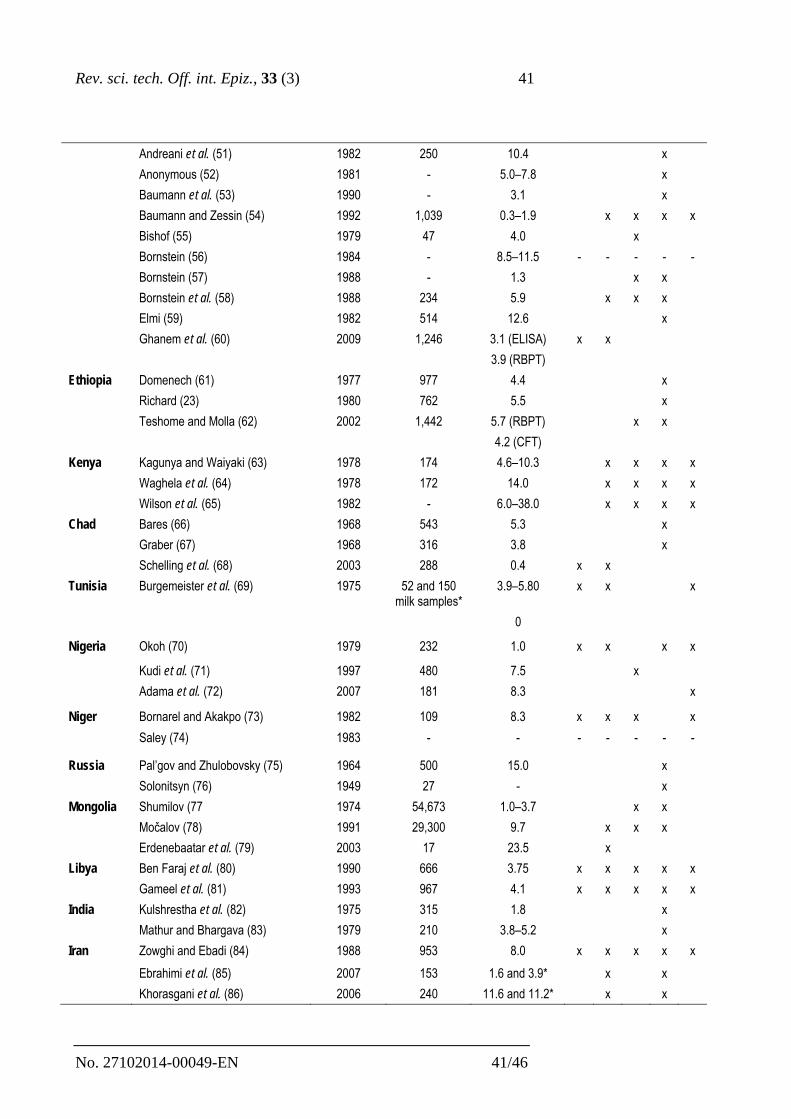
Rev. sci. tech. Off. int. Epiz., 33 (3) 41
No. 27102014-00049-EN 41/46
Andreani et al. (51) 1982 250 10.4 x
Anonymous (52) 1981 - 5.0–7.8 x
Baumann et al. (53) 1990 - 3.1 x
Baumann and Zessin (54) 1992 1,039 0.3–1.9 x x x x
Bishof (55) 1979 47 4.0 x
Bornstein (56) 1984 - 8.5–11.5 - - - - -
Bornstein (57) 1988 - 1.3 x x
Bornstein et al. (58) 1988 234 5.9 x x x
Elmi (59) 1982 514 12.6 x
Ghanem et al. (60) 2009 1,246 3.1 (ELISA) x x
3.9 (RBPT)
Ethiopia Domenech (61) 1977 977 4.4 x
Richard (23) 1980 762 5.5 x
Teshome and Molla (62) 2002 1,442 5.7 (RBPT) x x
4.2 (CFT)
Kenya Kagunya and Waiyaki (63) 1978 174 4.6–10.3 x x x x
Waghela et al. (64) 1978 172 14.0 x x x x
Wilson et al. (65) 1982 - 6.0–38.0 x x x x
Chad Bares (66) 1968 543 5.3 x
Graber (67) 1968 316 3.8 x
Schelling et al. (68) 2003 288 0.4 x x
Tunisia Burgemeister et al. (69) 1975 52 and 150 milk samples*
3.9–5.80 x x x
0
Nigeria Okoh (70) 1979 232 1.0 x x x x
Kudi et al. (71) 1997 480 7.5 x
Adama et al. (72) 2007 181 8.3 x
Niger Bornarel and Akakpo (73) 1982 109 8.3 x x x x
Saley (74) 1983 - - - - - - -
Russia Pal’gov and Zhulobovsky (75) 1964 500 15.0 x
Solonitsyn (76) 1949 27 - x
Mongolia Shumilov (77 1974 54,673 1.0–3.7 x x
Močalov (78) 1991 29,300 9.7 x x x
Erdenebaatar et al. (79) 2003 17 23.5 x
Libya Ben Faraj et al. (80) 1990 666 3.75 x x x x x
Gameel et al. (81) 1993 967 4.1 x x x x x
India Kulshrestha et al. (82) 1975 315 1.8 x
Mathur and Bhargava (83) 1979 210 3.8–5.2 x
Iran Zowghi and Ebadi (84) 1988 953 8.0 x x x x x
Ebrahimi et al. (85) 2007 153 1.6 and 3.9* x x
Khorasgani et al. (86) 2006 240 11.6 and 11.2* x x

Rev. sci. tech. Off. int. Epiz., 33 (3) 42
No. 27102014-00049-EN 42/46
Iraq Al-Ani et al. (6) 1998 215 7.0–17.0 x x x x
Jawad (87) 1984 235 3.8 x x x x
Saudi Arabia
Radwan et al. (88 1983 116 2.8–3.5 x
Radwan et al. (3) 1992 2,630 8.0 x x
Radwan et al. (15) 1995 2,536 8.0 x x x x
Hashim et al. (89) 1987 146 1.4 x x x
3.02 x x x x
1.9
4.0
Alshaikh et al. (90) 2007 859 3.14 x x x
Kuwait Al-Khalaf and El-Khaladi (91) 1989 698 serum samples
14.8 x x x x
209 milk samples
8.0 x
Oman Harby and Ismaily (92) 1995 550 3.6 x
Yagoub et al. (93) 1990 1,502 7.0 x
Ismaily et al. (94) 1988 550 3.6 x
UAE Afzal and Sakkir (95) 1994 392 racing 0.76 x x x
Moustafa et al. (96) 1998 7,899 0.01 x x x x x
Wernery and Wernery (97) 1990 196 breeding 2.0 (Breeding) x x
6.6 (Racing) x x
Wernery et al. (98) 2007 348 racing 1.9 x x x
Gwida et al. (camels from Sudan) (99)
2011 1,119 60 x x x x
895 x x x x
Taha (100) 2007 812 x x x
Pakistan Ajmal et al. (101) 1989 81 2.0 x x
Peru Acosta et al. (alpacas) (21) 1972 1,449 21 x x
Jordan Al-Majali (102) 2006 590 17 x x
Al-Majali et al. (103) 2008 412 13.1 (Herd 35.1)
x x
CFT: complement fixation test
MRT: milk ring test
RBT: Rose Bengal test
SAT: serum agglutination test
UAE: United Arab Emirates
f.: female
m.: male
-: not available
* samples taken from different herds in different locations

Rev. sci. tech. Off. int. Epiz., 33 (3) 43
No. 27102014-00049-EN 43/46
Table III
Brucella species isolated from camelids in different countries
Country Author (Reference no.) Year Species Organs
Jordan Al-Majali (102) 2006 B. melitensis biovar 3 Aborted fetuses vaginal swab
Russia Solonitsyn (76) 1949 B. abortus n/a
Pal’gov (110) 1950 B. abortus Bactrian, fetuses
Iran Zowghi and Ebadi (84) 1988 B. melitensis biovar 1 Lymph nodes
Kuwait Zowghi and Ebadi (84) 1988 B. melitensis biovar 3 Lymph nodes
Libya Al-Khalaf and El-Khaladi
(91)
1989 B. abortus biovar 1 Fetal stomach
Gameel et al. (81) 1993 B. melitensis biovar 1 Milk
Saudia Arabia Gameel et al. (81) 1993 B. melitensis biovar 1 Milk, vaginal swab, aborted
fetus
Radwan et al. (111) 1992 B. melitensis biovar 1 and 2 Milk
Radwan et al. (15) 1995 B. melitensis biovars 1, 2, 3 Milk
Ramadan et al. (112) 1998 B. melitensis Carpal hygroma
Al Dubaib (113) 2007 B. melitensis n/a
Sudan Agab et al. (114)
Musa et al. (115)
1996
2008
B. abortus biovar 3
B. abortus
biovar 6
B. melitensis
biovar 3
Teats, lymph nodes, vaginal
swab, testis
Lymph nodes
Omer et al. (46) 2010 B. abortus biovar 6 Lymph nodes, testis
Peru Acosta et al. (alpacas)
(21)
1972 B. melitensis Organs
UAE Wernery et al. (camels
from Sudan) (116)
2007 B. melitensis biovars 1 and 3 Milk, lymphnodes, placenta

Rev. sci. tech. Off. int. Epiz., 33 (3) 44
No. 27102014-00049-EN 44/46
Moustafa et al. (96) 1998 B. melitensis Milk
Senegal Verger et al. (117) 1979 Brucella abortus biovars 1
and 3
n/a
Egypt El-Seedy et al. (36) 2000 B. abortus biovars 1 and 7
B. melitensis biovar 3
Organs
n/a: information not available
UAE: United Arab Emirates

Rev. sci. tech. Off. int. Epiz., 33 (3) 45
No. 27102014-00049-EN 45/46
Table IV
Sensitivity and specificity of serological tests for brucellosis
Serological test Sensitivity in % Specificity in %
SAT 81.5 98.9
CFT 90–91.8 99.7–99.9
RBT 87 97.8
cELISA 95.2 99.7
iELISA 97.2 97.1–99.8
FPA 96.6 99.1
MRT 88.5 77.4
cELISA: complement enzyme-linked immunosorbent assay
CFT: complement fixation test
FPA: fluorescence polarisation assay
iELISA: indirect enzyme-linked immunosorbent assay
MRT: milk ring test
RBT: Rose Bengal test
SAT: serum agglutination test

Rev. sci. tech. Off. int. Epiz., 33 (3) 46
No. 27102014-00049-EN 46/46
Table V
Serology results for 23 seropositive dromedaries 36 months after
antibiotic treatment for brucellosis
Tests Positive Negative
cELISA 20 3
CFT 16 7
RBT 14 9
SAT 7 16
cELISA: complement-enzyme linked immuno sorbent assay
CFT: complement fixation test
RBT: Rose Bengal test
SAT: serum agglutination test