Embed Size (px)
Citation preview

593
Journal of Vertebrate Paleontology 22(3):593–611, September 2002q 2002 by the Society of Vertebrate Paleontology
CALSOYASUCHUS VALLICEPS, A NEW CROCODYLIFORM FROM THE EARLY JURASSICKAYENTA FORMATION OF ARIZONA
RONALD S. TYKOSKI1,2, TIMOTHY B. ROWE1,2,3, RICHARD A. KETCHAM1,3, and MATTHEW W. COLBERT1,2,3
1Jackson School of Geosciences, [email protected];2Texas Memorial Museum Vertebrate Paleontology Laboratory;
3High Resolution X-ray CT Facility, The University of Texas at Austin, Austin, Texas 78712
ABSTRACT—We describe a new fossil crocodyliform archosaur from the Early Jurassic Kayenta Formation of theNavajo Nation that is surprisingly derived for so ancient a specimen. High-resolution X-ray CT analysis reveals thatits long snout houses an extensive system of pneumatic paranasal cavities. These are among the most distinctive featuresof modern crocodylians, yet the evolutionary history of this unique system has been obscured by the inaccessibility ofinternal structures in most fossil crania. Preliminary phylogenetic analysis indicates that the new species is the oldestknown member of a monophyletic Goniopholididae, and within this lineage to be the sister taxon of Eutretauranosu-chus, from the Late Jurassic Morrison formation of Colorado. Goniopholididae became extinct at the end of theCretaceous, but it is more closely related to living crocodylians than are several lineages known only from Cretaceousand younger fossils. The new taxon nearly doubles the known length of goniopholid history and implies a deep, asyet undiscovered, Mesozoic history for several crocodyliform lineages that were once thought to have relatively com-plete fossil records.
INTRODUCTION
In the summer of 1997, a collaboration involving the TexasMemorial Museum of The University of Texas at Austin, theMuseum of Comparative Zoology of Harvard University, andthe Seba Dalkai Navajo Nation School sent crews to prospectexposures of the Early Jurassic Kayenta Formation. These ex-posures lie on lands of the Navajo Nation in northeastern Ari-zona. The Navajo Nation requires permits to conduct paleon-tological work on its lands, and our work was carried out underpermits kindly granted by the Navajo Nation Minerals Division.The Kayenta Formation is generally considered to be Early Ju-rassic in age (Sinemurian–Pliensbachian; Clark and Fastovsky,1986; Sues et al., 1994). Its beds previously yielded a rich di-versity of fossil tetrapod taxa (Clark, 1986; Clark and Fastov-sky, 1986; Sues et al., 1994), including a caecilian (Jenkins andWalsh, 1993), an anuran (Shubin and Jenkins, 1995; Jenkinsand Shubin, 1998), a turtle (Gaffney et al., 1987), several cro-codylomorphs (Crompton and Smith, 1980; Clark, 1986, 1994),a pterosaur (Padian, 1984), a sauropodomorph (Attridge et al.,1985), theropods (Welles, 1954, 1970, 1984; Rowe, 1989; Ty-koski, 1998), ornithischians (Colbert, 1981), tritylodontids(Kermack, 1982; Sues, 1985, 1986a, 1986b), and morganuco-dontids (Jenkins et al., 1983; Crompton and Luo, 1993).
A number of new specimens were recovered in the courseof our collaboration, including a partial skull of a distinctivenew crocodylomorph that is unlike any previously reportedfrom Early Jurassic sediments. Representing a new species, thistaxon adds to a diverse list of Kayenta crocodylomorphs thatincludes Eopneumatosuchus colberti (Crompton and Smith,1980), three undescribed protosuchian-grade taxa, and an un-described sphenosuchid-grade form (Clark, 1986; Clark andFastovsky, 1986; Sues et al., 1994).
Our study of the new specimen was greatly assisted by scan-ning it at The University of Texas High Resolution X-ray Com-puted Tomography (CT) Facility (Rowe et al., 1997; Ketchamand Carlson, 2001). The CT imagery revealed the internal anat-omy of extensive paranasal pneumatic cavities within the ros-trum, and it helped distinguish between cracks and sutures. Itwas also used to generate several of the illustrations (Figs. 1–
7) for this report. Two of these figures (Figs. 1A, C, 2A, C)include volumetric reconstructions of the skull that were com-puter-generated from the original CT slices. Although they looksuperficially like conventional photographs, volumetric recon-structions show far more structural detail than photos (Figs. 7–9), particularly the presence of numerous minute fractures thatare invisible to both the camera and the naked eye. The photos(Figs. 7–9) convey a more readily interpretable picture of therelatively good quality of preservation of the specimen.
All of our original high-resolution X-ray CT data accompanythis article on a supplemental CD-ROM (Table 1). Also includ-ed on the CD-ROM are serial section animations and animated3D volumetric reconstructions of the skull. Reduced-resolutionversions of the slice and volumetric animations are available onthe World Wide Web (http://www.digiMorph.org/specimens/Calsoyasuchusvalliceps/). Also included on the CD-ROM is asurface model of the specimen in stereolithography (STL) for-mat, which can be interactively viewed with an appropriateplayer. Stereolithography files have now been published for theendocast of the Triassic cynodont Thrinaxodon (Rowe et al.,1995) and the skull of Alligator mississippiensis (Rowe et al.,1999a, b). They are of special interest to the readership of thisjournal because stereolithography files can be rendered using agrowing array of rapid prototyping technologies that transformdigital 3D objects into physical 3D objects (e.g., Juricic andBarr, 1996). These models can be scaled up or down and ren-dered at any desired size (within the physical limits of the par-ticular rapid prototyping device used). Thus, with the appro-priate equipment, a detailed physical replica resembling a con-ventional cast of the specimen can be generated from the fileon the accompanying CD-ROM.
A series of analyses of crocodylomorph phylogeny publishedover the last two decades provided a strong framework in whichto diagnose and evaluate the affinities of the new taxon (Clark,1986, 1994; Benton and Clark, 1988; Clark and Norell, 1992;Brochu, 1997a, b, 1999; Wu et al., 1997; Buckley and Brochu,1999; Buckley et al., 2000). We relied heavily on these studiesto guide our estimation of the phylogenetic position of the newtaxon. Until now, wide stratigraphic and morphological gapsseparated the Late Jurassic and younger mesoeucrocodyliforms

594 JOURNAL OF VERTEBRATE PALEONTOLOGY, VOL. 22, NO. 3, 2002
TABLE 1. Contents of supplemental CD-ROM imagery archive of the Calsoyasuchus holotype.
● CTpData folder—CT slice files, 436 slice images in coronal slice plane, 8 and 16-bit TIF format● STL folder—contains one 3D surface model of Calsoyasuchus holotype in stereolithography (STL) format● Movies folder—contains two folders with animations requiring Quicktime software 3.0 or higher for viewing:
C 3D folder—contains six animated volumetric renderings of the holotype:n Yaw: two movies in which skull rotates about vertical axis (l 5 large format, s 5 small format)n Roll: two movies in which skull rotates about long horizontal axis (l 5 large format, s 5 small format)n Pitch: two movies in which skull rotates about short horizontal axis (l 5 large format, s 5 small format)
C Slices folder—contains five slice-by-slice animations through complete CT data stacks; slice thickness is one millimeter; interslice spacingis 0.9 mm (i.e., 10 percent overlap of slices):n Coronal (COR)—one movie through 436 slices taken in coronal plane, viewed from anterior to posteriorn Horizontal (HOR): two movies of 107 horizontal slices, viewed from dorsal to ventral, in large and small formatsn Sagittal (SAG): two movies of 138 vertical slices, viewed from left to right, in large and small formats
● Readme Describes in detail the contents and operation of files on the CD-ROM in Microsoft Word and Text formats.● UTCT inspeCTor—java applet allowing interactive viewing of the scan data on the orthogonal axes.
from Triassic and Early Jurassic protosuchian-grade and sphen-osuchian-grade crocodylomorphs (Langston, 1973). The newspecies is the oldest member of Goniopholididae, a clade withweak but unequivocal support in our analysis. The new speciespossesses a combination of primitive and derived features thathelps to fill wide morphological and temporal gaps in earlymesoeucrocodylian history. Its age and phylogenetic positionsuggest that the mesoeucrocodylian lineage, which was thoughtto possess one of the best records among fossil terrestrial ver-tebrates (Markwick, 1998), is far less complete than generallybelieved.
MATERIALS AND METHODS
Type Specimen
The holotype consists of a partial skull (Figs. 1A–D, 2A–D)found lying palate-side up, wedged between two pieces of pet-rified wood at the base of a trough scour within a thick, cross-bedded channel sandstone bed. The skull was preserved inlight-green, medium to coarse-grained sandstone, with hemati-tic crust over much of its surface. The posterior end of the skullwas reduced by erosion to several dozen weathered pieces ofsurface float, which included portions of the braincase, suspen-sorium, and palate. These fragments are too weathered to re-assemble with confidence, and are not described here. No post-cranial elements were preserved.
Anatomical Abbreviations Anatomical nomenclature inour description is based on Witmer (1995, 1997), Brochu(1999), and Rowe et al. (1999a, b): acc, accessory cavity; am,anterior palatal process of maxilla; aof, antorbital fenestra; ccr,caviconchal recess; dv, dorsal valley on nasals and frontal;dpm, dorsal process of premaxilla; en, external naris; f, frontal;iaf, internal antorbital fenestra; ins, internarial septum; j, jugal;l, lacrimal; laf, lacrimal antorbital fossa; ls, laterosphenoid; m,maxilla; ma (1–29), maxillary alveolus; maf, maxillary antor-bital fossa; md (1–29), maxillary tooth; mdep, maxillary de-presssion; mmr, median maxillary ridge; n, nasal; ncp, nasalcavity proper; nlc, nasolacrimal canal; npd, nasopharyngealduct; nv, nasal vestibule; occ, occlusal pit/pore; olf, impressionof olfactory tracts on frontal; orb, orbit; pal, palatine; p, pari-etal; pd (1–5), premaxillary tooth; pm, premaxilla; pma, pre-maxillary alveolus; po, postorbital; ppm, palatal process of pre-maxilla; pch, primary choana; prf, prefrontal; pt, pterygoid;pvr, postvestibular recess; q, quadrate; sac, secondary acces-sory cavity; sq, squamosal; stf, supratemporal fenestra; V, pas-sage for maxillary branch of trigeminal nerve; vo, vomer. Leftand right sides are differentiated by the prefixes ‘‘l’’ and ‘‘r’’.
Citations in the text with the prefixes COR, HOR, and SAGrefer respectively to relevant slice numbers in coronal, horizon-tal, and sagittal animations on the CD-ROM supplement.
Institutional Abbreviations CMNH, Cleveland Museum
of Natural History, Cleveland, Ohio; TMM, Texas MemorialMuseum, The University of Texas at Austin; YPM, Yale Pea-body Museum, Yale University, New Haven.
CT Scanning
The specimen was scanned at the High-Resolution X-rayComputed Tomography (CT) Facility at the University of Texasat Austin, which is described by Ketcham and Carlson (2001).X-ray energies were set to 420 kV and 4.7 mA, with a focal spotsize of 1.8 mm. X-rays were pre-filtered to reduce beam-hard-ening artifacts using two brass plates with a total thickness of3.175 mm. X-ray intensities were measured using an RLS detec-tor with 2,048 channels spaced at 0.05 mm intervals. Forty chan-nels were unsuitable for imaging, and the remaining channelswere averaged into groups of 4 to reduce image noise, resultingin 502 effective channels with a spacing of 0.2 mm. Each slicewas acquired using 1,800 views (angular orientations), each viewhaving an acquisition time of 64 ms, and detector gain was setto 8 to maximize count rate. The resulting acquisition time wasapproximately 2 minutes per slice. The sample was scanned ina 190% offset mode (Ketcham and Carlson, 2001) with a slicethickness of 1.0 mm and an inter-slice spacing of 0.9 mm. Theimage field of reconstruction was 130 mm, and reconstructionparameters were calibrated to maximize usage of the 12-bit rangeof grayscales available in the output images.
At the time of the scan the detector showed behavior inwhich it would first drift rapidly out of calibration once dataacquisition commenced, then drift more slowly. As a result itwas decided to obtain the entire data set in one pass withoutrecalibration during the scan. In order to minimize drift, thedetector as exposed to X-rays for two hours before the scanbegan, and a very long signal calibration (3,600 views, or 3.84minutes) was obtained. This procedure eliminated major prob-lems, but nevertheless a drift in grayscale values of up to 7–8% occurred over the course of the scan. In general, the driftresulted in darkening in the center and brightening of the mar-gins of the images as scanning progressed. The majority of thiseffect was removed from the data set by comparing initial andfinal images and analyzing intervening images to discern thepattern and course of the drift. Routines to perform this analysisand apply the subsequent correction were written in IDL ver-sion 5.3.1. For easier handling, the 12-bit data stored in 16-bitTIFF-format data files were exported to 8-bit format by dividingall grayscale values by 16. The data are archived as individualslices.
The coronal slice-by-slice animation on the CD presents theoriginal CT data. Owing to the length of the skull, the horizon-tal and sagittal animations are presented in reduced as well asfull-sized versions. The latter may be too large to display com-pletely on some current computer monitors. The coronal movie
595TYKOSKI ET AL.—NEW EARLY JURASSIC CROCODYLIFORM
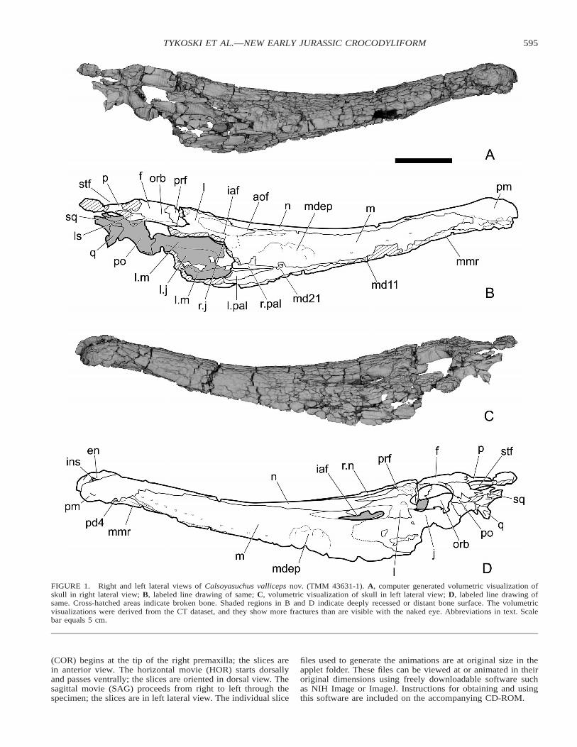
FIGURE 1. Right and left lateral views of Calsoyasuchus valliceps nov. (TMM 43631-1). A, computer generated volumetric visualization ofskull in right lateral view; B, labeled line drawing of same; C, volumetric visualization of skull in left lateral view; D, labeled line drawing ofsame. Cross-hatched areas indicate broken bone. Shaded regions in B and D indicate deeply recessed or distant bone surface. The volumetricvisualizations were derived from the CT dataset, and they show more fractures than are visible with the naked eye. Abbreviations in text. Scalebar equals 5 cm.
(COR) begins at the tip of the right premaxilla; the slices arein anterior view. The horizontal movie (HOR) starts dorsallyand passes ventrally; the slices are oriented in dorsal view. Thesagittal movie (SAG) proceeds from right to left through thespecimen; the slices are in left lateral view. The individual slice
files used to generate the animations are at original size in theapplet folder. These files can be viewed at or animated in theiroriginal dimensions using freely downloadable software suchas NIH Image or ImageJ. Instructions for obtaining and usingthis software are included on the accompanying CD-ROM.

596 JOURNAL OF VERTEBRATE PALEONTOLOGY, VOL. 22, NO. 3, 2002
FIGURE 2. Dorsal and ventral views of Calsoyasuchus valliceps nov. (TMM 43631-1). A, computer generated volumetric visualization of skull indorsal view; B, labeled line drawing of same; C, volumetric visualization of skull in ventral view; D, labeled line drawing of same. Cross-hatchedareas indicate broken bone. Shaded regions in B and D indicate deeply recessed or distant bone surface. The volumetric visualizations were derivedfrom the CT dataset, and they show more fractures than are visible with the naked eye. Abbreviations in text. Scale bar equals 5 cm.

597TYKOSKI ET AL.—NEW EARLY JURASSIC CROCODYLIFORM
FIGURE 3. Calsoyasuchus valliceps nov. (TMM 43631-1). Diagram showing the relative positions and orientations of the slice planes shownin Figures 4–6.
Phylogenetic Analysis
The pioneering work of Clark (1986, 1994) provided the firstthorough cladistic analysis of ingroup relationships among cro-codyliform archosaurs. Clark’s work has served as the basis forseveral other cladistic analyses of crocodyliform relationships(Wu et al., 1997; Buckley and Brochu, 1999; Buckley et al.,2000), as well as for the one presented here. The aim of thepresent study is simply to estimate the position of the new taxonwithin the phylogenetic framework already established by theseearlier studies. Consequently, our character list (Appendix 1)and matrix (Appendix 2) were modified minimally from thoseof Buckley et al. (2000). Some changes were made to reflectour reinterpretation of the anatomy of Eutretauranosuchus andGoniopholis. The new Kayenta taxon and the Asian taxon Su-nosuchus (scored from the literature) were added to the char-acter matrix, resulting in 27 taxa in the analysis (three outgrouptaxa: 24 ingroup taxa). Two new characters were also added tothe character list, bringing the total number to 119 (Appendix1). Only about 43 percent of cranial characters (35 percent oftotal characters) could be scored for the new taxon owing to itsincompleteness. We note that nearly 20% of these charactersproved uninformative and consequently were ignored in ouranalyses. In addition, while scoring the character matrix for ournew taxon, doubts arose over the independence among severalcharacters (e.g., characters 11 and 12; characters 13 and 14).These problems may affect the precision of diagnoses for in-group clades, but they are probably not serious problems inarriving at a tree topology. A more thorough analysis and treat-ment of these issues lies beyond the scope of the current work.
The comparative framework for our study of the new speciesfollows Buckley et al. (2000) by including a sample of ingrouptaxa consisting of Metriorhynchidae, Teleosauridae, Pelagosau-rus, Hsisosuchus, Comahuesuchus, Baurusuchus, Sebecus, Li-bycosuchus, Notosuchus, Malawisuchus, Uruguaysuchus, Ar-aripesuchus, Trematochampsa, Peirosauridae, Mahajangasu-
chus, Alligatorium, Theriosuchus, Goniopholis, Sunosuchus,Eutretauranosuchus, Bernissartia, and Crocodylia. Compari-sons were also drawn with an unnamed Early Cretaceous taxonwidely discussed in the literature and known informally as the‘‘Fruita taxon,’’ a designation that we follow here. The newtaxon was part of the ingroup as well.
We performed three analyses, all of which yielded identicaltree topologies (see Discussion, below). In the first, all char-acters were treated as unordered, and uninformative characterswere ignored. This yielded three trees of equal length (262steps, CI 5 0.462, RI 5 0.652). Following earlier authors, andat the urging of one reviewer, we also ordered several charactersin two subsequent runs. In the first of these analyses, characters15 and 49 were ordered, with no effect on tree topology. Threetrees of equal length were found (L 5 262, CI 5 0.462, RI 50.657). In the last analysis, characters 15, 37, 49, 67, and 77were ordered and all other characters were unordered. This toohad no effect on tree topology, but it lengthened the tree bytwo steps (L 5 264, CI 5 0.458, RI 5 0.655), and it alteredthe diagnoses of the various taxa found by the analysis. Thetaxon diagnoses presented below are based on analysis numberone, in which all characters were treated as unordered. Ouranalysis was run on an Apple Macintosh G4 computer withPAUP 3.0s (Swofford, 1991), using a random, stepwise addi-tion, heuristic search algorithm.
SYSTEMATIC PALEONTOLOGY
CROCODYLOMORPHA Walker, 1970CROCODYLIFORMES Hay, 1930
MESOEUCROCODYLIA Whetstone and Whybrow, 1983GONIOPHOLIDIDAE Cope, 1875
CALSOYASUCHUS VALLICEPS, sp. nov.(Figs. 1–9)
Etymology Calsoyasuchus, ‘‘Calsoyas’’ to honor Dr. KyrilCalsoyas, former principal of Seba Dalkai Navajo Tribal

598 JOURNAL OF VERTEBRATE PALEONTOLOGY, VOL. 22, NO. 3, 2002
FIGURE 4. Calsoyasuchus valliceps nov. (TMM 43631-1). Selected CT slice images (slice numbers 24, 90, 156, 201, 279, and 313) throughrostrum in coronal plane. See Figure 3 for position of slices through skull. Abbreviations in text.
School, our friend and gracious host in the Navajo Nation anda great champion of education, and souchos (Gr., derived fromthe Egyptian word for crocodile); valliceps, from combinationof valles (L. valley), and cephale, (Gr. head), in reference tothe deep median valley present on the dorsal surface of nasalsand frontal bones.
Holotype TMM 43631-1. Incomplete skull of a medium-sized crocodyliform, missing the occiput, braincase, most of thesuspensorium, posterior portions of the palate, and jaws.
Occurrence Locality TMM 43631 (‘‘Calsoyasuchus hill’’),field number TR 97/09, located in the northern part of the GoldSpring drainage basin, Adeii Eechii Cliffs, Navajo Nation, Co-conino County, Arizona, in the middle third of the silty facies(Harshbarger et al., 1957; Clark and Fastovsky, 1986) of theLower Jurassic (Early Jurassic: Sinemurian to Pliensbachian)Kayenta Formation.
Diagnosis based on our phylogenetic analyses, a gonio-pholid with following apomorphies: lacrimal–nasal contact oc-curs only along anterior edge of lacrimal; frontal does not reachsupratemporal fenestra; and teeth that are finely serrated. It isalso equivocally diagnosed by a long, narrow internal antorbitalfenestra whose length is slightly less than the orbit diameter.Other apomorphic features not scored in our analyses include:snout bowed downward and back upward so the tip of the ros-trum is as high or more dorsally placed than skull table (whenskull table is held horizontal); an internarial process rises fromnarial floor to partially divide external naris; the medial edgesof the maxillary palatal processes curve ventrally, forming me-dian ridge that descends below anterior alveolar border; medialmaxillary accessory cavities very elongate; posterior end ofmaxilla divergent laterally, medially exposing the jugal anter-oventral to the orbit; a deep pocket in medial surface of lacrimalanterior to the orbit; a deep and narrow median valley on pos-terior part of nasals and anterior third of frontal; lateral part ofpterygoid anterior process with inverted ‘‘U’’ cross-section pos-terior to primary choana.
DESCRIPTION
General Appearance
As preserved, the skull is approximately 380 mm long fromthe anterior tip of the rostrum to the most posterior preservededge of the parietal. The braincase, occiput, the right and allbut the anterodorsal end of the left quadrate, both quadratoju-gals, nearly all of the right squamosal, the posterior part of theleft squamosal, posterior parts of the palatines, all but the an-terior processes of the pterygoids, and most of the right and theposterior half of the left jugal were all eroded away prior todiscovery (Figs. 1, 2). Identification of some sutures is difficultowing to numerous cracks and deeply sculptured ornamenta-tion. Many of the cranial sutures are tightly closed. The originalexternal shape of the skull is largely preserved and shows onlyminor distortion. CT imagery reveals that the geometry of thenasal cavity proper is largely preserved as well, and that thebones surrounding the nasal cavity are extensively pneumati-cized (Figs. 3–6).
The skull is long and low in lateral view (Fig. 1A, D). Whenthe skull table is oriented horizontally, the rostrum curvesdownwards from the orbits and then rises dorsally towards therostral tip until it is slightly higher than the skull table. Thesnout is wider than tall throughout its length, although it is onlyslightly wider than tall near the premaxilla-maxilla contact. Incoronal sections (Fig. 4) it has a rounded dorsal surface fromthe external naris to a plane just before the orbits, where a deepcleft marks the dorsal midline (Fig. 4, slice 313). The orbits arealmost circular and are dorsolaterally oriented. An elongate ex-ternal antorbital fenestra (sensu Witmer, 1995, 1997) excavatesthe posterior quarter of the snout. A narrow, elliptical internalantorbital fenestra (sensu Witmer, 1995, 1997) perforates theskull in the center of the external antorbital fenestra. Its longaxis is almost equal in length to the orbit diameter. Both theexternal and internal antorbital fenestrae face more dorsallythan laterally. Anteroventral to the antorbital fenestra is a de-

599TYKOSKI ET AL.—NEW EARLY JURASSIC CROCODYLIFORM
FIGURE 5. Calsoyasuchus valliceps nov. (TMM 43631-1). Selected CT slice images (slice numbers 48, 57, and 69) through rostrum in sagittalplane. See Figure 3 for position of slices through skull. Abbreviations in text.
pression on the maxilla that lacks the rough sculpturing seenon the rest of the skull. The cranial roofing bones form a well-developed, flat skull table dorsal and posterior to the orbits.
In dorsal and ventral views (Fig. 2A, D) the snout is con-stricted at the premaxilla-maxilla junction. Behind this constric-tion the lateral margins of the snout are nearly straight as theydiverge posteriorly toward the orbits. Adjacent to the fifththrough seventh alveoli, the maxilla curves slightly outward toaccommodate enlarged tooth roots. Between the 20th and 22ndmaxillary alveoli, the skull broadens more noticeably, but notas sharply or to the degree seen in protosuchids and many long-snouted taxa (e.g., Gavialis; Iordansky, 1973; Langston, 1973;Crompton and Smith, 1980). The supratemporal fenestra mea-sures only about half the diameter of the orbit. In this featureC. valliceps differs greatly from the other described Kayentacrocodylomorph, Eopnuematosuchus colberti (Crompton andSmith, 1980).
Ventrally, (Fig. 2C, D) the secondary palate forms an elon-gated floor beneath the nasal cavity. Posteriorly, the secondarypalate ends at the primary choanae, but laterally the maxillaeand palatines form broad shelves along either side of the ven-trally open nasopharyngeal duct. Each primary choana isbounded anteriorly by the maxilla, laterally by the palatine, and
medially by the anterior ramus of the pterygoid. The primarychoanae open ventromedially into the nasopharyngeal duct. TheCT imagery indicates that the most of the individual pneumaticcavities within the snout interconnect and become confluentwith the nasal cavity proper in the vicinity of the primary cho-anae (COR 244-262).
Bones of the Skull
Premaxilla The right premaxilla is broken through the fifthalveolus and displaced slightly dorsally and anteriorly. The leftis unbroken and lies naturally articulated with the maxilla (Fig.7A, B). The premaxillae are enlarged to form a swollen rostraltip that is wider than high, which accentuates a constriction inthe snout at the premaxilla–maxilla juncture. At least four emp-ty alveoli are visible in the right premaxilla, and five are presentin the left (Figs. 2D, 7B). The first, second, and fifth alveoliare small. The third alveolus is the largest in the premaxillawhereas the fourth alveolus is intermediate in size between thesecond and third alveoli. CT imagery indicates that the alveoliinvade the premaxilla deeply and reach almost to its dorsalsurface (SAG 055-086; HOR 040-068). The poorly preservedbase of the fourth tooth is present in the left premaxilla. The

600 JOURNAL OF VERTEBRATE PALEONTOLOGY, VOL. 22, NO. 3, 2002
FIGURE 6. Calsoyasuchus valliceps nov. (TMM 43631-1). Selected CT slice images (slice numbers 62, 67, 74, and 82) through rostrum inhorizontal plane. See Figure 3 for position of slices through skull. Abbreviations in text.

601TYKOSKI ET AL.—NEW EARLY JURASSIC CROCODYLIFORM
FIGURE 7. Calsoyasuchus valliceps nov. (TMM 43631-1). Photographs of anterior end of rostrum in A, dorsal; and B, ventral views. Rightpremaxilla is broken and displaced slightly dorsally and anteriorly. Median maxillary ridge drops from palatal surface between the rostralconstriction to a plane roughly even with the sixth maxillary alveolus. Abbreviations in text. Scale bar equals 5 cm.
broken tip of a replacement tooth protrudes through the oldertooth base near the medial edge of this alveolus.
The external naris forms a sub-triangular aperture that opensdorsally and slightly anteriorly (Fig. 7A). The naris is partiallyseparated by a spike-like internarial process that angles dorsallyfrom the rear part of the floor of the nasal vestibule. The boneis broken, but CT imagery (Fig. 4, slice 24: COR 017-COR026) shows this process may be a dorsal extension of the pre-maxillary palatal processes. This weak division of the naris inCalsoyasuchus represents an apomorphic condition rather thanretention of the primitive internarial septum that separates theright and left nares in basal archosaurs. It differs greatly fromthe internarial septum present in some members of extant Cro-codylia (Osteolaemus tetraspis, Alligator mississppiensis, A. si-nensis; Iordansky, 1973; Rowe et al., 1999a, b), in which thebar is formed from paired anterior processes of the nasals andshort posterior processes of the premaxillae.
Laterally, the premaxilla meets the maxilla with a strong su-ture within the rostral constriction. Dorsally, the premaxillaemeet behind the naris, excluding the nasals from the narial ap-erture. Flat dorsal processes of the right and left premaxillaeextend posteriorly between the maxillae, to meet the nasals ata plane even with the eighth maxillary alveoli.
Each premaxilla bears a large paramedian occlusal pit justbehind the anterior alveoli (Fig. 4, slice 24; Fig. 7B). This ex-cavation probably received the tip of an enlarged symphysealdentary tooth. A second, smaller occlusal pit lies posterolateralto the first, between and medial to the third and fourth premax-illary alveoli. A third smaller occlusal pit lies posteromedial tothe fifth alveolus of the left premaxilla. There is no evidenceof an incisive foramen, but breakage and displacement of theright premaxilla renders interpretation of this area ambiguous.The premaxillary palatal processes separate just posterior to the
large anterior occlusal pits, and are divided by anterior pro-cesses of the maxillae (Fig. 2D). The premaxilla-maxilla sutureextends posterolaterally from the palatal midline, then anglesanteriorly through the third occlusal pit to almost reach the fifthpremaxillary alveolus. From there the suture travels dorsally,through the rostral constriction and onto the roof of the snout.Coinciding with the constriction at the premaxilla-maxilla con-tact is an interruption in the tooth row. There is no laterallyopen gap in the snout at this junction and the surface is faintlysculptured in the constriction, contrasting with the condition inbasal crocodylomorphs, protosuchids, and the Fruita taxon(Crompton and Smith, 1980; Clark, 1986, 1994; Walker, 1990;Wu et al., 1997).
Maxilla The maxilla is long and low, and it forms most ofthe elongated rostrum. Its posterior extent is indeterminable ow-ing to breakage and erosion. As described above, the maxillais rigidly attached to the premaxilla, and dorsally it meets theposterodorsal process of the premaxilla and the nasal along sim-ple contacts. A small dorsal process forms the anterior and partof the dorsal border of the external antorbital fenestra. The dor-sal process meets the nasal just above the rim of the externalantorbital fenestra and it meets the lacrimal within the fenestra.Ventral to the fenestra the maxilla is overlapped by the jugal(Fig. 4, slice 279). As in other archosaurs with a fenestratedsnout, the bone texture within the antorbital fossa is smooth,contrasting with the sculpturing over most of the rest of theexternal skull surface.
The maxilla extends posteriorly beyond the antorbital fenes-tra, and there bifurcates into a dorsal ramus that borders theorbit medial to the jugal and a ventral ramus that contains thealveoli. The orbital ramus can be seen in the anteroventral rimof the orbit only in ventral and medial views, being hidden bythe overlapping lacrimal and jugal in lateral view. The alveolar

602 JOURNAL OF VERTEBRATE PALEONTOLOGY, VOL. 22, NO. 3, 2002
ramus, which is incomplete on both sides, extends posteriorlyat least as far as a plane almost even with the anterior marginof the orbit. The jugal does not split the two maxillary rami,but rather overlaps them and walls in an internal medial fossa(Fig. 1A, B).
The lateral surface of the maxilla is marked by an irregulardepression anteroventral to the antorbital fenestra and just dor-sal to the alveolar border (Fig. 1A, D). The texture within thedepression is smoother than the surrounding maxillary surface,and within the depression are several smaller fossae. The de-pression’s posterior edge is even with the anterior rim of theinternal antorbital fenestra.
The maxillae form most of the osseous secondary palate (Fig.2C, D). From the rostral constriction, each maxilla sends a me-dial palatal process forward and together they broadly separatethe right and left premaxillary palatal processes. The maxillarypalatal processes curve ventrally to meet along the midline inthe vicinity of the rostrum constriction, forming a median ridgeon the palatal surface that descends below the alveolar border(Figs. 1B, D, 7B). The palatal processes are flat for a shortdistance posterior to the sixth alveoli, before arching postero-dorsally towards the primary choanae and the nasopharyngealduct.
Each maxillary palatal process divides to form the anteriorborder of the primary choana. One ramus diverges medially,where it rises toward the midline to meet the vomer and anteriorprocess of the pterygoid in the roof of the nasopharyngeal duct.This contributes to the separation of the right and left primarychoanae (Fig. 8A, B). The other ramus diverges laterally and,flanking the palatine, forms the lateral portion of the palatalshelf. The palatine thus excludes the maxillary lateral ramusfrom the nasopharyngeal duct.
There are at least 29 alveoli present in the more completeleft maxilla, but the full dental count is unknown. The firstalveolus is the smallest in the maxilla, and alveolar diameterincreases gradually until the eighth alveolus. The fifth througheighth alveoli are the largest in the maxilla, which swells slight-ly outward around them. CT images show that these alveoli arealso the deepest in the maxilla and that they arc posteriorlywithin the bone (Fig. 5, slice 48: SAG 037-055; HOR 055-075),also an indication that they held the longest teeth in maxilla.From the ninth tooth position posteriorly, the alveoli decreaseprogressively in diameter. The more posterior teeth have shal-lower implantations that lie beneath the caviconchal recess (seebelow).
The only maxillary teeth preserved and visible in externalview are a broken right 11th maxillary tooth, the base of theright 21st maxillary tooth, and the barely visible base of theleft ninth maxillary tooth. The partial crown of the 11th max-illary tooth is split just labial to its meso-distal axis. The crownis short and displays little labio-lingual compression. Both me-sial and distal edges bear fine serrations.
Nasal The nasal interdigitates with the dorsal process ofthe premaxilla near the plane through the 8th maxillary alveoli.It broadens toward the orbit and is divided posteriorly by theprefrontal. The medial process twists on its long axis, slopingtoward the midline and forming the deep dorsal valley on theskull roof (Figs. 2A, B, 9). The degree of slope increases pos-teriorly, deepening the median depression to a point just anter-odorsal to the orbits. Here the surfaces of the nasals and thefrontal are nearly vertical, and the opposing sides pinch to con-tact at one point on the frontal (COR 317). The nasofrontalsuture lies within the valley, just anterior to its deepest point.The valley shallows posteriorly and disappears between the or-bits. Other crocodyliforms such as Theriosuchus (Clark, 1986),Hsisosuchus (Li et al., 1994) and some thalattosuchians have amedian depression or groove on the nasals and frontal, but notas deep as in Calsoyasuchus. The lateral process of the nasal
meets the anterior and dorsomedial edges of the lacrimal abovethe external antorbital fenestra. The nasal laterally contacts themaxilla along most of its length. The nasal bones meet eachother medially along a simple edge to edge contact.
Prefrontal The prefrontal is a wedge-shaped bone thatforms the anterodorsal quarter of the orbital rim, where it meetsthe frontal and lacrimal. Its pointed anterior tip extends forwardto a point almost even with the anterior margin of the internalantorbital fenestra, splitting the posterior end of the nasal. Al-though no palpebral bones were found with the specimen, asingle faint facet marks the articulation of at least one palpebralalong the upper margin of the orbit. Impressions of short butbroad prefrontal pillars (5descending process of prefrontal)were preserved in matrix ventral to the interorbital region ofthe skull roof. It is impossible to tell whether the prefrontalpillars contacted the palatines.
Lacrimal The lacrimal is a dorsoventrally short bone witha deeply excavated lacrimal antorbital fossa on its anterior andlateral surfaces (Figs. 1, 9). The lacrimal meets the maxillawithin the external antorbital fenestra and anteriorly it meetsthe nasal. It also meets the prefrontal dorsally. The lacrimalforms most of the dorsal and posterior border of the internalantorbital fenestra. Its medial surface bears a deep recess be-tween the internal antorbital fenestra and orbit that may haveheld the lacrimal gland or pneumatic tissue in life. A smallforamen connects the deep medial fossa and the lateral surfaceof the lacrimal. The nasolacrimal canal originates dorsal to thismedian fossa and passes anteriorly through the lacrimal, exitingthrough the medial surface of the bone above the internal an-torbital fenestra (COR 331-293). Ventrally the lacrimal meetsthe jugal and maxilla, overlapping the latter.
Jugal The anterior and posterior borders of the jugal arenot well defined, owing to poor preservation. The jugal extendsanteriorly beneath the external antorbital fenestra and sends asmall triangular flange into the antorbital fenestra. The jugal isbordered dorsally by the lacrimal, and ventrally and anteriorlyby the maxilla. The jugal overlaps divergent posterior rami ofthe maxilla laterally, spanning the space between these pro-cesses and forming the lateral wall of a large medial fossa (Figs.1A, B, 4, slice 313). Medially, the jugal meets the dorsal max-illary ramus in the anteroventral corner of the orbit, contributingto the suborbital bar. A remnant of the broken dorsal (5post-orbital) process exhibits very faint pits and grooves, and it as-cends the anteriolateral edge of the postorbital bar.
Frontal On its dorsal surface the frontal appears to be asingle element, an apomorphic condition shared with the Fruitataxon and all other adult mesoeucrocodylians (Clark, 1986,1994; Benton and Clark, 1988). CT imagery indicates that thefrontal arose in ontogeny as a pair of bones that were not com-pletely fused at time of death; a persistent, faint median suturebetween the two is discernible ventrally (COR 317-374). Theanterior (5nasal) process of the frontal meets the nasal withinthe skull’s median dorsal valley (Figs. 2A, B, 9). The dorsalsurface of the frontal is flat posterior to the valley. The frontalbroadens to meet the frontal processes of the postorbital pos-terior to the orbits. The frontal does not contribute to the borderof the supratemporal fenestra. Dorsally the frontal meets theparietal between the anterior rims the supratemporal fenestrae.The frontal continues posteriorly deep to the parietal, extendingbetween the supratemporal fenestrae. The pineal fossa forms arounded pit in the ventral surface of the frontal, just anterior tothe fronto–parietal junction. It does not penetrate to the surface.From the dorsal rim of the orbit the frontal curves ventrome-dially. The bone is deeply grooved ventrally between the orbits,marking the path of the olfactory tract.
Parietal There is a single median parietal element and allbut its posterior edge is preserved. The parietal forms the me-dial border of the small supratemporal fenestrae. A thin antero-

603TYKOSKI ET AL.—NEW EARLY JURASSIC CROCODYLIFORM
FIGURE 8. Calsoyasuchus valliceps nov. (TMM 43631-1). A, stereophotograph pair focusing on palatal shelves, primary choanae, and naso-pharyngeal duct. B, labeled drawing of posterior palate, with detail of bones roofing the nasopharyngeal duct. Cross hatched areas indicate brokenbone. Non-stippled areas represent missing surface bone or cracks. Abbreviations in text. A, scale bar equals 5 cm. B, scale bar equals 2 cm.
lateral process contacts the postorbital, thereby excluding thefrontal from the supratemporal fenestra. Between the fenestraethe parietal is flat and deeply sculptured, and it broadens tomeet the squamosal. It is unknown how much the parietal con-tributes to the occiptial surface of the skull. The endocranialsurface of the parietal is embossed with a shallow impressionof the dorsal surface of the brain.
Postorbital Most of the left postorbital is present, althoughits anterodorsal corner is poorly preserved. The ventral (5jugal)process descends medial to the dorsal process of the jugal. Thelateral surface of the process is marked by a small round de-pression but otherwise it is not sculptured. However, the brokendorsal process of the jugal exhibits faint pits and grooves. The
postorbital bar is very thick in section, and is inset slightlybeneath the skull table (COR 376-393). As the bar descends, itcurves laterally to meet the posterior ramus of the jugal. Thepostorbital meets the squamosal posteriorly on the skull table.This contact continues around the lateral margin of the skulltable.
Quadrate Only the anterodorsal end of the left quadrate ispresent in the specimen, where it meets the postorbital andsquamosal. The quadrate contact with the postorbital is a fea-ture shared with other crocodyliforms. There is no trace of thequadratojugal.
Squamosal Pieces of both squamosals are present. The leftsquamosal meets the postorbital anteriorly and the quadrate

604 JOURNAL OF VERTEBRATE PALEONTOLOGY, VOL. 22, NO. 3, 2002
FIGURE 9. Calsoyasuchus valliceps nov. (TMM 43631-1). Stereophotograph pair of skull dorsal surface from the antorbital fenestrae to middleof supratemporal fenestrae. Abbreviations in text. Scale bar equals 5 cm.
ventrally. The remaining part of the right squamosal meets theparietal posterior to the supratemporal fenestra. The flat dorsalsurface of the squamosal contributes to the skull table.
Palatine The palatine is divided into two distinct laminaeor processes. A medial process of elaborate architecture risesupwards into the nasophayrngeal duct. It forms most of thelateral border of the primary choana. Posterior to the choana, acurved dorsal palatine lamina forms the wall and lateral part ofthe roof of the nasopharyngeal duct. The dorsal lamina curvesmedially to meet the anterior process of the pterygoid on theroof of the nasopharyngeal duct, along an abutting contact thatforms a parasagittal ridge which projects down into the duct(Fig. 4, slice 279: COR 265-284). The palatal process of thepalatine is narrow anteriorly and widens posteriorly. It formsthe medial surface of the palatal shelf, excluding the maxillafrom the nasopharyngeal duct (Figs. 2C, D, 8A, B).
Pterygoid Only the anterior palatal process of each ptery-goid is preserved. The anterior process is a very thin bar thatcontacts the vomer dorsally and the maxilla anteriorly in theroof of nasopharyngeal duct. Along the midline, the anteriorprocesses separate the right and left primary choanae (Fig. 8A,B). Toward the front of the choanae, these processes clasp theposterior median processes of the maxillae. Further posteriorly,each process widens into a thin plate that forms the posteriorborder of the primary choana and medial part of the roof of thenasopharyngeal duct. This thin process is strongly convex dor-sally, forming a deep parasagittal arch (Figs. 4, slice 279; 8A,B: COR 261-290).
Vomer Only a thin splint of the vomer is visible in ventralview, between the anterior processes of the pterygoids; mostinformation about this bone was provided by the CT imagery.The vomers appear to be unfused. Each forms a longitudinalbar that rises from the floor of the nasal cavity (HOR 063-075).The vomers may originate as far forward as the anterior mar-gins of the primary choanae (COR 234; HOR 68). In this areathey together form a median U-shaped trough along the floorof the nasal cavity. The vomers become thinner and straighterposteriorly, forming a Y-shaped bony brace dorsal to and be-
tween the long anterior processes of the pterygoids in the roofof the nasopharyngeal duct (Fig. 4, slice 279: COR 259-296).
Nasal Cavity and Paranasal Maxillary Cavities CT im-agery reveals that the maxilla forms most of the bony tubularwalls of the nasal cavity (COR 033-245). The maxilla is alsoexcavated by an extensive array of pneumatic paranasal cavi-ties, similar to the condition in extant Crocodylia (Wegner,1958; Witmer, 1995; Rowe et al., 1999a, b). The nasal cavityis a long, median, bone-enclosed tube that connects the nasalvestibule with the primary choana. The cavity is conical, itsdiameter almost doubling from the nasal vestibule posteriorlyto the primary choana, and it fills roughly 20% of the volumeof the rostrum. The cavity is roofed by the flat dorsal processesof the premaxilla back to near the 8th maxillary alveolus, andby the nasals posterior to that. A descending process from thenasal may form part of the dorsolateral wall of the nasal cavity.
The postvestibular recess (Witmer, 1995) extends anteriorlyas far as above the 11th alveolus and terminates posteriorlydorsal to the caviconchal recess, above alveolus 15 or 16 (Fig.4, slice 156: COR 155-188; SAG 039-050, 074-081; HOR 064-073). The recess is more posteriorly located than in extant Cro-codylia (Witmer, 1995:fig. 12C, B), but this may be a result ofthe relative elongation of the snout in Calsoyasuchus. The re-cess is entirely dorsal and medial to the alveoli. The postves-tibular recess opens directly into the nasal cavity through aventrally directed aperture (COR 172-183; HOR 066-068 [leftside], 070-073 [right side]).
A triangular cavity, the cavioconchal recess (Witmer, 1995),excavates the maxilla posterior to the postvestibular recess (Fig.4, slice 201: COR 155-240; SAG 028-049, 074-086; HOR 054-073). The cavioconchal recess also remains dorsal to the alveoliand expands posteriorly as the snout becomes wider (Fig. 4,slice 201: COR 118-257; SAG 027-050, 071-089). The recessis subdivided into chambers that are more or less separatedfrom each other by thin, incomplete transverse septa (Fig. 6,slice 67). The most posterior subchamber opens through thecaviconchal aperture into the posterior end of the nasal cavity,near the primary choana.

605TYKOSKI ET AL.—NEW EARLY JURASSIC CROCODYLIFORM
FIGURE 10. Strict consensus cladogram of 3 equally most parsimonious trees generated by this analysis. In three different analyses, tree lengthranged from 262 to 264 steps (see text for other statistics). Outgroups are Protosuchus, Hemiprotosuchus, and Orthosuchus.
Longitudinal accessory cavities (Witmer, 1995) pass throughthe maxillary palatal processes, beneath the nasal cavity (Fig.4, slices 90, 156, 201; Fig. 5, slices 48, 57, 69: COR 047-221,SAG 040-077, HOR 061). Each originates from the caviconchalrecess, near the primary choanae and passes anteriorly throughthe length of the palatal process of the maxilla (COR 046-231),where they end blindly. The accessory cavities produce a dou-ble-walled secondary palate similar to that described for Cro-codylia (Wegner, 1958; Witmer, 1995).
A third set of pneumatic cavities extends into the lateral pal-atal shelf of the maxilla, which is ‘honeycombed’ by severalsmall interconnecting cavities (Fig. 4, slice 279: COR 224-301).They evidently communicated with the nasal cavity in front ofthe choana. It is not clear whether this set of recesses is ho-mologous to the caudolateral recess associated with the palatinebone of Crocodylia. At this time it cannot be determined ifthese cavities communicated directly with the nasal cavity orvia some other recess. Until their exact affinities and homolo-gies are established we tentatively and collectively refer to theseas secondary accessory cavities. The palatine is variably pneu-matized in several extant taxa (Wegner, 1958; Witmer, 1995)but the preserved portions appear to be apneumatic in Calsoy-asuchus.
The osseus canal for the maxillary branch of the trigeminalnerve (V) can be easily traced through the body of the maxillafrom as far anteriorly as the second maxillary alveolus to justanterior to the primary choana (COR 081-229). The bony canalfor the maxillary nerve remains dorsolateral to the alveoli inthe anterior and middle parts of the snout (Fig. 4, slices 90,156), but shifts to a relatively more lateral position posteriorly(Fig. 4, slice 201). The diameter of the canal increases in thevicinity of alveoli 11–14. The passage remains ventrolateral tothe postvestibular and caviconchal recesses.
DISCUSSION
All three of our phylogenetic analyses resulted in three equal-ly most parsimonious trees (Fig. 10), which differed only in the
relative positions of Libycosuchus and Notosuchus. The treetopology is essentially the same as found by Buckley et al.(2000), from which the character matrix was adapted. Diag-noses for the taxa found in our analyses varied slightly whencertain characters were treated as ordered (see Methods). Thediagnoses presented below are based on the analysis in whichall characters were treated as un-ordered, but the findings ofthe other two analyses are noted below as equivocal characters.
All of our analyses found weak but unambiguous support fora monophyletic Goniopholididae consisting of Goniopholis, Su-nosuchus, Calsoyasuchus, and Eutretauranosuchus. The cladeis unambiguously diagnosed by the following: the nasal doesnot take part in the narial border; the choana is divided by aseptum; and by a distinctive depression on the lateral surfaceof the maxilla. An equivocally diagnostic character involves thepalatal rami of the premaxillae, which meet posteriorly alongtheir contact with the maxillae. Some of these characters arealso independently present in non-goniopholidid taxa.
Although occurring later in time, Goniopholis was found tobe the sister taxon to the other goniopholidids. Sunosuchus,Calsoyasuchus, and Eutretauranosuchus are united unambigu-ously by secondary choanae (sensu Witmer, 1995) that are morethan three times longer than wide. These three taxa are alsoequivocally united by the presence of one large palpebral bone,and by the presence of the mandibular fenestra.
The less-inclusive goniopholid clade comprising Calsoyasu-chus and Eutretauranosuchus is diagnosed by palatines thatform palatal shelves that do not meet; and by long anteriorprocesses of the pterygoids that contact the maxillae antero-medial to the primary choanae. This clade is equivocally di-agnosed by an antorbital fenestra that is no more than half thesize of the orbit (much smaller in E. delfsi).
The skull of Calsoyasuchus compares closely with the typespecimen of ‘‘Goniopholis’’ felix (YPM 517, 5Diplosaurus fe-lix Marsh), and with an uncatalogued goniopholid from Dino-saur National Monument, referable to Eutretauranosuchus delf-si (pers. obs.). The skull of ‘‘Goniopholis’’ felix exhibits similar

606 JOURNAL OF VERTEBRATE PALEONTOLOGY, VOL. 22, NO. 3, 2002
FIGURE 11. Phylogenetic relationships of the crocodyliform taxa analyzed in this report, superimposed upon the geologic time scale. Heavylines indicate the geologic ranges of lineages represented by fossils; thin lines represent inferred history (ghost lineages) based on the phylogeneticanalysis (Fig. 10).
downward and upward bowing that elevates the rostrum tip toa level almost even with the skull table. The retention of anantorbital fenestra, albeit very small, in the Dinosaur NationalMonument specimen and possibly also in ‘‘Goniopholis’’ felixmay represent reduction of a Calsoyasuchus-like antorbital fe-nestra. The long, tapered snout of Calsoyasuchus more closelyresembles the sleder-snouted goniopholids such as Sunosuchusand Vectisuchus leptognathus than relatively broad-snoutedgoniophholids (e.g., Goniopholis simus) (Buffetaut and Hutt,1980; Wu et al., 1996). These resemblances suggest that as amore complete fossil record of goniopholidids becomes known,reanalysis of the clade may alter the relationships found in ourpreliminary analyses, and that Calsoyasuchus may eventuallyfind a more basal position among goniopholidids.
The Calsoyasuchus type specimen provides a clear view ofthe primary choanae, the nasopharyngeal duct, and the sur-rounding bones, a luxury not always available in such ancientcrocodyliforms. The nasopharyngeal duct is open ventrally inboth Calsoyasuchus and Eutretauranosuchus. It was first re-ported that the palatines of E. delfsi contact each other, resultingin anterior and posterior choanal openings in the palate (Mook,1967), but later work demonstrated a separation between thepalatines in this taxon (Langston (1973: fig. 6C). The well-preserved Dinosaur National Monument E. delfsi confirms thatthe palatines closely approach on the midline, but do not ac-tually contact each other nor the anterior processes of the pter-
ygoids. The exact position of the primary choana is unknownin E. delfsi because the nasopharyngeal duct is filled with ma-trix in both the type (CMNH 8028) and Dinosaur NationalMonument specimens. The long anterior processes of the pter-ygoids contact the maxillae in both taxa, dividing the nasopha-ryngeal duct. Both taxa differ from Sunosuchus, in which thepalatines are in broad contact medially, resulting in a pair ofanteriorly placed palatal openings and posteriorly positioned butelongated secondary choanae (Wu et al., 1996:fig. 4B).
This analysis finds the goniopholidid clade to be the sistertaxon of an unnamed lineage consisting of Eusuchia 1 Bernis-sartia. The Early Cretaceous taxon Bernissartia is currently theoldest known member of that lineage. Teeth and osteodermsfrom Middle Jurassic (Bathonian) deposits of England were re-ferred to Goniopholididae (Evans and Milner, 1994), but insofaras there are no dental apomorphies of the clade these referralscan be only tentative. Diagnostic goniopholidid remains areknown from Late Jurassic through Late Cretaceous sediments.
Calsoyasuchus extends goniopholidid history into the EarlyJurassic (Sinemurian-Pliensbachian), and by implication therange of its sister lineage as well. The new specimen substan-tially increases the length of the goniopholidid fossil record,and suggests that the unnamed eusuchian stem lineage is farolder than its currently known fossil record indicates (Fig. 11).Moreover, goniopholidids share a closer ancestry with Eusuchiathan several clades currently known exclusively from Creta-
607TYKOSKI ET AL.—NEW EARLY JURASSIC CROCODYLIFORM
ceous and Paleocene fossil records. Contrary to the popularview that crocodyliforms possess an exceptionally complete re-cord (e.g., Markwick, 1998), our phylogenetic analysis suggeststhat the crocodyliform Mesozoic fossil record is still punctuatedby large gaps.
Calsoyasuchus valliceps is the fifth crocodylomorph collect-ed from the silty facies of the Kayenta Formation of northeast-ern Arizona (Clark, 1986; Clark and Fastovsky, 1986; Sues etal., 1994). Until now, wide temporal and morphological gapsseparated Late Jurassic and younger crocodyliformes from thearchaic protosuchian-grade taxa of the Triassic and Early Ju-rassic. Calsoyasuchus is transitional in both time and morphol-ogy. Although plesiomorphic in retaining an external antorbitalfenestra and a ventrally open nasopharyngeal duct, Calsoyas-uchus is surprisingly derived in other respects such as its ex-tensive system of paranasal pneumatic cavities in the snout andpalate, which compares very closely with that present in extantcrocodylians.
ACKNOWLEDGMENTS
We thank the Navajo Nations Minerals Department for grant-ing us permits to work and collect on lands of the Navajo Na-tion. We also thank Farish A. Jenkins, Jr. (MCZ) for invaluableassistance throughout this project. We are indebted to Dr. KyrilCalsoyas of Seba Dalkai School for his advice, assistance, andhospitality during our visits to Arizona. We thank Mr. TommyAnderson, Director of the Navajo Eco-Scouts, for help duringall phases of our work. We are grateful to the many membersof the Joe, Manygoats, Nez, and Yazzi clans, who have allowedus to share the spectacular beauty of their land, and to Mr.Harry Manygoats, for sharing much of its history with us. Weare deeply grateful to Dr. Wann Langston, Jr., Dr. Jim Clark,and Dr. Chris Brochu for generous access to unpublished notes,manuscripts, specimens, and for freely sharing their encyclo-pedic knowledge of extinct crocodylomorphs. Mr. Chuck Schaff(MCZ) and Dr. John Merck, Jr. helped collect the type specimenand provided transcendental advice throughout our field season.Dr. Pamela Owen provided assistance with catalog and collec-tion issues. This work was supported by the Langston and Lowefunds of the Geology Foundation of The University of Texasat Austin, and National Science Foundation grant IIS 9874781.
LITERATURE CITED
Attridge, J., A. W. Crompton, and F. A. Jenkins, Jr. 1985. The southernAfrican Liassic prosauropod Massospondylus discovered in NorthAmerica. Journal of Vertebrate Paleontology 5:128–132.
Benton, M. J., and J. M. Clark. 1988. Archosaur phylogeny and therelationships of the Crocodylia; pp. 295–338 in M. J. Benton (ed.),The Phylogeny and Classification of the Tetrapods, Vol. 1. Am-phibians, Reptiles, Birds. Systematics Association Special VolumeNo. 35A. Clarendon Press, Oxford.
Brochu, C. A. 1997a. Morphology, fossils, divergence timing, and thephylogenetic relationships of Gavialis. Systematic Biology 46:479–522.
——— 1997b. A review of ‘‘Leidyosuchus’’ (Crocodyliformes, Eusu-chia) from the Cretaceous through Eocene of North America. Jour-nal of Vertebrate Paleontology 17:679–697.
——— 1999. Phylogenetics, taxonomy, and historical biogeography ofAlligatoroidea; pp. 9–100 in T. Rowe, C. A. Brochu, and K. Kishi(eds.), Cranial Morphology of Alligator and Phylogeny of Alliga-toroidae. Society of Vertebrate Paleontology Memoir 6, Journal ofVertebrate Paleontology 19, supplement to number 2.
Buckley, G. A., and C. A. Brochu. 1999. An enigmatic new crocodilefrom the Upper Cretaceous of Madagascar; pp. 149–175 in D. Un-win (ed.), Cretaceous Terrestrial Vertebrates. Special Papers in Pa-laeontology, No. 60.
———, C. A. Brochu, D. W. Krause, and D. Pol. 2000. A pug-nosedcrocodyliform from the Late Cretaceous of Madagascar. Nature405:941–944.
Buffetaut, E., and S. Hutt. 1980. Vectisuchus leptognathus, n.g. n. sp.,a slender-snouted goniopholid crocodilian from the Wealden of theIsle of Wight. Neues Jahrbuch fur Geologie und PalaeontologieMonatshefte 1980:385–390.
Clark, J. M. 1986. Phylogenetic relationships of the crocodylomorpharchosaurs. Ph.D. dissertation, University of Chicago, Chicago, 556pp.
——— 1994. Patterns of evolution in Mesozoic Crocodyliformes; pp.84–97 in N. C. Fraser and H.-D. Sues (eds.), In the Shadow of theDinosaurs: Early Mesozoic Tetrapods. Cambridge University Press,Cambridge.
———, and D. E. Fastovsky. 1986. Vertebrate biostratigraphy of theGlen Canyon Group in northern Arizona; pp. 285–301 in K. Padian(ed.), The Beginning of the Age of Dinosaurs: Faunal changeacross the Triassic–Jurassic boundary. Cambridge University Press,Cambridge.
———, and M. Norrell. 1992. The Early Cretaceous crocodylomorphHylaeochampsa vectiana from the Wealden or the Isle of Wight.American Museum Novitates 3032:1–19.
Colbert, E. H. 1981. A primitive ornithischian dinosaur from the Kay-enta Formation of Arizona. Bulletin of the Museum of NorthernArizona 53:1–61.
Crompton, A. W., and K. K. Smith. 1980. A new genus and species ofcrocodilian from the Kayenta Formation (Late Triassic?) of North-ern Arizona; pp. 193–217 in L. L. Jacobs (ed.), Aspects of Verte-brate History: Essays in Honor of Edwin Harris Colbert. Museumof Northern Arizona Press.
———, and Z. Luo. 1993. Relationships of the Liassic mammals Sin-oconodon, Morganucodon, and Dinnetherium; pp. 30–62 in F. S.Szalay, M. J. Novacek, and M. C. McKenna (eds.), MammalianPhylogeny. Springer-Verlag, New York.
Evans, S. E., and A. R. Milner. 1994. Middle Jurassic microvertebrateassemblages from the British Isles; pp. 303–321 in N. C. Fraserand H.-D. Sues (eds.), In the Shadow of the Dinosaurs: Early Me-sozoic Tetrapods. Cambridge University Press, Cambridge.
Gaffney, E. S., J. H. Hutchinson, F. A. Jenkins, Jr., and L. J. Meeker.1987. Modern turtle origins: the oldest known cryptodire. Science237:289–291.
Harshbarger, J. W., C. A. Repenning, and J. H. Irwin. 1957. Stratigraphyof the uppermost Triassic and Jurassic rocks of the Navajo country.United States Geological Survey Professional Paper 291:1–74.
Iordansky, N. N. 1973. The skull of the Crocodilia; pp. 201–262 in C.Gans and T. S. Parsons (eds.), Biology of the Reptilia, Vol. 4.Moprhology D. Academic Press, London.
Jenkins, F. A., Jr., A. W. Crompton, and W. R. Downs. 1983. Mesozoicmammals from Arizona: new evidence in mammalian evolution.Science 222:1,233–1,235.
———, and N. H. Shubin. 1998. Prosalirus bitis and the anuran cau-dopelvic mechanism. Journal of Vertebrate Paleontology 18:495–510.
———, and D. M. Walsh. 1993. An Early Jurassic caecilian with limbs.Nature 365:246–250.
Juricic, D., and R. E. Barr. 1996. Extending engineering design graphicslaboratories to have a CAD/CAM component: implementation is-sues. Engineering and Design Graphics Journal 60:26–41.
Kermack, D. M. 1982. A new tritylodontid from the Kayenta Formationof Arizona. Zoological Journal of the Linnean Society 76:1–17.
Ketcham, R. A., and W. D. Carlson. 2001. Acquisition, optimization,and interpretation of X-ray computed tomographic imagery: appli-cations to the geosciences. Computers & Geosciences 27:381–400.
Langston, W. 1973. The crocodilian skull in historical perspective; pp.263–284 in C. Gans and T. S. Parsons (eds.), Biology of the Rep-tilia, Vol. 4. Morphology D. Academic Press, London.
Li, J., X. Wu, and X. Li. 1994. New material of Hsisosuchus chungkin-gensis from Sichuan, China. Vertebrata PalAsiatica 32:107–126.[Chinese with English translation]
Luo, Z., and X.-C. Wu. 1994. The small tetrapods of the Lower LufengFormation, Yunnan, China; pp. 251–270 in N. C. Fraser and H.-D.Sues (eds.), In the Shadow of the Dinosaurs: Early Mesozoic Tet-rapods. Cambridge University Press, Cambridge.
Markwick, P. J. 1998. Crocodilian diversity in space and time: the roleof climate in paleoecology and its implication for understanding K/T extinctions. Paleobiology 24:470–497.
Mook, C. C. 1967. Preliminary description of a new goniopholid croc-odilian. Kirtlandia 2:1–10.

608 JOURNAL OF VERTEBRATE PALEONTOLOGY, VOL. 22, NO. 3, 2002
Padian, K. 1984. Pterosaur remains from the Kayenta Formation (?EarlyJurassic) of Arizona. Palaeontology 27:407–413.
Rowe, T. 1989. A new species of the theropod Syntarsus from the EarlyJurassic Kayenta Formation of Arizona. Journal of Vertebrate Pa-leontology 9:125–136.
———, J. Kappelman, W. D. Carlson, R. A. Ketcham, and C. Denison.1997. High-Resolution Computed Tomography: a breakthroughtechnology for Earth scientists. Geotimes 42:23–27.
———, C. A. Brochu, and K. Kishi (eds.). 1999a. Cranial Morphologyof Alligator and Phylogeny of Alligatoroidae. Society of VertebratePaleontology Memoir 6, Journal of Vertebrate Paleontology 19,supplement to number 2:1–8.
———, C. A. Brochu, K. Kishi, J. W. Merck, Jr., M. W. Colbert, E.Saglamer, and S. Warren. 1999b. Alligator: Digital Atlas of theSkull; CD-ROM in Society of Vertebrate Paleontology Memior 6.Journal of Vertebrate Paleontology 19, supplement to number 2.
Shubin, N. H., and F. A. Jenkins. 1995. An early Jurassic jumping frog.Nature 377:49–52.
Sues, H.-D. 1985. First record of the tritylodontid Oligokyphus (Syn-apsida) from the Lower Jurassic of western North America. Journalof Vertebrate Paleontology 5:328–335.
——— 1986a. Dinnebitodon amarali, a new tritylodontid (Synapsida)from the Lower Jurassic of western North America. Journal of Pa-leontology 60:758–762.
——— 1986b. The skull and dentition of two tritylodontid synapsidsfrom the Lower Jurassic of western North America. Bulletin of theMuseum of Comparative Zoology 151:217–268.
———, J. M. Clark, and F. A. Jenkins, Jr. 1994. A review of the EarlyJurassic tetrapods from the Glen Canyon Group of the AmericanSouthwest; pp. 284–294 in N. C. Fraser and H.-D. Sues (eds.), Inthe Shadow of the Dinosaurs: Early Mesozoic Tetrapods, Cam-bridge University Press, Cambridge.
Swofford, D. L. 1991. PAUP: Phylogenetic Analysis Using Parsimony,version 3.0s. Computer program distributed by the Illinois NaturalHistory Survey, Champagne.
Tykoski, R. S. 1998. The osteology of Syntarsus kayentakatae and itsimplications for ceratosaurid phylogeny. M.S. thesis, The Univer-sity of Texas at Austin, Austin, 217 pp.
Walker, A. D. 1990. A revision of Sphenosuchus acutus Haughton, acrocodylomorph reptile from the Elliot Formation (late Triassic orearly Jurassic) of South Africa. Philosophical Transactions of theRoyal Society of London, B, 257:323–372.
Wegner, R. N. 1958. Die Nebenholen der Nase bei an Krokodilen. Wis-senschaftlichen Zeitschrift der Ernst Moritz Arndt-UniversitatGreifswald 7:1–39.
Welles, S. P. 1954. New Jurassic dinosaur from the Kayenta Fomationof Arizona. Bulletin of the Geological Society of America 65:591–598.
——— 1970. Dilophosaurus (Reptilia: Saurischia), a new name for adinosaur. Journal of Paleontology 44:989.
——— 1984. Dilophosaurus wetherilli (Dinosauria, Theropoda) oste-ology and comparisons. Palaeontographica Abt. A, 185:85–180.
Witmer, L. M. 1995. Homology of facial structures in extant archosaurs(birds and crocodilians), with special reference to paranasal pneu-maticity and nasal conchae. Journal of Morphology 225:269–327.
——— 1997. The Evolution of the Antorbital Cavity of Archosaurs: Astudy in Soft Tissue Reconstruction in the Fossil Record with anAnalysis of the Function of Pneumaticity. Society of VertebratePaleontology Memoir 3, Journal of Vertebrate Paleontology, sup-plement to number 1:1–73.
Wu, X.-C., and S. Chatterjee. 1993. Dibothrosuchus elaphros, a cro-codylomorph from the Early Jurassic of China and the phylogenyof the Sphenosuchia. Journal of Vertebrate Paleontology 13:58–89.
———, D. B. Brinkman, and J.-C. Lu. 1994. A new species of Shan-tungosuchus from the Lower Cretaceous of Inner Mongolia, north-ern China, with comments on S. chuhsienensis Young, 1961, andthe phylogenetic position of the genus. Journal of Vertebrate Pa-leontology 14:210–229.
———, D. B. Brinkman, and A. P. Russell. 1996. Sunosuchus jung-garensis sp. nov. (Archosauria: Crocodyliformes) from the UpperJurassic of Xinjiang, People’s Republic of China. Canadian Journalof Earth Sciences 33:606–630.
———, H.-D. Sues, and Z. M. Dong. 1997. Sichuanosuchus shuna-nensis, a new? Early Cretaceous protosuchian (Archosauria: Cro-
codyliformes) from Sichuan (China), and the monophyly of Pro-tosuchia. Journal of Vertebrate Paleontology 17:89–103.
Received 29 September 1999; accepted 16 October 2001.
APPENDIX 1
The 119 characters and state assignments used in this phylogeneticanalysis are listed below. Characters were modified minimally fromthose of Buckley et al. (2000) but we note that nearly 20% of thesecharacters proved uninformative in our analyses. For Eutretauranosu-chus, several character states are scored differently than in Buckley etal. (2000), based upon examination of a referred specimen from Dino-saur National Monument, as well as from photographs of and notesconcerning the holotype of E. delfsi made available to us by Dr. WannLangston, Jr. An additional state (2) was added to character 12; Char-acter 42 modified from Buckley et al. (2000). Characters 118 and 119are new. Coding for character states: 0 (ancestral), 1, 2, and 3 (derived),‘‘?’’ (state unknown), ‘‘N’’ (not applicable).
We ran three analyses, all of which yielded identical tree topologies(Fig. 10). In the first, all characters were treated as unordered, anduninformative characters were ignored. This yielded three trees of equallength (262 steps, CI 5 0.462, RI 5 0.652).
1. External surface of cranial and mandibular bones smooth (0) orheavily sculptured with deep grooves and pits (1).
2. Rostrum narrow anterior to orbits, broadening abruptly at orbits(0) or broad throughout (1).
3. Rostrum higher than wide (0), or nearly tubular (1), or wider thanhigh (2).
4. Premaxilla forms at least ventral half of internarial bar (0) or formslittle, if any, of internarial bar (1).
5. Premaxilla narrow anterior to naris (0) or broad similar in widthto part lateral to naris (1).
6. Dorsal part of premaxilla vertical, naris laterally oriented (0) ordorsal part of premaxilla nearly horizontal, naris dorsolaterally ordorsally oriented (1).
7. Palatal parts of premaxillae do not meet posterior to incisive fo-ramen (0) or meet posteriorly along contact with maxillae (1).
8. Premaxilla loosely overlying maxilla on face (0) or premaxilla andmaxilla sutured along butt joint (1).
9. Premaxilla and maxilla with broad contact on face, rostrum doesnot narrow at contact (0), or broad, laterally open notch betweenmaxilla and premaxilla (1), or rostrum constricted at contact withpremaxilla and maxilla, forming narrow slit (2) or rostrum con-stricted at contact, forming broad, laterally directed concavity (3).
10. Posterior ends of maxillae do not meet on palate anterior to pal-atines (0) or ends do meet (1).
11. Nasals contact lacrimal (0) or do not (1).12. Lacrimal contacts nasal along medial edge only (0), or on medial
and anterior edges (1), or along anterior edge only (2).13. Nasal takes part in narial border (0) or does not (1).14. Nasal contacts premaxilla (0) or does not (1).15. Descending process of prefrontal does not contact palate (0), or
contacts palate (1), or contacts palate in robust suture (2).16. Postorbital anterior to jugal on postorbital bar (0), or postorbital
medial to jugal, (1), or postorbital lateral to jugal (2).17. Anterior process of jugal as broad as posterior process (0) or about
twice as broad as posterior process (1).18. Jugal transversely flattened beneath infratemporal fenestra (0) or
rod-shaped beneath fenestra (1).19. Quadratojugal narrows dorsally, contacting only a small part of
postorbital (0) or extends dorsally as a broad sheet contacting mostof the postorbital portion of postorbital bar (1).
20. Frontals narrow between orbits (similar in breadth to nasals) (0)or are broad, about twice nasal breadth (1).
21. Frontals paired (0) or fused (1).22. Dorsal surface of frontal and parietal flat (0) or with narrow mid-
line ridge (1).23. Frontal extends well into supratemporal fossa (0), or extends slight-
ly or not at all (1).24. Supratemporal roof with complex dorsal surface (0) or dorsally
flat ‘‘skull table’’ developed, with flat shelves extending laterallybeyond quadrate contacts (1).
25. Postorbital bar weak, lateral surface sculptured (if skull sculptured)(0) or postorbital bar robust, unsculptured (1).

609TYKOSKI ET AL.—NEW EARLY JURASSIC CROCODYLIFORM
26. Postorbital bar transversely flattened, unsupported by ectoptery-goid (0) or postorbital bar columnar, supported by ectopterygoid(1).
27. Vascular opening on lateral edge of dorsal part of postorbital barabsent (0) or present (1).
28. Postorbital bar without anterolateral process (0) or with anterolat-eral process (1).
29. Dorsal part of postorbital with anterior and lateral edges only (0)or with anterolaterally facing edge (1).
30. Dorsal end of postorbital bar broadens dorsally, continuous withdorsal part of postorbital (0) or dorsal part of postorbital bar con-stricted, distinct from dorsal part of postorbital (1).
31. Bar between orbit and supratemporal fossa broad and solid, withbroadly sculptured dorsal surface (0) or bar narrow, with sculp-turing on anterior part only (1).
32. Parietal without broad occipital portion (0) or with broad occipitalportion (1).
33. Parietal with broad, sculptured region separating supratemporalfossae (0) or with sagittal crest between fossae (1).
34. Postparietal (dermosupraoccipital) a distinct element (0) or notdistinct (fused with parietal?) (1).
35. Posterodorsal corner of squamosal squared off, lacking extra‘‘lobe’’ (0), or with unsculptured ‘‘lobe’’ (1).
36. Posterior edge of squamosal nearly flat (0) or posterolateral edgeof squamosal extending posteriorly as a long process (1).
37. Palatines do not meet on palate below narial passage (0), or formpalatal shelves that do not meet (1), or meet ventral to narial pas-sage, forming part of secondary palate (2).
38. Pterygoid restricted to palate and suspensorium, joints with quad-rate and basisphenoid overlapping (0) or quadrate ramus of pter-ygoid extends dorsally to contact laterosphenoid and form ventro-lateral edge of trigeminal foramen, strongly sutured to quadrateand laterosphenoid (1).
39. Choana opens ventrally from palate (0) or opens posteriorly intomidline depression (1).
40. Palatal surface of pterygoid smooth (0) or sculptured (1).41. Pterygoids separate posterior to choana (0) or are fused (1).42. Choana of moderate size, less than one-fourth of skull breadth (0),
or choana extremely large, nearly half of skull breadth (1), or cho-ana (secondary choana) very narrow and elongate, more than threetimes longer than wide (2).
43. Pterygoids do not enclose choana (0) or enclose choana (1).44. Choana situated near anterior edge of pterygoid (or anteriorly) (0)
or in middle of pterygoid (1).45. Quadrate without fenestrae (0), or with single fenestra (1), or with
three or more fenestrae on dorsal and posteromedial surfaces (2).46. Posterior edge of quadrate broad medial to tympanum, gently con-
cave (0) or posterior edge narrow dorsal to otoccipital contact,strongly concave (1).
47. Dorsal, primary head of quadrate articulates with squamosal, otoc-cipital, and prootic (0) or with prootic and laterosphenoid (1).
48. Ventrolateral contact of otoccipital with quadrate very narrow (0)or broad (1).
49. Quadrate, squamosal, and otoccipital do not meet to enclose cran-ioquadrate passage (0), enclose cranioquadrate passage near lateraledge of skull (1), or meet broadly lateral to passage (2).
50. Pterygoid ramus of quadrate with flat ventral edge (0) or with deepgroove along ventral edge (1).
51. Ventromedial part of quadrate does not contact otoccipital (0), orcontacts otoccipital to enclose carotid artery and form passage forcranial nerves IX–XI (1).
52. Eustachian tubes not enclosed between basioccipital and basisphe-noid (0) or entirely enclosed (1).
53. Basisphenoid rostrum (cultriform process) slender (0) or dorso-ventrally expanded (1).
54. Basipterygoid process prominent, forming movable joint with pter-ygoid (0) basipterygoid process small or absent, with basipterygoidclosed suturally (1).
55. Basisphenoid similar in length to basioccipital, with flat or concaveventral surface (0) or basisphenoid shorter than basioccipital (1).
56. Basioccipital exposed on ventral surface of braincase (0) or vir-tually excluded from ventral surface by pterygoid and basioccipital(1).
57. Basioccipital without well-developed bilateral tuberosities (0) orwith large, pendulous tubera (1).
58. Otoccipital without laterally concave descending flange ventral tosubcapsular process (0) or with flange (1).
59. Cranial nerves IX–XI pass through common large foramen vagiin otoccipital (0) or cranial nerve IX passes medial to nerves Xand XI in separate passage (1).
60. Otoccipital without large ventrolateral part ventral to paroccipitalprocess (0) or with large ventrolateral part (1).
61. Crista interfenestralis between fenestrae psuedorotunda and ovalisnearly vertical (0) or horizontal (1).
62. Supraoccipital forms dorsal edge of foramen magnum (0) or otoc-cipitals broadly meet dorsal to foramen magnum, separating su-praoccipital from foramen (2).
63. Mastoid antrum does not extend into supraoccipital (0) or extendsthrough transverse canal in supraoccipital to connect middle earregions (1).
64. Posterior surface of supraoccipital nearly flat (0) or with bilateralposterior prominence (1).
65. One small palpebral present in orbit (0), or two large palpebralspresent (1), or one large palpebral present (2).
66. External naris divided (0) or confluent (1).67. Antorbital fenestra as large as orbit (0), or about half the diameter
of orbit (1), or much smaller than orbit (2), or absent (3).68. Supratemporal fenestrae much longer than orbits (0) or equal in
length or much shorter than orbits (1).69. Choana confluent (0) or divided by septum (1).70. Dentary extends posteriorly under mandibular fenestra (0) or does
not extend beneath fenestra (1).71. Retroarticular process very short and robust (0), or absent (1), or
short, robust and ventrally situated (2), or posterodorsally curvingand elongate (3), or posteroventrally projecting and paddle-shaped(4), or posteriorly projecting from ventral part of mandible andattenuating (5).
72. Prearticular present (0) or absent (1).73. Articular without medial process articulating with otoccipital and
basisphenoid (0) or with process (1).74. Dorsal edge of surangular flat (0) or arched dorsally (1).75. Mandibular fenestra present (0) or absent (1).76. Insertion area for M. pterygoideus posterior does not extend onto
lateral surface of angular (0) or extends onto lateral surface ofangular (1).
77. Splenial not involved with symphysis (0), or involved slightly insymphysis (1), or involved extensively in symphysis (2).
78. Posterior two premaxillary teeth similar in size to anterior teeth(0) or much longer (1).
79. Maxillary teeth homodont, with lateral edge of maxilla straight (0),or teeth enlarged in middle of tooth row, with edge of maxillaextending outward at these loci (1), or teeth enlarged and edge ofmaxilla curved in two waves (‘‘festooned’’) (2).
80. Anterior dentary teeth opposite premaxilla-maxilla contact no morethan twice the length of other dentary teeth (0) or more than twicethe length (1).
81. Dentary teeth posterior to tooth opposite premaxilla-maxilla con-tact homodont (0) or enlarged opposite smaller teeth in maxillarytooth row (1).
82. Anterior and posterior scapular edges symmetrical in lateral view(0) or anterior edge more strongly concave than posterior edge (1).
83. Coracoid no more than half the length of scapula (0) or aboutequal in length to the scapula (1).
84. Anterior process of ilium similar in length to posterior process (0)or one-quarter or less the length of the posterior process (1).
85. Pubis rod-like, without expanded distal end (0) or with expandeddistal end (1).
86. Pubis forms anterior half of ventral edge of acetabulum (0) or pubisat least partially excluded from acetabulum by anterior process ofischium (1).
87. Distal end of femur with large lateral facet for fibula (0) or withvery small facet (1).
88. Fifth pedal digit either with (0) or without (1) phalanges.89. Atlas intercentrum broader than long (0) or as long as broad (1).90. Neural spine on posterior cervical vertebrae as broad as those on
anterior cervical vertebrae (0) or anteroposteriorly narrow, rod-like(1).
91. Cervical vertebrae without well-developed hypapophyses (0) orwith well-developed hypapophyses (1).
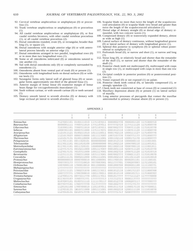
610 JOURNAL OF VERTEBRATE PALEONTOLOGY, VOL. 22, NO. 3, 2002
APPENDIX 2
10
20
30
40
50
60
NotosuchusBaurusuchusLibycosuchusSebecusAraripesuchusAlligatoriumTheriosuchusPelagosaurusTeleosauridaeMetriorhynchidaeEutretauranosuchus
?12?0011010000001111?10?0011011000?011011121011121112101?03?112101?131111?011101011?011101011?011101112??1?1?1
0100111010??00?11010??00?110100000?1100001002110100000?100000100110000001102010100110101000011?201010010211000
11?110?0111??110?01110?110?01111?111?0?11011110011101111?00?1101111001000000000010000000001000000000100111?00?
0000002??00?????2?10000?002???00010121100001?02110100?10???010011021100001012110001100211000110021100001001110
?00020112011??111120??00?111201100?11120100011112???00??11??10001?11?010000001101000?00110?000?00110?200?11120
11?1000?1011?1000??011?1010??01111000??011?1000??01??1000???1111000???11110010011111001011111100101111?1010??0
GoniopholisBernissartiaCrocodyliaProtosuchusHemiprotosuchusOrthosuchusMahajangasuchusPeirosauridaeHsisosuchusTrematochampsa
112?01?131112?11113111201111310000000010?00??0?1??100?0001101?????????112?111121100??????1112?0011?1
?010?11000??00?1100000002110000000?00010?????100101?00010010??????????1?002?10?01?00?0001000??211??0
100111?00110?111?00110011110010011000001?0??00?0010011000001??????????100111?01110011?0011100111?011
0001002?10000?002???00010021100011?001010011????010001000?00??????????00010121100001012110000101????
1000?11120??0001112?10110111200000201101??0020?00?0000001100??????????11001?112?1000101?11????11112?
11?1010?1011?10100?01111010010111110010111?11001011111100??1??????????1111000?0011?1000?0?11????0000
UruguaysuchusMalawisuchusComahuesuchusSimosuchusSunosuchusCalsoyasuchus
011?0??10?010?00?10100??0??1011120101100112?010131112?010131
??00??1??10100?111101?00?????01?00?000101N10?110000210?1???0
1???????1?11111?011??11???????110110?011100111100110111?1001
1??1?12?1?0001012??0?001002???0001011?1000010022100?????1?1?
?00011????100020?11?????1??1??110001?1101200111120?200??????
???????????????00?????????????01???0001111110100?0??????????
92. Cervical vertebrae amphicoelous or amphiplatyan (0) or procoe-lous (1).
93. Trunk vertebrae amphicoelous or amphiplatyan (0) or procoelous(1).
94. All caudal vertebrae amphicoelous or amphiplatyan (0), or firstcaudal vertebra biconvex, with other caudal vertebrae procoelous(1), or all caudal vertebrae procoelous (2).
95. Dorsal osteoderms rounded, ovate (0), or rectangular, broader thanlong (1), or square (2).
96. Dorsal osteoderms with straight anterior edge (0) or with antero-lateral process laterally on anterior edge (1).
97. Dorsal osteoderms arranged in two parallel, longitudinal rows (0)or in more than two longitudinal rows (1).
98. Some or all osteoderms imbricated (0) or osteoderms sutured toone another (1).
99. Tail with dorsal osteoderms only (0) or completely surrounded byosteoderms (1).
100. Osteoderms absent from ventral part of trunk (0) or present (1).101. Osteoderms with longitudinal keels on dorsal surfaces (0) or with-
out keels (1).102. Surangular forms only lateral wall of glenoid fossa (0) or suran-
gular forms approximately one-third of the glenoid fossa (1).103. Anterior margin of femur linear (0) oranterior margin of femur
bears flange for coccygeofemoralis musculature (1).104. Teeth without carinae, or with smooth carinae (0) or teeth serrated
(1).105. Dentary smooth lateral to seventh alveolus (0) or dentary with
large occlusal pit lateral to seventh alveolus (1).
106. Scapular blade no more than twice the length of the scapulocora-coid articulation (0) or scapular blade very broad and greater thantwice the length of the scapulocoracoid articulation (1).
107. Dorsal edge of dentary straight (0) or dorsal edge of dentary si-nusoidal, with two concave waves (1).
108. Compressed dentary (0) or transversely expanded dentary, almostas wide as high (1).
109. Lateral surface of dentary continuous, without longitudinal groove(0) or lateral surface of dentary with longitudinal groove (1).
110. Splenial thin posterior to symphysis (0) or splenial robust poster-odorsal to symphysis (1).
111. Prefrontals broad (0), or narrow and short (1), or narrow and long(1).
112. Snout long (0), or relatively broad and shorter than the remainderof the skull (1), or narrow and shorter than the remainder of theskull (1).
113. Posterior cheek teeth not multicusped (0), multicusped with cuspsin single row (1), or multicusped with cusps in more than one row(2).
114. Occipital condyle in posterior position (0) or posteroventral posi-tion (1).
115. Vomer exposed (0) or not exposed (1) on palate.116. Posterior cheek teeth conical (0), or laterally compressed (1), or
strongly spatulate (2).117. Cheek teeth not constricted at base of crown (0) or constricted (1)118. Maxillary depression absent (0) or present (1) on lateral surface
of maxilla.119. Long anterior processes of pterygoids that contact the maxillae
anteromedial to primary choanae absent (0) or present (1).

611TYKOSKI ET AL.—NEW EARLY JURASSIC CROCODYLIFORM
APPENDIX 2, continued
70
80
90
100
110
119
NotosuchusBaurusuchusLibycosuchusSebecusAraripesuchusAlligatoriumTheriosuchusPelagosaurusTeleosauridaeMetriorhynchidaeEutretauranosuchus
?110??11???11??03?01???0?0?1???1??103100?11?101100????10?1????1?10210??10001200??1?001200??1?001200??1?0?12110
5?010?110?5?010111015?0001?00051000110005?000110104?00101?104?0010101030000120003?0001200030001120004?00001010
0??1?????00?????????0????????00????????011111111?011011111001101111??00110111?0000101111?000101?11?0111???1??0
0?0???????0?????????0?0????????00???????10002?0???????1?0010011?1100100000110001000?110001000??????0??0?11????
?0??0?0000?0?1??0011???1??00???0010?001??0100111000000001100000000110010000001000000000100?0000001001000001100
0101?100001???1000?10????00010011000000101000010????0?0000100000000100000000?0000?00010000000010011
GoniopholisBernissartiaCrocodyliaProtosuchusHemiprotosuchusOrthosuchusMahajangasuchusPeirosauridaeHsisosuchusTrematochampsaUruguaysuchus
?1?011311??1???131??111001310001101011?1??1?1?11???1?01011?0?????????1?1?0??2000?1?02?1001?1?0??10????????111?
4?001?10104?0010?010310001101020110011012?11????01?000?0010051010????0????0?1?1?1?000?1?105?0???1???????0??000
11?1??1??011?1?11??011111111010100011100?????????0010001110011?1111?01??????????010?01??????????????1??1?1????
?00?1100?100021010111111101011000?110011????1?00?1000?11001010000010????????????????1?0011100020?0????????????
100000110000000011001000001100000000000??00000000?000000000??111111001??????11?100?00000???1111?1100??11??0???
0000100100000100000000100000200?00000200?0?00200000000??0??000??????10002000?0000???0?0000?011?2100
MalawisuchusComahuesuchusSimosuchusSunosuchusCalsoyasuchus
????1?21001????1?1?0?1?0102111?21021311??????1111?
41?00?111?????0?101041010?00004100011010???????01?
1?????????0?????????0???????011??1111?0???????????
100?????????????????000?2010?100001100?1??????????
1??0??0??????00??00?00?20?00001????????????1??????
1121?1100?10??0?00111102100?00?1??1000??1??11