Embed Size (px)
Citation preview
8/4/2019 Callus Re Generations 02
http://slidepdf.com/reader/full/callus-re-generations-02 1/4
Plantlet regeneration from immature inflorescence of Dioscorea hispida Dennst. 1Journal of Root Crops, 2008, Vol. 34 No. 1, pp. 1-4
Indian Society for Root Crops
ISSN 0378-2409
Plantlet Regeneration from Immature
Inflorescence of Dioscorea hispida Dennst.M. Unnikrishnan, Binu Hariprakash and C. MohanCentral Tuber Crops Research Institute, Sreekariyam
Thiruvananthapuram 695 017, India
Corresponding author: M. Unnikrishnan, e-mail: [email protected]
Abstract
Organogenesis and subsequent plantlet regeneration were observed in nodes with female flower initials
cultured on Murashige and Skoog medium supplemented with NAA (0.1µM), BAP (0.5µM) and GA3(0.3µM)
inDioscorea hispida Dennst. Rooting was also observed simultaneously from the base of the shoots developed
from the floral buds that got modified into vegetative buds. High frequency plantlet regeneration was achieved
by this method which helped in the micropropagation and in vitro conservation of this medicinal yam.
Introduction
Dioscorea hispida Dennst. (Intoxicating yam, Nami) isa wild growing, dioecious left twiner which has an AsiaOceanic origin. The plant is distinct from other species
by its trifoliate leaf, short sharp spiny stem, axillary panicle with small flowers and is often described as D . daemonaRoxb., D . hirsuta Blume. and D . triphylla L. The flesh
and sap of tubers are yellow and are highly toxic due tothe presence of an alkaloid- dioscorine, which can bedetoxified by boiling. The tubers are also source of foodas well as starch (Rao and Beri, 1952 a, b).
Callus culture, somatic embryogenesis and embryo culturehave been studied in Dioscorea species (Kohmura et al.,1995; Ammirato, 1984; Okezie, 1987; Viana and Mantell,
1989). However, only limited reports are available on therole of immature inflorescence in regeneration. The only report available is of Dioscorea floribunda where plantlet
regeneration was obtained from male inflorescence culturedin medium supplemented with kinetin (Borthakur et al., 2002).
The aim of the present study was to test the regenerationpotential of female inflorescence for somatic embryogenesisin vitro and also to check the effect of growth regulatorsin initiating plantlet regeneration in Dioscorea hispida.
Materials and Methods
Plant material
D. hispida female plant maintained in shade net house of
Division of Crop Improvement, CTCRI, Trivandrum were the source of explants. Two types of nodes viz., one
set with flower bud initials (A) and the other with vegetative buds (B) were used as the explants (Table 1).
Surface sterilization
The explants were washed thoroughly under running
tap water for 10 minutes followed by the treatment with 1% teepol for 20 minutes and Bavistin (BASF) 1%for 15 minutes. The explants were surface sterilized with 0.1% (w/v) mercuric chloride for 15 minutes andthen washed five times with sterile distilled water. Axenic cultures were raised by inoculating a single nodein each tube containing media. Single nodes with intact inflorescence was also inoculated separately.
Culture media
The explants were inoculated in MS media containing
standard MS salts and vitamins (HIMEDIA), 4% (w/v)sucrose and gelled with 0.8% (w/v) agar. The media wassupplemented with NAA (0.1µM), BAP (0.5µM) and
GA 3(0.3µM) and autoclaved at 121 o C at 15 lbs after
maintaining the pH at 5.8.
Culture condition
All the cultures were kept in 25 ± 2oC temperature andat 8h photoperiod. The materials were subcultured at five weeks intervals on fresh medium containing MS with
NAA, BAP and GA 3
as described earlier.
F O R
R O O
T
C R O
P S
I
N D I A
N
S O C I E
T Y
P R O D U C E T U BER REDUC E H U N
G E R
8/4/2019 Callus Re Generations 02
http://slidepdf.com/reader/full/callus-re-generations-02 2/4
2 M. Unnikrishnan, Binu Hariprakash and C. Mohan
Results and Discussion
Bud development was very slow and initiated only after
38 days in nodal explants with flower initials. Sixty per cent of these nodes differentiated into well developed dark
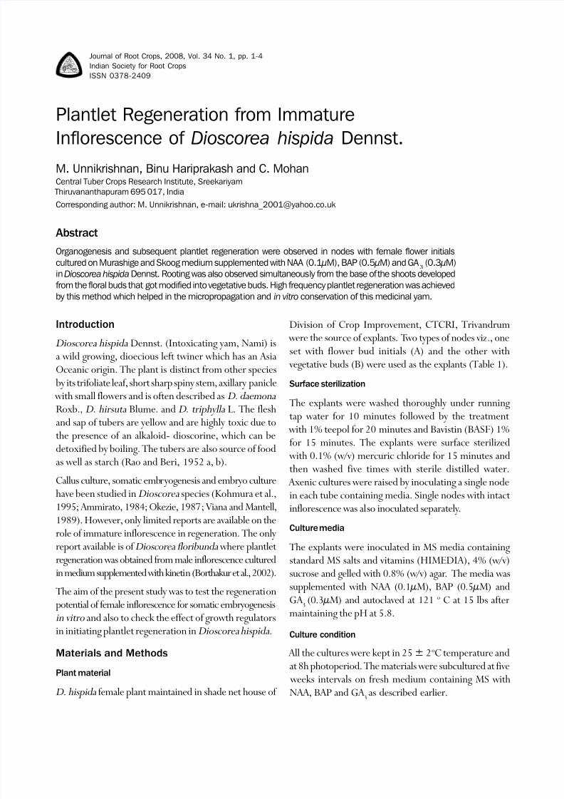
green inflorescence possessing secondary branches while40% differentiated into only callus with flower buds onit. Regeneration was observed after 70 days. Greenembryoids developed from the flower bud region whichsubsequently developed into plantlets within 14 days(Table 1).
Each plantlet had long slender independent roots (1-2 nos)hanging from the base, into the medium. The number of
embryoids produced per culture ranged from 5 to 7. Theinflorescence grew from 1.5 to 2cm (Table 2).
The shoots regenerated from flower buds showed root development also in the same medium.
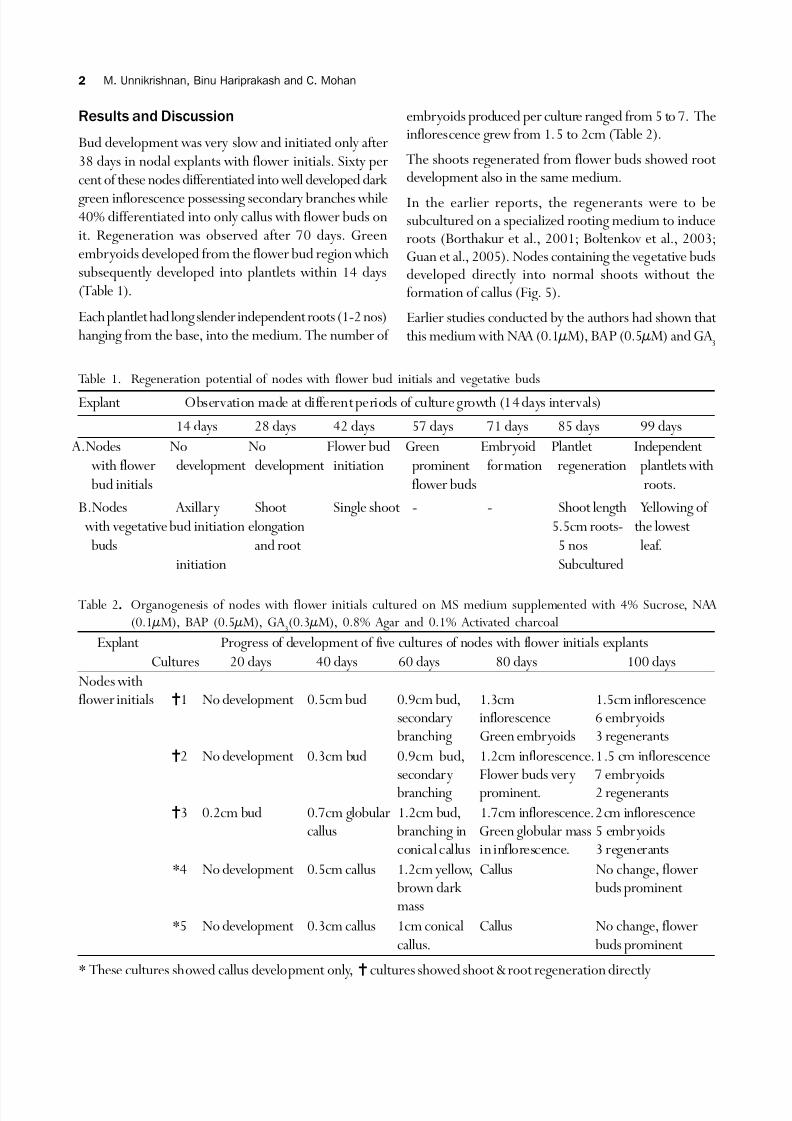
In the earlier reports, the regenerants were to besubcultured on a specialized rooting medium to induceroots (Borthakur et al., 2001; Boltenkov et al., 2003;Guan et al., 2005). Nodes containing the vegetative budsdeveloped directly into normal shoots without theformation of callus (Fig. 5).
Earlier studies conducted by the authors had shown that this medium with NAA (0.1µM), BAP (0.5µM) and GA
3
Table 2..... Organogenesis of nodes with flower initials cultured on MS medium supplemented with 4% Sucrose, NAA
(0.1µM), BAP (0.5µM), GA 3(0.3µM), 0.8% Agar and 0.1% Activated charcoal
Explant Progress of development of five cultures of nodes with flower initials explants
Cultures 20 days 40 days 60 days 80 days 100 days
Nodes withflower initials ?1 No development 0.5cm bud 0.9cm bud, 1.3cm 1.5cm inflorescence
secondary inflorescence 6 embryoidsbranching Green embryoids 3 regenerants
?2 No development 0.3cm bud 0.9cm bud, 1.2cm inflorescence.1.5 cm inflorescencesecondary Flower buds very 7 embryoidsbranching prominent. 2 regenerants
?3 0.2cm bud 0.7cm globular 1.2cm bud, 1.7cm inflorescence.2cm inflorescencecallus branching in Green globular mass 5 embryoids
conical callus in inflorescence. 3 regenerants
*4 No development 0.5cm callus 1.2cm yellow, Callus No change, flower brown dark buds prominent mass
*5 No development 0.3cm callus 1cm conical Callus No change, flower
callus. buds prominent
* These cultures showed callus development only, "? cultures showed shoot & root regeneration directly
Table 1. Regeneration potential of nodes with flower bud initials and vegetative buds
Explant Observation made at different periods of culture growth (14 days intervals)
14 days 28 days 42 days 57 days 71 days 85 days 99 days
A.Nodes No No Flower bud Green Embryoid Plantlet Independent
with flower development development initiation prominent formation regeneration plantlets with
bud initials flower buds roots.
B.Nodes Axillary Shoot Single shoot - - Shoot length Yellowing of
with vegetative bud initiation elongation 5.5cm roots- the lowest
buds and root 5 nos leaf.
initiation Subcultured
8/4/2019 Callus Re Generations 02
http://slidepdf.com/reader/full/callus-re-generations-02 3/4
Plantlet regeneration from immature inflorescence of Dioscorea hispida Dennst. 3
Fig. 1. Vegetative shoots emerging from
the flower bud region
Fig. 2. Formation of shoot from flower bud
region
Fig. 3. Roots emerging from the
base of shoots
(0.3µM) was effective for the micro propagation of thethree edible species of Dioscorea: D . alata, D . esculentaand D . rotundata, (Unnikrishnan et al., 2005). Thepresent study indicated the effectiveness of this medium
in inducing shoot and root regeneration even from thefloral buds. The cultures which showed only callus formationdid not develop into embryoids (Table 2). In the cultures with inflorescence, explant showed heavy phenolic
Fig. 4. Fully grown shoot from flower
bud initial
Fig. 5. Fully grown plantlet in node
with vegetative bud
Fig. 6. Callus formation in the flower bud
region
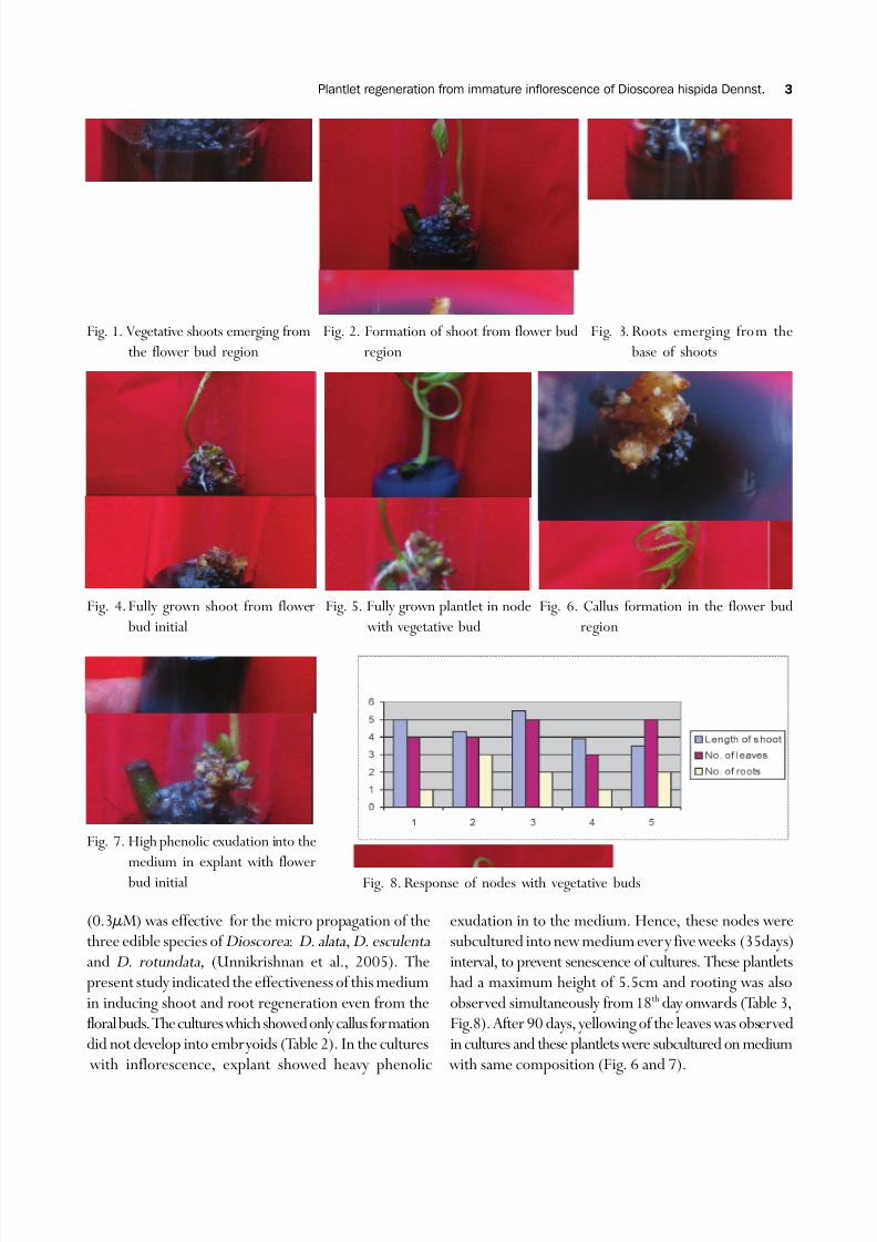
Fig. 8. Response of nodes with vegetative buds
Fig. 7. High phenolic exudation into the
medium in explant with flower bud initial
exudation in to the medium. Hence, these nodes weresubcultured into new medium ever y five weeks (35days)interval, to prevent senescence of cultures. These plantletshad a maximum height of 5.5cm and rooting was also
observed simultaneously from 18th day onwards (Table 3,Fig.8). After 90 days, yellowing of the leaves was observedin cultures and these plantlets were subcultured on medium with same composition (Fig. 6 and 7).
8/4/2019 Callus Re Generations 02
http://slidepdf.com/reader/full/callus-re-generations-02 4/4