Embed Size (px)
Citation preview
J. Funct. Biomater. 2010, 1, 22-107; doi:10.3390/jfb1010022
Journal of
Functional
Biomaterials ISSN 2079-4983
www.mdpi.com/journal/jfb/
Review
Calcium Orthophosphates as Bioceramics: State of the Art
Sergey V. Dorozhkin
Kudrinskaja sq. 1-155, Moscow 123242, Russia; E-Mail: [email protected];
Tel.: +7-499-255-4460
Received: 21 October 2010; in revised form: 16 November 2010 / Accepted: 25 November 2010 /
Published: 30 November 2010
Abstract: In the late 1960s, much interest was raised in regard to biomedical applications
of various ceramic materials. A little bit later, such materials were named bioceramics.
This review is limited to bioceramics prepared from calcium orthophosphates only, which
belong to the categories of bioactive and bioresorbable compounds. There have been a
number of important advances in this field during the past 30–40 years. Namely, by
structural and compositional control, it became possible to choose whether calcium
orthophosphate bioceramics were biologically stable once incorporated within the skeletal
structure or whether they were resorbed over time. At the turn of the millennium, a new
concept of calcium orthophosphate bioceramics—which is able to promote regeneration of
bones—was developed. Presently, calcium orthophosphate bioceramics are available in the
form of particulates, blocks, cements, coatings, customized designs for specific
applications and as injectable composites in a polymer carrier. Current biomedical
applications include artificial replacements for hips, knees, teeth, tendons and ligaments, as
well as repair for periodontal disease, maxillofacial reconstruction, augmentation and
stabilization of the jawbone, spinal fusion and bone fillers after tumor surgery. Exploratory
studies demonstrate potential applications of calcium orthophosphate bioceramics as
scaffolds, drug delivery systems, as well as carriers of growth factors, bioactive peptides
and/or various types of cells for tissue engineering purposes.
Keywords: calcium orthophosphates; hydroxyapatite; bioceramics; biomaterials;
biomedical applications; bone grafts; tissue engineering
OPEN ACCESS
J. Funct. Biomater. 2010, 1
23
1. Introduction
One of the most exciting and rewarding research areas of material science involves various
applications to health care. Examples are sutures, catheters, heart valves, pacemakers, breast implants,
fracture fixation plates, nails and screws in orthopedics, dental filling materials, orthodontic wires, as
well as total joint replacement prostheses. Furthermore, during recent decades, both an ageing
population and a democratization of high-risk sports have led to a surge of bone-related diseases and
bone fractures, which must be treated. However, in order to be accepted by the living body, all
implantable items must be prepared from a special class of materials, called biomedical materials or
biomaterials, in short.
In general, all solids are divided into four major groups of materials: metals, polymers ceramics and
composites thereof. Similarly, all biomaterials are also divided into the same major groups: biometals,
biopolymers, bioceramics and biocomposites. All of them play very important roles in replacement
and regeneration of human tissues. However, due to a great number of publications, this review is
limited to bioceramics only. In general, the modern bioceramics comprise various polycrystalline
materials, glasses, glass-ceramics, as well as ceramic-filled bioactive composites. All of them might be
manufactured in both porous and dense forms in bulk, as well as in the forms of powders, granules
and/or coatings. An expansion of bioceramics to health care has been characterized by a significant
increase in the number of publications and patents in this field and an ever-increasing number of major
international conferences and themed meetings [1-5].
Interestingly, the chemical elements used to manufacture bioceramics form just a small set of the
Periodic Table. Namely, bioceramics might be prepared from alumina, zirconia, carbon,
silica-contained and calcium-contained compounds, as well as some other chemicals [3]; however, this
review is limited to calcium orthophosphates only. Calcium orthophosphate-based biomaterials and
bioceramics are now used for a number of different applications throughout the body, covering all
areas of the skeleton. Applications include dental implants, percutaneous devices and use in
periodontal treatment, healing of bone defects, fracture treatment, total joint replacement (bone
augmentation), orthopedics, cranio-maxillofacial reconstruction, otolaryngology and spinal
surgery [2-6]. Depending upon the required properties, different calcium orthophosphates might be
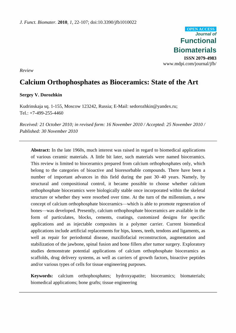
used. For example, Figure 1 shows some randomly chosen samples of the commercially available
calcium orthophosphate bioceramics for bone graft applications.
In this review, the focus has been placed upon applications of calcium orthophosphates as medical
implants to repair and reconstruct damaged or diseased hard tissues of the body (usually, those of the
musculo-skeletal system, such as bones or teeth) and to describe some of the major developments in
this field during the past ~40 years. To narrow the subject further, with a few important exceptions,
bioceramics prepared from undoped and un-substituted calcium orthophosphates have been considered
and discussed only. Furthermore, calcium orthophosphate bioceramics prepared from biological
resources, such as bones, teeth, corals, etc., are not considered either. Readers interested in these topics
are advised to read the original papers [7-37].
J. Funct. Biomater. 2010, 1
24
Figure 1. Several examples of the commercial calcium orthophosphate-based bioceramics.
2. General Knowledge on Biomaterials and Bioceramics
A number of definitions have been developed for the term ―biomaterials‖. Until recently, the
consensus definition developed by the experts in this field has been the following: biomaterials are
synthetic or natural materials used to replace parts of a living system or to function in intimate contact
with living tissues [38]. However, in September 2009, a more advanced definition was introduced: ―A
biomaterial is a substance that has been engineered to take a form which, alone or as part of a complex
system, is used to direct, by control of interactions with components of living systems, the course of
any therapeutic or diagnostic procedure, in human or veterinary medicine‖ [39]. In any case,
biomaterials are intended to interface with biological systems to evaluate, treat, augment or replace
any tissue, organ or function of the body and are now used in a number of different applications
throughout the body [4,5,40]. The major difference between biomaterials and other classes of materials
is the ability of biomaterials to remain in a biological environment without damaging the surroundings
and without being damaged themselves in the process. Thus, biomaterials are solely associated with
the health care domain and must have an interface with tissues or tissue components. One should stress
that any artificial materials that are simply in contact with skin, such as hearing aids and wearable
artificial limbs, are not included in the definition of biomaterials since the skin acts as a protective
barrier between the body and the external world.
The biomaterials discipline is founded in the knowledge of the synergistic interaction of material
science, biological science, chemical science, medical science and mechanical science and requires
input and comprehension from all these areas so that implanted biomaterials perform adequately in a
living body and interrupt normal body functions as little as possible [41]. As biomaterials mainly deal
with all aspects of material synthesis and processing, the knowledge in chemistry, material science and
engineering is essential. On the other hand, as clinical applications are the main purposes of
biomaterials, biomedical sciences become a key part of the research. These include cell and molecular
biology, anatomy and animal and human physiology. The final aim is to achieve the ideal biological
interaction of implanted biomaterials with living tissues of a host. In order to achieve these goals,
several stages have to be performed, namely: material synthesis, design and manufacturing of
J. Funct. Biomater. 2010, 1
25
prostheses, followed by various types of tests. Furthermore, any potential biomaterial must also pass
all regulatory requirements before its clinical application [42].
Biomaterials must be distinguished from biological materials because the former are the materials that
are accepted by living tissues and, therefore, they might be used for tissue replacements, while the latter
are the materials being produced by various biological systems (wood, cotton, bones, chitin, etc.) [43]. In
addition, there are biomimetic materials, which are not made by living organisms but have similar
composition, structure and properties to biological materials. Further, bioceramics (or biomedical
ceramics) might be defined as biomaterials of the ceramic origin [44]. In general, bioceramics can
have structural functions as joint or tissue replacements, can be used as coatings to improve the
biocompatibility [45] of metal implants, as well as function as resorbable lattices, providing temporary
structures and frameworks those are dissolved and/or replaced as the body rebuilds the damaged
tissues [46-51]. Some types of bioceramics even feature a drug-delivery capability [52,53].
A progressive deterioration of all tissues with age is the major contributor to the need for spare
parts for the body. Bone is especially vulnerable to fracture in older people due to a loss of density and
strength with age. This effect is especially severe in women due to the hormonal changes associated
with menopause. A graphical representation of the effect of time on bone strength and density from the
age of 30 years onward is available in literature [Ref. 48, Figure 1]. Bone density decreases because
bone-growing cells (osteoblasts) become progressively less productive in making new bone and
repairing micro-fractures. The lower density greatly deteriorates the strength of bones and an
unfortunate consequence is that many old people fracture their hips or have collapsed vertebrae and
spinal problems [48].
Surface reactivity is one of the common characteristics of bioceramics. It contributes to their bone
bonding ability and their enhancing effect on bone tissue formation. During implantation, various
reactions occur at the material/tissue interfaces that lead to time-dependent changes in the surface
characteristics of the implanted bioceramics and the surrounding tissues [54]. Bioceramics are needed
to alleviate pain and restore functions to diseased or damaged calcified tissues (bones and teeth) of the
body. A great challenge facing the medical application of bioceramics is to replace old, deteriorating
bone with a material that can function the remaining years of the patient’s life and, ideally, be replaced
by a new mature bone without transient loss of mechanical support [1]. Because the average life span
of humans is now 80+ years and the major need for spare parts begins at about 60 years of age, the
implanted non-resorbable bioceramics need to last, at least, for 20+ years. This demanding
requirement of survivability is under conditions of use that are especially harsh to implanted materials:
corrosive saline solutions at 37 °C under variable, multiaxial and cyclical mechanical loads. The
excellent performance of the specially designed bioceramics that have survived these clinical
conditions represents one of the most remarkable accomplishments of research, development,
production and quality assurance during the past century [48].
J. Funct. Biomater. 2010, 1
26
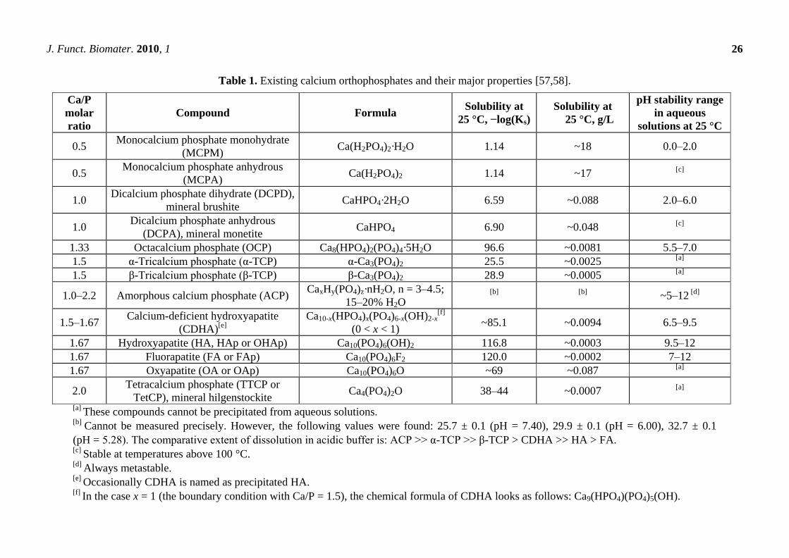
Table 1. Existing calcium orthophosphates and their major properties [57,58].
Ca/P
molar
ratio
Compound Formula Solubility at
25 °C, −log(Ks)
Solubility at
25 °C, g/L
pH stability range
in aqueous
solutions at 25 °C
0.5 Monocalcium phosphate monohydrate
(MCPM) Ca(H2PO4)2·H2O 1.14 ~18 0.0–2.0
0.5 Monocalcium phosphate anhydrous
(MCPA) Ca(H2PO4)2 1.14 ~17
[c]
1.0 Dicalcium phosphate dihydrate (DCPD),
mineral brushite CaHPO4·2H2O 6.59 ~0.088 2.0–6.0
1.0 Dicalcium phosphate anhydrous
(DCPA), mineral monetite CaHPO4 6.90 ~0.048
[c]
1.33 Octacalcium phosphate (OCP) Ca8(HPO4)2(PO4)4·5H2O 96.6 ~0.0081 5.5–7.0
1.5 α-Tricalcium phosphate (α-TCP) α-Ca3(PO4)2 25.5 ~0.0025 [a]
1.5 β-Tricalcium phosphate (β-TCP) β-Ca3(PO4)2 28.9 ~0.0005 [a]
1.0–2.2 Amorphous calcium phosphate (ACP) CaxHy(PO4)z·nH2O, n = 3–4.5;
15–20% H2O [b] [b]
~5–12 [d]
1.5–1.67 Calcium-deficient hydroxyapatite
(CDHA)[e]
Ca10-x(HPO4)x(PO4)6-x(OH)2-x[f]
(0 < x < 1) ~85.1 ~0.0094 6.5–9.5
1.67 Hydroxyapatite (HA, HAp or OHAp) Ca10(PO4)6(OH)2 116.8 ~0.0003 9.5–12
1.67 Fluorapatite (FA or FAp) Ca10(PO4)6F2 120.0 ~0.0002 7–12
1.67 Oxyapatite (OA or OAp) Ca10(PO4)6O ~69 ~0.087 [a]
2.0 Tetracalcium phosphate (TTCP or
TetCP), mineral hilgenstockite Ca4(PO4)2O 38–44 ~0.0007
[a]
[a] These compounds cannot be precipitated from aqueous solutions.
[b] Cannot be measured precisely. However, the following values were found: 25.7 ± 0.1 (pH = 7.40), 29.9 ± 0.1 (pH = 6.00), 32.7 ± 0.1
(pH = 5.28). The comparative extent of dissolution in acidic buffer is: ACP >> α-TCP >> β-TCP > CDHA >> HA > FA. [c]
Stable at temperatures above 100 °C. [d]
Always metastable. [e]
Occasionally CDHA is named as precipitated HA. [f]
In the case x = 1 (the boundary condition with Ca/P = 1.5), the chemical formula of CDHA looks as follows: Ca9(HPO4)(PO4)5(OH).
J. Funct. Biomater. 2010, 1
27
3. General Knowledge on Calcium Orthophosphates
The main driving force behind the use of calcium orthophosphates as bone substitute materials is
their chemical similarity to the mineral component of mammalian bones and teeth [55-58]. As a result,
in addition to being non-toxic, they are biocompatible, not recognized as foreign materials in the body
and, most importantly, exhibit both bioactive behavior [59] and integrate into living tissue by the same
processes active in remodeling healthy bone. This leads to an intimate physicochemical bond between
the implants and bones, termed osteointegration [60]. More to the point, calcium orthophosphates are
also known to be osteoconductive (able to provide a scaffold or template for new bone formation) and
support osteoblast adhesion and proliferation [61,62]. Even so, the major limitations to use calcium
orthophosphates as load-bearing bioceramics are their mechanical properties; namely, they are brittle
with a poor fatigue resistance [46-48,63]. The poor mechanical behavior is even more evident for
highly porous bioceramics and scaffolds because porosity greater than ~100 µm is considered as the
requirement for proper vascularization and bone cell colonization [64-66]. Thus, for biomedical
applications, calcium orthophosphates are used primarily as fillers and coatings, rendering it
impossible to use them for repair of large osseous defects [57,58].
The complete list of known calcium orthophosphates, including their standard abbreviations and the
major properties, is given in Table 1, while detailed information on their synthesis, structure,
chemistry, other properties and biomedical application has been comprehensively reviewed
recently [57,58]; interested readers are referred here. Additional thorough information on various
calcium orthophosphates can be found in books and monographs [67-75]. One should note that among
the existing calcium orthophosphates (Table 1), only certain compounds are useful for biomedical
applications, because those having a Ca/P ionic ratio less than 1 are not suitable for implantation due
to their high solubility and acidity. Due to its basicity, TTCP is not suitable either. However, to be
used in medicine, the ―unsuitable‖ calcium orthophosphates might successfully be combined with
either other calcium orthophosphates or other chemicals.
4. Bioceramics of Calcium Orthophosphates
4.1. History
The performance of living tissues is the result of millions of years of evolution, while the
performance of acceptable artificial substitutions that humankind has designed to repair damaged
tissues are only a few decades old. However, attempts to repair the human body with the use of
implant materials are recorded in the early medical writings of the Hindu, Egyptian and Greek
civilizations. The earliest successful implants were in the skeletal system. Historically, selection of the
materials was based on their availability and an ingenuity of the individual making and applying the
prosthetic [76]. Archaeological findings exhibited in museums showed that materials used to replace
missing human bones and teeth included animal or human (from corpses) bones and teeth, shells,
corals, ivory (elephant tusk), wood, as well as some metals (gold or silver). For instance, the Etruscans
learned to substitute missing teeth with bridges made from artificial teeth carved from the bones of
oxen, while in ancient Phoenicia loose teeth were bound together with gold wires, tying artificial ones
to neighboring teeth. In the 17th century, a piece of dog skull was successfully transplanted into the
J. Funct. Biomater. 2010, 1
28
damaged skull of a Dutch duke. The Chinese recorded the first use of dental amalgam to repair
decayed teeth in the year 659 AD, while pre-Columbian civilizations used gold sheets to heal cranial
cavities following trepanation [77]. Furthermore, in 1970, Amadeo Bobbio discovered Mayan skulls, some
of then more than ~4000 years old, in which missing teeth had been replaced by nacre substitutes [78].
Unfortunately, due to the practice of cremation in many societies, little is known about prehistoric
materials used to replace bone lost to accident or disease.
The first widely tested artificial bioceramic was plaster of Paris. However, in the past, many
implantations failed due to infections, which tended to be exacerbated in the presence of implants,
since they provided a region inaccessible to the body’s immunologically competent cells. Thus, the use
of biomaterials did not become practical until the advent of an aseptic surgical technique developed by
J. Lister in the 1860s. Furthermore, there was a lack of knowledge about the toxicity of selected
materials. In this frame, application of calcium orthophosphates appears to be logical due to their
similarity with the mineral phases of bones and teeth [55,56,69,79,80]. Calcium orthophosphates are
not toxic and do not cause cell death in the surrounding tissues. However, according to available
literature, the first attempt to use them (it was TCP) as an artificial material to repair surgically created
defects in rabbits was performed in 1920 [81]. Although this may be the first scientific study on use of
a calcium orthophosphate for bone defects repair, it remains unclear whether the calcium
orthophosphate was a precipitated or a ceramic material and whether it was in a powder or granular
form. The second clinical report was published 30 years later [82]. More than 20 years afterwards, the
first dental application of a calcium orthophosphate (erroneously described as TCP) in surgically
created periodontal defects [83] and the use of dense HA cylinders for immediate tooth root
replacement [84] were reported. According to the available databases, the first paper with the term
―bioceramics‖ in the abstract was published in 1971 [85], and with the term in the title in 1972 [86,87].
However, application of the ceramic materials as prostheses had been known before [88-91]. Further
historical details might be found in literature [92,93]. On April 26, 1988, the first international
symposium on bioceramics was held in Kyoto, Japan.
Commercialization of the dental and surgical applications of calcium orthophosphate (mainly, HA)
bioceramics occurred in the 1980s, largely due to the pioneering efforts by Jarcho [94-97] in the USA,
De Groot [67,98,99] in Europe and Aoki [100-103] in Japan. Shortly afterwards, HA became a
bioceramic of reference in the field of calcium orthophosphates for biomedical applications.
Preparation and biomedical applications of apatites derived from sea corals (coralline HA) [104-106]
and bovine bone [107] were reported at the same time [108].
4.2. Chemical Composition and Preparation
Currently, calcium orthophosphate bioceramics can be prepared from various sources [109-116].
Unfortunately, up until now, all attempts to synthesize bone replacement materials for clinical
applications featuring physiological tolerance, biocompatibility and a long-term stability have had only
relative success; showing the superiority and a complexity of the natural structures [117].
In general, calcium orthophosphate bioceramics should be characterized from many viewpoints
such as the chemical composition (stoichiometry and purity), homogeneity, phase distribution,
morphology, grain sizes and shape, grain boundaries, crystallite size, crystallinity, pores, cracks,
J. Funct. Biomater. 2010, 1
29
surface, etc. From the chemical point of view, the vast majority of calcium orthophosphate
bioceramics is based on HA, β-TCP, α-TCP and/or biphasic calcium phosphate (BCP, which is an
intimate mixture of either β-TCP + HA [118-130] or α-TCP + HA [7-11]) [131-139]. One should note
that recently the concept of BCP has been extended by preparation and characterization of biphasic
TCP, consisting of α-TCP and β-TCP phases [140-144]. The biphasic TCP is usually prepared by
heating ACP precursors [142-144], in which the α-TCP/β-TCP ratio can be controlled by aging time
and pH value during synthesis of the amorphous precursor [143]. Furthermore, very recently, a
triphasic formulation, consisting of HA, α-TCP and β-TCP has been prepared [145]. The preparation
techniques of various calcium orthophosphates have been extensively reviewed in literature [57,58,67-75]
and references therein. When compared to both α- and β-TCP, HA is a more stable phase under the
physiological conditions, as it has a lower solubility (Table 1) and, thus, a slower resorption
kinetics [69,131,132]. Therefore, the BCP concept is determined by the optimum balance of a more
stable phase of HA and a more soluble TCP. Due to a higher biodegradability of the α- or β-TCP
component, the reactivity of BCP increases with increasing TCP/HA ratio. Thus, in vivo
bioresorbability of BCP can be controlled through the phase composition [127]. Similar conclusions
are also valid for both the biphasic TCP (in which α-TCP is a more soluble phase) and the triphasic
(HA, α-TCP and β-TCP) formulation.
As implants made of calcined HA are found in bone defects for many years after implantation,
bioceramics made of more soluble calcium orthophosphates [7-11,118-130,133-147] are preferable for
biomedical purposes. Furthermore, experimental results showed that BCP had a higher ability to
adsorb fibrinogen, insulin or type I collagen than HA [148]. Thus, according to both observed and
measured bone formation parameters, calcium orthophosphates have been ranked as follows: low
sintering temperature BCP (rough and smooth) ≈medium sintering temperature BCP ≈ TCP > calcined
low sintering temperature HA > non-calcined low sintering temperature HA > high sintering
temperature BCP (rough and smooth) > high sintering temperature HA (calcined and non-calcined) [149].
This sequence was developed in 2000 and, thus, neither biphasic TCP, nor triphasic (HA, α-TCP and
β-TCP) formulation have been included. Recent developments in processing and surface modification
of HA have been reviewed elsewhere [150].
4.3. Forming and Shaping
In order to fabricate bioceramics in more and more complex shapes, scientists are investigating the
use of old and new manufacturing techniques. These techniques range from an adaptation of age-old
pottery techniques to the latest manufacturing methods for high-temperature ceramic parts for airplane
engines. For example, reverse engineering and rapid prototyping technologies have revolutionized a
generation of physical models, allowing an engineer to efficiently and accurately produce physical
models and customized implants with high levels of geometric intricacy [151-153]. Combined with the
computer-aided design and manufacturing (CAD/CAM), complex physical objects of the anatomical
structure can be fabricated in a variety of sizes. In a typical application, an image of a bone defect in a
patient can be taken and used to develop a three-dimensional (3D) CAD computer model [154-156]. A
computer can then reduce the model to slices or layers. The 3D objects are constructed layer-by-layer
using rapid prototyping techniques such as fused deposition modeling [157,158], selective laser
J. Funct. Biomater. 2010, 1
30
sintering [159-161], 3D printing [162-170] or stereo lithography [171-174]. A custom-made implant of
actual dimensions would reduce the time it takes to perform the medical implantation procedure and
subsequently lower the risk to the patient. Another advantage of a prefabricated, exact-fitting implant
is that it can be used more effectively and applied directly to the damaged site rather than a
replacement that is formulated during surgery from a paste or granular material [175-177]. In some
cases, laser processing can be applied as well [178].
The manufacturing technique depends greatly on the ultimate application of the bioceramic device,
whether it is for a hard-tissue replacement or integration of the device within the surrounding tissues.
In general, three types of processing technologies are used: (1) employment of a lubricant and a liquid
binder with ceramic powders for shaping and subsequent firing; (2) application of self-setting and
self-hardening properties of water-wet molded powders (cementation); (3) melting of materials to form
a liquid and shaping during cooling and solidification [179-182]. Since calcium orthophosphates are
either thermally unstable (MCPM, MCPA, DCPA, DCPD, OCP, ACP, CDHA) or have a melting point
at temperatures exceeding ~1400 °C (α-TCP, β-TCP, HA, FA, TTCP), only the first and second
consolidation approaches are used to prepare bulk bioceramics and scaffolds. The methods include
uniaxial compaction [183,184], isostatic pressing (cold or hot) [185-191], granulation [192], loose
packing [193], slip casting [194-196], gel casting [173,174,197-202], pressure mold forming [203],
injection molding [204], polymer replication [205-208], extrusion [209-211], slurry dipping and
spraying [212]. In addition, formation of ceramic sheets from slurries tape casting [130,199,213,214],
doctor blade [215] and colander methods might be employed [63,179-182]. Furthermore, some of
these processes might be performed under the magnetic field, which helps crystal aligning [216-219].
Powders are usually pressed damp in metal dies or dry in lubricated dies at pressures high enough to
form sufficiently strong structures to hold together until they are sintered. An organic binder such as
polyvinyl alcohol helps to bind the powder together [185]. Drying at about 100 °C is a critical step in
preparing damp-formed pieces for firing. Too much or too little water in the compacts can lead to
blowing apart the ware on heating or crumbling, respectively. The binder is removed by heating in air
to oxidize the organic phases to carbon dioxide and water [179-182].
Furthermore, forming and shaping of any ceramic products require a proper selection of the raw
materials in terms of particle sizes and size distribution. Namely, tough and strong bioceramics consist
of pure, fine and homogeneous microstructures. To attain this, pure powders with small average size
and high surface area must be used as the starting sources. However, for maximum packing and least
shrinkage after firing, mixing of ~70% coarse and ~30% fine powders have been suggested [182].
Mixing is usually carried out in a ball mill for uniformity of properties and reaction during subsequent
firing. Mechanical die forming, or sometimes extrusion through a die orifice, can be used to produce a
fixed cross-section. Drying involves removal of water and subsequent shrinkage of the product.
However, due to local variations in water content, warping and even cracks may be developed during
drying. Dry pressing and hydrostatic molding can minimize these problems [182]. Afterwards, the
manufactured green samples are sintered.
Finally, to produce the accurate shaping, necessary for the fine design of bioceramics, machine
finishing might be essential [156,179,220]. Unfortunately, cutting tools developed for metals are
usually useless for bioceramics due to their fragility; therefore, grinding and polishing appear to be the
J. Funct. Biomater. 2010, 1
31
convenient finishing techniques [156,179]. Furthermore, the surface of bioceramics might be modified
by various additional treatments [221].
4.4. Sintering and Firing
A sintering (or firing) procedure appears to be of a great importance to manufacture bulk
bioceramics with the required properties. Usually, this stage is carried out according to controlled
temperature programs of electric furnaces in adjusted ambience of air with necessary additional gasses;
however, always at temperatures below the melting points of the materials. The firing step can include
temporary holds at intermediate temperatures to burn out organic binders [179-182]. The heating rate,
sintering temperature and holding time depend on the starting materials. For example, in the case of
HA, these values are in the ranges of 0.5–3 °C/min, 1000–1250 °C and 2–5 h, respectively [222]. In
the majority cases, sintering allows a structure to retain its shape. However, this process might be
accompanied by a considerable degree of shrinkage [107], which must be accommodated in the
fabrication process. The sintering mechanism is controlled by both surface and volume diffusion at
grain boundaries. In general, when solids heat to high temperatures, the constituent ions or atoms are
driven to move to fill up pores and open channels between the grains of powders, as well as to
compensate for the surface energy differences among their convex and concave surfaces. At the initial
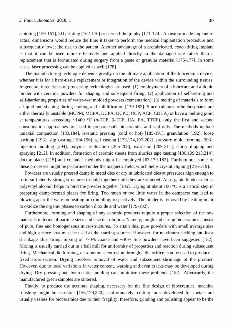
stages, bottlenecks are formed and grow among the particles (Figure 2). Existing vacancies tend to
flow away from the surfaces of sharply curved necks; this is an equivalent of a material flow towards
the necks, which grow as the voids shrink. Small contact areas among the particles expand and, at the
same time, a density of the compact increases and the total void volume decreases. As the pores and
open channels are closed during a heat treatment, the particles become tightly bonded together and
density, strength and fatigue resistance of the sintered object improve greatly. Grain-boundary
diffusion was identified as the dominant mechanism for densification [223]. Furthermore, strong
chemical bonds form among the particles and loosely compacted green bodies are hardened to denser
materials [179-182].
Figure 2. A schematic diagram representing the changes occurring with particles
under sintering.
In the case of calcium orthophosphates, several specific processes occur during sintering. Firstly,
moisture, carbonates and all other volatile chemicals remaining from the synthesis stage, such as
ammonia, nitrates and any organic compounds, are removed as gaseous products. Secondly, unless
powders are sintered, the removal of these gases facilitates production of denser ceramics with
subsequent shrinkage of the samples (Figure 3). Thirdly, all chemical changes are accompanied by a
concurrent increase in crystal size and a decrease in the specific surface area. Fourthly, a chemical
J. Funct. Biomater. 2010, 1
32
decomposition of all acidic orthophosphates and their transformation into other phosphates
(e.g., 2HPO42−
→ P2O74−
+ H2O↑) takes place.
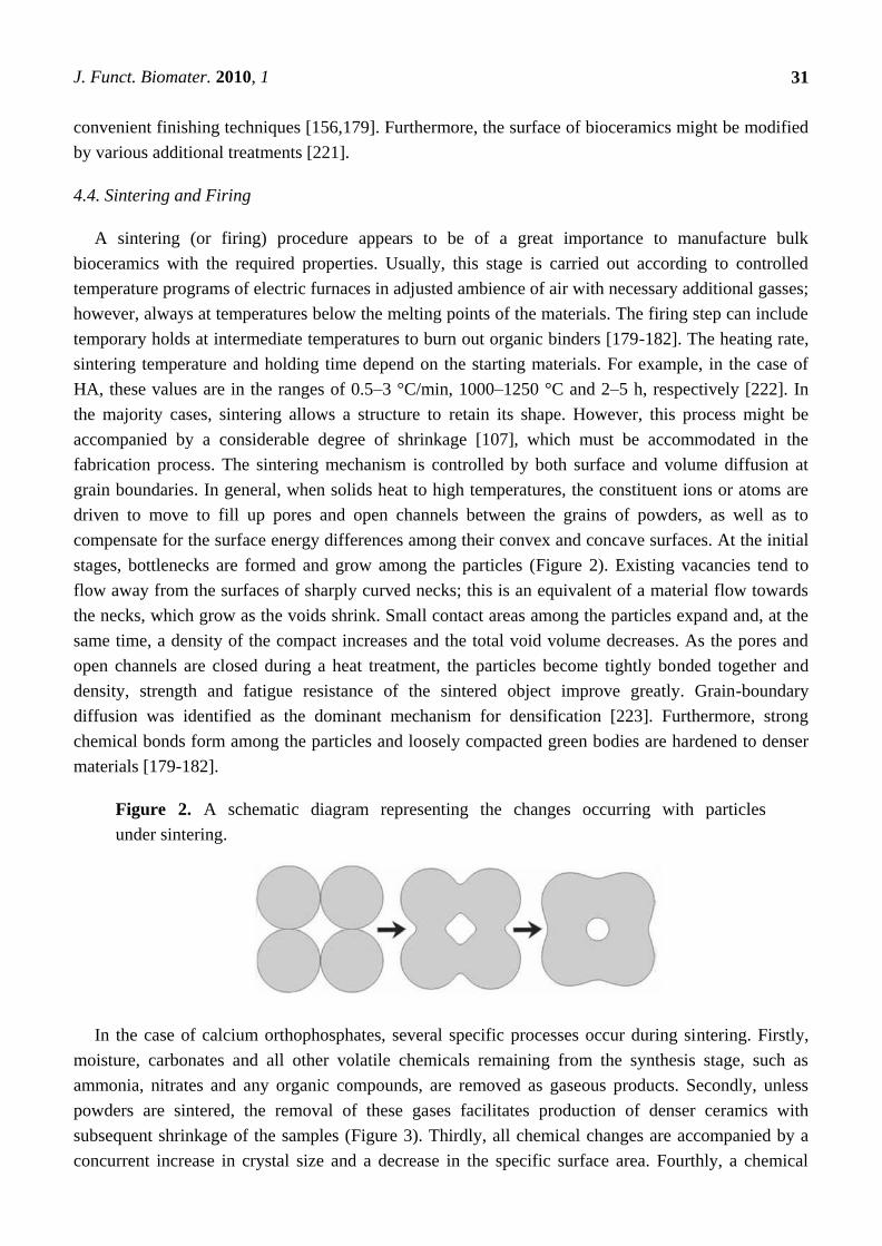
Figure 3. Linear shrinkage of the compacted ACP powders that were converted into
β-TCP, BCP (50% HA + 50% β-TCP) and HA upon heating. According to the authors: ―At
1300 °C, the shrinkage reached a maximum of approximately ~25, ~30 and ~35% for the
compacted ACP powders that converted into HA, BCP 50/50 and β-TCP, respectively‖ [224].
Reprinted from [224] with permission.
In addition, sintering causes toughening [225], densification [226], as well as increasing the
mechanical strength [227,228]. The latter events are due to presence of air and other gases filling gaps
among the unsintered powders. At sintering, the gases move towards the outside of powders and green
bodies shrink owing to decrease of distances among powders. However, in the case of FA sintering, a
linear shrinkage was found to occur at ~715 °C and the material reached its final density at ~890 °C.
Above this value, grain growth became important and induced an intra-granular porosity, which was
responsible for density decrease. At ~1180 °C, a liquid phase forms due to formation of a binary
eutectic between FA and fluorite contained in the powder as impurity. This liquid phase further
promoted the coarsening process and induced formation of large pores at high temperatures [229].
Sintering of a biologically formed apatite has been investigated [230,231] as well, and the obtained
products have been characterized [232,233]. In all cases, the numerical value of the Ca/P ratio in
sintered apatites of biological origin was higher than that of the stoichiometric HA. One should
mention that in the vast majority of cases, calcium orthophosphates with Ca/P ratio < 1.5 (Table 1) are
not sintered, since these compounds are thermally unstable, while sintering of CDHA and ACP leads
to their transformation into BCP (HA + β-TCP [234] or HA + α-TCP [235]).
An extensive study on the effects of sintering temperature and time on the properties of HA
bioceramics revealed a correlation between these parameters and density, porosity, grain size,
chemical composition and strength of the scaffolds [236]. Namely, sintering below ~1000 °C was
found to result in initial particle coalescence, with little or no densification and a significant loss of the
J. Funct. Biomater. 2010, 1
33
surface area and porosity. The degree of densification appeared to depend on the sintering temperature,
whereas the degree of ionic diffusion was governed by the period of sintering [236]. Furthermore,
various sintering additives might be added to calcium orthophosphate bioceramics to enhance
sinterability [237-240]. Unexpectedly, a magnetic field during sintering was found to influence the
growth of HA grains [241].
HA powders can be pressurelessly sintered up to the theoretical density at 1000–1200 °C.
Processing at higher temperatures may lead to exaggerated grain growth and decomposition because
HA becomes unstable at temperatures exceeding ~1300 °C [67-75,242]. The decomposition
temperature of HA bioceramics is a function of the partial pressure of water vapor. Moreover,
processing under vacuum leads to an earlier decomposition of HA, while processing under high partial
pressure of water prevents the decomposition. On the other hand, the presence of water in the sintering
atmosphere was reported to inhibit densification of HA and accelerate grain growth [63,243]. A
definite correlation between hardness, density and grain size in sintered HA bioceramics was found:
despite exhibiting high bulk density, hardness started to decrease at a certain critical grain size
limit [244,245].
Hot pressing [245-251], hot isostatic pressing (HIP) [189,190] or hot pressing with
post-sintering [252,253] processes make it possible to decrease the temperature of the densification
process, diminish the grain size, as well as achieve higher densities. This leads to finer microstructures,
higher thermal stability of calcium orthophosphates and subsequently better mechanical properties of
bulk bioceramics. Microwave [254-261] and spark plasma [262-270] sintering techniques are
alternative methods to the conventional sintering, hot pressing and HIP. Both techniques were found to
be time and energy efficient densification methods. Recently, a hydrothermal hot pressing method was
developed to fabricate OCP bioceramics without thermal dehydration and/or thermal
decomposition [271]. Further details on the sintering and firing processes of calcium orthophosphate
bioceramics are available in literature [47,63,69,70,272,273].
To conclude this part, one should mention an excellent recent review on various ceramic
manufacturing techniques [274], to which interested readers are referred to extend their knowledge on
ceramic processing.
5. The Major Properties
5.1. Mechanical Properties
Ideally, a bone substitute should be replaced by a mature bone without transient loss of mechanical
support. Unfortunately for material scientists, a human body provides one of the most inhospitable
environments for implanted materials. It is warm, wet and both chemically and biologically active.
Furthermore, the body is capable of generating quite massive force concentrations and the variance in
such characteristics among individuals might be enormous. Therefore, all types of potential
biomaterials and bioceramics must sustain attacks of a great variety of aggressive conditions.
Regrettably, there is presently no material fulfilling all these requirements.
On the other hand, any ceramics, when they fail, tend to do so in a dramatic manner. Namely, the
brittle nature of calcium orthophosphate bioceramics is attributed to high strength ionic bonds. Thus, it
is not possible for plastic deformation to happen prior to failure, as a slip cannot occur. Consequently,
J. Funct. Biomater. 2010, 1
34
if a crack is initiated, its progress will not be hindered by the deformation of material ahead of the
crack, as would be the case in a ductile material (e.g., a metal). The crack will continue to propagate,
rapidly resulting in a catastrophic failure [180].
Accordingly, from the mechanical point of view, calcium orthophosphate bioceramics appear to be
brittle polycrystalline materials for which the mechanical properties are governed by crystallinity,
grain size, grain boundaries, porosity and composition [188]. It appears to be very sensitive to slow
crack growth [275]. For dense bioceramics, the strength is a function of the grain size. Finer grain size
materials have smaller flaws at the grain boundaries and thus are stronger than bioceramics with larger
grain sizes. In general, the mechanical properties decrease significantly with increasing content of an
amorphous phase, microporosity and grain size, while a high crystallinity, a low porosity and small
grain size tend to give a higher stiffness, a higher compressive and tensile strength and a greater
fracture toughness. Thus, calcium orthophosphate bioceramics possess poor mechanical properties (for
instance, a low impact and fracture resistances) that do not allow use in load-bearing areas, such as
artificial teeth or bones [46-52,276]. For example, fracture toughness [277] of HA bioceramics does
not exceed ~1.2 MPa·m1/2
[278] (human bone: 2–12 MPa·m1/2
). It decreases almost linearly with
increasing porosity [63]. Generally, fracture toughness increases with decreasing grain size. However,
in some materials, especially non-cubic ceramics, fracture toughness reaches the maximum and rapidly
drops with decreasing grain size. For example, Halouani et al. investigated fracture toughness of pure
hot pressed HA with grain sizes of 0.2–1.2 µm [251]. There appeared to be two distinct trends, where
fracture toughness decreased with increasing grain size above ~0.4 µm and subsequently decreased
with decreasing grain size. The maximum fracture toughness measured was 1.20 ± 0.05 MPa·m1/2
at
~0.4 µm [251]. Fracture energy of HA bioceramics is in the range of 2.3–20 J/m2, while the Weibull
modulus [279] is low (~5–12) in wet environments, which means that HA behaves as a typical brittle
ceramics and indicates low reliability of HA implants [63]. Interestingly, three peaks of internal friction
were found at temperatures about −40, 80 and 130 °C for HA but no internal friction peaks were obtained
for FA in the measured temperature range; this effect was attributed to the differences of the positions of
F- and OH
- in FA and HA, respectively [280].
Bending, compressive and tensile strengths of dense HA bioceramics are in the range of
38–250 MPa, 120–900 MPa and 38–300 MPa, respectively. Similar values for porous HA bioceramics
are in the range of 2–11 MPa, 2–100 MPa and ~3 MPa, respectively [63]. These wide variations in the
properties are due to both structural variations (e.g., an influence of remaining microporosity, grain
sizes, presence of impurities, etc.) and manufacturing processes, as well as caused by a statistical
nature of the strength distribution. Strength [281] was found to increase with increasing Ca/P ratio,
reaching a maximum value around Ca/P ~1.67 (stoichiometric HA) and to decrease suddenly when
Ca/P > 1.67 [63]. Furthermore, strength decreases almost exponentially with increasing
porosity [119,120]. However, by changing the pore geometry, it is possible to influence the strength of
porous bioceramics. It is also worth mentioning that porous HA bioceramics are considerably less
fatigue [282] resistant than dense ones. Both grain sizes and porosity are reported to influence the
fracture path, which itself has little effect on the fracture toughness of calcium orthophosphate
bioceramics [188,283]. Furthermore, no obvious decrease in mechanical properties was found after
calcium orthophosphate bioceramics had been aged in various solutions for different time
periods [284].
J. Funct. Biomater. 2010, 1
35
Young’s (or elastic) modulus [285] of dense HA bioceramics is in the range of 35–120 GPa, which
is more or less similar to those of the most resistant components of the natural calcified tissues (dental
enamel: ~74 GPa, dentine: ~21 GPa, compact bone: ~18–22 GPa). Nevertheless, dense bulk compacts
of HA have mechanical resistances of the order of 100 MPa versus ~300 MPa of human bones,
diminishing drastically their resistance in the case of porous bulk compacts [286]. Young’s modulus
measured in bending is between 44 and 88 GPa. Recently, a considerable anisotropy in the stress-strain
behavior of the perfect HA crystals was found by ab initio calculations [287]. The crystals appeared to
be brittle for tension along the z-axis with the maximum stress of ~9.6 GPa at 10% strain. Furthermore,
the structural analysis of the HA crystal under various stages of tensile strain revealed that the
deformation behavior manifested itself mainly in the rotation of PO4 tetrahedrons with concomitant
movements of both the columnar and axial Ca ions [287]. Vickers hardness [288] of dense HA
bioceramics is within 3–7 GPa, while the Poisson’s ratio [289] for the synthetic HA is about 0.27,
which is close to that of bones (~0.3). At temperatures within 1000–1100 °C, dense HA bioceramics
were found to exhibit superplasticity with a deformation mechanism based on grain boundary sliding.
Furthermore, both the wear resistance and friction coefficient of dense HA bioceramics are comparable
to those of dental enamel [63].
Due to high brittleness (associated to a low crack resistance), biomedical applications of calcium
orthophosphate bioceramics are focused on production of non-load-bearing implants, such as pieces
for middle ear surgery, filling of bone defects in oral or orthopedic surgery, as well as coating of dental
implants and metallic prosthesis (see below) [117,290,291]. In order to improve the reliability of
calcium orthophosphate bioceramics, diverse reinforcements (ceramics, metals or polymers) have been
applied to manufacture various biocomposites and hybrid biomaterials [292], but that is another story.
However, successful hybrid formulations consisting of calcium orthophosphates only should be
mentioned [293-298]. For example, bulk HA bioceramics might be reinforced by HA
whiskers [294-297]. Furthermore, a superior superplasticity of HA/β-TCP composites (i.e., BCP) to
HA bioceramics has been detected [298].
Another approach to improve the mechanical properties of calcium orthophosphate bioceramics is
to coat the items by a polymeric layer [299,300]; however, this is still other story. Interested readers
are referred to further details on the mechanical properties of calcium orthophosphate bioceramics
available elsewhere [63,301].
5.2. Electrical Properties
Occasionally, interest is expressed in the electrical properties of calcium orthophosphate
bioceramics. For example, a surface ionic conductivity of both porous and dense HA bioceramics was
examined for humidity sensor applications, since the room temperature conductivity was influenced by
relative humidity [302]. Namely, the ionic conductivity of HA has been a subject of research for its
possible use as an alcohol [303], carbon dioxide [303] or carbon monoxide [304] gas sensors.
Electrical measurements have also been used as a characterization tool to study the evolution of
microstructure in HA bioceramics [305]. More to the point, Valdes et al. examined the dielectric
properties of HA to understand its decomposition to β-TCP [306]. In the case of CDHA, the electrical
properties, in terms of ionic conductivity, were found to increase after compression of the samples at
J. Funct. Biomater. 2010, 1
36
15 t/cm2, which was attributed to establishment of some order within the apatitic network [307]. The
conductivity mechanism of CDHA appeared to be multiple [308]. Furthermore, there is an attempt to
develop CDHA whisker electrets for biomedical utilization [309].
Interestingly, the electrical properties of calcium orthophosphate bioceramics appear to influence
their biomedical applications. For example, there is an interest in polarization of HA bioceramics to
generate a surface charge by the application of electric fields at elevated temperatures [310,311]. The
presence of surface charges on HA bioceramics was shown to have a significant effect on both in vitro
and in vivo crystallization of biological apatite [312-316]. Furthermore, growth of both biomimetic
calcium orthophosphates and bones was found to be accelerated on negatively charged surfaces and
decelerated on positively charged surfaces [316-325]. In addition, the electrical polarization of HA
bioceramics was found to accelerate a cytoskeleton reorganization of osteoblast-like cells [326-328],
extend bioactivity [329] and enhance bone ingrowth through the pores of porous HA implants [330].
There is an interesting study on the interaction of a blood coagulation factor on electrically polarized
HA surfaces [331]. Further details on the electrical properties of calcium orthophosphate-based
bioceramics can be found in [258,332-336].
5.3. Possible Transparency
Single crystals of all calcium orthophosphates are optically transparent for visible light. As
bioceramics of calcium orthophosphates have a polycrystalline nature with a random orientation of big
amounts of small crystals they are opaque and of white color, unless colored dopants have been added.
However, in some cases, transparency is convenient to provide some essential advantages (e.g., to
enable direct viewing of living cells in a transmitted light). Thus, transparent calcium orthophosphate
bioceramics have been prepared and investigated [189,191,267,270,337-344]. The preparation
techniques, for example, include hot isostatic pressing [189,191], ambient-pressure sintering [337], gel
casting coupled with a low-temperature sintering [340,343], pulse electric current sintering [341], as
well as spark plasma sintering [267,270]. Fully dense, transparent calcium orthophosphate bioceramics
were obtained at temperatures above ~800 °C. Depending on the preparation technique, the transparent
calcium orthophosphate bioceramics have a uniform grain size ranging from ~0.2 μm [337] to
~250 μm [340] and are always pore-free; the latter is not good for biomedical applications.
5.4. Porosity
Porosity is defined as the percentage of void spaces in solids and it is a morphological property
independent of the material. The surface area of porous bodies is much higher, which guarantees a
good mechanical fixation in addition to providing sites on the surface that allow chemical bonding
between the bioceramics and bones [345]. Furthermore, a porous material may have both closed
(isolated) pores and open (connected) pores. Connected pores look like tunnels and are accessible by
gases, liquids and particulate suspensions [346]. The open-cell nature of reticulated materials is a
unique characteristic essential in many applications. Furthermore, dimensions of open pores are
directly related to bone formation, since such pores grant both the surface and space for cell adhesion
and bone ingrowth. On the other hand, pore interconnection provides the way for cell distribution and
migration, as well as allowing efficient in vivo blood vessel formation suitable for sustaining bone
J. Funct. Biomater. 2010, 1
37
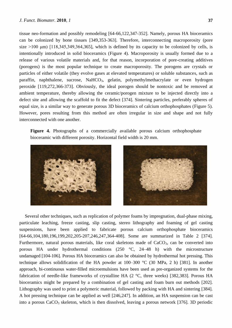
tissue neo-formation and possibly remodeling [64-66,122,347-352]. Namely, porous HA bioceramics
can be colonized by bone tissues [349,353-363]. Therefore, interconnecting macroporosity (pore
size >100 μm) [118,345,349,364,365], which is defined by its capacity to be colonized by cells, is
intentionally introduced in solid bioceramics (Figure 4). Macroporosity is usually formed due to a
release of various volatile materials and, for that reason, incorporation of pore-creating additives
(porogens) is the most popular technique to create macroporosity. The porogens are crystals or
particles of either volatile (they evolve gases at elevated temperatures) or soluble substances, such as
paraffin, naphthalene, sucrose, NaHCO3, gelatin, polymethylmethacrylate or even hydrogen
peroxide [119,272,366-373]. Obviously, the ideal porogen should be nontoxic and be removed at
ambient temperature, thereby allowing the ceramic/porogen mixture to be injected directly into a
defect site and allowing the scaffold to fit the defect [374]. Sintering particles, preferably spheres of
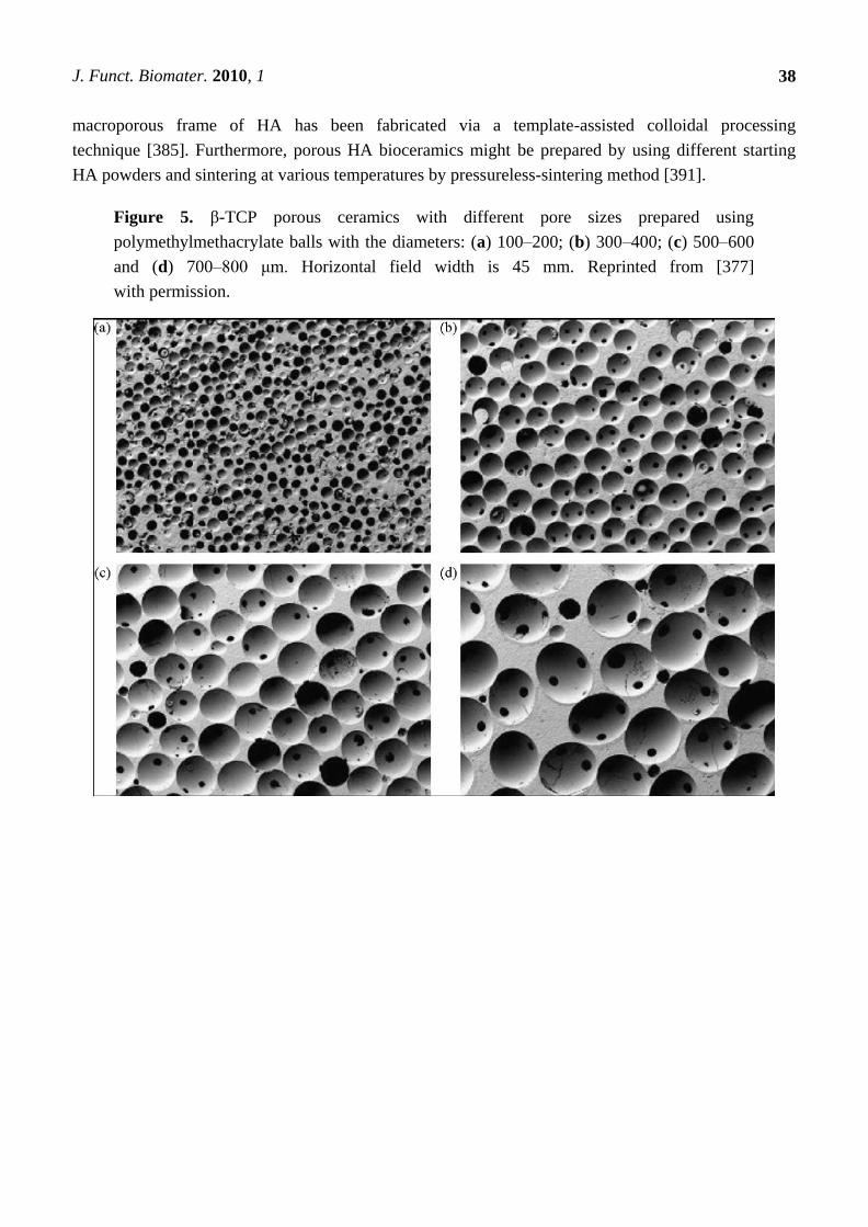
equal size, is a similar way to generate porous 3D bioceramics of calcium orthophosphates (Figure 5).
However, pores resulting from this method are often irregular in size and shape and not fully
interconnected with one another.
Figure 4. Photographs of a commercially available porous calcium orthophosphate
bioceramic with different porosity. Horizontal field width is 20 mm.
Several other techniques, such as replication of polymer foams by impregnation, dual-phase mixing,
particulate leaching, freeze casting, slip casting, stereo lithography and foaming of gel casting
suspensions, have been applied to fabricate porous calcium orthophosphate bioceramics
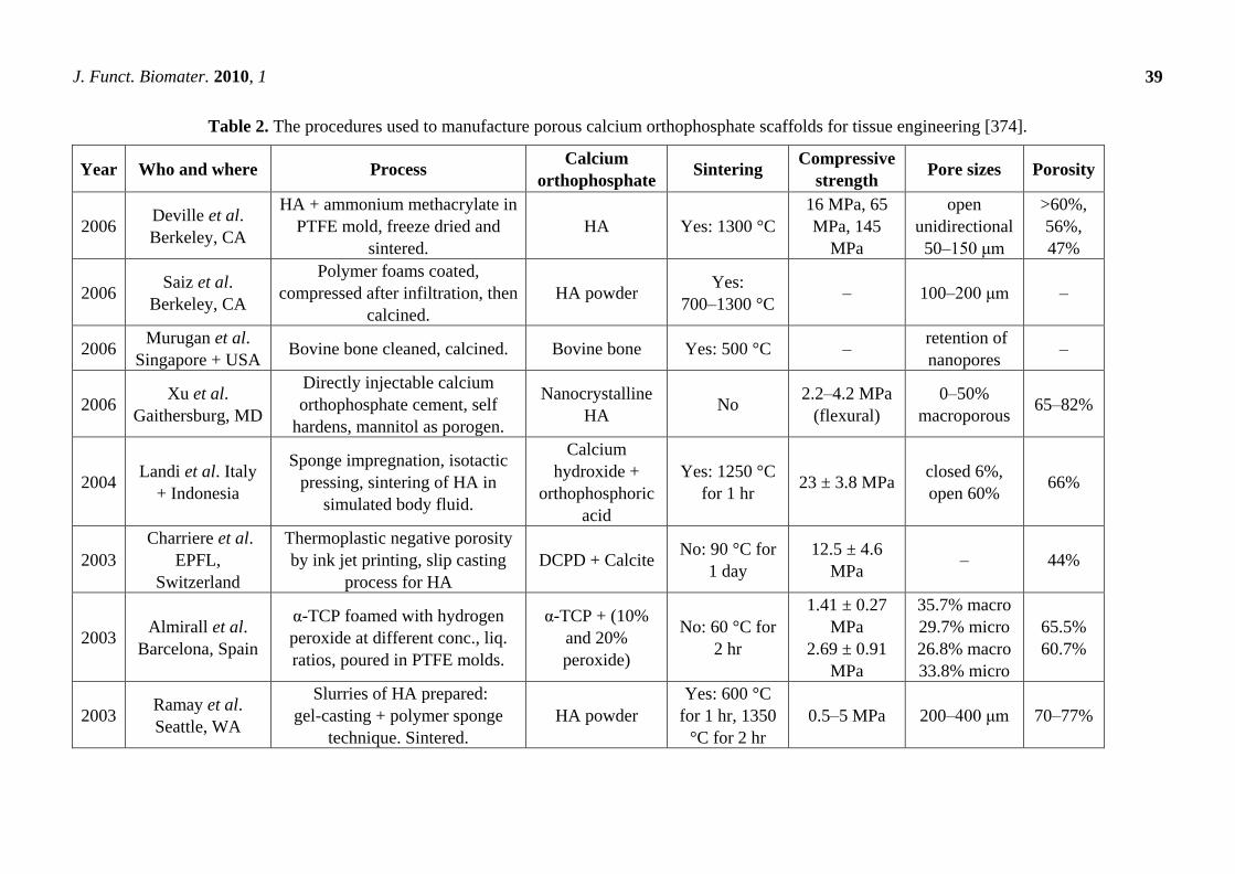
[64-66,104,180,196,199,202,205-207,246,247,364-408]. Some are summarized in Table 2 [374].
Furthermore, natural porous materials, like coral skeletons made of CaCO3, can be converted into
porous HA under hydrothermal conditions (250 °C, 24–48 h) with the microstructure
undamaged [104-106]. Porous HA bioceramics can also be obtained by hydrothermal hot pressing. This
technique allows solidification of the HA powder at 100–300 °C (30 MPa, 2 h) [381]. In another
approach, bi-continuous water-filled microemulsions have been used as pre-organized systems for the
fabrication of needle-like frameworks of crystalline HA (2 °C, three weeks) [382,383]. Porous HA
bioceramics might be prepared by a combination of gel casting and foam burn out methods [202].
Lithography was used to print a polymeric material, followed by packing with HA and sintering [384].
A hot pressing technique can be applied as well [246,247]. In addition, an HA suspension can be cast
into a porous CaCO3 skeleton, which is then dissolved, leaving a porous network [376]. 3D periodic
J. Funct. Biomater. 2010, 1
38
macroporous frame of HA has been fabricated via a template-assisted colloidal processing
technique [385]. Furthermore, porous HA bioceramics might be prepared by using different starting
HA powders and sintering at various temperatures by pressureless-sintering method [391].
Figure 5. β-TCP porous ceramics with different pore sizes prepared using
polymethylmethacrylate balls with the diameters: (a) 100–200; (b) 300–400; (c) 500–600
and (d) 700–800 μm. Horizontal field width is 45 mm. Reprinted from [377]
with permission.
J. Funct. Biomater. 2010, 1
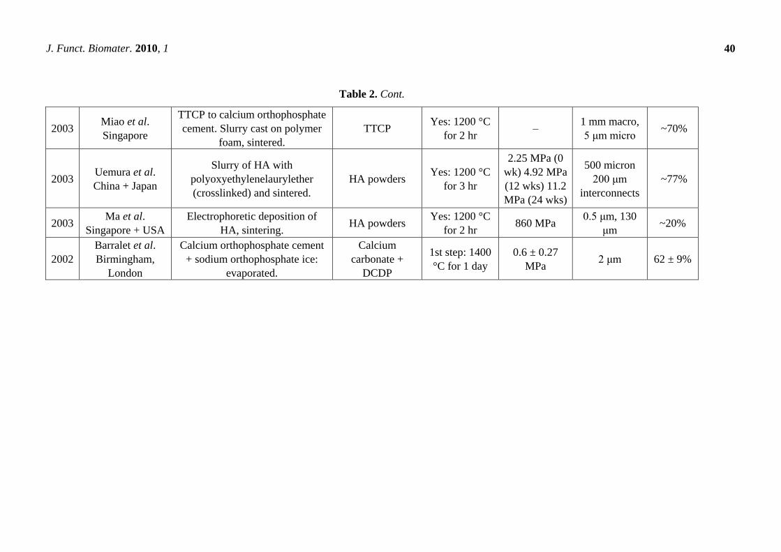
39
Table 2. The procedures used to manufacture porous calcium orthophosphate scaffolds for tissue engineering [374].
Year Who and where Process Calcium
orthophosphate Sintering
Compressive
strength Pore sizes Porosity
2006 Deville et al.
Berkeley, CA
HA + ammonium methacrylate in
PTFE mold, freeze dried and
sintered.
HA Yes: 1300 °C
16 MPa, 65
MPa, 145
MPa
open
unidirectional
50–150 μm
>60%,
56%,
47%
2006 Saiz et al.
Berkeley, CA
Polymer foams coated,
compressed after infiltration, then
calcined.
HA powder Yes:
700–1300 °C – 100–200 μm –
2006 Murugan et al.
Singapore + USA Bovine bone cleaned, calcined. Bovine bone Yes: 500 °C –
retention of
nanopores –
2006 Xu et al.
Gaithersburg, MD
Directly injectable calcium
orthophosphate cement, self
hardens, mannitol as porogen.
Nanocrystalline
HA No
2.2–4.2 MPa
(flexural)
0–50%
macroporous 65–82%
2004 Landi et al. Italy
+ Indonesia
Sponge impregnation, isotactic
pressing, sintering of HA in
simulated body fluid.
Calcium
hydroxide +
orthophosphoric
acid
Yes: 1250 °C
for 1 hr 23 ± 3.8 MPa
closed 6%,
open 60% 66%
2003
Charriere et al.
EPFL,
Switzerland
Thermoplastic negative porosity
by ink jet printing, slip casting
process for HA
DCPD + Calcite No: 90 °C for
1 day
12.5 ± 4.6
MPa – 44%
2003 Almirall et al.
Barcelona, Spain
α-TCP foamed with hydrogen
peroxide at different conc., liq.
ratios, poured in PTFE molds.
α-TCP + (10%
and 20%
peroxide)
No: 60 °C for
2 hr
1.41 ± 0.27
MPa
2.69 ± 0.91
MPa
35.7% macro
29.7% micro
26.8% macro
33.8% micro
65.5%
60.7%
2003 Ramay et al.
Seattle, WA
Slurries of HA prepared:
gel-casting + polymer sponge
technique. Sintered.
HA powder
Yes: 600 °C
for 1 hr, 1350
°C for 2 hr
0.5–5 MPa 200–400 μm 70–77%
J. Funct. Biomater. 2010, 1
40
Table 2. Cont.
2003 Miao et al.
Singapore
TTCP to calcium orthophosphate
cement. Slurry cast on polymer
foam, sintered.
TTCP Yes: 1200 °C
for 2 hr –
1 mm macro,
5 μm micro ~70%
2003 Uemura et al.
China + Japan
Slurry of HA with
polyoxyethylenelaurylether
(crosslinked) and sintered.
HA powders Yes: 1200 °C
for 3 hr
2.25 MPa (0
wk) 4.92 MPa
(12 wks) 11.2
MPa (24 wks)
500 micron
200 μm
interconnects
~77%
2003 Ma et al.
Singapore + USA
Electrophoretic deposition of
HA, sintering. HA powders
Yes: 1200 °C
for 2 hr 860 MPa
0.5 μm, 130
μm ~20%
2002
Barralet et al.
Birmingham,
London
Calcium orthophosphate cement
+ sodium orthophosphate ice:
evaporated.
Calcium
carbonate +
DCDP
1st step: 1400
°C for 1 day
0.6 ± 0.27
MPa 2 μm 62 ± 9%
J. Funct. Biomater. 2010, 1
41
Porous bioceramics with an improved strength might be fabricated from calcium orthophosphate fibers
or whiskers. In general, fibrous porous materials are known to exhibit improved strength due to fiber
interlocking, crack deflection and/or pullout [386]. Namely, porous bioceramics with well-controlled open
pores were processed by sintering of fibrous HA particles [387]. In another approach, porosity was
achieved by firing apatite-fiber compacts mixed with carbon beads and agar. By varying the compaction
pressure, firing temperature and carbon/HA ratio, the total porosity was controlled in the ranges from
~40% to ~85% [378]. Additional examples are available in literature [364,367,374-380,389-408].
In vivo response of calcium orthophosphate bioceramics of different porosity was investigated and
hardly any effect of macropore dimensions (~150, ~260, ~510 and ~1220 μm) was observed [409]. In
another study, a greater differentiation of mesenchymal stem cells was observed when cultured on
~200 μm pore size HA scaffolds when compared to those on ~500 μm pore size HA [410]. The latter
finding was attributed to the fact that a higher pore volume in ~500 μm macropore scaffolds might
contribute to a lack of cell confluency, leading to the cells proliferating before beginning
differentiation. In addition, the authors hypothesized that bioceramics having less than optimal pore
dimensions induced quiescence in differentiated osteoblasts due to reduced cell confluency [410].
Already in 1979, Holmes suggested that the optimal pore range was 200–400 μm with the average
human osteon size of ~223 μm [105]. In 1997, Tsurga and coworkers implied that the optimal pore
size of bioceramics that supported ectopic bone formation was 300–400 μm [411]. Thus, there is no
need to create calcium orthophosphate bioceramics with very big pores; however, the pores must be
interconnected [108,352,364,365]. Interconnectivity governs a depth of cells or tissue penetration into
the porous bioceramics, as well as allowing development of blood vessels required for new bone
nourishing and waste removal [412,413].
Bioceramic microporosity (pore size <10 μm), which is defined by its capacity to be impregnated
by biological fluids [412], results from the sintering process, while the pore dimensions mainly depend
on the material composition, thermal cycle and sintering time. The microporosity provides both a
greater surface area for protein adsorption and increased ionic solubility. Nanoporous (average pore
sizes of less than 100 nm) HA bioceramics might be fabricated as well [414]. Differences in porogens
influence the macroporosity, while differences in sintering temperatures and conditions affect the
percentage of microporosity. Usually, the higher the sintering temperature, the lower both the
microporosity content and the specific surface area of bioceramics. Namely, HA bioceramics sintered
at ~1200 °C shows significantly less microporosity and a dramatic change in crystal sizes, if compared
with those sintered at ~1050 °C (Figure 6). Furthermore, the average shape of pores was found to
transform from strongly oblate to round at higher sintering temperatures [416]. The total porosity
(macroporosity + microporosity) of calcium orthophosphate bioceramics was reported to be about 70%
of the bioceramic volume [417]. In the case of coralline HA or bovine-derived apatites, the porosity of
the original biologic material (coral or bovine bone) is usually preserved during processing [107]. To
conclude this topic, creation of the desired porosity in calcium orthophosphate bioceramics is a rather
complicated engineering task and interested readers are referred to [65,119,368,382,418-442].
Studies revealed that increasing both the specific surface area and pore volume of bioceramics
might greatly accelerate the in vivo process of apatite deposition and, therefore, enhance bone-forming
bioactivity. More importantly, a precise control over the porosity, pore dimensions and internal pore
architecture of bioceramics on different length scales is essential for understanding the
J. Funct. Biomater. 2010, 1
42
structure-bioactivity relationship and the rational design of better bone-forming biomaterials
[439,443,444]. Namely, in antibiotic charging experiments, a nanoporous calcium orthophosphate
bioceramic showed a much higher charging capacity (1621 μg/g) than that of commercially available
calcium orthophosphate (100 μg/g), which did not have any nanoporosity [434]. In other experiments,
porous blocks of HA were found to be viable carriers with sustained release profiles for drugs [445]
and antibiotics over 12 days [446] and 12 weeks [447], respectively. Unfortunately, the porosity
significantly decreases the strength of implants [63,283,301]. Thus, porous calcium orthophosphate
implants cannot be loaded and are used to fill only small bone defects. However, their strength
increases gradually when bones ingrow into the porous network of calcium orthophosphate
implants [448-451]. For example, Martin et al. reported bending strengths of 40–60 MPa for a porous
HA implant filled with 50–60% of cortical bone [448], while in another study an ingrown bone
increased strength of porous HA bioceramics by a factor of three to four [450].
To conclude this topic, filters for microbial filtration might be manufactured from porous HA [452].
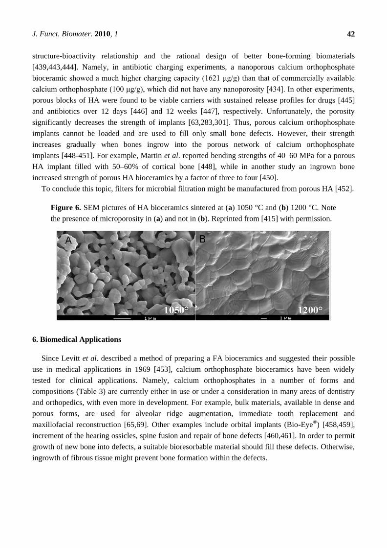
Figure 6. SEM pictures of HA bioceramics sintered at (a) 1050 °C and (b) 1200 °C. Note
the presence of microporosity in (a) and not in (b). Reprinted from [415] with permission.
6. Biomedical Applications
Since Levitt et al. described a method of preparing a FA bioceramics and suggested their possible
use in medical applications in 1969 [453], calcium orthophosphate bioceramics have been widely
tested for clinical applications. Namely, calcium orthophosphates in a number of forms and
compositions (Table 3) are currently either in use or under a consideration in many areas of dentistry
and orthopedics, with even more in development. For example, bulk materials, available in dense and
porous forms, are used for alveolar ridge augmentation, immediate tooth replacement and
maxillofacial reconstruction [65,69]. Other examples include orbital implants (Bio-Eye®
) [458,459],
increment of the hearing ossicles, spine fusion and repair of bone defects [460,461]. In order to permit
growth of new bone into defects, a suitable bioresorbable material should fill these defects. Otherwise,
ingrowth of fibrous tissue might prevent bone formation within the defects.
J. Funct. Biomater. 2010, 1
43
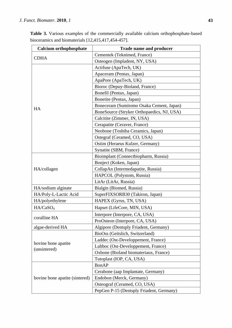
Table 3. Various examples of the commercially available calcium orthophosphate-based
bioceramics and biomaterials [12,415,417,454-457].
Calcium orthophosphate Trade name and producer
CDHA Cementek (Teknimed, France)
Osteogen (Impladent, NY, USA)
HA
Actifuse (ApaTech, UK)
Apaceram (Pentax, Japan)
ApaPore (ApaTech, UK)
Bioroc (Depuy-Bioland, France)
Bonefil (Pentax, Japan)
Bonetite (Pentax, Japan)
Boneceram (Sumitomo Osaka Cement, Japan)
BoneSource (Stryker Orthopaedics, NJ, USA)
Calcitite (Zimmer, IN, USA)
Cerapatite (Ceraver, France)
Neobone (Toshiba Ceramics, Japan)
Ostegraf (Ceramed, CO, USA)
Ostim (Heraeus Kulzer, Germany)
Synatite (SBM, France)
HA/collagen
Bioimplant (Connectbiopharm, Russia)
Bonject (Koken, Japan)
CollapAn (Intermedapatite, Russia)
HAPCOL (Polystom, Russia)
LitAr (LitAr, Russia)
HA/sodium alginate Bialgin (Biomed, Russia)
HA/Poly-L-Lactic Acid SuperFIXSORB30 (Takiron, Japan)
HA/polyethylene HAPEX (Gyrus, TN, USA)
HA/CaSO4 Hapset (LifeCore, MIN, USA)
coralline HA Interpore (Interpore, CA, USA)
ProOsteon (Interpore, CA, USA)
algae-derived HA Algipore (Dentsply Friadent, Germany)
bovine bone apatite
(unsintered)
BioOss (Geitslich, Switzerland)
Laddec (Ost-Developpement, France)
Lubboc (Ost-Developpement, France)
Oxbone (Bioland biomateriaux, France)
Tutoplast (IOP, CA, USA)
bovine bone apatite (sintered)
BonAP
Cerabone (aap Implantate, Germany)
Endobon (Merck, Germany)
Osteograf (Ceramed, CO, USA)
PepGen P-15 (Dentsply Friadent, Germany)
J. Funct. Biomater. 2010, 1
44
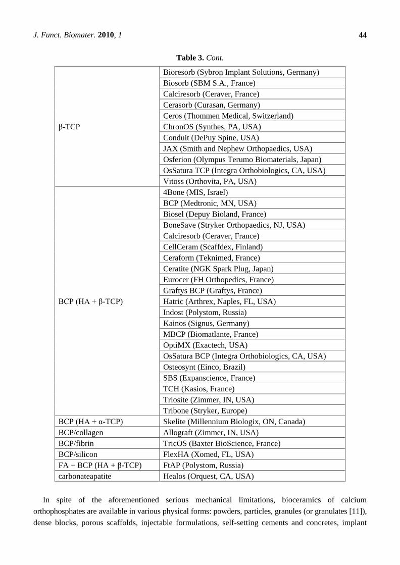
Table 3. Cont.
β-TCP
Bioresorb (Sybron Implant Solutions, Germany)
Biosorb (SBM S.A., France)
Calciresorb (Ceraver, France)
Cerasorb (Curasan, Germany)
Ceros (Thommen Medical, Switzerland)
ChronOS (Synthes, PA, USA)
Conduit (DePuy Spine, USA)
JAX (Smith and Nephew Orthopaedics, USA)
Osferion (Olympus Terumo Biomaterials, Japan)
OsSatura TCP (Integra Orthobiologics, CA, USA)
Vitoss (Orthovita, PA, USA)
BCP (HA + β-TCP)
4Bone (MIS, Israel)
BCP (Medtronic, MN, USA)
Biosel (Depuy Bioland, France)
BoneSave (Stryker Orthopaedics, NJ, USA)
Calciresorb (Ceraver, France)
CellCeram (Scaffdex, Finland)
Ceraform (Teknimed, France)
Ceratite (NGK Spark Plug, Japan)
Eurocer (FH Orthopedics, France)
Graftys BCP (Graftys, France)
Hatric (Arthrex, Naples, FL, USA)
Indost (Polystom, Russia)
Kainos (Signus, Germany)
MBCP (Biomatlante, France)
OptiMX (Exactech, USA)
OsSatura BCP (Integra Orthobiologics, CA, USA)
Osteosynt (Einco, Brazil)
SBS (Expanscience, France)
TCH (Kasios, France)
Triosite (Zimmer, IN, USA)
Tribone (Stryker, Europe)
BCP (HA + α-TCP) Skelite (Millennium Biologix, ON, Canada)
BCP/collagen Allograft (Zimmer, IN, USA)
BCP/fibrin TricOS (Baxter BioScience, France)
BCP/silicon FlexHA (Xomed, FL, USA)
FA + BCP (HA + β-TCP) FtAP (Polystom, Russia)
carbonateapatite Healos (Orquest, CA, USA)
In spite of the aforementioned serious mechanical limitations, bioceramics of calcium
orthophosphates are available in various physical forms: powders, particles, granules (or granulates [11]),
dense blocks, porous scaffolds, injectable formulations, self-setting cements and concretes, implant
J. Funct. Biomater. 2010, 1
45
coatings and composite component of different origin (natural, biological or synthetic) often with
specific shapes, such as implants, prostheses or prosthetic devices (Table 4) [1,108]. Furthermore,
bone grafts are also proposed as non-hardening pastes (=―putty‖). Generally, the latter materials
consist of a mixture of calcium orthophosphate granules and a ―glue‖, typically a highly viscous
hydrogel [1,292]. More to the point, custom-designed shapes like wedges for tibial opening osteotomy,
cones for spine and knee and inserts for vertebral cage fusion are also available [417]. Various
trademarks of the commercially available types of calcium orthophosphate-based bioceramics and
biomaterials are summarized in Table 3.
6.1. Cements and Concretes
The need of bioceramics for minimal invasive surgery has induced the development of a concept of
self-setting bone cements consisting of only calcium orthophosphates to be applied as injectable and/or
mouldable bone substitutes [149,366,367,384,431,462-470]. In addition, there are reinforced
formulations, which, in a certain sense, might be defined as calcium orthophosphate concretes [464].
Furthermore, porous formulations of both the cements and the concretes are available [367,384,465-468].
Calcium orthophosphate cements and concretes belong to low temperature bioceramics. They are
divided into two major groups. The first one is a dry mixture of two different calcium orthophosphates
(a basic one and an acidic one), in which, after being wetted, the setting reaction occurs according to
an acid-base reaction. The second group of the cements contains only one calcium orthophosphate.
Typical examples include ACP with Ca/P molar ratio within 1.50–1.67 and α-TCP: they form CDHA
upon contact with an aqueous solution [149,463,464]. The setting reaction (= hardening, curing) of
these materials is initiated by mixing the initial powder(s) with an aqueous solution. Chemically,
hardening is due to the successive dissolution and precipitation reactions. Mechanically, hardening
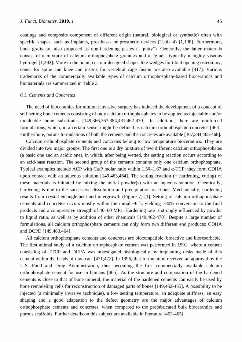
results from crystal entanglement and intergrowth (Figure 7) [1]. Setting of calcium orthophosphate
cements and concretes occurs mostly within the initial ~6 h, yielding ~80% conversion to the final
products and a compressive strength of 40–60 MPa. Hardening rate is strongly influenced by powder
to liquid ratio, as well as by addition of other chemicals [149,462-470]. Despite a large number of
formulations, all calcium orthophosphate cements can only form two different end products: CDHA
and DCPD [149,463,464].
All calcium orthophosphate cements and concretes are biocompatible, bioactive and bioresorbable.
The first animal study of a calcium orthophosphate cement was performed in 1991, where a cement
consisting of TTCP and DCPA was investigated histologically by implanting disks made of this
cement within the heads of nine cats [471,472]. In 1996, that formulation received an approval by the
U.S. Food and Drug Administration, thus becoming the first commercially available calcium
orthophosphate cement for use in humans [465]. As the structure and composition of the hardened
cements is close to that of bone mineral, the material of the hardened cements can easily be used by
bone remodeling cells for reconstruction of damaged parts of bones [149,462-465]. A possibility to be
injected (a minimally invasive technique), a low setting temperature, an adequate stiffness, an easy
shaping and a good adaptation to the defect geometry are the major advantages of calcium
orthophosphate cements and concretes, when compared to the prefabricated bulk bioceramics and
porous scaffolds. Further details on this subject are available in literature [463-465].
J. Funct. Biomater. 2010, 1
46
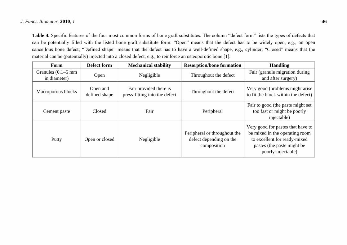
Table 4. Specific features of the four most common forms of bone graft substitutes. The column ―defect form‖ lists the types of defects that
can be potentially filled with the listed bone graft substitute form. ―Open‖ means that the defect has to be widely open, e.g., an open
cancellous bone defect; ―Defined shape‖ means that the defect has to have a well-defined shape, e.g., cylinder; ―Closed‖ means that the
material can be (potentially) injected into a closed defect, e.g., to reinforce an osteoporotic bone [1].
Form Defect form Mechanical stability Resorption/bone formation Handling
Granules (0.1–5 mm
in diameter) Open Negligible Throughout the defect
Fair (granule migration during
and after surgery)
Macroporous blocks Open and
defined shape
Fair provided there is
press-fitting into the defect Throughout the defect
Very good (problems might arise
to fit the block within the defect)
Cement paste Closed Fair Peripheral
Fair to good (the paste might set
too fast or might be poorly
injectable)
Putty Open or closed Negligible
Peripheral or throughout the
defect depending on the
composition
Very good for pastes that have to
be mixed in the operating room
to excellent for ready-mixed
pastes (the paste might be
poorly-injectable)

J. Funct. Biomater. 2010, 1
47
Figure 7. A typical microstructure of a calcium orthophosphate cement after hardening.
The mechanical stability is provided by the physical entanglement of crystals. Reprinted
from [1] with permission.
6.2. Coatings
For many years, the clinical application of calcium orthophosphate-based bioceramics has been
largely limited to non-load bearing parts of the skeleton due to their inferior mechanical properties.
One of the major innovations in the last ~30 years has been to coat mechanically strong bioinert and/or
biotolerant prostheses by calcium orthophosphates [60,473,474]. For example, metallic implants are
encountered in endoprostheses (total hip joint replacements) and artificial teeth sockets. The
requirement for a sufficient mechanical stability necessitates the use of a metallic body for such
devices. As metals do not undergo bone bonding, i.e., do not form a mechanically stable link between
the implant and bone tissue, methods have been sought to improve contacts at the interface. The major
way is to coat metals with calcium orthophosphate bioceramics that exhibit a bone-bonding ability
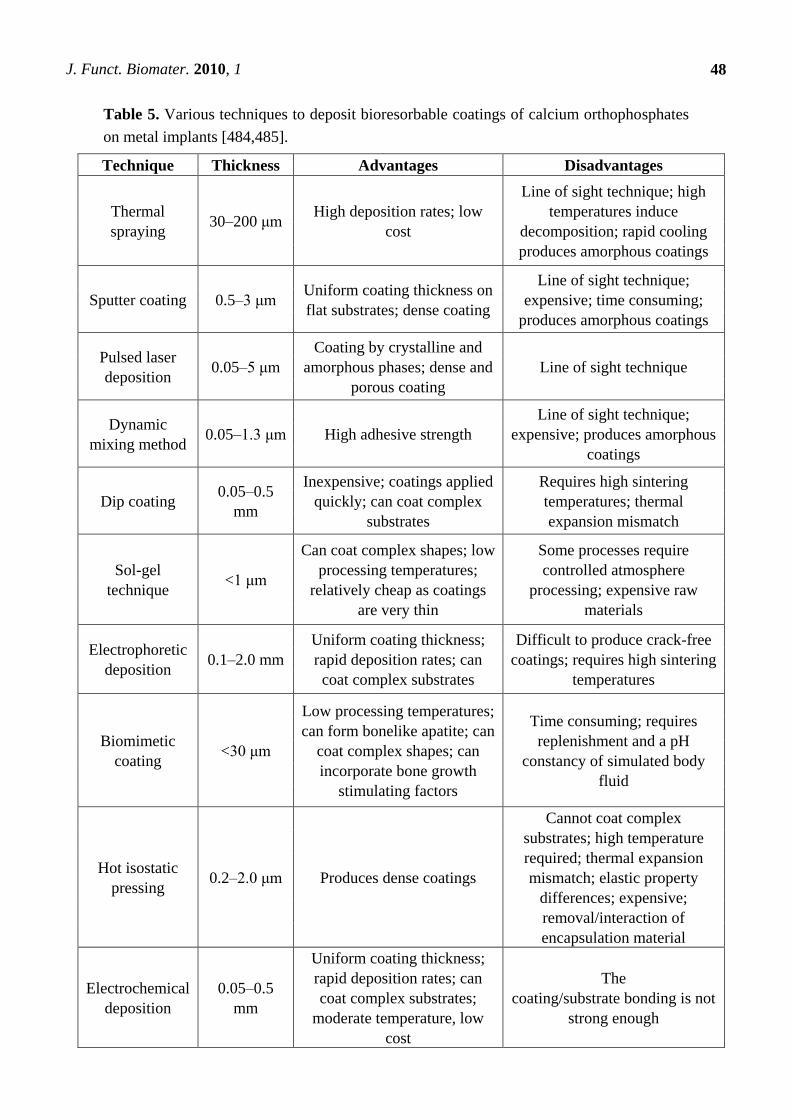
between the metal and bone [60,179,190,315,475-480]. Thickness of the coatings vary from submicron
dimensions to several hundreds microns (Table 5) and this parameter appears to be very important. For
example, if a calcium orthophosphate coating is too thick, it is easy to break. On the contrary, if the
coating is too thin, it is easy to dissolve, because resorbability of HA, which is the second slowest to
dissolve among calcium orthophosphates (Table 1), is about 15–30 μm per year [481]. One should
stress that calcium orthophosphate coatings are not limited to metals only; they can be applied on
carbon, bioinert ceramics and polymers as well [482]. Most important coating techniques are listed in
Table 5, while the main advantages and drawbacks of each coating technique, as well as the major
properties of the deposed calcium orthophosphates, are discussed in detail elsewhere
[60,179,221,272,473,483-497]. Unfortunately, none of these methods can provide the perfect covering
because each coating always contains cracks, pores, second phases and residual stresses that reduced
their durability and might lead to a partial or complete disintegration of the coating in body fluids. The
biomedical aspects of osteoconductive coatings for total joint arthroplasty have been reviewed
elsewhere [498].
J. Funct. Biomater. 2010, 1
48
Table 5. Various techniques to deposit bioresorbable coatings of calcium orthophosphates
on metal implants [484,485].
Technique Thickness Advantages Disadvantages
Thermal
spraying 30–200 μm
High deposition rates; low
cost
Line of sight technique; high
temperatures induce
decomposition; rapid cooling
produces amorphous coatings
Sputter coating 0.5–3 μm Uniform coating thickness on
flat substrates; dense coating
Line of sight technique;
expensive; time consuming;
produces amorphous coatings
Pulsed laser
deposition 0.05–5 μm
Coating by crystalline and
amorphous phases; dense and
porous coating
Line of sight technique
Dynamic
mixing method 0.05–1.3 μm High adhesive strength
Line of sight technique;
expensive; produces amorphous
coatings
Dip coating 0.05–0.5
mm
Inexpensive; coatings applied
quickly; can coat complex
substrates
Requires high sintering
temperatures; thermal
expansion mismatch
Sol-gel
technique <1 μm
Can coat complex shapes; low
processing temperatures;
relatively cheap as coatings
are very thin
Some processes require
controlled atmosphere
processing; expensive raw
materials
Electrophoretic
deposition 0.1–2.0 mm
Uniform coating thickness;
rapid deposition rates; can
coat complex substrates
Difficult to produce crack-free
coatings; requires high sintering
temperatures
Biomimetic
coating <30 μm
Low processing temperatures;
can form bonelike apatite; can
coat complex shapes; can
incorporate bone growth
stimulating factors
Time consuming; requires
replenishment and a pH
constancy of simulated body
fluid
Hot isostatic
pressing 0.2–2.0 μm Produces dense coatings
Cannot coat complex
substrates; high temperature
required; thermal expansion
mismatch; elastic property
differences; expensive;
removal/interaction of
encapsulation material
Electrochemical
deposition
0.05–0.5
mm
Uniform coating thickness;
rapid deposition rates; can
coat complex substrates;
moderate temperature, low
cost
The
coating/substrate bonding is not
strong enough
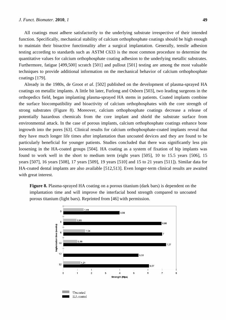
J. Funct. Biomater. 2010, 1
49
All coatings must adhere satisfactorily to the underlying substrate irrespective of their intended
function. Specifically, mechanical stability of calcium orthophosphate coatings should be high enough
to maintain their bioactive functionality after a surgical implantation. Generally, tensile adhesion
testing according to standards such as ASTM C633 is the most common procedure to determine the
quantitative values for calcium orthophosphate coating adhesion to the underlying metallic substrates.
Furthermore, fatigue [499,500] scratch [501] and pullout [501] testing are among the most valuable
techniques to provide additional information on the mechanical behavior of calcium orthophosphate
coatings [179].
Already in the 1980s, de Groot et al. [502] published on the development of plasma-sprayed HA
coatings on metallic implants. A little bit later, Furlong and Osborn [503], two leading surgeons in the
orthopedics field, began implanting plasma-sprayed HA stems in patients. Coated implants combine
the surface biocompatibility and bioactivity of calcium orthophosphates with the core strength of
strong substrates (Figure 8). Moreover, calcium orthophosphate coatings decrease a release of
potentially hazardous chemicals from the core implant and shield the substrate surface from
environmental attack. In the case of porous implants, calcium orthophosphate coatings enhance bone
ingrowth into the pores [63]. Clinical results for calcium orthophosphate-coated implants reveal that
they have much longer life times after implantation than uncoated devices and they are found to be
particularly beneficial for younger patients. Studies concluded that there was significantly less pin
loosening in the HA-coated groups [504]. HA coating as a system of fixation of hip implants was
found to work well in the short to medium term (eight years [505], 10 to 15.5 years [506], 15
years [507], 16 years [508], 17 years [509], 19 years [510] and 15 to 21 years [511]). Similar data for
HA-coated dental implants are also available [512,513]. Even longer-term clinical results are awaited
with great interest.
Figure 8. Plasma-sprayed HA coating on a porous titanium (dark bars) is dependent on the
implantation time and will improve the interfacial bond strength compared to uncoated
porous titanium (light bars). Reprinted from [46] with permission.
J. Funct. Biomater. 2010, 1
50
A number of factors appear to influence the properties of calcium orthophosphate coatings,
including coating thickness (this will influence coating adhesion and fixation—the agreed optimum
now seems to be within 50–100 µm), crystallinity (affecting the dissolution and biological behavior),
phase purity, chemical purity, porosity and adhesion [473,484]. Methods for the production of coatings
and their properties are now largely standardized and, over recent years, calcium
orthophosphate-coated implants have found highly successful clinical application, particularly in
younger patients [514-516]. Further details on calcium orthophosphate coatings can be found in
excellent reviews [517,518].
6.3. Functionally Graded Bioceramics
In general, functionally gradient materials (FGMs) are defined as materials having either a gradient
of compositional or structural changes from their surface to the interior. The idea of FGMs allows one
device to possess two different properties. One of the most important combinations for the biomedical
field is that of mechanical strength and biocompatibility. Namely, only surface properties govern
biocompatibility of the entire device. In contrast, the strongest material determines the mechanical
strength of the entire device. Although, this subject belongs to the coatings section (above), in a certain
sense, metallic implants covered by calcium orthophosphates can be considered as a FGM. The surface
shows excellent biocompatibility because it consists of calcium orthophosphates, while the metallic
core provides excellent mechanical strength. The gradient change from calcium orthophosphate to
metal is important, for example, from the point of thermal expansion.
Functionally graded bioceramics consisting of calcium orthophosphates only [519] have been
developed [377,438,440,522-529]. For example, dense sintered bodies with gradual compositional
changes from α-TCP to HA were prepared by sintering diamond-coated HA compacts at 1280 °C
under a reduced pressure, followed by heating under atmospheric conditions [522]. The content of
α-TCP gradually decreased, while the content of HA increased with increasing depth from the surface.
This functionally gradient bioceramic consisting of an HA core and α-TCP surface showed potential
value as bone-substituting biomaterial [522]. Two types of functionally gradient FA/β-TCP
biocomposites were prepared in another study [523]. As shown in Figure 9, one of the graded
biocomposites was in the shape of a disk and contained four different layers of about 1 mm thickness.
The other graded biocomposite was also in the shape of a disk but contained two sets of four layers,
each layer being 0.5 mm thick, controlled by using a certain amount of the mixed powders. The final
FA/β-TCP graded structures were formed at 100 MPa and sintered at 1300 °C for 2 h [523].
Furthermore, it is known that the bone cross-section from cancellous to cortical bone is non-uniform in
porosity and pore dimensions. Thus, in various attempts to mimic the porous structure of bones, calcium
orthophosphate bioceramics with graded porosity have been fabricated [377,438,440,522-529]. Since
diverse biomedical applications require different configurations and shapes, the graded (or gradient)
porous bioceramics can be grouped according to both the overall shape and the structural
configuration [346]. The basic shapes include rectangular blocks and cylinders (or disks). For the
cylindrical shape, there are configurations of dense core-porous layer, less porous core-more porous
layer, dense layer-porous core and less porous layer-more porous core. For the rectangular shape, in
the gradient direction i.e., the direction with varying porosity, pore size or composition, there are
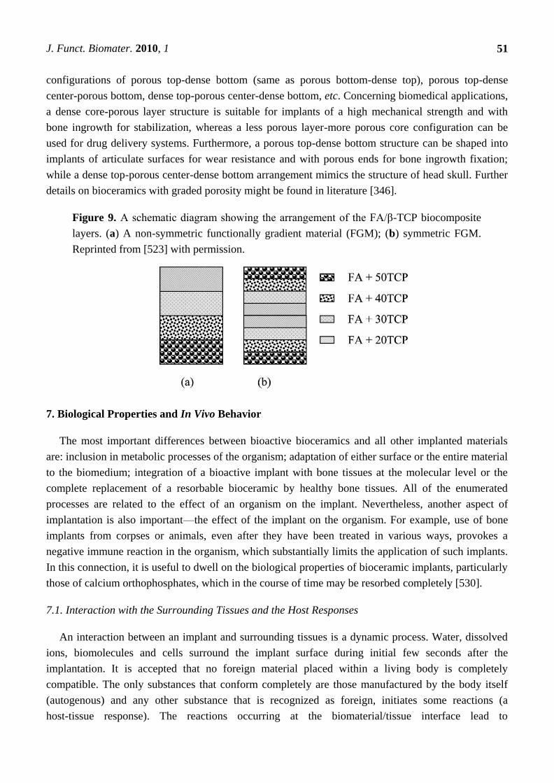
J. Funct. Biomater. 2010, 1
51
configurations of porous top-dense bottom (same as porous bottom-dense top), porous top-dense
center-porous bottom, dense top-porous center-dense bottom, etc. Concerning biomedical applications,
a dense core-porous layer structure is suitable for implants of a high mechanical strength and with
bone ingrowth for stabilization, whereas a less porous layer-more porous core configuration can be
used for drug delivery systems. Furthermore, a porous top-dense bottom structure can be shaped into
implants of articulate surfaces for wear resistance and with porous ends for bone ingrowth fixation;
while a dense top-porous center-dense bottom arrangement mimics the structure of head skull. Further
details on bioceramics with graded porosity might be found in literature [346].
Figure 9. A schematic diagram showing the arrangement of the FA/β-TCP biocomposite
layers. (a) A non-symmetric functionally gradient material (FGM); (b) symmetric FGM.
Reprinted from [523] with permission.
7. Biological Properties and In Vivo Behavior
The most important differences between bioactive bioceramics and all other implanted materials
are: inclusion in metabolic processes of the organism; adaptation of either surface or the entire material
to the biomedium; integration of a bioactive implant with bone tissues at the molecular level or the
complete replacement of a resorbable bioceramic by healthy bone tissues. All of the enumerated
processes are related to the effect of an organism on the implant. Nevertheless, another aspect of
implantation is also important—the effect of the implant on the organism. For example, use of bone
implants from corpses or animals, even after they have been treated in various ways, provokes a
negative immune reaction in the organism, which substantially limits the application of such implants.
In this connection, it is useful to dwell on the biological properties of bioceramic implants, particularly
those of calcium orthophosphates, which in the course of time may be resorbed completely [530].
7.1. Interaction with the Surrounding Tissues and the Host Responses
An interaction between an implant and surrounding tissues is a dynamic process. Water, dissolved
ions, biomolecules and cells surround the implant surface during initial few seconds after the
implantation. It is accepted that no foreign material placed within a living body is completely
compatible. The only substances that conform completely are those manufactured by the body itself
(autogenous) and any other substance that is recognized as foreign, initiates some reactions (a
host-tissue response). The reactions occurring at the biomaterial/tissue interface lead to
J. Funct. Biomater. 2010, 1
52
time-dependent changes in the surface characteristics of both the implanted biomaterials and the
surrounding tissues [54,531].
In order to develop new biomaterials, it is necessary to understand the in vivo host responses. Like
any other species, biomaterials and bioceramics react chemically with their environment and, ideally,
they should neither induce any changes nor provoke undesired reactions in the neighboring or distant
tissues. In general, living organisms can treat artificial implants as biotoxic (or bioincompatible [50]),
bioinert (or biostable [42]), biotolerant (or biocompatible [50]), bioactive and bioresorbable
materials [3-6,38-40,46-50,91,478,530-532]. Biotoxic (e.g., alloys containing cadmium, vanadium,
lead and other toxic elements) materials release substances to the body in toxic concentrations and/or
trigger the formation of antigens that may cause immune reactions ranging from simple allergies to
inflammation to septic rejection with the associated severe health consequences. They cause atrophy,
pathological change or rejection of living tissue near the material due to chemical, galvanic or other
processes. Bioinert [533] (e.g., zirconia, alumina, carbon and titanium) and biotolerant
(e.g., polymethylmethacrylate, titanium and Co-Cr alloy) materials do not release any toxic
constituents but also do not show positive interaction with living tissue. They evoke a physiological
response to form a fibrous capsule, thus, isolating the material from the body. In such cases, thickness
of the layer of fibrous tissue separating the material from other tissues of an organism can serve as a
measure of bioinertness. Generally, both bioactivity and bioresorbability phenomena are fine examples
of chemical reactivity and calcium orthophosphates (both non-substituted and ion-substituted ones) fall
into these two categories of bioceramics [3-6,38-40,46-50,91,478,530-532]. A bioactive material will
dissolve slightly but promote formation of a surface layer of biological apatite before interfacing
directly with the tissue at the atomic level, which results in formation of a direct chemical bonds to
bones. Such an implant provides good stabilization for materials that are subject to mechanical
loading. A bioresorbable material will dissolve over time (regardless of the mechanism leading to the
material removal) and allow a newly formed tissue to grow into any surface irregularities but may not
necessarily interface directly with the material. Consequently, the functions of bioresorbable materials
are to participate in dynamic processes of formation and re-absorption occurring in bone tissues; thus,
bioresorbable materials are used as scaffolds or filling spacers allowing their infiltration and
substitution to the tissues [47,272,534-537].
A distinction between bioactive and bioresorbable bioceramics might be associated with a structural
factor only. For example, bioceramics made from non-porous, dense and highly crystalline HA
behaves as a bioinert (but a bioactive) material and are retained in an organism for at least 5–7 years
without noticeable changes, while a highly porous bioceramics of the same composition can be
resorbed approximately within a year. Furthermore, submicron-sized HA powders are biodegraded
even faster than the highly porous HA scaffolds. Other examples of bioresorbable materials include
porous bioceramic scaffolds made of BCP (which is an intimate mixture of either β-TCP + HA [118-130],
or α-TCP + HA [7-11]) or bone grafts (dense or porous) made of CDHA [538], TCP [377,539,540]
and/or ACP [418,541]. One must stress that recently the concepts of bioactive and bioresorbable
materials have converged and bioactive materials are made bioresorbable, while bioresorbable
materials are made bioactive [542].
In certain in vivo experiments an inflammatory reaction was observed after implantation of calcium
orthophosphate bioceramics [543-545]. Despite this, the general conclusion on using calcium
J. Funct. Biomater. 2010, 1
53
orthophosphates with a Ca/P ionic ratio within 1.0–1.7 is that all types of implants (bioceramics of
various porosities and structures, powders or granules) are not only nontoxic but also induce neither
inflammatory nor foreign-body reactions [546]. The biological response to implanted calcium
orthophosphates follows a similar cascade observed in fracture healing. This cascade includes
hematoma formation, inflammation, neovascularization, osteoclastic resorption and new bone
formation. An intermediate layer of fibrous tissue between the implants and bones has never been
detected. Furthermore, calcium orthophosphate implants display the ability to directly bond to
bones [2-6,38,42,46-52,54,60,530]. For further details, interested readers are referred to a good review
on cellular perspectives of bioceramic scaffolds for bone tissue engineering [374].
One should note that the aforementioned rare cases of inflammatory reactions to calcium
orthophosphate bioceramics were caused by ―other‖ reasons. For example, a high rate of wound
inflammation occurred when highly porous HA was used. In that particular case, the inflammation was
explained by sharp implant edges, which irritated surrounding soft tissues [544]. Another reason for
inflammation produced by porous HA could be due to micro movements of the implants, leading to
simultaneous disruption of a large number of micro-vessels, which grow into the pores of the
bioceramics. This would immediately produce an inflammatory reaction. Additionally, problems could
arise in clinical tests connected with migration of granules used for alveolar ridge augmentation, because
it might be difficult to achieve mechanical stability of implants at the implantation sites [544].
7.2. Osteoinduction
Until recently, it was generally considered, that alone, any type of synthetic bioceramics possessed
neither osteogenic [547] nor osteoinductive [548] properties and demonstrated minimal immediate
structural support. When attached to healthy bones, an osteoid [550] is produced directly onto the
surfaces of bioceramics in the absence of a soft tissue interface. Consequently, the osteoid is
mineralized and the resulting new bone undergoes remodeling [549]. However, several reports have
already shown osteoinductive properties of certain types of calcium orthophosphate
bioceramics [168,415,457,551-563]. Namely, bone formation was found to occur in dog muscle inside
porous calcium orthophosphates with surface microporosity, while bone was not observed on the
surface of dense bioceramics [559]. Furthermore, implantation of porous β-TCP bioceramics appeared
to induce bone formation in soft tissues of dogs, while no bone formation was detected in any α-TCP
implants [556]. More to the point, titanium implants coated by a microporous layer of OCP were found
to induce ectopic bone formation in goat muscles, while a smooth layer of carbonated apatite on the
same implants was not able to induce bone formation there [562,563].
Although the mechanisms of intrinsic osteoinduction of calcium orthophosphate bioceramics are
not unraveled, the dissolution/precipitation behavior of calcium orthophosphates [479], as well as their
microporosity [564,565] and specific surface area [565] have been pointed out as the relevant
parameters. A positive effect of increased microporosity on ectopic bone formation could be direct and
indirect. Firstly, an increased microporosity is directly related to the changes in surface topography,
i.e., increases a surface roughness, which might affect cellular differentiation. Secondly, an increased
microporosity indirectly means a larger surface is exposed to the body fluids, leading to elevated
dissolution/precipitation phenomena as compared to non-microporous surfaces. Furthermore, other
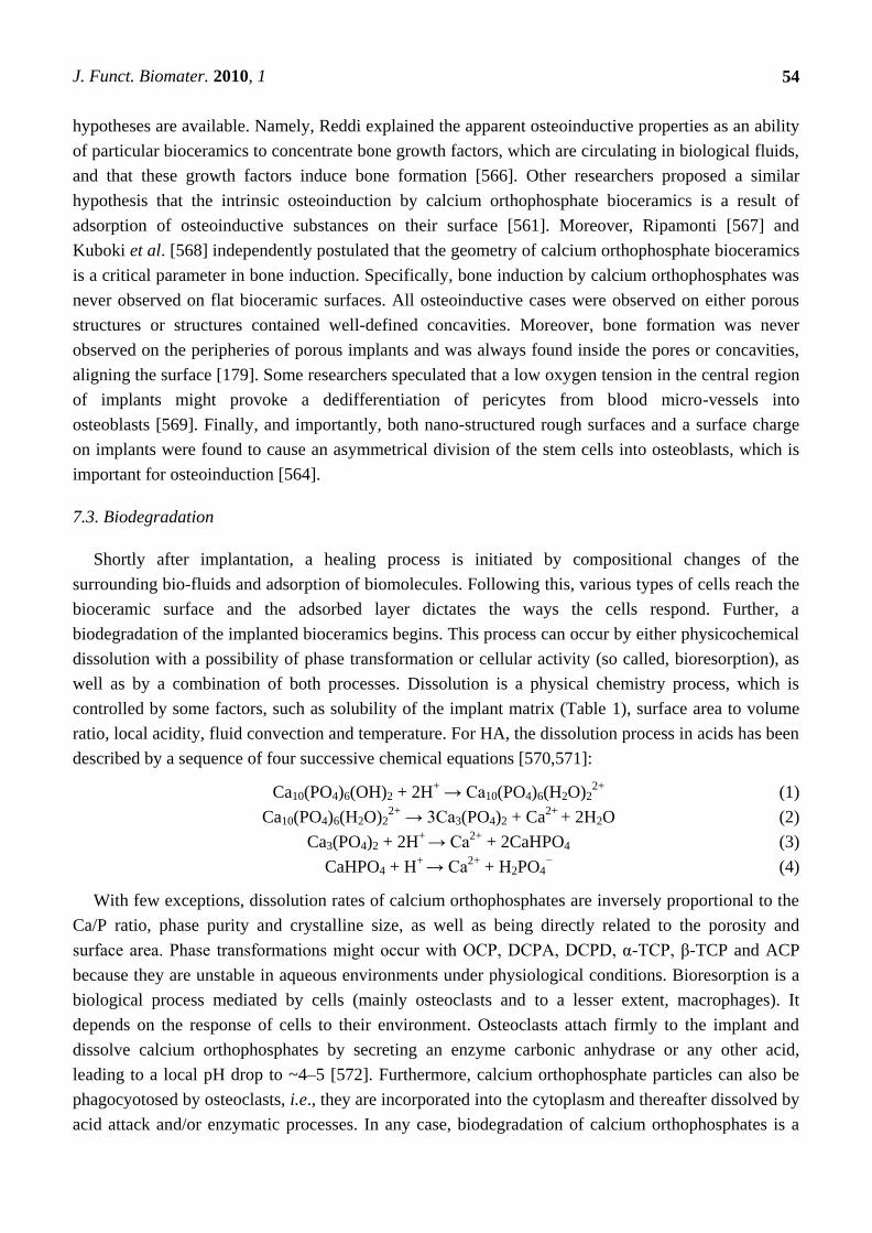
J. Funct. Biomater. 2010, 1
54
hypotheses are available. Namely, Reddi explained the apparent osteoinductive properties as an ability
of particular bioceramics to concentrate bone growth factors, which are circulating in biological fluids,
and that these growth factors induce bone formation [566]. Other researchers proposed a similar
hypothesis that the intrinsic osteoinduction by calcium orthophosphate bioceramics is a result of
adsorption of osteoinductive substances on their surface [561]. Moreover, Ripamonti [567] and
Kuboki et al. [568] independently postulated that the geometry of calcium orthophosphate bioceramics
is a critical parameter in bone induction. Specifically, bone induction by calcium orthophosphates was
never observed on flat bioceramic surfaces. All osteoinductive cases were observed on either porous
structures or structures contained well-defined concavities. Moreover, bone formation was never
observed on the peripheries of porous implants and was always found inside the pores or concavities,
aligning the surface [179]. Some researchers speculated that a low oxygen tension in the central region
of implants might provoke a dedifferentiation of pericytes from blood micro-vessels into
osteoblasts [569]. Finally, and importantly, both nano-structured rough surfaces and a surface charge
on implants were found to cause an asymmetrical division of the stem cells into osteoblasts, which is
important for osteoinduction [564].
7.3. Biodegradation
Shortly after implantation, a healing process is initiated by compositional changes of the
surrounding bio-fluids and adsorption of biomolecules. Following this, various types of cells reach the
bioceramic surface and the adsorbed layer dictates the ways the cells respond. Further, a
biodegradation of the implanted bioceramics begins. This process can occur by either physicochemical
dissolution with a possibility of phase transformation or cellular activity (so called, bioresorption), as
well as by a combination of both processes. Dissolution is a physical chemistry process, which is
controlled by some factors, such as solubility of the implant matrix (Table 1), surface area to volume
ratio, local acidity, fluid convection and temperature. For HA, the dissolution process in acids has been
described by a sequence of four successive chemical equations [570,571]:
Ca10(PO4)6(OH)2 + 2H+ → Ca10(PO4)6(H2O)2
2+ (1)
Ca10(PO4)6(H2O)22+
→ 3Ca3(PO4)2 + Ca2+
+ 2H2O (2)
Ca3(PO4)2 + 2H+
→ Ca2+
+ 2CaHPO4 (3)
CaHPO4 + H+
→ Ca2+
+ H2PO4− (4)
With few exceptions, dissolution rates of calcium orthophosphates are inversely proportional to the
Ca/P ratio, phase purity and crystalline size, as well as being directly related to the porosity and
surface area. Phase transformations might occur with OCP, DCPA, DCPD, α-TCP, β-TCP and ACP
because they are unstable in aqueous environments under physiological conditions. Bioresorption is a
biological process mediated by cells (mainly osteoclasts and to a lesser extent, macrophages). It
depends on the response of cells to their environment. Osteoclasts attach firmly to the implant and
dissolve calcium orthophosphates by secreting an enzyme carbonic anhydrase or any other acid,
leading to a local pH drop to ~4–5 [572]. Furthermore, calcium orthophosphate particles can also be
phagocyotosed by osteoclasts, i.e., they are incorporated into the cytoplasm and thereafter dissolved by
acid attack and/or enzymatic processes. In any case, biodegradation of calcium orthophosphates is a
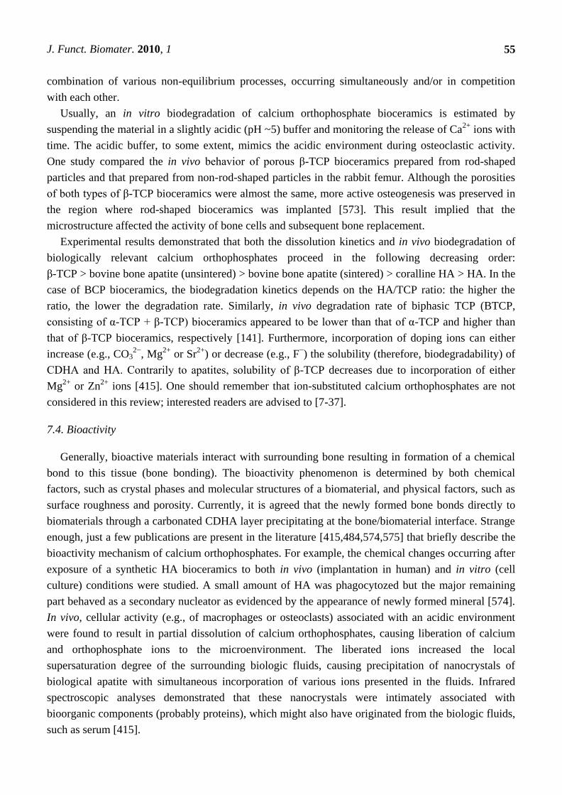
J. Funct. Biomater. 2010, 1
55
combination of various non-equilibrium processes, occurring simultaneously and/or in competition
with each other.
Usually, an in vitro biodegradation of calcium orthophosphate bioceramics is estimated by
suspending the material in a slightly acidic (pH ~5) buffer and monitoring the release of Ca2+
ions with
time. The acidic buffer, to some extent, mimics the acidic environment during osteoclastic activity.
One study compared the in vivo behavior of porous β-TCP bioceramics prepared from rod-shaped
particles and that prepared from non-rod-shaped particles in the rabbit femur. Although the porosities
of both types of β-TCP bioceramics were almost the same, more active osteogenesis was preserved in
the region where rod-shaped bioceramics was implanted [573]. This result implied that the
microstructure affected the activity of bone cells and subsequent bone replacement.
Experimental results demonstrated that both the dissolution kinetics and in vivo biodegradation of
biologically relevant calcium orthophosphates proceed in the following decreasing order:
β-TCP > bovine bone apatite (unsintered) > bovine bone apatite (sintered) > coralline HA > HA. In the
case of BCP bioceramics, the biodegradation kinetics depends on the HA/TCP ratio: the higher the
ratio, the lower the degradation rate. Similarly, in vivo degradation rate of biphasic TCP (BTCP,
consisting of α-TCP + β-TCP) bioceramics appeared to be lower than that of α-TCP and higher than
that of β-TCP bioceramics, respectively [141]. Furthermore, incorporation of doping ions can either
increase (e.g., CO32−
, Mg2+
or Sr2+
) or decrease (e.g., F−) the solubility (therefore, biodegradability) of
CDHA and HA. Contrarily to apatites, solubility of β-TCP decreases due to incorporation of either
Mg2+
or Zn2+
ions [415]. One should remember that ion-substituted calcium orthophosphates are not
considered in this review; interested readers are advised to [7-37].
7.4. Bioactivity
Generally, bioactive materials interact with surrounding bone resulting in formation of a chemical
bond to this tissue (bone bonding). The bioactivity phenomenon is determined by both chemical
factors, such as crystal phases and molecular structures of a biomaterial, and physical factors, such as
surface roughness and porosity. Currently, it is agreed that the newly formed bone bonds directly to
biomaterials through a carbonated CDHA layer precipitating at the bone/biomaterial interface. Strange
enough, just a few publications are present in the literature [415,484,574,575] that briefly describe the
bioactivity mechanism of calcium orthophosphates. For example, the chemical changes occurring after
exposure of a synthetic HA bioceramics to both in vivo (implantation in human) and in vitro (cell
culture) conditions were studied. A small amount of HA was phagocytozed but the major remaining
part behaved as a secondary nucleator as evidenced by the appearance of newly formed mineral [574].
In vivo, cellular activity (e.g., of macrophages or osteoclasts) associated with an acidic environment
were found to result in partial dissolution of calcium orthophosphates, causing liberation of calcium
and orthophosphate ions to the microenvironment. The liberated ions increased the local
supersaturation degree of the surrounding biologic fluids, causing precipitation of nanocrystals of
biological apatite with simultaneous incorporation of various ions presented in the fluids. Infrared
spectroscopic analyses demonstrated that these nanocrystals were intimately associated with
bioorganic components (probably proteins), which might also have originated from the biologic fluids,
such as serum [415].
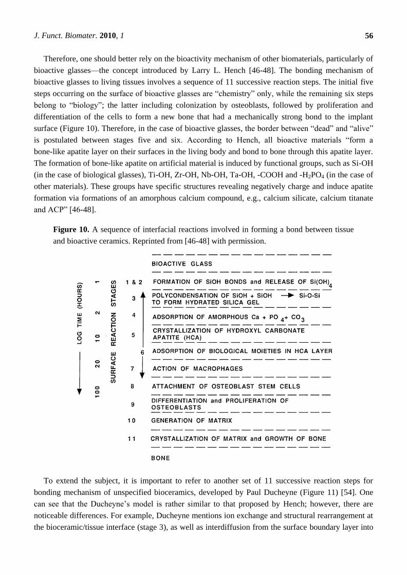
J. Funct. Biomater. 2010, 1
56
Therefore, one should better rely on the bioactivity mechanism of other biomaterials, particularly of
bioactive glasses—the concept introduced by Larry L. Hench [46-48]. The bonding mechanism of
bioactive glasses to living tissues involves a sequence of 11 successive reaction steps. The initial five
steps occurring on the surface of bioactive glasses are ―chemistry‖ only, while the remaining six steps
belong to ―biology‖; the latter including colonization by osteoblasts, followed by proliferation and
differentiation of the cells to form a new bone that had a mechanically strong bond to the implant
surface (Figure 10). Therefore, in the case of bioactive glasses, the border between ―dead‖ and ―alive‖
is postulated between stages five and six. According to Hench, all bioactive materials ―form a
bone-like apatite layer on their surfaces in the living body and bond to bone through this apatite layer.
The formation of bone-like apatite on artificial material is induced by functional groups, such as Si-OH
(in the case of biological glasses), Ti-OH, Zr-OH, Nb-OH, Ta-OH, -COOH and -H2PO4 (in the case of
other materials). These groups have specific structures revealing negatively charge and induce apatite
formation via formations of an amorphous calcium compound, e.g., calcium silicate, calcium titanate
and ACP‖ [46-48].
Figure 10. A sequence of interfacial reactions involved in forming a bond between tissue
and bioactive ceramics. Reprinted from [46-48] with permission.
To extend the subject, it is important to refer to another set of 11 successive reaction steps for
bonding mechanism of unspecified bioceramics, developed by Paul Ducheyne (Figure 11) [54]. One
can see that the Ducheyne’s model is rather similar to that proposed by Hench; however, there are
noticeable differences. For example, Ducheyne mentions ion exchange and structural rearrangement at
the bioceramic/tissue interface (stage 3), as well as interdiffusion from the surface boundary layer into
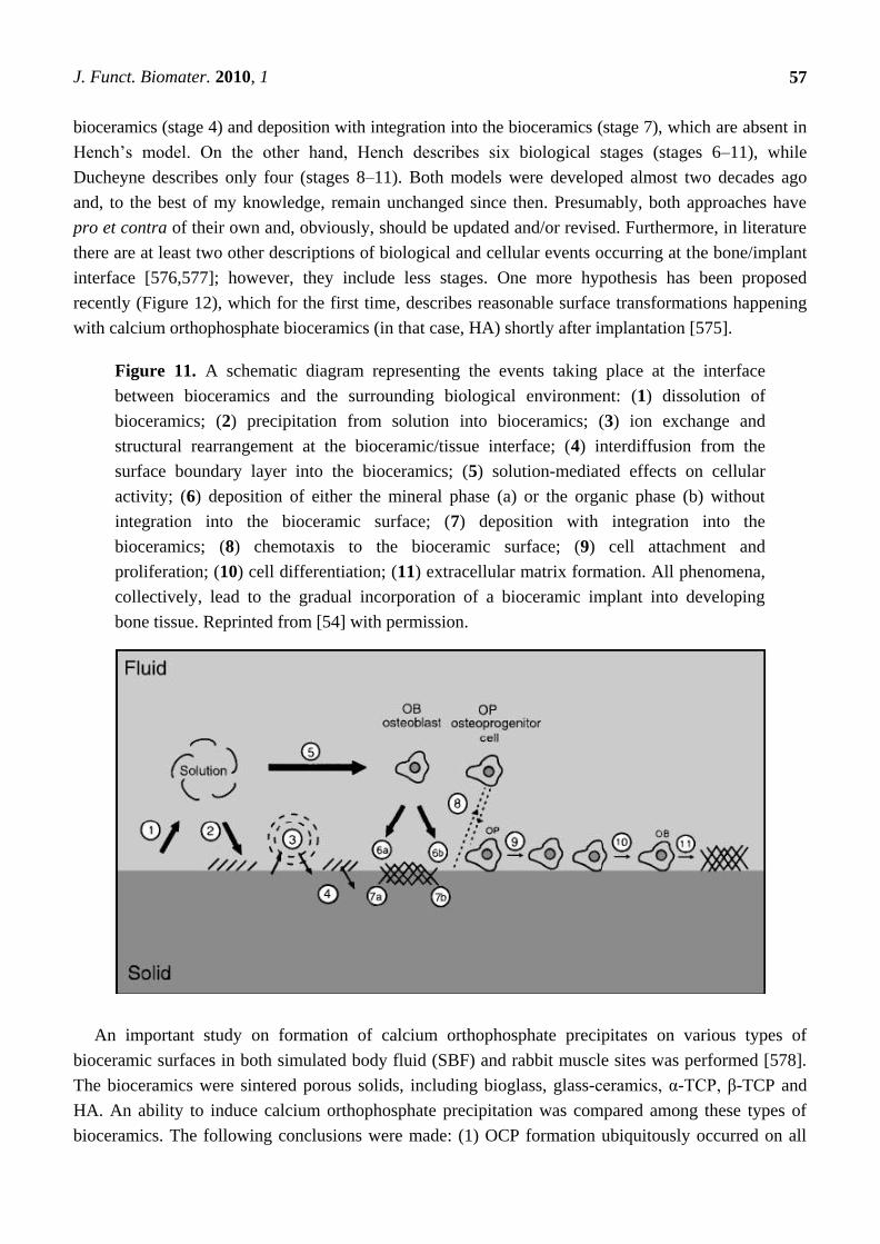
J. Funct. Biomater. 2010, 1
57
bioceramics (stage 4) and deposition with integration into the bioceramics (stage 7), which are absent in
Hench’s model. On the other hand, Hench describes six biological stages (stages 6–11), while
Ducheyne describes only four (stages 8–11). Both models were developed almost two decades ago
and, to the best of my knowledge, remain unchanged since then. Presumably, both approaches have
pro et contra of their own and, obviously, should be updated and/or revised. Furthermore, in literature
there are at least two other descriptions of biological and cellular events occurring at the bone/implant
interface [576,577]; however, they include less stages. One more hypothesis has been proposed
recently (Figure 12), which for the first time, describes reasonable surface transformations happening
with calcium orthophosphate bioceramics (in that case, HA) shortly after implantation [575].
Figure 11. A schematic diagram representing the events taking place at the interface
between bioceramics and the surrounding biological environment: (1) dissolution of
bioceramics; (2) precipitation from solution into bioceramics; (3) ion exchange and
structural rearrangement at the bioceramic/tissue interface; (4) interdiffusion from the
surface boundary layer into the bioceramics; (5) solution-mediated effects on cellular
activity; (6) deposition of either the mineral phase (a) or the organic phase (b) without
integration into the bioceramic surface; (7) deposition with integration into the
bioceramics; (8) chemotaxis to the bioceramic surface; (9) cell attachment and
proliferation; (10) cell differentiation; (11) extracellular matrix formation. All phenomena,
collectively, lead to the gradual incorporation of a bioceramic implant into developing
bone tissue. Reprinted from [54] with permission.
An important study on formation of calcium orthophosphate precipitates on various types of
bioceramic surfaces in both simulated body fluid (SBF) and rabbit muscle sites was performed [578].
The bioceramics were sintered porous solids, including bioglass, glass-ceramics, α-TCP, β-TCP and
HA. An ability to induce calcium orthophosphate precipitation was compared among these types of
bioceramics. The following conclusions were made: (1) OCP formation ubiquitously occurred on all
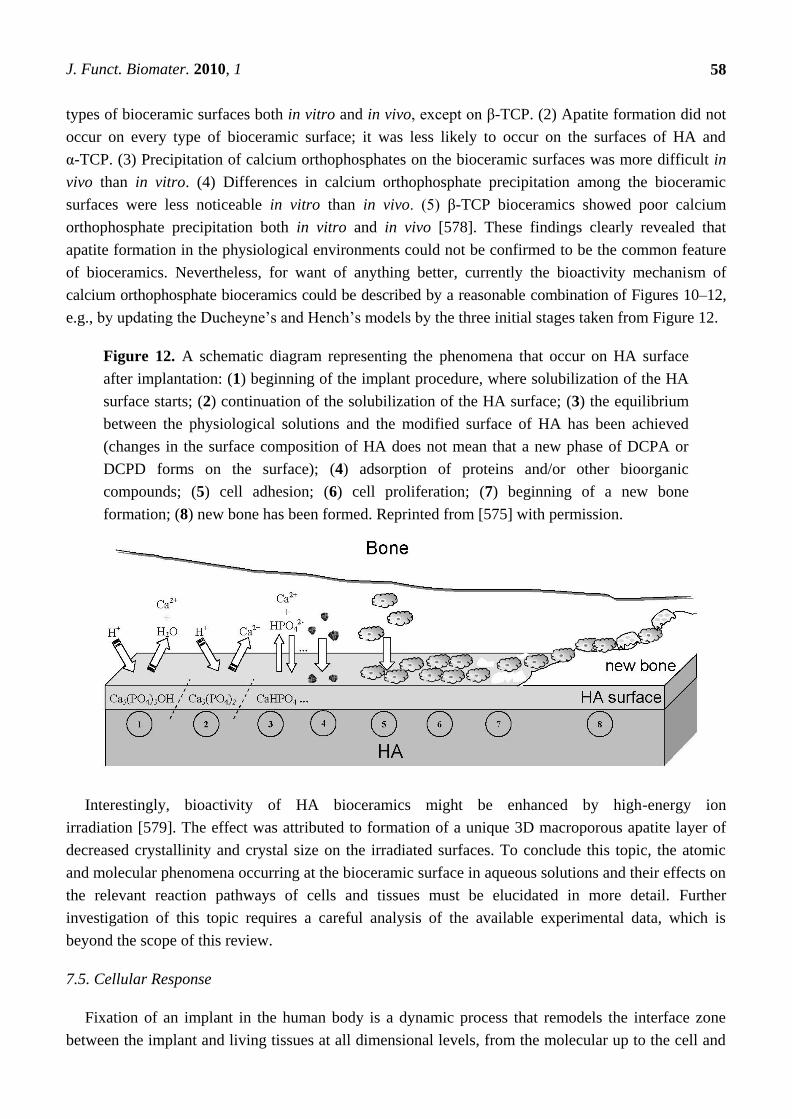
J. Funct. Biomater. 2010, 1
58
types of bioceramic surfaces both in vitro and in vivo, except on β-TCP. (2) Apatite formation did not
occur on every type of bioceramic surface; it was less likely to occur on the surfaces of HA and
α-TCP. (3) Precipitation of calcium orthophosphates on the bioceramic surfaces was more difficult in
vivo than in vitro. (4) Differences in calcium orthophosphate precipitation among the bioceramic
surfaces were less noticeable in vitro than in vivo. (5) β-TCP bioceramics showed poor calcium
orthophosphate precipitation both in vitro and in vivo [578]. These findings clearly revealed that
apatite formation in the physiological environments could not be confirmed to be the common feature
of bioceramics. Nevertheless, for want of anything better, currently the bioactivity mechanism of
calcium orthophosphate bioceramics could be described by a reasonable combination of Figures 10–12,
e.g., by updating the Ducheyne’s and Hench’s models by the three initial stages taken from Figure 12.
Figure 12. A schematic diagram representing the phenomena that occur on HA surface
after implantation: (1) beginning of the implant procedure, where solubilization of the HA
surface starts; (2) continuation of the solubilization of the HA surface; (3) the equilibrium
between the physiological solutions and the modified surface of HA has been achieved
(changes in the surface composition of HA does not mean that a new phase of DCPA or
DCPD forms on the surface); (4) adsorption of proteins and/or other bioorganic
compounds; (5) cell adhesion; (6) cell proliferation; (7) beginning of a new bone
formation; (8) new bone has been formed. Reprinted from [575] with permission.
Interestingly, bioactivity of HA bioceramics might be enhanced by high-energy ion
irradiation [579]. The effect was attributed to formation of a unique 3D macroporous apatite layer of
decreased crystallinity and crystal size on the irradiated surfaces. To conclude this topic, the atomic
and molecular phenomena occurring at the bioceramic surface in aqueous solutions and their effects on
the relevant reaction pathways of cells and tissues must be elucidated in more detail. Further
investigation of this topic requires a careful analysis of the available experimental data, which is
beyond the scope of this review.
7.5. Cellular Response
Fixation of an implant in the human body is a dynamic process that remodels the interface zone
between the implant and living tissues at all dimensional levels, from the molecular up to the cell and
J. Funct. Biomater. 2010, 1
59
tissue morphology level, and at all time scales, from the first second up to several years after
implantation. Immediately following implantation, a space filled with bio-fluids appears next to the
implant surface. With time, proteins adsorb at the bioceramic surface and gives rise to osteoinduction
by cell proliferation and their differentiation towards bone cells, revascularization and eventual gap
closing. Ideally, a strong bond forms between the implant and surrounding tissues [50]. A detailed
study on interfacial interactions between calcined HA and substrates has been performed
recently [580].
Osteoblasts cultured on HA bioceramics are generally reported to be completely flattened, leading
to difficulties in distinguishing the cytoplasmic edge from the HA surfaces after ~2 hours
incubation [581]. These observations underscore an expected bioactivity of HA and make HA
bioceramics well suited for bone reconstruction. Osteoblasts cultured on porous HA bioceramics
appeared to exhibit higher adhesion, enhanced differentiation and suppressed proliferation rates when
compared to the non-porous controls [582,583]. Furthermore, formation of distinct resorption pits on
HA [584] and β-TCP [573] surfaces in the presence of osteoclasts was observed. Moreover, a surface
roughness of calcium orthophosphate bioceramics was reported to strongly influence the activation of
mononuclear precursors to mature osteoclasts [584].
Cellular biodegradation of calcium orthophosphate bioceramics is known to depend on its phases.
For example, a higher solubility of β-TCP prevented L-929 fibroblast cell adhesion, thereby leading to
damage and rupture of the cells [585]. A mouse ectopic model study indicated the maximal bone
growth for the 80:20 β-TCP:HA biphasic formulations preloaded with human mesenchymal stem cells
when compared to other calcium orthophosphates [586]. The effects of substrate microstructure and
crystallinity have been corroborated with an in vivo rabbit femur model, where rod-like crystalline
β-TCP was reported to enhance osteogenesis when compared to non-rod like crystalline β-TCP [573].
Additionally, using a dog mandibular defect model, a higher bone formation on a scaffold surface
coated by nano-dimensional HA was observed when compared to that coated by a micro-dimensional
HA [587]. Furthermore, recent studies revealed a stronger stress signaling response by osteoblast
precursor cells in 3D scaffolds when compared to 2D surfaces [588].
Mesenchymal stem cells are one of the most attractive cellular lines for application as bone
grafts [589]. Early investigations by Okumura et al. indicated an adhesion, proliferation and
differentiation, which ultimately became new bone and integrated with porous HA bioceramics [590].
Recently, Unger et al. showed a sustained co-culture of endothelial cells and osteoblasts on HA
scaffolds for up to six weeks [591]. Furthermore, a release of factors by endothelial and osteoblast
cells in co-culture supported proliferation and differentiation was suggested to ultimately result in
microcapillary-like vessel formation and supported a neo-tissue growth within the scaffold [374].
More to the point, investigation of rat calvaria osteoblasts cultured on transparent HA bioceramics, as
well as the analysis of osteogenic-induced human bone marrow stromal cells at different time points of
culturing, indicated a good cytocompatibility of HA bioceramics and revealed favorable cell
proliferation [343]. Positive results for other types of cells have been obtained in other
studies [191,338,339,342].
Interestingly, HA scaffolds with marrow stromal cells in a perfused environment were reported to
result in ~85% increase in mean core strength, a ~130% increase in failure energy and a ~355%
increase in post-failure strength. The increase in mineral quantity and promotion of uniform mineral
J. Funct. Biomater. 2010, 1
60
distribution in that study was suggested to attribute to the perfusion effect [449]. Additionally, other
investigators indicated mechanical properties increased for other calcium orthophosphate scaffolds
after induced osteogenesis [448,451].
Furthermore, the dimensions, extent and interconnectivity of pores in bioceramics are known to
influence bone in-growth, blood vessels formation and canaliculi networks [409,410,457]. Initial reports
have estimated a minimum pore size of ~50 μm for blood vessel formation and a minimum pore size of
~200 μm for osteonal in-growth [457]. Pore dimensions of ~100 μm and even ~50 μm [592] were
reported in later studies to support bone in-growth. Additionally, vascularization, cell migration and
nutrient diffusion required for sustained cell viability and tissue function are possible if pores within the
scaffolds are well interconnected. For example, an essential mean pore interconnection size of ~10 μm
was necessary to allow cell migration between the pores [593]. As such, both porosity and general
architecture are critical in determining the rate of fluid transport through porous bioceramics, which, in
turn, determines the rate and degree of bone ingrowth in vivo [122,412,413,594].
8. Calcium Orthophosphate Bioceramics in Tissue Engineering
8.1. Tissue Engineering
All modern orthopedic implants lack three of the most critical abilities of living tissues:
(i) self-repairing; (ii) maintaining blood supply; (iii) self-modifying their structure and properties in
response to external aspects such as a mechanical load [429]. Needless to mention, bones not only
possess all of these properties but, in addition, are self-generating, hierarchical, multifunctional,
nonlinear, composite and biodegradable; therefore, the ideal artificial bone grafts must possess similar
properties [117].
The last decades have seen a surge in creative ideas and technologies developed to tackle the
problem of repairing or replacing diseased and damaged tissues, leading to the emergence of a new
field in healthcare technology now referred to as tissue engineering. This is an interdisciplinary field
that exploits a combination of living cells, engineering materials and suitable biochemical factors to
improve, replace, restore, maintain or enhance living tissues and whole organs [595,596]. However, as
two of three major components (namely, cells and biochemical factors) of the tissue engineering
subject appear to be far beyond the scope of this review, the topic of tissue engineering is limited to
the engineering materials prepared from calcium orthophosphate bioceramics only.
Regeneration, rather than repair, is the central goal of any tissue engineering strategy [597]. Thus,
tissue engineering has potential to create tissues and organs de novo. This field of science [599] started
more than two decades ago [600,601] and a famous review article by Langer and Vacanti [602] has
greatly contributed to the promotion of tissue engineering research worldwide. The field of tissue
engineering, particularly when applied to bone substitutes where tissues often function in a
mechanically demanding environment [603], requires a collaboration of excellence in cell and
molecular biology, biochemistry, material sciences, bioengineering and clinical research. For the
success, it is necessary that researchers with expertise in one area have an appreciation of the
knowledge and challenges of the other areas. However, since the technical, regulatory and commercial
challenges might be substantial, the introduction of new products is likely to be slow [598].
J. Funct. Biomater. 2010, 1
61
Nowadays tissue engineering is at full research potential due to the following key advantages:
(i) the solutions it provides are long-term, much safer than other options and cost-effective as well;
(ii) the need for a donor tissue is minimal, which eliminates the immuno-suppression problems;
(iii) the presence of residual foreign material is eliminated as well.
8.2. Scaffolds and Their Properties
It would be very convenient to both patients and physicians if devastated tissues or organs of
patients could be regenerated by simple cell injections to the target sites, but such cases are rare. The
majority of large-sized tissues and organs with distinct 3D form require a support for their formation
from cells. The support is named a scaffold [604], template and/or artificial extracellular
matrix (ECM) [151,152,386,600,603,605-608]. The major function of scaffolds is similar to that of the
natural ECM that assists proliferation, differentiation and biosynthesis of cells. In addition, scaffolds
placed at the regeneration sites will prevent disturbing cells from invasion into the sites of
action [609,610]. The role of scaffolds was perfectly described by Andrés Segovia (1893–1987), a
Spanish classical guitarist: ―When one puts up a building one makes an elaborate scaffold to get
everything into its proper place. But when one takes the scaffold down, the building must stand by
itself with no trace of the means by which it was erected. That is how a musician should work.‖
The idea behind tissue engineering is to create or engineer autografts by either expanding
autologous cells in vitro guided by a scaffold or implanting an acellular template in vivo and allowing
the patient’s cells to repair the tissue guided by the scaffold. The first phase is the in vitro formation of
a tissue construct by placing the chosen cells and scaffolds in a metabolically and mechanically
supportive environment with growth media (in a bioreactor), in which the cells proliferate and
elaborate extracellular matrix. It is expected that cells infiltrate into the porous matrix and
consequently proliferate and differentiate therein. In the second phase, the construct is implanted in the
appropriate anatomic location, where remodeling in vivo is intended to recapitulate the normal
functional architecture of an organ or a tissue [611,612]. The key processes occurring during both in
vitro and in vivo phases of tissue formation and maturation are: (1) cell proliferation, sorting and
differentiation, (2) extracellular matrix production and organization, (3) biodegradation of the scaffold,
(4) remodeling and potentially growth of the tissue.
To achieve the goal of tissue reconstruction, the scaffolds must meet several specific
requirements [151,152,605]. A reasonable surface roughness is necessary to facilitate cell seeding and
fixation [613,614]. A sufficient mechanical strength and stiffness are mandatory to oppose contraction
forces and later for the remodeling of damaged tissues. A high porosity and adequate pore dimensions
(Tables 2 and 6) are very important to allow cell migration, vascularization, as well as diffusion of
nutrients [352]. Namely, scaffolds should have a network of interconnected pores where more than
~60% of the pores should have a size ranging from ~150 μm to ~400 μm and at least ~20% should be
smaller than ~20 μm [11,105,352,362,362,409-415,457,615-621]. Scaffolds must be manufactured
from materials with controlled biodegradability and/or bioresorbability, such as calcium
orthophosphate bioceramics, so that new bone will eventually replace the scaffold [622]. Furthermore,
the resorption rate has to coincide as much as possible with the rate of bone formation (i.e., between a
few months and about two years) [623]. This means that while cells are fabricating their own natural
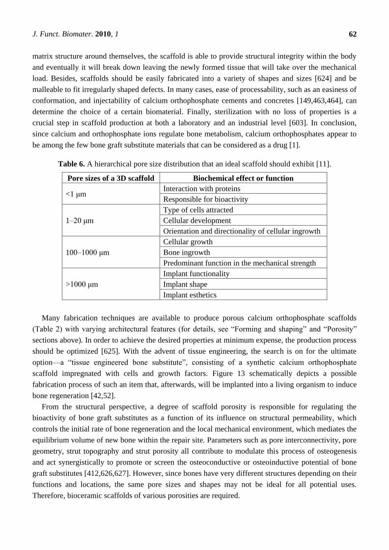
J. Funct. Biomater. 2010, 1
62
matrix structure around themselves, the scaffold is able to provide structural integrity within the body
and eventually it will break down leaving the newly formed tissue that will take over the mechanical
load. Besides, scaffolds should be easily fabricated into a variety of shapes and sizes [624] and be
malleable to fit irregularly shaped defects. In many cases, ease of processability, such as an easiness of
conformation, and injectability of calcium orthophosphate cements and concretes [149,463,464], can
determine the choice of a certain biomaterial. Finally, sterilization with no loss of properties is a
crucial step in scaffold production at both a laboratory and an industrial level [603]. In conclusion,
since calcium and orthophosphate ions regulate bone metabolism, calcium orthophosphates appear to
be among the few bone graft substitute materials that can be considered as a drug [1].
Table 6. A hierarchical pore size distribution that an ideal scaffold should exhibit [11].
Pore sizes of a 3D scaffold Biochemical effect or function
<1 μm Interaction with proteins
Responsible for bioactivity
1–20 μm
Type of cells attracted
Cellular development
Orientation and directionality of cellular ingrowth
100–1000 μm
Cellular growth
Bone ingrowth
Predominant function in the mechanical strength
>1000 μm
Implant functionality
Implant shape
Implant esthetics
Many fabrication techniques are available to produce porous calcium orthophosphate scaffolds
(Table 2) with varying architectural features (for details, see ―Forming and shaping‖ and ―Porosity‖
sections above). In order to achieve the desired properties at minimum expense, the production process
should be optimized [625]. With the advent of tissue engineering, the search is on for the ultimate
option—a ―tissue engineered bone substitute‖, consisting of a synthetic calcium orthophosphate
scaffold impregnated with cells and growth factors. Figure 13 schematically depicts a possible
fabrication process of such an item that, afterwards, will be implanted into a living organism to induce
bone regeneration [42,52].
From the structural perspective, a degree of scaffold porosity is responsible for regulating the
bioactivity of bone graft substitutes as a function of its influence on structural permeability, which
controls the initial rate of bone regeneration and the local mechanical environment, which mediates the
equilibrium volume of new bone within the repair site. Parameters such as pore interconnectivity, pore
geometry, strut topography and strut porosity all contribute to modulate this process of osteogenesis
and act synergistically to promote or screen the osteoconductive or osteoinductive potential of bone
graft substitutes [412,626,627]. However, since bones have very different structures depending on their
functions and locations, the same pore sizes and shapes may not be ideal for all potential uses.
Therefore, bioceramic scaffolds of various porosities are required.
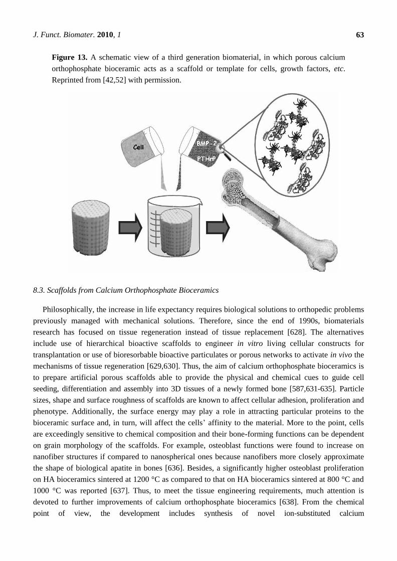
J. Funct. Biomater. 2010, 1
63
Figure 13. A schematic view of a third generation biomaterial, in which porous calcium
orthophosphate bioceramic acts as a scaffold or template for cells, growth factors, etc.
Reprinted from [42,52] with permission.
8.3. Scaffolds from Calcium Orthophosphate Bioceramics
Philosophically, the increase in life expectancy requires biological solutions to orthopedic problems
previously managed with mechanical solutions. Therefore, since the end of 1990s, biomaterials
research has focused on tissue regeneration instead of tissue replacement [628]. The alternatives
include use of hierarchical bioactive scaffolds to engineer in vitro living cellular constructs for
transplantation or use of bioresorbable bioactive particulates or porous networks to activate in vivo the
mechanisms of tissue regeneration [629,630]. Thus, the aim of calcium orthophosphate bioceramics is
to prepare artificial porous scaffolds able to provide the physical and chemical cues to guide cell
seeding, differentiation and assembly into 3D tissues of a newly formed bone [587,631-635]. Particle
sizes, shape and surface roughness of scaffolds are known to affect cellular adhesion, proliferation and
phenotype. Additionally, the surface energy may play a role in attracting particular proteins to the
bioceramic surface and, in turn, will affect the cells’ affinity to the material. More to the point, cells
are exceedingly sensitive to chemical composition and their bone-forming functions can be dependent
on grain morphology of the scaffolds. For example, osteoblast functions were found to increase on
nanofiber structures if compared to nanospherical ones because nanofibers more closely approximate
the shape of biological apatite in bones [636]. Besides, a significantly higher osteoblast proliferation
on HA bioceramics sintered at 1200 °C as compared to that on HA bioceramics sintered at 800 °C and
1000 °C was reported [637]. Thus, to meet the tissue engineering requirements, much attention is
devoted to further improvements of calcium orthophosphate bioceramics [638]. From the chemical
point of view, the development includes synthesis of novel ion-substituted calcium
J. Funct. Biomater. 2010, 1
64
orthophosphates [7-37]. From the material point of view, the major research topics include
nanodimensional and nanocrystalline structures [639-643], organic-inorganic biocomposites and hybrid
biomaterials [292], fibers, whiskers and filaments [644-656], micro- and nanospheres and beads [656-669],
porous 3D scaffolds made of ACP [418], TCP [396,397], HA [167,364,365,398,400,441,625,670-675] and
various biphasic formulations [440,667,676-680], structures with graded porosity [377,438,440,524-529]
and hierarchically organized ones [681,682]. Furthermore, an addition of defects through an intensive
milling [683,684] or their removal by a thermal treatment [685] can be used to modify a chemical
reactivity of calcium orthophosphates. In addition, more attention should be paid to crystallographically
aligned calcium orthophosphate bioceramics [686].
There are three principal therapeutic strategies for treating diseased or injured tissues in patients:
(i) implantation of freshly isolated or cultured cells; (ii) implantation of tissues assembled in vitro from
cells and scaffolds; (iii) in situ tissue regeneration. For cellular implantation, individual cells or small
cellular aggregates from the patient or a donor are either injected into the damaged tissue directly or
are combined with a degradable scaffold in vitro and then implanted. For tissue implantation, a
complete 3D tissue is grown in vitro using patient or donor cells and a bioresorbable scaffold and then
is implanted into the patients to replace diseased or damaged tissues. For in situ regeneration, a
scaffold implanted directly into the injured tissue stimulates the body’s own cells to promote local
tissue repair [595,687]. In any case, simply trapping cells at the particular point on a surface is not
enough: the cells must be encouraged to differentiate, which is impossible without the presence of
suitable biochemical factors [688]. All the previously mentioned clearly indicate that for the purposes
of tissue engineering, calcium orthophosphate bioceramics play an auxiliary role; namely, they act as a
suitable material to manufacture the appropriate 3D templates, substrates or scaffolds to be colonized
by living cells before the successive implantation. The in vitro evaluation of potential calcium
orthophosphate scaffolds for tissue engineering has been described elsewhere [689], and data on the
mechanical properties of calcium orthophosphate bioceramics for use in tissue engineering are also
available [690,691]. The effect of a HA-based biomaterial on gene expression in osteoblast-like cells
was reported as well [692]. To conclude, the excellent biocompatibility of calcium orthophosphate
bioceramics, their possible osteoinductivity [168,415,457,551-563] and a high affinity for drugs,
proteins and cells make them very functional for tissue engineering applications. The feasible
production of scaffolds with tailored structures and properties opens up a spectacular future for
calcium orthophosphates [692-699].
8.4. A Clinical Experience
During the last decade, several groups have made steps towards a clinical application of cell-seeded
calcium orthophosphate bioceramics for bone tissue engineering of humans. For example,
Quarto et al. [700] were the first to report treatment of large (4–7 cm) bone defects of the tibia, ulna
and humerus in three patients from 16 to 41 years old, where conventional surgical therapies had
failed. The authors implanted a custom-made unresorbable porous HA scaffold seeded with in vitro
expanded autologous bone marrow stromal cells. In all three patients, radiographs and computed
tomographic scans revealed abundant callus formation along the implants and good integration at the
interfaces with the host bones by the second month after surgery [700]. In the same year,
J. Funct. Biomater. 2010, 1
65
Vacanti et al. [701] reported the case of a man who had a traumatic avulsion of the distal phalanx of a
thumb. The phalanx was replaced with a specially treated natural coral (porous HA; 500-pore
ProOsteon (see Table 3)) implant that was previously seeded with in vitro expanded autologous
periosteal cells. The procedure resulted in the functional restoration of a stable and biomechanically
sound thumb of normal length, without the pain and complications that are usually associated with
harvesting a bone graft.
Morishita et al. [702] treated a defect resulting from surgery of benign bone tumors in three patients
using HA scaffolds seeded with in vitro expanded autologous bone marrow stromal cells after
osteogenic differentiation of the cells. Two bone defects in a tibia and one defect in a femur were
treated. Although ectopic implants in nude mice showed the osteogenicity of the cells, details such as
the percentage of the implants containing bone and at what quantities were not reported. Furthermore,
cell-seeded calcium orthophosphate scaffolds were found to be superior to autograft, allograft or cell-
seeded allograft in terms of bone formation at ectopic implantation sites [703].
To conclude this part, one should mention that a cell seeding method for practical clinical
experience is not enough. Although cell seeding into scaffolds at high density is closely associated
with enhancement of tissue formation in 3D constructs (i.e., cartilage [704] and bone [705]), effective
and high-density cell seeding into 3D scaffolds is difficult to achieve. Technical difficulties in cell
seeding are caused by the complex structure of the scaffold and insufficient migration into the
scaffolds due to pore size and material, which prolongs the culture period because of the shortage of
initially seeded cells. Recently, Shimizu et al. [706] proposed a methodology for tissue engineering
using magnetite nanoparticles and magnetic force. This method has been applied to a cell seeding
process and attractive results have been reported.
9. Conclusions and Outlook
The available chronology of the search for a suitable bioceramic for bone substitutes is as follows:
since the 1950s, the first aim was to use bioinert [533] bioceramics, which had no reaction with living
tissues. Later on, in the 1980s, the trend changed towards the opposite: the idea was to implant
bioceramics that reacted with the surrounding tissues by producing newly formed bone. These two
stages have been referred to as the first and the second generations of bioceramics, respectively [707].
In the current century, we search for the third generation of bioceramics [687], which will be able to
regenerate bone tissues by stimulating specific responses at the molecular level [42,52]. One should
note that these three generations should not be interpreted as the chronological but the conceptual,
since each generation represents an evolution on the requirements and properties of the biomaterials
involved. This means that at present, research and development is still devoted to biomaterials and
bioceramics that, according to their properties, could be considered to be of the first or the second
generations, because the second generation of bioceramics with added porosity is one of the initial
approaches in developing the third generation of bioceramics [708]. Furthermore, there is another
classification of the history of biomaterials introduced by James M. Anderson. According to Anderson,
between 1950–1975, researchers studied biomaterials, between 1975–2000 they studied biomaterials
and since 2000 the time for biomaterials has been coming [709]; here, the italicized letters emphasize
the major direction of the research efforts in the complex subject of biomaterials. As bioceramics are
J. Funct. Biomater. 2010, 1
66
biomaterials of the ceramic origin (see Section 2), Anderson’s historical classification appears to be
applicable to the bioceramics field.
The field of biomaterials is in the midst of a revolutionary change in which the life sciences are
becoming equal in importance to materials science and engineering as the foundation of the field.
Simultaneously, advances in engineering (for example nanotechnology) are greatly increasing the
sophistication with which biomaterials can be designed, allowing fabrication of biomaterials with
increasingly complex functions [76]. Specifically, during the last ~40 years, calcium orthophosphate
bioceramics has become an integral and vital segment of our modern health care delivery system. In
the modern fields of the third generation bioceramics (Hench) or BIOceramics (Anderson), the full
potential of calcium orthophosphates has only begun to be recognized. Namely, calcium
orthophosphates, which were intended as osteoconductive bioceramics in the past, stand for materials
to fabricate osteoinductive implants nowadays [168,415,457,551-563]. The initial steps in this
direction have been already made by both fabricating BCP-based scaffolds for bone tissue engineering
through the design of controlled 3D-porous structures and increasing the biological activity through
development of novel ion-substituted calcium orthophosphate bioceramics [11,417]. In the future, the
composition, microstructure and molecular surface chemistry of various types of calcium
orthophosphates will be tailored to match the specific biological and metabolic requirements of tissues
or disease states. This new generation of calcium orthophosphate bioceramics should enhance the
quality of life of millions of people as they grow older [710].
In spite of the great progress, there is still great potential for major advances to be made in the field
of calcium orthophosphate bioceramics [4]. This includes requirements for:
• Improvement of the mechanical performance of existing types of bioceramics;
• Enhanced bioactivity in terms of gene activation;
• Improvement in the performance of biomedical coatings in terms of their mechanical
stability and ability to deliver biological agents;
• Development of smart biomaterials capable of combining sensing with bioactivity;
• Development of improved biomimetic composites.
Furthermore, there is still need for a better understanding of the biological systems. For example,
the bonding mechanism between the bone mineral and collagen remains unclear. It is also unclear
whether a rapid repair that is elicited by the new generation of bioceramics results from the
enhancement of mineralization per se or whether there is a more complex signaling process involving
proteins in collagen. If we were able to understand the fundamentals of bone response to specific ions
and the signals they activate, then we could design better bioceramics for the future [4].
To finalize this review, it is obvious that the present status of research and development in the field
of calcium orthophosphate bioceramics is still at the starting point for the solution of new problems at
the confluence of materials science, biology and medicine, concerned with the restoration of damaged
functions in humans. A large increase in active elderly people has dramatically raised the need for
load-bearing bone graft substitutes, for example for bone reconstruction during revision arthroplasty or
for the reinforcement of osteoporotic bones. Strategies applied in the last four decades towards this
goal have failed. New strategies, possibly based on self-assembling and/or nanofabrication, have to be
proposed and developed [711]. Furthermore, it should be feasible to design a new generation of
J. Funct. Biomater. 2010, 1
67
gene-activating calcium orthophosphate based scaffolds tailored for specific patients and disease states
in the future. Perhaps, bioactive stimuli will be used to activate genes in a preventative treatment to
maintain the health of aging tissues. Currently this concept seems impossible. However, we need to
remember that only ~40 years ago the concept of a material that would not be rejected by living tissues
also seemed impossible [542].
Acknowledgements
Many thanks to the contributors, who kindly agreed to permit reproducing the figures.
References and Notes
1. Bohner, M. Resorbable biomaterials as bone graft substitutes. Mater. Today 2010, 13, 24-30.
2. Doremus, R.H. Bioceramics. J. Mater. Sci. 1992, 27, 285-297.
3. Vallet-Regí, M. Ceramics for medical applications. J. Chem. Soc. Dalton Trans. 2001, 97-108,
DOI: 10.1039/b007852m.
4. Best, S.M.; Porter, A.E.; Thian, E.S.; Huang, J. Bioceramics: Past, present and for the future.
J. Eur. Ceram. Soc. 2008, 28, 1319-1327.
5. Vallet-Regí, M. Evolution of bioceramics within the field of biomaterials. C. R. Chimie 2010, 13,
174-185.
6. Rahaman, M.N.; Yao, A.; Bal, B.S.; Garino, J.P.; Ries, M.D. Ceramics for prosthetic hip and
knee joint replacement. J. Am. Ceram. Soc. 2007, 90, 1965-1988.
7. Langstaff, S.D.; Sayer, M.; Smith, T.J.N.; Pugh, S.M.; Hesp, S.A.M.; Thompson, W.T.
Resorbable bioceramics based on stabilized calcium phosphates. Part I: Rational design, sample
preparation and material characterization. Biomaterials 1999, 20, 1727-1741.
8. Langstaff, S.D.; Sayer, M.; Smith, T.J.N.; Pugh, S.M. Resorbable bioceramics based on
stabilized calcium phosphates. Part II: Evaluation of biological response. Biomaterials 2001, 22,
135-150.
9. Sayer, M.; Stratilatov, A.D.; Reid, J.W.; Calderin, L.; Stott, M.J.; Yin, X.; MacKenzie, M.;
Smith, T.J.N.; Hendry, J.A.; Langstaff, S.D. Structure and composition of silicon-stabilized
tricalcium phosphate. Biomaterials 2003, 24, 369-382.
10. Reid, J.W.; Pietak, A.M.; Sayer, M.; Dunfield, D.; Smith, T.J.N. Phase formation and evolution
in the silicon substituted tricalcium phosphate/apatite system. Biomaterials 2005, 26, 2887-2897.
11. Sanchez-Sálcedo, S.; Arcos, D.; Vallet-Regí, M. Upgrading calcium phosphate scaffolds for
tissue engineering applications. Key Eng. Mater. 2008, 377, 19-42.
12. Ito, A.; LeGeros, R.Z. Magnesium- and zinc-substituted beta-tricalcium phosphates as potential
bone substitute biomaterials. Key Eng. Mater. 2008, 377, 85-98.
13. Ergun, C.; Webster, T.J.; Bizios, R.; Doremus, R.H. Hydroxylapatite with substituted
magnesium, zinc, cadmium, and yttrium. I. Structure and microstructure. J. Biomed. Mater. Res.
2002, 59, 305-311.
14. Webster, T.J.; Ergun, C.; Doremus, R.H.; Bizios, R. Hydroxylapatite with substituted
magnesium, zinc, cadmium, and yttrium. II. Mechanisms of osteoblast adhesion. J. Biomed.
Mater. Res. 2002, 59, 312-317.
J. Funct. Biomater. 2010, 1
68
15. Kim, S.R.; Lee, J.H.; Kim, Y.T.; Riu, D.H.; Jung, S.J.; Lee, Y.J.; Chung, S.C.; Kim, Y.H.
Synthesis of Si, Mg substituted hydroxyapatites and their sintering behaviors. Biomaterials 2003,
24, 1389-1398.
16. Patel, N.; Best, S.M.; Bonfield, W. Characterisation of hydroxyapatite and
substituted-hydroxyapatites for bone grafting. J. Australas. Ceram. Soc. 2005, 41, 1-22.
17. Vallet-Regí, M.; Arcos, D. Silicon substituted hydroxyapatites. A method to upgrade calcium
phosphate based implants. J. Mater. Chem. 2005, 15, 1509-1516.
18. Landi, E.; Celotti, G.; Logroscino, G.; Tampieri, A. Carbonated hydroxyapatite as bone
substitute. J. Eur. Ceram. Soc. 2003, 23, 2931-2937.
19. Gbureck, U.; Thull, R.; Barralet, J.E. Alkali ion substituted calcium phosphate cement formation
from mechanically activated reactants. J. Mater. Sci. Mater. Med. 2005, 16, 423-427.
20. Kannan, S.; Ventura, J.M.; Ferreira, J.M.F. In situ formation and characterization of
flourine-substituted biphasic calcium phosphate ceramics of varied F-HAP / β-TCP ratios. Chem.
Mater. 2005, 17, 3065-3068.
21. Tas, A.C.; Bhaduri S.B.; Jalota, S. Preparation of Zn-doped β-tricalcium phosphate
(β-Ca3(PO4)2) bioceramics. Mater. Sci. Eng. C 2007, 27, 394-401.
22. Gbureck, U.; Knappe, O.; Grover, L.M.; Barralet, J.E. Antimicrobial potency of alkali ion
substituted calcium phosphate cements. Biomaterials 2005, 26, 6880-6886.
23. Tardei, C.; Grigore, F.; Pasuk, I.; Stoleriu, S. The study of Mg2+
/Ca2+
substitution of β-tricalcium
phosphate. J. Optoelectron. Adv. Mater. 2006, 8, 568-571.
24. Pietak, A.M.; Reid, J.W.; Stott, M.J.; Sayer, M. Silicon substitution in the calcium phosphate
bioceramics. Biomaterials 2007, 28, 4023-4032.
25. Yao, Z.P.; Liu, W.G.; Ni, G.X. Biology characteristics and clinical application of strontium
substituted hydroxyapatite bone cement. J. Clin. Rehabil. Tissue Eng. Res. 2008, 12, 7151-7154.
26. Landi, E.; Tampieri, A.; Mattioli-Belmonte, M.; Celotti, G.; Sandri, M.; Gigante, A.; Fava, P.;
Biagini, G. Biomimetic Mg- and Mg,CO3-substituted hydroxyapatites: Synthesis characterization
and in vitro behaviour. J. Eur. Ceram. Soc. 2006, 26, 2593-2601.
27. Landi, E.; Tampieri, A.; Celotti, G.; Sprio, S.; Sandri, M.; Logroscino, G. Sr-substituted
hydroxyapatites for osteoporotic bone replacement. Acta Biomater. 2007, 3, 961-969.
28. Kannan, S.; Ventura, J.M.G.; Ferreira, J.M.F. Synthesis and thermal stability of potassium
substituted hydroxyapatites and hydroxyapatite/β-tricalcium phosphate mixtures. Ceram. Int.
2007, 33, 1489-1494.
29. Kannan, S.; Rebelo, A.; Lemos, A.F.; Barba, A.; Ferreira, J.M.F. Synthesis and mechanical
behaviour of chlorapatite and chlorapatite/β-TCP composites. J. Eur. Ceram. Soc. 2007, 27,
2287-2294.
30. Kannan, S.; Goetz-Neunhoeffer, F.; Neubauer, J.; Ferreira, J.M.F. Ionic substitutions in biphasic
hydroxyapatite and β-tricalcium phosphate mixtures: Structural analysis by Rietveld refinement.
J. Am. Ceram. Soc. 2008, 91, 1-12.
31. Lafon, J.P.; Champion, E.; Bernache-Assollant, D. Processing of AB-type carbonated
hydroxyapatite Ca10−x(PO4)6−x(CO3)x(OH)2−x−2y(CO3)y ceramics with controlled composition. J.
Eur. Ceram. Soc. 2008, 28, 139-147.
J. Funct. Biomater. 2010, 1
69
32. Ren, F.; Xin, R.; Ge, X.; Leng, Y. An experimental and computational study on Zn-substituted
hydroxyapatite. Adv. Mater. Res. 2008, 47-50, 1379-1382.
33. Meejoo, S.; Pon-On, W.; Charnchai, S.; Amornsakchai, T. Substitution of iron in preparation of
enhanced thermal property and bioactivity of hydroxyapatite. Adv. Mater. Res. 2008, 55-57,
689-692.
34. Kannan, S.; Goetz-Neunhoeffer, F.; Neubauer, J.; Ferreira, J.M.F. Synthesis and structure
refinement of zinc-doped β-tricalcium phosphate powders. J. Am. Ceram. Soc. 2009, 92,
1592-1595.
35. Natasha, A.N.; Sopyan, I.; Zuraida, A. Fourier transform infrared study on sol-gel derived
manganese-doped hydroxyapatite. Adv. Mater. Res. 2008, 47-50, 1185-1188.
36. Matsumoto, N.; Yoshida, K.; Hashimoto, K.; Toda, Y. Thermal stability of β-tricalcium
phosphate doped with monovalent metal ions. Mater. Res. Bull. 2009, 44, 1889-1894.
37. Boanini, E.; Gazzano, M.; Bigi, A. Ionic substitutions in calcium phosphates synthesized at low
temperature. Acta Biomater. 2010, 6, 1882-1894.
38. Williams, D.F. The Williams Dictionary of Biomaterials; Liverpool University Press: Liverpool,
UK, 1999; p. 368.
39. Williams, D.F. On the nature of biomaterials. Biomaterials 2009, 30, 5897-5909.
40. Jandt, K.D. Evolutions, revolutions and trends in biomaterials science—A perspective. Adv. Eng.
Mater. 2007, 9, 1035-1050.
41. Biomimetic Materials Chemistry; Mann, S., Ed.; Wiley-VCH: Weinheim, Germany, 1996; p.400.
42. Vallet-Regí, M. Bioceramics: Where do we come from and which are the future expectations.
Key Eng. Mater. 2008, 377, 1-18.
43. Meyers, M.A.; Chen, P.Y.; Lin, A.Y.M.; Seki, Y. Biological materials: Structure and mechanical
properties. Prog. Mater. Sci. 2008, 53, 1-206.
44. According to Wikipedia, the free encyclopedia: ―The word ceramic comes from the Greek word
κεραμικός (keramikos) meaning pottery, which is said to derive from the Indo-European word
ker, meaning heat. A ceramic is an inorganic, non-metallic solid prepared by the action of heat
and subsequent cooling. Ceramic materials may have a crystalline or partly crystalline structure,
or may be amorphous (e.g., a glass). Because most common ceramics are crystalline, the
definition of ceramic is often restricted to inorganic crystalline materials, as opposed to the
non-crystalline glasses. Ceramic may be used as an adjective describing a material, product or
process; or as a singular noun, or, more commonly, as a plural noun, ceramics.‖ Available online:
http://en.wikipedia.org/wiki/Ceramics (accessed on 11 November 2010)
45. Biocompatibility is the ability of a material to perform with an appropriate host response in a
specific application [46-48].
46. Hench, L.L. Bioceramics: From concept to clinic. J. Am. Ceram. Soc. 1991, 74, 1487-1510.
47. Cao, W.; Hench, L.L. Bioactive materials. Ceram. Int. 1996, 22, 493-507.
48. Hench, L.L. Bioceramics. J. Am. Ceram. Soc. 1998, 81, 1705-1728.
49. Pinchuk, N.D.; Ivanchenko, L.A. Making calcium phosphate biomaterials. Powder Metall. Metal
Ceram. 2003, 42, 357-371.
50. Heimann, R.B. Materials science of crystalline bioceramics: A review of basic properties and
applications. CMU J. 2002, 1, 23-46.
J. Funct. Biomater. 2010, 1
70
51. Dorozhkin, S.V. Bioceramics based on calcium orthophosphates. Glass Ceram. 2007, 64,
442-447.
52. Salinas, A.J.; Vallet-Regí, M. Evolution of ceramics with medical applications. Z. Anorg. Allg.
Chem. 2007, 633, 1762-1773.
53. Tomoda, K.; Ariizumi, H.; Nakaji, T.; Makino, K. Hydroxyapatite particles as drug carriers for
proteins. Colloids Surf. B Biointerfaces 2010, 76, 226-235.
54. Ducheyne, P.; Qiu, Q. Bioactive ceramics: The effect of surface reactivity on bone formation and
bone cell function. Biomaterials 1999, 20, 2287-2303.
55. Lowenstam, H.A.; Weiner, S. On Biomineralization; Oxford University Press: Oxford, UK,
1989; p. 324.
56. Weiner, S.; Wagner, H.D. Material bone: Structure-mechanical function relations. Ann. Rev.
Mater. Sci. 1998, 28, 271-298.
57. Dorozhkin, S.V. Calcium orthophosphates. J. Mater. Sci. 2007, 42, 1061-1095.
58. Dorozhkin, S.V. Calcium orthophosphates in nature, biology and medicine. Materials 2009, 2,
399-498.
59. Bioactivity is defined as the property of the material to develop a direct, adherent and strong
bonding with bone [46-48].
60. Ong, J.L.; Chan, D.C.N. Hydroxyapatite and their use as coatings in dental implants: A review.
Crit. Rev. Biomed. Eng. 1999, 28, 667-707.
61. Davies, J.E. In vitro modeling of the bone/implant interface. Anat. Rec. 1996, 245, 426-445.
62. Anselme, K. Osteoblast adhesion on biomaterials. Biomaterials 2000, 21, 667-681.
63. Suchanek, W.L.; Yoshimura, M. Processing and properties of hydroxyapatite-based biomaterials
for use as hard tissue replacement implants. J. Mater. Res. 1998, 13, 94-117.
64. Gauthier, O.; Bouler, J.M.; Weiss, P.; Bosco, J.; Daculsi, G.; Aguado, E. Kinetic study of bone
ingrowth and ceramic resorption associated with the implantation of different injectable calcium-
phosphate bone substitutes. J. Biomed. Mater. Res. 1999, 47, 28-35.
65. Hing, K.A.; Best, S.M.; Bonfield, W. Characterization of porous hydroxyapatite. J. Mater. Sci.
Mater. Med. 1999, 10, 135-145.
66. Carotenuto, G.; Spagnuolo, G.; Ambrosio, L.; Nicolais, L. Macroporous hydroxyapatite as
alloplastic material for dental applications. J. Mater. Sci. Mater. Med. 1999, 10, 671-676.
67. Bioceramics of Calcium Phosphate; De Groot, K., Ed.; CRC Press: Boca Raton, FL, USA, 1983;
p. 146.
68. Narasaraju, T.S.B.; Phebe, D.E. Some physico-chemical aspects of hydroxylapatite. J. Mater.
Sci. 1996, 31, 1-21.
69. LeGeros, R.Z. Calcium Phosphates in Oral Biology and Medicine; Karger: Basel, Switzerland,
1991; p. 201.
70. Elliott, J.C. Structure and chemistry of the apatites and other calcium orthophosphates. In Studies
in Inorganic Chemistry; Elsevier: Amsterdam, The Netherlands, 1994; Volume 18, p. 389.
71. Hydroxyapatite and Related Materials; Brown, P.W., Constantz, B., Eds.; CRC Press: Boca
Raton, FL, USA, 1994; p. 343.
72. Calcium Phosphates in Biological and Industrial Systems; Amjad, Z., Ed.; Kluwer Academic
Publishers: Boston, MA, USA, 1997; p. 529.
J. Funct. Biomater. 2010, 1
71
73. Phosphates: Geochemical, Geobiological and Materials Importance; Hughes, J.M., Kohn, M.,
Rakovan, J., Eds.; Mineralogical Society of America: Washington, DC, USA, 2002; p.742.
74. Octacalcium phosphate. In Monographs in Oral Science; Chow, L.C., Eanes, E.D., Eds.;
S. Karger AG: Basel, Switzerland, 2001; Vol. 18, p. 168.
75. Les Matériaux en Phosphate de Calcium. Aspects fondamentaux/Calcium Phosphate Materials.
Fundamentals; Brès, E., Hardouin, P., Eds.; Sauramps Medical: Montpellier, France, 1998; p. 176.
76. Huebsch N.; Mooney, D.J. Inspiration and application in the evolution of biomaterials. Nature
2009, 462, 426-432.
77. Ring, M.E. Dentistry: An Illustrated History; Harry, N., Ed.; Abrams: New York, NY, USA,
1992; p. 320.
78. Bobbio, A. The first endosseous alloplastic implant in the history of man. Bull. Hist. Dent. 1970,
20, 1-6.
79. Weiner, S.; Dove, P.M. An overview of biomineralization processes and the problem of the vital
effect. In Biomineralization, Series: Reviews in Mineralogy and Geochemistry; Dove, P.M.,
De Yoreo, J.J., Weiner, S., Eds.; Mineralogical Society of America: Washington, DC, USA,
2003; Volume 54, pp. 1-29.
80. Weiner, S.; Traub, W.; Wagner, H.D. Lamellar bone: Structure-function relations. J. Struct. Biol.
1999, 126, 241-255.
81. Albee, F.H. Studies in bone growth: Triple calcium phosphate as a stimulus to osteogenesis. Ann.
Surg. 1920, 71, 32-39.
82. Ray, R.D.; Ward, A.A., Jr. A preliminary report on studies of basic calcium phosphate in bone
replacement. Surg. Form. 1951, 3, 429-434.
83. Nery, E.B.; Lynch, K.L.; Hirthe W.M.; Mueller; K.H. Bioceramic implants in surgically
produced infrabony defects. J. Periodontol. 1975, 46, 328-347.
84. Denissen, H.W.; De Groot, K. Immediate dental root implants from synthetic dense calcium
hydroxylapatite. J. Prosthet. Dent. 1979, 42, 551-556.
85. Blakeslee, K.C.; Condrate, R.A., Sr. Vibrational spectra of hydrothermally prepared
hydroxyapatites. J. Am. Ceram. Soc. 1971, 54, 559-563.
86. Garrington, G.E.; Lightbody, P.M. Bioceramics and dentistry. J. Biomed. Mater. Res. 1972, 6,
333-343.
87. Cini, L.; Sandrolini, S.; Paltrinieri, M.; Pizzoferrato, A.; Trentani, C. Materiali bioceramici in
funzione sostitutiva. Nota preventiva. (Bioceramic materials for replacement purposes.
Preliminary note). Chir. Organi Mov. 1972, 60, 423-430.
88. Rivault, M.A. Evolution, conception et technologie des travaux de prothese fixe , realises en
ceramo-metallique. (Evolution, conception and technology of fixed prosthesis made of ceramic
and metal). Rev. Fr. Odontostomatol. 1966, 13, 1367-1402.
89. Dumont, A.; Appel, M.; Favard, E. Protheses plurales en ceramique sur metal . Soudage et
artifices de jonction. (Multiple prostheses made of ceramics on metal. Soldering and artifacts of
the junction). Ann. Odontostomatol. (Lyon) 1968, 25, 231-240.
90. Hulbert, S.F.; Young, F.A.; Mathews, R.S.; Klawitter, J.J.; Talbert, C.D.; Stelling, F.H. Potential
of ceramic materials as permanently implantable skeletal prostheses. J. Biomed. Mater. Res.
1970, 4, 433-456.

J. Funct. Biomater. 2010, 1
72
91. Hench, L.L.; Splinter, R.J.; Allen, W.C.; Greenlee, T.K. Bonding mechanisms at the interface of
ceramic prosthetic materials. J. Biomed. Mater. Res. 1971, 2, 117-141.
92. Hulbert, S.F.; Hench, L.L.; Forbers, D.; Bowman, L.S. History of bioceramics. Ceram. Int. 1982,
8, 131-140.
93. Hulbert, S.F.; Hench, L.L.; Forbers, D.; Bowman, L.S. History of bioceramics. In Ceramics in
Surgery; Vincenzini, P., Ed.; Elsevier: Amsterdam, The Netherlands, 1983; pp. 3-29.
94. Jarcho, M.; Bolen, C.H.; Thomas, M.B.; Bobick, J.; Kay, J.F. Doremus, R.H. Hydroxylapatite
synthesis and characterization in dense polycrystalline form. J. Mater. Sci. 1976, 11, 2027-2035.
95. Jarcho, M.; O’Connor, J.R.; Paris, D.A. Ceramic hydroxylapatite as a plaque growth and drug
screening substrate. J. Dental Res. 1977, 56, 151-156.
96. Jarcho, M.; Salsbury, R.L.; Thomas, M.B.; Doremus, R.H. Synthesis and fabrication of
β-tricalcium phosphate ceramics for potential prosthetic applications. J. Mater. Sci. 1979, 14,
142-150.
97. Jarcho, M. Calcium phosphate ceramics as hard tissue prosthetics. Clin. Orthop. Rel. Res. 1981,
157, 259-278.
98. Rejda, B.V.; Peelen, J.G.J.; De Groot, K. Tricalcium phosphate as a bone substitute. J. Bioeng.
1977, 1, 93-97.
99. De Groot, K. Bioceramics consisting of calcium phosphate salts. Biomaterials 1980, 1, 47-50.
100. Aoki, H.; Kato, K.M.; Ogiso, M.; Tabata, T. Studies on the application of apatite to dental
materials. J. Dent. Eng. 1977, 18, 86-89.
101. Kato, K.; Aoki, H.; Tabata, T.; Ogiso, M. Biocompatibility of apatite ceramics in mandibles.
Biomater. Med. Dev. Artif. Organs 1979, 7, 291-297.
102. Akao, M.; Aoki, H.; Kato, K. Mechanical properties of sintered hydroxyapatite for prosthetic
applications. J. Mater. Sci. 1981, 16, 809-812.
103. Akao, M.; Aoki, H.; Kato, K.; Sato, A. Dense polycrystalline β-tricalcium phosphate for
prosthetic applications. J. Mater. Sci. 1982, 17, 343-346.
104. Roy, D.M.; Linnehan, S.K. Hydroxyapatite formed from coral skeletal carbonate by
hydrothermal exchange. Nature 1974, 247, 220-222.
105. Holmes, R.E. Bone regeneration within a coralline hydroxyapatite implant. Plast. Reconstr. Surg.
1979, 63, 626-633.
106. Elsinger, E.C.; Leal, L. Coralline hydroxyapatite bone graft substitutes. J. Foot Ankle Surg.
1996, 35, 396-399.
107. Shipman, P.; Foster, G.; Schoeninger, M. Burnt bones and teeth: An experimental study of color,
morphology, crystal structure and shrinkage. J. Archaeol. Sci. 1984, 11, 307-325.
108. LeGeros, R.Z. LeGeros, J.P. Calcium phosphate bioceramics: Past, present, future. Key Eng.
Mater. 2003, 240-242, 3-10.
109. Rivera, E.M.; Araiza, M.; Brostow, W.; Castaño, V.M.; Díaz-Estrada, J.R.; Hernández, R.;
Rodríguez, J.R. Synthesis of hydroxyapatite from eggshells. Mater. Lett. 1999, 41, 128-134.
110. Lee, S.J.; Oh, S.H. Fabrication of calcium phosphate bioceramics by using eggshell and
phosphoric acid. Mater. Lett. 2003, 57, 4570-4574.
111. Balazsi, C.; Weber, F.; Kover, Z.; Horvath, E.; Nemeth, C. Preparation of calcium-phosphate
bioceramics from natural resources. J. Eur. Ceram. Soc. 2007, 27, 1601-1606.
J. Funct. Biomater. 2010, 1
73
112. Murugan, R.; Ramakrishna, S. Crystallographic study of hydroxyapatite bioceramics derived
from various sources. Cryst. Growth Des. 2005, 5, 111-112.
113. Ruksudjarit, A.; Pengpat, K.; Rujijanagul, G.; Tunkasiri, T. Synthesis and characterization of
nanocrystalline hydroxyapatite from natural bovine bone. Curr. Appl. Phys. 2008, 8, 270-272.
114. Ooi, C.Y.; Hamdi, M.; Ramesh, S. Properties of hydroxyapatite produced by annealing of bovine
bone. Ceram. Int. 2007, 33, 1171-1177.
115. Lee, S.J.; Lee, Y.C.; Yoon, Y.S. Characteristics of calcium phosphate powders synthesized from
cuttlefish bone and phosphoric acid. J. Ceram. Proc. Res. 2007, 8, 427-430.
116. Oktar, F.N. Microstructure and mechanical properties of sintered enamel hydroxyapatite. Ceram.
Int. 2007, 33, 1309-1314.
117. Vallet-Regí, M.; González-Calbet, J.M. Calcium phosphates as substitution of bone tissues.
Progr. Solid State Chem. 2004, 32, 1-31.
118. Lecomte, A.; Gautier, H.; Bouler, J.M.; Gouyette, A.; Pegon, Y.; Daculsi, G.; Merle, C. Biphasic
calcium phosphate: A comparative study of interconnected porosity in two ceramics. J. Biomed.
Mater. Res. B Appl. Biomater. 2008, 84B, 1-6.
119. Tancret, F.; Bouler, J.M.; Chamousset, J.; Minois, L.M. Modelling the mechanical properties of
microporous and macroporous biphasic calcium phosphate bioceramics. J. Eur. Ceram. Soc.
2006, 26, 3647-3656.
120. Bouler, J.M.; Trecant, M.; Delecrin, J.; Royer, J.; Passuti, N.; Daculsi, G. Macroporous biphasic
calcium phosphate ceramics: Influence of five synthesis parameters on compressive strength.
J. Biomed. Mater. Res. 1996, 32, 603-609.
121. Kivrak, N.; Tas, A.C. Synthesis of calcium hydroxyapatite—tricalcium phosphate (HA-TCP)
composite bioceramic powders and their sintering behavior. J. Am. Ceram. Soc. 1998, 81,
2245-2252.
122. Gauthier, O.; Bouler, J.M.; Aguado, E.; Pilet, P.; Daculsi, G. Macroporous biphasic calcium
phosphate ceramics: Influence of macropore diameter and macroporosity percentage on bone
ingrowth. Biomaterials 1998, 19, 133-139.
123. Wang, J.; Chen, W.; Li, Y.; Fan, S.; Weng, J.; Zhang, X. Biological evaluation of biphasic
calcium phosphate ceramic vertebral laminae. Biomaterials 1998, 19, 1387-1392.
124. Daculsi, G. Biphasic calcium phosphate concept applied to artificial bone, implant coating and
injectable bone substitute. Biomaterials 1998, 19, 1473-1478.
125. Daculsi, G.; Weiss, P.; Bouler, J.M.; Gauthier, O.; Millot, F.; Aguado, E. Biphasic calcium
phosphate/hydrosoluble polymer composites: A new concept for bone and dental substitution
biomaterials. Bone 1999, 25, Suppl. 2, 59S-61S.
126. LeGeros, R.Z. Lin, S.; Rohanizadeh, R.; Mijares, D.; LeGeros, J.P. Biphasic calcium phosphate
bioceramics: Preparation, properties and applications. J. Mater. Sci. Mater. Med. 2003, 14, 201-209.
127. Daculsi, G.; Laboux, O.; Malard, O.; Weiss, P. Current state of the art of biphasic calcium
phosphate bioceramics. J. Mater. Sci. Mater. Med. 2003, 14, 195-200.
128. Alam, I.; Asahina, I.; Ohmamiuda, K.; Enomoto, S. Comparative study of biphasic calcium
phosphate ceramics impregnated with rhBMP-2 as bone substitutes. J. Biomed. Mater. Res. 2001,
54, 129-138.
J. Funct. Biomater. 2010, 1
74
129. Daculsi, G. Biphasic calcium phosphate granules concept for injectable and mouldable bone
substitute. Adv. Sci. Technol. 2006, 49, 9-13.
130. Tanimoto, Y.; Shibata, Y.; Murakami, A.; Miyazaki, T.; Nishiyama, N. Effect of varying
HAP/TCP ratios in tape-cast biphasic calcium phosphate ceramics on responcce in vitro. J. Hard
Tissue Biology 2009, 18, 71-76.
131. O’Neill, W.C. The fallacy of the calcium—phosphorus product. Kidney Int. 2007, 72, 792-796.
132. da Silva, R.V.; Bertran, C.A.; Kawachi, E.Y.; Camilli, J.A. Repair of cranial bone defects with
calcium phosphate ceramic implant or autogenous bone graft. J. Craniofac. Surg. 2007, 18,
281-286.
133. Metsger, D.S.; Driskell, T.D.; Paulsrud, J.R. Tricalcium phosphate ceramic—a resorbable bone
implant: Review and current status. J. Am. Dent. Assoc. 1982, 105, 1035-1038.
134. Reid, J.W.; Tuck, L.; Sayer, M.; Fargo, K.; Hendry, J.A. Synthesis and characterization of single-
phase silicon substituted α-tricalcium phosphate. Biomaterials 2006, 27, 2916-2925.
135. Yin, X.; Stott, M.J.; Rubio, A. α- and β-tricalcium phosphate: A density functional study. Phys.
Rev. B 2003, 68, 205205-205211.
136. Yin, X.; Stott, M.J. Theoretical insights into bone grafting Si-stabilized α-tricalcium phosphate.
J. Chem. Phys. 2005, 122, 024709-024717.
137. Ruan, J.M.; Zou, J.P.; Zhou, J.N.; Hu, J.Z. Porous hydroxyapatite—tricalcium phosphate
bioceramics. Powder Metall. 2006, 49, 66-69.
138. Yin, X.; Stott, M.J. Surface and adsorption properties of α-tricalcium phosphate. J. Chem. Phys.
2006, 124, 124701-124709.
139. Kamitakahara, M.; Ohtsuki, C.; Miyazaki, T. Review paper: Behavior of ceramic biomaterials
derived from tricalcium phosphate in physiological condition. J. Biomater. Appl. 2008, 23,
197-212.
140. Oishi, M.; Ohtsuki, C.; Kitamura, M.; Kamitakahara, M.; Ogata, S.; Miyazaki, T.; Tanihara, M.
Fabrication and chemical durability of porous bodies consisting of biphasic tricalcium
phosphates. Phosphorus Res. Bull. 2004, 17, 95-100.
141. Kamitakahara, M.; Ohtsuki, C.; Oishi, M.; Ogata, S.; Miyazaki, T.; Tanihara, M. Preparation of
porous biphasic tricalcium phosphate and its in vivo behavior. Key Eng. Mater. 2005, 284-286,
281-284.
142. Wang, R.; Weng, W.; Deng, X.; Cheng, K.; Liu, X.; Du, P.; Shen, G.; Han, G. Dissolution
behavior of submicron biphasic tricalcium phosphate powders. Key Eng. Mater. 2006, 309-311,
223-226.
143. Li, Y.; Weng, W.; Tam, K.C. Novel highly biodegradable biphasic tricalcium phosphates
composed of α-tricalcium phosphate and β-tricalcium phosphate. Acta Biomater. 2007, 3,
251-254.
144. Li, Y.; Li, D.; Weng, W. In vitro dissolution behavior of biphasic tricalcium phosphate
composite powders composed of α-tricalcium phosphate and β-tricalcium phosphate. Key Eng.
Mater. 2008, 368-372, 1206-1208.
145. Vani, R.; Girija, E.K.; Elayaraja, K.; Prakash Parthiban, S.; Kesavamoorthy, R.; Narayana
Kalkura, S. Hydrothermal synthesis of porous triphasic hydroxyapatite/(α and β) tricalcium
phosphate. J. Mater. Sci. Mater. Med. 2009, 20, Suppl. 1, 43-48.
J. Funct. Biomater. 2010, 1
75
146. Daculsi, G.; Bouler, J.M.; LeGeros, R.Z. Adaptive crystal formation in normal and pathological
calcifications in synthetic calcium phosphate and related biomaterials. Int. Rev. Cytol. 1997, 172,
129-191.
147. Astala, R.; Calderin, L.; Yin, X.; Stott, M.J. Ab initio simulation of Si-doped hydroxyapatite.
Chem. Mater. 2006, 18, 413-422.
148. Zhu, X.D.; Zhang, H.J.; Fan, H.S.; Li, W.; Zhang, X.D. Effect of phase composition and
microstructure of calcium phosphate ceramic particles on protein adsorption. Acta Biomater.
2010, 6, 1536-1541.
149. Bohner, M. Calcium orthophosphates in medicine: From ceramics to calcium phosphate cements.
Injury 2000, 31, Suppl. 4, D37-D47.
150. Norton, J.; Malik, K.R.; Darr, J.A.; Rehman, I. Recent developments in processing and surface
modification of hydroxyapatite. Adv. Appl. Ceram. 2006, 105, 113-139.
151. Yang, S.; Leong, K.F.; Du, Z.; Chua, C.K. The design of scaffolds for use in tissue engineering.
Part II. Rapid prototyping techniques. Tissue Eng. 2002, 8, 1-11.
152. Yeong, W.Y.; Chua, C.K.; Leong, K.F.; Chandrasekaran, M. Rapid prototyping in tissue
engineering: Challenges and potential. Trends Biotechnol. 2004, 22, 643-652.
153. Hieu, L.C.; Zlatov, N.; Sloten, J.V.; Bohez, E.; Khanh, L.; Binh, P.H.; Oris, P.; Toshev, Y.
Medical rapid prototyping applications and methods. Assembly Autom. 2005, 25, 284-292.
154. Eufinger, H.; Wehniöller, M.; Machtens, E.; Heuser, L.; Harders, A.; Kruse, D. Reconstruction of
craniofacial bone defects with individual alloplastic implants based on CAD/CAM-manipulated
CT-data. J. Craniomaxillofac. Surg. 1995, 23, 175-181.
155. Klein, M.; Glatzer, C. Individual CAD/CAM fabricated glass-bioceramic implants in
reconstructive surgery of the bony orbital floor. Plast. Reconstr. Surg. 2006, 117, 565-570.
156. Yin, L.; Song, X.F.; Song, Y.L.; Huang, T.; Li, J. An overview of in vitro abrasive finishing &
CAD/CAM of bioceramics in restorative dentistry. Int. J. Mach. Tools Manuf. 2006, 46,
1013-1026.
157. Yardimci, M.A.; Guceri, S.I.; Danforth, S.C. Process modeling for fused deposition of ceramics.
Ceram. Eng. Sci. Proc. 1996, 17, 78-82.
158. Bellini, A.; Shor, L.; Guceri, S.I. New developments in fused deposition modeling of ceramics.
Rapid Prototyping J. 2005, 11, 214-220.
159. Tan, K.H.; Chua, C.K.; Leong, K.F.; Cheah, C.M.; Cheang, P.; Abu Bakar, M.S.; Cha, S.W.
Scaffold development using selective laser sintering of polyetheretherketone-hydroxyapatite
biocomposite blends. Biomaterials 2003, 24, 3115-3123.
160. Wiria, F.E.; Leong, K.F.; Chua, C.K.; Liu, Y. Poly-ε-caprolactone/hydroxyapatite for tissue
engineering scaffold fabrication via selective laser sintering. Acta Biomater. 2007, 3, 1-12.
161. Zhou, W.Y.; Lee, S.H.; Wang, M.; Cheung, W.L.; Ip, W.Y. Selective laser sintering of porous
tissue engineering scaffolds from poly(L-lactide)/carbonated hydroxyapatite nanocomposite
microspheres. J. Mater. Sci. Mater. Med. 2008, 19, 2535-2540.
162. Sachs, E.; Cima, M.; Williams, P.; Brancazio, D.; Cornie, J. Three dimensional printing. Rapid
tooling and prototypes directly from a CAD model. J. Eng. Ind. 1992, 114, 481-488.
163. Gbureck, U.; Hölzel, T.; Doillon, C.J.; Müller, F.A.; Barralet, J.E. Direct printing of bioceramic
implants with spatially localized angiogenic factors. Adv. Mater. 2007, 19, 795-800.
J. Funct. Biomater. 2010, 1
76
164. Gbureck, U.; Hölzel, T.; Klammert, U.; Würzler, K.; Müller, F.A.; Barralet, J.E. Resorbable
dicalcium phosphate bone substitutes prepared by 3D powder printing. Adv. Funct. Mater. 2007,
17, 3940-3945.
165. Vorndran, E.; Klarner, M.; Klammert, U.; Grover, L.M.; Patel, S.; Barralet, J.E.; Gbureck, U. 3D
powder printing of β-tricalcium phosphate ceramics using different strategies. Adv. Eng. Mater.
2008, 10, B67-B71.
166. Seitz, H.; Rieder, W.; Irsen, S.; Leukers, B.; Tille, C. Three-dimensional printing of porous
ceramic scaffolds for bone tissue engineering. J. Biomed. Mater. Res. B 2005, 74B, 782-788.
167. Leukers, B.; Gülkan, H.; Irsen, S.H.; Milz, S.; Tille, C.; Schieker, M.; Seitz, H. Hydroxyapatite
scaffolds for bone tissue engineering made by 3D printing. J. Mater. Sci. Mater. Med. 2005, 16,
1121-1124.
168. Habibovic, P.; Gbureck, U.; Doillon, C.J.; Bassett, D.C.; Van Blitterswijk, C.A.; Barralet, J.E.
Osteoconduction and osteoinduction of low-temperature 3D printed bioceramic implants.
Biomaterials 2008, 29, 944-953.
169. Fierz, F.C.; Beckmann, F.; Huser, M.; Irsen, S.H.; Leukers, B.; Witte, F.; Degistirici, O.;
Andronache, A.; Thie, M.; Müller, B. The morphology of anisotropic 3D-printed hydroxyapatite
scaffolds. Biomaterials 2008, 29, 3799-3806.
170. Seitz, H.; Deisinger, U.; Leukers, B.; Detsch, R.; Ziegler, G. Different calcium phosphate granules
for 3-D printing of bone tissue engineering scaffolds. Adv. Eng. Mater. 2009, 11, B41-B46.
171. Chu, T.M.G.; Halloran, J.W.; Hollister, S.J.; Feinberg, S.E. Hydroxyapatite implants with
designed internal architecture. J. Mater. Sci. Mater. Med. 2001, 12, 471-478.
172. Li, X.; Li, D.; Lu, B.; Tang, Y.; Wang, L.; Wang, Z. Design and fabrication of CAP scaffolds by
indirect solid free form fabrication. Rapid Prototyping J. 2005, 11, 312-318.
173. Woesz, A.; Rumpler, M.; Stampfl, J.; Varga, F.; Fratzl-Zelman, N.; Roschger, P.; Klaushofer, K.;
Fratzl, P. Towards bone replacement materials from calcium phosphates via rapid prototyping
and ceramic gel casting. Mater. Sci. Eng. C 2005, 25, 181-186.
174. Li, X.; Li, D.; Lu, B.; Wang, C. Fabrication of bioceramic scaffolds with pre-designed internal
architecture by gel casting and indirect stereolithography techniques. J. Porous Mater. 2008, 15,
667-671.
175. Leong, K.F.; Cheah, C.M.; Chua, C.K. Solid freeform fabrication of three-dimensional scaffolds
for engineering replacement tissues and organs. Biomaterials 2003, 24, 2363-2378.
176. Bone Grafts and Bone Substitutes: Basic Science and Clinical Applications; Nather, A., Ed.;
World Scientific: Singapore, 2005; p. 592.
177. Bio-Materials and Prototyping Applications in Medicine; Bártolo, P.; Bidanda, B., Eds.;
Springer: New York, NY, USA, 2008; p. 216.
178. Narayan, R.J.; Jin, C.; Doraiswamy, A.; Mihailescu, I.N.; Jelinek, M.; Ovsianikov, A.; Chichkov, B.;
Chrisey, D.B. Laser processing of advanced bioceramics. Adv. Eng. Mater. 2005, 7, 1083-1098.
179. Bioceramics and Their Clinical Applications; Kokubo, T., Ed.; Woodhead Publishing: Sawston,
UK, 2008; p. 784.
180. Biomedical Materials; Narayan, R., Ed.; Springer, New York, NY, USA, 2009; p. 566.
181. Raksujarit, A.; Pengpat, K.; Rujijanagul, G.; Tunkasiri, T. Processing and properties of
nanoporous hydroxyapatite ceramics. Mater. Des. 2010, 31, 1658-1660.
J. Funct. Biomater. 2010, 1
77
182. Park, J. Bioceramics: Properties, Characterizations, and Applications; Springer: Berlin,
Germany, 2008; p. 364.
183. Rodriguez-Lorenzo, L.M.; Vallet-Regi, M.; Ferreira, J.M.F. Fabrication of hydroxyapatite bodies
by uniaxial pressing from a precipitated powder. Biomaterials 2001, 22, 583-588.
184. Nazarpak, M.H.; Solati-Hashjin, M.; Moztarzadeh, F. Preparation of hydroxyapatite ceramics for
biomedical applications. J. Ceram. Proc. Res. 2009, 10, 54-57.
185. Itoh, H.; Wakisaka, Y.; Ohnuma, Y.; Kuboki, Y. A new porous hydroxyapatite ceramic prepared
by cold isostatic pressing and sintering synthesized flaky powder. Dental Mater. 1994, 13, 25-35.
186. Gautier, H.; Merle, C.; Auget, J.L.; Daculsi, G. Isostatic compression, a new process for
incorporating vancomycin into biphasic calcium phosphate: Comparison with a classical method.
Biomaterials 2000, 21, 243-249.
187. Tadic, D.; Epple, M. Mechanically stable implants of synthetic bone mineral by cold isostatic
pressing. Biomaterials 2003, 24, 4565-4571.
188. Pecqueux, F.; Tancret, F.; Payraudeau, N.; Bouler, J.M. Influence of microporosity and
macroporosity on the mechanical properties of biphasic calcium phosphate bioceramics:
Modelling and experiment. J. Eur. Ceram. Soc. 2010, 30, 819-829.
189. Uematsu, K.; Takagi, M.; Honda, T.; Uchida, N.; Saito, K. Transparent hydroxyapatite prepared
by hot isostatic pressing of filter cake. J. Am. Ceram. Soc. 1989, 72, 1476-1478.
190. Onoki, T.; Hashida, T. New method for hydroxyapatite coating of titanium by the hydrothermal
hot isostatic pressing technique. Surf. Coat. Technol. 2006, 200, 6801-6807.
191. Takikawa, K.; Akao, M. Fabrication of transparent hydroxyapatite and application to bone
marrow derived cell/hydroxyapatite interaction observation in-vivo. J. Mater. Sci. Mater. Med.
1996, 7, 439-445.
192. Viana, M.; Désiré, A.; Chevalier, E.; Champion, E.; Chotard, R.; Chulia, D. Interest of high shear
wet granulation to produce drug loaded porous calcium phosphate pellets for bone filling. Key
Eng. Mater. 2009, 396-398, 535-538.
193. Reikerås, O.; Johansson, C.B.; Sundfeldt, M. Bone ingrowths to press-fit and loose-fit implants:
Comparisons between titanium and hydroxyapatite. J. Long. Term. Eff. Med. Implants 2006, 16,
157-164.
194. Rao, R.R.; Kannan, T.S. Dispersion and slip casting of hydroxyapatite. J. Am. Ceram. Soc. 2001,
84, 1710-1716.
195. Sakka, Y.; Takahashi, K.; Matsuda, N.; Suzuki, T.S. Effect of milling treatment on texture
development of hydroxyapatite ceramics by slip casting in high magnetic field. Mater. Trans.
2007, 48, 2861-2866.
196. Zhang, Y.; Yokogawa, Y.; Feng, X.; Tao, Y.; Li, Y. Preparation and properties of bimodal
porous apatite ceramics through slip casting using different hydroxyapatite powders. Ceram. Int.
2010, 36, 107-113.
197. Sepulveda, P.; Ortega, F.S.; Innocentini, M.D.M.; Pandolfelli, V.C. Properties of highly porous
hydroxyapatite obtained by the gel casting of foams. J. Am. Ceram. Soc. 2000, 83, 3021-3024.
198. Padilla, S.; Vallet-Regi, M.; Ginebra, M.P.; Gil, F.J. Processing and mechanical properties of
hydroxyapatite pieces obtained by the gel-casting method. J. Eur. Ceram. Soc. 2005, 25, 375-383.

J. Funct. Biomater. 2010, 1
78
199. Sanchez-Salcedo, S.; Werner, J.; Vallet-Regi, M. Hierarchical pore structure of calcium
phosphate scaffolds by a combination of gel-casting and multiple tape-casting methods. Acta
Biomater. 2008, 4, 913-922.
200. Chen, B.; Zhang, T.; Zhang, J.; Lin, Q.; Jiang, D. Microstructure and mechanical properties of
hydroxyapatite obtained by gel-casting process. Ceram. Int. 2008, 34, 359-364.
201. Marcassoli, P.; Cabrini, M.; Tirillò, J.; Bartuli, C.; Palmero, P.; Montanaro, L. Mechanical
characterization of hydroxiapatite micro/macro-porous ceramics obtained by means of innovative
gel-casting process. Key Eng. Mater. 2010, 417-418, 565-568.
202. Padilla, S.; Roman, J.; Vallet-Regí, M. Synthesis of porous hydroxyapatites by combination of
gel casting and foams burn out methods. J. Mater. Sci. Mater. Med. 2002, 13, 1193-1197.
203. Fomin, A.S.; Barinov, S.M.; Ievlev, V.M.; Smirnov, V.V.; Mikhailov, B.P.; Belonogov, E.K.;
Drozdova, N.A. Nanocrystalline hydroxyapatite ceramics produced by low-temperature sintering
after high-pressure treatment. Dokl. Chem. 2008, 418, 22-25.
204. Kankawa, Y.; Kaneko, Y.; Saitou, K. Injection molding of highly-purified hydroxylapatite and
TCP utilizing solid phase reaction method. J. Ceram. Soc. Jpn. 1991, 99, 438-442.
205. Kwon, S.H.; Jun, Y.K.; Hong, S.H.; Lee, I.S.; Kim, H.E.; Won, Y.Y. Calcium phosphate
bioceramics with various porosities and dissolution rates. J. Am. Ceram. Soc. 2002, 85,
3129-3131.
206. Fooki, A.C.B.M.; Aparecida, A.H.; Fideles, T.B.; Costa, R.C.; Fook, M.V.L. Porous
hydroxyapatite scaffolds by polymer sponge method. Key Eng. Mater. 2009, 396-398, 703-706.
207. Sopyan, I.; Kaur, J. Preparation and characterization of porous hydroxyapatite through polymeric
sponge method. Ceram. Int. 2009, 35, 3161-3168.
208. Bellucci, D.; Cannillo, V.; Sola, A. Shell scaffolds: A new approach towards high strength
bioceramic scaffolds for bone regeneration. Mater. Lett. 2010, 64, 203-206.
209. Velayudhan, S.; Ramesh, P.; Sunny, M.C.; Varma, H.K. Extrusion of hydroxyapatite to clinically
significant shapes. Mater. Lett. 2000, 46, 142-146.
210. Yang, H.Y.; Thompson, I.; Yang, S.F.; Chi, X.P.; Evans, J.R.G.; Cook, R.J. Dissolution
characteristics of extrusion freeformed hydroxyapatite—tricalcium phosphate scaffolds.
J. Mater. Sci. Mater. Med. 2008, 19, 3345-3353.
211. Yang, S.; Yang, H.; Chi, X.; Evans, J.R.G.; Thompson, I.; Cook, R.J.; Robinson, P. Rapid
prototyping of ceramic lattices for hard tissue scaffolds. Mater. Des. 2008, 29, 1802-1809.
212. Muthutantri, A.I.; Huang, J.; Edirisinghe, M.J.; Bretcanu, O.; Boccaccini, A.R. Dipping and
electrospraying for the preparation of hydroxyapatite foams for bone tissue engineering. Biomed.
Mater. 2008, 3, 25009-25022.
213. Roncari, E.; Galassi, C.; Pinasco, P. Tape casting of porous hydroxyapatite ceramics. J. Mater.
Sci. Lett. 2000, 19, 33-35.
214. Tian, T.; Jiang, D.; Zhang, J.; Lin, Q. Aqueous tape casting process for hydroxyapatite. J. Eur.
Ceram. Soc. 2007, 27, 2671-2677.
215. Suzuki, S.; Itoh, K.; Ohgaki, M.; Ohtani, M.; Ozawa, M. Preparation of sintered filter for ion
exchange by a doctor blade method with aqueous slurries of needlelike hydroxyapatite. Ceram.
Int. 1999, 25, 287-291.
J. Funct. Biomater. 2010, 1
79
216. Inoue, K.; Sassa, K.; Yokogawa, Y.; Sakka, Y.; Okido, M.; Asai, S. Control of crystal orientation
of hydroxyapatite by imposition of a high magnetic field. Mater. Trans. 2003, 44, 1133-1137.
217. Iwai, K.; Akiyama, J.; Asai, S. Structure control of hydroxyapatite using a magnetic field. Mater.
Sci. Forum 2007, 561-565, 1565-1568.
218. Iwai, K.; Akiyama, J.; Tanase, T.; Asai, S. Alignment of HAp crystal using a sample rotation in a
static magnetic field. Mater. Sci. Forum 2007, 539-543, 716-719.
219. Sakka, Y.; Takahashi, K.; Suzuki, T.S.; Ito, S.; Matsuda, N. Texture development of
hydroxyapatite ceramics by colloidal processing in a high magnetic field followed by sintering.
Mater. Sci. Eng. A 2008, 475, 27-33.
220. Kang, J.; Hadfield, M. Parameter optimization by Taguchi methods for finishing advanced
ceramic balls using a novel eccentric lapping machine. Proc. Inst. Mech. Eng. B 2001, 215, 69-78.
221. Kurella, A.; Dahotre, N.B. Surface modification for bioimplants: The role of laser surface
engineering. J. Biomater. Appl. 2005, 20, 5-50.
222. Oktar, F.N.; Genc, Y.; Goller, G.; Erkmen, E.Z.; Ozyegin, L.S.; Toykan, D.; Demirkiran, H.;
Haybat, H. Sintering of synthetic hydroxyapatite compacts. Key Eng. Mater. 2004, 264-268,
2087-2090.
223. He, Z.; Ma, J.; Wang, C. Constitutive modeling of the densification and the grain growth of
hydroxyapatite ceramics. Biomaterials 2005, 26, 1613-1621.
224. Fellah, B.H.; Layrolle, P. Sol-gel synthesis and characterization of macroporous calcium
phosphate bioceramics containing microporosity. Acta Biomater. 2009, 5, 735-742.
225. Khalil, K.A.; Kim, S.W.; Dharmaraj, N.; Kim, K.W.; Kim, H.Y. Novel mechanism to improve
toughness of the hydroxyapatite bioceramics using high-frequency induction heat sintering. J.
Mater. Proc. Technol. 2007, 187-188, 417-420.
226. Landi, E.; Tampieri, A.; Celotti, G.; Sprio, S. Densification behaviour and mechanisms of
synthetic hydroxyapatites. J. Eur. Ceram. Soc. 2000, 20, 2377-2387.
227. Ruys, A.J.; Wei, M.; Sorrell, C.C.; Dickson, M.R.; Brandwood, A.; Milthorpe, B.K. Sintering
effect on the strength of hydroxyapatite. Biomaterials 1995, 16, 409-415.
228. Van Landuyt, P.; Li, F.; Keustermans, J.P.; Streydio, J.M.; Delannay, F.; Munting, E. The
influence of high sintering temperatures on the mechanical properties of hydroxylapatite.
J. Mater. Sci. Mater. Med. 1995, 6, 8-13.
229. Ben Ayed, F.; Bouaziz, J.; Bouzouita, K. Pressureless sintering of fluorapatite under oxygen
atmosphere. J. Eur. Ceram. Soc. 2000, 20, 1069-1076.
230. Haberko, K.; Bućko, M.M.; Brzezińska-Miecznik, J.; Haberko, M.; Mozgawa, W.; Panz, T.;
Pyda, A.; Zarebski, J. Natural hydroxyapatite—its behaviour during heat treatment. J. Eur.
Ceram. Soc. 2006, 26, 537-542.
231. Haberko, K.; Bućko, M.M.; Mozgawa, W.; Pyda, A.; Brzezińska-Miecznik, J.; Carpentier, J.
Behaviour of bone origin hydroxyapatite at elevated temperatures and in O2 and CO2
atmospheres. Ceram. Int. 2009, 35, 2537-2540.
232. Janus, A.M.; Faryna, M.; Haberko, K.; Rakowska, A.; Panz, T. Chemical and microstructural
characterization of natural hydroxyapatite derived from pig bones. Mikrochim. Acta 2008, 161,
349-353.
J. Funct. Biomater. 2010, 1
80
233. Bahrololoom, M.E.; Javidi, M.; Javadpour, S.; Ma, J. Characterisation of natural hydroxyapatite
extracted from bovine cortical bone ash. J. Ceram. Proc. Res. 2009, 10, 129-138.
234. Dorozhkina, E.I.; Dorozhkin, S.V. Mechanism of the solid-state transformation of a
calcium-deficient hydroxyapatite (CDHA) into biphasic calcium phosphate (BCP) at elevated
temperatures. Chem. Mater. 2002, 14, 4267-4272.
235. Li, Y.; Kong, F.; Weng, W. Preparation and characterization of novel biphasic calcium
phosphate powders (α-TCP/HA) derived from carbonated amorphous calcium phosphates.
J. Biomed. Mater. Res. B Appl. Biomater. 2009, 89B, 508-517.
236. Mostafa, N.Y. Characterization, thermal stability and sintering of hydroxyapatite powders
prepared by different routes. Mater. Chem. Phys. 2005, 94, 333-341.
237. Suchanek, W.; Yashima, M.; Kakihana, M.; Yoshimura, M. Hydroxyapatite ceramics with
selected sintering additives. Biomaterials 1997, 18, 923-933.
238. Kalita, S.J.; Bose, S.; Bandyopadhyay, A.; Hosick, H.L. Oxide based sintering additives for HAp
ceramics. Ceram. Trans. 2003, 147, 63-72.
239. Kalita, S.J.; Bose, S.; Hosick, H.L.; Bandyopadhyay, A. CaO-P2O5-Na2O-based sintering
additives for hydroxyapatite (HAp) ceramics. Biomaterials 2004, 25, 2331-2339.
240. Safronova, T.V.; Putlyaev, V.I.; Shekhirev, M.A.; Tretyakov, Y.D.; Kuznetsov, A.V.; Belyakov,
A.V. Densification additives for hydroxyapatite ceramics. J. Eur. Ceram. Soc. 2009, 29,
1925-1932.
241. Chen, S.; Wang, W.; Kono, H.; Sassa, K.; Asai, S. Abnormal grain growth of hydroxyapatite
ceramic sintered in a high magnetic field. J. Cryst. Growth 2010, 312, 323-326.
242. Muralithran, G.; Ramesh, S. Effects of sintering temperature on the properties of hydroxyapatite.
Ceram. Int. 2000, 26, 221-230.
243. Bernache-Assollant, D.; Ababou, A.; Champion, E.; Heughebaert, M. Sintering of calcium
phosphate hydroxyapatite Ca10(PO4)6(OH)2 I. Calcination and particle growth. J. Eur. Ceram.
Soc. 2003, 23, 229-241.
244. Ramesh, S.; Tan, C.Y.; Bhaduri, S.B.; Teng, W.D.; Sopyan I. Densification behaviour of
nanocrystalline hydroxyapatite bioceramics. J. Mater. Proc. Technol. 2008, 206, 221-230.
245. Kobayashi, S.; Kawai, W.; Wakayama, S. The effect of pressure during sintering on the strength
and the fracture toughness of hydroxyapatite ceramics. J. Mater. Sci. Mater. Med. 2006, 17,
1089-1093.
246. Kasuga, T.; Ota, Y.; Tsuji, K.; Abe, Y. Preparation of high-strength calcium phosphate ceramics
with low modulus of elasticity containing β-Ca(PO3)2 fibers. J. Am. Ceram. Soc. 1996, 79,
1821-1824.
247. Suchanek, W.L.; Yoshimura, M. Preparation of fibrous, porous hydroxyapatite ceramics from
hydroxyapatite whiskers. J. Am. Ceram. Soc. 1998, 81, 765-767.
248. Kim, Y.; Kim, S.R.; Song, H.; Yoon, H. Preparation of porous hydroxyapatite/TCP composite
block using a hydrothermal hot pressing method. Mater. Sci. Forum 2005, 486-487, 117-120.
249. Li, J.G.; Hashida, T. Preparation of hydroxyapatite ceramics by hydrothermal hot-pressing
method at 300 °C. J. Mater. Sci. 2007, 42, 5013-5019.
250. Li, J.G.; Hashida, T. In situ formation of hydroxyapatite-whisker ceramics by hydrothermal hot-
pressing method. J. Am. Ceram. Soc. 2006, 89, 3544-3546.
J. Funct. Biomater. 2010, 1
81
251. Halouani, R.; Bernache-Assolant, D.; Champion, E.; Ababou, A. Microstructure and related
mechanical properties of hot pressed hydroxyapatite ceramics. J. Mater. Sci. Mater. Med. 1994,
5, 563-568.
252. Nakahira, A.; Murakami, T.; Onoki, T.; Hashida, T.; Hosoi, K. Fabrication of porous
hydroxyapatite using hydrothermal hot pressing and post-sintering. J. Am. Ceram. Soc. 2005, 88,
1334-1336.
253. Auger, M.A.; Savoini, B.; Muñoz, A.; Leguey, T.; Monge, M.A.; Pareja, R.; Victoria, J.
Mechanical characteristics of porous hydroxyapatite/oxide composites produced by post-sintering
hot isostatic pressing. Ceram. Int. 2009, 35, 2373-2380.
254. Fang, Y.; Agrawal, D.K.; Roy, D.M.; Roy, R. Microwave sintering of hydroxyapatite ceramics.
J. Mater. Res. 1994, 9, 180-187.
255. Yang, Y.; Ong, J.L.; Tian, J. Rapid sintering of hydroxyapatite by microwave processing.
J. Mater. Sci. Lett. 2002, 21, 67-69.
256. Nath, S.; Basu, B.; Sinha, A. A comparative study of conventional sintering with microwave
sintering of hydroxyapatite synthesized by chemical route. Trends Biomater. Artif. Organs 2006,
19, 93-98.
257. Ramesh, S.; Tan, C.Y.; Bhaduri, S.B.; Teng, W.D. Rapid densification of nanocrystalline
hydroxyapatite for biomedical applications. Ceram. Int. 2007, 33, 1363-1367.
258. Silva, C.C.; Graça, M.P.F.; Sombra, A.S.B.; Valente, M.A. Structural and electrical study of
calcium phosphate obtained by a microwave radiation assisted procedure. Phys. Rev. B—
Condens. Matter. 2009, 404, 1503-1508.
259. Chanda, A.; Dasgupta, S.; Bose, S.; Bandyopadhyay, A. Microwave sintering of calcium
phosphate ceramics. Mater. Sci. Eng. C 2009, 29, 1144-1149.
260. Veljović, D.; Zalite, I.; Palcevskis, E.; Smiciklas, I.; Petrović, R.; Janaćković, D. Microwave
sintering of fine grained HAP and HAP/TCP bioceramics. Ceram. Int. 2010, 36, 595-603.
261. Kalita, S.J.; Verma, S. Nanocrystalline hydroxyapatite bioceramic using microwave radiation:
Synthesis and characterization. Mater. Sci. Eng. C 2010, 30, 295-303.
262. Gu, Y.W.; Loh, N.H.; Khor, K.A.; Tor, S.B.; Cheang, P. Spark plasma sintering of
hydroxyapatite powders. Biomaterials 2002, 23, 37-43.
263. Guo, X.; Xiao, P.; Liu, J.; Shen, Z. Fabrication of nanostructured hydroxyapatite via
hydrothermal synthesis and spark plasma sintering. J. Am. Ceram. Soc. 2005, 88, 1026-1029.
264. Drouet, C.; Largeot, C.; Raimbeaux, G.; Estournès, C.; Dechambre, G.; Combes, C.; Rey, C.
Bioceramics: Spark plasma sintering (SPS) of calcium phosphates. Adv. Sci. Technol. 2006, 49,
45-50.
265. Li, H.; Khor, K.A.; Chow, V.; Cheang, P. Nanostructural characteristics, mechanical properties
and osteoblast response of spark plasma sintered hydroxyapatite. J. Biomed. Mater. Res. A 2007,
82A, 296-303.
266. Nakamura, T.; Fukuhara, T.; Izui, H. Mechanical properties of hydroxyapatites sintered by spark
plasma sintering. Ceram. Trans. 2006, 194, 265-272.
267. Kawagoe, D.; Ioku, K.; Fujimori, H.; Goto, S. Transparent β-tricalcium phosphate ceramics
prepared by spark plasma sintering. J. Ceram. Soc. Jpn. 2004, 112, 462-463.
J. Funct. Biomater. 2010, 1
82
268. Zhang, F.; Lin, K.; Chang, J.; Lu, J.; Ning, C. Spark plasma sintering of macroporous calcium
phosphate scaffolds from nanocrystalline powders. J. Eur. Ceram. Soc. 2008, 28, 539-545.
269. Grossin, D.; Rollin-Martinet, S.; Estournès, C.; Rossignol, F.; Champion, E.; Combes, C.; Rey,
C.; Geoffroy, C.; Drouet, C. Biomimetic apatite sintered at very low temperature by spark
plasma sintering: Physico-chemistry and microstructure aspects. Acta Biomater. 2010, 6,
577-585.
270. Chesnaud, A.; Bogicevic, C.; Karolak, F.; Estournes, C.; Dezanneau, G. Preparation of
transparent oxyapatite ceramics by combined use of freeze-drying and spark-plasma sintering.
Chem. Comm. 2007, 1550-1552.
271. Ishihara, S.; Matsumoto, T.; Onoki, T.; Sohmura, T.; Nakahira, A. New concept bioceramics
composed of octacalcium phosphate (OCP) and dicarboxylic acid-intercalated OCP via
hydrothermal hot-pressing. Mater. Sci. Eng. C 2009, 29, 1885-1888.
272. Gross, K.A.; Berndt C.C. Biomedical application of apatites. In Phosphates: Geochemical,
Geobiological And Materials Importance, Series: Reviews in Mineralogy and Geochemistry.
Hughes, J.M.; Kohn, M.; Rakovan, J. Eds.; Mineralogical Society of America: Washington, DC,
USA, 2002; Volume 48, pp. 631-672.
273. LeGeros, R.Z.; LeGeros, J.P. Dense hydroxyapatite. In An Introduction to Bioceramics;
Hench, L.L.; Wilson, J. Eds.; World Scientific: London, UK, 1993; pp. 139-180.
274. Evans, J.R.G. Seventy ways to make ceramics. J. Eur. Ceram. Soc. 2008, 28, 1421-1432.
275. Benaqqa, C.; Chevalier, J.; Saädaoui, M.; Fantozzi, G. Slow crack growth behaviour of
hydroxyapatite ceramics. Biomaterials 2005, 26, 6106-6112.
276. Linhart, W.; Briem, D.; Amling, M.; Rueger, J.M.; Windolf, J. Mechanical failure of porous
hydroxyapatite ceramics 7.5 years after implantation in the proximal tibial. Unfallchirurg. 2004,
107, 154-157.
277. In materials science, fracture toughness is a property, which describes the ability of a material
containing a crack to resist fracture and is one of the most important properties of any material
for virtually all design applications.
278. Ramesh, S.; Tan, C.Y.; Sopyan, I.; Hamdi, M.; Teng, W.D. Consolidation of nanocrystalline
hydroxyapatite powder. Sci. Technol. Adv. Mater. 2007, 8, 124-130.
279. The nature of flaws in most ceramics is statistical. As such, the strength of ceramics is not one
specific value, but a distribution of strengths. The Weibull modulus is a measure of the
distribution of flaws, usually for a brittle material. The modulus is a dimensionless number
corresponding to the variability in measured strength and reflects the distribution of flaws in the
material.
280. Suzuki, S.; Sakamura, M.; Ichiyanagi, M.; Ozawa, M. Internal friction of hydroxyapatite and
fluorapatite. Ceram. Int. 2004, 30, 625-627.
281. Strength can be defined as the maximum tensile stresses that a material can endure before
fracture occurs. It might also be defined as force per unit area required to initiate and propagate a
crack to the fracture point [179].
282. In materials science, fatigue is the progressive and localized structural damage that occurs when
a material is subjected to cyclic loading.
J. Funct. Biomater. 2010, 1
83
283. le Huec, J.C.; Schaeverbeke, T.; Clement, D.; Faber, J.; le Rebeller, A. Influence of porosity on
the mechanical resistance of hydroxyapatite ceramics under compressive stress. Biomaterials
1995, 16, 113-118.
284. Hsu, Y.H.; Turner, I.G.; Miles, A.W. Mechanical properties of three different compositions of
calcium phosphate bioceramic following immersion in Ringer’s solution and distilled water. J.
Mater. Sci. Mater. Med. 2009, 20, 2367-2374.
285. The elastic modulus is the ratio of stress to strain below the proportional limit. It represents the
magnitude of the interatomic stiffness of a material within the elastic range when tensile or
compressive forces are applied. It is also an indication of the amount of reversible deformation
that will occur in a structure when a load is applied [179].
286. De Aza, P.N.; De Aza, A.H; De Aza, S. Crystalline bioceramic materials. Bol. Soc. Esp. Ceram.
V. 2005, 44, 135-145.
287. Ching, W.Y.; Rulis, P.; Misra, A. Ab initio elastic properties and tensile strength of crystalline
hydroxyapatite. Acta Biomater. 2009, 5, 3067-3075.
288. Hardness of a material usually is considered as a measure of the resistance to permanent
indentation. A standardized force or weight is applied to an indenter pressing into the surface of a
material to create a symmetrical shaped indentation [179].
289. Poisson’s ratio (ν), named after Simeon Poisson, is the ratio, when a sample object is stretched,
of the contraction or transverse strain (perpendicular to the applied load), to the extension or
axial strain (in the direction of the applied load).
290. Burger, E.L.; Patel, V. Calcium phosphates as bone graft extenders. Orthopedics 2007, 30,
939-942.
291. Rodriguez-Lorenzo, L.M.; Vallet-Regí, M.; Ferreira, J.M.F.; Ginebra, M.P.; Aparicio, C.;
Planell, J. A hydroxyapatite ceramic bodies with tailored mechanical properties for different
applications. J. Biomed. Mater. Res. 2002, 60, 159-166.
292. Dorozhkin, S.V. Calcium orthophosphate-based biocomposites and hybrid biomaterials.
J. Mater. Sci. 2009, 44, 2343-2387.
293. Bouslama, N.; Ben Ayed, F.; Bouaziz, J. Sintering and mechanical properties of tricalcium
phosphate – fluorapatite composites. Ceram. Int. 2009, 35, 1909-1917.
294. Suchanek, W.; Yashima, M.; Kakihana, M.; Yoshimura, M. Processing and mechanical
properties of hydroxyapatite reinforced with hydroxyapatite whiskers. Biomaterials 1996, 17,
1715-1723.
295. Suchanek, W.; Yashima, M.; Kakihana, M.; Yoshimura, M. Hydroxyapatite/hydroxyapatite-whisker
composites without sintering additives: Mechanical properties and microstructural evolution.
J. Am. Ceram. Soc. 1997, 80, 2805-2813.
296. Simsek, D.; Ciftcioglu, R.; Guden, M.; Ciftcioglu, M.; Harsa, S. Mechanical properties of
hydroxyapatite composites reinforced with hydroxyapatite whiskers. Key Eng. Mater. 2004,
264-268, 1985-1988.
297. Bose, S.; Banerjee, A.; Dasgupta, S.; Bandyopadhyay, A. Synthesis, processing, mechanical, and
biological property characterization of hydroxyapatite whisker-reinforced hydroxyapatite
composites. J. Am. Ceram. Soc. 2009, 92, 323-330.
J. Funct. Biomater. 2010, 1
84
298. Shiota, T.; Shibata, M.; Yasuda, K.; Matsuo, Y. Influence of β-tricalcium phosphate dispersion
on mechanical properties of hydroxyapatite ceramics. J. Ceram. Soc. Jpn. 2009, 116, 1002-1005.
299. Dorozhkin, S.V.; Ajaal, T. Toughening of porous bioceramic scaffolds by bioresorbable
polymeric coatings. Proc. Inst. Mech. Eng. Part Η: J. Eng. Med. 2009, 223, 459-470.
300. Dorozhkin, S.V.; Ajaal, T. Strengthening of dense bioceramic samples using bioresorbable
polymers—A statistical approach. J. Biomim. Biomater. Tissue Eng. 2009, 4, 27-39.
301. He, L.H.; Standard, O.C.; Huang, T.T.; Latella, B.A.; Swain, M.V. Mechanical behaviour of
porous hydroxyapatite. Acta Biomater. 2008, 4, 577-586.
302. Nagai, M.; Nishino, T. Surface conduction of porous hydroxyapatite ceramics at elevated
temperatures. Solid State Ionics 1988, 28-30, 1456-1461.
303. Yamashita, K.; Owada, H.; Umegaki, T.; Kanazawa, T.; Futagamu, T. Ionic conduction in apatite
solid solutions. Solid State Ionics 1988, 28-30, 660-663.
304. Mahabole, M.P.; Aiyer, R.C.; Ramakrishna, C.V.; Sreedhar, B.; Khairnar, R.S. Synthesis,
characterization and gas sensing property of hydroxyapatite ceramic. Bull. Mater. Sci. 2005, 28,
535-545.
305. Fanovich, M.A.; Castro, M.S.; Lopez, J.M.P. Analysis of the microstructural evolution in
hydroxyapatite ceramics by electrical characterisation. Ceram. Int. 1999, 25, 517-522.
306. Valdes, J.J.P.; Rodriguez, A.V.; Carrio, J.G. Dielectric properties and structure of hydroxyapatite
ceramics sintered by different conditions. J. Mater. Res. 1995, 10, 2174-2177.
307. Bensaoud, A.; Bouhaouss, A.; Ferhat, M. Electrical properties in compressed poorly crystalline
apatite. J. Solid State Electrochem. 2001, 5, 362-365.
308. Tanaka, Y.; Nakamura, M.; Nagai, A.; Toyama, T.; Yamashita, K. Ionic conduction mechanism
in Ca-deficient hydroxyapatite whiskers. Mater. Sci. Eng. B 2009, 161, 115-119.
309. Tanaka, Y.; Takata, S.; Shimoe, K.; Nakamura, M.; Nagai, A.; Toyama, T.; Yamashita, K.
Conduction properties of non-stoichiometric hydroxyapatite whiskers for biomedical use.
J. Ceram. Soc. Jpn. 2008, 116, 815-821.
310. Nakamura, S.; Takeda, H.; Yamashita, K. Proton transport polarization and depolarization of
hydroxyapatite ceramics. J. Appl. Phys. 2001, 89, 5386-5392.
311. Gittings, J.P.; Bowen, C.R.; Turner, I.G.; Baxter, F.R.; Chaudhuri, J.B. Polarisation behaviour of
calcium phosphate based ceramics. Mater. Sci. Forum 2008, 587-588, 91-95.
312. Itoh, S.; Nakamura, S.; Kobayashi, T.; Shinomiya, K.; Yamashita, K.; Itoh, S. Effect of electrical
polarization of hydroxyapatite ceramics on new bone formation. Calcif. Tissue Int. 2006, 78,
133-142.
313. Iwasaki, T.; Tanaka, Y.; Nakamura, M.; Nagai, A.; Hashimoto, K.; Toda, Y.; Katayama, K.;
Yamashita, K. Rate of bonelike apatite formation accelerated on polarized porous
hydroxyapatite. J. Am. Ceram. Soc. 2008, 91, 3943-3949.
314. Itoh, S.; Nakamura, S.; Kobayashi, T.; Shinomiya, K.; Yamashita, K. Enhanced bone ingrowth
into hydroxyapatite with interconnected pores by electrical polarization. Biomaterials 2006, 27,
5572-5579.
315. Kobayashi, T., Itoh, S., Nakamura, S., Nakamura, M., Shinomiya, K., Yamashita, K. Enhanced
bone bonding of hydroxyapatite-coated titanium implants by electrical polarization. J. Biomed.
Mater. Res. A 2007, 82A, 145-151.
J. Funct. Biomater. 2010, 1
85
316. Bodhak S.; Bose S.; Bandyopadhyay A. Role of surface charge and wettability on early stage
mineralization and bone cell-materials interactions of polarized hydroxyapatite. Acta Biomater.
2009, 5, 2178-2188.
317. Yamashita, K.; Oikawa, N.; Umegaki, T. Acceleration and deceleration of bone-like crystal
growth on ceramic hydroxyapatite by electric poling. Chem. Mater. 1996, 8, 2697-2700.
318. Teng, N.C.; Nakamura, S.; Takagi, Y.; Yamashita, Y.; Ohgaki, M.; Yamashita, K. A new
approach to enhancement of bone formation by electrically polarized hydroxyapatite. J. Dental
Res. 2001, 80, 1925-1929.
319. Kobayashi, T.; Nakamura, S.; Yamashita, K. Enhanced osteobonding by negative surface
charges of electrically polarized hydroxyapatite. J. Biomed. Mater. Res. 2001, 57, 477-484.
320. Park, Y.J.; Hwang, K.S.; Song, J.E.; Ong, J.L., Rawls, H.R. Growth of calcium phosphate on
poling treated ferroelectric BaTiO3 ceramics. Biomaterials 2002, 23, 3859-3864.
321. Hwang, K.S.; Song, J.E.; Jo, J.W.; Yang, H.S.; Park, Y.J.; Ong, J.L.; Rawls, H.R. Effect of
poling conditions on growth of calcium phosphate crystal in ferroelectric BaTiO3 ceramics. J.
Mater. Sci. Mater. Med. 2002, 13, 133-138.
322. Yamashita, K. Enhanced bioactivity of electrically poled hydroxyapatita ceramics and coatings.
Mater. Sci. Forum 2003, 426-432, 3237-3242.
323. Nakamura, S.; Kobayashi, T.; Yamashita, K. Highly orientated calcification in newly formed
bones on negatively charged hydroxyapatite electrets. Key Eng. Mater. 2005, 284-286, 897-900.
324. Kato, R.; Nakamura, S.; Katayama, K.; Yamashita, K. Electrical polarization of
plasma-spray-hydroxyapatite coatings for improvement of osteoconduction of implants.
J. Biomed. Mater. Res. A 2005, 74A, 652-658.
325. Nakamura, S.; Kobayashi, T.; Nakamura, M.; Itoh, S.; Yamashita, K. Electrostatic surface charge
acceleration of bone ingrowth of porous hydroxyapatite/β-tricalcium phosphate ceramics.
J. Biomed. Mater. Res. A 2010, 92A, 267-275.
326. Itoh, S.; Nakamura, S.; Nakamura, M.; Shinomiya, K.; Yamashita, K. Enhanced bone
regeneration by electrical polarization of hydroxyapatite. Artif. Organs 2006, 30, 863-869.
327. Nakamura, M.; Nagai, A.; Ohashi, N.; Tanaka, Y.; Sekilima, Y.; Nakamura, S. Regulation of
osteoblast-like cell behaviors on hydroxyapatite by electrical polarization. Key Eng. Mater. 2008,
361-363, 1055-1058.
328. Nakamura, M.; Nagai, A.; Tanaka, Y.; Sekilima, Y.; Yamashita, K. Polarized hydroxyapatite
promotes spread and motility of osteoblastic cells. J. Biomed. Mater. Res. A 2010, 92A, 783-790.
329. Nakamura, S.; Kobayashi, T.; Yamashita, K. Extended bioactivity in the proximity of
hydroxyapatite ceramic surfaces induced by polarization charges. J. Biomed. Mater. Res. 2002,
61, 593-599.
330. Wang, W.; Itoh, S.; Tanaka, Y.; Nagai, A.; Yamashita, K. Comparison of enhancement of bone
ingrowth into hydroxyapatite ceramics with highly and poorly interconnected pores by electrical
polarization. Acta Biomater. 2009, 5, 3132-3140.
331. Nakamura, M.; Niwa, K.; Nakamura, S.; Sekijima, Y.; Yamashita, K. Interaction of a blood
coagulation factor on electrically polarized hydroxyapatite surfaces. J. Biomed. Mater. Res. B
Appl. Biomater. 2007, 82B, 29-36.
J. Funct. Biomater. 2010, 1
86
332. Nagai, M.; Shibuya, Y.; Nishino, T.; Saeki, T.; Owada, H.; Yamashita, K.; Umegaki, T.
Electrical conductivity of calcium phosphate ceramics with various Ca/P ratios. J. Mater. Sci.
1991, 26, 2949-2953.
333. Laghzizil, A.; Elherch, N.; Bouhaouss, A.; Lorente, G.; Coradin, T.; Livage, J. Electrical behavior
of hydroxyapatites M10(PO4)6(OH)2 (M = Ca, Pb, Ba). Mater. Res. Bull. 2001, 36, 953-962.
334. Louati, B.; Guidara, K.; Gargouri, M. Dielectric and ac ionic conductivity investigations in the
monetite. J. Alloys Compd. 2009, 472, 347-351.
335. Gittings, J.P.; Bowen, C.R.; Dent, A.C.; Turner, I.G.; Baxter, F.R.; Chaudhuri, J.B. Electrical
characterization of hydroxyapatite-based bioceramics. Acta Biomater. 2009, 5, 743-754.
336. Tofail, S.A.M.; Baldisserri, C.; Haverty, D.; McMonagle, J.B.; Erhart, J. Pyroelectric surface
charge in hydroxyapatite ceramics. J. Appl. Phys. 2009, 106, 106104.
337. Fang, Y.; Agrawal, D.K.; Roy, D.M.; Roy, R. Fabrication of transparent hydroxyapatite ceramics
by ambient-pressure sintering. Mater. Lett. 1995, 23, 147-151.
338. Kotobuki, N.; Kawagoe, D.; Fujimori, H.; Goto, S.; Loku, K.; Ohgushi, H. In vitro osteogenic
activity of rat bone marrow derived mesenchymal stem cells cultured on transparent
hydroxyapatite ceramics. Key Eng. Mater. 2004, 254-256, 1055-1058.
339. Kotobuki, N.; Ioku, K.; Kawagoe, D.; Nomura, D.; Fujimori, H.; Goto, S.; Ohgushi, H. In vitro
osteogenic activity of rat mesenchymal cells cultured on transparent β-tricalcium phosphate
ceramics. Key Eng. Mater. 2005, 284-286, 663-666.
340. Varma, H.; Vijayan, S.P.; Babu, S.S. Transparent hydroxyapatite ceramics through gel-casting
and low-temperature sintering. J. Am. Ceram. Soc. 2002, 85, 493-495.
341. Watanabe, Y.; Ikoma, T.; Monkawa, A.; Suetsugu, Y.; Yamada, H.; Tanaka, J.; Moriyoshi, Y.
Fabrication of transparent hydroxyapatite sintered body with high crystal orientation by pulse
electric current sintering. J. Am. Ceram. Soc. 2005, 88, 243-245.
342. Kotobuki, N.; Ioku, K.; Kawagoe, D.; Fujimori, H.; Goto, S.; Ohgushi, H. Observation of
osteogenic differentiation cascade of living mesenchymal stem cells on transparent
hydroxyapatite ceramics. Biomaterials 2005, 26, 779-785.
343. John, A.; Varma, H.K.; Vijayan, S.; Bernhardt, A.; Lode, A.; Vogel, A.; Burmeister, B.; Hanke, T.;
Domaschke, H.; Gelinsky, M. In vitro investigations of bone remodeling on a transparent
hydroxyapatite ceramic. Biomed. Mater. 2009, 4, 015007-015015.
344. Kotobuki, N.; Kawagoe, D.; Nomura, D.; Katou, Y.; Muraki, K.; Fujimori, H.; Goto, S.; Ioku,
K.; Ohgushi, H. Observation and quantitative analysis of rat bone marrow stromal cells cultured
in vitro on newly formed transparent β-tricalcium phosphate. J. Mater. Sci. Mater. Med. 2006,
17, 33-41.
345. Tancred, D.C.; McCormack, B.A.; Carr, A.J. A synthetic bone implant macroscopically identical
to cancellous bone. Biomaterials 1998, 19, 2303-2311.
346. Miao, X.; Sun, D. Graded/gradient porous biomaterials. Materials 2010, 3, 26-47.
347. Bucholz, R.W.; Carlton, A.; Holmes, R. Interporous hydroxyapatite as a bone graft substitute in
tibial plateau fractures. Clin. Orthop. 1989, 240, 53-62.
348. Cavagna, R.; Daculsi, G.; Bouler, J.M. Macroporous calcium phosphate ceramic: A prospective
study of 106 cases in lumbar spinal fusion. J. Long Term Eff. Med. Implants 1999, 9, 403-412.
J. Funct. Biomater. 2010, 1
87
349. Lu, J.X.; Flautre, B.; Anselme, K.; Hardouin, P.; Gallur, A.; Descamps, M.; Thierry, B. Role of
interconnections in porous bioceramics on bone recolonization in vitro and in vivo. J. Mater. Sci.
Mater. Med. 1999, 10, 111-120.
350. Ayers, R.A.; Simske, S.J.; Nunes, C.R.; Wolford, L.M. Long-term bone ingrowth and residual
microhardness of porous block hydroxyapatite implants in humans. J. Oral Maxillof. Surg. 1998,
56, Suppl. 5, 1297-1302.
351. Jones, A.C.; Arns, C.H.; Sheppard, A.P.; Hutmacher, D.W.; Milthorpe, B.K.; Knackstedt, M.A.
Assessment of bone ingrowth into porous biomaterials using MICRO-CT. Biomaterials 2007, 28,
2491-2504.
352. Karageorgiou, V.; Kaplan, D. Porosity of 3D biomaterial scaffolds and osteogenesis.
Biomaterials 2005, 26, 5474-5491.
353. Ohgushi, H.; Goldberg, V.M.; Caplan, A.I. Heterotopic osteogenesis in porous ceramics induced
by marrow cells. J. Orthop. Res. 1989, 7, 568-578.
354. Cheung, H.S.; Haak, M.H. Growth of osteoblasts on porous calcium phosphate ceramic: An in
vitro model for biocompatibility study. Biomaterials 1989, 10, 63-67.
355. Zyman, Z.; Ivanov, I.; Glushko, V.; Dedukh, N.; Malyshkina, S. Inorganic phase composition of
remineralisation in porous CaP ceramics. Biomaterials 1998, 19, 1269-1273.
356. Yoshikawa, T.; Ohgushi, H.; Tamai, S. Immediate bone forming capability of prefabricated
osteogenic hydroxyapatite. J. Biomed. Mater. Res. 1996, 32, 481-492.
357. Chang, B.S.; Lee, C.K.; Hong, K.S.; Youn, H.J.; Ryu, H.S.; Chung, S.S.; Park, K.
Osteoconduction at porous hydroxyapatite with various pore configurations. Biomaterials 2000,
21, 1291-1298.
358. Flautre, B.; Descamps, M.; Delecourt, C.; Blary, M.C.; Hardouin, P. Porous HA ceramic for
bone replacement: Role of the pores and interconnections-experimental study in the rabbits.
J. Mater. Sci. Mater. Med. 2001, 12, 679-682.
359. McAfee, P.C.; Cunningham, B.W.; Orbegoso, D.O.; Sefter, J.C.; Dmitriev, A.E.; Fedder, I.L.
Analysis of porous ingrowth in intervertebral disc prostheses. Spine 2003, 28, 332-340.
360. Tamai, N.; Myoui, A.; Tomita, T.; Nakase, T.; Tanaka, J.; Ochi, T.; Yoshikawa, H. Novel
hydroxyapatite ceramics with an interconnective porous structure exhibit superior
osteoconduction in vivo. J. Biomed. Mater. Res. 2002, 59, 110-117.
361. Mastrogiacomo, M.; Scaglione, S.; Martinetti, R.; Dolcini, L.; Beltrame, F.; Cancedda, R.;
Quarto, R. Role of scaffold internal structure on in vivo bone formation in macroporous calcium
phosphate bioceramics. Biomaterials 2006, 27, 3230-3237.
362. Okamoto, M.; Dohi, Y.; Ohgushi, H.; Shimaoka, H.; Ikeuchi, M.; Matsushima, A.; Yonemasu,
K.; Hosoi, H. Influence of the porosity of hydroxyapatite ceramics on in vitro and in vivo bone
formation by cultured rat bone marrow stromal cells. J. Mater. Sci. Mater. Med. 2006, 17, 327-336.
363. Zhang, L.; Hanagata, N.; Maeda, M.; Minowa, T.; Ikoma, T.; Fan, H.; Zhang, X. Porous
hydroxyapatite and biphasic calcium phosphate ceramics promote ectopic osteoblast
differentiation from mesenchymal stem cells. Sci. Technol. Adv. Mater. 2009, 10, 025003-025011.
364. Omae, H.; Mochizuki, Y.; Yokoya, S.; Adachi, N.; Ochi, M. Effects of interconnecting porous
structure of hydroxyapatite ceramics on interface between grafted tendon and ceramics.
J. Biomed. Mater. Res. A 2006, 79A, 329-337.
J. Funct. Biomater. 2010, 1
88
365. Yoshikawa, H.; Tamai, N.; Murase, T.; Myoui, A. Interconnected porous hydroxyapatite
ceramics for bone tissue engineering. J. R. Soc. Interface 2009, 6, S341-S348.
366. Takagi, S.; Chow, L.C. Formation of macropores in calcium phosphate cement implants.
J. Biomed. Mater. Res. 2001, 12, 135-139.
367. Walsh, D.; Tanaka, J. Preparation of a bone-like apatite foam cement. J. Mater. Sci. Mater. Med.
2001, 12, 339-344.
368. Chevalier, E.; Chulia, D.; Pouget, C.; Viana, M. Fabrication of porous substrates: A review of
processes using pore forming agents in the biomaterial field. J. Pharm. Sci. 2008, 97, 1135-1154.
369. Komlev, V.S.; Barinov, S.M. Porous hydroxyapatite ceramics of bi-modal pore size distribution.
J. Mater. Sci. Mater. Med. 2002, 13, 295-299.
370. Sepulveda, P.; Binner, J.G.; Rogero, S.O.; Higa, O.Z.; Bressiani, J.C. Production of porous
hydroxyapatite by the gel-casting of foams and cytotoxic evaluation. J. Biomed. Mater. Res.
2000, 50, 27-34.
371. Hsu, Y.H.; Turner, I.G.; Miles, A.W. Mechanical characterization of dense calcium phosphate
bioceramics with interconnected porosity. J. Mater. Sci. Mater. Med. 2007, 18, 2319-2329.
372. Zhang, H.G.; Zhu, Q. Preparation of porous hydroxyapatite with interconnected pore
architecture. J. Mater. Sci. Mater. Med. 2007, 18, 1825-1829.
373. le Ray, A.M.; Gautier, H.; Bouler, J.M.; Weiss, P.; Merle, C. A new technological procedure
using sucrose as porogen compound to manufacture porous biphasic calcium phosphate ceramics
of appropriate micro- and macrostructure. Ceram. Int. 2010, 36, 93-101.
374. Guda, T.; Appleford, M.; Oh, S.; Ong, J.L. A cellular perspective to bioceramic scaffolds for
bone tissue engineering: The state of the art. Curr. Topics Med. Chem. 2008, 8, 290-299.
375. Ota, Y.; Kasuga, T.; Abe, Y. Preparation and compressive strength behaviour of porous ceramics
with β-Ca3(PO3)2 fiber skeletons. J. Am. Ceram. Soc. 1997, 80, 225-231.
376. White, E.; Shors, E.C. Biomaterial aspects of Interpore-200 porous hydroxyapatite. Dent. Clin.
North Am. 1986, 30, 49-67.
377. Descamps, M.; Duhoo, T.; Monchau, F.; Lu, J.; Hardouin, P.; Hornez, J.C.; Leriche, A.
Manufacture of macroporous β-tricalcium phosphate bioceramics. J. Eur. Ceram. Soc. 2008, 28,
149-157.
378. Engin, N.O.; Tas, A.C. Manufacture of macroporous calcium hydroxyapatite bioceramics. J. Eur.
Ceram. Soc. 1999, 19, 2569-2572.
379. Liu, D.M. Fabrication of hydroxyapatite ceramic with controlled porosity. J. Mater. Sci. Mater.
Med. 1997, 8, 227-232.
380. Rivera-Munoz, E.; Diaz, J.R.; Rodriguez, J.R.; Brostow, W.; Castano, V.M. Hydroxyapatite
spheres with controlled porosity for eye ball prosthesis: Processing and characterization.
J. Mater. Sci. Mater. Med. 2001, 12, 305-311.
381. Hosoi, K.; Hashida, T.; Takahashi, H.; Yamasaki, N.; Korenaga, T. New processing technique
for hydroxyapatite ceramics by the hydrothermal hot-pressing method. J. Am. Ceram. Soc. 1996,
79, 2771-2774.
382. Walsh, D.; Hopwood, J.D.; Mann, S. Crystal tectonics: Construction of reticulated calcium
phosphate frameworks in bicontinuous reverse microemulsions. Science 1994, 264, 1576-1578.
J. Funct. Biomater. 2010, 1
89
383. Walsh, D.; Mann, S. Chemical synthesis of microskeletal calcium phosphate in bicontinuous
microemulsions. Chem. Mater. 1996, 8, 1944-1953.
384. Charriere, E.; Lemaitre, J.; Zysset, P. Hydroxyapatite cement scaffolds with controlled
macroporosity: Fabrication protocol and mechanical properties. Biomaterials 2003, 24, 809-817.
385. Zhou, L.; Wang, D.; Huang, W.; Yao, A.; Kamitakahara, M.; Ioku, K. Preparation and
characterization of periodic porous frame of hydroxyapatite. J. Ceram. Soc. Jpn. 2009, 117, 521-524.
386. Moroni, L.; De Wijn, J.R.; Van Blitterswijk, C.A. Integrating novel Technologies to fabricate
smart scaffolds. J. Biomater. Sci. Polymer Ed. 2008, 19, 543-572.
387. Aizawa, M.; Howell, S.F.; Itatani, K.; Yokogawa, Y.; Nishizawa, K.; Toriyama, M.; Kameyama, T.
Fabrication of porous ceramics with well-controlled open pores by sintering of fibrous
hydroxyapatite particles. J. Ceram. Soc. Jpn. 2000, 108, 249-253.
388. Kawata, M.; Uchida, H.; Itatani, K.; Okada, I.; Koda, S.; Aizawa, M. Development of porous
ceramics with well-controlled porosities and pore sizes from apatite fibers and their evaluations.
J. Mater. Sci. Mater. Med. 2004, 15, 817-823.
389. Li, S.H.; De Wijn, J.R.; Layrolle, P.; De Groot, K. Novel method to manufacture porous
hydroxyapatite by dual-phase mixing. J. Am. Ceram. Soc. 2003, 86, 65-72.
390. Koh, Y.H.; Kim, H.W.; Kim, H.E.; Halloran, J.W. Fabrication of macrochannelled-
hydroxyapatite bioceramic by a coextrusion process. J. Am. Ceram. Soc. 2002, 85, 2578-2580.
391. Nakahira, A.; Tamai, M.; Sakamoto, K.; Yamaguchi, S. Sintering and microstructure of porous
hydroxyapatite. J. Ceram. Soc. Jpn. 2000, 108, 99-104.
392. De Sousa, F.C.G.; Evans, J.R.G. Tubular hydroxyapatite scaffolds. Adv. Appl. Ceram. 2005, 104,
30-34.
393. Kitamura, M.; Ohtsuki, C.; Ogata, S.I.; Kamitakahara, M.; Tanihara, M.; Miyazaki, T.
Mesoporous calcium phosphate via post-treatment of α-TCP. J. Am. Ceram. Soc. 2005, 88, 822-826.
394. Walsh, D.; Boanini, E.; Tanaka, J.; Mann, S. Synthesis of tri-calcium phosphate sponges by
interfacial deposition and thermal transformation of self-supporting calcium phosphate films.
J. Mater. Chem. 2005, 15, 1043-1048.
395. Gonzalez-McQuire, R.; Green, D.; Walsh, D.; Hall, S.; Chane-Ching, J.Y.; Oreffo, R.O.C.;
Mann, S. Fabrication of hydroxyapatite sponges by dextran sulphate/amino acid templating.
Biomaterials 2005, 26, 6652-6656.
396. Ioku, K.; Kawachi, G.; Nakahara, K.; Ishida, E.H.; Minagi, H.; Okuda, T.; Yonezawa, I.;
Kurosawa, H.; Ikeda, T. Porous granules of β-tricalcium phosphate composed of rod-shaped
particles. Key Eng. Mater. 2006, 309-311, 1059-1062.
397. Kitamura, M.; Ohtsuki, C.; Ogata, S.; Kamitakahara, M.; Tanihara, M. Microstructure and
bioresorbable properties of α-TCP ceramic porous body fabricated by direct casting method.
Mater. Trans. 2004, 45, 983-988.
398. Ramay, H.R.; Zhang, M. Preparation of porous hydroxyapatite scaffolds by combination of the
gel-casting and polymer sponge methods. Biomaterials 2003, 24, 3293-3302.
399. Potoczek, M.; Zima, A.; Paszkiewicz, Z.; Ślósarczyk, A. Manufacturing of highly porous
calcium phosphate bioceramics via gel-casting using agarose. Ceram. Int. 2009, 35, 2249-2254.
J. Funct. Biomater. 2010, 1
90
400. Xu, S.; Li, D.; Lu, B.; Tang, Y.; Wang, C.; Wang, Z. Fabrication of a calcium phosphate scaffold
with a three dimensional channel network and its application to perfusion culture of stem cells.
Rapid Prototyping J. 2007, 13, 99-106.
401. Li, S.H.; De Wijn J.R.; Layrolle, P.; De Groot, K. Synthesis of macroporous hydroxyapatite
scaffolds for bone tissue engineering. J. Biomed. Mater. Res. 2002, 61, 109-120.
402. Almirall, A.; Larrecq, G.; Delgado, J.A.; Martínez, S.; Planell, J.A.; Ginebra, M.P. Fabrication of
low temperature macroporous hydroxyapatite scaffolds by foaming and hydrolysis of an α-TCP
paste. Biomaterials 2004, 25, 3671-3680.
403. Kamitakahara, M.; Ohtsuki, C.; Kawachi, G.; Wang, D.; Ioku, K. Preparation of hydroxyapatite
porous ceramics with different porous structures using a hydrothermal treatment with different
aqueous solutions. J. Ceram. Soc. Jpn. 2008, 116, 6-9.
404. Peña, J.; Román, J.; Cabañas, M.V.; Vallet-Regí, M. An alternative technique to shape scaffolds
with hierarchical porosity at physiological temperature. Acta Biomater. 2010, 6, 1288-1296.
405. Marcassoli, P.; Cabrini, M.; Tirillò, J.; Bartuli, C.; Palmero, P.; Montanaro, L. Mechanical
characterization of hydroxyapatite micro/macro-porous ceramics obtained by means of
innovative gel-casting process. Key Eng. Mater. 2010, 417-418, 565-568.
406. Nakamura, S.; Nakahira, A. Synthesis and evaluation of porous hydroxyapatite prepared by
hydrothermal treatment and subsequent sintering method. J. Ceram. Soc. Jpn. 2008, 116, 42-45.
407. Song, H.Y.; Islam, S.; Lee, B.T. A novel method to fabricate unidirectional porous
hydroxyapatite body using ethanol bubbles in a viscous slurry. J. Am. Ceram. Soc. 2008, 91,
3125-3127.
408. Impens, S.; Schelstraete, R.; Luyten, J.; Mullens, S.; Thijs, I.; Van Humbeeck, J.; Schrooten, J.
Production and characterisation of porous calcium phosphate structures with controllable
hydroxyapatite/β-tricalcium phosphate ratios. Adv. Appl. Ceram. 2009, 108, 494-500.
409. Von Doernberg, M.C.; Von Rechenberg, B.; Bohner, M.; Grünenfelder, S.; Van Lenthe, G.H.;
Müller, R.; Gasser, B.; Mathys, R.; Baroud, G.; Auer, J. In vivo behavior of calcium phosphate
scaffolds with four different pore sizes. Biomaterials 2006, 27, 5186-5198.
410. Mygind, T.; Stiehler, M.; Baatrup, A.; Li, H.; Zou, X.; Flyvbjerg, A.; Kassem, M.; Bunger, C.
Mesenchymal stem cell ingrowth and differentiation on coralline hydroxyapatite scaffolds.
Biomaterials 2007, 28, 1036-1047.
411. Tsuruga, E.; Takita, H.; Wakisaka, Y.; Kuboki, Y. Pore size of porous hydoxyapatite as the cell-
substratum controls BMP-induced osteogenesis. J. Biochem. 1997, 121, 317-324.
412. Hing, K.; Annaz, B.; Saeed, S.; Revell, P.; Buckland, T. Microporosity enhances bioactivity of
synthetic bone graft substitutes. J. Mater. Sci. Mater. Med. 2005, 16, 467-475.
413. Woodard, J.R.; Hilldore, A.J.; Lan, S.K.; Park, C.J.; Morgan, A.W.; Eurell, J.A.C.; Clark, S.G.;
Wheeler, M.B.; Jamison, R.D.; Wagoner, J.A.J. The mechanical properties and osteoconductivity
of hydroxyapatite bone scaffolds with multi-scale porosity. Biomaterials 2007, 28, 45-54.
414. Ruksudjarit, A.; Pengpat, K.; Rujijanagul, G.; Tunkasiri, T. The fabrication of nanoporous
hydroxyapatite ceramics. Adv. Mater. Res. 2008, 47-50, 797-800.
415. LeGeros, R.Z. Calcium phosphate-based osteoinductive materials. Chem. Rev. 2008, 108,
4742-4753.
J. Funct. Biomater. 2010, 1
91
416. Prokopiev, O.; Sevostianov, I. Dependence of the mechanical properties of sintered
hydroxyapatite on the sintering temperature. Mater. Sci. Eng. A 2006, 431, 218-227.
417. Daculsi, G.; Jegoux F.; Layrolle, P. The micro macroporous biphasic calcium phosphate concept
for bone reconstruction and tissue engineering. In Advanced Biomaterials: Fundamentals,
Processing and Applications; Basu, B., Katti, D.S., Kumar, A. Eds.; Wiley-American Ceramic
Society: Hoboken, NJ, USA, 2009; p. 768.
418. Tadic, D.; Beckmann, F.; Schwarz, K.; Epple, M. A novel method to produce hydroxylapatite
objects with interconnecting porosity that avoids sintering. Biomaterials 2004, 25, 3335-3340.
419. Woyansky, J.S.; Scott, C.E.; Minnear, W.P. Processing of porous ceramics. Am. Ceram. Soc.
Bull. 1992, 71, 1674-1682.
420. Vaz, L.; Lopes, A.B.; Almeida, M. Porosity control of hydroxyapatite implants. J. Mater. Sci.
Mater. Med. 1999, 10, 239-242.
421. Yao, X.; Tan, S.; Jiang, D. Improving the properties of porous hydroxyapatite ceramics by
fabricating methods. J. Mater. Sci. 2005, 40, 4939-4942.
422. Zhang, J.; Fujiwara, M.; Xu, Q.; Zhu, Y.; Iwasa, M.; Jiang, D. Synthesis of mesoporous calcium
phosphate using hybrid templates. Micropor. Mesopor. Mater. 2008, 111, 411-416.
423. Simske, S.J.; Ayers R.A.; Bateman T.A. Porous materials for bone engineering. Mater. Sci.
Forum 1997, 250, 151-182.
424. Wang, H.; Zhai, L.; Li, Y.; Shi, T. Preparation of irregular mesoporous hydroxyapatite. Mater.
Res. Bull. 2008, 43, 1607-1614.
425. Cyster, L.A.; Grant, D.M.; Howdle, S.M.; Rose, F.R.A.J.; Irvine, D.J.; Freeman, D.; Scotchford,
C.A.; Shakesheff, K.M. The influence of dispersant concentration on the pore morphology of
hydroxyapatite ceramics for bone tissue engineering. Biomaterials 2005, 26, 697-702.
426. Tian, J.; Tian, J. Preparation of porous hydroxyapatite. J. Mater. Sci. 2001, 36, 3061-3066.
427. Rodriguez-Lorenzo, L.M.; Vallet-Regí, M.; Ferreira, J.M.F. Fabrication of porous
hydroxyapatite bodies by a new direct consolidation method: Starch consolidation. J. Biomed.
Mater. Res. 2002, 60, 232-240.
428. Lee, E.J.; Koh, Y.H.; Yoon, B.H.; Kim, H.E.; Kim, H.W. Highly porous hydroxyapatite
bioceramics with interconnected pore channels using camphene-based freeze casting. Mater.
Lett. 2007, 61, 2270-2273.
429. Jones, J.R.; Hench, L.L. Regeneration of trabecular bone using porous ceramics. Curr. Opin.
Solid State Mater. Sci. 2003, 7, 301-307.
430. Ota, T.; Eitsuka, T.; Yoshida, H.; Adachi, N. Porous apatite ceramics derived from woods. Adv.
Mater. Res. 2006, 11-12, 247-250.
431. Tas, A.C. Preparation of porous apatite granules from calcium phosphate cement. J. Mater. Sci.
Mater. Med. 2008, 19, 2231-2239.
432. Fu, Q.; Rahaman, M.N.; Dogan, F.; Bal, B.S. Freeze casting of porous hydroxyapatite scaffolds.
I. Processing and general microstructure. J. Biomed. Mater. Res. B Appl. Biomater. 2008, 86B,
125-135.
433. Tang, Y.J.; Tang, Y.F.; Lv, C.T.; Zhou, Z.H. Preparation of uniform porous hydroxyapatite
biomaterials by a new method. Appl. Surf. Sci. 2008, 254, 5359-5362.
J. Funct. Biomater. 2010, 1
92
434. Fan, J.; Lei, J.; Yu, C.; Tu, B.; Zhao, D. Hard-templating synthesis of a novel rod-like
nanoporous calcium phosphate bioceramics and their capacity as antibiotic carriers. Mater.
Chem. Phys. 2007, 103, 489-493.
435. Sopyan, I.; Mel, M.; Ramesh, S.; Khalid, K.A. Porous hydroxyapatite for artificial bone
applications. Sci. Technol. Adv. Mater. 2007, 8, 116-123.
436. Rice, R.W. Porosity of ceramics. Marcel Dekker, New York, NY, USA, 1998, 560 pp.
437. Fu, Y.C.; Ho, M.L.; Wu, S.C.; Hsieh, H.S.; Wang, C.K. Porous bioceramic bead prepared by
calcium phosphate with sodium alginate gel and PE powder. Mater. Sci. Eng. C 2008, 28,
1149-1158.
438. Hsu, Y.H.; Turner, I.G.; Miles, A.W. Fabrication of porous bioceramics with porosity gradients
similar to the bimodal structure of cortical and cancellous bone. J. Mater. Sci. Mater. Med. 2007,
18, 2251-2256.
439. Munch, E.; Franco, J.; Deville, S.; Hunger, P.; Saiz, E.; Tomsia, A.P. Porous ceramic scaffolds
with complex architectures. JOM 2008, 60, 54-59.
440. Macchetta, A.; Turner, I.G.; Bowen, C.R. Fabrication of HA/TCP scaffolds with a graded and
porous structure using a camphene-based freeze-casting method. Acta Biomater. 2009, 5,
1319-1327.
441. Deville, S.; Saiz, E.; Tomsia, A.P. Freeze casting of hydroxyapatite scaffolds for bone tissue
engineering. Biomaterials 2006, 27, 5480-5489.
442. Zuo, K.H.; Zeng, Y.P.; Jiang, D. Effect of polyvinyl alcohol additive on the pore structure and
morphology of the freeze-cast hydroxyapatite ceramics. Mater. Sci. Eng. C 2010, 30, 283-287.
443. Yan, X.; Yu, C.; Zhou, X.; Tang, J.; Zhao, D. Highly ordered mesoporous bioactive glasses with
superior in vitro bone-forming bioactivities. Angew. Chem. Int. Ed. Engl. 2004, 43, 5980-5984.
444. Izquierdo-Barba, I.; Ruiz-González, L.; Doadrio, J.C.; González-Calbet, J.M.; Vallet-Regí, M.
Tissue regeneration: A new property of mesoporous materials. Solid State Sci. 2005, 7, 983-989.
445. Cosijns, A.; Vervaet, C.; Luyten, J.; Mullens, S.; Siepmann, F.; Van Hoorebeke, L.; Masschaele,
B.; Cnudde, V.; Remon, J.P. Porous hydroxyapatite tablets as carriers for low-dosed drugs. Eur.
J. Pharm. Biopharm. 2007, 67, 498-506.
446. Uchida, A.; Shinto, Y.; Araki, N.; Ono, K. Slow release of anticancer drugs from porous calcium
hydroxyapatite ceramic. J. Orthop. Res. 1992, 10, 440-445.
447. Shinto, Y.; Uchida, A.; Korkusuz, F.; Araki, N.; Ono, K. Calcium hydroxyapatite ceramic used
as a delivery system for antibiotics. J. Bone. Joint. Surg. Br. 1992, 74, 600-604.
448. Martin, R.B.; Chapman, M.W.; Sharkey, N.A.; Zissimos, S.L.; Bay, B.; Shors, E.C. Bone
ingrowth and mechanical properties of coralline hydroxyapatite 1 yr after implantation.
Biomaterials 1993, 14, 341-348.
449. Kazakia, G.J.; Nauman, E.A.; Ebenstein, D.M.; Halloran, B.P., Keaveny, T.M. Effects of in vitro
bone formation on the mechanical properties of a trabeculated hydroxyapatite bone substitute.
J. Biomed. Mater. Res. A 2006, 77A, 688-699.
450. Hing, K.A.; Best, S.M.; Tanner, K.E.; Bonfield, W.; Revell, P.A. Mediation of bone ingrowth in
porous hydroxyapatite bone graft substitutes. J. Biomed. Mater. Res. A 2004, 68A, 187-200.
J. Funct. Biomater. 2010, 1
93
451. Vuola, J.; Taurio, R.; Goransson, H.; Asko-Seljavaara, S. Compressive strength of calcium
carbonate and hydroxyapatite implants after bone marrow induced osteogenesis. Biomaterials
1998, 19, 223-227.
452. Yang, L.; Ning, X.; Chen, K.; Zhou, H. Preparation and properties of hydroxyapatite filters for
microbial filtration. Ceram. Int. 2007, 33, 483-489.
453. Levitt, G.E; Crayton, P.H.; Monroe, E.A.; Condrate, R.A. Forming methods for apatite
prosthesis. J. Biomed. Mater. Res. 1969, 3, 683-685.
454. Tadic, D.; Epple, M. A thorough physicochemical characterization of 14 calcium phosphate-
based bone substitution materials in comparison to natural bone. Biomaterials 2004, 25, 987-994.
455. Bodde, E.W.H.; Wolke, J.G.C.; Kowalski, R.S.Z.; Jansen, J.A. Bone regeneration of porous
β-tricalcium phosphate (Conduit™
TCP) and of biphasic calcium phosphate ceramic (Biosel®
) in
trabecular defects in sheep. J. Biomed. Mater. Res. A 2007, 82A, 711-722.
456. le Guéhennec, L.; Layrolle, P.; Daculsi, G. A review of bioceramics and fibrin sealant. Eur. Cell
Mater. 2004, 8, 1-11.
457. LeGeros, R.Z. Properties of osteoconductive biomaterials: Calcium phosphates. Clin. Orthop.
Rel. Res. 2002, 395, 81-98.
458. Jordan, D.R.; Gilberg, S.; Bawazeer, A. Coralline hydroxyapatite orbital implant (Bio-Eye):
Experience with 158 patients. Ophthal. Plast. Reconstr. Surg. 2004, 20, 69-74.
459. Yoon, J.S.; Lew, H.; Kim, S.J.; Lee, S.Y. Exposure rate of hydroxyapatite orbital implants a
15-year experience of 802 cases. Ophthalmology 2008, 115, 566-572.
460. Schnettler, R.; Stahl, J.P.; Alt, V.; Pavlidis, T.; Dingeldein, E.; Wenisch, S. Calcium
phosphate-based bone substitutes. Eur. J. Trauma 2004, 4, 219-229.
461. Zyman, Z.Z.; Glushko, V.; Dedukh, N.; Malyshkina, S.; Ashukina, N. Porous calcium phosphate
ceramic granules and their behaviour in differently loaded areas of skeleton. J. Mater. Sci. Mater.
Med. 2008, 19, 2197-2205.
462. Constantz, B.R.; Ison, I.C.; Fulmer, M.T.; Poser, R.D.; Smith, S.T.; Vanwagoner, M.; Ross, J.;
Goldstein, S.A.; Jupiter, J.B.; Rosenthal, D.I. Skeletal repair by in situ formation of the mineral
phase of bone. Science 1995, 267, 1796-1799.
463. Dorozhkin, S.V. Calcium orthophosphate cements for biomedical application. J. Mater. Sci.
2008, 43, 3028-3057.
464. Dorozhkin, S.V. Calcium orthophosphate cements and concretes. Materials 2009, 2, 221-291.
465. Chow, L.C. Next generation calcium phosphate-based biomaterials. Dent. Mater. J. 2009, 28, 1-10.
466. Del Real, R.P.; Wolke, J.G.C.; Vallet-Regí, M.; Jansen, J.A. A new method to produce
macropores in calcium phosphate cements. Biomaterials 2002, 23, 3673-3680.
467. Victor, S.P.; Kumar, T.S.S. Processing and properties of injectable porous apatitic cements.
J. Ceram. Soc. Jpn. 2008, 116, 105-107.
468. Hesaraki, S.; Nemati, R.; Nosoudi, N. Preparation and characterisation of porous calcium
phosphate bone cement as antibiotic carrier. Adv. Appl. Ceram. 2009, 108, 231-240.
469. Weiss, D.D.; Sachs, M.A.; Woodard, C.R. Calcium phosphate bone cements: A comprehensive
review. J. Long Term Eff. Med. Implants 2003, 13, 41-47.
470. Ambard, A.J.; Mueninghoff, L. Calcium phosphate cement: Review of mechanical and biological
properties. J. Prosthodont. 2006, 15, 321-328.
J. Funct. Biomater. 2010, 1
94
471. Costantino, P.D.; Friedman, C.D.; Jones, K.; Chow, L.C.; Pelzer, H.J.; Sisson, G.A.
Hydroxyapatite cement. I—Basic chemistry and histologic properties. Arch. Otolaryngol. Head
Neck Surg. 1991, 117, 379-384.
472. Costantino, P.D.; Friedman, C.D.; Jones, K.; Chow, L.C.; Pelzer, H.J.; Sisson, G.A.
Hydroxyapatite cement. II—Obliteration and reconstruction of the cat frontal sinus. Arch.
Otolaryngol. Head Neck Surg. 1991, 117, 385-389.
473. De Groot, K.; Wolke, J.G.C.; Jansen, J.A. Calcium phosphate coatings for medical implants.
Proc. Inst. Mech. Eng. Part Η: J. Eng. Med. 1998, 212, 137-147.
474. Campbell, A.A. Bioceramics for implant coatings. Mater. Today 2003, 6, 26-30.
475. Epinette, J.A.M.D.; Geesink, R.G.T. Hydroxyapatite Coated Hip and Knee Arthroplasty;
Elsevier: Amsterdam, The Netherlands, 1995; p. 394.
476. Willmann, G. Coating of implants with hydroxyapatite—Material connections between bone and
metal. Adv. Eng. Mater. 1999, 1, 95-105.
477. Schliephake, H.; Scharnweber, D.; Roesseler, S.; Dard, M.; Sewing, A.; Aref, A. Biomimetic
calcium phosphate composite coating of dental implants. Int. J. Oral Max. Impl. 2006, 21,
738-746.
478. Kokubo, T.; Kim, H.M.; Kawashita, M. Novel bioactive materials with different mechanical
properties. Biomaterials 2003, 24, 2161-2175.
479. Habibovic, P.; Li, J.; Van der Valk, C. M.; Meijer, G.; Layrolle, P.; Van Blitterswijk, C.A.;
De Groot, K. Biological performance of uncoated and octacalcium phosphate-coated Ti6Al4V.
Biomaterials 2005, 26, 23-36.
480. Hahn, B.D.; Park, D.S.; Choi, J.J.; Ryu, J.; Yoon, W.H.; Kim, K.H.; Park, C.; Kim, H.E. Dense
nanostructured hydroxyapatite coating on titanium by aerosol deposition. J. Am. Ceram. Soc.
2009, 92, 683-687.
481. Gineste, L.; Gineste, M.; Ranz, X.; Ellefterion, A.; Guilhem, A.; Rouquet, N.; Frayssinet, P.
Degradation of hydroxylapatite, fluorapatite, and fluorhydroxyapatite coatings of dental implants
in dogs. J. Biomed. Mater. Res. 1999, 48, 224-234.
482. Okada, M.; Furuzono, T. Hydroxyapatite nanocrystal coating on biodegradable microspheres.
Mater. Sci. Eng. B 2010, 173, 199-203.
483. Habibovic, P.; Barrere, F.; Van Blitterswijk, C.A.; De Groot, K., Layrolle, P. Biomimetic
hydroxyapatite coating on metal implants. J. Am. Ceram. Soc. 2002, 85, 517-522.
484. Sun, L.; Berndt, C.C.; Gross, K.A.; Kucuk, A. Review: Material fundamentals and clinical
performance of plasma sprayed hydroxyapatite coatings. J. Biomed. Mater. Res. Appl. Biomater.
2001, 58, 570-592.
485. Yang, Y.; Kim, K.H.; Ong, J.L. A review on calcium phosphate coatings produced using a
sputtering process—An alternative to plasma spraying. Biomaterials 2005, 26, 327-337.
486. Oliveira, A.L.; Mano, J.F.; Reis, R.L. Nature-inspired calcium phosphate coatings: Present status
and novel advances in the science of mimicry. Curr. Opin. Solid State Mater. Sci. 2003, 7,
309-318.
487. Manso, M.; Jiménez, C.; Morant, C.; Herrero, P.; Martínez-Duart, J. Electrodeposition of
hydroxyapatite coatings in basic conditions. Biomaterials 2000, 21, 1755-1761.
J. Funct. Biomater. 2010, 1
95
488. Kuo, M.C.; Yen, S.K. The process of electrochemical deposited hydroxyapatite coatings on
biomedical titanium at room temperature. Mater. Sci. Eng. C 2002, 20, 153-160.
489. Eliaz, N.; Sridhar, T.M.; Mudali, U.K.; Raj, B. Electrochemical and electrophoretic deposition of
hydroxyapatite for orthopaedic applications. Surf. Eng. 2005, 21, 238-242.
490. Ma, M.; Ye, W.; Wang, X.X. Effect of supersaturation on the morphology of hydroxyapatite
crystals deposited by electrochemical deposition on titanium. Mater. Lett. 2008, 62, 3875-3877.
491. Ong, J.L.; Appleford, M.; Oh, S.; Yang, Y.; Chen, W.H.; Bumgardner, J.D.; Haggard, W.O. The
characterization and development of bioactive hydroxyapatite coatings. JOM 2006, 58, 67-69.
492. Qu, H.; Wei, M. The effect of temperature and initial pH on biomimetic apatite coating.
J. Biomed. Mater. Res. B Appl. Biomater. 2008, 87B, 204-212.
493. Esfehanian, M.; Raissi-Dehkordi, B.; Moztarzadeh, F. Electrophoretic deposition of
hydroxyapatite on metal implants. Inter. Ceram. 2005, 54, 92-98.
494. Narayanan, R.; Seshadri, S.K.; Kwon, T.Y.; Kim, K.H. Calcium phosphate-based coatings on
titanium and its alloys. J. Biomed. Mater. Res. B Appl. Biomater. 2008, 85B, 279-299.
495. Sugino, A.; Tsuru, K.; Hayakawa, S.; Kikuta, K.; Kawachi, G.; Osaka, A.; Ohtsuki, C. Induced
deposition of bone-like hydroxyapatite on thermally oxidized titanium substrates using a spatial
gap in a solution that mimics a body fluid. J. Ceram. Soc. Jpn. 2009, 117, 515-520.
496. Ye, G.; Troczynski, T. Hydroxyapatite coatings by pulsed ultrasonic spray pyrolysis. Ceram. Int.
2008, 34, 511-516.
497. Zheng, M.; Fan, D.; Li, X.K.; Zhang, J.B.; Liu, Q.B. Microstructure and in vitro bioactivity of
laser-cladded bioceramic coating on titanium alloy in a simulated body fluid. J. Alloys Compd.
2010, 489, 211-214.
498. Geesink, R.G.T. Osteoconductive coating for total joint arthroplasty. Clin. Orthop. Rel. Res.
2002, 395, 53-65.
499. Mukherjee, D.P.; Dorairaj, N.R.; Mills, D.K.; Graham, D.; Krauser, J.T. Fatigue properties of
hydroxyapatite-coated dental implants after exposure to a periodontal pathogen. J. Biomed.
Mater. Res. 2000, 53, 467-474.
500. Gledhill, H.C.; Turner, I.G.; Doyle, C. In vitro fatigue behaviour of vacuum plasma and
detonation gun sprayed hydroxyapatite coatings. Biomaterials 2001, 22, 1233-1240.
501. Cheng, K.; Ren, C.; Weng, W.; Du, P.; Shen, G.; Han, G.; Zhang, S. Bonding strength of
fluoridated hydroxyapatite coatings: A comparative study on pull-out and scratch analysis. Thin
Solid Films 2009, 517, 5361-5364.
502. De Groot, K.; Geesink, R.G.T.; Klein, C.P.A.T.; Serekian, P. Plasma sprayed coatings of
hydroxylapatite. J. Biomed. Mater. Res. 1987, 21, 1375-1381.
503. Furlong, R.J.; Osborn, J.F. Fixation of hip prostheses by hydroxyapatite ceramic coating. J. Bone
Joint Surg. B 1991, 73, 741-745.
504. Saithna, A. The influence of hydroxyapatite coating of external fixator pins on pin loosening and
pin track infection: A systematic review. Injury 2010, 41, 128-132.
505. Chang, J.K.; Chen, C.H.; Huang, K.Y.; Wang, G.J. Eight-year results of hydroxyapatite-coated
hip arthroplasty. J. Arthroplasty 2006, 21, 541-546.
J. Funct. Biomater. 2010, 1
96
506. Matsumine, A.; Myoui, A.; Kusuzaki, K.; Araki, N.; Seto, M.; Yoshikawa, H.; Uchida, A.
Calcium hydroxyapatite ceramic implants in bone tumor surgery. A long-term follow-up study.
J. Bone Joint Surg. B 2004, 86, 719-725.
507. Shetty, A.A.; Slack, R.; Tindall, A.; James, K.D.; Rand, C. 1 Results of a hydroxyapatite-coated
(Furlong) total hip replacement. A 13- to 15-year follow-up. J. Bone Joint Surg. B 2005, 87,
1050-1054.
508. Buchanan, J.M. 16 year review of hydroxyapatite ceramic coated hip implants—A clinical and
histological evaluation. Key Eng. Mater. 2005, 284-286, 1049-1052.
509. Buchanan, J.M. 17 year review of hydroxyapatite ceramic coated hip implants—A clinical and
histological evaluation. Key Eng. Mater. 2006, 309-311, 1341-1344.
510. Buchanan, J.M.; Goodfellow, S. Nineteen years review of hydroxyapatite ceramic coated hip
implants: A clinical and histological evaluation. Key Eng. Mater. 2008, 361-363, 1315-1318.
511. Rajaratnam, S.S.; Jack, C.; Tavakkolizadeh, A.; George, M.D.; Fletcher, R.J.; Hankins, M.;
Shepperd, J.A.N. Long-term results of a hydroxyapatite-coated femoral component in total hip
replacement: A 15- to 21-year follow-up study. J. Bone Joint Surg. B 2008, 90, 27-30.
512. Binahmed, A.; Stoykewych, A.; Hussain, A.; Love, B.; Pruthi, V. Long-term follow-up of
hydroxyapatite-coated dental implants—A clinical trial. Int. J. Oral Max. Impl. 2007, 22, 963-968.
513. Iezzi, G.; Scarano, A.; Petrone, G.; Piattelli, A. Two human hydroxyapatite-coated dental
implants retrieved after a 14-year loading period: A histologic and histomorphometric case
report. J. Periodontol. 2007, 78, 940-947.
514. Bauer, T.W.; Geesink, R.G.T.; Zimmerman, R.; McMahon, J.T. Hydroxyapatite-coated femoral
stems. Histological analysis of components retrieved at autopsy. J. Bone Joint Surg. A 1991, 73,
1439-1452.
515. Buma, P.; Gardeniers, J.W. Tissue reactions around a hydroxyapatite-coated hip prostheses: Case
report of a retrieveal specimen. J. Arthroplasty 1995, 10, 389-395.
516. Capello, W.D.; D’Antonio, J.A.; Feinberg, J.R.; Manley, M.T. Hydroxyapatite-coated total hip
femoral components in patients less than fifty years old. Clinical and radiographic results after
five to eight years of follow-up. J. Bone Joint Surg. A 1997, 79, 1023-1029.
517. Paital, S.R.; Dahotre, N.B. Calcium phosphate coatings for bio-implant applications: Materials,
performance factors, and methodologies. Mater. Sci. Eng. R 2009, 66, 1-70.
518. Thin Calcium Phosphate Coatings for Medical Implants; León, B., Jansen, J.A., Eds.; Springer:
Berlin, Germany, 2009; p. 326.
519. To narrow the subject of this review, functionally graded bioceramics prepared from calcium
orthophosphates with various dopants is not considered. The interested readers are advised to
read the original papers [292,520,521].
520. Manjubala, I.; Kumar, T.S.S. Effect of TiO2—Ag2O additives on the formation of calcium
phosphate based functionally graded bioceramics. Biomaterials 2000, 21, 1995-2002.
521. Wei, T.; Ruys, A.; Milthorpe, B. Hydroxyapatite-zirconia functionally graded bioceramics
prepared by hot isostatic pressing. Key Eng. Mater. 2003, 240-242, 591-594.
522. Kon, M.; Ishikawa, K.; Miyamoto, Y.; Asaoka, K. Development of calcium phosphate based
functional gradient bioceramics. Biomaterials 1995, 16, 709-714.
J. Funct. Biomater. 2010, 1
97
523. Wong, L.H.; Tio, B.; Miao, X. Functionally graded tricalcium phosphate/fluoroapatite composites.
Mater. Sci. Eng. C 2002, 20, 111-115.
524. Tampieri, A.; Celotti, G.; Sprio, S.; Delcogliano, A.; Franzese, S. Porosity-graded hydroxyapatite
ceramics to replace natural bone. Biomaterials 2001, 22, 1365-1370.
525. Lu, W.W.; Zhao, F.; Luk, K.D.K.; Yin, Y.J.; Cheung, K.M.C.; Cheng, G.X.; Yao, K.D.; Leong,
J.C.Y. Controllable porosity hydroxyapatite ceramics as spine cage: Fabrication and properties
evaluation. J. Mater. Sci. Mater. Med. 2003, 14, 1039-1046.
526. Werner, J.; Linner-Krcmar, B.; Friess, W.; Greil, P. Mechanical properties and in vitro cell
compatibility of hydroxyapatite ceramics with graded pore structure. Biomaterials 2002, 23,
4285-4294.
527. Kaito, T.; Mukai, Y.; Nishikawa, M.; Ando, W.; Yoshikawa, H.; Myoui, A. Dual hydroxyapatite
composite with porous and solid parts: Experimental study using canine lumbar interbody fusion
model. J. Biomed. Mater. Res. B 2006, 78B, 378-384.
528. Rodriguez-Lorenzo, L.M.; Ferreira, J.M.F. Development of porous ceramic bodies for
applications in tissue engineering and drug delivery systems. Mater. Res. Bull. 2004, 39, 83-91.
529. Lu, W.W.; Zhao, F.; Luk, K.D.K.; Yin, Y.J.; Cheung, K.M.C.; Cheng, G.X.; Yao, K.D.; Leong,
J.C.Y. Controllable porosity hydroxyapatite ceramics as spine cage: Fabrication and properties
evaluation. J. Mater. Sci. Mater. Med. 2003, 14, 1039-1046.
530. Dubok, V.A. Bioceramics—Yesterday, today, tomorrow. Powder Metallurgy Metal Ceram.
2000, 39, 381-394.
531. Heness, G.; Ben-Nissan, B. Innovative bioceramics. Mater. Forum 2004, 27, 104-114.
532. Ohtsuki C.; Kamitakahara, M.; Miyazaki, T. Bioactive ceramic-based materials with designed
reactivity for bone tissue regeneration. J. R. Soc. Interface 2009, 6, S349-S360.
533. The term ―bioinert‖ should be used with care, since it is clear that any material introduced into
the physiological environment will induce a response. However, for the purposes of biomedical
implants, the term can be defined as a minimal level of response from the host tissue [4].
534. Yuan, H.; Fernandes, H.; Habibovic, P.; De Boer J.; Barradas, A.M.C.; De Ruiter, A.; Walsh,
W.R.; Van Blitterswijk, C.A.; De Bruijn, J.D. Osteoinductive ceramics as a synthetic alternative
to autologous bone grafting. Proc. Natl. Acad. Sci. USA 2010, 107, 13614-13619.
535. Greenspan, D.C. Bioactive ceramic implant materials. Curr. Opin. Solid State Mater. Sci. 1999,
4, 389-393.
536. Blokhuis, T.J.; Termaat, M.F.; Den Boer, F.C.; Patka, P.; Bakker, F.C.; Haarman, H.J.T.M.
Properties of calcium phosphate ceramics in relation to their in vivo behavior. J. Trauma 2000,
48, 179-189.
537. Kim, H.M. Bioactive ceramics: Challenges and perspectives. J. Ceram. Soc. Jpn. 2001, 109,
S49-S57.
538. Okuda, T.; Ioku, K.; Yonezawa, I.; Minagi, H.; Gonda, Y.; Kawachi, G.; Kamitakahara, M.;
Shibata, Y.; Murayama, H.; Kurosawa, H.; Ikeda, T. The slow resorption with replacement by
bone of a hydrothermally synthesized pure calcium-deficient hydroxyapatite. Biomaterials 2008,
29, 2719-2728.
539. Seeley, Z.; Bandyopadhyay, A.; Bose, S. Tricalcium phosphate based resorbable ceramics:
Influence of NaF and CaO addition. Mater. Sci. Eng. C 2008, 28, 11-17.
J. Funct. Biomater. 2010, 1
98
540. Descamps, M.; Richart, O.; Hardouin, P.; Hornez. J.C.; Leriche A. Synthesis of macroporous
β-tricalcium phosphate with controlled porous architectural. Ceram. Int. 2008, 34, 1131-1137.
541. Cushnie, E.K.; Khan, Y.M.; Laurencin, C.T. Amorphous hydroxyapatite-sintered polymeric
scaffolds for bone tissue regeneration: Physical characterization studies. J. Biomed. Mater. Res.
A 2008, 84A, 54-62.
542. Hench, L.L. Challenges for bioceramics in the 21st century. Am. Ceram. Soc. Bull. 2005, 84, 18-21.
543. Nagase, M.; Baker, D.G.; Schumacher, H.R. Prolonged inflammatory reactions induced by
artificial ceramics in the rat pouch model. J. Rheumatol. 1988, 15, 1334-1338.
544. Rooney, T.; Berman, S.; Indersano, A.T. Evaluation of porous block hydroxylapatite for
augmentation of alveolar ridges. J. Oral Maxillof. Surg. 1988, 46, 15-18.
545. Prudhommeaux, F.; Schiltz, C.; Lioté, F.; Hina, A.; Champy, R.; Bucki, B.; Ortiz-Bravo, E.;
Meunier, A.; Rey, C.; Bardin, T. Variation in the inflammatory properties of basic calcium
phosphate crystals according to crystal type. Arthritis Rheum. 1996, 39, 1319-1326.
546. Uchida, A.; Araki, N.; Shinto, Y.; Yoshikawa, H.; Kurisaki, E.; Ono, K. The use of calcium
hydroxyapatite ceramic in bone tumour surgery. J. Bone Joint Surg. B 1990, 72, 298-302.
547. Osteogenesis is the process of laying down new bone material by osteoblasts.
548. Osteoinduction is the property of the material to induce bone formation de novo or ectopically
(i.e., in non-bone forming sites). It happens due to a stimulation and activation of host
mesenchymal stem cells from the surrounding tissues, which differentiate into bone-forming
osteoblasts [549].
549. Giannoudis P.V.; Dinopoulos, H.; Tsiridis, E. Bone substitutes: An update. Injury 2005, 36,
Suppl. 1, S20-S27.
550. Osteoid is a bioorganic portion of the matrix of bone tissue. Osteoblasts begin the process of
forming bone tissue by secreting the osteoid as several specific proteins. When the osteoid
becomes mineralized, it and the adjacent bone cells have developed into new bone tissues.
551. Yamasaki, H.; Sakai, H. Osteogenic response to porous hydroxyapatite ceramics under the skin
of dogs. Biomaterials 1992, 13, 308-312.
552. Klein, C.; De Groot, K.; Chen, W.; Li, Y.; Zhang, X. Osseous substance formation induced in
porous calcium phosphate ceramics in soft tissues. Biomaterials 1994, 15, 31-34.
553. Yuan, H.; Yang, Z.; Li, Y.; Zhang, Z.; De Bruijn, J.D.; De Groot, K. Osteoinduction by calcium
phosphate biomaterials. J. Mater. Sci. Mater. Med. 1998, 9, 723-726.
554. Ripamonti, U. Osteoinduction in porous hydroxyapatite implanted in heterotopic sites of
different animal models. Biomaterials 1996, 17, 31-35.
555. Ripamonti, U.; Crooks, J.; Kirkbride, A.N. Sintered porous hydroxyapatites with intrinsic
osteoinductive activity: Geometric induction of bone formation. S. Afr. J. Sci. 1999, 95, 335-343.
556. Yuan, H.P.; De Bruijn, J.D.; Li, Y.B.; Feng, J.Q.; Yang, Z.J.; De Groot, K.; Zhang, X.D. Bone
formation induced by calcium phosphate ceramics in soft tissue of dogs: A comparative study
between porous α-TCP and β-TCP. J. Mater. Sci. Mater. Med. 2001, 12, 7-13.
557. Matsushita, N.; Terai, H.; Okada, T.; Nozaki, K.; Inoue, H.; Miyamoto, S.; Takaoka, K. A new
bone-inducing biodegradable porous β-tricalcium phosphate. J. Biomed. Mater. Res. A 2004,
70A, 450-458.
J. Funct. Biomater. 2010, 1
99
558. Le Nihouannen, D.; Daculsi, G.; Saffarzadeh, A.; Gauthier, O.; Delplace, S.; Pilet, P.; Layrolle, P.
Ectopic bone formation by microporous calcium phosphate ceramic particles in sheep muscles.
Bone 2005, 36, 1086-1093.
559. Yuan, H.; Kurashina, K.; De Bruijn, D.J.; Li, Y.; De Groot, K.; Zhang, X. A preliminary study of
osteoinduction of two kinds of calcium phosphate bioceramics. Biomaterials 1999, 20, 1799-1806.
560. Gosain, A.K.; Song, L.; Riordan, P.; Amarante, M.T.; Nagy, P.G.; Wilson, C.R.; Toth, J.M.;
Ricci, J.L. A 1-year study of osteoinduction in hydroxyapatite-derived biomaterials in an adult
sheep model: Part I. Plast. Reconstr. Surg. 2002, 109, 619-630.
561. Cheng, L.; Ye, F.; Yang, R.; Lu, X.; Shi, Y.; Li, L.; Fan, H.; Bu, H. Osteoinduction of
hydroxyapatite/β-tricalcium phosphate bioceramics in mice with a fractured fibula. Acta
Biomater. 2010, 6, 1569-1574.
562. Barrere, F.; Van der Valk, C.M.; Dalmeijer, R.A.; Meijer, G.; Van Blitterswijk, C.A.; De Groot,
K.; Layrolle, P. Osteogenecity of octacalcium phosphate coatings applied on porous titanium.
J. Biomed. Mater. Res. A 2003, 66A, 779-788.
563. Habibovic, P.; Van der Valk, C.M.; Van Blitterswijk, C.A.; De Groot, K.; Meijer, G. Influence of
octacalcium phosphate coating on osteoinductive properties of biomaterials. J. Mater. Sci. Mater.
Med. 2004, 15, 373-380.
564. Habibovic, P.; Yuan, H.; Van der Valk, C.M.; Meijer, G.; Van Blitterswijk, C.A.; De Groot, K.
3D microenvironment as essential element for osteoinduction by biomaterials. Biomaterials
2005, 26, 3565-3575.
565. Habibovic, P.; Sees, T.M.; Van den Doel, M.A.; Van Blitterswijk, C.A.; De Groot, K.
Osteoinduction by biomaterials—physicochemical and structural influences. J. Biomed. Mater.
Res. A 2006, 77A, 747-762.
566. Reddi, A.H. Morphogenesis and tissue engineering of bone and cartilage: Inductive signals, stem
cells and biomimetic biomaterials. Tissue Eng. 2000, 6, 351-359.
567. Ripamonti, U. The morphogenesis of bone in replicas of porous hydroxyapatite obtained by
conversion of calcium carbonate exoskeletons of coral. J. Bone Joint Surg. A 1991, 73, 692-703.
568. Kuboki, Y.; Takita, H.; Kobayashi, D. BMP-induced osteogenesis on the surface of
hydroxyapatite with geometrically feasible and nonfeasible structures: Topology of osteogenesis.
J. Biomed. Mater. Res. 1998, 39, 190-199.
569. Diaz-Flores, L.; Gutierrez, R.; Lopez-Alonso, A.; Gonzalez, R.; Varela, H. Pericytes as a
supplementary source of osteoblasts in periosteal osteogenesis. Clin. Orthop. Relat. Res. 1992,
275, 280-286.
570. Dorozhkin, S.V. Inorganic chemistry of the dissolution phenomenon, the dissolution mechanism
of calcium apatites at the atomic (ionic) level. Comment Inorg. Chem. 1999, 20, 285-299.
571. Dorozhkin, S.V. A review on the dissolution models of calcium apatites. Prog. Cryst. Growth
Charact. 2002, 44, 45-61.
572. Teitelbaum, S.L. Bone resorption by osteoclasts. Science 2000, 289, 1504-1508.
573. Okuda, T.; Ioku, K.; Yonezawa, I.; Minagi, H.; Kawachi, G.; Gonda, Y.; Murayama, H.; Shibata, Y.;
Minami, S.; Kamihara, S.; Kurosawa, H.; Ikeda, T. The effect of the microstructure of
β-tricalcium phosphate on the metabolism of subsequently formed bone tissue. Biomaterials
2007, 28, 2612-2621.
J. Funct. Biomater. 2010, 1
100
574. Orly, I.; Gregoire, M.; Menanteau, J.; Heughebaert, M.; Kerebel, B. Chemical changes in
hydroxyapatite biomaterial under in vivo and in vitro biological conditions. Calcif. Tissue Int.
1989, 45, 20-26.
575. Bertazzo, S.; Zambuzzi, W.F.; Campos, D.D.P.; Ogeda, T.L.; Ferreira, C.V.; Bertran, C.A.
Hydroxyapatite surface solubility and effect on cell adhesion. Colloids Surf. B Biointerfaces
2010, 78, 177-184.
576. Schwartz, Z.; Boyan, B.D. Underlying mechanisms at the bone-biomaterial interface. J. Cell.
Biochem. 1994, 56, 340-347.
577. Puleo, D.A.; Nanci, A. Understanding and controlling the bone-implant interface. Biomaterials
1999, 20, 2311-2321.
578. Xin, R.; Leng, Y.; Chen, J.; Zhang, Q. A comparative study of calcium phosphate formation on
bioceramics in vitro and in vivo. Biomaterials 2005, 26, 6477-6486.
579. Girija, E.K.; Parthiban, S.P.; Suganthi, R.V.; Elayaraja, K.; Joshy, M.I.A.; Vani, R.; Kularia, P.;
Asokan, K.; Kanjilal, D.; Yokogawa, Y.; Narayana Kalkura, S. High energy irradiation—A tool
for enhancing the bioactivity of hydroxyapatite. J. Ceram. Soc. Jpn. 2008, 116, 320-324.
580. Okada, M.; Furukawa, K.; Serizawa, T.; Yanagisawa, Y.; Tanaka, H.; Kawai, T.; Furuzono, T.
Interfacial interactions between calcined hydroxyapatite nanocrystals and substrates. Langmuir
2009, 25, 6300-6306.
581. Malik, M.A.; Puleo, D.A.; Bizios, R.; Doremus, R.H. Osteoblasts on hydroxyapatite, alumina
and bone surfaces in vitro: Morphology during the first 2 h of attachment. Biomaterials 1992, 13,
123-128.
582. Norman, M.E.; Elgendy, H.M.; Shors, E.C.; El-Amin, S.F.; Laurencin, C.T. An in-vitro
evaluation of coralline porous hydroxyapatite as a scaffold for osteoblast growth. Clin. Mater.
1994, 17, 85-91.
583. Shu, R.; McMullen, R.; Baumann, M.J.; McCabe, L.R. Hydroxyapatite accelerates differentiation
and suppresses growth of MC3T3-E1 osteoblasts. J. Biomed. Mater. Res. A 2003, 67A, 1196-1204.
584. Gomi, K.; Lowenberg, B.; Shapiro, G.; Davies, J.E. Resorption of sintered synthetic
hydroxyapatite by osteoclasts in vitro. Biomaterials 1992, 20, 91-96.
585. Suzuki, T.; Ohashi, R.; Yokogawa, Y.; Nishizawa, K.; Nagata, F.; Kawamoto, Y.; Kameyama, T.;
Toriyama, M. Initial anchoring and proliferation of fibroblast L-929 cells on unstable surface of
calcium phosphate ceramics. J. Biosci. Bioeng. 1999, 87, 320-327.
586. Arinzeh, T.L.; Tran, T.; McAlary, J.; Daculsi, G. A comparative study of biphasic calcium
phosphate ceramics for human mesenchymal stem-cell-induced bone formation. Biomaterials
2005, 26, 3631-3638.
587. Oh, S.; Oh, N.; Appleford, M.; Ong, J.L. Bioceramics for tissue engineering applications—A
review. Am. J. Biochem. Biotechnol. 2006, 2, 49-56.
588. Appleford, M.; Oh, S.; Cole, J.A.; Carnes, D.L.; Lee, M.; Bumgardner, J.D.; Haggard, W.O.;
Ong, J.L. Effects of trabecular calcium phosphate scaffolds on stress signaling in osteoblast
precursor cells. Biomaterials 2007, 28, 2747-2753.
589. Ohgushi, H.; Caplan, A.I. Stem cell technology and bioceramics: From cell to gene engineering.
J. Biomed. Mater. Res. 1999, 48, 913-927.
J. Funct. Biomater. 2010, 1
101
590. Okumura, M.; Ohgushi, H.; Tamai, S. Bonding osteogenesis in coralline hydroxyapatite
combined with bone marrow cells. Biomaterials 1990, 12, 28-37.
591. Unger, R.E.; Sartoris, A.; Peters, K.; Motta, A.; Migliaresi, C.; Kunkel, M.; Bulnheim, U.;
Rychly, J.; Kirkpatrick, C.J. Tissue-like self-assembly in cocultures of endothelial cells and
osteoblasts and the formation of microcapillary like structures on three-dimensional porous
biomaterials. Biomaterials 2007, 28, 3965-3976.
592. Saiz, E.; Gremillard, L.; Menendez, G.; Miranda, P.; Gryn, K.; Tomsia, A.P. Preparation of
porous hydroxyapatite scaffolds. Mater. Sci. Eng. C 2007, 27, 546-550.
593. Tamai, N.; Myoui, A.; Hirao, M.; Kaito, T.; Ochi, T.; Tanaka, J.; Takaoka, K.; Yoshikawa, H. A
new biotechnology for articular cartilage repair: Subchondral implantation of a composite of
interconnected porous hydroxyapatite, synthetic polymer (PLAPEG), and bone morphogenetic
protein-2 (rhBMP-2). Osteoarthritis Cartilage 2005, 13, 405-417.
594. Bignon, A.; Chouteau, J.; Chevalier, J.; Fantozzi, G.; Carret, J.P.; Chavassieux, P.; Boivin, G.;
Melin, M.; Hartmann, D. Effect of micro- and macroporosity of bone substitutes on their
mechanical properties and cellular response. J. Mater. Sci. Mater. Med. 2003, 14, 1089-1097.
595. Griffith, L.G.; Naughton, G. Tissue engineering—Current challenges and expanding opportunities.
Science 2002, 295, 1009-1014.
596. Tissue Engineering; Van Blitterswijk, C.A., Thomsen, P., Hubbell, J., Cancedda R., De Bruijn,
J.D., Lindahl, A., Sohier, J., Williams, D.F., Eds.; Academic Press: Burlington, MA, USA, 2008;
p. 760.
597. Repair is usually a rapid occurrence that is required for the survival of the individual but is not
necessary for its optimal function. It usually results in a dense connective tissue scar that fills the
space; however, it may not be responsive to the highly loaded mechanical environment required
of musculoskeletal tissue. Regeneration is a slow process that is usually characterized by the
recapitulation of aspects of embryonic development [598].
598. Goldberg, V.M.; Caplan, A.I. Orthopedic Tissue Engineering Basic Science and Practice;
Marcel Dekker: New York, NY, USA, 2004; p. 338.
599. In 2003, the NSF published a report titled: ―The emergence of tissue engineering as a research
field‖, which provides a thorough description of the history of this field. Available online:
http://www.nsf.gov/pubs/2004/nsf0450/start.htm (accessed on October 2010).
600. Ikada, Y. Challenges in tissue engineering. J. R. Soc. Interface 2006, 3, 589-601.
601. Cima, L.G.; Langer, R. Engineering human tissue. Chem. Eng. Prog. 1993, 89, 46-54.
602. Langer, R.; Vacanti, J.P. Tissue engineering. Science 1993, 260, 920-926.
603. El-Ghannam, A. Bone reconstruction: From bioceramics to tissue engineering. Expert Rev. Med.
Dev. 2005, 2, 87-101.
604. In general, a scaffold is a temporary structure used to support people and material in the
construction or repair of buildings and other large structures. It is usually a modular system of
metal pipes (termed tubes in Britain), although it can be made out of other materials.
605. Yang, S.; Leong, K.F.; Du, Z.; Chua, C.K. The design of scaffolds for use in tissue engineering.
Part I—Traditional factors. Tissue Eng. 2001, 7, 679-689.
606. Hutmacher, D.W. Scaffolds in tissue engineering bone and cartilage. Biomaterials 2000, 21,
2529-2543.
J. Funct. Biomater. 2010, 1
102
607. Ma, P.X. Scaffolds for tissue fabrication. Mater. Today 2004, 7, 30-40.
608. Yasuhiko, T. Biomaterial Technology for tissue engineering applications. J. R. Soc. Interface
2009, 6, S311-S324.
609. Scaffolding in Tissue Engineering; Ma, P.X., Elisseeff, J., Eds.; CRC Press: Boca Raton, FL,
USA, 2006; p. 638.
610. Schieker, M.; Seitz, H.; Drosse, I.; Seitz, S.; Mutschler, W. Biomaterials as scaffold for bone
tissue engineering. Eur. J. Trauma 2006, 32, 114-124.
611. Okumura, M.; Ohgushi, H.; Dohi, Y.; Katuda, T.; Tamai, S.; Koerten, H.K.; Tabata, S.
Osteoblastic phenotype expression on the surface of hydroxyapatite ceramics. J. Biomed. Mater.
Res. 1997, 37, 122-129.
612. Service, R.F. Tissue engineers build new bone. Science 2000, 289, 1498-1500.
613. Fini, M.; Giardino, R.; Borsari, V.; Torricelli, P.; Rimondini, L.; Giavaresi, G.; Aldini, N.N. In
vitro behaviour of osteoblasts cultured on orthopaedic biomaterials with different surface
roughness, uncoated and fluorohydroxyapatite-coated, relative to the in vivo osteointegration
rate. Int. J. Artif. Organs 2003, 26, 520-528.
614. Sato, M.; Webster, T.J. Designing orthopedic implant surfaces: Harmonization of nanotopographical
and chemical aspects. Nanomedicine 2006, 1, 351-354.
615. Annaz, B.; Hing, K.A.; Kayser, M.; Buckland, T.; Di Silvio, L. Porosity in phase-pure
hydroxyapatite. J. Microscopy 2004, 216, 97-109.
616. Li, S.; De Wijn, J.R.; Li, J.; Layrolle, P.; De Groot, K. Macroporous biphasic calcium phosphate
scaffold with high permeability/porosity ratio. Tissue Eng. 2003, 9, 535-548.
617. Ebaretonbofa, E.; Evans, J.R. High porosity hydroxyapatite foam scaffolds for bone substitute.
J. Porous Mater. 2002, 9, 257-263.
618. Specchia, N.; Pagnotta, A.; Cappella, M.; Tampieri, A.; Greco, F. Effect of hydroxyapatite
porosity on growth and differentiation of human osteoblast-like cells. J. Mater. Sci. 2002, 37,
577-584.
619. Malmstro m, J.; Adolfsson, E.; Arvidsson, A.; Thomsen, P. Bone response inside free-form
fabricated macroporous hydroxyapatite scaffolds with and without an open microporosity. Clin.
Implant Dentistry Rel. Res. 2007, 9, 79-88.
620. Teixeira, C.C.; Nemelivsky, Y.; Karkia, C.; LeGeros, R.Z. Biphasic calcium phosphate: A
scaffold for growth plate chondrocyte maturation. Tissue Eng. 2006, 12, 2283-2289.
621. Teixeira, S.; Oliveira, S.; Ferraz, M.P.; Monteiro, F.J. Three dimensional macroporous calcium
phosphate scaffolds for bone tissue engineering. Key Eng. Mater. 2008, 361-363, 947-950.
622. Stevens, M.M. Biomaterials for bone tissue engineering. Mater. Today 2008, 11, 18-25.
623. Artzi, Z.; Weinreb, M.; Givol, N.; Rohrer, M.D.; Nemcovsky, C.E.; Prasad, H.S.; Tal, H.
Biomaterial resorbability and healing site morphology of inorganic bovine bone and beta
tricalcium phosphate in the canine: A 24-month longitudinal histologic study and morphometric
analysis. Int. J. Oral Max. Impl. 2004, 19, 357-368.
624. Burg, K.J.L.; Porter, S.; Kellam, J.F. Biomaterial developments for bone tissue engineering.
Biomaterials 2000, 21, 2347-2359.
625. Ajaal, T.T.; Smith, R.W. Employing the Taguchi method in optimizing the scaffold production
process for artificial bone grafts. J. Mater. Proc. Technol. 2009, 209, 1521-1532.
J. Funct. Biomater. 2010, 1
103
626. Boyde, A.; Corsi, A.; Quarto, R.; Cancedda, R.; Bianco, P. Osteoconduction in large
macroporous hydroxyapatite ceramic implants: Evidence for a complementary integration and
disintegration mechanism. Bone 1999, 24, 579-589.
627. Hing, K.A. Bioceramic bone graft substitutes: Influence of porosity and chemistry. Int. J. Appl.
Ceram. Technol. 2005, 2, 184-199.
628. Peppas, N.A.; Langer, R. New challenges in biomaterials. Science 1994, 263, 1715-1720.
629. Hench, L.L. Biomaterials: A forecast for the future. Biomaterials 1998, 19, 1419-1423.
630. Barrère, F.; Mahmood, T.A.; De Groot, K.; Van Blitterswijk, C.A. Advanced biomaterials for
skeletal tissue regeneration: Instructive and smart functions. Mater. Sci. Eng. R 2008, 59, 38-71.
631. Dellinger, J.G.; Eurell, J.A.C.; Jamison, R.D. Bone response to 3D periodic hydroxyapatite
scaffolds with and without tailored microporosity to deliver bone morphogenetic protein 2.
J. Biomed. Mater. Res. A 2006, 76A, 366-376.
632. Pal, K.; Pal, S. Development of porous hydroxyapatite scaffolds. Mater. Manuf. Proc. 2006, 21,
325-328.
633. Bae, C.J.; Kim, H.W.; Koh, Y.H.; Kim, H.E. Hydroxyapatite (HA) bone scaffolds with
controlled macrochannel pores. J. Mater. Sci. Mater. Med. 2006, 17, 517-521.
634. Guo, H.; Wei, J.; Kong, H.; Liu, C.; Pan, K. Biocompatibility and osteogenesis of calcium
phosphate cement scaffolds for bone tissue engineering. Adv. Mater. Res. 2008, 47-50, 1383-1386.
635. Guo, H.; Su, J.; Wei, J.; Kong, H.; Liu, C. Biocompatibility and osteogenicity of degradable
Ca-deficient hydroxyapatite scaffolds from calcium phosphate cement for bone tissue
engineering. Acta Biomater. 2009, 5, 268-278.
636. Liu, H.; Webster, T.J. Nanomedicine for implants: A review of studies and necessary
experimental tools. Biomaterials 2007, 28, 354-369.
637. Wang, C.; Duan, Y.; Markovic, B.; Barbara, J.; Howlett, C.R.; Zhang, X.; Zreiqat, H.
Proliferation and bone-related gene expression of osteoblasts grown on hydroxyapatite ceramics
sintered at different temperature. Biomaterials 2004, 25, 2949-2956.
638. Matsumoto, T.; Okazaki, M.; Nakahira, A.; Sasaki, J.; Egusa, H.; Sohmura, T. Modification of
apatite materials for bone tissue engineering and drug delivery carriers. Curr. Med. Chem. 2007,
14, 2726-2733.
639. Traykova, T.; Aparicio, C.; Ginebra, M.P.; Planell, J.A. Bioceramics as nanomaterials.
Nanomedicine 2006, 1, 91-106.
640. Kalita, S.J.; Bhardwaj, A.; Bhatt, H.A. Nanocrystalline calcium phosphate ceramics in
biomedical engineering. Mater. Sci. Eng. C 2007, 27, 441-449.
641. Dorozhkin, S.V. Nano-sized and nanocrystalline calcium orthophosphates in biomedical
engineering. J. Biomimetics Biomaterials Tissue Eng. 2009, 3, 59-92.
642. Dorozhkin, S.V. Nanodimensional and nanocrystalline apatites and other calcium orthophosphates
in biomedical engineering, biology and medicine. Materials 2009, 2, 1975-2045.
643. Dorozhkin, S.V. Nanosized and nanocrystalline calcium orthophosphates. Acta Biomater. 2010,
6, 715-734.
644. Aizawa, M.; Porter, A.E.; Best, S.M.; Bonfield, W. Ultrastructural observation of single-crystal
apatite fibres. Biomaterials 2005, 26, 3427-3433.
J. Funct. Biomater. 2010, 1
104
645. Park, Y.M.; Ryu, S.C.; Yoon, S.Y.; Stevens, R.; Park, H.C. Preparation of whisker-shaped
hydroxyapatite/β-tricalcium phosphate composite. Mater. Chem. Phys. 2008, 109, 440-447.
646. Morisue, H.; Matsumoto, M.; Chiba, K.; Matsumoto, H.; Toyama, Y.; Aizawa, M.; Kanzawa, N.;
Fujimi, T.J.; Uchida, H.; Okada, I. A novel hydroxyapatite fiber mesh as a carrier for
recombinant human bone morphogenetic protein-2 enhances bone union in rat posterolateral
fusion model. Spine 2006, 31, 1194-1200.
647. Matsuda, A.; Ikoma, T.; Kobayashi, H.; Tanaka, J. Preparation and mechanical property of core-
shell type chitosan/calcium phosphate composite fiber. Mater. Sci. Eng. C 2004, 24, 723-728.
648. Tas, A.C. Molten salt synthesis of calcium hydroxyapatite whiskers. J. Am. Ceram. Soc. 2001,
84, 295-300.
649. Aizawa, M.; Ueno, H.; Itatani, K.; Okada, I. Syntheses of calcium-deficient apatite fibres by a
homogeneous precipitation method and their characterizations. J. Eur. Ceram. Soc. 2006, 26,
501-507.
650. Wu, Y.; Hench, L.L.; Du, J.; Choy, K.L.; Guo, J. Preparation of hydroxyapatite fibers by
electrospinning technique. J. Am. Ceram. Soc. 2004, 87, 1988-1991.
651. Ramanan, S.R.; Venkatesh, R. A study of hydroxyapatite fibers prepared via sol-gel route.
Mater. Lett. 2004, 58, 3320-3323.
652. Seo, D.S.; Lee, J.K. Synthesis of hydroxyapatite whiskers through dissolution-reprecipitation
process using EDTA. J. Cryst. Growth 2008, 310, 2162-2167.
653. Tas, A.C. Formation of calcium phosphate whiskers in hydrogen peroxide (H2O2) solutions at
90 °C. J. Am. Ceram. Soc. 2007, 90, 2358-2362.
654. Neira, I.S.; Guitian, F.; Taniguchi, T.; Watanabe, T.; Yoshimura, M. Hydrothermal synthesis of
hydroxyapatite whiskers with sharp faceted hexagonal morphology. J. Mater. Sci. 2008, 43,
2171-2178.
655. Yang, H.Y.; Yang, S.F.; Chi, X.P.; Evans, J.R.G.; Thompson, I.; Cook, R.J.; Robinson, P.
Sintering behaviour of calcium phosphate filaments for use as hard tissue scaffolds. J. Eur.
Ceram. Soc. 2008, 28, 159-167.
656. Zhang, H.; Zhu, Q. Synthesis of nanospherical and ultralong fibrous hydroxyapatite and
reinforcement of biodegradable chitosan/hydroxyapatite composite. Modern Phys. Lett. B 2009,
23, 3967-3976.
657. Ribeiro, C.C.; Barrias, C.C.; Barbosa, M.A. Preparation and characterisation of
calcium-phosphate porous microspheres with a uniform size for biomedical applications.
J. Mater. Sci. Mater. Med. 2006, 17, 455-463.
658. Ribeiro, C.C.; Barrias, C.C.; Barbosa, M.A. Calcium phosphate-alginate microspheres as enzyme
delivery matrices. Biomaterials 2004, 25, 4363-4373.
659. Zhou, W.Y.; Wang, M.; Cheung, W.L.; Guo, B.C.; Jia, D.M. Synthesis of carbonated
hydroxyapatite nanospheres through nanoemulsion. J. Mater. Sci. Mater. Med. 2008, 19, 103-110.
660. Kim, H.W.; Gu, H.J.; Lee, H.H. Microspheres of collagen-apatite nanocomposites with
osteogenic potential for tissue engineering. Tissue Eng. 2007, 13, 965-973.
661. Kawai, T.; Sekikawa, H.; Unuma, H. Preparation of hollow hydroxyapatite microspheres
utilizing poly(divinylbenzene) as a template. J. Ceram. Soc. Jpn. 2009, 117, 340-343.
J. Funct. Biomater. 2010, 1
105
662. Descamps, M.; Hornez, J.C.; Leriche, A. Manufacture of hydroxyapatite beads for medical
applications. J. Eur. Ceram. Soc. 2009, 29, 369-375.
663. Cho, J.S.; Jung, D.S.; Han, J.M.; Kang, Y.C. Spherical shape hydroxyapatite powders prepared
by flame spray pyrolysis. J. Ceram. Proc. Res. 2008, 9, 348-352.
664. Kimura, I.; Honma, T.; Riman, R.E. Preparation of hydroxyapatite microspheres by interfacial
reaction in a multiple emulsion. J. Ceram. Soc. Jpn. 2007, 115, 888-893.
665. Yao, A.; Ai, F.; Liu, X.; Wang, D.; Huang, W.; Xu, W. Preparation of hollow hydroxyapatite
microspheres by the conversion of borate glass at near room temperature. Mater. Res. Bull. 2010,
45, 25-28.
666. Jiang, D.; Chen, M.; Li, D.; Zhu, J.; Lü, X.; Xie, J. One-pot synthesis of hierarchical fluorapatite
hollow microparticles. Mater. Lett. 2009, 63, 2639-2642.
667. Lim, J.H.; Park, J.H.; Park, E.K.; Kim, H.J.; Park, I.K.; Shin, H.Y.; Shin, H.I. Fully
interconnected globular porous biphasic calcium phosphate ceramic scaffold facilitates
osteogenic repair. Key Eng. Mater. 2008, 361-363, 119-122.
668. Cho, J.S.; Ko, Y.N.; Koo, H.Y.; Kang, Y.C. Synthesis of nano-sized biphasic calcium phosphate
ceramics with spherical shape by flame spray pyrolysis. J. Mater. Sci. Mater. Med. 2010, 21,
1143-1149.
669. Ye, F.; Guo, H.; Zhang, H.; He, X. Polymeric micelle-templated synthesis of hydroxyapatite
hollow nanoparticles for a drug delivery system. Acta Biomater. 2010, 6, 2212-2218.
670. Simon, J.L.; Michna, S.; Lewis, J.A.; Rekow, E.D.; Thompson, V.P.; Smay, J.E.; Yampolsky, A.;
Parsons, J.R.; Ricci, J.L. In vivo bone response to 3D periodic hydroxyapatite scaffolds
assembled by direct ink writing. J. Biomed. Mater. Res. A 2007, 83A, 747-758.
671. Chu, T.M.; Orton, D.G.; Hollister, S.J.; Feinberg, S.E.; Halloran, J.W. Mechanical and in vivo
performance of hydroxyapatite implants with controlled architectures. Biomaterials 2002, 23,
1283-1293.
672. Yoshikawa, H.; Myoui, A. Bone tissue engineering with porous hydroxyapatite ceramics.
J. Artif. Organs 2005, 8, 131-136.
673. Detsch, R.; Uhl, F.; Deisinger, U.; Ziegler, G. In vitro-studies of cell growth on three differently
fabricated hydroxyapatite ceramic scaffolds for bone tissue engineering. Key Eng. Mater. 2008,
361-363, 1181-1184.
674. Min, S.H.; Jin, H.H.; Park, H.Y.; Park, I.M.; Park, H.C.; Yoon, S.Y. Preparation of porous
hydroxyapatite scaffolds for bone tissue engineering. Mater. Sci. Forum 2006, 510-511, 754-757.
675. Deville, S.; Saiz, E.; Nalla, R.K.; Tomsia, A.P. Strong biomimetic hydroxyapatite scaffolds. Adv.
Sci. Technol. 2006, 49, 148-152.
676. Yang, W.; Zhou, D.; Yin, G.; Chen, H. Progress of biphasic calcium phosphate bioceramic as
scaffold materials of bone tissue engineering. J. Chinese Ceram. Soc. 2004, 32, 1143-1149.
677. Ramay, H.R.R.; Zhang, M. Biphasic calcium phosphate nanocomposite porous scaffolds for
load-bearing bone tissue engineering. Biomaterials 2004, 25, 5171-5180.
678. Yuan, H.; Van Blitterswijk, C.A.; De Groot, K.; De Bruijn, J.D. Cross-species comparison of
ectopic bone formation in biphasic calcium phosphate (BCP) and hydroxyapatite (HA) scaffolds.
Tissue Eng. 2006, 12, 1607-1615.
J. Funct. Biomater. 2010, 1
106
679. Chen, G.; Li, W.; Zhao, B.; Sun, K. A novel biphasic bone scaffold: β-calcium phosphate and
amorphous calcium polyphosphate. J. Am. Ceram. Soc. 2009, 92, 945-948.
680. Guo, D.; Xu, K.; Han, Y. The in situ synthesis of biphasic calcium phosphate scaffolds with
controllable compositions, structures, and adjustable properties. J. Biomed. Mater. Res. A 2009,
88A, 43-52.
681. Furuichi, K.; Oaki, Y.; Ichimiya, H.; Komotori, J.; Imai, H. Preparation of hierarchically
organized calcium phosphate-organic polymer composites by calcification of hydrogel. Sci.
Technol. Adv. Mater. 2006, 7, 219-225.
682. Wei, J.; Jia, J.; Wu, F.; Wei, S.; Zhou, H.; Zhang, H.; Shin, J.W.; Liu, C. Hierarchically
microporous/macroporous scaffold of magnesium-calcium phosphate for bone tissue
regeneration. Biomaterials 2010, 31, 1260-1269.
683. Gbureck, U.; Grolms, O.; Barralet, J.E.; Grover, L.M.; Thull, R. Mechanical activation and
cement formation of β-tricalcium phosphate. Biomaterials 2003, 24, 4123-4131.
684. Gbureck, U.; Barralet, J.E.; Hofmann, M.; Thull, R. Mechanical activation of tetracalcium
phosphate. J. Am. Ceram. Soc. 2004, 87, 311-313.
685. Bohner, M.; Luginbühl, R.; Reber, C.; Doebelin, N.; Baroud, G.; Conforto, E. A physical
approach to modify the hydraulic reactivity of α-tricalcium phosphate powder. Acta Biomater.
2009, 5, 3524-3535.
686. Hagio, T.; Tanase, T.; Akiyama, J.; Iwai, K.; Asai, S. Formation and biological affinity
evaluation of crystallographically aligned hydroxyapatite. J. Ceram. Soc. Jpn. 2008, 116, 79-82.
687. Hench, L.L.; Polak, J.M. Third-generation biomedical materials. Science 2002, 295, 1014-1017.
688. Blawas, A.S.; Reichert, W.M. Protein patterning. Biomaterials 1998, 19, 595-609.
689. Sánchez-Salcedo, S.; Izquierdo-Barba, I.; Arcos, D.; Vallet-Regí, M. In vitro evaluation of
potential calcium phosphate scaffolds for tissue engineering. Tissue Eng. 2006, 12, 279-290.
690. Meganck, J.A.; Baumann, M.J.; Case, E.D.; McCabe, L.R.; Allar, J.N. Biaxial flexure testing of
calcium phosphate bioceramics for use in tissue engineering. J. Biomed. Mater. Res. A 2005,
72A, 115-126.
691. Case, E.D.; Smith, I.O.; Baumann, M.J. Microcracking and porosity in calcium phosphates and
the implications for bone tissue engineering. Mater. Sci. Eng. A 2005, 390, 246-254.
692. Sibilla, P.; Sereni, A.; Aguiari, G.; Banzi, M.; Manzati, E.; Mischiati, C.; Trombelli, L.; Del
Senno, L. Effects of a hydroxyapatite-based biomaterial on gene expression in osteoblast-like
cells. J. Dent. Res. 2006, 85, 354-358.
693. Ono, I.; Ohura, T.; Murata, M.; Yamaguchi, H.; Ohnuma, Y.; Kuboki, Y. A study on bone
induction in hydroxyapatite combined with bone morphogenetic protein. Plast. Reconstr. Surg.
1992, 90, 870-879.
694. Ono, I.; Yamashita, T.; Jin, H.Y.; Ito, Y.; Hamada, H.; Akasaka, Y.; Nakasu, M.; Ogawa, T.;
Jimbow, K. Combination of porous hydroxyapatite and cationic liposomes as a vector for BMP-2
gene therapy. Biomaterials 2004, 25, 4709-4718.
695. Sawyer, A.A.; Hennessy, K.M.; Bellis, S.L. The effect of adsorbed serum proteins, RGD and
proteoglycan-binding peptides on the adhesion of mesenchymal stem cells to hydroxyapatite.
Biomaterials 2007, 28, 383-392.
J. Funct. Biomater. 2010, 1
107
696. Mastrogiacomo, M.; Muraglia, A.; Komlev, V.; Peyrin, F.; Rustichelli, F.; Crovace, A.;
Cancedda, R. Tissue engineering of bone: Search for a better scaffold. Orthod. Craniofac. Res.
2005, 8, 277-284.
697. Schek, R.M.; Taboas, J.M.; Hollister, S.J.; Krebsbach, P.H. Tissue engineering osteochondral
implants for temporomandibular joint repair. Orthod. Craniofac. Res. 2005, 8, 313-319.
698. Krylova, E.A.; Ivanov, A.A.; Krylov, S.E.; Plashchina, I.G.; Grigorjan, A.S.; Goldstein, D.V.;
Pulin, A.A.; Fatkhudinov, T.H. Hydroxyapatite—Alginate structure as living cells supporting
system. Minerva Biotecnol. 2006, 18, 17-22.
699. Nishikawa, M.; Myoui, A.; Ohgushi, H.; Ikeuchi, M.; Tamai, N.; Yoshikawa, H. Bone tissue
engineering using novel interconnected porous hydroxyapatite ceramics combined with marrow
mesenchymal cells: Quantitative and three-dimensional image analysis. Cell Transplant. 2004, 3,
367-376.
700. Quarto, R.; Mastrogiacomo, M.; Cancedda, R.; Kutepov, S.M.; Mukhachev, V; Lavroukov, A.;
Kon, E.; Marcacci, M. Repair of large bone defects with the use of autologous bone marrow
stromal cells. N. Engl. J. Med. 2001, 344, 385-386.
701. Vacanti, C.A.; Bonassar, L.J.; Vacanti, M.P.; Shufflebarger, J. Replacement of an avulsed
phalanx with tissue-engineered bone. N. Engl. J. Med. 2001, 344, 1511-1514.
702. Morishita, T.; Honoki, K.; Ohgushi, H.; Kotobuki, N.; Matsushima, A.; Takakura, Y. Tissue
engineering approach to the treatment of bone tumors: Three cases of cultured bone grafts
derived from patients’ mesenchymal stem cells. Artif. Organs 2006, 30, 115-118.
703. Eniwumide, J.O.; Yuan, H.; Cartmell, S.H.; Meijer, G.J.; De Bruijn, J.D. Ectopic bone formation
in bone marrow stem cell seeded calcium phosphate scaffolds as compared to autograft and (cell
seeded) allograft. Eur. Cell Mater. 2007, 14, 30-39.
704. Freed, L.E.; Langer, R.; Martin, I.; Pellis, N.R.; Vunjak-Novakovic, G. Tissue engineering of
cartilage in space. Proc. Natl. Acad. Sci. USA 1997, 94, 13885-13890.
705. Holy, C.E.; Shoichet, M.S.; Davies, J.E. Engineering three-dimensional bone tissue in vitro using
biodegradable scaffolds: Investigating initial cell-seeding density and culture period. J. Biomed.
Mater. Res. 2000, 51, 376-382.
706. Shimizu, K., Ito, A., Honda, H. Enhanced cell-seeding into 3D porous scaffolds by use of
magnetite nanoparticles. J. Biomed. Mater. Res. B Appl. Biomater. 2006, 77B, 265-272.
707. Hench, L.L.; Wilson, J. Surface-active biomaterials. Science 1984, 226, 630-636.
708. Navarro, M.; Michiardi, A.; Castano O.; Planell, J.A. Biomaterials in orthopaedics. J. R. Soc.
Interface 2008, 5, 1137-1158.
709. Anderson, J.M. The future of biomedical materials. J. Mater. Sci. Mater. Med. 2006, 17, 1025-1028.
710. Chevalier, J.; Gremillard, L. Ceramics for medical applications: A picture for the next 20 years.
J. Eur. Ceram. Soc. 2009, 29, 1245-1255.
711. Hartgerink, J.D.; Beniash, E.; Stupp, S.I. Self-assembly and mineralization of peptide-amphiphile
nanofibers. Science 2001, 294, 1684-1688.
© 2010 by the authors; licensee MDPI, Basel, Switzerland. This article is an open access article
distributed under the terms and conditions of the Creative Commons Attribution license
(http://creativecommons.org/licenses/by/3.0/).