Embed Size (px)
Citation preview
Calcitonin Gene-Related
Peptide and Migraine:
Implications for Therapy
Udayasankar Arulmani

Calcitonin Gene-Related Peptide and Migraine: Implications for Therapy
Thesis, Erasmus University, Rotterdam. With summary in Dutch
ISBN 90-77595-47-3
© U. Arulmani 2004
All rights reserved. Save exceptions stated by the law, no part of this publication may
be reproduced, stored in a retrieval system of any nature, or transmitted in any form or
means, electronic, mechanical, photocopying, recording or otherwise, included a
complete or partial transcription, without the prior written permission of the author,
application of which should be addressed to Udayasankar Arulmani, Department of
Pharmacology, Erasmus Medical Centre, P.O. Box 1738, 3000 DR Rotterdam, The
Netherlands.
An electronic version of this thesis is available in Adobe PDF-format on the following
internet address: http://www.eur.nl/fgg/pharm/
Printed by: Optima, Rotterdam

Calcitonin Gene-Related Peptide
and Migraine:
Implications for Therapy
Calcitonine gen-gerelateerde peptide
en migraine: Implicaties voor therapie
Proefschrift
ter verkrijging van de graad van doctor aan de Erasmus Universiteit Rotterdam
op gezag van de Rector Magnificus Prof.dr. S.W.J. Lamberts
en volgens besluit van het College voor Promoties
De openbare verdediging zal plaatsvinden op vrijdag 11 juni 2004 om 11.00 uur
door
Udayasankar Arulmani
geboren te Chennai (Madras), India

Promotiecommissie
Promotor: Prof.dr. P.R. Saxena
Overige leden: Prof.dr. C.M. Villalón
Prof.dr. D.J. Duncker
Prof.dr. C.I. de Zeeuw
Financial support of the following institutions and companies is gratefully
acknowledged:
AngloDutch Migraine Association (ADMA, Amsterdam, The Netherlands),
Boehringer Ingelheim Pharma KG, Erasmus Universiteit Rotterdam (Rotterdam, The
Netherlands), GlaxoSmithkline B.V., Harlan Nederland, J. E. Jurriaanse Stichting,
Janssen-Cilag B.V, Merck Sharp & Dohme B.V., Nederlandse Hoofdpijn Vereniging
(Breda, The Netherlands).
TABLE OF CONTENTS
1 INTRODUCTION ----------------------------------------------------------------------8 1.1 HISTORICAL PERSPECTIVE ...............................................................................................8 1.2 EPIDEMIOLOGY...............................................................................................................10 1.3 CO-MORBIDITY...............................................................................................................10 1.4 DIAGNOSTIC CRITERIA ...................................................................................................11 1.5 PATHOPHYSIOLOGY OF MIGRAINE.................................................................................16 1.6 EXPERIMENTAL MODELS FOR ACUTELY-ACTING ANTIMIGRAINE DRUGS.......................19 1.7 MANAGEMENT OF MIGRAINE .........................................................................................26 1.8 CALCITONIN GENE-RELATED PEPTIDE............................................................................30 1.9 CGRP RECEPTORS..........................................................................................................35 1.10 CGRP PEPTIDE ASSAY...................................................................................................43 1.11 PHYSIOLOGICAL FUNCTIONS OF CGRP .........................................................................43 1.12 THERAPEUTIC POTENTIALS OF CGRP RECEPTOR LIGANDS...........................................46 1.13 CGRP AND MIGRAINE; POTENTIAL TARGETS FOR MIGRAINE THERAPY........................48 1.14 AIMS OF THIS THESIS......................................................................................................52 2 EFFECTS OF THE CGRP RECEPTOR ANTAGONIST BIBN4096BS ON
CAPSAICIN-INDUCED CAROTID HAEMODYNAMIC CHANGES IN ANAESTHETISED PIGS ----------------------------------------------------------- 54
2.1 INTRODUCTION...............................................................................................................54 2.2 MATERIALS AND METHODS ............................................................................................55 2.3 RESULTS .........................................................................................................................61 2.4 DISCUSSION ....................................................................................................................69 3 EFFECTS OF BIBN4096BS ON CARDIAC OUTPUT DISTRIBUTION
AND ON CGRP-INDUCED CAROTID HAEMODYNAMIC RESPONSES IN THE PIG---------------------------------------------------------------------------- 74
3.1 INTRODUCTION...............................................................................................................74 3.2 MATERIALS AND METHODS ............................................................................................76 3.3 RESULTS .........................................................................................................................80 3.4 DISCUSSION ....................................................................................................................88 4 EFFECTS OF SUMATRIPTAN ON CAPSAICIN-INDUCED CAROTID
HAEMODYNAMIC CHANGES AND CGRP RELEASE IN ANAESTHETISED PIGS ----------------------------------------------------------- 94
4.1 INTRODUCTION...............................................................................................................94 4.2 MATERIALS AND METHODS ............................................................................................95 4.3 RESULTS .......................................................................................................................100 4.4 DISCUSSION ..................................................................................................................110 5 EFFECTS OF THE CGRP RECEPTOR ANTAGONIST BIBN4096BS ON
α-CGRP-INDUCED REGIONAL HAEMODYNAMIC CHANGES IN ANAESTHETISED RATS ---------------------------------------------------------116
5.1 INTRODUCTION.............................................................................................................116 5.2 MATERIALS AND METHODS ..........................................................................................117 5.3 RESULTS .......................................................................................................................120 5.4 DISCUSSION ..................................................................................................................124

6 DISCUSSION -------------------------------------------------------------------------128 6.1 GENERAL ......................................................................................................................128 6.2 CAPSAICIN-INDUCED CAROTID HAEMODYNAMIC RESPONSES AND CGRP RELEASE ...129 6.3 EFFECTS OF BIBN4096BS ON CAPSAICIN-INDUCED CAROTID HAEMODYNAMIC
RESPONSES ...................................................................................................................129 6.4 EFFECTS OF SUMATRIPTAN ON CAPSAICIN-INDUCED CAROTID HAEMODYNAMIC
RESPONSES ...................................................................................................................130 6.5 EFFECTS OF BIBN4096BS ON α-CGRP-INDUCED CAROTID HAEMODYNAMIC
RESPONSES ...................................................................................................................130 6.6 ROLE OF ENDOGENOUS CGRP IN REGULATING BASAL VASCULAR TONE....................132 6.7 PRE- AND POST-JUNCTIONAL CGRP MODULATION: IMPLICATIONS FOR MIGRAINE
TREATMENT .................................................................................................................133 6.8 IMPLICATIONS FOR FUTURE ANTIMIGRAINE THERAPY .................................................134 6.9 CONCLUSION ................................................................................................................139 7 SUMMARY ---------------------------------------------------------------------------142 7.1 SUMMARY IN ENGLISH .................................................................................................142 7.2 SAMENVATTING IN HET NEDERLANDS (SUMMARY IN DUTCH) ...................................144 8 APPENDIX----------------------------------------------------------------------------148 8.1 ACKNOWLEDGEMENT...................................................................................................148 8.2 ABOUT THE AUTHOR ....................................................................................................151 8.3 PUBLICATIONS..............................................................................................................152 8.4 LIST OF ABBREVIATIONS ..............................................................................................156 8.5 REFERENCES.................................................................................................................160

CHAPTER 1
Introduction

General Introduction Chapter 1
8
1 Introduction 1.1 Historical Perspective
It is clearly evident from the literature that headache has troubled mankind from the
dawn of civilization (Rapoport & Edmeads, 2000). A variety of methods have been
used throughout the ages in an attempt to alleviate or cure this pain; these may have
been the most appropriate at that time, and were probably seen as “cutting edge”.
Today they seem at best amusing, and at worst cruel and barbaric.
The earliest concepts in migraine were those of the supernatural, with
migraine believed to be due to malevolent beings within the head; treatment based on
this idea included incantations and application to the head of substances intended to
drive out the demons and spirits (Edmeads,
1991). These were also driven out physically,
as in the Neolithic period (8500-7000 BC). The
people living in this time used the method of
trepanation, a kind of neurosurgery, which
involved removing circular chunks of skull so
that the spirits causing the headache could
escape. Over 50% of the trepanned skulls have
shown evidence of healing, indicating a high
survival rate for this operation. Although the
scientific rationale behind trepanation is not
understood, it is surprising that this procedure
was performed as a treatment for migraine as
late as the mid 17th century (Edmeads, 1991;
Rapoport & Edmeads, 2000).
The oldest known medical manuscript, the Ebers Papyrus (dating back to
about 1200 BC and discovered in the necropolis of Thebes), contains an ancient
Egyptian prescription for migraine based on earlier medical documents including an
Egyptian papyrus of 2500 BC. Believing the Gods could cure their ailments, a clay
effigy of a sacred crocodile with herbs stuffed into its mouth was firmly bound to the
head of the patient by a linen strip (Figure 1.1). Admittedly, this process may have
Figure 1.1. Egyptian papyrus (2500 BC), which describes bandaging a clay crocodile (with herbs stuffed into its mouth) to the head of the sufferer and praying

General Introduction Chapter 1
9
relieved the headache by collapsing distended cranial blood vessels, which were
causing the pain.
Around 400 BC, the ancient Greek physician, Hippocrates, released migraine
from the realms of the supernatural by attributing it to vapours rising from the
stomach to the head and described, for the first time, the visual symptoms (“aura”) of
migraine (Edmeads, 1991; Rapoport & Edmeads, 2000). No further progress was
reported, but in the 2nd century (AD) Galen wrote of “a painful disorder affecting
approximately one-half of the head” (Critchley, 1967). His term for this,
“hemicrania”, was gradually transmuted into “migraine”. Galen, like Hippocrates,
believed that this headache was caused by vapours rising from the stomach to the head
(Critchley, 1967). The hippocratic/galenic concept of migraine survived into the 17th
century, when Thomas Willis published in 1664 his hypothesis that “megrim” was
due to dilatation of blood vessels within the head (the first enunciation of a vascular
theory) (Edmeads, 1991; Rapoport & Edmeads, 2000). In the years to follow,
migraine intensity was decreased by a compression of the superficial temporal artery.
In the 19th century, however, the vascular origin of migraine was undermined by a
conflicting theory that the prime event was a neurological dysfunction. Thus, in 1873,
Edward Liveing proposed that migraine was due to “nerve storms evolved out of the
optic thalamus” (Edmeads, 1991). Like the vascular theory, there was nothing but
conjecture to support this neurogenic theory (Edmeads, 1991; Rapoport & Edmeads,
2000). Towards the end of the 19th century attempts were made to reconcile both
theories. Thus, Moebius stated in 1898 that “parenchyma is the master, circulation the
servant”, and that both brain and blood vessels dysfunctions were necessary to
produce an attack of migraine (Edmeads, 1991). Almost simultaneously, ergot (the
product of the fungus Claviceps purpurea that grows upon rye) was introduced in
1884 by W.H. Thomson as an effective remedy for migraine (Thompson, 1894);
physicians, however, were aware of the intoxication risk when taken frequently
(ergotism or St. Antony’s Fire), with descriptions dating back to the Middle Ages
(Peroutka, 1995). Ergotism is characterised by gangrene on the feet, legs, hands and
arms due to a potent and long-lasting vasoconstriction. Thus, the introduction of ergot
and the subsequent isolation of the first pure ergot alkaloid, ergotamine, by Stoll in
1920 (Stoll, 1920), represented a remarkable accomplishment as the beginning of an
effective therapy for the treatment of migraine. However, the wide array of
General Introduction Chapter 1
10
cardiovascular unwanted effects produced by this ergot (Villalón et al., 2002)
prompted the search for more selective antimigraine agents. These attempts
ultimately led to the development of sumatriptan as the first selective 5-HT1 receptor
agonist effective in the acute treatment of migraine (Feniuk et al., 1991; Humphrey &
Feniuk, 1991). However, its short half-life and low oral bioavailability stimulated the
development of compounds with longer half-life and higher oral bioavailability,
presently known as “second-generation triptans” (Goadsby et al., 2002b).
1.2 Epidemiology
Migraine is a public health problem that has major effects on the individual sufferer,
his/her surrounding environment (including family and work) and society. Moreover,
the impact of migraine on health care utilisation is well marked and it has been
reported that 1% of all visits to physicians (over 10 million visits a year in U.S.A.
only) were for headache (Silberstein & Silberstein, 1990). Migraine affects a
substantial proportion (16%) of the population (Rasmussen et al., 1991) and is more
prevalent in females than in males (15-18% vs. 6%) (Stewart et al., 1992). The
incidence of migraine begins earlier in males than in females, and Migraine With
Aura begins earlier than Migraine Without Aura (Stewart et al., 1993).
1.3 Co-morbidity
Migraine is co-morbid with a number of neurological and psychiatric disorders,
including, amongst others, stroke, epilepsy, depression and anxiety disorders (Low &
Merikangas, 2003). Understanding the co-morbidity related with migraine is
important in diagnosing and treating this syndrome (Low & Merikangas, 2003). For
example, the association between migraine and stroke is well described, as strokes in
younger age groups were attributed to migraine (Schwaag et al., 2003); moreover,
stroke appears more often with migraine with aura than migraine without aura
(Rothrock et al., 1993; Welch, 1994). Analogous to stroke, the median prevalence of
epilepsy in migraine patients (6%) exceeds the population prevalence (0.5%)
(Andermann, 1987; Hauser et al., 1991). The risk of getting migraine attacks is
higher with partial and generalised seizures and highest in post-traumatic epileptics
(Lipton et al., 1994; Petzold, 2003). Migraine is also co-morbid with major
depression, anxiety and panic disorders (Merikangas et al., 1990; Breslau et al.,

General Introduction Chapter 1
11
1991). The lifetime rates for affective and anxiety disorders are elevated in
migraineurs and, in patients with psychiatric disorders, anxiety precedes the onset of a
migraine attack, whereas the onset of depression usually follows migraine
(Merikangas et al., 1990). Moreover, migraine with aura was more strongly
associated with various psychiatric disorders than migraine without aura (Breslau et
al., 1991).
1.4 Diagnostic criteria
1.4.1 Based on clinical features
Migraine is a neurovascular disorder, diverse in its expression, complex
in manifestation and with an elusive pathophysiology (Villalón et al., 2002).
Migraine is characterised by intense, throbbing and pulsatile headache, which is often
unilateral in onset; and accompanied by anorexia, nausea, vomiting and photo-and/or
phonophobia; in some are preceded by, or associated with, conspicuous sensory,
motor and mood disturbances; and are often familial (Elkind & Friedman, 1962;
Villalón et al., 2002). Based on clinical features, migraine can be divided into three
different phases namely, Premonitory phase (Phase I: occurs hours or days before the
headache), Main attack phase (Phase II: an aura phase precedes or occurs with the
headache and headache phase) and Post-drome (resolution) phase (Phase III)
(Goadsby et al., 2002b).
(I) PHASE I: PREMONITORY (PRODROME) PHASE
A trigger, usually unknown, can bring about migraine attacks if an individual is
susceptible to migraine (Villalón et al., 2002). About 25% of the patients suffering
from migraine have reported symptoms like elation, irritability, depression, hunger,
thirst or drowsiness during 24 hours preceding headache, indicating a hypothalamic
site for their origin (Goadsby et al., 2002b). The premonitory phenomena occur hours
to days before the onset of headache and about 60% of the migraineurs experience
these premonitory symptoms. These symptoms are seen both in patients with aura or
without aura (Goadsby et al., 2002b).
General Introduction Chapter 1
12
(II) PHASE II: MAIN ATTACK PHASE
(i) Phase IIA: aura phase
The migraine aura is a complex of focal neurological symptoms that precedes or
accompanies migraine in about 30% of patients (Ziegler & Hassanein, 1990). Most
aura symptoms develop over 5-20 min and last about 60 min. This type of attack is
also termed as migraine with aura or classical migraine. The aura symptoms may
consist of the following characteristics:
• Visual (flashing jagged lights (photopsia) or visual loss), • Sensory (pins and needle feeling or numbness), • Motor (weakness or incoordination), • Language problems (difficulty in finding or using words), • Brainstem disturbances (vertigo or double vision).
Almost any symptom and sign of brain dysfunction may be a feature of the aura, but
the most common aura is a visual, followed by sensory, aphasic and motor symptoms
(Russell & Olesen, 1996). However, the majority of migraineurs do not experience
the above associated symptoms: this is generally known as migraine without aura or
common migraine (Ferrari, 1998).
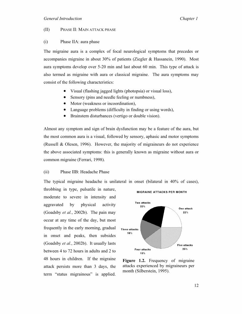
(ii) Phase IIB: Headache Phase
The typical migraine headache is unilateral in onset (bilateral in 40% of cases),
throbbing in type, pulsatile in nature,
moderate to severe in intensity and
aggravated by physical activity
(Goadsby et al., 2002b). The pain may
occur at any time of the day, but most
frequently in the early morning, gradual
in onset and peaks, then subsides
(Goadsby et al., 2002b). It usually lasts
between 4 to 72 hours in adults and 2 to
48 hours in children. If the migraine
attack persists more than 3 days, the
term “status migrainous” is applied.
Figure 1.2. Frequency of migraineattacks experienced by migraineurs permonth (Silberstein, 1995).

General Introduction Chapter 1
13
Frequency varies among individuals from a few in a lifetime to several times in a
week, with an average of 1-3 a month (Figure 1.2) (Silberstein, 1995; Goadsby et al.,
2002b).
PHASE III: POST-DROME (RESOLUTION) PHASE
As the pain lessens, patients feel tired, washed out, irritable and may have impaired
concentration, scalp tenderness or mood changes. Some patients feel unusually
refreshed or euphoric after the attack while others experience depression and malaise
(Goadsby et al., 2002b).
1.4.2 Formal Classification and Diagnostic Criteria for Migraine
The International Headache Society (IHS) formally classified the headaches in order
to improve clinical practise and research. In 1988, IHS published the first edition of
the International Classification of Headache Disorders (ICHD-I) and it was later
redefined in 2004 (ICHD-II) (Olesen et al., 2003b). Migraine was grouped under the
primary headaches based on their symptoms, as true aetiological classification is not
possible like secondary headaches. This scheme stipulates that certain characteristic
features are necessary to establish a diagnosis of migraine.
The IHS system recognises six subtypes of migraine with two major
varieties, namely, migraine without aura and migraine with aura. Tables 1.1 and 1.2
show the classification for migraine and diagnostic criteria for migraine with or
without aura proposed by the International Headache Society (IHS' 2004) (Olesen et
al., 2003b).

General Introduction Chapter 1
14
Table 1.1. The international classification of migraine (ICHD’2004)
Migraine
1. Migraine without aura
2. Migraine with aura 2.1 Typical aura with migraine headache 2.2 Typical aura with non-migraine headache 2.3 Typical aura without headache 2.4 Familial hemiplegic migraine (FHM) 2.5 Sporadic hemiplegic migraine 2.6 Basilar-type migraine
3. Childhood periodic syndromes that are common precursors of migraine 3.1 Cyclical vomiting 3.2 Abdominal migraine 3.3 Benign paroxysmal vertigo of childhood
4. Retinal migraine
5. Complications of migraine 5.1 Chronic migraine 5.2 Status migrainosus 5.3 Persistent aura without infarction 5.4 Migrainous infarction 5.5 Migraine-triggered seizure
6. Probable migraine 6.1 Probable migraine without aura 6.2 Probable migraine with aura 6.3 Probable chronic migraine

General Introduction Chapter 1
15
Table 1.2. Diagnostic criteria proposed by the International Headache society (ICHD' 2004)
Migraine with aura (Previously used terms: Classic, ophthalmic, hemiparaesthetic, hemiplegic or aphasic, complicated migraine) A. At least two attacks fulfilling B. B. At least following three of the following four characteristics • One or more fully reversible aura symptoms indicating focal cerebral cortical and/or
brain stem dysfunction. • At least one aura symptom develops gradually over more than 4 min, or two or more
symptoms occur in succession. • No aura symptom last more than 60 min. • Headaches follow aura with a free interval of less than 60 min. C. At least one of the following • History, physical and neurological examinations do not suggest secondary headache
disorders. • History and/or physical and/or neurological examinations do suggest such disorder,
but it is ruled out by appropriate investigations. • Such disorder is present, but migraine attacks do not occur for the first time in close
temporal relation to the disorder. Migraine without aura (Previously used terms: Common migraine, hemicrania simplex) A. At least five attacks fulfilling B-D. B. Headache attacks lasting 4 to 72 hours (untreated or unsuccessfully treated). C. Headache has at least two of the following characteristics: • Unilateral location. • Pulsatile quality. • Moderate or severe intensity (inhibits or prohibits daily activities). • Aggravation by or causing avoidance of routine physical activity (e.g., walking stairs
or similar routine physical activity). D. During headache at least one of the following • Nausea and/or vomiting. • Photophobia and phonophobia. E. At least one of the following • History, physical and neurological examinations do not suggest secondary headache
disorders. • History and/or physical and/or neurological examinations do suggest such disorder,
but it is ruled out by appropriate investigations. • Such disorder is present, but migraine attacks do not occur for the first time in close
temporal relation to the disorder.
General Introduction Chapter 1
16
1.5 Pathophysiology of Migraine
A migraine attack is believed to be an inherited instability in the brain sensory control
system (i.e., hyperexcitable brain); when this system malfunctions either due to
accumulation of unknown triggers or other mechanisms, results in migraine headache
(Bigal et al., 2002). Based on its clinical features, three distinct phases of migraine
can be discerned, namely, a trigger, an aura and a headache phase.
1.5.1 Trigger phase including premonitory symptoms
Although limited information regarding the trigger is available, there is a better
conception about the pathophysiology of migraine (Ferrari, 1998; Villalón et al.,
2002). Moreover, it is believed that an initiating trigger arises from the brain stem
known as "migraine generator" and may also be due to a genetic predisposition
(Ophoff et al., 1996; Ferrari, 1998). The subsequent events following the trigger
phase leading to the symptoms observed during the aura and headache phases can be
explained on the basis of the neurovascular hypothesis (Ferrari & Saxena, 1993b;
Villalón et al., 2002).
1.5.2 Aura Phase
As mentioned in the Figure 1.3 (Tfelt-Hansen et al., 2000), once the brain stem gets
activated (i.e., the brain generator has been switched on), there is a decrease in the
regional cerebral blood flow, possibly following a wave of cortical spreading
depression (Goadsby et al., 2002b). When the cerebral blood flow decreases beyond a
critical level, the corresponding aura symptoms occur. Most clinicians believe that
the migraine aura is due to a neuronal dysfunction rather than ischaemia and it is
probably the clinical manifestation of a cortical spreading depression (Olesen, 1991a).
The majority of migraine patients do not experience aura, but the following
disturbances: (i) scintillating scotoma, flashing of lights that move across the visual
field, etc.; (ii) paraesthesias; or (iii) other neurological signs (Goadsby et al., 2002b).
The decrease in the cerebral blood flow begins usually in the occipital lobe, but this
reduction enlarges and may involve the whole hemisphere. This spreading oligemia
does not respect the vascular territories and it is unlikely due to vasoconstriction
(Olesen, 1991a).

General Introduction Chapter 1
17

General Introduction Chapter 1
18
1.5.3 Headache Phase
The cerebral oligemia is subsequently followed by a reflex vasodilatation of the
cranial blood vessels and arteriovenous anastomoses, probably due to changes in the
neuronal activity that innervates the cranial extracerebral blood vessels and
arteriovenous anastomoses (e.g., dura mater, base of the skull and scalp region).
Tracing studies have shown that the fibres innervating the cerebral blood vessels arise
from within the trigeminal ganglion containing several vasoactive neurotransmitters
including substance P, calcitonin-gene related peptide (CGRP), 5-hydroxytryptamine
(5-HT), vasoactive intestinal peptide (VIP), nitric oxide (NO) and neurokinin A
(Goadsby et al., 2002b). This profuse cranial vasodilatation leads to an enhanced
blood volume following each cardiac stroke and rapid diastolic run off, with a
consequent augmentation in carotid pulsations within the affected blood vessels.
These augmented pulsations can then be sensed by so-called "stretch" receptors in the
vessel wall thereby activating the perivascular (trigeminal) sensory nerves (De Vries
et al., 1999a; De Vries et al., 1999b). This nociceptive information is conveyed to
central neurons in the trigeminal sensory nucleus that in turn relays the pain signals to
higher centers where headache pain is perceived (Williamson & Hargreaves, 2001;
Edvinsson, 2003). In addition, stimulation of trigeminal nerves may also release
neuropeptides, thus reinforcing vasodilatation and perivascular nerve activity
(Villalón et al., 2002).
Acutely-acting antimigraine compounds constrict dilated cranial
extracerebral blood vessels (Saxena & Ferrari, 1989; Feniuk et al., 1991; Ferrari &
Saxena, 1993a) and inhibit neuropeptide release, plasma protein extravasation across
dural blood vessels (Buzzi et al., 1992) and impulse transmission within the
trigeminovascular system (Goadsby et al., 2002b).

General Introduction Chapter 1
19
1.6 Experimental models for acutely-acting antimigraine drugs
The experimental models currently known for the discovery and development of
antimigraine drugs are based on the vascular or neurogenic involvement in migraine
(De Vries et al., 1999a): (i) vasoconstriction of the dilated extracranial blood vessels
including carotid arteriovenous anastomoses (e.g., carotid vasculature or isolated
blood vessels; vascular hypothesis); (ii) inhibition of the trigeminal system (e.g.,
blockade of plasma protein extravasation and/or central trigeminal inhibition;
neurogenic hypothesis); and (iii) combination of both (e.g., inhibition of neurogenic
vasodilatation).
1.6.1 Experimental models based on the vascular involvement
(i) Constriction of carotid arteriovenous anastomoses (Figure 1.4) in anaesthetised animals
Although a complete understanding of the migraine pathogenesis remains elusive,
there seems to be little doubt that the dilatation of cranial blood vessels, including
carotid arteriovenous anastomoses, is involved in the headache phase of migraine (De
Vries et al., 1999a). In addition to
headache, migraine patients also
experience facial paleness, reduction in
the facial temperature, increase in the
temporal artery pulsations and swelling
of the frontal vein on the side of the
headache (Drummond & Lance, 1983;
Drummond & Lance, 1984). Based on
these findings, Heyck (Heyck, 1969)
investigated the potential underlying
mechanisms involve in migraine, by
measuring the oxygen saturation
difference between the arterial
(femoral) and venous (external jugular) blood samples (A-V SO2 difference) during
and after the headache phase of migraine and compared it with the healthy control
groups (Heyck, 1969). Interestingly, he observed that the A-V SO2 difference was
Figure 1.4. A schematic representationof an arteriovenous anastomosis

General Introduction Chapter 1
20
abnormally decreased during the headache phase of migraine, likely due to dilatation
of the carotid arteriovenous anastomoses, and this decrease was normalised after
spontaneous or drug-induced (ergotamine) alleviation of the headache (Heyck, 1969).
Arteriovenous anastomoses are precapillary communications between the
arteries and veins (Figure 1.4); they are predominantly located in the head skin, ears,
nasal mucosa, eyes and dura mater in several species, including humans and pigs
(Saxena, 1995). In conscious pigs, the arteriovenous anastomoses are constricted
being under a strong influence of the sympathetic neuronal tone, thereby shunting
only a small (<3%) fraction of the total carotid blood flow (Hales, 1974). In contrast,
under pentobarbital anaesthesia, ~80% of the total carotid blood flow is shunted via
arteriovenous anastomoses into the jugular venous circulation (Den Boer et al., 1993).
Consequently, opening of the carotid arteriovenous anastomoses during migraine
shunts a large quantity of oxygenated blood directly into the veins thereby resulting in
facial pallor, lowering of skin temperature and increase in vascular pulsations
(Saxena, 1995). This increase in vascular pulsations stimulate the so-called ‘stretch
receptors’ present in the wall of blood vessels, with ensuing activation of perivascular
trigeminal nerves containing peptides (e.g. CGRP) (De Vries et al., 1999a; De Vries
et al., 1999b). The fifth cranial nerve conveys nociceptive information to central
trigeminal nuclei that in turn relay the pain signals to higher centres where headache
pain is perceived (Williamson & Hargreaves, 2001; Edvinsson, 2003).
In line with the above findings, it is reasonable to assume that the
constriction of dilated carotid arteriovenous anastomoses may abort migraine.
Therefore, we developed an animal experimental model using radioactive
microspheres to determine carotid arteriovenous anastomostic blood flow and the
effects of antimigraine drugs on carotid arteriovenous anastomoses (Saxena, 1990;
Saxena, 1995). Over the years, this model has proven predictive of antimigraine
activity in the clinic (Saxena, 1995). Another major advantage of this model is that
one can simultaneously study different vascular beds in order to evaluate the
cranioselectivity of current or prospective antimigraine drugs (De Vries et al., 1999a;
De Vries et al., 1999b). Based on this notion, we have previously shown that
conventional antimigraine agents like ergotamine and sumatriptan as well as second-
generation triptans potently constrict the porcine carotid arteriovenous anastomoses
(Willems et al., 1998; De Vries et al., 1999a; Tom et al., 2002). Moreover, we have

General Introduction Chapter 1
21
recently demonstrated that α1- and α2-adrenoreceptors mediate porcine carotid
vasoconstriction and suggested that selective agonists at these receptors might provide
a promising novel avenue for the development of acute antimigraine drugs (Willems
et al., 2003).
Several lines of evidence indicate that CGRP, a potent vasodilator released
from the trigeminal sensory nerves may play an important role in the pathophysiology
of migraine (Goadsby et al., 2002b). Indeed, CGRP receptors are widely distributed
in several vascular beds including the carotid vasculature (Gardiner et al., 1990; Van
Gelderen et al., 1995; Shen et al., 2001). Moreover, triptans abort migraine not only
by constricting the dilated cranial blood vessels via 5-HT1B receptors, but also by
inhibiting CGRP release by activation of 5-HT1D receptors (Goadsby et al., 2002b).
Therefore, it is admissible to propose that CGRP receptors may be involved in the
vascular tone of the carotid circulation, which may provide a novel target for
developing new antimigraine compounds (Edvinsson, 2003). The recently introduced
potent and selective CGRP receptor antagonist, BIBN4096BS (Doods et al., 2000),
may be a useful as a pharmacological tool to evaluate the potential role of CGRP
receptors in migraine (Edvinsson, 2003). The following chapters (Chapters 2 and 3)
will discuss the effects of BIBN4096BS on porcine carotid haemodynamics as well as
its cardiac output distribution.
(ii) Contraction of isolated cranial blood vessels
Several in vitro studies using a number of isolated blood vessels have shown that the
acute antimigraine compounds contract these blood vessels via 5-HT1 receptors, (De
Vries et al., 1999a; Villalón et al., 2002). This contractile effect is more marked in
cranial blood vessels than in peripheral blood vessels where 5-HT2 receptors are
predominant (Longmore et al., 1997). It is noteworthy that the pharmacological
profile of the above contractile 5-HT1 receptors correlates with the 5-HT1B, but not the
5-HT1D or 5-ht1F receptor subtypes (De Vries et al., 1998; Verheggen et al., 1998;
Cohen et al., 1999). In addition, dipeptide CGRP receptor antagonists, such as
BIBN4096BS and compound 1, potently antagonise CGRP-induced vasorelaxations
in cerebral arteries (Edvinsson, 2001a; Edvinsson, 2002; Moreno et al., 2002;
Verheggen et al., 2002).
General Introduction Chapter 1
22
1.6.2 Experimental models based on the neurogenic involvement
The basic perception behind the development of neurogenic models for migraine is
that migraine pain is due to a sterile neurogenic inflammation within the meninges
and consequent activation of trigeminal nerve terminals (Williamson & Hargreaves,
2001). The activated trigeminal nerves release several neuropeptides (including
substance P, neurokinin A and CGRP), which cause subsequent features of migraine
rather than merely dilatation of cranial blood vessels (Goadsby et al., 2002b).
Therefore, the efficacy of an antimigraine drug is believed to be due to a presynaptic
action on sensory nerves thereby inhibiting the neuropeptide release and the process
and/or consequences of "neurogenic inflammation" (Buzzi et al., 1991; Buzzi et al.,
1992). Moreover, mechanisms, which do not seem to be mediated solely by the 5-
HT1B receptor have also been implicated in migraine relief (Goadsby, 1998). These
mechanisms include inhibition of the trigemino-vascular system peripherally and/or
centrally (Goadsby, 1998; May et al., 1998).
(i) Inhibition of plasma protein extravasation after trigeminal stimulation
Migraine pain is believed to be a form of sterile neurogenic inflammation, which is
characterised by plasma proteins extravasation across the dura mater and associated
structural changes in the dura mater, such as increases in endothelial permeability and
mast cell degranualation (Williamson & Hargreaves, 2001). The concept of plasma
protein extravasation gained importance in migraine pathogenesis following a study
demonstrating plasma protein extravasation following antidromic stimulation of
trigeminal ganglion/sensory nerve in rats and guinea pigs (Moskowitz, 1993).
Clinically effective antimigraine agents, such as ergots, triptans, opioids and
valporate, inhibited this sterile neurogenic inflammation, suggesting that plasma
protein extravasation inhibition could be predictive of antimigraine therapeutic
activity (Moskowitz, 1993).
Based on this finding, the compounds inhibiting plasma protein extravasation
were investigated as new approaches in migraine treatment. The conventional
antimigraine drug, sumatriptan, inhibits plasma protein extravasation and this effect
was attenuated by the 5-HT1B/1D receptor antagonist GR127935 in both rats and
guinea pigs, implying the involvement of 5-HT1B/1D receptor subtypes (Williamson &
Hargreaves, 2001). However, in mice this effect resembles 5-HT1B receptors whereas
General Introduction Chapter 1
23
in guinea pigs and rats, it is a 5-HT1D-mediated effect (Shepheard et al., 1997; Yu et
al., 1997; Williamson & Hargreaves, 2001). Although triptans have high affinity for
both 5-HT1B and 5-HT1D receptor subtypes, they may also act on other subtypes
(Williamson & Hargreaves, 2001). In this respect, plasma protein extravasation
inhibition can also be induced by 5-carboxamidotryptamine and CP122288 in 5-HT1B
receptor knockout mice and this effect is not prevented by GR127935 in guinea pigs
(Yu et al., 1996; Yu et al., 1997). Moreover, CP122288 shows a higher potency than
sumatriptan in rats and this did not correlate with its affinity for 5-HT1B or 5-HT1D
receptors, suggesting that at least part of the CP122288 action may be via 5-ht1F
receptors, where it displays a high affinity (Shepheard et al., 1997; Williamson &
Hargreaves, 2001). Moreover, a number of 5-HT1 receptor agonists that inhibit
plasma protein extravasation in guinea pig dura mater displaying a rank order of
potency that correlates with their affinity towards 5-ht1F rather than 5-HT1B or 5-HT1D
receptor subtypes (Williamson & Hargreaves, 2001). Based on this finding, a
selective 5-ht1F receptor agonist, LY344864, was developed (Johnson et al., 1997).
This compound, besides proving effective in inhibiting plasma protein extravasation
produced by trigeminal ganglion stimulation in rats, was apparently effective in acute
migraine treatment (Goldstein et al., 2001b). Despite the clinical effectiveness of
LY344864, it was not clear whether plasma protein extravasation inhibition was
responsible for its antimigraine properties. Moreover, LY344864 inhibits the
activation of brainstem neurons in response to the stimulation of dura mater as well as
the c-fos expression in trigeminal nucleus caudalis; this suggests that the primary
mechanism of LY344864 is central (i.e. interruption of the ascending pain pathways)
rather than peripheral (inhibition of plasma protein extravasation) (Williamson &
Hargreaves, 2001). Similarly, the selective 5-HT1D receptor agonist PNU-142633F,
which blocks plasma protein extravasation in guinea pigs (Cutrer et al., 1999), was
ineffective in migraine treatment (Cutrer et al., 2000). Furthermore, other studies
have shown that plasma protein extravasation can be inhibited by the CGRP receptor
antagonist CGRP (8-37) (O'Shaughnessy & Connor, 1994; Brandli et al., 1996).
Importantly, plasma protein extravasation models do not always predict
antimigraine efficacy (Goadsby, 2000) as clearly evidenced by the failure of several
compounds in clinical trials, including: (i) the NK1 receptor antagonist, lanipetant
(Goldstein et al., 1997); (ii) specific plasma protein extravasation inhibitors such as

General Introduction Chapter 1
24
CP122,288 and 4991W93 (Roon et al., 2000); (iii) the ETA/B receptor antagonist
bosentan (May et al., 1996); and (iv) the neurosteroid ganaxolone (Data et al., 1998).
Moreover, the clinical antimigraine predictability of plasma protein extravasation
assays became questionable following an elegant clinical study in migraine showing
no increases in retinal or choroid permeability (May et al., 1998); this contrasts with
the increase in retinal or choroid permeability following trigeminal ganglion
stimulation in rats (Williamson & Hargreaves, 2001).
(ii) Inhibition of cranial vasodilatation (carotid, dural and cortical) induced by trigeminal stimulation
Electrical stimulation of the trigeminal nerve in humans evokes the release of CGRP
in cranial venous blood (Goadsby et al., 2002b). Moreover, during the headache
phase of migraine, plasma levels of CGRP, but not substance P, were elevated in the
jugular venous blood (Goadsby et al., 2002b). Therefore, CGRP released from
trigeminal sensory nerves that innervate cranial blood vessels produces vasodilatation
thereby causing headache (Williamson & Hargreaves, 2001). Based on this, several
animal models were developed to demonstrate cranial vasodilatation associated to
CGRP release produced by trigeminal stimulation as well as to study the effects of
antimigraine drugs on it (Williamson & Hargreaves, 2001). It is known that triptans
attenuate cranial vasodilatation induced by trigeminal stimulation as well as CGRP
release in rats (Williamson & Hargreaves, 2001). However, carotid vasodilatation in
guinea pigs following trigeminal ganglion stimulation is mediated by vasoactive
intestinal peptide, which was not amenable to blockade by antagonists at CGRP or
tachykinin receptors (Beattie & Connor, 1994; Raval et al., 1999).
Therefore, another model was developed in which trigeminal sensory
Aδ-fibres were stimulated following short, low intensity electrical stimulation (which
releases CGRP only); the dural blood vessel diameter was measured by an intravital
microscope through a closed cranial window (Williamson & Hargreaves, 2001;
Akerman et al., 2003). Electrical stimulation of this cranial window as well as
intravenous infusion of substance P and α-CGRP in rats evoked an increase (80%) in
dural blood vessel diameter (Shepheard et al., 1997; Williamson & Hargreaves,
2001). Interestingly, the NK1 receptor antagonist, RP 67580 clearly antagonised
substance P-induced vasodilatation, but not the neurogenic vasodilatation (Williamson
General Introduction Chapter 1
25
& Hargreaves, 2001). However, the CGRP receptor antagonist CGRP(8-37) completely
antagonised the vasodilatation induced by both α-CGRP and neurogenic stimulation
(Akerman et al., 2003); this suggests that the neurogenic vasodilatation is mediated by
endogenous CGRP released from trigeminal sensory nerves. This observation is
consistent with clinical data showing that CGRP, but not substance P, levels are
elevated during the headache phase of migraine (Goadsby et al., 1990).
Significantly, triptans attenuated the neurogenic dural vasodilatation
following trigeminal stimulation, probably via presynaptic inhibition of CGRP release
(Williamson & Hargreaves, 2001). This observation mimicked clinical situations
since sumatriptan normalised elevated plasma CGRP levels with resolution of the
headache (Williamson & Hargreaves, 2001). It was suggested that the above
inhibitory effect of sumatriptan is mediated via prejunctional 5-HT1B receptors in rats
and 5-HT1D receptors in guinea pigs, cats and humans (Williamson & Hargreaves,
2001).
(iii) Central trigeminal neuronal inhibition
The importance of brainstem in the pathogenesis of migraine was emphasised
following its activation during migraine attacks (Bahra et al., 2001). During migraine,
blood flow was increased in the cerebral hemispheres (cingulate, auditory and visual
cortex) as well as in brain stem (Bahra et al., 2001). Sumatriptan relieved the
headache and other symptoms as well as reversed the increase in cerebral blood flow,
but not in brain stem; this indicates that persistent brain stem activation is due to other
factors including increased activity of the endogenous antinociceptive system (Bahra
et al., 2001). Moreover, the brain stem activation may be inherent of the migraine
process itself, and continuous activation of brain stem (despite symptom resolution by
sumatriptan) may account for the headache recurrence (Bahra et al., 2001).
Based on this finding, animal migraine models were developed to study c-fos
activation of the trigeminal nucleus caudalis; interestingly, this effect was not altered
by sumatriptan (Goadsby, 1997a; Goadsby, 1997b; Goadsby & Hoskin, 1997;
Goadsby & Knight, 1997). However, the second generation triptans, such as
zolmitriptan (Goadsby & Boes, 2001), naratriptan (Donaldson et al., 2002) and
eletriptan (Donaldson et al., 2002; Lambert et al., 2002) inhibited the action potentials
generated in the trigeminal nucleus caudalis after superior saggital sinus stimulation in
General Introduction Chapter 1
26
cats and dural stimulation in rats (Cumberbatch et al., 1997). This difference in
effects could be due to the poor central penetrating effects of sumatriptan (Ferrari &
Saxena, 1993a; Ferrari & Saxena, 1993b; Saxena & Tfelt-Hansen, 1993) as compared
to second generation triptans, which are known to have central inhibitory effects
(Saxena & Tfelt-Hansen, 2000; Goadsby et al., 2002b). Consequently, it has been
argued that the blood brain barrier may be disrupted during migraine (Harper et al.,
1977); indeed, under experimental disruption of the blood brain barrier by
hyperosmolar mannitol, sumatriptan produced central inhibitory effects (Shepheard et
al., 1995). However, there is little or no evidence for a disrupted blood brain barrier
based on computerised tomography or MRI findings in migraine patients (Alvarez-
Cermeno et al., 1986; Hamalainen et al., 1996).
Several lines of pharmacological evidence indicate that potent antimigraine
agents act on the second order trigeminal neurons to reduce cell activity, suggesting
that trigeminocervical complex neurons in the caudal brain stem could be a possible
target for antimigraine activity (Bahra et al., 2001). It is likely that this central
inhibitory effect is mediated by 5-HT1B/1D receptors since the central inhibitory effect
of eletriptan in cats is amenable to blockade by GR127935. In addition, the
involvement of 5-HT1D receptors rather than 5-HT1B receptors is crucial for this effect
(Donaldson et al., 2002; Lambert et al., 2002). Moreover, CGRP mediates sensory
nerve transmission between the first and second order afferent input from the cranial
blood vessels, and CGRP receptor antagonists may attenuate sensory nerve
transmission (Gulbenkian et al., 2001; Williamson & Hargreaves, 2001; Conner et al.,
2002; Goadsby et al., 2002b; Poyner et al., 2002; Smith et al., 2002). Recently,
adenosine A1 receptors were localised in human trigeminal ganglia, suggesting a
potential usage of adenosine A1 receptor agonists to inhibit trigeminal nociception
(Welch, 2003).
1.7 Management of migraine
The drugs used in the treatment of migraine can be divided into two groups: agents
that abolish the acute migraine headache (acute antimigraine drugs; e.g. ergotamine
and sumatriptan) and agents aimed at its prevention (prophylactic drugs; e.g.
methysergide). Patients who experience frequent headaches may require both forms
of treatment.
General Introduction Chapter 1
27
1.7.1 Acute treatment
Though several acutely acting antimigraine drugs are available, administration of
these drugs depends upon: (i) severity; (ii) frequency of the attacks; (iii) associated
symptoms; and (iv) co-morbid conditions interrelated with a migraine attack (Villalón
et al., 2002). Acutely acting antimigraine drugs attempt to abort the headache and
they can be specific or non-specific in action. Non-specific medications control the
pain and associated symptoms of migraine or other pain disorders; in contrast, the
specific medications control the headache attack, but are not useful for non-headache
pain disorders (Goadsby et al., 2002b).
(I) NON-SPECIFIC MEDICATIONS
Non-specific medications are prescribed for mild to moderate headaches; the most
commonly used drugs include non-steroidal antiinflammatory drugs (NSAIDs; e.g.
aspirin or acetaminophen) either alone or combination with caffeine. NSAIDs are the
most popular agents because, in addition to being cheap, effective and easy to
administer, they allow a control over own therapy. Unfortunately, they produce
headache after a long-term use (Villalón et al., 2002). For associated symptoms (e.g.
nausea or vomiting), antiemetics such as domperidone or metoclopromide are
administered (Goadsby et al., 2002b).
(II) SPECIFIC-MEDICATIONS
Specific antimigraine drugs abort the migraine headache by constricting the dilated
extracranial blood vessels, including the external carotid bed (Olesen, 1991a; Olesen,
1991b; Rasmussen et al., 1991; Saxena & Den Boer, 1991). The most commonly
used specific antimigraine compounds are ergot alkaloids and triptans.
a. Ergot alkaloids
Ergotamine and its derivative dihydroergotamine are used to treat moderate to severe
migraine attacks, particularly if NSAIDs fail to alleviate the headache (Saxena & Den
Boer, 1991). Dihydroergotamine has fewer side effects than ergotamine and they are
effective in most of the patients with low recurrence rate. Ergot and its derivatives are
contraindicated in patients with uncontrolled hypertension, hepatic or renal failure,
vascular disease (coronary, cerebral and/or peripheral) and in pregnancy (Villalón et
al., 2002).

General Introduction Chapter 1
28
b. Selective 5-HT agonists
Based on the vascular involvement in migraine and with the supporting role of 5-HT
in the migraine pathogenesis (Saxena & Tfelt-Hansen, 2000), a new tryptamine
derivative was synthesised to achieve selectivity at the craniovascular 5-HT1 like
receptors; this search culminated in the design and development of sumatriptan, a
selective 5-HT1B/1D receptor agonist (Humphrey et al., 1989). Sumatriptan constricts
cranial arteries and displays much less activity in other vascular systems (Dahlöf &
Saxena, 2000; MaassenVanDenBrink et al., 2000a; MaassenVanDenBrink et al.,
2000b; Saetrum Opgaard et al., 2000; Saxena & Tfelt-Hansen, 2000; Tfelt-Hansen et
al., 2000; Villalón et al., 2002). Sumatriptan relieves headache, nausea, vomiting and
restores the persons back to normal situations (Goadsby et al., 2002b). Sumatriptan is
available in subcutaneous or nasal spray forms; they are used for immediate relief of
headache, while the oral form is used in patients with gradual onset of headache
(Goadsby et al., 2002b). Despite its advantage over other conventional antimigraine
compounds, it has low oral bioavailability, short half-life and high headache
recurrence (up to 40%) and is contraindicated in patients suffering from coronary
arterial disease (Maassen VanDenBrink et al., 1999; Goadsby et al., 2002b).
Therefore, in order to overcome these limitations, several new 5-HT1B/1D receptor
agonists (referred to as second-generation triptans) have been developed with an
action similar to that of sumatriptan, but different in their pharmacokinetic properties
(particularly higher oral bioavailability, longer half-life, low headache recurrence,
etc.).
1.7.2 Preventive (prophylactic) treatment
Patients who experience migraine attacks that are frequent, severe, long lasting,
unresponsive to acute antimigraine agents and that cause substantial disability are the
candidates for preventive therapy (Villalón et al., 2002). Preventive antimigraine
drugs are taken every day (whether or not the headache is present), to reduce the
frequency and severity of migraine attacks (Villalón et al., 2002). The mechanism by
which the preventive antimigraine drugs work is still unclear, but they are believed to
modify the sensitivity of the brain that underlies migraine pathogenesis (Goadsby et
al., 2002b). The commonly used preventive antimigraine agents include, β-adrenergic
antagonists, calcium channel blockers, antidepressants, serotonin antagonists,

General Introduction Chapter 1
29
anticonvulsants and NSAIDs (Goadsby et al., 2002b). Moreover, botulinum toxin
(type A) appears to be safe, tolerable and possibly effective drug for migraine
prevention, with almost no systemic adverse events (Ashkenazi & Silberstein, 2003).
Interestingly, angiotensin converting enzyme receptor blocker candesartan appears to
be effective and highly tolerable in the prevention of migraine, but needs to be further
evaluated (Ashkenazi & Silberstein, 2003). If preventive medication is indicated, the
drug should be chosen from one of the above categories based on side-effect profiles
and co-existent morbidities (Silberstein & Lipton, 1994; Silberstein, 1995). It has
been reported that on average, about two-thirds of the patients with the above
preventive medications had a significant reduction (50%) in the frequency and
severity of the attacks (Goadsby et al., 2002b). Natural products such as riboflavin
(Vitamin B2: used for mitochondrial dysfunction), niacin, magnesium and leukotriene
antagonists (montelukast) have shown some promising results in reducing the
frequency and severity of migraine attacks (Bigal et al., 2002; Velling et al., 2003).
In conclusion, though the pathophysiological basis of migraine still remains elusive,
the development of migraine models has remarkably contributed to the understanding
of migraine pathophysiology and to the development of more effective antimigraine
drugs. Hopefully, the advent of novel antimigraine compounds and the introduction of
molecular biology techniques will shed further light on this complex scenario.

General Introduction Chapter 1
30
1.8 Calcitonin gene-related peptide
1.8.1 Introduction
The calcitonin family of peptides comprises five members, namely calcitonin, amylin,
calcitonin gene-related peptide (CGRP; two forms α-CGRP and β-CGRP), and
adrenomedullin (Poyner & Marshall, 2001). They all have a six-aminoacid ring
structure (seven for calcitonin) close to their N-terminal, formed by an intermolecular
disulfide bond. This is followed by a region of potential amphipathic α-helix and
C amidated terminals (Poyner & Marshall, 2001). These peptides are widely
distributed and induce multiple biological effects, such as potent vasodilatation
(CGRP and amylin), reduction in nutrient intake (amylin) and decrease in bone
resorption (calcitonin) (Poyner & Marshall, 2001). Due to their similarities in
structure and biological activities, it is suggested that these peptides interact with
similar G-protein coupled receptors (Poyner & Marshall, 2001).
CGRP was first identified in 1983 in rats, where serially transplanted rat cells
from a medullary thyroid carcinoma showed a spontaneous ability to switch from a
high to a low calcitonin (calcitonin) producing state, by increasing the size of the
mRNA (Rosenfeld et al., 1983); this was later determined to be an “altered mRNA”
derived from the calcitonin/CGRP gene (Wimalawansa, 2001). The alternative
splicing of this primary mRNA transcript of the calcitonin/CGRP gene, which is
encoded on the short arm of chromosome 11p14, will lead to the translation of CGRP
and calcitonin gene in a tissue specific manner (Wimalawansa, 2001). For example,
in the central nervous system (CNS), splicing of the calcitonin/CGRP gene mRNA
transcript will produce CGRP, whereas in the C cells of the thyroid gland, calcitonin
is predominantly formed (Hoppener et al., 1985).
In 1984, based on the sequence of rat α-CGRP, a similar peptide (human
CGRP-α; hα-CGRP) from a human medullary carcinoma was demonstrated (Morris
et al., 1984). Subsequently, a second CGRP gene (β-CGRP), also located in
chromosome 11 and thought to have arisen by the exon duplication, was identified by
further analysis of the rat and human cDNA clones (Amara et al., 1985;
Wimalawansa, 1996). The r-α-CGRP differs from r-β-CGRP by one aminoacid and
the h-β-CGRP differs by three aminoacids from homologous h-α-CGRP (Steenbergh

General Introduction Chapter 1
31
et al., 1984; Amara et al., 1985); both α-form and β-form of CGRP are very similar in
their biological activities (Poyner & Marshall, 2001).
1.8.2 Molecular genetics
A schematic representation of the human α-calcitonin/CGRP gene is illustrated in
Figure 1.5. The α-calcitonin/CGRP gene (located in chromosome 11) contains six
exons of which first three exons are constitutively spliced in both mRNAs (calcitonin
and CGRP). The exon I is untranslated, whereas the exons II and III code for the
signal peptide and N-terminal propeptide, respectively. The calcitonin and CGRP
sequences are localised in exons IV and V, respectively; exon VI is a part of α-CGRP
mRNA, but untranslated (Wimalawansa, 1996). The primary mRNA transcript
includes all six exons, and the calcitonin or CGRP mRNA is formed subsequently (see
Figure 1.1) (Steenbergh et al., 1984). The splicing of the first three exons with exons
V and VI produces CGRP containing mRNA. The exon V encodes CGRP, while the
exon VI encodes the 3’-untranslated region of the CGRP mRNA and polyadenylation
(poly A) signal (Wimalawansa, 1996). This mRNA is translated to generate the pro-
CGRP peptide, which is subsequently cleaved at the paired dibasic aminoacids to
release the 37-aminoacid CGRP (Amara et al., 1985).
The organization of the β-CGRP gene in the chromosome 11 is similar to that
of α-calcitonin/CGRP gene (Steenbergh et al., 1984). While exon I is untranslated,
exon II encodes for the signal peptide and exon III, which is 92% homologous to exon
II of the α-calcitonin/CGRP gene, encodes for the N-terminal propeptide. The exon
IV of the β-CGRP gene is 67% homologous to the same region of the
α-calcitonin/CGRP gene that gives rise to calcitonin. The exon IV lacks
polyadenylation, thereby preventing the alternative splicing. Consequently, the
transcript from this gene produces only CGRP; therefore it is considered as a
pseudogene for calcitonin (Proudfoot & Brownlee, 1976; Alevizaki et al., 1986).

General Introduction Chapter 1
32
General Introduction Chapter 1
33
1.8.3 Structure
All species variants of CGRP have 37 aminoacids, constituted as a single polypeptide
chain (Bell & McDermott, 1996). The structure of α-CGRP (Figure 1.6) comprises a
N-terminal disulfide bridge between the positions 2 and 7 (Cys2 and Cys7), a well-
defined α-helix between the residues 8 and 18. This is followed by either a β- or a γ-
turn in the region of residues 19 to 21 and phenylalanylamide C-terminus in the
regions of residues 28 and 30, and also in 32 and 34 (Conner et al., 2002). The β-
CGRP differs from the α-form by one and three aminoacids in rats and humans,
respectively. CGRP shares ~50% homology in their sequence of aminoacids with
amylin and some homology with amylin (Chantry et al., 1991; Kitamura et al., 1993).
1.8.4 Structure-activity relationships
It has been suggested that an intact peptide is required for the full biological activity
of a CGRP molecule (Zaidi et al., 1990). The N-terminal loop (disulphide-bonded
loop) is principally involved in triggering the signal transduction and receptor
activation (Conner et al., 2002). The α-helix plays an important role in the interaction
of the molecule with the receptor (receptor binding) and its deletion causes
approximately 100 fold loss of affinity, (Conner et al., 2002). The residues of 19-27
are necessary as a spacer or hinge region. The C-terminal region (residues 28-37)
shows a weak binding to CGRP receptors. However, by making a few more
aminoacid substitutions, a high affinity antagonist such as CGRP(8-37) can be generated
(Conner et al., 2002). The C-terminal region is probably necessary for the peptide to
assume the right conformation in the interaction with its receptor (Conner et al.,
2002).

General Introduction Chapter 1
34
General Introduction Chapter 1
35
1.8.5 Distribution and localisation
CGRP and its receptors are widely distributed in the peripheral and central nervous
systems as well as in the cardiovascular system (Wimalawansa, 2001). In the
periphery, CGRP is abundantly present in the posterior horn cells. In primary sensory
ganglia, CGRP is often co-stored with substance P (Lundberg et al., 1985) and in
motor neurons CGRP is co-stored with acetylcholine (Gibson et al., 1988; Roa &
Changeux, 1991). In most neurons, the α-and β-forms of CGRP co-exist, but the β-
CGRP form is predominantly seen in the enteric nervous system (Mulderry et al.,
1988) and in the human pituitary gland (Jonas et al., 1985).
In the cardiovascular system, CGRP-containing nerve fibres are more
abundant around the arteries than around the veins (Bell & McDermott, 1996); in the
arterial system, they are predominantly seen in the junction of the adventia and media
(Wimalawansa, 2001). Moreover, CGRP-containing nerve fibres are seen more in the
atria than in ventricles; within the atria, they are localised in the sino-atrial node, the
atrio-ventricular node and the specilaised conduction system (Wimalawansa, 2001).
In addition, the myocardium is less densely innervated than the epicardium,
endocardium and pericardium (Wimalawansa, 2001). In the periphery,
CGRP-containing nerve fibres are often associated with vascular smooth muscle such
as: (i) most parts of gastrointestinal tract, including the excretory ducts of the parotid
gland, over the epithelium of the fundic glands of stomach, endocrine cells of the
duodenum and ileum and some myenteric ganglia; (ii) lung; (iii) thyroid gland (close
to C cells); (iv) splenic vein and sinusoids; (v) human skin; and (vi) pituitary gland
(Hagner et al., 2002a; Hagner et al., 2002b; Hagner et al., 2002c). Moreover, i-CGRP
has been found in the plasma of some patients with medullary thyroid and lung
carcinoma and also in normal human beings (Girgis et al., 1985; Takami et al., 1985).
1.9 CGRP receptors
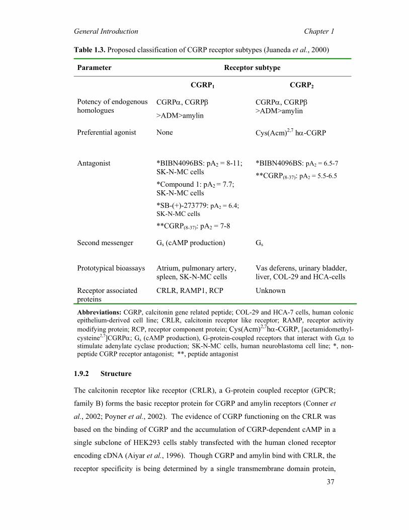
1.9.1 Classification
Based on functional studies (by using the C-terminal fragment of α-CGRP, α-
CGRP(8-37) and linear CGRP analogues, [Cys(Acm)2,7]- and [Cys(Et)2,7]h-α-CGRP),
CGRP receptors are classified into CGRP1 and CGRP2 subtypes (Table 1.3) (Conner
et al., 2002; Poyner et al., 2002). Experimental evidence has shown that α-CGRP(8-37)

General Introduction Chapter 1
36
behaved as a more potent antagonist on CGRP-induced responses in guinea pig atria
(high affinity pA2=7-8) than in those induced in rat vas deferens (lower affinity pA2 =
5.5-6.5) (Poyner et al., 2002). In contrast, linear CGRP analogues, such as
[Cys(Acm)2,7]- and [Cys(Et)2,7] hα-CGRP, have higher affinity for the rat vas deferens
(EC50 = 234, 8.32 nM, respectively) than for the guinea pig atria (Conner et al., 2002;
Poyner et al., 2002). Based on this evidence, it was proposed that the CGRP-induced
responses in the guinea pig atria are mediated via CGRP1 receptors and in the rat vas
deferens by CGRP2 receptors (Conner et al., 2002; Poyner et al., 2002). Furthermore,
cell lines have now been characterised as uniquely enriched with the CGRP1 (e.g.
human SK-N-MC cells and rat L6 skeletal myocytes) and CGRP2 receptors (e.g.
COL-29 and HCA-7 colonic epithelium cells) (Juaneda et al., 2000). Moreover, a
potent and selective CGRP receptor antagonist, BIBN4096BS (Table 1.3), showed a
10-fold higher affinity for CGRP receptors in rat left atrium compared to than in rat
vas deferens, supporting the existence of CGRP receptor subtypes in these two tissues.
Interestingly, this study evidenced the presence of two CGRP-like receptor subtypes
in rat vas deferens namely: (i) the CGRP2 receptor; and (ii) a "novel" receptor that
displays low efficacy for CGRP and that is selectively stimulated by [Cys(Et)2,7]h-
α-CGRP or amylin and which can be blocked with high affinity by BIBN4096BS
(Wu et al., 2000; Wu et al., 2002). Moreover, BIBN4096BS also revealed additional
functional differences between the actions of α-CGRP and β-CGRP in the pig left
anterior descending coronary artery and in the cerebral basilar artery, indicating the
existence of different CGRP receptor subtypes (Wu et al., 2002). Notwithstanding the
above findings, the molecular nature of both CGRP receptor subtypes remains far
from clear and final demonstration must come from their respective cloning and the
development of fully selective agonists and antagonists.
General Introduction Chapter 1
37
Table 1.3. Proposed classification of CGRP receptor subtypes (Juaneda et al., 2000)
Parameter Receptor subtype
CGRP1 CGRP2
Potency of endogenous homologues
CGRPα, CGRPβ
>ADM>amylin
CGRPα, CGRPβ >ADM>amylin
Preferential agonist None Cys(Acm)2,7 hα-CGRP
Antagonist *BIBN4096BS: pA2 = 8-11; SK-N-MC cells
*Compound 1: pA2 = 7.7; SK-N-MC cells
*SB-(+)-273779: pA2 = 6.4; SK-N-MC cells
**CGRP(8-37): pA2 = 7-8
*BIBN4096BS: pA2 = 6.5-7
**CGRP(8-37): pA2 = 5.5-6.5
Second messenger Gs (cAMP production) Gs
Prototypical bioassays Atrium, pulmonary artery, spleen, SK-N-MC cells
Vas deferens, urinary bladder, liver, COL-29 and HCA-cells
Receptor associated proteins
CRLR, RAMP1, RCP Unknown
Abbreviations: CGRP, calcitonin gene related peptide; COL-29 and HCA-7 cells, human colonic epithelium-derived cell line; CRLR, calcitonin receptor like receptor; RAMP, receptor activity modifying protein; RCP, receptor component protein; Cys(Acm)2,7hα-CGRP, [acetamidomethyl-cysteine2,7]CGRPα; Gs (cAMP production), G-protein-coupled receptors that interact with Gsα to stimulate adenylate cyclase production; SK-N-MC cells, human neuroblastoma cell line; *, non-peptide CGRP receptor antagonist; **, peptide antagonist
1.9.2 Structure
The calcitonin receptor like receptor (CRLR), a G-protein coupled receptor (GPCR;
family B) forms the basic receptor protein for CGRP and amylin receptors (Conner et
al., 2002; Poyner et al., 2002). The evidence of CGRP functioning on the CRLR was
based on the binding of CGRP and the accumulation of CGRP-dependent cAMP in a
single subclone of HEK293 cells stably transfected with the human cloned receptor
encoding cDNA (Aiyar et al., 1996). Though CGRP and amylin bind with CRLR, the
receptor specificity is being determined by a single transmembrane domain protein,

General Introduction Chapter 1
38
termed as the receptor activity modifying protein (RAMP) (McLatchie et al., 1998;
Mallee et al., 2002). The RAMPs (150-177 aminoacids in size) are cleavable signal
peptides, with a relatively large N-terminal extracellular domain, one transmembrane
spanning domain and nine aminoacid intracellular C-terminal domains (Fitzsimmons
et al., 2003). The RAMPs have been
localised in the endoplasmic reticulum
and they are required to facilitate the
intracellular translocation of the CRLR-
maturing protein and its insertion into
plasma membranes (McLatchie et al.,
1998; Hilairet et al., 2001; Mallee et al.,
2002). Moreover, RAMPs can alter the
pharmacology of the given CRLR by
providing a mechanism whereby a cell
could dynamically change its sensitivity
from one receptor to another (McLatchie et al., 1998; Mallee et al., 2002). Three
RAMPs have been identified in the human tissues namely, RAMP1, RAMP2 and
RAMP3 (McLatchie et al., 1998; Mallee et al., 2002). Co-expression of CRLR with
RAMP1 reveals CGRP receptors (Figure 1.7), whereas co-expression of CRLR with
RAMP2 and RAMP3 forms adrenomedullin receptors (McLatchie et al., 1998; Mallee
et al., 2002). The mechanism of action of RAMPs in CGRP/adrenomedullin binding
is not clear, but in chimaeric RAMPs, it has been shown that the N-terminus of
RAMP1 is the key determinant for CGRP binding, which could be due to the
interaction of CRLR with N-terminus (Foord et al., 1999). Similarly, in the human
RAMP1, the extracellular domain of RAMP1 is sufficient for normal CRLR
association and efficacy, while the specific sequences of the transmembrane domain
contribute to CGRP affinity and specificity. The tail domain of RAMP1 is
unnecessary for CRLR function (Fitzsimmons et al., 2003). Moreover, in the human
RAMP1, substitution of tryptophan at position 74 with lysine (as found in rat RAMP1)
confers low affinity and vice-versa suggesting important determinants for small
molecule antagonists (Mallee et al., 2002). In addition to RAMPs, the CGRP receptor
complex requires another chaperone protein named as the receptor component protein
(RCP) to function optimally (McLatchie et al., 1998). The RCP is a 148-aminoacid,
Figure 1.7. Schematic representation ofCGRP1 receptor showing interactions ofRAMP1 protein with CRLR and a receptorcomponent protein (RCP). This complex istightly coupled to Gs to promote cAMPproduction.
αβ γαβ γ AC
cAMP
Gs
RCP
CRLR
CGRP
RAMP

General Introduction Chapter 1
39
intracellular protein that is required for G-protein-coupled signal transduction at the
CGRP receptors (Prado et al., 2002). The RCP is well expressed in CGRP responsive
tissues and RCP expression correlates to the biological efficacy of CGRP in vivo
(Evans et al., 2000).
The structure of CGRP2 receptor subtypes is unclear and little has been done
so far to characterise the structural requirements for the CGRP binding to CGRP2
receptors (Poyner & Marshall, 2001). The linear CGRP analogue
Cys(Acm)2,7hα-CGRP has been used to classify CGRP2 receptor subtypes, but an
agonist may not definitively characterise a receptor (Poyner & Marshall, 2001). The
mRNA for the CRLR receptor is present in the rat vas deferens as well as in other
tissues showing pharmacological properties similar to those of the putative CGRP2
receptor subtypes, e.g. the porcine coronary artery (Conner et al., 2002; Hay et al.,
2002; Poyner et al., 2002). Moreover, other studies have demonstrated that, in
general, the affinity of ligands for CGRP1 receptors is higher than for CGRP2
receptors, which could be attributable to tissue factors such as proteases (Rorabaugh
et al., 2001) and/or to a deficiency of one or more ligand-binding contacts (Conner et
al., 2002). However, further work is necessary to confirm CGRP2 receptors structure,
activity and effects in the tissues.
1.9.3 Distribution and binding
CENTRAL NERVOUS SYSTEM
The distribution of CGRP receptors has been well documented and they generally
match with CGRP binding studies (Wimalawansa, 1996). In the CNS, both high and
low binding sites for CGRP have been reported (Wimalawansa, 1996; Morara et al.,
2000; Segond von Banchet et al., 2002). For example, moderate to high density of
CGRP receptors was found in the piriform-insular cortex complex, while low to
moderate receptors were seen in the medial preoptic area (Wimalawansa, 1996). In
contrast, very high levels of CGRP receptors are seen in the solitary, vagus,
hypoglossus, dorsal medullary reticular nuclei and in the area postrema
(Wimalawansa, 1996). The highest density of CGRP receptors is found in the
cerebellum and dorsal spinal cord (Wimalawansa, 1996). In cerebellum, its molecular
layer contains the highest density of CGRP receptors, whilst the Purkinje cell layer
contains a moderate number; in contrast, its granular layer is devoid of CGRP
General Introduction Chapter 1
40
receptors (Morara et al., 1998; Morara et al., 2000; Ueda et al., 2001). In dorsal root
ganglion and other neurons, CGRP receptors co-exist with receptors for other
neurotransmitters and neuromodulators such as, substance P, noradrenaline,
neuropeptide Y, vasoactive intestinal peptide, histidine (van Rossum et al., 1997;
Ohtori et al., 2002). Moreover, in the nonadrenergic and noncholinergic (NANC)
fibres and in the coronary vessels, CGRP receptors co-exist with receptors for
tachykinins (Ursell et al., 1991) and substance-P, respectively (Wiesenfeld-Hallin et
al., 1984).
CARDIOVASCULAR SYSTEM
The highest density of CGRP binding sites is present in the heart and in the blood
vessels (intima and media layers) (Sigrist et al., 1986; Wimalawansa, 2001).
Moreover, in the heart, high affinity binding sites for CGRP are found in the atrial and
ventricular preparations (Wimalawansa, 2001). Regardless of the species, the density
of the CGRP binding sites in atria invariably exceeds that of ventricles (Chang et al.,
2001). Autoradiographic studies in the hearts of rats (Chang et al., 2001), guinea pigs
and humans (Coupe et al., 1990; Hasbak et al., 2003) have shown the highest density
of CGRP binding sites in the coronary arteries, coronary veins and in the heart valves,
while a lower density is found in the coronary arterioles and endocardium
(Wimalawansa, 2001).
CGRP receptors are also abundantly present in the thyroid gland,
gastrointestinal tract, parotid gland, adrenals, pituitary, exocrine pancreas, kidneys,
bones, skin and skeletal muscles (Wimalawansa, 1996; Hagner et al., 2002a; Hagner
et al., 2002b; Hagner et al., 2002c; Irie et al., 2002; Rossi et al., 2003).
1.9.4 Signal transduction mechanisms
The CGRP-induced vascular responses are mediated by both endothelium-dependent
and endothelium-independent mechanisms (Marshall, 1992; Wimalawansa, 1996)
(Figure 1.8). In many blood vessels including the thoracic aorta, renal, pulmonary
and cerebral arteries, the endothelium is absolutely required for CGRP-induced
vasodilatory responses (Wimalawansa, 1996). This endothelium-dependent pathway
begins with CGRP binding to its receptors in the endothelial cells, activating adenylyl
cyclase thereby increasing cAMP levels. This increase in cAMP levels may then
activate the enzyme nitric oxide synthase (NOS), leading to an increase level of nitric

General Introduction Chapter 1
41
oxide (NO) (Lincoln, 1989). However, CGRP may also elevate NO directly without
the involvement of adenlycyclase. This elevated NO acts on the smooth muscle cells
by activating guanylate-cyclase with an ensuing production of cGMP leading to
smooth muscle relaxation (Wimalawansa, 1996).
In the endothelium-independent pathway, CGRP bypasses the endothelium
and directly binds to CGRP receptors on the smooth muscle cells, activating adenylyl
cyclase; this, in turn, will increase cAMP levels leading to vascular relaxation
(Wimalawansa, 1996). This endothelium-independent vasorelaxation has been
demonstrated in several arteries, including the (i) feline cerebral artery; (ii) porcine
splenic artery; (iii) human skeletal muscle artery; (iv) rabbit mesenteric artery; (v) rat
gastric, splenic and hepatic arteries; and (vi) guinea pig pulmonary artery
(Wimalawansa, 1996; van Rossum et al., 1997). Interestingly, blood vessels such as
the rat basilar and superior mesenteric arteries require both endothelium-dependent
and endothelium-independent mediated mechanisms. CGRP also act indirectly by
stimulating protein kinase A that activates K+ATP channels (in rabbit arterial smooth
muscle) (van Rossum et al., 1997).
Evidently, CGRP-induced responses involve multiple second messengers,
including cAMP, nitric oxide-cGMP and K+ channels (van Rossum et al., 1997).
Notwithstanding, irrespective of second messengers involved, the final common
pathway for CGRP-induced vasorelaxation depends ultimately upon the decrease in
intracellular calcium (Wimalawansa, 1996).

General Introduction Chapter 1
42
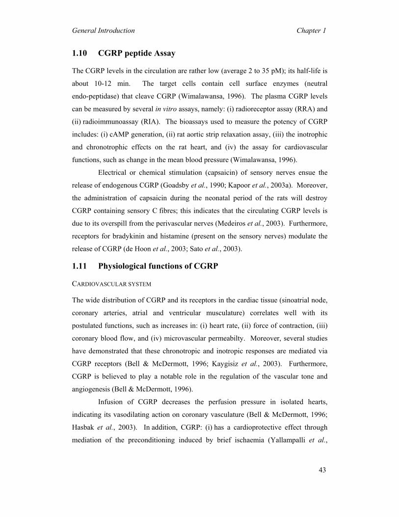
General Introduction Chapter 1
43
1.10 CGRP peptide Assay
The CGRP levels in the circulation are rather low (average 2 to 35 pM); its half-life is
about 10-12 min. The target cells contain cell surface enzymes (neutral
endo-peptidase) that cleave CGRP (Wimalawansa, 1996). The plasma CGRP levels
can be measured by several in vitro assays, namely: (i) radioreceptor assay (RRA) and
(ii) radioimmunoassay (RIA). The bioassays used to measure the potency of CGRP
includes: (i) cAMP generation, (ii) rat aortic strip relaxation assay, (iii) the inotrophic
and chronotrophic effects on the rat heart, and (iv) the assay for cardiovascular
functions, such as change in the mean blood pressure (Wimalawansa, 1996).
Electrical or chemical stimulation (capsaicin) of sensory nerves ensue the
release of endogenous CGRP (Goadsby et al., 1990; Kapoor et al., 2003a). Moreover,
the administration of capsaicin during the neonatal period of the rats will destroy
CGRP containing sensory C fibres; this indicates that the circulating CGRP levels is
due to its overspill from the perivascular nerves (Medeiros et al., 2003). Furthermore,
receptors for bradykinin and histamine (present on the sensory nerves) modulate the
release of CGRP (de Hoon et al., 2003; Sato et al., 2003).
1.11 Physiological functions of CGRP
CARDIOVASCULAR SYSTEM
The wide distribution of CGRP and its receptors in the cardiac tissue (sinoatrial node,
coronary arteries, atrial and ventricular musculature) correlates well with its
postulated functions, such as increases in: (i) heart rate, (ii) force of contraction, (iii)
coronary blood flow, and (iv) microvascular permeabilty. Moreover, several studies
have demonstrated that these chronotropic and inotropic responses are mediated via
CGRP receptors (Bell & McDermott, 1996; Kaygisiz et al., 2003). Furthermore,
CGRP is believed to play a notable role in the regulation of the vascular tone and
angiogenesis (Bell & McDermott, 1996).
Infusion of CGRP decreases the perfusion pressure in isolated hearts,
indicating its vasodilating action on coronary vasculature (Bell & McDermott, 1996;
Hasbak et al., 2003). In addition, CGRP: (i) has a cardioprotective effect through
mediation of the preconditioning induced by brief ischaemia (Yallampalli et al.,

General Introduction Chapter 1
44
2002); and (ii) directly affects the capacitance blood vessels thereby producing
peripheral vasodilatation (Wimalawansa, 1996).
CGRP receptors are abundantly present on renal blood vessels
(Wimalawansa & MacIntyre, 1988), where they have several functions, namely: (i) to
increase the renal blood flow and glomerular filtration rate; (ii) to relax the afferent
arterioles in the glomeruli; (iii) to increase the renin production; and (iv) to stimulate
the release of atrial naturetic peptide (Wimalawansa, 1996).
CENTRAL NERVOUS SYSTEM
In the CNS, CGRP plays an important role in various functions including the motor,
sensory and integrative systems (van Rossum et al., 1997). Moreover, CGRP
modulates several senses including audition, olfaction, vision, feeding and
behavioural effects (van Rossum et al., 1997). The wide distributions of i-CGRP in
the parabrachial area (a relay centre for autonomic-related functions) indicate its role
in regulating the cardiovascular, respiratory and sleep functions (van Rossum et al.,
1997). Consistent with this anatomical distribution, microinjection of CGRP into the
central nucleus of amygdala increased the heart rate and mean arterial blood pressure;
this is due to catecholamines release following sympathetic stimulation via CGRP
receptors in the CNS (Kuo et al., 1994; Oh-hashi et al., 2001).
The CGRP receptors are widely distributed in the parabrachial nucleus and in
the nucleus tractus solitarii; this suggests that CGRP receptors may be involved in
relaying the visceral sensory information from the vagus and glossopharyngeal nerves
(Sykes et al., 1994). Moreover, the association of CGRP receptors with the vagus and
glassopharyngeal nerves may be responsible for the CGRP-induced responses on the
cardiovascular functions including baroreceptor reflexes (Sykes et al., 1994; van
Rossum et al., 1997).
Amongst other effects, CGRP seems to regulate: (i) growth hormone
secretion; (ii) hyperthermia; (iii) catalepsy; (iv) motor activity; and (v) nociceptive
responses (van Rossum et al., 1997). Furthermore, CGRP potentiates excitatory
actions by enhancing the release of substance P as well as of excitatory aminoacids
from the primary afferent fibres (Oku et al., 1987; Kangrga et al., 1990), hence the
possibility of leading to an increased activity of these transmitters.
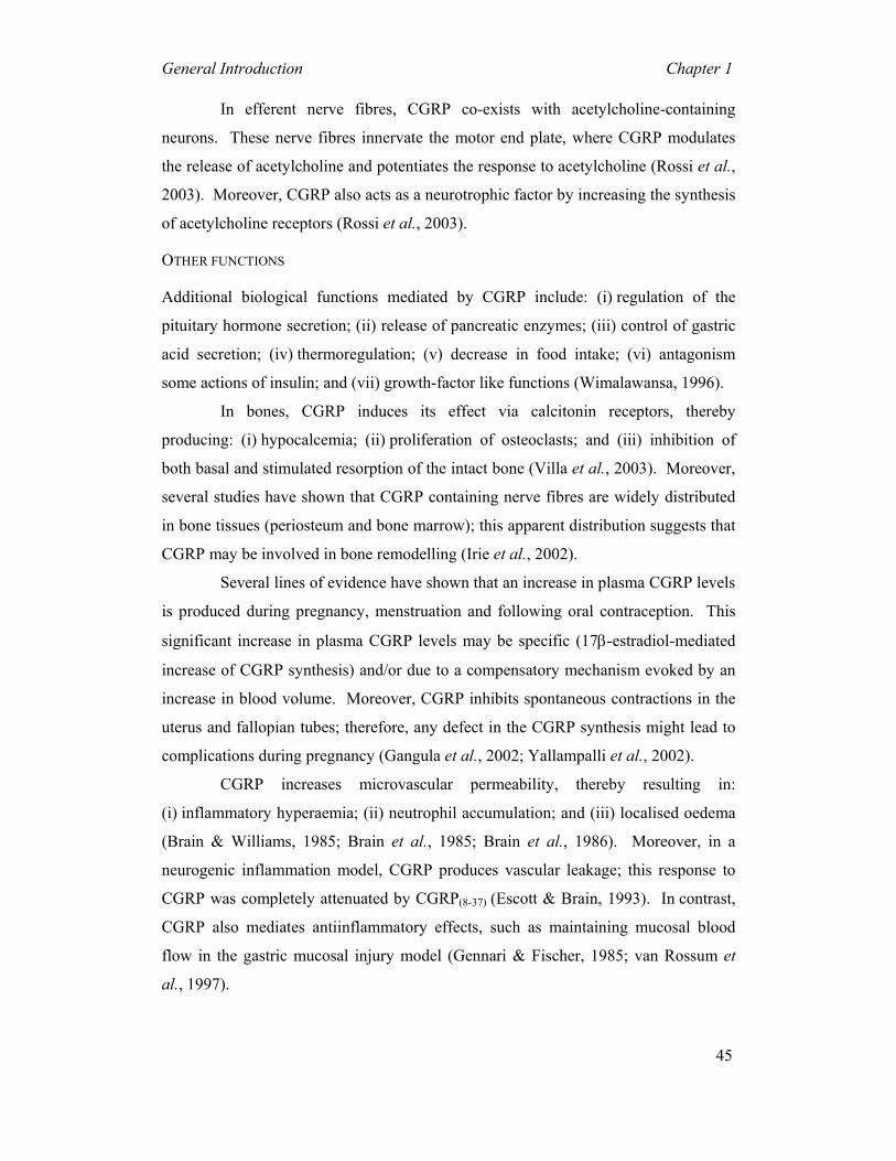
General Introduction Chapter 1
45
In efferent nerve fibres, CGRP co-exists with acetylcholine-containing
neurons. These nerve fibres innervate the motor end plate, where CGRP modulates
the release of acetylcholine and potentiates the response to acetylcholine (Rossi et al.,
2003). Moreover, CGRP also acts as a neurotrophic factor by increasing the synthesis
of acetylcholine receptors (Rossi et al., 2003).
OTHER FUNCTIONS
Additional biological functions mediated by CGRP include: (i) regulation of the
pituitary hormone secretion; (ii) release of pancreatic enzymes; (iii) control of gastric
acid secretion; (iv) thermoregulation; (v) decrease in food intake; (vi) antagonism
some actions of insulin; and (vii) growth-factor like functions (Wimalawansa, 1996).
In bones, CGRP induces its effect via calcitonin receptors, thereby
producing: (i) hypocalcemia; (ii) proliferation of osteoclasts; and (iii) inhibition of
both basal and stimulated resorption of the intact bone (Villa et al., 2003). Moreover,
several studies have shown that CGRP containing nerve fibres are widely distributed
in bone tissues (periosteum and bone marrow); this apparent distribution suggests that
CGRP may be involved in bone remodelling (Irie et al., 2002).
Several lines of evidence have shown that an increase in plasma CGRP levels
is produced during pregnancy, menstruation and following oral contraception. This
significant increase in plasma CGRP levels may be specific (17β-estradiol-mediated
increase of CGRP synthesis) and/or due to a compensatory mechanism evoked by an
increase in blood volume. Moreover, CGRP inhibits spontaneous contractions in the
uterus and fallopian tubes; therefore, any defect in the CGRP synthesis might lead to
complications during pregnancy (Gangula et al., 2002; Yallampalli et al., 2002).
CGRP increases microvascular permeability, thereby resulting in:
(i) inflammatory hyperaemia; (ii) neutrophil accumulation; and (iii) localised oedema
(Brain & Williams, 1985; Brain et al., 1985; Brain et al., 1986). Moreover, in a
neurogenic inflammation model, CGRP produces vascular leakage; this response to
CGRP was completely attenuated by CGRP(8-37) (Escott & Brain, 1993). In contrast,
CGRP also mediates antiinflammatory effects, such as maintaining mucosal blood
flow in the gastric mucosal injury model (Gennari & Fischer, 1985; van Rossum et
al., 1997).

General Introduction Chapter 1
46
CGRP may induce angiogenesis by promoting migration of the endothelial
cells during physiological and pathological conditions such as ischaemia,
inflammation and wound healing (Bell & McDermott, 1996; Ackermann et al., 2002).
1.12 Therapeutic potentials of CGRP receptor ligands
1.12.1 CGRP agonists
Several studies have shown that CGRP receptor agonists are under consideration for
many clinical conditions (Wimalawansa, 1996). Because of its potent vasodilatory
effects, it is now being considered that CGRP receptor agonists may be used in
vascular diseases, such as coronary heart disease and myocardial ischaemia (CGRP
relieves arterial vasospasm) (Wimalawansa, 1996). Infusions of CGRP: (i) improve
exercise tolerance; (ii) increase the diameter of epicardial coronary artery (at the site
of atheromatous stenoses); and (iii) delay the onset of myocardial ischaemia (Bell &
McDermott, 1996). Moreover, in patients with congestive cardiac failure, CGRP
increases cardiac output and decreases blood pressure, without altering the heart rate
(Bell & McDermott, 1996).
It has been suggested that during myocardial infarction, there is: (i) an
upregulation of CGRP receptors in sympathetic ganglia; and (ii) an increase in plasma
CGRP levels. These findings indicate that CGRP may be involved in the regulation
of heart ischaemia (Bell & McDermott, 1996; Roudenok & Schmitt, 2001).
Moreover, infusions of CGRP reduce markers of myocardial ischaemia, such as
creatinine phosphokinase and glutamine-oxaloacetic transaminase (Bell &
McDermott, 1996; Roudenok & Schmitt, 2001). Furthermore, CGRP agonists can be
used as antiarrhythmic agents, because they reduce the degree of arterioventricular
blockade and protect against ventricular fibrillation (Bell & McDermott, 1996).
The clinical application of CGRP agonists for hypertension is of great
interest; as an experimental rat model for hypertension demonstrated a significant
decrease in: (i) plasma CGRP levels; (ii) CGRP contents in perivascular nerves; and
(iii) the vascular sensitivity to CGRP (Wimalawansa, 2001). Moreover, infusions of
β-CGRP in the hypertensive patients significantly decrease the blood pressure
(Wimalawansa, 2001).
Other clinical applications of CGRP agonists are: (i) Raynaud’s syndrome;
(ii) peripheral vascular diseases (thrombo-embolism or diabetic vascular disease);

General Introduction Chapter 1
47
(iii) subarchanoid haemorrhage; (iv) nerve and neuromuscular regeneration; (v)
erectile dysfunction; (vi) pulmonary hypertension; (vii) pre-eclamptic toxaemia and
preterm labour; and (viii) venous stasis ulcer (Bivalacqua et al., 2001; Wimalawansa,
2001; Ackermann et al., 2002; Ellington et al., 2002; Knerr et al., 2002; Qing &
Keith, 2003).
In view of wide variety of effects produced by CGRP, its systemic
administration would produce an array of side effects. Therefore, oral and long acting
CGRP-mimetics may be useful in disorders where CGRP administration has been
shown to be beneficial. Some known CGRP mimetics are capsaicin/vanilloid receptor
agonists and gene transfer of an adenoviral vector that encodes prepro-CGRP
(Doggrell, 2001). Examples include, capsiate (a capsaicin-like ingredient of a non-
pungent cultivar of red pepper,), anandamide, etc.
1.12.2 CGRP receptor antagonists
Several lines of evidence have shown that an inappropriate release of CGRP is a
potential causative factor in several diseases, including: (i) migraine (discussed
below); (ii) inflammation (as meningitis); (iii) cardiogenic shock associated with
sepsis; and (iv) thermal injury (Wimalawansa, 1996; Hoffmann et al., 2002).
Moreover, other studies have demonstrated that CGRP receptor antagonists can be
used in treating insulin resistant type II diabetes mellitus. (Miyamoto et al., 2001).
The role of CGRP in inflammation, pain and nociception is well established;
CGRP potentiates oedema formation by stimulating mediators of vascular
permeability and chemotactic factors leading to neutrophil accumulation
(Wimalawansa, 1996; de Hoon et al., 2003; Jarvikallio et al., 2003; Kawasaki et al.,
2003; Low & Merikangas, 2003; Ma et al., 2003; Sato et al., 2003). Other findings in
arthritic rats suggest a marked increase in i-CGRP in the dorsal horn (Wimalawansa,
1996); this indicates that CGRP may be involved in the nociception associated with
cutaneous inflammation (Sun et al., 2003). Furthermore, CGRP and substance P
containing nerve fibres are abundantly seen in atopic dermatitis and nummular
eczema (Jarvikallio et al., 2003). Therefore, CGRP receptor antagonists may dampen
an inflammatory response, neurogenic inflammation and/or pain transmission (de
Hoon et al., 2003; Jarvikallio et al., 2003; Kawasaki et al., 2003; Low & Merikangas,
2003; Ma et al., 2003; Sato et al., 2003).

General Introduction Chapter 1
48
1.13 CGRP and migraine; potential targets for migraine therapy
Several studies have shown that the stimulation of trigeminal ganglia/sensory nerves
releases CGRP, which dilates cranial blood vessels and stimulates sensory nerve
transmission (Goadsby et al., 2002b; Edvinsson, 2003). Interestingly, during the
headache phase of migraine, plasma concentrations of CGRP, but not other
neuropeptides, are elevated. Hence, trigeminal CGRP release is considered as a
marker for migraine that can be measured in a venous blood sample; the decrease in
this marker seems to be highly predictive of antimigraine activity in humans (Goadsby
et al., 2002b; Edvinsson, 2003; Hasbak et al., 2003). In line with this finding, CGRP
is believed to play a central role in migraine pathophysiology. Therefore, it is
reasonable to assume that a substance capable of inhibiting trigeminal CGRP release
or antagonising vascular CGRP receptors may be an effective antimigraine strategy
(Goadsby et al., 2002b; Edvinsson, 2003; Hasbak et al., 2003). Either strategy would
ultimately result in the prevention of cranial vasodilatation, as clearly demonstrated
for essentially all acute antimigraine agents, such as the triptans and ergot derivatives
(Villalón et al., 2002). In this respect, it is noteworthy that the acute antimigraine
agents have been reported to abort migraine attacks by at least two main mechanisms,
namely: (i) constriction of dilated cranial arteries and arteriovenous anastomoses via
the stimulation of 5-HT1B receptor (De Vries et al., 1999b; Villalón et al., 2002); and
(ii) inhibition of CGRP release as well as of nociceptive transmission on peripheral
and central trigeminal sensory nerves via 5-HT1B/1D receptors (Bigal et al., 2002;
Goadsby et al., 2002b; Tepper et al., 2002)
1.13.1 Inhibition of CGRP release
Cerebral blood vessels are innervated by sensory nerves that store several
neuropeptides of which CGRP is the most abundant (Williamson & Hargreaves,
2001). As discussed above, it has been proposed that triptans alleviate migraine by:
normalising the elevated plasma CGRP levels (Goadsby et al., 2002b). Though
triptans represent a significant advance in migraine therapy, they are ineffective in
some patients and have limitations due to their potential side effects (Maassen
VanDenBrink et al., 1999; MaassenVanDenBrink et al., 2000a). Therefore, the
crucial improvement in antimigraine therapy would seem to be the development of an
antimigraine agent with no (cardio) vascular side effects (Goadsby et al., 2002b), but

General Introduction Chapter 1
49
still capable of inhibiting the trigeminal release of CGRP. In this context, selective
agonists at 5-HT1D (e.g. PNU-109291) (Ennis et al., 1998) and 5-ht1F (e.g. LY344864,
LY334370) (Johnson et al., 1997; Phebus et al., 1997; Ramadan et al., 2003)
receptors are devoid of contractile effects on coronary and cerebral blood vessels
(Bouchelet et al., 2000; Villalón et al., 2002). However, PNU-142633 proved to be
ineffective in the acute treatment of migraine (Gómez-Mancilla et al., 2001), whilst
LY334370 did show some efficacy when used in doses, which interact with 5-HT1B
receptors (Ramadan et al., 2003).
On the basis of the above, the potential role of α2-adrenoceptor subtypes and
adenosine A1 receptors in inhibiting the CGRP release may prove vital in developing
a potent antimigraine compound (Goadsby et al., 2002a; Willems et al., 2003).
Indeed, several experimental studies have shown that a selective adenosine A1
receptor agonist, GR79236, inhibits trigeminal nociception and CGRP release
(Goadsby et al., 2002a; Giffin et al., 2003). Other inhibitors of CGRP release include
antagonists at capsaicin/vanilloid receptors and CGRP receptor (see below) (Doggrell,
2001).
1.13.2 Antagonism of CGRP receptors
Truncated fragments of CGRP, such as CGRP(8-37), function as CGRP receptor
antagonists (Juaneda et al., 2000). However, CGRP(8-37) proved ineffective in
migraine treatment due to its low potency and shorter half-life (Chiba et al., 1989;
Rist et al., 1998). An important breakthrough in the field of CGRP is the
development of potent CGRP receptor antagonists (Figure 1.9), namely:
(i) BIBN4096BS (Doods et al., 2000); (ii) compound 1 (Hasbak et al., 2001; Hasbak
et al., 2003); and (iii) SB-(+)-273779 (Aiyar et al., 2001). Both BIBN4096BS and
compound 1 are 2-3 log units more potent in human tissues than in those of other
animals (Edvinsson, 2003; Hasbak et al., 2003). BIBN4096BS demonstrates
extremely high affinity for human CGRP receptors expressed in SK-NM-C cells (Ki =
14.4±6.3 pM) (Hay et al., 2002; Schindler & Doods, 2002; Wu et al., 2002). Several
studies have shown that BIBN4096BS clearly attenuates: (i) the vasodilatation
induced by trigeminal stimulation in marmosets (Doods et al., 2000); and (ii)
capsaicin and α-CGRP-induced porcine carotid vasodilator responses (Kapoor et al.,
2003a; Kapoor et al., 2003b). Moreover, in human cerebral vessels, BIBN4096BS
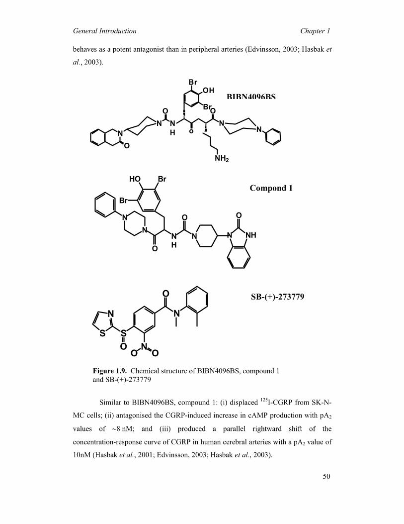
General Introduction Chapter 1
50
behaves as a potent antagonist than in peripheral arteries (Edvinsson, 2003; Hasbak et
al., 2003).
Similar to BIBN4096BS, compound 1: (i) displaced 125I-CGRP from SK-N-
MC cells; (ii) antagonised the CGRP-induced increase in cAMP production with pA2
values of ∼8 nM; and (iii) produced a parallel rightward shift of the
concentration-response curve of CGRP in human cerebral arteries with a pA2 value of
10nM (Hasbak et al., 2001; Edvinsson, 2003; Hasbak et al., 2003).
Figure 1.9. Chemical structure of BIBN4096BS, compound 1 and SB-(+)-273779
NH2
NO
N NH
O ON
Br
BrOH
o N
BIBN4096BS
NHN
ONN
NH
HO Br
Br
ON
O
Compond 1
ON
S
N
SO
N
O
O
SB-(+)-273779
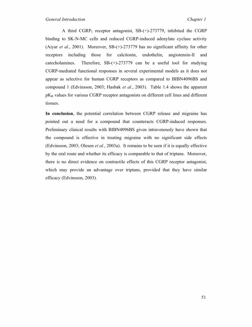
General Introduction Chapter 1
51
A third CGRP1 receptor antagonist, SB-(+)-273779, inhibited the CGRP
binding to SK-N-MC cells and reduced CGRP-induced adenylate cyclase activity
(Aiyar et al., 2001). Moreover, SB-(+)-273779 has no significant affinity for other
receptors including those for calcitonin, endothelin, angiotensin-II and
catecholamines. Therefore, SB-(+)-273779 can be a useful tool for studying
CGRP-mediated functional responses in several experimental models as it does not
appear as selective for human CGRP receptors as compared to BIBN4096BS and
compound 1 (Edvinsson, 2003; Hasbak et al., 2003). Table 1.4 shows the apparent
pKB values for various CGRP receptor antagonists on different cell lines and different
tissues.
In conclusion, the potential correlation between CGRP release and migraine has
pointed out a need for a compound that counteracts CGRP-induced responses.
Preliminary clinical results with BIBN4096BS given intravenously have shown that
the compound is effective in treating migraine with no significant side effects
(Edvinsson, 2003; Olesen et al., 2003a). It remains to be seen if it is equally effective
by the oral route and whether its efficacy is comparable to that of triptans. Moreover,
there is no direct evidence on contractile effects of this CGRP receptor antagonist,
which may provide an advantage over triptans, provided that they have similar
efficacy (Edvinsson, 2003).

General Introduction Chapter 1
52
1.14 Aims of this thesis
• To study in anaesthetised vagosympathectomised pigs, the effects of
BIBN4096BS (a potent and selective CGRP receptor antagonist) and
sumatriptan (a 5-HT1B/D receptor agonist with established antimigraine
properties) on capsaicin-induced: (i) carotid haemodynamic responses; and
(ii) plasma CGRP concentrations.
• To investigate, in anaesthetised vagosympathectomised pigs, the effects of
BIBN4096BS on: (i) α-CGRP induced changes in carotid haemodynamics;
and (ii) cardiac output distribution, in order to establish BIBN4096BS
cardiovascular safety.
• To examine the cardiovascular distribution of CGRP receptors in
anaesthetised rats by investigating the effects of BIBN4096BS on
α-CGRP-induced changes in cardiac output distribution and regional
haemodynamics.
CHAPTER 2
Effects of the CGRP receptor antagonist BIBN4096BS on
capsaicin-induced carotid haemodynamic changes in
anaesthetised pigs
Based on: Kapoor, K., Arulmani, U., Heiligers, J.P.C., Garrelds, I.M., Willems, E.W.,
Doods, H., Villalon, C.M & Saxena, P.R. (2003). Effects of the CGRP receptor
antagonist BIBN4096BS on capsaicin-induced carotid haemodynamic changes in
anaesthetised pigs. Br. J. Pharmacol., 140, 329-338

BIBN4096BS on capsaicin… Chapter 2
54
2 Effects of the CGRP receptor antagonist BIBN4096BS on capsaicin-induced carotid haemodynamic changes in anaesthetised pigs
Abstract: Calcitonin gene-related peptide (CGRP), a potent vasodilator released from capsaicin-sensitive trigeminal sensory nerves, seems to be involved in the pathogenesis of migraine. Hence, CGRP receptor antagonists may serve as a novel treatment for migraine. This study was therefore designed to investigate the effects of BIBN4096BS (100, 300 and 1000 µg kg-1, i.v.), a potent and selective CGRP receptor antagonist, on capsaicin-induced carotid haemodynamic changes in anaesthetised pigs. Both vagosympathetic trunks were cut and phenylephrine was infused into the carotid artery (i.c.) to support carotid vascular tone. Infusions of capsaicin (0.3, 1, 3 and 10 µg kg-1 min-1, i.c.) did not alter heart rate, but dose-dependently increased mean arterial blood pressure. This moderate hypertensive effect was not modified by BIBN4096BS. Capsaicin infusion (10 µg kg-1 min-1, i.c.) increased total carotid, arteriovenous anastomotic and tissue blood flows and conductances as well as carotid pulsations, but decreased the difference between arterial and jugular venous oxygen saturations. These responses to capsaicin were dose-dependently blocked by BIBN4096BS. Capsaicin infusion (10 µg kg-1 min-1, i.c.) more than doubled jugular venous plasma concentration of CGRP. This effect was not blocked, but rather increased, by BIBN4096BS. The above results show that BIBN4096BS behaves as a potent antagonist of capsaicin-induced carotid haemodynamic changes that are mediated via the release of CGRP. Therefore, this compound may prove effective in the treatment of migraine.
2.1 Introduction
Although a complete understanding of the pathogenesis of migraine remains elusive
thus far, there seems little doubt that dilatation of cranial blood vessels, including
carotid arteriovenous anastomoses, is involved in the headache phase (De Vries et al.,
1999a). Moreover, evidence is accumulating that a release of vasoactive
neuropeptides from the trigeminal sensory nerves may be an important factor in the
genesis of migraine (Goadsby et al., 2002b). In this respect, a high circulating plasma
concentration of calcitonin gene related peptide (CGRP) has been demonstrated
during migraine headache (Goadsby et al., 1990) and these concentrations can be
normalised by triptans in parallel with alleviation of headache (Goadsby et al., 1990;
Ashina et al., 2000). Indeed, CGRP is widely distributed in the body, including the
central and peripheral parts of the trigeminovascular system (Brain et al., 1985; van
Rossum et al., 1997; Juaneda et al., 2000; Poyner & Marshall, 2001), where it is
co-localised with substance P, neurokinin A and/or 5-HT1D receptors (Gulbenkian et
BIBN4096BS on capsaicin… Chapter 2
55
al., 1995; Gulbenkian et al., 2001; Smith et al., 2002). CGRP can mediate neurogenic
dilatation of cranial blood vessels as well as sensory nerve transmission between the
first and second order afferent input from these vessels during migraine headache
(Gulbenkian et al., 2001; Williamson & Hargreaves, 2001; Goadsby et al., 2002b;
Smith et al., 2002). Thus, it follows that inhibition of CGRP-mediated cranial
vasodilatation and sensory nerve transmission with a potent and selective CGRP
receptor antagonist may prove a novel strategy in treating migraine.
The recent discovery of a di-peptide CGRP receptor antagonist BIBN4096BS
(1-piperidinecarboxamide, N-[2-[[5-amino-1[[4-(4-pyridinyl)-1-piperazinyl]carbonyl]
pentyl]amino]-1-[(3,5-dibromo-4-hydroxyphenyl)methyl]-2-oxoethyl]-4-(1,4 dihydro-
2-oxo-3(2H)-quinazolinyl)-, (Doods et al., 2000; Doods, 2001) represents a
significant advance in exploring the pathophysiological role of CGRP in migraine.
BIBN4096BS displays a very high affinity for human CGRP receptors (Doods et al.,
2000; Wu et al., 2000; Edvinsson et al., 2002; Moreno et al., 2002). This compound
is undergoing clinical trials for aborting migraine headache and the clinical results are
awaited with great interest.
Using an animal model that seems to be predictive of antimigraine activity
(Spierings & Saxena, 1980; Villalón & Terrón, 1994; Saxena, 1995; Saxena et al.,
1998; De Vries et al., 1999a; Tfelt-Hansen et al., 2000), the present study in
anaesthetised pigs was designed (i) to investigate the effects of capsaicin (pungent
substance in red chilli pepper), which releases neuropeptides, including CGRP
(Alving et al., 1991; Jansen-Olesen et al., 1996; Szallasi & Blumberg, 1999; Eltorp et
al., 2000), on systemic and carotid haemodynamics, and (ii) to establish if
BIBN4096BS is able to attenuate the responses induced by capsaicin. A preliminary
account of this investigation was presented at the XIVth World Congress of
Pharmacology (Kapoor et al., 2002).
2.2 Materials and methods
2.2.1 General
After an overnight fast, a total of 22 pigs (Yorkshire x Landrace, females, 10-14 kg;
n=11 each for vehicle and BIBN4096BS) were sedated with azaperone (120 mg, i.m.)
and midazolam hydrochloride (10 mg, i.m.) and then anaesthetised with sodium
pentobarbital (600 mg, i.v.). After tracheal intubation, the animals were connected to

BIBN4096BS on capsaicin… Chapter 2
56
a respirator (BEAR 2E, BeMeds AG, Baar, Switzerland) for intermittent positive
pressure ventilation with a mixture of room air and oxygen. Respiratory rate, tidal
volume and oxygen supply were adjusted to keep arterial blood gas values within
physiological limits (pH: 7.35-7.48; pCO2: 35-48 mmHg; pO2: 100-120 mmHg).
Anaesthesia was maintained with a continuous i.v. infusion of sodium pentobarbital
(12-20 mg kg-1 h-1). This anaesthetic regimen, together with bilateral
vagosympathectomy (see below), increases heart rate and markedly dilates carotid
arterioles and arteriovenous anastomoses due to a loss of parasympathetic and
sympathetic tone, respectively. Consequently, carotid blood flow, particularly its
arteriovenous anastomotic fraction, is considerably higher in these pigs than in
conscious or thiopental-anaesthetised pigs (Den Boer et al., 1993).
Heart rate was measured with a tachograph (CRW, Erasmus University,
Rotterdam, The Netherlands) triggered by electrocardiogram signals. Both common
carotid arteries were dissected free and the accompanying vagosympathetic trunks
were cut between two ligatures to prevent any possible influence via baroreceptor
reflexes on the carotid vascular responses produced by capsaicin. Pulsatile and mean
carotid blood flows were measured in the right common carotid artery with a flow
probe (internal diameter: 2.5 mm) connected to a sine-wave electromagnetic flow
meter (Transflow 601-system, Skalar, Delft, The Netherlands). The amplitude of
carotid blood flow signals provided an index of carotid flow pulse. Subsequently,
three hub-less needles, connected to a polyethylene tube, were inserted into the right
common carotid artery for the administration of capsaicin, radioactive microspheres
and the α1-adrenoceptor agonist phenylephrine. The use of phenylephrine is
necessitated by the fact that the carotid arterioles and arteriovenous anastomoses are
already in a dilated state under the present anaesthetic regime (Den Boer et al., 1993)
and, therefore, to study the effects of vasodilator agents (in the present case capsaicin)
one has to constrict them first. As described earlier (Willems et al., 1999),
phenylephrine decreases total carotid conductance exclusively by constricting carotid
arteriovenous anastomoses, which results in an increase in the difference between
arterial and jugular venous oxygen saturations (A-V SO2 difference) (Saxena, 1987).
Lastly, catheters were placed in the right external jugular vein for the
withdrawal of venous blood samples to measure blood gases (ABL-510; Radiometer,
Copenhagen, Denmark) and plasma concentrations of CGRP (see below), inferior

BIBN4096BS on capsaicin… Chapter 2
57
vena cava (via the left femoral vein) for the administration of the vehicle or
BIBN4096BS and aortic arch (via the left femoral artery) for the measurement of
arterial blood pressure (Combitrans disposable pressure transducer; Braun,
Melsungen, Germany) as well as withdrawal of arterial blood samples to measure
blood gases.
Heart rate and systolic, diastolic and mean arterial blood pressures as well as
mean and pulsatile carotid artery blood flows were continuously monitored on a
polygraph (CRW, Erasmus University, Rotterdam, The Netherlands). Vascular
conductances were calculated by dividing respective blood flows (ml min-1) by mean
arterial blood pressure (mmHg), multiplied by one hundred and expressed as
10-2 ml min-1 mmHg-1. During the experiment, body temperature was maintained at
37±1°C by a heating pad and the animal was infused with physiological saline to
compensate for fluid losses.
2.2.2 Distribution of carotid blood flow
The distribution of common carotid blood flow into tissue (capillary) and
arteriovenous anastomotic fractions was determined in 13 pigs (later receiving
vehicle, n=7 or BIBN4096BS, n=6) with radioactive microspheres (diameter:
15.5±0.1 µm; S.D.), labelled with 141Ce, 113Sn, 103Ru, 95Nb or 46Sc (NEN Dupont,
Boston, USA). For each measurement, a suspension of about 200,000 microspheres,
labelled with one of the isotopes, was mixed and injected into the carotid artery. At
the end of the experiment, the animal was killed using an overdose of pentobarbital
and the heart, kidneys, lungs and different cranial tissues were dissected out, weighed
and put in vials. The radioactivity in these vials was counted for 5 min in a
γ-scintillation counter (Packard, Minaxi autogamma 5000), using suitable windows
for discriminating the different isotopes (141Ce: 120-167 KeV, 113Sn: 355-435 KeV, 103Ru: 450-548 KeV, 95Nb: 706-829 KeV and 46Sc: 830-965 KeV). All data were
processed by a set of specially designed computer programs (Saxena et al., 1980).
The distribution of total carotid blood flow to different tissues (Qtis) was
calculated by the formula: Qtis = (Itis/Itotal) x Qcarotid, where Itis is tissue radioactivity,
Itotal is the sum of radioactivity counted in tissues and Qcarotid is the total common
carotid blood flow at the time of microsphere injection. Since little or no radioactivity
was detected in the heart or kidneys, it can be assumed that all microspheres trapped

BIBN4096BS on capsaicin… Chapter 2
58
in lungs reach the lungs from the venous side after escaping via carotid arteriovenous
anastomoses. Therefore, the amount of radioactivity in the lungs can be used as an
index of the arteriovenous anastomotic fraction of carotid blood flow (Saxena et al.,
1980; 1982).
2.2.3 Determination of plasma concentration of CGRP
Jugular venous blood samples were obtained from 12 pigs, receiving vehicle or
BIBN4096BS (n=6 each). Four of these animals (2 each for vehicle and
BIBN4096BS) had been used for carotid haemodynamic experiments, while the other
8 were separate experiments using the same protocol except that the radioactive
microspheres were not used. Blood was transferred immediately into a polypropylene
tube containing ethylene dinitro-tetraacetic acid (1 mg ml-1 of blood) and aprotinin
(500 KIU ml-1 of blood). Aprotinin was used to inhibit endogenous plasma proteases,
since we observed that CGRP is not detectable in biological samples without aprotinin
(unpublished). After centrifugation at 1600 g for 15 min, plasma samples were coded
and stored at -80°C until CGRP measurements were performed. The person
measuring CGRP concentrations remained blind to the treatments, until all data had
been collated.
CGRP was extracted from plasma using a C18 SEP-COLUMN, dried by
lypholisation, and measured by radioimmunoassay (Dwenger, 1984), as per protocol
of the Peninsula Laboratories, Inc (Belmont, CA, U.S.A.). The recovery of CGRP
from the extraction procedure was ascertained by assaying control samples paired
with a duplicate sample spiked with known quantities of CGRP. The column
recovery values were 85, 79, 81, 89 and 92% (Mean=85.2; Standard deviation=5.4;
Coefficient of variation=6.3%). The CGRP concentrations measured in the actual
samples were, however, not corrected for the loss in the extraction procedure.
2.2.4 Experimental protocol
Following surgery and after haemodynamic condition of the animals (n=22) had been
stable for 15-20 min (heart rate: 107±4 beats min-1, mean arterial blood pressure:
95±2 mmHg, mean carotid blood flow: 120±12 ml min-1 and A-V SO2 difference:
7.6±1.1%), phenylephrine was infused into the right common carotid artery at a rate
of 10 µg kg-1 min-1 for 10 min, followed by 3-6 µg kg-1 min-1 throughout the rest of
the experiment. The latter dose of phenylephrine was chosen so that the external

BIBN4096BS on capsaicin… Chapter 2
59
jugular venous oxygen saturation was between 60-70% and mean carotid blood flow
was about 40% of the original value. After a period during which haemodynamic
variables remained constant for at least 60 min (heart rate: 130±4 beats min-1, mean
arterial blood pressure: 105±2 mmHg, mean carotid blood flow: 48±5 ml min-1 and
A-V SO2 difference: 31±2.3%; n=22), the animals received consecutive infusions
(0.15, 0.45, 1.5 and 4.5 ml, i.c. during 3 min each) of capsaicin vehicle (see
Compounds and kits section). It is important to mention that the vehicle of capsaicin
was devoid of any systemic and carotid haemodynamic responses (data not shown).
Five to 10 min after the last infusion of capsaicin vehicle, blood samples
were obtained for the measurements of blood gases and CGRP concentration and
values of heart rate, arterial blood pressure and total carotid blood flow and
conductance were collated (baseline values; 11 pigs each for vehicle and
BIBN4096BS). In 12 of the 22 pigs (6 each for vehicle and BIBN4096BS) the first
batch of radioactive microspheres was injected for determining the baseline
distribution of carotid blood flow. The animals then received consecutive infusions
of capsaicin (0.3, 1, 3 and 10 µg kg-1 min-1, i.c. for 3 min each) and heart rate, arterial
blood pressure and total carotid blood flow were determined at the end of each
infusion. In addition, after the last infusion of capsaicin (10 µg kg-1 min-1), blood
gases, plasma CGRP concentration and carotid blood flow distribution were measured
as described above (control values). Subsequently, a recovery period of 20 min was
allowed until all haemodynamic parameters had returned to baseline levels. At this
point, the animals were divided into two groups, which were treated with i.v.
infusions (rate: 0.5 ml min-1 for 10 min) of either vehicle (three times 5 ml of acidified
distilled water) or BIBN4096BS (100, 300 and 1000 µg kg-1). Ten min after each
infusion, capsaicin was given and haemodynamic and biochemical variables were
measured again, as described above.
2.2.5 Data presentation and statistical analysis
All data are presented as mean±s.e.mean, unless stated otherwise. The statistical
analysis was performed using the SPSS package for windows (version 10.0; SPSS
Inc., Chicago, IL, USA). The significance of changes within one group (vehicle or
BIBN4096BS) was analysed with repeated-measures ANOVA, followed by
Greenhouse-Geisser correction for serial autocorrelation (Ludbrook, 1994) and

BIBN4096BS on capsaicin… Chapter 2
60
Bonferroni correction for multiple comparisons (Overall & Doyle, 1996). The
significance of the between-group changes (vehicle versus BIBN4096BS treatments)
was first analysed with repeated-measures ANOVA, including baseline measurements
as a covariate (Overall & Doyle, 1994) and the Greenhouse-Geisser correction. If the
two groups differed significantly, pairwise comparisons of corresponding values in
the vehicle- and BIBN4096BS-treated groups were performed using univariate
analysis (Overall & Atlas, 1999), followed by Bonferroni correction. Statistical
significance was accepted at P<0.05 (two-tailed).
2.2.6 Ethical approval
The Ethics Committee of the Erasmus MC, Rotterdam, dealing with the use of
animals in scientific experiments, approved the protocols for this investigation.
2.2.7 Compounds and kits
The following compounds were used: aprotinin (5850 KIU mg-1; Roth, Karlsruhe,
Germany), azaperone (Stresnil®; Janssen Pharmaceuticals, Beerse, Belgium),
BIBN4096BS (gift from Boehringer Ingelheim Pharma KG, Biberach, Germany),
capsaicin, tween 80, ethanol and phenylephrine hydrochloride (all from Sigma-
Aldrich Chemie b.v., Zwijndrecht, The Netherlands), ethylene dinitro-tetraacetic acid
(Merck, Darmstadt, Germany), heparin sodium (to prevent blood clotting in catheters;
Leo Pharmaceutical Products, Weesp, The Netherlands), midazolam hydrochloride
(Dormicum®; Hoffmann La Roche b.v., Mijdrecht, The Netherlands), phenylephrine
hydrochloride (Sigma-Aldrich Chemie b.v., Zwijndrecht, The Netherlands) and
sodium pentobarbital (Sanofi Sante b.v., Maasluis, The Netherlands). The
radioimmunoassay kit for CGRP was purchased from Peninsula Laboratories, Inc.
(Belmont, CA, U.S.A.).
Capsaicin was initially dissolved in tween 80, ethanol and physiological
saline in the ratio of 0.5:1:8.5 ml, respectively. Phenylephrine was dissolved in
distilled water, while BIBN4096BS was initially dissolved in 0.5 ml of 1N HCl, then
diluted with 4 ml of distilled water and adjusted to pH 6.5 by 1N NaOH.
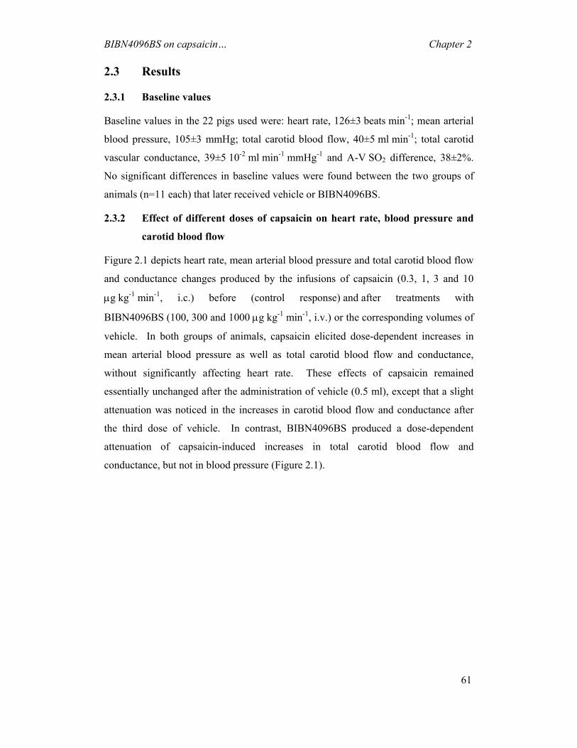
BIBN4096BS on capsaicin… Chapter 2
61
2.3 Results
2.3.1 Baseline values
Baseline values in the 22 pigs used were: heart rate, 126±3 beats min-1; mean arterial
blood pressure, 105±3 mmHg; total carotid blood flow, 40±5 ml min-1; total carotid
vascular conductance, 39±5 10-2 ml min-1 mmHg-1 and A-V SO2 difference, 38±2%.
No significant differences in baseline values were found between the two groups of
animals (n=11 each) that later received vehicle or BIBN4096BS.
2.3.2 Effect of different doses of capsaicin on heart rate, blood pressure and
carotid blood flow
Figure 2.1 depicts heart rate, mean arterial blood pressure and total carotid blood flow
and conductance changes produced by the infusions of capsaicin (0.3, 1, 3 and 10
µg kg-1 min-1, i.c.) before (control response) and after treatments with
BIBN4096BS (100, 300 and 1000 µg kg-1 min-1, i.v.) or the corresponding volumes of
vehicle. In both groups of animals, capsaicin elicited dose-dependent increases in
mean arterial blood pressure as well as total carotid blood flow and conductance,
without significantly affecting heart rate. These effects of capsaicin remained
essentially unchanged after the administration of vehicle (0.5 ml), except that a slight
attenuation was noticed in the increases in carotid blood flow and conductance after
the third dose of vehicle. In contrast, BIBN4096BS produced a dose-dependent
attenuation of capsaicin-induced increases in total carotid blood flow and
conductance, but not in blood pressure (Figure 2.1).
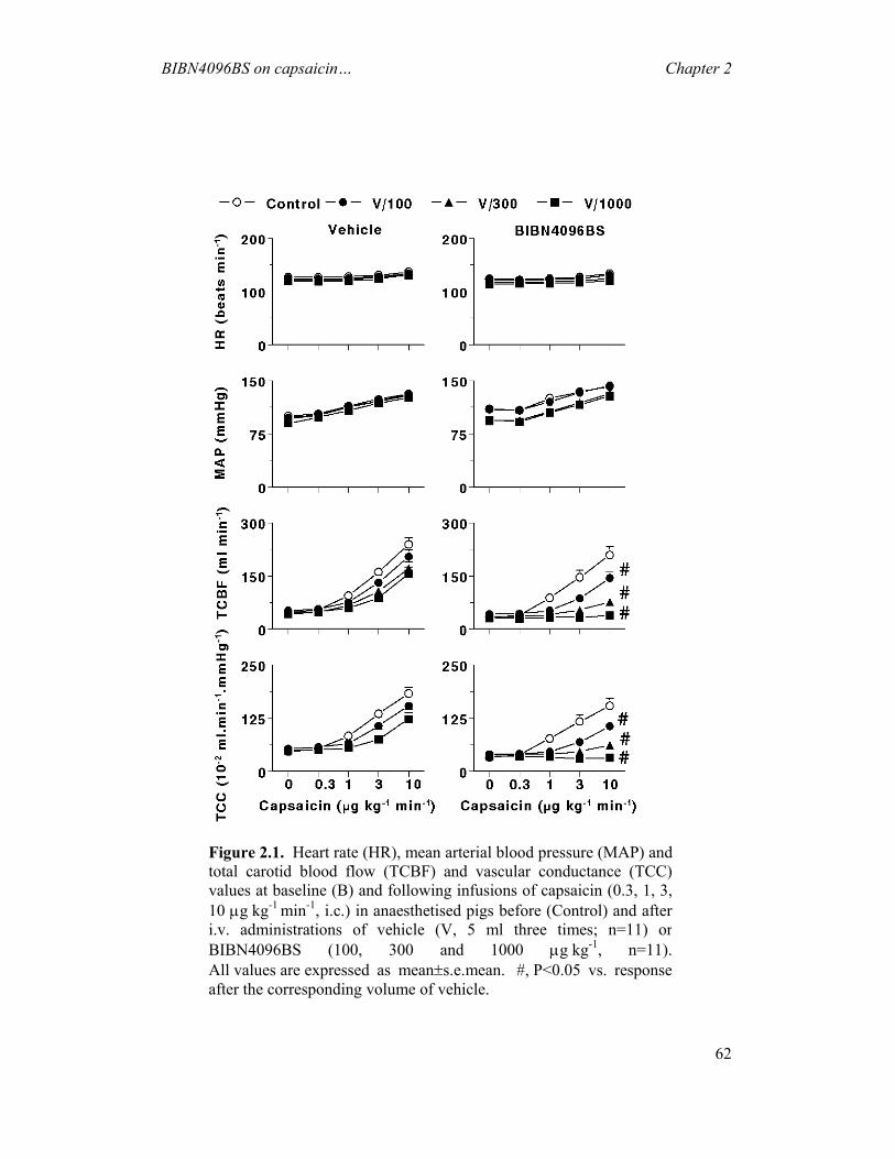
BIBN4096BS on capsaicin… Chapter 2
62
Figure 2.1. Heart rate (HR), mean arterial blood pressure (MAP) andtotal carotid blood flow (TCBF) and vascular conductance (TCC)values at baseline (B) and following infusions of capsaicin (0.3, 1, 3,10 µg kg-1 min-1, i.c.) in anaesthetised pigs before (Control) and afteri.v. administrations of vehicle (V, 5 ml three times; n=11) or BIBN4096BS (100, 300 and 1000 µg kg-1, n=11). All values are expressed as mean±s.e.mean. #, P<0.05 vs. response after the corresponding volume of vehicle.
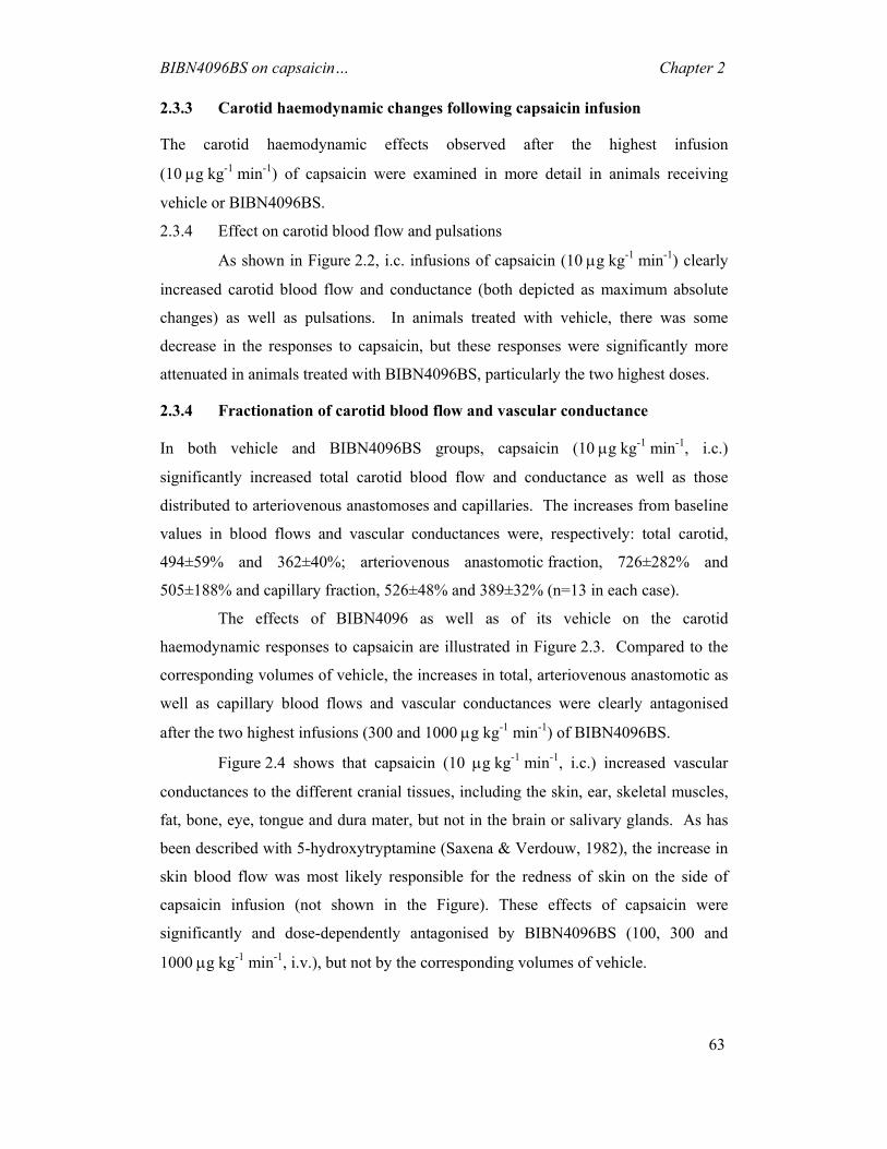
BIBN4096BS on capsaicin… Chapter 2
63
2.3.3 Carotid haemodynamic changes following capsaicin infusion
The carotid haemodynamic effects observed after the highest infusion
(10 µg kg-1 min-1) of capsaicin were examined in more detail in animals receiving
vehicle or BIBN4096BS.
2.3.4 Effect on carotid blood flow and pulsations
As shown in Figure 2.2, i.c. infusions of capsaicin (10 µg kg-1 min-1) clearly
increased carotid blood flow and conductance (both depicted as maximum absolute
changes) as well as pulsations. In animals treated with vehicle, there was some
decrease in the responses to capsaicin, but these responses were significantly more
attenuated in animals treated with BIBN4096BS, particularly the two highest doses.
2.3.4 Fractionation of carotid blood flow and vascular conductance
In both vehicle and BIBN4096BS groups, capsaicin (10 µg kg-1 min-1, i.c.)
significantly increased total carotid blood flow and conductance as well as those
distributed to arteriovenous anastomoses and capillaries. The increases from baseline
values in blood flows and vascular conductances were, respectively: total carotid,
494±59% and 362±40%; arteriovenous anastomotic fraction, 726±282% and
505±188% and capillary fraction, 526±48% and 389±32% (n=13 in each case).
The effects of BIBN4096 as well as of its vehicle on the carotid
haemodynamic responses to capsaicin are illustrated in Figure 2.3. Compared to the
corresponding volumes of vehicle, the increases in total, arteriovenous anastomotic as
well as capillary blood flows and vascular conductances were clearly antagonised
after the two highest infusions (300 and 1000 µg kg-1 min-1) of BIBN4096BS.
Figure 2.4 shows that capsaicin (10 µg kg-1 min-1, i.c.) increased vascular
conductances to the different cranial tissues, including the skin, ear, skeletal muscles,
fat, bone, eye, tongue and dura mater, but not in the brain or salivary glands. As has
been described with 5-hydroxytryptamine (Saxena & Verdouw, 1982), the increase in
skin blood flow was most likely responsible for the redness of skin on the side of
capsaicin infusion (not shown in the Figure). These effects of capsaicin were
significantly and dose-dependently antagonised by BIBN4096BS (100, 300 and
1000 µg kg-1 min-1, i.v.), but not by the corresponding volumes of vehicle.
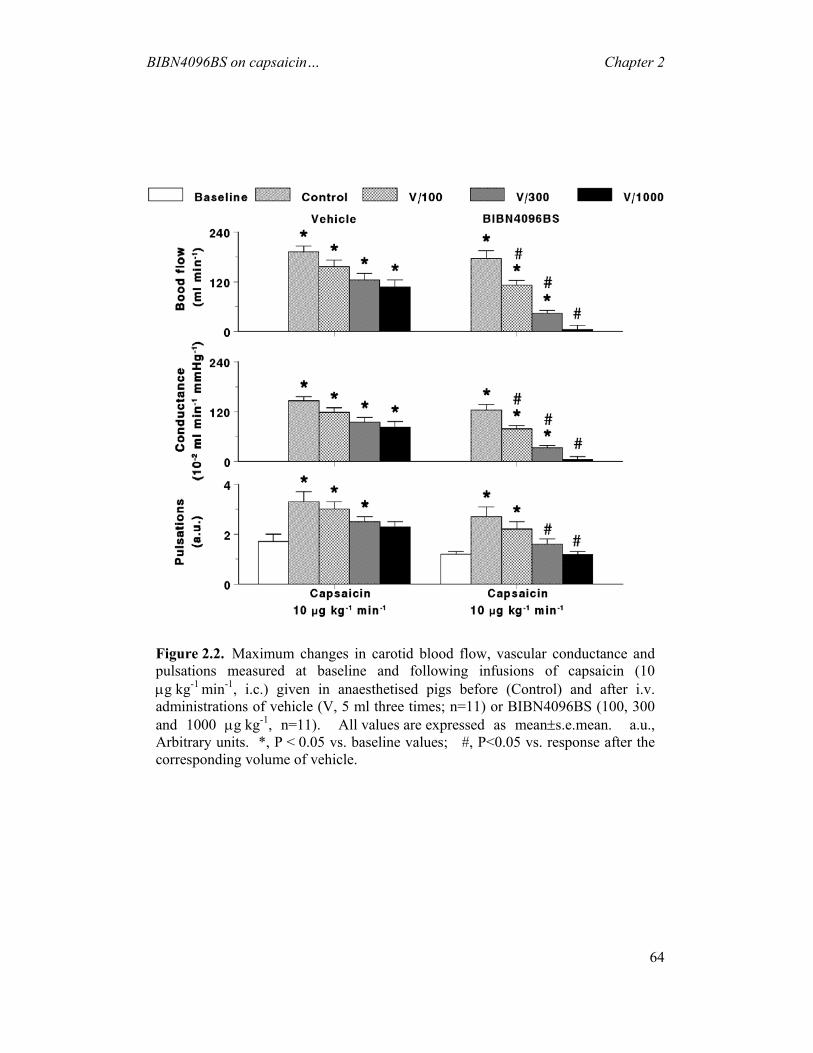
BIBN4096BS on capsaicin… Chapter 2
64
Figure 2.2. Maximum changes in carotid blood flow, vascular conductance andpulsations measured at baseline and following infusions of capsaicin (10µg kg-1 min-1, i.c.) given in anaesthetised pigs before (Control) and after i.v.administrations of vehicle (V, 5 ml three times; n=11) or BIBN4096BS (100, 300and 1000 µg kg-1, n=11). All values are expressed as mean±s.e.mean. a.u., Arbitrary units. *, P < 0.05 vs. baseline values; #, P<0.05 vs. response after thecorresponding volume of vehicle.

BIBN4096BS on capsaicin… Chapter 2
65
Figure 2.3. Total carotid, arteriovenous anastomotic (AVA) and capillaryblood flows (left panel) and vascular conductances (right panel) measured at baseline and following infusions of capsaicin (10 µg kg-1 min-1, i.c.) given in anaesthetised pigs before (Control) and after i.v. administrationsof vehicle (V, 5 ml three times; n=7) or BIBN4096BS (100, 300 and 1000µg kg-1, n=6). All values are expressed as mean±s.e.mean. *, P < 0.05 vs. baseline values; #, P<0.05 vs. response after the corresponding volume ofvehicle.

BIBN4096BS on capsaicin… Chapter 2
66
Figure 2.4. Distribution of carotid vascular conductances to head tissues measuredat baseline (Bas) and following infusions of capsaicin (10 µg kg-1 min-1, i.c.) givenin anaesthetised pigs before (Con) and after i.v. administrations of vehicle (V, 5 mlthree times; n=7) or BIBN4096BS (100, 300 and 1000 µg kg-1, n=6).All values are expressed as mean±s.e.mean. *, P < 0.05 vs. baseline values;#, P<0.05 vs. response after the corresponding volume of vehicle.
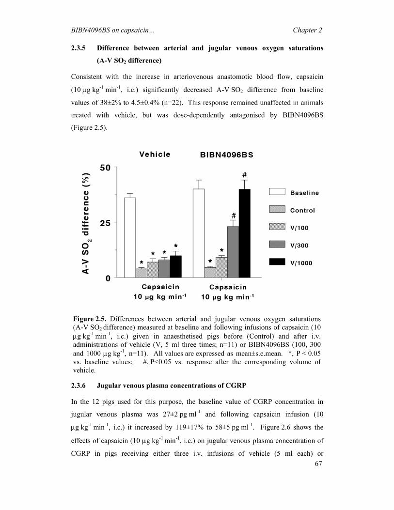
BIBN4096BS on capsaicin… Chapter 2
67
2.3.5 Difference between arterial and jugular venous oxygen saturations
(A-V SO2 difference)
Consistent with the increase in arteriovenous anastomotic blood flow, capsaicin
(10 µg kg-1 min-1, i.c.) significantly decreased A-V SO2 difference from baseline
values of 38±2% to 4.5±0.4% (n=22). This response remained unaffected in animals
treated with vehicle, but was dose-dependently antagonised by BIBN4096BS
(Figure 2.5).
2.3.6 Jugular venous plasma concentrations of CGRP
In the 12 pigs used for this purpose, the baseline value of CGRP concentration in
jugular venous plasma was 27±2 pg ml-1 and following capsaicin infusion (10
µg kg-1 min-1, i.c.) it increased by 119±17% to 58±5 pg ml-1. Figure 2.6 shows the
effects of capsaicin (10 µg kg-1 min-1, i.c.) on jugular venous plasma concentration of
CGRP in pigs receiving either three i.v. infusions of vehicle (5 ml each) or
Figure 2.5. Differences between arterial and jugular venous oxygen saturations(A-V SO2
difference) measured at baseline and following infusions of capsaicin (10µg kg-1 min-1, i.c.) given in anaesthetised pigs before (Control) and after i.v.administrations of vehicle (V, 5 ml three times; n=11) or BIBN4096BS (100, 300and 1000 µg kg-1, n=11). All values are expressed as mean±s.e.mean. *, P < 0.05vs. baseline values; #, P<0.05 vs. response after the corresponding volume ofvehicle.
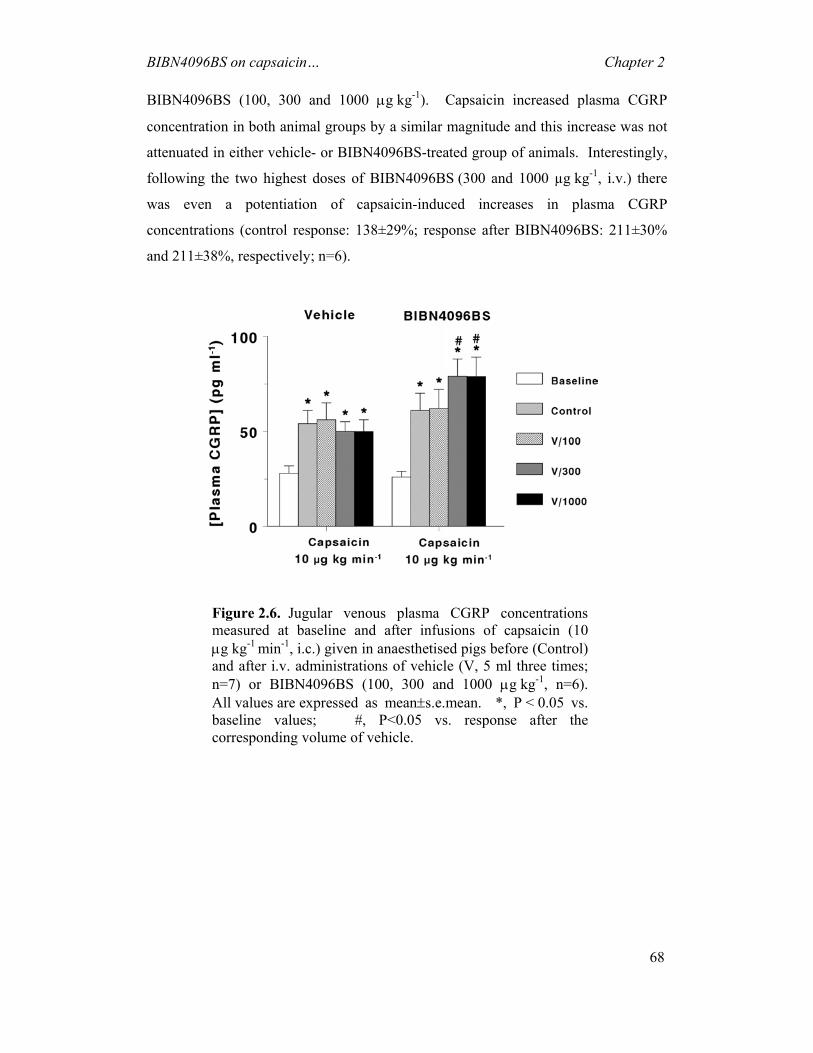
BIBN4096BS on capsaicin… Chapter 2
68
BIBN4096BS (100, 300 and 1000 µg kg-1). Capsaicin increased plasma CGRP
concentration in both animal groups by a similar magnitude and this increase was not
attenuated in either vehicle- or BIBN4096BS-treated group of animals. Interestingly,
following the two highest doses of BIBN4096BS (300 and 1000 µg kg-1, i.v.) there
was even a potentiation of capsaicin-induced increases in plasma CGRP
concentrations (control response: 138±29%; response after BIBN4096BS: 211±30%
and 211±38%, respectively; n=6).
Figure 2.6. Jugular venous plasma CGRP concentrationsmeasured at baseline and after infusions of capsaicin (10µg kg-1 min-1, i.c.) given in anaesthetised pigs before (Control) and after i.v. administrations of vehicle (V, 5 ml three times;n=7) or BIBN4096BS (100, 300 and 1000 µg kg-1, n=6). All values are expressed as mean±s.e.mean. *, P < 0.05 vs. baseline values; #, P<0.05 vs. response after the corresponding volume of vehicle.

BIBN4096BS on capsaicin… Chapter 2
69
2.4 Discussion
2.4.1 General
Although there is much debate about the pathogenesis of migraine, there seems to be a
general agreement regarding its neurovascular nature (Goadsby & Edvinsson, 1993;
De Vries et al., 1999a; Goadsby et al., 2002b; Villalón et al., 2002). Thus, there is a
release of vasoactive peptides producing intense cranial vasodilatation, increased
arterial pulsations and a sterile inflammatory reaction with pain (Moskowitz et al.,
1989; De Vries et al., 1999a). Amongst these neuropeptides, CGRP is considered as a
biological marker in migraine pathogenesis (van Rossum et al., 1997; Goadsby et al.,
2002b; Hagner et al., 2002c). Moreover, stimulation of trigeminal sensory neurones
with electrical procedures or chemical substances, like capsaicin, releases
endogenously-stored CGRP (Buzzi et al., 1995; Eltorp et al., 2000) that, in turn,
dilates cranial vessels (Williamson & Hargreaves, 2001), including carotid
arteriovenous anastomoses (Van Gelderen et al., 1995). In addition, CGRP may also
facilitate sensory nerve transmission between the first and second order afferent input
from these vessels during migraine headache (Gulbenkian et al., 2001; Goadsby et al.,
2002b; Smith et al., 2002). On this basis, it is reasonable to assume that CGRP
receptor antagonists can be a novel approach to antimigraine therapy. In this respect,
recent in vitro studies have shown that, BIBN4096BS, a potent and ‘silent’ CGRP
receptor antagonist (Doods et al., 2000), inhibits CGRP-induced dilatation of isolated
cranial blood vessels (Edvinsson et al., 2002; Verheggen et al., 2002). BIBN4096BS
can also effectively antagonise CGRP-induced carotid vasodilatation in anaesthetised
pigs (Kapoor et al., 2003b). Therefore, it seems important to investigate the effects of
BIBN4096BS on the carotid haemodynamic responses produced by endogenous
CGRP released by capsaicin in a porcine model predictive of antimigraine activity
(Saxena, 1995; De Vries et al., 1999a; Tfelt-Hansen et al., 2000). Our results show
that: (i) i.c. administration of capsaicin increased blood pressure, but dilated carotid
arteriovenous anastomoses and arterioles, together with an increase in carotid
pulsations and a narrowing of A-V SO2 difference as well as an elevation of jugular
venous plasma CGRP concentration; and (ii) BIBN4096BS dose-dependently
antagonised the changes in carotid haemodynamics and A-VSO2 difference caused by
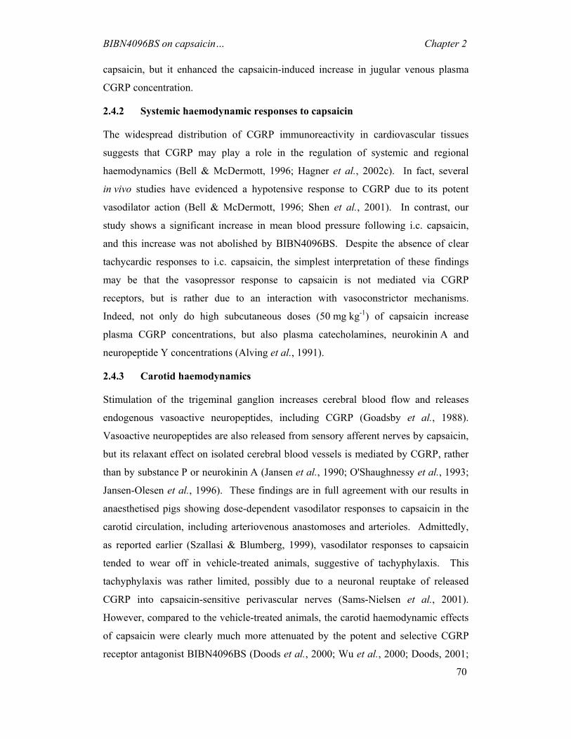
BIBN4096BS on capsaicin… Chapter 2
70
capsaicin, but it enhanced the capsaicin-induced increase in jugular venous plasma
CGRP concentration.
2.4.2 Systemic haemodynamic responses to capsaicin
The widespread distribution of CGRP immunoreactivity in cardiovascular tissues
suggests that CGRP may play a role in the regulation of systemic and regional
haemodynamics (Bell & McDermott, 1996; Hagner et al., 2002c). In fact, several
in vivo studies have evidenced a hypotensive response to CGRP due to its potent
vasodilator action (Bell & McDermott, 1996; Shen et al., 2001). In contrast, our
study shows a significant increase in mean blood pressure following i.c. capsaicin,
and this increase was not abolished by BIBN4096BS. Despite the absence of clear
tachycardic responses to i.c. capsaicin, the simplest interpretation of these findings
may be that the vasopressor response to capsaicin is not mediated via CGRP
receptors, but is rather due to an interaction with vasoconstrictor mechanisms.
Indeed, not only do high subcutaneous doses (50 mg kg-1) of capsaicin increase
plasma CGRP concentrations, but also plasma catecholamines, neurokinin A and
neuropeptide Y concentrations (Alving et al., 1991).
2.4.3 Carotid haemodynamics
Stimulation of the trigeminal ganglion increases cerebral blood flow and releases
endogenous vasoactive neuropeptides, including CGRP (Goadsby et al., 1988).
Vasoactive neuropeptides are also released from sensory afferent nerves by capsaicin,
but its relaxant effect on isolated cerebral blood vessels is mediated by CGRP, rather
than by substance P or neurokinin A (Jansen et al., 1990; O'Shaughnessy et al., 1993;
Jansen-Olesen et al., 1996). These findings are in full agreement with our results in
anaesthetised pigs showing dose-dependent vasodilator responses to capsaicin in the
carotid circulation, including arteriovenous anastomoses and arterioles. Admittedly,
as reported earlier (Szallasi & Blumberg, 1999), vasodilator responses to capsaicin
tended to wear off in vehicle-treated animals, suggestive of tachyphylaxis. This
tachyphylaxis was rather limited, possibly due to a neuronal reuptake of released
CGRP into capsaicin-sensitive perivascular nerves (Sams-Nielsen et al., 2001).
However, compared to the vehicle-treated animals, the carotid haemodynamic effects
of capsaicin were clearly much more attenuated by the potent and selective CGRP
receptor antagonist BIBN4096BS (Doods et al., 2000; Wu et al., 2000; Doods, 2001;
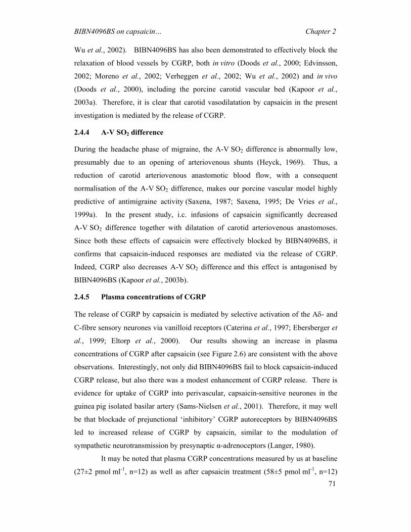
BIBN4096BS on capsaicin… Chapter 2
71
Wu et al., 2002). BIBN4096BS has also been demonstrated to effectively block the
relaxation of blood vessels by CGRP, both in vitro (Doods et al., 2000; Edvinsson,
2002; Moreno et al., 2002; Verheggen et al., 2002; Wu et al., 2002) and in vivo
(Doods et al., 2000), including the porcine carotid vascular bed (Kapoor et al.,
2003a). Therefore, it is clear that carotid vasodilatation by capsaicin in the present
investigation is mediated by the release of CGRP.
2.4.4 A-V SO2 difference
During the headache phase of migraine, the A-V SO2 difference is abnormally low,
presumably due to an opening of arteriovenous shunts (Heyck, 1969). Thus, a
reduction of carotid arteriovenous anastomotic blood flow, with a consequent
normalisation of the A-V SO2 difference, makes our porcine vascular model highly
predictive of antimigraine activity (Saxena, 1987; Saxena, 1995; De Vries et al.,
1999a). In the present study, i.c. infusions of capsaicin significantly decreased
A-V SO2 difference together with dilatation of carotid arteriovenous anastomoses.
Since both these effects of capsaicin were effectively blocked by BIBN4096BS, it
confirms that capsaicin-induced responses are mediated via the release of CGRP.
Indeed, CGRP also decreases A-V SO2 difference and this effect is antagonised by
BIBN4096BS (Kapoor et al., 2003b).
2.4.5 Plasma concentrations of CGRP
The release of CGRP by capsaicin is mediated by selective activation of the Aδ- and
C-fibre sensory neurones via vanilloid receptors (Caterina et al., 1997; Ebersberger et
al., 1999; Eltorp et al., 2000). Our results showing an increase in plasma
concentrations of CGRP after capsaicin (see Figure 2.6) are consistent with the above
observations. Interestingly, not only did BIBN4096BS fail to block capsaicin-induced
CGRP release, but also there was a modest enhancement of CGRP release. There is
evidence for uptake of CGRP into perivascular, capsaicin-sensitive neurones in the
guinea pig isolated basilar artery (Sams-Nielsen et al., 2001). Therefore, it may well
be that blockade of prejunctional ‘inhibitory’ CGRP autoreceptors by BIBN4096BS
led to increased release of CGRP by capsaicin, similar to the modulation of
sympathetic neurotransmission by presynaptic α-adrenoceptors (Langer, 1980).
It may be noted that plasma CGRP concentrations measured by us at baseline
(27±2 pmol ml-1, n=12) as well as after capsaicin treatment (58±5 pmol ml-1, n=12)

BIBN4096BS on capsaicin… Chapter 2
72
are in agreement with those previously reported in pigs (Table 2.1) (Alving et al.,
1991; Arden et al., 1994; Kallner et al., 1998).
Table 2.1. Plasma CGRP concentration range (pg ml-1) in pigs
Baseline Capsaicin Sampled from Reference
10 36 Femoral artery Alving et al. (Alving et
al., 1991)
11-16 Not measured Femoral artery and
interventricular vein
Kallner et al. (Kallner
et al., 1998)
4-12 Not measured Carotid artery Arden et al. (Arden et
al., 1994)
14-38 27-88 External jugular vein Present investigation
2.4.6 Possible clinical implications
Lastly, we would like to consider the possible clinical implications of our results with
BIBN4096BS within the context of antimigraine therapy. Indeed, the
trigeminovascular system, a functional network of cranial blood vessels and their
trigeminal innervation, seems to be activated during migraine (Goadsby et al., 2002b),
thereby provoking CGRP release and cranial blood vessel dilatation. Thus, a
blockade of the release and/or the effects of CGRP are likely to provide novel avenues
for developing antimigraine drugs without associated vasoconstriction. BIBN4096BS
may be such a compound and the present findings demonstrating that it effectively
antagonises the carotid vasodilator responses elicited by capsaicin are indeed
encouraging. Obviously, the results of currently undergoing clinical trials with
BIBN4096BS are awaited with great interest; these would be crucial in determining
not only the role of CGRP in the pathophysiology of migraine, but also of such
compounds as therapeutic agents.

CHAPTER 3
Effects of BIBN4096BS on cardiac output distribution
and on CGRP-induced carotid haemodynamic responses in the pig
Based on: Kapoor, K., Arulmani, U., Heiligers, J.P.C., Willems, E.W., Doods, H.,
Villalon, C.M & Saxena, P.R. (2003). Effects of BIBN4096BS on cardiac output
distribution and on CGRP-induced carotid haemodynamic responses in the pig
Eur. J. Pharmacol., 475, 69-77

BIBN4096BS on cardiac output… Chapter 3
74
3 Effects of BIBN4096BS on cardiac output distribution and on CGRP-induced carotid haemodynamic responses in the pig
Abstract: Calcitonin gene related peptide (CGRP) seems to be involved in the pathogenesis of migraine, since plasma CGRP levels increase during the headache phase. In the present study, we investigated the effects of a novel CGRP receptor antagonist, BIBN4096BS (1-piperidinecarboxamide, N-[2-[[5-amino-1-[[4-(4-pyridinyl)-1-pipera-zinyl]carbonyl] pentyl] amino]-1-[(3,5-dibromo-4-hydroxyphenyl) methyl]- 2-oxoethyl]-4-(1,4-dihydro-2-oxo-3(2H)-quinazolinyl) (Kapoor et al., 2003b), on the regional cardiac output distribution and on the carotid haemodynamic changes induced by α-CGRP in anaesthetised pigs. Treatment with BIBN4096BS (100, 300 and 1000 µg.kg-1, i.v.) did not affect heart rate, mean arterial blood pressure or systemic vascular conductance, but a small decrease in cardiac output was noticed; the latter was, however, not significantly different from that in vehicle-treated animals. The highest dose of BIBN4096BS moderately decreased vascular conductance in the lungs, kidneys, spleen and adrenals. Vascular conductance in other tissues, including the brain, heart, gastrointestinal system, skin and skeletal muscles remained unchanged. Intracarotid artery infusions of α-CGRP (10, 30 and 100 pmol.kg-1.min-1 during 3 min) increased total carotid blood flow and conductance, but decreased arterial blood pressure. These responses were dose-dependently blocked by BIBN4096BS. The above results show that BIBN4096BS is a CGRP receptor antagonist in the porcine carotid and systemic circulations, but the endogenous CGRP does not seem to play an important physiological role in regulating basal vascular tone. These findings suggest that BIBN4096BS may have therapeutic usefulness in migraine.
3.1 Introduction
Calcitonin gene related peptide (CGRP), a 37 amino acid neuropeptide generated by
alternative splicing of the calcitonin gene (Amara et al., 1982), is widely distributed in
the body, including in trigeminal sensory nerve fibres innervating central and
peripheral blood vessels, where it is co-localised with other vasoactive neuropeptides,
such as substance P and neurokinin A (Gulbenkian et al., 1995; Gulbenkian et al.,
2001). CGRP is a potent vasodilator agent in a wide variety of tissues (Brain &
Williams, 1985; van Rossum et al., 1997; Juaneda et al., 2000; Poyner & Marshall,
2001) and, although exogenous α-CGRP has potent systemic and regional
haemodynamic effects (Gardiner et al., 1990), the physiological role of endogenous
CGRP is not clear (Shen et al., 2001). This is mainly due to the unavailability of
potent and selective CGRP receptor antagonists; the most widely used CGRP receptor

BIBN4096BS on cardiac output… Chapter 3
75
antagonist thus far, CGRP-(8-37) is not very potent and displays partial agonist
properties (Wisskirchen et al., 1998; Waugh et al., 1999). Clearly, the advent of
‘silent’, selective and potent non-peptide CGRP receptor antagonists would be
valuable in this regard.
Interestingly, CGRP has been implicated in the pathogenesis of migraine
(Goadsby et al., 1990; Ashina et al., 2000; Edvinsson, 2001b; Durham & Russo,
2002), and it can mediate neurogenic dilatation of cranial blood vessels as well as
sensory nerve transmission between the first and second order afferent input from
these vessels during migraine headache (Gulbenkian et al., 2001; Williamson &
Hargreaves, 2001; Goadsby et al., 2002b; Smith et al., 2002). Significantly, plasma
levels of CGRP, but not of other neurotransmitter (e.g., neuropeptide Y, vasoactive
intestinal peptide or substance P), are elevated during migraine and, after sumatriptan,
these levels are normalised paralleling the resolution of headache (Goadsby et al.,
1990; Goadsby & Hoskin, 1999). Therefore, inhibition of α-CGRP release or
blockade of α-CGRP-induced vasodilatation may be a novel approach in the
management of acute migraine headache.
Doods and colleagues (2000) have recently described a small molecule
CGRP receptor antagonist, BIBN4096BS (1-piperidinecarboxamide, N-[2-[[5-amino-
1-[[4-(4-pyridinyl)-1-piperazinyl]carbonyl]pentyl] amino]- 1-[(3,5-dibromo-4-
hydroxyphenyl) methyl]- 2-oxoethyl]-4-(1,4-dihydro-2-oxo-3(2H)-quinazolinyl)
(Kapoor et al., 2003b), which possesses over 200 fold higher affinity for human (SK-
N-MC cells; Ki: 14 pM) than for rat (spleen; Ki: 3.4 nM) CGRP receptors.
BIBN4096BS as well as the endogenous ligand CGRP and its analogues
concentration-dependently displace (Kawasaki et al., 1990; Bard et al., 1993;
Goadsby & Knight, 1997; Yu et al., 1997; Bhalla et al., 2002)BIBN4096BS from SK-
N-MC cell membranes with the rank order of affinity: BIBN4096BS >
human α-CGRP = human β-CGRP > [Cys(Et)2,7] human α-CGRP > adrenomedullin
(high affinity site) = human α-CGRP8-37 = human β-CGRP8-37 >> calcitonin = amylin
(Schindler & Doods, 2002). The compound inhibits vasodilatation evoked by
trigeminal ganglion stimulation in marmosets (Doods et al., 2000) and by CGRP in
several human isolated blood vessels (Edvinsson et al., 2002; Moreno et al., 2002;
Verheggen et al., 2002). The purpose of the present study in anaesthetised pigs was to
investigate the effects of BIBN4096BS on: (i) the complete distribution of cardiac

BIBN4096BS on cardiac output… Chapter 3
76
output to assess the potential role of endogenous CGRP in regulating basal vascular
tone and thereby the cardiovascular safety of BIBN4096BS, and
(ii) the haemodynamic responses produced by intracarotid arterial (i.c.) infusion of
α-CGRP in a model predictive of antimigraine activity (Saxena, 1995; De Vries et al.,
1999a).
3.2 Materials and methods
3.2.1 General
After an overnight fast, 25 domestic pigs (Yorkshire x Landrace, females, 10-14 kg)
were sedated with intramuscular injections of azaperone (120 mg) and midazolam
hydrochloride (10 mg) and then anaesthetised with sodium pentobarbital (600 mg,
i.v.). After tracheal intubation, the animals were connected to a respirator (BEAR 2E,
BeMeds AG, Baar, Switzerland) for intermittent positive pressure ventilation with a
mixture of room air and oxygen. Respiratory rate, tidal volume and oxygen supply
were adjusted to keep arterial blood gas values within physiological limits
(pH: 7.35-7.48; pCO2: 35-48 mmHg; pO2: 100-120 mmHg). Anaesthesia was
maintained with a continuous i.v. infusion of sodium pentobarbital
(12-20 mg.kg-1.h-1). Heart rate was measured with a tachograph (CRW, Erasmus
University, Rotterdam, The Netherlands) triggered by electrocardiogram signals. A
catheter was placed in the inferior vena cava via the right femoral vein for the
administration of vehicle and BIBN4096BS. Another catheter was placed in the
aortic arch via the left femoral artery for the measurement of arterial blood pressure
(Combitrans disposable pressure transducer; Braun, Melsungen, Germany) and
arterial blood withdrawal for the measurement of blood gases (ABL-510; Radiometer,
Copenhagen, Denmark). During the experiment, body temperature was kept around
37°C and the animal was continuously infused with physiological saline to
compensate for fluid losses.
Heart rate and systolic, diastolic and mean arterial blood pressure as well as
the pulsatile and mean carotid artery blood flows (see later) were continuously
monitored on a polygraph (CRW, Erasmus University, Rotterdam, The Netherlands).

BIBN4096BS on cardiac output… Chapter 3
77
3.2.2 Cardiac output and its distribution
Cardiac output was measured by the thermodilution method using a 6F Swan-Ganz
catheter (Braun Melsungen AG, Melsungen, Germany) introduced into the pulmonary
artery via the left femoral vein.
The distribution of cardiac output was determined with 15.5±0.1 (S.D.) µm
diameter microspheres labelled with 141Ce, 113Sn, 103Ru, 95Nb or 46Sc (NEN Dupont,
Boston, USA). For each measurement, a suspension of about 1,000,000
microspheres, labelled with one of the isotopes, was injected into the left ventricle via
a catheter guided by way of the left carotid artery. Starting 15 s before microsphere
injection and lasting 70 s, a reference arterial blood sample was withdrawn
(Withdrawal pump, Harvard Apparatus Company, Southnatick, Mass, USA; rate:
6 ml.min-1) via a catheter placed into the right femoral artery. An infusion of the
corresponding volume of Haemaccel compensated blood loss during this procedure.
At the end of the experiment, the animal was killed using an overdose of
pentobarbital. Subsequently, a number of tissues (lungs, kidneys, heart, stomach,
small intestine, spleen, liver, adrenals, brain, skin and skeletal muscles) were dissected
out, weighed and put into vials. The radioactivity in these vials was counted for 5 min
in a γ-scintillation counter (Packard, Minaxi autogamma 5000), using suitable
windows for the discrimination of the different isotopes (141Ce: 120-167 KeV, 113Sn: 355-435 KeV, 103Ru: 450-548 KeV, 95Nb: 706-829 KeV and 46Sc: 830-965
KeV). All data were processed by a set of specially designed computer programs
(Saxena et al., 1980), using a personal computer. Tissue blood flows were calculated
by multiplying the ratio of tissue and reference blood sample radioactivities by the
blood withdrawal rate (6 ml.min-1) and normalised to 100 g tissue weight. Systemic
and tissue vascular conductances were calculated by dividing cardiac output (ml.min-
1) and tissue blood flows (ml.min-1/100 g tissue), respectively, by mean arterial blood
pressure (mmHg). Radioactivity in the lungs mainly represents peripheral
arteriovenous anastomotic blood flow (the non-nutrient part of the cardiac output),
although a small part (1-1.5% of cardiac output) is derived from the bronchial arteries
(Baile et al., 1982).

BIBN4096BS on cardiac output… Chapter 3
78
3.2.3 Carotid haemodynamic responses to CGRP
Both common carotid arteries and the external jugular veins were dissected free and
the accompanying vagosympathetic trunks were cut between two ligatures in order to
prevent a possible influence of CGRP via baroreceptor reflexes. Pulsatile and mean
blood flows were measured in the right common carotid artery with a flow probe
(internal diameter: 2.5 mm) connected to a sine-wave electromagnetic flow meter
(Transflow 601-system, Skalar, Delft, The Netherlands). The amplitude of carotid
blood flow signals provided an index of carotid flow pulse. Carotid vascular
conductance was calculated by dividing carotid blood flow (ml.min-1) by mean
arterial blood pressure (mmHg).
The right external jugular vein was catheterised for obtaining jugular venous
blood samples to determine blood gases. Two hub-less needles, connected to
polyethylene tubes, were inserted into the right common carotid artery and used for
intracarotid (i.c.) infusions of phenylephrine (α1-adrenoceptor agonist) and α-CGRP,
respectively. It should be noted that under pentobarbital anaesthesia carotid
arteriovenous anastomoses are dilated (Den Boer et al., 1993) and, therefore, to elicit
vasodilator responses to CGRP, a continuous infusion of phenylephrine was used
throughout the experiment. We have previously reported that phenylephrine
decreases total carotid blood flow and conductance exclusively due to constriction of
carotid arteriovenous anastomoses (Willems et al., 1999), resulting in an increase in
the difference between arterial and jugular venous oxygen saturations (A-V SO2
difference) (Saxena, 1987).
3.2.4 Experimental protocols
In the case of cardiac output distribution experiments (n=12), baseline values of heart
rate, mean arterial blood pressure, cardiac output and its distribution to the various
tissues (see above) were determined after a stabilisation period of at least 90 min. The
animals were then divided into two groups (n=6 each) receiving three i.v. infusions
(rate: 0.5 ml.min-1) of either BIBN4096BS (100, 300 and 1000 µg.kg-1) or its vehicle
(5 ml of acidified distilled water); each dose was given over 10 min with an
intervening period of 10 min before the next dose. At the end of each infusion, the
above mentioned haemodynamic variables were collated again. Lastly, the final

BIBN4096BS on cardiac output… Chapter 3
79
measurements were made 40 min after the third dose of vehicle or BIBN4096BS
(recovery).
In the case of the carotid artery experiments (n=13), phenylephrine
(10 µg.kg-1.min-1 for 10 min, followed by 3-6 µg.kg-1.min-1 throughout the rest of the
experiment) was infused into the right common carotid artery to maintain carotid
blood flow at a constant low level. After a stabilisation period of at least 90 min,
values of heart rate, arterial blood pressure, total carotid blood flow and A-V SO2
difference were collated. The animal was then given three sequential i.c. infusions
(rate: 0.083-1 ml.min-1, depending on the weight of the animal) of CGRP (10, 30 and
100 pmol.kg-1.min-1) for 3 min and the above variables (except the A-V SO2
difference, which was determined only after the highest dose) were collated again.
After the highest dose of α-CGRP, a recovery period of 20 min was allowed to elapse
when all haemodynamic parameters returned to baseline levels. At this point, the
animals were divided into two groups receiving three i.v. infusions (rate: 0.5 ml.min-1)
of either BIBN4096BS (100, 300 and 1000 µg.kg-1; n=7) or its vehicle (5 ml of
acidified distilled water; n=6); each dose was given over a period of 10 min with an
intervening period of about 10 min before the next dose. Ten min after each
treatment, the values of mean arterial blood pressure, heart rate, total carotid blood
flow and A-V SO2 difference were collated. CGRP was infused as above after each
treatment and data were collated again.
It may be mentioned that the vehicle of α-CGRP (distilled water) was devoid
of any systemic and carotid haemodynamic responses (data not shown).
3.2.5 Data presentation and statistical analysis
All data have been expressed as mean±s.e.mean, unless stated otherwise. The
significance of changes from baseline values within one group (vehicle or
BIBN4096BS) was evaluated with Duncan's new multiple range test, once an analysis
of variance (randomised block design) had revealed that the samples represented
different populations (Saxena et al., 1980; Steel & Torrie, 1980). The differences in
baseline haemodynamic values and percent change (from baseline values) in
haemodynamic variables by corresponding doses of the vehicle and BIBN4096BS
(between group comparisons) were evaluated by Student's unpaired t-test. Student's
unpaired t-test was also applied to compare the changes in the effects of CGRP

BIBN4096BS on cardiac output… Chapter 3
80
observed after different corresponding doses of the vehicle and BIBN4096BS.
Statistical significance was accepted at P<0.05 (two-tailed).
3.2.6 Ethical approval
The Ethics Committee of the Erasmus MC, Rotterdam, dealing with the use of
animals in scientific experiments, approved investigation protocols, which adhere to
EEC guidelines.
3.2.7 Compounds
The following compounds were used: azaperone (Stresnil®; Janssen Pharmaceuticals,
Beerse, Belgium), BIBN4096BS and human α-CGRP (Boehringer Ingelheim Pharma
KG, Biberach, Germany), heparin sodium (to prevent blood clotting in catheters;
Leo Pharmaceutical Products, Weesp, The Netherlands), midazolam hydrochloride
(Dormicum®; Hoffmann La Roche b.v., Mijdrecht, The Netherlands), phenylephrine
hydrochloride (Sigma-Aldrich Chemie b.v., Zwijndrecht, The Netherlands) and
sodium pentobarbital (Sanofi Sante b.v., Maasluis, The Netherlands).
Phenylephrine and α-CGRP were dissolved in distilled water, while
BIBN4096BS was initially dissolved in 0.5 ml of 1N HCl and subsequently diluted
with 4 ml of distilled water, and then adjusted to pH 6.5 with 1N NaOH.
3.3 Results
3.3.1 Effect of BIBN4096BS on cardiac output and its distribution
BASELINE VALUES
Baseline values of heart rate, mean arterial blood pressure, cardiac output (expressed
as cardiac index) and systemic vascular conductance in anaesthetised pigs (n=12)
were: 108±3 beats.min-1, 102±2 mmHg, 133±4 ml.min-1.kg-1 and 1491±54
ml.min-1.mmHg-1, respectively. Baseline values of regional vascular conductances
(ml.min-1.mmHg-1/100 g tissue) were: brain, 31±3; heart, 104±10; liver, 34±8;
stomach, 24±2; lungs (mainly systemic arteriovenous anastomoses), 229±37; adrenals,
138±10; kidneys, 263±14; spleen, 126±15; skeletal muscles, 3.3±0.3; and skin, 11±2.

BIBN4096BS on cardiac output… Chapter 3
81
SYSTEMIC AND REGIONAL HAEMODYNAMIC CHANGES
Systemic haemodynamic values collated at baseline, after vehicle or BIBN4096BS
(100, 300 and 1000 µg.kg-1, i.v.) and after a 40-min recovery period are shown in
Figure 3.1. There were no statistically significant differences (P>0.05) in baseline
values in the vehicle and BIBN4096BS groups. Except for small decreases in heart
rate by the vehicle (maximum change: 4±1%) and cardiac index by BIBN4096BS
(maximum change: 19±8%), no other changes were observed. The changes in cardiac
index by BIBN4096BS did not differ significantly (P>0.05) from those in the vehicle-
treated animals (maximum change: 7±3%).
Figure 3.2 presents regional vascular conductances in a number of tissues in
animals treated with either vehicle or BIBN4096BS (100, 300 and 1000 µg.kg-1, i.v.).
Baseline values in the two groups were not significantly different (P>0.05) in any of
the tissues, including the liver, lungs and skin. Apart from decreases in liver
conductance, no other changes in regional vascular conductances were noticed in the
vehicle-treated group. BIBN4096BS produced small decreases in vascular
conductance to liver, and with the highest dose (1000 µg.kg-1) in lungs, adrenals,
kidneys and spleen. Only the latter changes were significant when compared with the
corresponding changes in the vehicle-treated animals.
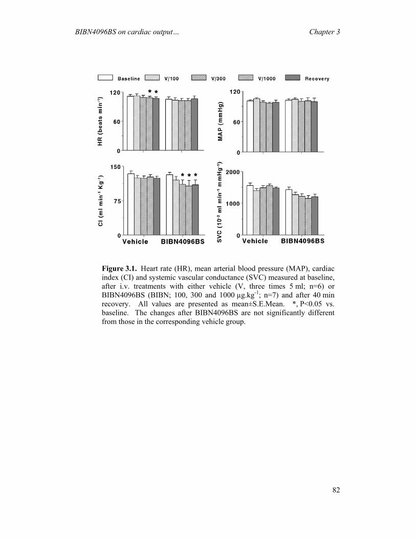
BIBN4096BS on cardiac output… Chapter 3
82
Figure 3.1. Heart rate (HR), mean arterial blood pressure (MAP), cardiacindex (CI) and systemic vascular conductance (SVC) measured at baseline,after i.v. treatments with either vehicle (V, three times 5 ml; n=6) orBIBN4096BS (BIBN; 100, 300 and 1000 µg.kg-1; n=7) and after 40 min recovery. All values are presented as mean±S.E.Mean. *, P<0.05 vs. baseline. The changes after BIBN4096BS are not significantly differentfrom those in the corresponding vehicle group.

BIBN4096BS on cardiac output… Chapter 3
83
Figure 3.2. Regional vascular conductances at baseline (Bas), after i.v.treatments with either vehicle (V, three times 5 ml; n=6) or BIBN4096BS (100,300, and 1000 µg.kg-1, i.v.; n=6) and after 40 min recovery (Rec). All valuesare presented as mean±s.e.mean. *, P<0.05 vs. baseline. #, P<0.05 vs. thecorresponding change in animals treated with vehicle.

BIBN4096BS on cardiac output… Chapter 3
84
3.3.2 Effect of BIBN4096BS on the haemodynamic responses to i.c. infusions
of α-CGRP
BASELINE VALUES
Baseline values in anaesthetised pigs (n=13) were: heart rate, 129±5 beats.min-1; mean
arterial blood pressure, 122±4 mmHg; carotid flow pulse, 1.7±0.1 arbitrary units
(a.u.); total carotid blood flow, 67±7 ml.min-1; total carotid vascular conductance,
56±5 10-2 ml.min-1.mmHg-1 and A-V SO2 difference, 26±3%. Baseline values in the
two groups of animals (vehicle and BIBN4096BS) did not differ significantly.
SYSTEMIC AND CAROTID HAEMODYNAMIC RESPONSES
Figure 3.3 shows the original tracings illustrating the systemic (blood pressure and
heart rate) and carotid (flow pulse and total carotid blood flow) haemodynamic
responses in anaesthetised pigs obtained with α-CGRP (10, 30 and
100 pmol.kg-1.min-1, i.c.) before and after i.v. treatments with three doses of vehicle
(5 ml each time; upper panel) or BIBN4096BS (100, 300 and 1000 µg.kg-1; lower
panel). The infusions of α-CGRP did not affect heart rate, but decreased arterial
blood pressure and increased carotid flow pulse and blood flow. These changes were
accompanied by a redness of head skin and ears on the side of infusion (not shown in
the Figure). The effects of α-CGRP were clearly attenuated in the animals receiving
BIBN4096BS, but not in the ones treated with vehicle.
The effects of α-CGRP (10, 30 and 100 pmol.kg-1.min-1, i.c.) in the animals
treated with vehicle or BIBN4096BS (100, 300 and 1000 µg.kg-1, i.v.) were quantified
as percent changes from baseline values (Figure 3.4). In both groups, infusions of
α-CGRP before treatments with vehicle or BIBN4096 (control infusions) produced
dose-dependent decreases in mean arterial blood pressure and increases in total
carotid blood flow (data not shown) and conductance; heart rate was not affected (data
not shown). These responses to α-CGRP remained unaffected after vehicle, but, in
contrast, were dose-dependently antagonised by BIBN4096BS (Figure 3.4).
As shown in Figure 3.5, infusions of α-CGRP (100 pmol.kg-1.min-1, i.c.)
clearly increased carotid blood flow (depicted as the maximum changes) and carotid
blood flow pulsations (compare baseline and control values). While there was little
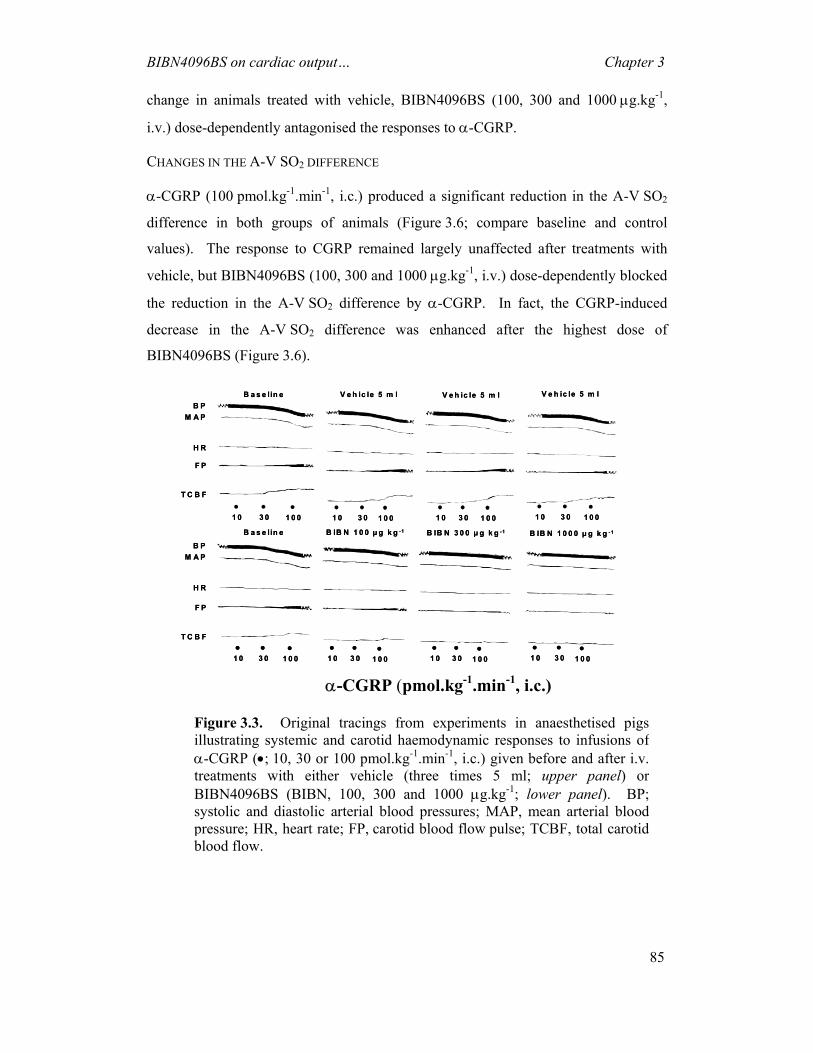
BIBN4096BS on cardiac output… Chapter 3
85
change in animals treated with vehicle, BIBN4096BS (100, 300 and 1000 µg.kg-1,
i.v.) dose-dependently antagonised the responses to α-CGRP.
CHANGES IN THE A-V SO2 DIFFERENCE
α-CGRP (100 pmol.kg-1.min-1, i.c.) produced a significant reduction in the A-V SO2
difference in both groups of animals (Figure 3.6; compare baseline and control
values). The response to CGRP remained largely unaffected after treatments with
vehicle, but BIBN4096BS (100, 300 and 1000 µg.kg-1, i.v.) dose-dependently blocked
the reduction in the A-V SO2 difference by α-CGRP. In fact, the CGRP-induced
decrease in the A-V SO2 difference was enhanced after the highest dose of
BIBN4096BS (Figure 3.6).
B a s e lin e V e h ic le 5 m l V e h ic le 5 m l V e h ic le 5 m l
B a s e lin e B IB N 1 0 0 µ g k g -1 B IB N 3 0 0 µ g k g -1 B IB N 1 0 0 0 µ g k g -1
B PM A P
H R
F P
T C B F
B PM A P
H R
F P
T C B F
•3 0
•1 0
•1 0 0
•3 0
•1 0
•1 0 0
•3 0
•1 0
•1 0 0
•3 0
•1 0
•1 0 0
•3 0
•1 0
•1 0 0
•3 0
•1 0
•1 0 0
•3 0
•1 0
•1 0 0
•3 0
•1 0
•1 0 0
α-CGRP (pmol.kg-1.min-1, i.c.)
Figure 3.3. Original tracings from experiments in anaesthetised pigsillustrating systemic and carotid haemodynamic responses to infusions ofα-CGRP (•; 10, 30 or 100 pmol.kg-1.min-1, i.c.) given before and after i.v.treatments with either vehicle (three times 5 ml; upper panel) orBIBN4096BS (BIBN, 100, 300 and 1000 µg.kg-1; lower panel). BP;systolic and diastolic arterial blood pressures; MAP, mean arterial bloodpressure; HR, heart rate; FP, carotid blood flow pulse; TCBF, total carotidblood flow.
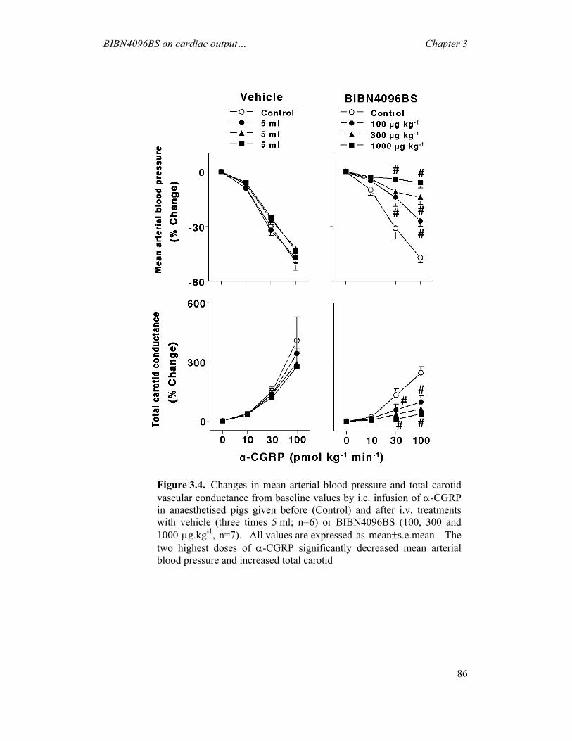
BIBN4096BS on cardiac output… Chapter 3
86
Figure 3.4. Changes in mean arterial blood pressure and total carotidvascular conductance from baseline values by i.c. infusion of α-CGRP in anaesthetised pigs given before (Control) and after i.v. treatmentswith vehicle (three times 5 ml; n=6) or BIBN4096BS (100, 300 and 1000 µg.kg-1, n=7). All values are expressed as mean±s.e.mean. The two highest doses of α-CGRP significantly decreased mean arterial blood pressure and increased total carotid

BIBN4096BS on cardiac output… Chapter 3
87
Figure 3.5. Maximum carotid blood flow changes and carotid bloodflow pulsations measured at baseline and following infusions of α-CGRP (100 pmol.kg-1.min-1, i.c.) given in anaesthetised pigs before (Control) and after i.v. treatments with vehicle (V, 5 ml three times; n=6) orBIBN4096BS (100, 300 and 1000 µg.kg-1, n=7). All values are expressed as mean±s.e.mean. a.u., Arbitrary units. *, P < 0.05 vs. baseline values; #, P<0.05 vs. response after the corresponding volume of vehicle.

BIBN4096BS on cardiac output… Chapter 3
88
3.4 Discussion
3.4.1 General
Undoubtedly, a remarkable progress has been achieved in acute antimigraine therapy
(De Vries et al., 1996a). Notwithstanding, the exact pathophysiological mechanisms
underlying migraine remain unclear. There is, however, evidence supporting the
involvement of the trigeminovascular system in migraine pathophysiology (Goadsby,
1997b; Goadsby, 1999; Hargreaves et al., 1999; Williamson & Hargreaves, 2001).
Thus, activation of the trigeminovascular system leads to neuropeptide release,
including that of CGRP, and neurogenic dural vasodilatation (Williamson &
Figure 3.6. Differences between arterial and jugular venous oxygen saturations(A-V SO2
difference) measured at baseline and after infusions of α-CGRP(100 pmol.kg-1.min-1, i.c.) given in anaesthetised pigs before (Control) and afteri.v. treatments with vehicle (Veh, 5 ml three times; n=6) or BIBN4096BS (100,300 and 1000 µg.kg-1, n=7). All values are expressed as mean±s.e.mean. *,P < 0.05 vs. baseline values; #, P<0.05 vs. response after the correspondingvolume of vehicle.

BIBN4096BS on cardiac output… Chapter 3
89
Hargreaves, 2001). Of particular relevance is the finding that plasma concentration of
CGRP is elevated during the headache phase of migraine, and this is normalised after
treatment with sumatriptan (Goadsby et al., 1990; Goadsby, 1997b; Goadsby, 1999).
Hence, it is reasonable to assume that a potent CGRP receptor antagonist, such as
BIBN4096BS (Doods et al., 2000), might be useful in migraine therapy.
BIBN4096BS behaves as a ‘silent’ competitive antagonist at CGRP receptors
mediating relaxation of human temporal, cranial and coronary arteries (Edvinsson et
al., 2002; Moreno et al., 2002; Verheggen et al., 2002). The present study in
anaesthetised pigs was designed: (i) to analyse, using BIBN4096BS, the potential role
of endogenous CGRP in regulating vascular tone in vivo; and (ii) to investigate the
effects of BIBN4096BS on the systemic and carotid haemodynamic responses
produced by α-CGRP.
3.4.2 Systemic and regional haemodynamic effects of BIBN4096BS
It is well known that CGRP-immunoreactive nerve fibres are widely distributed in the
cardiovascular system, with a higher preponderance in arteries than in veins (Bell &
McDermott, 1996). CGRP decreases blood pressure and has positive inotropic and
chronotropic effects on the heart (Wimalawansa, 1996), which are mainly mediated
via CGRP1 receptors (Bell & McDermott, 1996; Saetrum Opgaard et al., 1999;
Saetrum Opgaard et al., 2000). Though CGRP has diverse biological actions within
the cardiovascular system, our experiments showing few systemic haemodynamic
changes with BIBN4096BS do not support a major role for CGRP in the regulation of
cardiovascular function in the anaesthetised pig.
As far as regional haemodynamics is concerned, a moderate decrease
(compared to vehicle) in vascular conductances in the lungs, adrenals, kidneys and
spleen was observed with the highest dose (1000 µg.kg-1) of BIBN4096BS
(Figure 3.2). Similarly, renal vasoconstriction was noticed in conscious rats with a
high (300 nmol.kg-1.min-1), but not with a low (30 nmol.kg-1.min-1) dose of CGRP- (8-
37) (Gardiner et al., 1990). Since both BIBN4096BS and CGRP8-37 caused renal
changes only in doses that were considerably higher than those needed for CGRP
antagonism, it does not appear that endogenous CGRP regulates renal vascular tone.
Also, Shen et al. (Wu et al., 2001) recently reported that 30 µg.kg-1.min-1
(~10 nmol.kg-1.min-1) of CGRP-(8-37) which antagonised CGRP-induced
BIBN4096BS on cardiac output… Chapter 3
90
haemodynamic responses, caused little regional haemodynamic effects in conscious
dogs as well as anaesthetised rats, thereby not supporting an important physiological
role for endogenous CGRP in regulating vascular tone. Although we cannot rule out
the involvement of CGRP in certain other circumstances, for example, cardiac
preconditioning or coronary artery disease (Lu et al., 1999; Peng et al., 2000; Wu et
al., 2001), the present results imply cardiovascular safety of BIBN4096BS.
Nevertheless, one will have to explore the role of CGRP in cardiovascular
pathophysiology before establishing whether or not CGRP receptor antagonists are
completely safe in patients afflicted with cardiovascular disorders.
3.4.3 CGRP-induced haemodynamic responses and antagonism by
BIBN4096BS
Activation of CGRP receptors elicits dilatation in different vascular beds in several
species (Gardiner et al., 1990; Van Gelderen et al., 1995; Shen et al., 2001).
Consistent with these studies, our experiments show that i.c. infusions of α-CGRP
produced a marked vasodilatation in the porcine carotid circulation, with
accompanying fall in arterial blood pressure. The fact that the animals were
systematically vagosympathectomised may explain why the hypotension was not
accompanied by a baroreflex-mediated tachycardia, as reported earlier (Van Gelderen
et al., 1995). Interestingly, the ipsilateral skin redness, together with the marked
decrease in A-V SO2 difference by CGRP, indicates that porcine carotid arteriovenous
anastomoses dilated in response to α-CGRP (Saxena, 1987). However, we previously
reported that i.c. infusions of α-CGRP failed to increase porcine arteriovenous
anastomotic blood flow, despite a marked increase in the total carotid and capillary
blood flows (Van Gelderen et al., 1995). Admittedly, arteriovenous anastomotic
blood flow was not directly measured in these experiments, but we have recently
observed that i.c. infusions capsaicin, which released CGRP, did increase carotid
arteriovenous anastomotic blood flow with a concomitant decrease in the A-V SO2
difference (Tom et al., 2002). Thus, it appears that the discrepancy between the two
investigations may be due to different anaesthetic regimens employed (pentobarbital
and fentanyl/thiopental, respectively) and, particularly, the use of phenylephrine in the
present experiments. Phenylephrine potently constricts arteriovenous anastomoses
(Willems et al., 1999).

BIBN4096BS on cardiac output… Chapter 3
91
In the present experimental study in anaesthetised pigs, BIBN4096BS proved
to be an effective antagonist at the CGRP receptors mediating the systemic
(hypotension) as well as the carotid (increased blood flow, pulsations and skin
redness) haemodynamic responses to α-CGRP. The fact that BIBN4096BS also
abolished α-CGRP-induced decreases in the A-V SO2 difference suggests its action
on carotid arteriovenous anastomoses; for further considerations, see Saxena (1987).
Interestingly, BIBN4096BS also antagonised the capsaicin-induced increases in
carotid arteriovenous anastomotic blood flow as well as decreases in the A-V SO2
difference, but not the plasma CGRP concentrations (Kapoor et al., 2003a).
One cannot be certain about the nature of CGRP receptors that mediate
porcine carotid vascular responses, but cardiac inotropic and vasodilator responses are
mediated predominantly by CGRP1 receptors (Saetrum Opgaard et al., 1999), where
BIBN4096BS has a very high affinity (Doods et al., 2000; Poyner & Marshall, 2001).
3.4.4 Potential therapeutic efficacy of BIBN4096BS in the treatment migraine
Considering that plasma CGRP levels are elevated during the headache phase of
migraine (Goadsby, 1997b) and that BIBN4096BS dose-dependently blocked
α-CGRP-induced carotid haemodynamic responses, it is likely that BIBN4096BS
may be effective in migraine. The compound is presently under clinical investigation
for the acute treatment of migraine and the results are awaited with great interest.
In conclusion, our study clearly demonstrates that BIBN4096BS is an effective
antagonist at vascular CGRP receptors in anaesthetised pigs, but has little
haemodynamic effects of its own, a finding that negates a major physiological role for
CGRP in cardiovascular regulation. The potent blockade of the carotid
haemodynamic effects of CGRP does suggest that BIBN4096BS may be effective in
migraine treatment.


CHAPTER 4
Effects of sumatriptan on capsaicin-induced carotid haemodynamic changes and CGRP release
in anaesthetised pigs
Based on: Arulmani, U., Heiligers, J.P.C., Garrelds, I.M., Sánchez-López, A,
Willems, E.W., Villalón, C.M & Saxena, P.R. (2004). Effects of sumatriptan on
capsaicin-induced carotid haemodynamic changes and CGRP release in anaesthetised
pigs. Cephalalgia (in press)
Sumatriptan on capsaicin induced carotid haemodynamics Chapter 4
94
4 Effects of sumatriptan on capsaicin-induced carotid haemodynamic changes and CGRP release in anaesthetised pigs
Abstract: It is suggested that during a migraine attack capsaicin-sensitive trigeminal sensory nerves release calcitonin gene related peptide (CGRP), resulting in cranial vasodilatation and central nociception. Hence, inhibition of trigeminal CGRP release may prevent the above vasodilatation and, accordingly, abort migraine headache. Therefore, this study investigated the effects of sumatriptan (100 and 300 µg kg-1, i.v.) on capsaicin-induced carotid haemodynamic changes and on CGRP release. Intracarotid (i.c.) infusions of capsaicin (10 µg kg-1 min-1, i.c.) increased total carotid, arteriovenous anastomotic and capillary conductances as well as carotid pulsations, but decreased the difference between arterial and jugular venous oxygen saturations. Except for some attenuation of arteriovenous anastomotic changes, the capsaicin-induced responses were not affected by sumatriptan. Moreover, i.c. infusions of capsaicin (0.3, 1, 3 and 10 µg kg-1 min-1, i.c.) dose-dependently increased the jugular venous plasma concentrations of CGRP, which also remained unaffected by sumatriptan. The above results support the contention that the therapeutic action of sumatriptan is mainly due to cranial vasoconstriction rather than trigeminal (CGRP release) inhibition.
4.1 Introduction
Migraine is a neurovascular disorder characterised by vasodilatation of cranial blood
vessels with activation of perivascular trigeminal sensory nerves (Edvinsson, 2003).
Thus, activation of trigeminal sensory nerves results in the release of several
neuropeptides, including neuropeptide Y, vasoactive intestinal peptide, substance P
and calcitonin gene related peptide (CGRP). Interestingly, plasma concentrations of
CGRP, but not of other neuropeptides, are elevated during the headache phase of
migraine and these levels are normalised by triptans in parallel with alleviation of
headache (Goadsby et al., 2002b). CGRP, the most potent endogenous vasodilator
described thus far, is predominantly located on sensory neurons and perivascular
nerves surrounding blood vessels, where it is co-localised with other vasoactive
neuropeptides, such as substance P and neurokinin A (Brain et al., 1985; van Rossum
et al., 1997). In view that an increase in circulating CGRP levels is considered as a
biological marker for migraine headache, it is reasonable to assume that a substance
capable of inhibiting the release of CGRP from trigeminal sensory nerves may be
effective in migraine therapy (Goadsby et al., 2002b).

Sumatriptan on capsaicin induced carotid haemodynamics Chapter 4
95
CGRP can be released from the sensory nerves by electrical or chemical
(capsaicin) stimuli (Edvinsson, 2001b; Hou et al., 2002). In this respect, electrical
stimulation of trigeminal sensory nerves evokes the release of CGRP in the cranial
venous blood of rats and cats, which was attenuated by sumatriptan (Goadsby &
Edvinsson, 1993; Buzzi et al., 1995). However, to the best of our knowledge, there is
no comprehensive in vivo evidence to show that capsaicin-induced CGRP release is
inhibited by triptans. Therefore, the main objective of this study in anaesthetised
vagosympathectomised pigs was to investigate the effects of sumatriptan on
capsaicin-induced: (i) carotid haemodynamic changes, and (ii) increase in plasma
CGRP release.
4.2 Materials and methods
4.2.1 General
After an overnight fast, 28 domestic pigs (Yorkshire x Landrace, female, 10-14 kg),
divided into two groups (n=15 and n=13 for vehicle and sumatriptan, respectively),
were sedated with azaperone (120 mg, i.m.), midazolam hydrochloride (10 mg, i.m.)
and then anaesthetised with sodium pentobarbital (600 mg, i.v.). After tracheal
intubation, the animals were connected to a respirator (BEAR 2E, BeMeds AG, Baar,
Switzerland) for intermittent positive pressure ventilation with a mixture of room air
and oxygen. Respiratory rate, tidal volume and oxygen supply were adjusted to keep
arterial blood gas values within physiological limits (pH: 7.35-7.48, pCO2: 35-48
mmHg, pO2: 100-120 mmHg). Anaesthesia was maintained with a continuous i.v.
infusion of sodium pentobarbital (12-20 mg kg-1 h-1). This anaesthetic regimen,
together with bilateral vagosympathectomy (see below), increases heart rate and
markedly dilates carotid arterioles and arteriovenous anastomoses. Consequently,
carotid blood flow, particularly its arteriovenous anastomotic fraction, is considerably
higher in these pigs than in conscious or thiopental-anaesthetised pigs (Den Boer et
al., 1993).
Heart rate was measured with a tachograph (CRW, Erasmus University,
Rotterdam, The Netherlands) triggered by electrocardiogram signals. Both common
carotid arteries were dissected free and the accompanying vagosympathetic trunks
were cut between two ligatures to prevent any possible influence via baroreceptor
reflexes on the carotid vascular responses produced by capsaicin. Pulsatile and mean
Sumatriptan on capsaicin induced carotid haemodynamics Chapter 4
96
carotid blood flows were measured in the right common carotid artery with a flow
probe (internal diameter: 2.5 mm) connected to a sine-wave electromagnetic flow
meter (Transflow 601-system, Skalar, Delft, The Netherlands). The amplitude of
carotid blood flow signals provided an index of carotid flow pulse. Subsequently,
three hub-less needles, connected to a polyethylene tube, were inserted into the right
common carotid artery for the administration of capsaicin, radioactive microspheres
and the α1-adrenoceptor agonist phenylephrine. The use of phenylephrine is
necessitated by the fact that the carotid arterioles and arteriovenous anastomoses are
in a dilated state under the present anaesthetic regimen (Den Boer et al., 1993);
therefore, to study the effects of vasodilator agents (in the present case, capsaicin),
one has to constrict these shunt vessels first. As described earlier (Willems et al.,
1999), phenylephrine decreases the total carotid conductance exclusively by
constricting carotid arteriovenous anastomoses, which results in an increase in the
difference between arterial and jugular venous oxygen saturations (A-V SO2
difference) (Saxena, 1987).
Lastly, catheters were placed in: (i) the right external jugular vein for the
withdrawal of venous blood samples to measure blood gases (ABL-510; Radiometer,
Copenhagen, Denmark) and plasma concentrations of CGRP (see below); (ii) the
inferior vena cava (via the left femoral vein) for the administration of vehicle or
sumatriptan; and (iii) the aortic arch (via the left femoral artery) for the measurement
of arterial blood pressure (Combitrans disposable pressure transducer; Braun,
Melsungen, Germany) as well as withdrawal of arterial blood samples to measure
blood gases.
Heart rate and systolic, diastolic and mean arterial blood pressures as well as
mean and pulsatile carotid artery blood flows were continuously monitored on a
polygraph (CRW, Erasmus University, Rotterdam, The Netherlands). Vascular
conductances were calculated by dividing the respective blood flows (ml min-1) by
mean arterial blood pressure (mmHg), multiplied by one hundred and expressed as
10-2 ml min-1 mmHg-1. During the experiment, body temperature was maintained at
37±1°C by a heating pad and each animal was infused with physiological saline to
compensate for fluid losses.

Sumatriptan on capsaicin induced carotid haemodynamics Chapter 4
97
4.2.2 Distribution of carotid blood flow
The distribution of common carotid blood flow into tissue (capillary) and
arteriovenous anastomotic fractions was determined in 14 pigs later receiving vehicle
(n=8) or sumatriptan (n=6) with radioactive microspheres (diameter: 15.5±0.1 µm;
S.D.), labelled with 141Ce, 103Ru, 95Nb or 46Sc (NEN Dupont, Boston, USA). For each
measurement, a suspension of about 200,000 microspheres, labelled with one of these
isotopes, was mixed and injected into the right carotid artery. At the end of the
experiment, the animal was killed using an overdose of sodium pentobarbital and the
heart, kidneys, lungs and different cranial tissues were dissected out, weighed and put
in vials. The radioactivity in these vials was counted for 5 min in a γ-scintillation
counter (Packard, Minaxi autogamma 5000 for 5 min), using suitable windows for
discriminating the different isotopes (141Ce: 120-167 KeV, 103Ru: 450-548 KeV, 95Nb: 706-829 KeV and 46Sc: 830-965 KeV). All data were processed by a set of
specially designed computer programs (Saxena et al., 1980).
The distribution of total carotid blood flow to the different tissues (Qtis) was
calculated by the formula: Qtis = (Itis/Itotal) x Qcarotid, where Itis is tissue radioactivity,
Itotal is the total radioactivity injected and Qcarotid is the total common carotid blood
flow at the time of microsphere injection. Since little or no radioactivity was detected
in the heart or kidneys, it can be assumed that all microspheres trapped in lungs reach
this tissue from the venous side after escaping via carotid arteriovenous anastomoses.
Therefore, the amount of radioactivity in the lungs can be used as an index of the
arteriovenous anastomotic fraction of carotid blood flow (Saxena et al., 1980; Saxena
& Verdouw, 1982).
4.2.3 Determination of plasma levels of CGRP
Jugular venous blood samples were obtained from the 28 pigs, receiving either vehicle
(n=15) or sumatriptan (n=13). Fourteen of these animals (8 and 6 animals from
vehicle and sumatriptan groups, respectively) were used for carotid haemodynamic
experiments, while the other fourteen were separate experiments using the same
protocol, except that the radioactive microspheres were not used. Blood was
transferred immediately into a polypropylene tube containing ethylene dinitro-
tetraacetic acid (1 mg ml-1 of blood) and aprotinin (500 KIU ml-1 of blood). Aprotinin
was used to inhibit endogenous plasma proteases, since our unpublished studies have

Sumatriptan on capsaicin induced carotid haemodynamics Chapter 4
98
shown that CGRP is not detectable in biological samples without aprotinin. After
centrifugation at 1600 g for 15 min, plasma samples were coded and stored at -80°C
until CGRP measurements were performed. The person measuring CGRP
concentrations remained blind to the treatments, until all data had been collated.
CGRP was extracted from plasma using a C18 SEP-COLUMN, dried by
lypholisation, and measured by radioimmunoassay (Dwenger, 1984), as per protocol
of the Peninsula Laboratories, Inc. (Belmont, CA, U.S.A.). The recovery of CGRP
from the extraction procedure was ascertained by assaying control samples paired
with the same sample spiked with known quantities of CGRP. The column recovery
values were 85, 79, 81, 89 and 92% (mean=85.2; standard deviation=5.4; coefficient
of variation=6.3%). The CGRP concentrations measured in the actual samples were,
however, not corrected for the loss in the extraction procedure.
4.2.4 Experimental protocol
Following surgery and after the haemodynamic condition of the animals (n=28) had
been stable for 15-20 min (heart rate: 109±3 beats min-1; mean arterial blood pressure:
101±2 mmHg; mean carotid blood flow: 108±5 ml min-1; and A-V SO2 difference:
7.6±1.3%) phenylephrine was infused into the right common carotid artery at a rate of
10 µg kg-1 min-1 for 10 min, followed by 3-6 µg kg-1 min-1 throughout the rest of the
experiment to maintain carotid blood flow at a constant low level. The latter dose of
phenylephrine was chosen so that the external jugular venous oxygen saturation was
between 60-70% and mean carotid blood flow was about 40% of the original value.
After a period during which the haemodynamic variables remained constant for at
least 60 min (heart rate: 151±4 beats min-1; mean arterial blood pressure:
110±2 mmHg; mean carotid blood flow: 63±5 ml min-1; and A-V SO2 difference:
23±1.7%; n=28), the animals received consecutive infusions (0.15, 0.45, 1.5 and 4.5
ml, i.c. for 3 min each) of capsaicin vehicle (see Compounds and kits section). It is
important to mention that the vehicle of capsaicin was devoid of any systemic and
carotid haemodynamic responses (see Results section).
Five to ten min after the last infusion of capsaicin vehicle, blood samples
were obtained for the measurements of blood gases and CGRP concentrations;
moreover, the values of heart rate, mean arterial blood pressure and total carotid blood
flow and conductance were collated (baseline values; 15 and 13 pigs for vehicle and

Sumatriptan on capsaicin induced carotid haemodynamics Chapter 4
99
sumatriptan, respectively). In 14 out of the 28 pigs (8 for vehicle and 6 for
sumatriptan) the first batch of radioactive microspheres was injected for determining
the baseline distribution of carotid blood flow. The animals then received 4
consecutive infusions of capsaicin (0.3, 1, 3 and 10 µg kg-1 min-1, i.c. for 3 min each)
and heart rate, arterial blood pressure and total carotid blood flow were determined at
the end of each infusion. In addition, after the last infusion of capsaicin
(10 µg kg-1 min-1), blood gases, plasma CGRP concentration and carotid blood flow
distribution were measured as described above (control values). Subsequently, a
recovery period of 20 min was allowed until all haemodynamic parameters had
returned to baseline levels. At this point, the animals were divided into two groups,
which were treated with i.v. infusions (rate: 0.5 ml min-1 for 10 min) of either vehicle
(two times 5 ml of acidified distilled water; n=15) or sumatriptan (100 and
subsequently 300 µg kg-1; n=13). Ten min after each infusion, capsaicin was given
and the haemodynamic and biochemical variables were measured again, as described
above.
In the remaining fourteen animals, subdivided into two subgroups (n=7 each;
for vehicle and sumatriptan, respectively), apart from determining the variables
described above, venous blood samples (for plasma CGRP concentration) were
withdrawn after each dose of capsaicin (0.3, 1 and 3 and 10 µg kg-1 min-1) given
before and after treatment with vehicle (n=7) or sumatriptan (300 µg kg-1; n=7).
4.2.5 Data presentation and statistical analysis
All data are presented as mean±s.e.mean. The statistical analysis was performed
using the SPSS package for windows (version 10.0; SPSS Inc., Chicago, IL, USA).
The significance of changes within one group (vehicle or sumatriptan) was analysed
with repeated-measures ANOVA, followed by Greenhouse-Geisser correction for
serial autocorrelation (Ludbrook, 1994) and Bonferroni correction for multiple
comparisons (Overall & Doyle, 1996). The significance of the between-group
changes (vehicle vs. sumatriptan treatments) was first analysed with repeated-
measures ANOVA, including baseline measurements as a covariate (Overall & Doyle,
1994). If the two groups differed significantly, pairwise comparisons between the
corresponding values in the vehicle- and sumatriptan-treated groups were performed

Sumatriptan on capsaicin induced carotid haemodynamics Chapter 4
100
using univariate analysis (Overall & Atlas, 1999), followed by Bonferroni correction.
Statistical significance was accepted at P<0.05 (two-tailed).
4.2.6 Ethical approval
The Ethics Committee of the Erasmus MC, Rotterdam, dealing with the use of
animals in scientific experiments, approved the protocols for this investigation.
4.2.7 Compounds and kits
The following compounds were used: aprotinin (5850 KIU mg-1; Roth, Karlsruhe,
Germany), azaperone (Stresnil®; Janssen Pharmaceuticals, Beerse, Belgium),
sumatriptan succinate (gift from Dr. H.E. Connor, Glaxo Group Research, Stevenage,
Hertfordshire, UK), capsaicin, tween 80, ethanol and phenylephrine hydrochloride (all
from Sigma-Aldrich Chemie b.v., Zwijndrecht, The Netherlands), ethylene dinitro-
tetraacetic acid (Merck, Darmstadt, Germany), heparin sodium (to prevent blood
clotting in catheters; Leo Pharmaceutical Products, Weesp, The Netherlands),
midazolam hydrochloride (Dormicum®; Hoffmann La Roche b.v., Mijdrecht,
The Netherlands) and sodium pentobarbital (Sanofi Sante b.v., Maasluis,
The Netherlands). The radioimmunoassay kit for CGRP was purchased from
Peninsula Laboratories, Inc. (Belmont, CA, U.S.A.).
Capsaicin was initially dissolved in tween 80, ethanol and physiological saline in the
ratio of 0.5:1:8.5 ml, respectively. Phenylephrine was dissolved in distilled water,
while sumatriptan was dissolved in physiological saline.
4.3 Results
4.3.1 Baseline values
Baseline values (i.e.,, after capsaicin vehicle infusion) in the 28 pigs used were: heart
rate, 133±3 beats min-1; mean arterial blood pressure, 108±2 mmHg; total carotid
blood flow, 47±4 ml min-1; total carotid conductance, 44±3 10-2 ml min-1 mmHg-1; A-
V SO2 difference, 36±2%; and plasma CGRP concentration, 13±1 pg ml-1. No
significant difference in the baseline values were found between the two groups that
subsequently received vehicle (n=15) or sumatriptan (n=13).
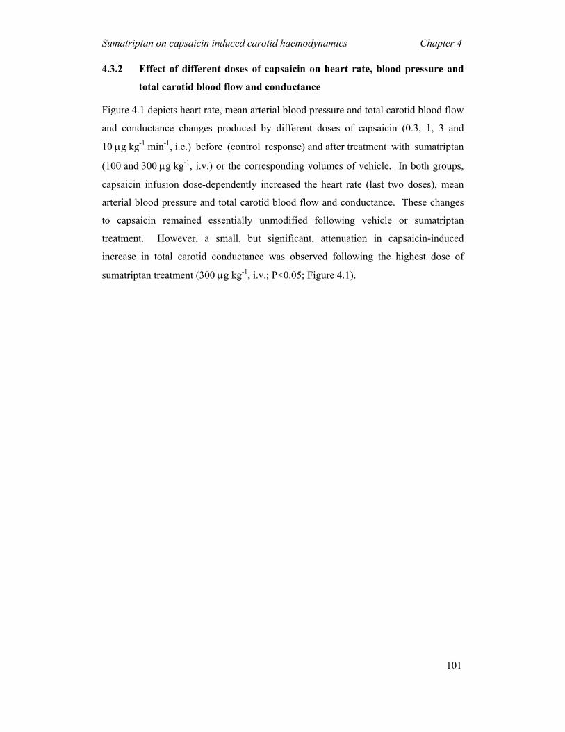
Sumatriptan on capsaicin induced carotid haemodynamics Chapter 4
101
4.3.2 Effect of different doses of capsaicin on heart rate, blood pressure and
total carotid blood flow and conductance
Figure 4.1 depicts heart rate, mean arterial blood pressure and total carotid blood flow
and conductance changes produced by different doses of capsaicin (0.3, 1, 3 and
10 µg kg-1 min-1, i.c.) before (control response) and after treatment with sumatriptan
(100 and 300 µg kg-1, i.v.) or the corresponding volumes of vehicle. In both groups,
capsaicin infusion dose-dependently increased the heart rate (last two doses), mean
arterial blood pressure and total carotid blood flow and conductance. These changes
to capsaicin remained essentially unmodified following vehicle or sumatriptan
treatment. However, a small, but significant, attenuation in capsaicin-induced
increase in total carotid conductance was observed following the highest dose of
sumatriptan treatment (300 µg kg-1, i.v.; P<0.05; Figure 4.1).

Sumatriptan on capsaicin induced carotid haemodynamics Chapter 4
102
Figure 4.1. Heart rate (HR), mean arterial blood pressure (MAP), total carotid bloodflow (TCBF) and total carotid vascular conductance (TCC) values at baseline (B) andfollowing infusions of capsaicin (0.3, 1, 3, 10 µg kg-1 min-1, i.c.) in anaesthetised pigsbefore (Control) and after i.v. administrations of vehicle (V, 5 ml two times; n=15) orsumatriptan (S100 and S300, 100 and 300 µg kg-1, respectively; n=13).All values are expressed as mean±s.e.mean. #, P<0.05 vs. response after thecorresponding volume of vehicle.

Sumatriptan on capsaicin induced carotid haemodynamics Chapter 4
103
4.3.3 Capsaicin-induced carotid haemodynamic changes
The effects of capsaicin (10 µg kg-1 min-1, i.c.) on carotid haemodynamics were
investigated in detail in animals receiving vehicle or sumatriptan.
CAROTID BLOOD FLOW, CONDUCTANCE AND PULSATIONS
The effects of capsaicin (10 µg kg-1 min-1, i.c.) on total carotid blood flow and
conductance (depicted as maximum absolute changes) as well as pulsations
(represented as arbitrary units; a.u.), before (control response) and after treatment with
sumatriptan (100 and 300 µg kg-1 min-1, i.v.) or the corresponding volumes of vehicle
are shown in Figure 4.2. In both treatment groups, the capsaicin infusion significantly
increased the carotid blood flow and conductance as well as pulsations. While vehicle
and sumatriptan (100 µg kg-1, i.v.) were devoid of any significant effect on capsaicin-
induced increases in carotid haemodynamics, a small, but significant, decrease in the
carotid vascular conductance was observed in the animals treated with the highest
dose of sumatriptan (300 µg kg-1, i.v.; P<0.05; Figure 4.2).
FRACTIONATION OF CAROTID VASCULAR CONDUCTANCE
As shown in Figure 4.3, the capsaicin infusion significantly increased total carotid,
arteriovenous anastomotic and capillary conductances. The capsaicin-induced
increases in conductances from baseline values (maximal percent changes) were,
respectively: total carotid, 329±39; arteriovenous anastomoses, 554±406; and
capillary fraction, 340±30. While vehicle as well as sumatriptan treatment did not
affect capsaicin-induced changes in total carotid and capillary fractions, the increase
in arteriovenous anastomotic conductance was markedly attenuated by sumatriptan.
Moreover, following the highest dose of sumatriptan (300 µg kg-1, i.v.), a small
decrease in capsaicin-induced increase in total carotid conductance was observed (#,
P<0.05; see Figures 4.1 and 4.3).
Furthermore, capsaicin infusion significantly increased the vascular
conductance to the different cranial tissues, including skin, ear, skeletal muscles, fat,
bone, salivary glands, eye, tongue and dura mater, but not that to brain. These
vasodilator responses to capsaicin remained unaltered after sumatriptan (100 and
300 µg kg-1, i.v.) or the corresponding volumes of vehicle (Figure 4.4).

Sumatriptan on capsaicin induced carotid haemodynamics Chapter 4
104
4.3.4 Difference between arterial and jugular venous oxygen saturations
(A-V SO2 difference)
Consistent with the increase in arteriovenous anastomotic conductance, capsaicin
infusion (10 µg kg-1 min-1, i.c.) significantly decreased the A-V SO2 difference from
baseline values from 39±4% to 4±0.6% (control response; n=28). This response to
capsaicin remained unaffected after treatment with vehicle or sumatriptan (Figure
4.5).
Figure 4.2. Maximum changes in carotid blood flow, vascular conductance andpulsations measured at baseline and following infusions of capsaicin (10 µg kg-1 min-1, i.c.) given in anaesthetised pigs before (Control) and after i.v. administrations of vehicle(V, 5 ml two times; n=15) or sumatriptan (S100 and S300, 100 and 300 µg kg-1, respectively; n=13). All values are expressed as mean±s.e.mean. a.u., Arbitrary units. *, P < 0.05 vs. baseline values; #, P<0.05 vs. response after the corresponding volume ofvehicle.
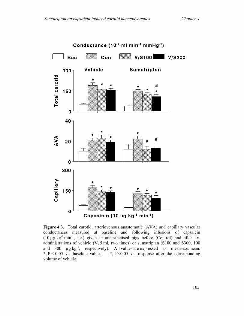
Sumatriptan on capsaicin induced carotid haemodynamics Chapter 4
105
Figure 4.3. Total carotid, arteriovenous anastomotic (AVA) and capillary vascularconductances measured at baseline and following infusions of capsaicin(10 µg kg-1 min-1, i.c.) given in anaesthetised pigs before (Control) and after i.v.administrations of vehicle (V, 5 ml, two times) or sumatriptan (S100 and S300, 100 and 300 µg kg-1, respectively). All values are expressed as mean±s.e.mean. *, P < 0.05 vs. baseline values; #, P<0.05 vs. response after the correspondingvolume of vehicle.
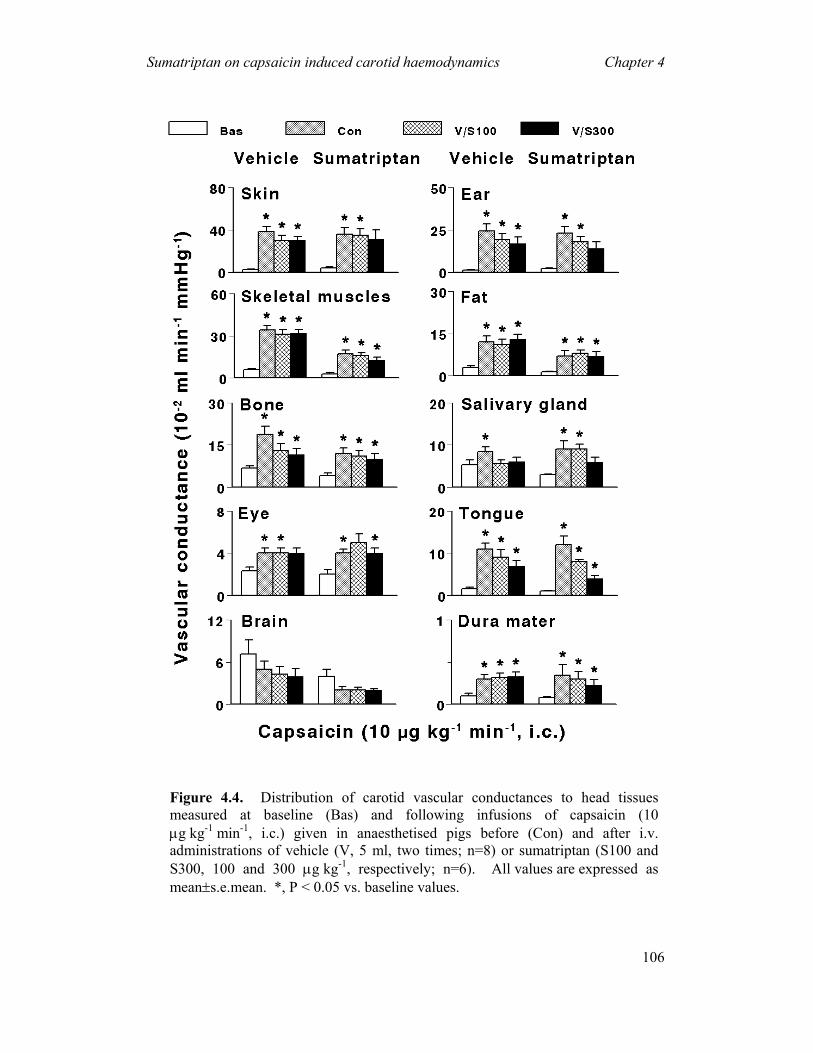
Sumatriptan on capsaicin induced carotid haemodynamics Chapter 4
106
Figure 4.4. Distribution of carotid vascular conductances to head tissuesmeasured at baseline (Bas) and following infusions of capsaicin (10µg kg-1 min-1, i.c.) given in anaesthetised pigs before (Con) and after i.v.administrations of vehicle (V, 5 ml, two times; n=8) or sumatriptan (S100 andS300, 100 and 300 µg kg-1, respectively; n=6). All values are expressed asmean±s.e.mean. *, P < 0.05 vs. baseline values.
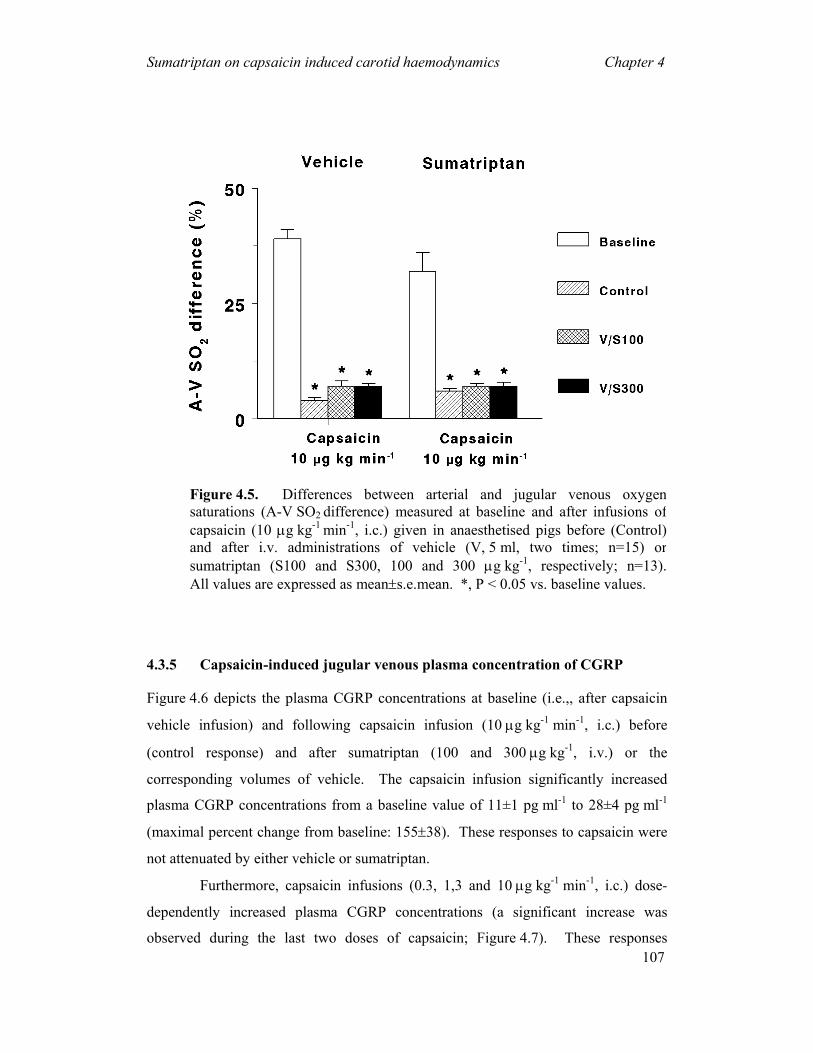
Sumatriptan on capsaicin induced carotid haemodynamics Chapter 4
107
4.3.5 Capsaicin-induced jugular venous plasma concentration of CGRP
Figure 4.6 depicts the plasma CGRP concentrations at baseline (i.e.,, after capsaicin
vehicle infusion) and following capsaicin infusion (10 µg kg-1 min-1, i.c.) before
(control response) and after sumatriptan (100 and 300 µg kg-1, i.v.) or the
corresponding volumes of vehicle. The capsaicin infusion significantly increased
plasma CGRP concentrations from a baseline value of 11±1 pg ml-1 to 28±4 pg ml-1
(maximal percent change from baseline: 155±38). These responses to capsaicin were
not attenuated by either vehicle or sumatriptan.
Furthermore, capsaicin infusions (0.3, 1,3 and 10 µg kg-1 min-1, i.c.) dose-
dependently increased plasma CGRP concentrations (a significant increase was
observed during the last two doses of capsaicin; Figure 4.7). These responses
Figure 4.5. Differences between arterial and jugular venous oxygensaturations (A-V SO2
difference) measured at baseline and after infusions ofcapsaicin (10 µg kg-1 min-1, i.c.) given in anaesthetised pigs before (Control)and after i.v. administrations of vehicle (V, 5 ml, two times; n=15) orsumatriptan (S100 and S300, 100 and 300 µg kg-1, respectively; n=13).All values are expressed as mean±s.e.mean. *, P < 0.05 vs. baseline values.
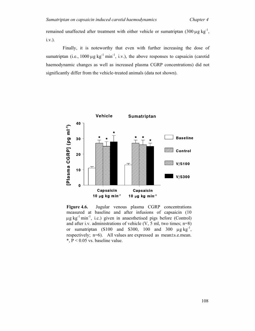
Sumatriptan on capsaicin induced carotid haemodynamics Chapter 4
108
remained unaffected after treatment with either vehicle or sumatriptan (300 µg kg-1,
i.v.).
Finally, it is noteworthy that even with further increasing the dose of
sumatriptan (i.e., 1000 µg kg-1 min-1, i.v.), the above responses to capsaicin (carotid
haemodynamic changes as well as increased plasma CGRP concentrations) did not
significantly differ from the vehicle-treated animals (data not shown).
Figure 4.6. Jugular venous plasma CGRP concentrationsmeasured at baseline and after infusions of capsaicin (10µg kg-1 min-1, i.c.) given in anaesthetised pigs before (Control)and after i.v. administrations of vehicle (V, 5 ml, two times; n=8)or sumatriptan (S100 and S300, 100 and 300 µg kg-1, respectively; n=6). All values are expressed as mean±s.e.mean. *, P < 0.05 vs. baseline value.

Sumatriptan on capsaicin induced carotid haemodynamics Chapter 4
109
Figure 4.7. Jugular venous plasma CGRP concentrations measuredat baseline (B) and after different doses of capsaicin infusion (0.3, 1,3 and 10 µg kg-1 min-1, i.c.) given in anaesthetised pigs before (Control) and after i.v. administrations of vehicle (Veh, 5 ml, twotimes; n=7) or sumatriptan (S300, 300 µg kg-1; n=7). All values are expressed as mean±s.e.mean. *, P < 0.05 vs. baseline values.

Sumatriptan on capsaicin induced carotid haemodynamics Chapter 4
110
4.4 Discussion
4.4.1 General
Migraine headache is a neurovascular syndrome in which the neural events seem to
involve stimulation of the trigeminal system and an ensuing release of CGRP from
perivascular trigeminal nerves (Goadsby et al., 2002b; Edvinsson, 2003).
The introduction of triptans (5-HT1B/1D/1F receptor agonists) in migraine
therapy has focussed on the role of serotonin receptors in migraine (Tfelt-Hansen et
al., 2000; Villalón et al., 2002). Triptans abort migraine attacks by several
mechanisms, including: (i) constriction of dilated cranial blood vessels and carotid
arteriovenous anastomoses via the stimulation of 5-HT1B receptors (De Vries et al.,
1998; De Vries et al., 1999c); and (ii) inhibition of CGRP release as well as of
nociceptive transmission on peripheral and central trigeminal sensory nerves via 5-
HT1B/1D receptors (Goadsby et al., 2002b; Tepper et al., 2002). Since trigeminal
inhibition of CGRP release may reduce cranial vasodilatation and nociception, the
present study set out to investigate the effects of sumatriptan on capsaicin-induced
porcine carotid haemodynamic changes and the associated increase on plasma CGRP
concentrations. Our results in anaesthetised pigs show that i.c. administration of
capsaicin: (i) increased total carotid (including arteriovenous anastomoses and
capillary) blood flows and conductances, carotid pulsations as well as jugular venous
plasma CGRP concentrations; and (ii) narrowed the A-V SO2 difference.
Interestingly, sumatriptan failed to modify capsaicin-induced: (i) carotid
haemodynamic changes, except carotid arteriovenous anastomoses; and (ii) increase
in plasma CGRP concentrations.
4.4.2 Systemic haemodynamic effects of capsaicin
The systemic haemodynamic effects of capsaicin have been investigated extensively
(Alving & Franco-Cereceda, 1993; Kapoor et al., 2003a). Indeed, the significant
increases in heart rate and mean arterial blood pressure observed in our study with
capsaicin are in accordance with these findings, which are due to a central activation
of the sympathetic outflow.

Sumatriptan on capsaicin induced carotid haemodynamics Chapter 4
111
4.4.3 Carotid haemodynamic changes to capsaicin
Several studies have shown that sensory nerves innervating the cerebral vasculature
contain substance P and CGRP (Asari et al., 2001; Edvinsson, 2001b). However,
capsaicin-induced vasorelaxation of guinea pig isolated basilar artery is mediated by
CGRP rather than by substance P (Jansen et al., 1990; O'Shaughnessy et al., 1993).
Moreover, BIBN4096BS, a potent CGRP receptor antagonist, abolished capsaicin-
induced porcine carotid haemodynamic responses (Kapoor et al., 2003a), a finding
which shows the involvement of CGRP in the responses to capsaicin.
Apart from some attenuation of arteriovenous anastomotic changes, the
capsaicin-induced responses were not affected by sumatriptan (Figures 4.1-4.4).
These results are in keeping with other findings showing that sumatriptan failed to
block capsaicin-induced relaxation of guinea pig isolated basilar artery
(O'Shaughnessy et al., 1993) as well as carotid vasodilatation induced by trigeminal
ganglion stimulation (Spokes & Middlefell, 1995; Lambert & Michalicek, 1996;
Raval et al., 1999).
It is noteworthy that the decrease in total carotid conductance by sumatriptan
is predominantly due to the decrease in carotid arteriovenous anastomotic
conductance, even when all triptans slightly increase the nutrient conductance (Den
Boer et al., 1992; De Vries et al., 1996b). Considering this, it is most likely that the
above apparent inhibition by sumatriptan on capsaicin-induced responses (Figure 4.3)
is due to physiological antagonism (i.e., vasoconstriction by sumatriptan) rather than
inhibition of CGRP release, since sumatriptan failed to modify this variable (see
Figures 4.6 and 4.7). In contrast, the apparent failure of sumatriptan to attenuate
capsaicin-induced increase in carotid conductance may have been due to: (i) massive
bursts of CGRP release produced by capsaicin; and/or (ii) an insufficient dose of
sumatriptan. However, the latter possibility can be excluded as treatment with an
even higher dose of sumatriptan (1000 µg kg-1 min-1, i.v.) did not modify capsaicin-
induced carotid haemodynamic changes (unpublished data); thus, the involvement of
5-HT1B/1D receptors, if any, seems to be rather limited under the present experimental
conditions.

Sumatriptan on capsaicin induced carotid haemodynamics Chapter 4
112
4.4.4 A-V SO2 difference
The dilatation of carotid arteriovenous anastomoses with an associated decrease in the
A-V SO2 difference is a characteristic feature observed during migraine headache
(Heyck, 1969). Consistent with the above finding, our study shows that capsaicin
decreased the A-V SO2 difference, which is presumably due to a CGRP-mediated
dilatation of carotid arteriovenous anastomoses (Kapoor et al., 2003b). As expected,
sumatriptan decreased the carotid arteriovenous anastomotic conductance in our study
(Figure 4.3); however, its failure to normalise the capsaicin-induced A-V SO2
difference may be explained in terms that capsaicin-induced increase in capillary
conductance may lead to oxygen saturation in the cranial tissues. Therefore, these
tissues cannot extract oxygen further, thereby shunting the oxygenated blood via
capillaries rather than via carotid arteriovenous anastomoses.
4.4.5 Plasma levels of CGRP
Several lines of evidence have shown that stimulation of the trigeminal system with
electrical or chemical (capsaicin) stimuli releases the endogenously stored CGRP
(Buzzi et al., 1991; Goadsby, 1993; O'Shaughnessy et al., 1993; Knight et al., 1999;
Eltorp et al., 2000; Limmroth et al., 2001; Kapoor et al., 2003a), which produces
cranial vasodilatation (Goadsby & Edvinsson, 1993; Akerman et al., 2003; Kapoor et
al., 2003a). Interestingly, triptans have been reported to attenuate CGRP release
elicited by both electrical (Buzzi et al., 1991; Goadsby, 1993; Williamson et al., 1997;
Knight et al., 1999; Limmroth et al., 2001) and chemical (capsaicin) (Eltorp et al.,
2000) stimulation; the latter effect was barely significant and observed with
sumatriptan in a concentration (50 µM) that may be considered far beyond the
therapeutic range (Eltorp et al., 2000). Indeed, this inhibitory effect on CGRP release
in cats has been associated with a blockade of the resulting cranial vasodilatation
(Goadsby & Edvinsson, 1993) although, admittedly, this may also reflect a
physiological antagonism produced by the triptans-induced vasoconstriction (De Vries
et al., 1999a). Considering these findings, it would seem reasonable to suggest that
trigeminal (CGRP release) inhibition may be an additional mechanism behind the
antimigraine action of triptans.
Notwithstanding, other lines of evidence seem to show just the opposite,
namely, that triptans do not modify the cranial vasodilatation produced by either

Sumatriptan on capsaicin induced carotid haemodynamics Chapter 4
113
trigeminal stimulation (Lambert & Michalicek, 1996; Raval et al., 1999) or capsaicin
(O'Shaughnessy et al., 1993). Admittedly, in these studies CGRP concentrations were
not measured in parallel and, consequently, no categorical conclusion can be drawn
regarding the antimigraine action of triptans. Our in vivo study sheds further light on
this matter by showing that: (i) sumatriptan failed to modify the capsaicin-induced
increases in plasma CGRP levels and the associated carotid vasodilatation; and (ii) the
carotid arteriovenous anastomotic vasoconstriction to sumatriptan is not associated
with a corresponding change in CGRP levels.
Admittedly, there is no clear-cut explanation why sumatriptan seems to
attenuate CGRP release upon electrical stimulation but not by capsaicin. It is known
that capsaicin activates a subset of small sensory fibres that cover a major proportion
of C and some Aδ fibres (Dray, 1992a; Dray, 1992b; Urban & Dray, 1992; Akerman
et al., 2003), while low intensity electrical stimulation recruits Aδ fibres alone
(Akerman et al., 2003). Thus, one explanation for the differential effect of
sumatriptan may be that, compared to electrical stimulation, capsaicin releases high
quantities of CGRP that can be potently antagonised by selective vanilloid VR1
antagonists (Caterina et al., 1997), but not by a presynaptic mechanism involving
sumatriptan. Another possibility is that, in contrast to the capsaicin-sensitive C and
δ fibres, the δ fibres recruited by electrical stimulation possess 5-HT1B/1D/1F receptors
stimulated by sumatriptan (Tfelt-Hansen et al., 2000; Goadsby et al., 2002b). We
have indeed shown that the mRNAs for 5-HT1B and 5-ht1F receptors are expressed in
the porcine trigeminal ganglia (Bhalla et al., 2001; Bhalla et al., 2002). Finally,
sumatriptan does not easily penetrate the blood brain barrier (Tfelt-Hansen et al.,
2000) and, should CGRP be mainly released from intracerebral sources, it will inhibit
such a release only if the blood brain barrier is disrupted.
4.4.6 Possible clinical implications
Finally, the possible clinical implications of our results with sumatriptan within the
context of antimigraine therapy must be considered. Therefore, based on our findings,
the blockade of the postjunctional effects of CGRP (with BIBN4096BS) (Kapoor et
al., 2003a) would seem to be a better therapeutic strategy to prevent neurogenic
vasodilatation rather than trigeminal inhibition of CGRP release (via the activation of
Sumatriptan on capsaicin induced carotid haemodynamics Chapter 4
114
prejunctional 5-HT1B/1D receptors by sumatriptan). Moreover, in view of the putative
pathophysiological role of arteriovenous anastomotic dilatation in migraine (Heyck,
1969; Saxena, 1995), the constriction of these non-nutrient vessels by sumatriptan in
our study may be responsible for the therapeutic action of this drug in migraine.
In conclusion, our results imply that prejunctional 5-HT1B/1D receptors (activated by
sumatriptan) do not inhibit capsaicin-induced: (i) vasodilatation of the porcine carotid
circulation; and (ii) increase in plasma CGRP concentrations. Therefore, the primary
mechanism behind the clinical efficacy of sumatriptan in migraine may be due to
vasoconstriction of cranial blood vessels rather than neurogenic inhibition of CGRP
release.

CHAPTER 5
Effects of the CGRP receptor antagonist BIBN4096BS on
α-CGRP-induced regional haemodynamic changes in
anaesthetised rats
Based on: Arulmani, U., Schuijt, M.P., Heiligers, J.P.C., Willems, E.W.,
Villalon, C.M & Saxena, P.R. (2004). Effects of the CGRP receptor antagonist
BIBN4096BS on α-CGRP-induced regional haemodynamic changes in
anaesthetised rats. Pharmacology & Toxicology (in press).
BIBN4096BS on α-CGRP-induced regional…. Chapter 5
116
5 Effects of the CGRP receptor antagonist BIBN4096BS on α-CGRP-induced regional haemodynamic changes in anaesthetised rats
Abstract: Several studies have suggested that a calcitonin gene-related peptide (CGRP) receptor antagonist may have antimigraine properties, most probably via the inhibition of CGRP-induced cranial vasodilatation. We have previously shown that BIBN4096BS, a potent and selective CGRP receptor antagonist, attenuated the CGRP-induced porcine carotid vasodilatation in a model predictive of antimigraine activity. In order to evaluate the potential safety of BIBN4096BS in migraine therapy, this study was designed to investigate the effects of intravenous (i.v.) BIBN4096BS on α-CGRP-induced systemic and regional haemodynamic changes in anaesthetised rats. In vehicle-pretreated animals, consecutive i.v. infusions of α-CGRP (0.25, 0.5 and 1 µg kg-1 min-1) dose-dependently decreased mean arterial blood pressure with an accompanying increase in heart rate and systemic vascular conductance whereas cardiac output remained unchanged. α-CGRP also increased the vascular conductance to the heart, brain, gastrointestinal tract, adrenals, skeletal muscle and skin, whilst that to the kidneys, spleen, mesentery/pancreas and liver remained unaltered. The above systemic and regional haemodynamic responses to α-CGRP were clearly attenuated in BIBN4096BS (3000 µg kg-1 min-1; i.v.)-pretreated animals. These lines of evidence indicate that exogenously administered α-CGRP dilates regional vascular beds via CGRP receptors on the basis of the antagonism produced by BIBN4096BS. Moreover, the fact that BIBN4096BS did not alter baseline haemodynamics suggests that endogenously produced CGRP does not play an important role in regulating the systemic and regional haemodynamics under resting conditions.
5.1 Introduction
Migraine is a neurovascular syndrome thought to be associated with profound dilation
of cranial blood vessels and activation of the trigeminovascular system (Saxena &
Tfelt-Hansen, 2000; Goadsby et al., 2002b). Several studies have shown that
vasoactive neuropeptides (e.g., neuropeptide Y, substance P, calcitonin gene-related
peptide; CGRP) may be involved in the aetiology of this disorder (Edvinsson, 2001b;
Goadsby et al., 2002b). Interestingly, circulating plasma levels of α-CGRP (a 37-
amino acid neuropeptide), but not of other neuropeptides, are significantly elevated
during the headache phase of a migraine attack (Ashina et al., 2000; Goadsby et al.,
2002b) and these elevated α-CGRP levels are normalised by antimigraine agents, such
as sumatriptan, with complete resolution of headache (Goadsby & Edvinsson, 1993).

BIBN4096BS on α-CGRP-induced regional…. Chapter 5
117
These findings suggest that CGRP may play a predominant role in migraine
pathogenesis, possibly by dilating large cranial blood vessels (Williamson &
Hargreaves, 2001; Goadsby et al., 2002b). Therefore, compounds inhibiting either the
CGRP release or its effects, particularly cranial vasodilatation, may be efficacious in
migraine therapy. In this context, BIBN4096BS, a potent and selective CGRP
receptor antagonist (Doods et al., 2000), completely attenuated carotid vasodilatation
by endogenously released (by capsaicin) and exogenously administered CGRP
(Kapoor et al., 2003a; Kapoor et al., 2003b) in an experimental animal model
predictive of antimigraine activity (Saxena, 1995; De Vries et al., 1999a). Besides its
potential efficacy, the therapeutic effectiveness of BIBN4096BS in acute migraine
treatment will also depend on its pharmacokinetic properties and potential side effects
in humans. With respect to the latter, it has been shown that BIBN4096BS attenuates
CGRP-induced dilatation of human isolated coronary arteries (Edvinsson et al., 2002),
a vascular bed that is largely affected by the currently used antimigraine agents
(Maassen VanDenBrink et al., 1999).
On the basis of the above, the present study set out to analyse in
anaesthetised rats, the effects produced by i.v. administration of BIBN4096BS on: (i)
baseline systemic haemodynamics (to investigate its the potential cardiovascular side
effects); and (ii) the systemic and regional haemodynamic responses to α-CGRP (to
ascertain CGRP receptor distribution).
5.2 Materials and methods
5.2.1 General
Experiments were carried out in 13 male Wistar rats (body weight: 356±35 g)
obtained from Harlan, Zeist, The Netherlands. The animals were initially
anaesthetised with an intraperitoneal (i.p) injection of sodium pentobarbitone
(60 mg kg-1, i.p), and additional i.v. bolus injections (5 mg kg-1, i.v.) were provided
every 20-30 min to maintain the anaesthesia. A catheter was placed in the trachea for
intermittent positive pressure ventilation with a mixture of oxygen and room air, using
a respiratory pump (small animal ventilator, Harvard Apparatus, Natick, MA, USA).
The ventilation rate was adjusted (40 strokes min-1) to keep the arterial blood gases
within the physiological range. The right common carotid artery was exposed and a
catheter connected to a pressure transducer (Combitrans disposable pressure
BIBN4096BS on α-CGRP-induced regional…. Chapter 5
118
transducer, Braun, Melsungen, Germany) was guided through the carotid artery into
the left ventricle. The presence of the catheter tip in the left ventricle was confirmed
by the observation of a sudden switch from an arterial to a ventricular pressure profile.
The right femoral artery was catheterised and connected to a pressure transducer
(Combitrans disposable pressure transducer, Braun, Melsungen, Germany) for
recording the blood pressure, while the left femoral artery was catheterised for the
withdrawal of reference blood samples. The heart rate was measured with a
tachograph (CRW, Erasmus Medical Centre, Rotterdam, The Netherlands) triggered
by electrocardiogram signals. Both blood pressure and heart rate were recorded
simultaneously on a polygraph (CRW, Erasmus Medical Centre, Rotterdam,
The Netherlands). The right external jugular vein was catheterised for the
administration of compounds (α-CGRP and BIBN4096BS or the corresponding
volume of vehicle).
5.2.2 Distribution of cardiac output
The distribution of cardiac output was determined with radioactive microspheres
(diameter: 15.5± 0.1 µm; S.D.), labelled with 141Ce, 103Ru, 95Nb or 46Sc (NEN Dupont,
Boston, USA). For each measurement, about 200,000 microspheres, suspended in 0.2
ml of physiological saline and labelled with one of the isotopes, was mixed and
injected into the left ventricle over a period of 15 s; the catheter was thoroughly
flushed with 0.5 ml of saline. Starting 10 s before each microsphere injection and
lasting 70 s, an arterial reference blood sample was drawn from the left femoral artery
at a constant rate of 0.5 ml min-1, using a withdrawal pump (Model 55, Harvard
apparatus, Natick, USA). At the end of the experiment, the animal was killed using
an overdose of sodium pentobarbitone and all tissues were dissected out, weighed and
put in vials. The following tissues were studied: skeletal muscle; carcass (consisting
of bone with skeletal muscle residue, fat, tail, eyes, and urogenital tract);
mesentery/pancreas (for practical reasons, these two tissues were not studied
separately); adrenals; lungs; kidneys; skin; heart; liver; brain; gastrointestinal tract and
spleen. Lungs were not evaluated further, because the amount of radioactivity in
these organs represents the microspheres that bypassed the peripheral vascular beds
(via arteriovenous anastomoses), rather than pulmonary blood flow (Baile et al.,
1982). The radioactivity in the reference blood samples and in the tissues was

BIBN4096BS on α-CGRP-induced regional…. Chapter 5
119
counted for 5 min in a γ-scintillation counter (Packard, Minaxi Auto-Gamma
5000 series), using suitable windows for the discrimination of the different isotopes
(141Ce: 120-167 KeV, 103Ru: 450-548 KeV, 95Nb: 706-829 KeV and 46Sc: 830-965 KeV). All data were processed by a set of specially designed computer
programs (Saxena et al., 1980).
The cardiac output was calculated by multiplying the ratio of total and
arterial blood sample radioactivity by the withdrawal rate of the arterial reference
blood sample (0.5 ml min-1). Accordingly, tissue blood flow was calculated by
multiplying the ratio of the tissue and total radioactivity by cardiac output (Saxena et
al., 1980). Systemic and regional vascular conductances (i.e., cardiac output and
regional blood flow corrected for mean arterial blood pressure) were calculated,
multiplied by hundred and expressed as 10-2 ml mmHg-1 min-1.
5.2.3 Experimental protocol
The experiments were started after a stabilisation period of about 30 min. At this
point, the animals were divided into two groups. The first group (n=6) was pre-
treated with the vehicle of BIBN4096BS (0.5 ml of acidified distilled water;
0.05 ml min-1 for 10 min; i.v.; see Compounds section), while the second group (n=7)
was pre-treated with BIBN4096BS (3000 µg kg-1, i.v.; also at a rate of 0.05 ml min-1
for 10 min). After a waiting period of 10 min, the baseline values of heart rate, mean
arterial blood pressure, cardiac output and its distribution to the various tissues (see
above) were determined and both groups received sequential i.v. infusions of α-CGRP
(0.25, 0.5 and 1 µg kg-1 min-1; each dose for 10 min). After each dose of α-CGRP
infusion, the above mentioned haemodynamic variables were reassessed.
5.2.4 Data presentation and statistical analysis
All data are presented as mean±s.e.mean. The statistical analysis was performed
using the SPSS package for windows (version 10.0; SPSS Inc., Chicago, IL, USA).
The significance of changes within one group (vehicle or BIBN4096BS) was analysed
with repeated-measures ANOVA, followed by Greenhouse-Geisser correction for
serial autocorrelation (Ludbrook, 1994) and Bonferroni correction for multiple
comparisons (Overall & Doyle, 1996). The significance of the between-group
changes (vehicle vs. BIBN4096BS treatments) was first analysed with repeated-
measures ANOVA, including baseline measurements as a covariate (Overall & Doyle,

BIBN4096BS on α-CGRP-induced regional…. Chapter 5
120
1994). If the two groups differed significantly, pairwise comparisons of the
corresponding values in the vehicle- and BIBN4096BS-treated groups were
performed using univariate analysis (Overall & Atlas, 1999), followed by Bonferroni
correction. Statistical significance was accepted at P<0.05 (two-tailed).
5.2.5 Ethical approval
The Ethics Committee of the Erasmus MC, Rotterdam, The Netherlands, dealing with
the use of animals in scientific experiments, approved the protocols for the present
investigation.
5.2.6 Compounds
The following compounds were used: sodium pentobarbitone (Sanofi Sante b.v.,
Maasluis, The Netherlands), heparin sodium (to prevent clotting of blood in the
catheters; Leo Pharmaceutical Products, Weesp, The Netherlands), α-CGRP and
BIBN4096BS (both gifts from Dr. H. Doods, Boehringer Ingelheim Pharma KG,
Biberach, Germany).
α-CGRP was dissolved in distilled water, while BIBN4096BS was initially dissolved
in 0.5 ml of 1N HCl, then diluted with 4 ml of distilled water, and then adjusted to pH
6.5 by 1N NaOH.
5.3 Results
5.3.1 Baseline values of systemic and regional haemodynamic variables
Baseline values of systemic haemodynamic variables in the 13 anaesthetised rats used
in this investigation were: heart rate, 266±10 beats min-1; mean arterial blood
pressure, 108±3 mmHg; cardiac output, 68±4 ml min-1 and systemic vascular
conductance, 64±4 10-2 ml min-1 mmHg-1. Baseline values of regional vascular
conductances (10-2 ml min-1 mmHg-1/100g tissue) were: brain, 32±4; heart, 409±58;
liver, 35±3; gastrointestinal tract, 124±24; mesentery/pancreas, 32±4; adrenals,
257±52; kidneys, 417±38; spleen, 92±24; skeletal muscle, 5±0.4; and skin, 5±1.
These values were similar to those reported earlier from our laboratories (Schuijt et
al., 1999).
The baseline values in the animals pre-treated with vehicle (n=6) and
BIBN4096BS (n=7) did not differ significantly: heart rate (275±19 vs. 259±8
beats min-1); mean arterial blood pressure (108±3 vs. 108±5 mmHg); cardiac output

BIBN4096BS on α-CGRP-induced regional…. Chapter 5
121
(68±6 vs. 69±6 ml min-1); systemic vascular conductance (63±6 vs. 65±7 10-
2 ml min-1 mmHg-1) and regional vascular conductances (10-2 ml min-1 mmHg-1/100g
tissue) in brain (32±5 vs. 33±6), heart (393±101 vs. 423±72), liver (33±6 vs. 36±2),
gastrointestinal tract (103±16 vs. 141±43), mesentery/pancreas (33±7 vs. 32±4),
adrenals (282±96 vs. 235±58), kidneys (445±56 vs. 394±54), spleen (100±50 vs.
85±16), skeletal muscle (5±0.5 vs. 4±0.5) and skin (6±1 vs. 4±0.7).
5.3.2 Systemic haemodynamic responses to α-CGRP
The absolute changes in systemic haemodynamics following consecutive i.v.
infusions of α-CGRP (0.25, 0.5 and 1 µg kg-1 min-1; i.v.) in the animals pre-treated
with BIBN4096BS (3 mg kg-1, i.v.) or the corresponding volume of vehicle (acidified
distilled water, 0.05 ml; i.v) are shown in Figure 5.1. The infusions of α-CGRP
dose-dependently decreased mean arterial blood pressure (maximum percent change
from baseline: 55±5) with an increase in systemic vascular conductance (maximum
percent change from baseline: 56±21) and heart rate (maximum percent change from
baseline: 17±3). BIBN4096BS pre-treatment produced an attenuation of the systemic
haemodynamic responses produced by α-CGRP. Under these conditions, the highest
dose of α-CGRP still elicited small, though significant, decreases (maximum percent
changes from baseline) in: (i) mean arterial blood pressure (28±4 vs. 51±7 in vehicle
pre-treated animals); and (ii) systemic vascular conductance (16±6 vs. 27±8 in vehicle
pre-treated animals).
5.3.3 Regional haemodynamic responses to α-CGRP
Figure 5.2 depicts absolute changes in regional vascular conductance following
consecutive i.v. infusions of α-CGRP (0.25, 0.5 and 1 µg kg-1 min-1; i.v.) in the
animals pre-treated with BIBN4096BS (3 mg kg-1, i.v.) or the corresponding volume
of vehicle. α-CGRP increased the vascular conductances (maximal percent change
from baseline) to brain (124±45), gastrointestinal tract (80±35), heart (74±31),
adrenals (87±37), muscle (79±27) and skin (154±37), whilst that to the
mesentery/pancreas, liver, spleen and kidneys remained unchanged. BIBN4096BS
attenuated the above regional haemodynamic changes induced by α-CGRP. The
vascular conductance to the kidneys decreased significantly in vehicle-pretreated
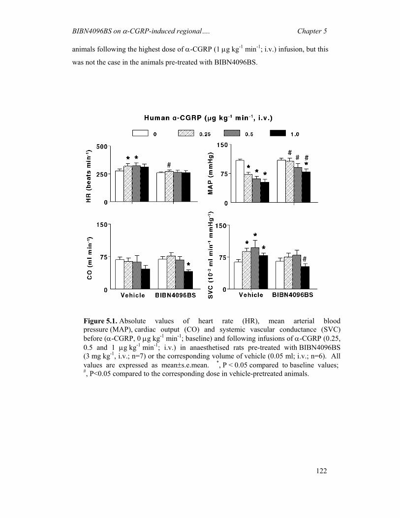
BIBN4096BS on α-CGRP-induced regional…. Chapter 5
122
animals following the highest dose of α-CGRP (1 µg kg-1 min-1; i.v.) infusion, but this
was not the case in the animals pre-treated with BIBN4096BS.
Figure 5.1. Absolute values of heart rate (HR), mean arterial bloodpressure (MAP), cardiac output (CO) and systemic vascular conductance (SVC)before (α-CGRP, 0 µg kg-1 min-1; baseline) and following infusions of α-CGRP (0.25,0.5 and 1 µg kg-1 min-1; i.v.) in anaesthetised rats pre-treated with BIBN4096BS(3 mg kg-1, i.v.; n=7) or the corresponding volume of vehicle (0.05 ml; i.v.; n=6). Allvalues are expressed as mean±s.e.mean. *, P < 0.05 compared to baseline values;#, P<0.05 compared to the corresponding dose in vehicle-pretreated animals.
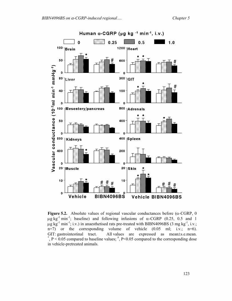
BIBN4096BS on α-CGRP-induced regional…. Chapter 5
123
Figure 5.2. Absolute values of regional vascular conductances before (α-CGRP, 0 µg kg-1 min-1; baseline) and following infusions of α-CGRP (0.25, 0.5 and 1 µg kg-1 min-1; i.v.) in anaesthetised rats pre-treated with BIBN4096BS (3 mg kg-1, i.v.; n=7) or the corresponding volume of vehicle (0.05 ml; i.v.; n=6). GIT: gastrointestinal tract. All values are expressed as mean±s.e.mean. *, P < 0.05 compared to baseline values; #, P<0.05 compared to the corresponding dose in vehicle-pretreated animals.

BIBN4096BS on α-CGRP-induced regional…. Chapter 5
124
5.4 Discussion
5.4.1 General
CGRP receptors are widely distributed throughout the body and are predominantly
expressed in the nervous system including perivascular nerves (Okimura et al., 1987;
Sternini et al., 1992). Therefore, CGRP may play an important role in regulating
peripheral vascular tone and in controlling blood flow to various organs (Poyner et al.,
2002). Several lines of evidence suggest that an increase in the release of CGRP is a
potential causative factor in some pathological conditions including
migraine (Wimalawansa, 1996). Hence, the advent of BIBN4096BS may represent an
important headway in treating migraine.
Apart from the implications discussed below, our study in anaesthetised rats
shows that BIBN4096BS: (i) had no effect on baseline systemic and regional
haemodynamics, confirming its cardiovascular safety; and (ii) antagonised α-
CGRP-induced changes in systemic and regional haemodynamics, demonstrating the
wide distribution of CGRP receptors.
5.4.2 Systemic haemodynamic changes to α-CGRP
The potential role of CGRP in regulating the systemic haemodynamics has been
studied extensively in several species, including humans (Franco-Cereceda et al.,
1987; Ventura et al., 2000; Rasmussen et al., 2001). Accordingly, CGRP decreases
systemic blood pressure through its potent vasodilator effect on the peripheral
vasculature (van Rossum et al., 1997). The fact that BIBN4096BS blocked the
hypotensive responses to α-CGRP (Figure 5.1) confirms a CGRP receptor-mediated
response. Interestingly, the hypotensive response to the highest dose of α-CGRP
(1 µg kg-1 min-1; i.v.) was just partly blocked by BIBN4096BS; this may be attributed,
at least in part, to the lower affinity of BIBN4096BS for rat CGRP receptors (Doods
et al., 2000). Consistent with our findings, it has been reported that BIBN4096BS is
more potent than CGRP8-37 at rat CGRP receptors (Poyner et al., 2002). However, a
non-specific vasodilatation to CGRP via the production of nitric oxide cannot be
categorically excluded in our experiments (Akerman et al., 2002; de Hoon et al.,
2003).
BIBN4096BS on α-CGRP-induced regional…. Chapter 5
125
On the other hand, with respect to the tachycardic responses to α-CGRP
observed in the present study, it is worthy of note that cardiac CGRP receptors are
more abundant in the sinoatrial node and atria than in the ventricles (Du et al., 1994;
Bell & McDermott, 1996; Wimalawansa, 1996; Saetrum Opgaard et al., 2000). Most
importantly, other lines of evidence have shown that this response is resistant to
β-adrenoceptor antagonists, suggesting a direct chronotropic action of α-CGRP in the
rat heart (Marshall et al., 1986). However, a baroreceptor reflex mechanism (which
cannot be entirely ruled out in our experimental set-up) may also be involved.
Noteworthily, CGRP is a potent inotropic agent in rabbits, pigs as well as
humans (Van Gelderen et al., 1995; Bell & McDermott, 1996), but not in rats
(Ishikawa et al., 1987; Bratveit et al., 1991). This may explain the lack of effect of α-
CGRP on cardiac output in our study (Figure 5.1).
5.4.3 Regional haemodynamic changes by α-CGRP
The regional haemodynamic responses to α-CGRP observed in our study clearly
demonstrate the vasodilator properties of CGRP in different vascular beds.
Furthermore, the CGRP-induced increases in vascular conductances in the heart,
brain, gastrointestinal tract, adrenals, skin and skeletal muscle were attenuated by
BIBN4096BS (Figure 5.2), indicating that these responses are mediated via CGRP
receptors. However, tissues such as liver, spleen, mesentery/pancreas and kidneys did
not apparently show any changes to CGRP. Several possible explanations for this lack
of effect of CGRP may include, amongst others: (i) tissue-dependent factors such as the
density of CGRP receptors and coupling efficiency; and/or (ii) a blunting effect via the
activation of compensatory pressor mechanisms triggered by the prominent reduction
in systemic blood pressure (DiPette et al., 1989; Gardiner et al., 1990).
5.4.4 Clinical implications of CGRP antagonism by BIBN4096BS
Lastly, we would like to consider the possible clinical implications of BIBN4096BS
safety in antimigraine therapy. BIBN4096BS, which is effective in antagonising
CGRP-induced responses in both in vivo and in vitro studies (Edvinsson et al., 2002;
Kapoor et al., 2002; Moreno et al., 2002; Verheggen et al., 2002; Kapoor et al.,
2003b), has been shown effective in a phase II clinical trial for acute antimigraine
therapy (Edvinsson, 2003; Olesen et al., 2003a). In this context, our study shows that

BIBN4096BS on α-CGRP-induced regional…. Chapter 5
126
BIBN4096BS did not compromise the blood flow in a number of tissues, even in a
dose that may be considered relatively higher than that used in the clinic. Thus,
BIBN4096BS does not show any unwanted cardiovascular effects in our experiments.
In conclusion, the present investigation demonstrates that: (i) exogenously
administered α-CGRP dilates several regional vascular beds in a dose-dependent
manner; and (ii) endogenous CGRP does not play an important role in regulating
systemic and regional haemodynamics.

CHAPTER 6
Discussion

General Discussion Chapter 6
128
6 DISCUSSION 6.1 General
Though the precise mechanism behind migraine pathogenesis is still far from clear,
some lines of evidence suggest an involvement of extracranial arterial vasodilatation,
extracranial neurogenic inflammation and/or a decreased inhibition of central pain
transmission (Spierings, 2003). It is undeniable that the cranial vasoconstrictor
activity of the triptans, mediated by 5-HT1B receptors, is associated with antimigraine
efficacy (De Vries et al., 1999a; De Vries et al., 1999b). Unfortunately, the 5-HT1B
receptor, being not exclusively confined to cranial blood vessels, is most likely also
responsible for the moderate hypertension and coronary constriction noticed with
these drugs (Villalón et al., 2002).
In an attempt to avoid coronary vasoconstriction, some new avenues are
being explored for the development of novel antimigraine agents, including the
antagonism of receptors for CGRP and the inhibition of trigeminal CGRP release.
Hence, the discovery and development of an antimigraine agent capable of inhibiting
trigeminal CGRP release (and the associated cranial vasodilatation) or of antagonising
selectively cranial CGRP receptors without producing vasoconstriction would be a
tremendous achievement in the clinical treatment of migraine (Goadsby et al., 2002b;
Villalón et al., 2002).
Several findings support the potential role of CGRP in migraine
pathogenesis, including its ability to dilate cranial blood vessels and to stimulate
central nociception transmission (Edvinsson, 2001b; Olesen & Lassen, 2001; Goadsby
et al., 2002b). With this in mind, the CGRP receptor antagonist, BIBN4096BS, which
has the highest selectivity for human CGRP receptors, was developed as an agent with
potential therapeutic usefulness in the acute treatment of migraine. Indeed, preclinical
studies demonstrated its efficacy as a potent and selective CGRP receptor antagonist
(Doods et al., 2000; Olesen et al., 2003a).
In view that vasoconstriction of porcine carotid arteriovenous anastomoses is
an experimental model highly predictive of antimigraine activity (Saxena, 1995; De
Vries et al., 1999a), the present thesis was, in principle, conducted to explore the role
of CGRP receptors in producing vasodilatation in the (cranial) carotid circulation,
which is one of the main features of migraine headache pathophysiology (Saxena, 1995).
General Discussion Chapter 6
129
Therefore, we investigated the effects of BIBN4096BS (Doods et al., 2000) on both
the capsaicin (which releases endogenously stored CGRP- and α-CGRP-induced
porcine carotid haemodynamic changes. In addition, we explored the effects of
sumatriptan on capsaicin-induced trigeminal CGRP release (see below).
6.2 Capsaicin-induced carotid haemodynamic responses and
CGRP release
Several studies have shown that stimulation of trigeminal ganglia/nerve fibres, which
innervate cranial blood vessels, release endogenous CGRP (Asari et al., 2001). It is
known that capsaicin, a pungent substance from red chilli pepper, can release several
neuropeptides, including: (i) CGRP; (ii) substance P; (iii) neuropeptide Y;
(iv) neurokinin A; and (v) catecholamines (Alving et al., 1991). Therefore, we
administered intracarotid infusions of capsaicin in anaesthetised pigs to stimulate the
release of endogenous CGRP.
Indeed, as described in Chapter 2, capsaicin-induced increase in plasma
CGRP concentration are in complete agreement with previously published studies
(Alving et al., 1991; Alving & Franco-Cereceda, 1993). Moreover, the increase in
plasma CGRP concentrations in our study is clearly associated with the vasodilatation
of carotid blood vessels including arteriovenous anastomoses. The use of
phenylephrine in our experiments is necessitated by the fact that the carotid arterioles
and arteriovenous anastomoses are in a dilated state under pentobarbital anaesthesia
(Den Boer et al., 1993); therefore, to study the effects of vasodilator agents
(e.g., capsaicin-induced CGRP release) one has to constrict them first. Thus,
phenylephrine was infused to decrease the total carotid conductance exclusively by
constricting carotid arteriovenous anastomoses (Willems et al., 1999). Moreover,
capsaicin was infused in increasing doses to construct dose-response curves and to
avoid desensitisation of sensory nerves to capsaicin (Szallasi, 2002).
6.3 Effects of BIBN4096BS on capsaicin-induced carotid
haemodynamic responses
As described in Chapter 2, capsaicin-induced carotid haemodynamic responses were
dose-dependently attenuated by BIBN4096BS; these findings clearly suggest that the
above responses are mediated via CGRP receptors. Interestingly, not only did

General Discussion Chapter 6
130
BIBN4096BS fail to block capsaicin-induced plasma CGRP release, but also there
was a modest enhancement of CGRP release. It is known that presynaptic CGRP
receptors are likely to be involved in neuronal CGRP uptake into perivascular and
capsaicin-sensitive neurones (Sams-Nielsen et al., 2001). Our findings clearly show
that following the blockade of presynaptic ‘inhibitory’ CGRP autoreceptors with
BIBN4096BS, a further increase in the capsaicin-induced CGRP release was
observed. This effect is similar to the modulation of sympathetic neurotransmission
by presynaptic α2-adrenoceptors (Langer, 1980). Therefore, these results imply that
BIBN4096BS behaves as a potent antagonist of capsaicin-induced carotid
haemodynamic changes that are mediated via the release of CGRP.
6.4 Effects of sumatriptan on capsaicin-induced carotid
haemodynamic responses
Furthermore, we investigated the effects of sumatriptan on capsaicin-induced carotid
haemodynamics and on CGRP release (see Chapter 4). Several studies have reported
that triptans inhibit trigeminal CGRP release via the activation of presynaptic 5-HT1D
receptors (Tfelt-Hansen et al., 2000; Goadsby et al., 2002b). However, this finding
was observed following electrical stimulation of the trigeminal system (Buzzi et al.,
1991; Moskowitz & Buzzi, 1991; Goadsby, 1993; Goadsby & Edvinsson, 1993;
Knight et al., 1999), but not by chemical (capsaicin) (O'Shaughnessy et al., 1993)
stimulation. To the best of our knowledge, our study (Chapter 5) seems to be the first
to show in vivo the failure of sumatriptan to inhibit capsaicin-induced carotid
vasodilatation (except a small reduction in carotid conductance and arteriovenous
anastomoses) and CGRP release. These findings are in complete agreement with
in vitro studies (O'Shaughnessy et al., 1993; Zimmermann et al., 2003). Therefore,
our study implies that the primary mechanism behind the clinical efficacy of
sumatriptan in migraine may be vasoconstriction of cranial blood vessels rather than
trigeminal inhibition of CGRP release.
6.5 Effects of BIBN4096BS on α-CGRP-induced carotid
haemodynamic responses
It has previously been shown that CGRP is a potent vasodilator in several vascular
beds, including the (cranial) carotid vasculature (Wimalawansa, 1996; Wimalawansa,

General Discussion Chapter 6
131
2001). Noteworthily, administration of α-CGRP in migraineurs causes a
migraine-like headache (Lassen et al., 2002). Therefore, we mimicked the above
pathophysiological feature of migraine in our porcine model by infusing α-CGRP and
investigated the effects of BIBN4096BS on α-CGRP-induced carotid haemodynamic
responses.
Our results (Chapter 3) reconfirm and extend previous findings observed in
anaesthetised pigs (Van Gelderen et al., 1995) and show that: (i) the carotid
circulation is markedly dilated in response to α-CGRP infusions; and (ii)
BIBN4096BS behaves as a “silent” antagonist of the carotid vasodilator responses to
α-CGRP. On this basis, the involvement of CGRP receptors is clearly established, as
previously demonstrated in isolated cranial blood vessels (Edvinsson et al., 2002;
Moreno et al., 2002). Significantly, the capability of BIBN4096BS to elicit a
complete blockade of the carotid vasodilator responses to α-CGRP in our study makes
the involvement of other receptors highly unlikely.
Considering the above findings in anaesthetised pigs, we finally decided to
evaluate in anaesthetised rats (Chapter 4) the potential cardiovascular safety of i.v.
BIBN4096BS on: (i) systemic and regional haemodynamics under resting conditions,
in order to ascertain the potential role of CGRP receptors in regulating these variables;
and (ii) α-CGRP-induced systemic and regional haemodynamic changes in order to
analyse the distribution of CGRP receptors in the cardiovascular system.
The fact that BIBN4096BS antagonised the increases in regional vascular
conductances to the different tissues indicates that CGRP receptors are widely
distributed in the cardiovascular system (Chapter 4). These findings are similar to
previously reported results in conscious dogs and anaesthetised rats (Shen et al.,
2001), although, admittedly, the peptide antagonist CGRP(8-37) (rather than
BIBN4096BS) was used; since this antagonist, unlike BIBN4096BS, possesses partial
agonist properties on CGRP receptors (Wimalawansa, 1996), no categorical
conclusion can be drawn regarding the distribution of CGRP receptors in the above
study.
Relevantly, in vitro findings have shown that BIBN4096BS possesses a
higher affinity for human and marmoset CGRP receptors than for rat CGRP receptors.
Our study carried out in anaesthetised rats is in complete agreement with these
General Discussion Chapter 6
132
findings as the α-CGRP-induced vasodepressor responses were just partly blocked by
BIBN4096BS.
6.6 Role of endogenous CGRP in regulating basal vascular tone
Several lines of evidence have shown that endogenous CGRP might be actively
released from primary afferent nerves that innervate the cardiovascular system; these
are activated in response to appropriate stimuli such as heating, ischaemia, pH
changes, etc (Bell & McDermott, 1996). Therefore, it is reasonable to assume that
endogenous CGRP might participate in the regulation of basal vascular tone.
Interestingly, our studies in anaesthetised pigs and rats (Chapters 3 and 5,
respectively) show that the baseline systemic and regional haemodynamics remained
unaltered after i.v. administration of BIBN4096BS. These findings suggest that under
normal resting conditions, endogenous CGRP does not play a significant role in the
regulation of vascular tone. Nevertheless, in anaesthetised pigs, the highest dose of
BIBN4096BS (1000 µg kg-1) moderately decreased vascular conductance in the lungs,
kidneys, spleen and adrenals (Chapter 3). These changes to BIBN4096BS may not be
due to blockade of CGRP receptors, which was clearly evident with lower doses of
BIBN4096BS (100 and 300 µg kg-1).
As described in Chapter 1, the plasma concentrations of CGRP are quite low
(Wimalawansa, 1996) and this may explain the lack of effects of BIBN4096BS on
systemic and regional haemodynamics with BIBN4096BS; alternatively, it could be
proposed that CGRP does not tonically regulate the (cardio) vascular tone. Moreover,
it has been reported that the potent vasodilatory responses to endogenous CGRP may
depend upon the innervation by nerve fibres containing high concentrations of CGRP
(Bell & McDermott, 1996). For example, in rats, the superior mesenteric artery
exhibits a dense network of CGRP-immunoreactive nerve fibres compared with the
femoral artery (Bell & McDermott, 1996); therefore, the superior mesenteric artery
may be more responsive to endogenous CGRP than the femoral artery (Bell &
McDermott, 1996). However, in in vivo situations, the endogenous feedback
mechanisms regulate the synthesis and release of CGRP (Bell & McDermott, 1996);
this may be an additional possible explanation for the lack of effect of BIBN4096BS
in our studies.

General Discussion Chapter 6
133
In contrast to the above, the role of endogenous CGRP in pathological
conditions, such as myocardial infarction, cerebral ischaemia, endotoxic shock and
preconditioning (induced by brief ischaemia/reperfusion) has been well documented
(Bell & McDermott, 1996; Gangula et al., 2002; Li & Peng, 2002; Yallampalli et al.,
2002). Moreover, in hyperdynamic conditions, such as in pregnancy, endogenous
CGRP maintains normal fetoplacental development, fetal survival and vascular
adaptations (Gangula et al., 2002).
6.7 Pre- and post-junctional CGRP modulation: implications for
migraine treatment
The hypothesis underlying the development of antimigraine compounds is based on
the involvement of vascular or neural components in migraine (Fusco et al., 2003). It
is well known that migraine pathogenesis involves the activation of trigeminal nerves,
which may release CGRP that, in turn, promotes neurogenic inflammation and cranial
vasodilatation (Goadsby et al., 2002b; Villalón et al., 2002). Moreover, conventional
antimigraine compounds, such as triptans, abort migraine attacks by several
mechanisms, including: (i) constriction of dilated cranial blood vessels and carotid
arteriovenous anastomoses via the stimulation of 5-HT1B receptors (De Vries et al.,
1999b; Saxena & Tfelt-Hansen, 2000; Tfelt-Hansen et al., 2000; Goadsby et al.,
2002b; Villalón et al., 2002); and (ii) inhibition of CGRP release as well as of
nociceptive transmission on peripheral and central trigeminal sensory nerves via 5-
HT1B/1D receptors (Bigal et al., 2002; Goadsby et al., 2002b; Tepper et al., 2002).
Several studies have reported that inhibition of trigeminal CGRP release may
underlie the therapeutic efficacy of triptans (Bigal et al., 2002; Goadsby et al., 2002b).
Indeed, findings in animals have shown that triptans inhibit trigeminal CGRP release;
this is further strengthened by clinical data showing that sumatriptan normalised the
elevated CGRP levels with alleviation of migraine headache (Goadsby & Edvinsson,
1993; Knight et al., 1999; Goadsby et al., 2002b). On the other hand, it has been
shown that compounds that inhibit neurogenic inflammation (e.g. NK1 receptor
antagonists) and the trigemino-vascular system (5-HT1D receptor agonist;
PNU142633) are ineffective in acute migraine treatment (McCall, 1999; Williamson
& Hargreaves, 2001). Therefore, it is not clear whether the inhibition of trigeminal
CGRP release is an important mechanism behind the therapeutic efficacy of

General Discussion Chapter 6
134
antimigraine agents. The above effect of triptans (inhibition of trigeminal CGRP
release) may be secondary to the alleviation of headache produced by cranial
vasoconstriction. Accordingly, it is tempting to suggest that vasoconstriction of
cranial blood vessels, including arteriovenous anastomoses, is the most important
effect of acutely-acting antimigraine drugs. This suggestion gains weight when
considering that: (i) sumatriptan poorly penetrates the central nervous system; (ii) the
5-HT1B/1D receptor agonists, alniditan and IS159, which have little affinity for 5-ht1F
receptors, are at least as effective as sumatriptan in aborting acute migraine attacks
(Goldstein et al., 1996; Chaveau & Delaage, 1997; Dingemanse et al., 1999); and (iii)
BIBN4096BS is reported to be effective in migraine based on its antagonism of
vascular CGRP receptors (Edvinsson, 2003; Olesen et al., 2003a) and on its failure to
block capsaicin-induced CGRP release (Chapter 2).
Finally, our experimental findings support the contention that the therapeutic
action of antimigraine compounds is mainly due to cranial vasoconstriction rather
than inhibition of trigeminal CGRP release. It would be interesting to investigate
whether BIBN4096BS (like triptans) is capable of: (i) inhibiting CGRP release
following electrical stimulation of the trigeminal system; and (ii) normalising the
elevated plasma CGRP levels in migraine patients.
6.8 Implications for future antimigraine therapy
Based on epidemiology data and other lines of evidence (Mathew, 2001), it seems that
people suffering from migraine headache worldwide are not adequately treated and
there remains a significant unmet need in migraine care. The real challenge to be
faced in near future in migraine treatment is to: (i) diagnose migraine early; and (ii)
deliver migraine-specific therapies (Brandes, 2002). Regarding the latter, a crucial
improvement should be aimed to avoid potent side effects such as coronary
vasoconstriction (Villalón et al., 2002). In this respect, the following possible
avenues are being explored: (i) 5-HT1D receptor agonists; (ii) 5-ht1F receptor agonists;
(iii) 5-HT7 receptor antagonists; (iv) antagonists at CGRP and substance P receptors;
(v) agonists at specific α-adrenoreceptor subtypes; (vi) selective adenosine A1
receptor agonists; (vii) NO synthesis inhibitors; and (viii) other possible avenues.

General Discussion Chapter 6
135
(I) 5-HT1D RECEPTOR AGONISTS
A series of isochroman-6-carboxamide derivatives, including PNU-109291, have been
described as highly selective 5-HT1D receptor agonists (pKi: 5.2 and 9.0 at 5-HT1B and
5-HT1D receptor, respectively) (Ennis et al., 1998). PNU-109291 is devoid of carotid
vasoconstrictor effects in the anaesthetised cat, but potently inhibits dural plasma
extravasation in the guinea pig (Ennis et al., 1998). Moreover, these 5-HT1D receptor
agonists do not produce vasoconstriction in in vivo (canine external and internal
carotid beds) (Centurión et al., 2001) or in vitro (cerebral arteries) (Bouchelet et al.,
2000) preparations. More recently, it has been shown that PNU-142633 (congener) is
ineffective in patients with migraine (Gómez-Mancilla et al., 2001). Clearly,
inhibition of dural plasma extravasation by itself is not predictive of antimigraine
activity.
5-HT1D receptors are found in the trigeminal sensory nerves and their ability
to shut down firing is dependent on both drug efficacy and firing frequency; hence,
the apparent failure of PNU-142633F to relieve migraine could be due to an
insufficient efficacy at 5-HT1D receptors (Williamson & Hargreaves, 2001). This
strategy may still represent a viable option for migraine therapy, but the compound
should lack activity at 5-HT1B receptors and should have an efficacy similar to
sumatriptan at 5-HT1D receptors (Villalón et al., 2002).
(II) 5-HT1F RECEPTOR AGONISTS
Two potent 5-ht1F receptor agonists, LY344864 (pKi: 6.3, 6.2 and 8.2 at 5-HT1B,
5-HT1D and 5-ht1F receptor, respectively; (Johnson et al., 1997; Phebus et al., 1997)
and LY334370 (pKi values: 6.9, 6.9 and 8.8 at 5-HT1B, 5-HT1D and 5-ht1F receptor,
respectively (Johnson et al., 1997; Phebus et al., 1997) have been described. Both
compounds potently inhibit dural plasma protein extravasation (Johnson et al., 1997;
Phebus et al., 1997), but are devoid of vasoconstrictor activity (Bouchelet et al.,
2000). Together with the fact that SB224289, which displays little affinity at the
5-ht1F receptor (Hagan et al., 1997), completely antagonises sumatriptan-induced
external carotid vasoconstrictor effects (De Vries et al., 1998; Saxena et al., 1998), it
is evident that the 5-ht1F receptor is not involved in the vasoconstrictor effects of
sumatriptan and the second-generation triptans. It is therefore implied that, if LY334370
turns out to be effective in migraine at doses devoid of 5-HT1B/1D receptor interaction, the

General Discussion Chapter 6
136
mechanism of action will not be via cranial vasoconstriction. In fact, it has recently been
reported that LY334370 is clinically effective to abort a migraine attacks (Goldstein et
al., 2001b). However, it has to be emphasised that LY3334370 displayed antimigraine
activity at doses that may interact with extracranial vasoconstrictor 5-HT1B receptors
(Goldstein et al., 2001b). In the absence of the importance of dural plasma protein
extravasation (see above), further experiments will be needed to explain the efficacy
of LY334370.
(III) 5-HT7 RECEPTOR ANTAGONISTS
Methysergide and lisuride, prophylactic antimigraine drugs, have high affinity for
5-HT7 receptors (Hoyer et al., 1994). In addition, it has been shown that 5-HT7
receptors mediate vasodilator responses in several vascular tissues (Eglen et al., 1997)
including the canine external carotid bed (Villalón et al., 1997). Thus, it would be
expected that selective antagonists at 5-HT7 receptors might have antimigraine
properties, although this remains to be determined.
(IV) ANTAGONISTS AT CGRP AND SUBSTANCE P RECEPTORS
Electrical stimulation of the trigeminal ganglion produces release of potent vasodilator
peptides such as substance P and CGRP (Goadsby, 1993; Goadsby et al., 2002b).
Further evidence suggests that during an attack of migraine an increase in plasma
levels of CGRP is observed (Ashina et al., 2000). The release of CGRP is blocked by
dihydroergotamine and sumatriptan (Goadsby, 1993; Goadsby et al., 2002b),
indicating that the blockade of this mechanism could be another strategy to develop
antimigraine drugs. Recently, it has been shown that the CGRP antagonist,
BIBN4096BS, potently and dose-dependently inhibited the increases in facial blood
flow induced by electrical stimulation of the trigeminal ganglion (Doods et al., 2000).
These findings, in conjunction with the ability of BIBN4096BS to antagonise
CGRP-induced vasorelaxation of the human temporal artery (Verheggen et al., 2002)
and porcine carotid arteriovenous anastomoses (Kapoor et al., 2003a; Kapoor et al.,
2003b) strongly suggest that blockade of vascular CGRP receptors may have potential
therapeutic usefulness in the treatment of migraine. In addition, a new nonpeptide
CGRP antagonist, SB-273779, which blocked the CGRP-induced hypotension in
anaesthetised rats (Aiyar et al., 2001), represents an opportunity to analyse its
potential antimigraine activity.
General Discussion Chapter 6
137
Lastly, it has been shown that lanepitant (Goldstein et al., 2001a) and
RPR 100893-201 (Diener et al., 1995), very potent substance P (NK1) receptor
antagonists, were not clinically effective in aborting migraine (Diener et al., 1995;
Goldstein et al., 2001a), although they inhibited the neurogenic dural inflammation.
Thus, these results suggest that blockade of NK1 receptors is not a fruitful strategy to
relieve migraine.
(V) AGONISTS AT SPECIFIC α-ADRENORECEPTOR SUBTYPES
It has been shown that specific α1- and α2-adrenoceptor subtypes mediate
vasoconstriction in the carotid circulation of anaesthetised dogs and pigs, implying
that selective agonists at these receptors may be effective in aborting migraine attacks
(Willems et al., 2003). Considering the possibility of α1-adrenoceptor subtypes, it is
reported that the α1B-adrenoreceptor subtype does not seem to play an important role
in cardiovascular regulation (Willems et al., 2003). Thus, a selective α1B-adrenoceptor
agonist may have advantages over the currently available acute antimigraine drugs,
which all constrict the human isolated coronary artery (Maassen VanDenBrink et al.,
1999); significantly, in this context, α1B-adrenoceptors are not present in the latter
vascular bed (Rudner et al., 1999). The possible antimigraine efficacy of selective
agonists at specific α2-adrenoceptor subtypes can be considered on the basis of our
findings showing that α2C-adrenoceptors mediate canine external carotid
vasoconstriction. Moreover, α2C-adrenoceptors exclusively mediate the
antinociceptive effect of α2-adrenoceptor agonists (such as clonidine) in rats (Khasar
et al., 1995). This property, together with the fact that these receptors mainly mediate
vasoconstriction in both anaesthetised pigs (Willems et al., unpublished observations)
and dogs (Willems et al., 2001), favours the potential usefulness of selective
α2C-adrenoceptor agonists in migraine therapy (Willems et al., 2003). Admittedly, the
above findings are mainly based on the effects of antagonists and, therefore, it is
crucial to develop potent and selective agonists at α1B- and α2C-adrenoceptor subtypes
as potential antimigraine agents.
(VI) SELECTIVE ADENOSINE A1 RECEPTOR AGONISTS
GR79236, a selective adenosine A1 receptor agonist, inhibits trigeminal nerve firing
and calcitonin gene-related peptide release without producing vasoconstriction

General Discussion Chapter 6
138
(Goadsby et al., 2002b; Giffin et al., 2003). Therefore, GR79236 may have
therapeutic potential in migraine and it would be interesting to investigate in migraine
patients
(VII) NO SYNTHESIS INHIBITORS
Several lines of evidence have shown that NO may play a pivotal role in migraine
pain (Goadsby et al., 2002b). Moreover, migraineurs are hypersensitive to
nitroglycerin, and the NO donor nitroglycerin (glyceryl trinitrate) triggers genuine
migraine attacks (Olesen et al., 1994). Apart from potent vasodilating effects, NO: (i)
mediates central processing of pain by interacting with central nervous system NMDA
(N-methyl-D-aspartate) receptors (Hibbs et al., 1988; Kolesnikov et al., 1992); and
(ii) causes CGRP release from perivascular nerve endings (Strecker et al., 2002;
Strecker & Messlinger, 2003). Therefore, a substance that inhibits NO production
may be a useful in acute migraine treatment (Lassen et al., 1997; Goadsby et al.,
2002b). This hypothesis was explored by studying the effect of NOS inhibitor,
L-Na-methylarginine hydrochloride (546C88) in migraine patients; the study reported
that NOS inhibitor was effective in headache relief (Lassen et al., 1997). However,
the results are from a small group of patients and further clinical investigations are
warranted to prove the efficacy of NOS inhibitors in migraine therapy.
(VIII) OTHER POSSIBLE AVENUES
There are several agents that are explored for antimigraine potential. Among those:
(i) civamide (chemically related to capsaicin) (Doggrell, 2001), a vanilloid receptor
agonist (ii) neuroactive steroids (e.g., ganaxolone) (Ramadan, 2001); and (iii)
octreotide (Kapicioglu et al., 1997), a somatostatin analogue have shown promising
results in clinical investigations.
Finally, to attain a maximal therapeutic efficacy, the antimigraine compounds
should act via several mechanisms (De Vries et al., 1999a). However, an
antimigraine agent, which elicits its effect on the trigeminal system without producing
cranial vasoconstriction, may not be effective in acute migraine management.
Therefore, the future goal is to focus more on the development of selective
vasoconstrictor compounds with minor side effects.
General Discussion Chapter 6
139
6.9 Conclusion
In conclusion, the results of the present thesis show that: (i) BIBN4096BS is a potent
CGRP receptor antagonist devoid of important effects per se in the cardiovascular
system; (ii) endogenous CGRP does not play an important role in regulating the basal
vascular tone in anaesthetised rats and pigs; (iii) CGRP receptors are widely
distributed in the cardiovascular system of anaesthetised rats; (iv) the antimigraine
potential of BIBN4096BS involves CGRP receptor antagonism and (v) the therapeutic
action of sumatriptan is mainly due to cranial vasoconstriction rather than trigeminal
(CGRP release) inhibition.


CHAPTER 7
Summary
Summary Chapter 7
142
7 SUMMARY 7.1 Summary in English
Chapter 1 is divided into three sections, namely: (i) migraine (Part I); (ii) the putative
role of CGRP in migraine pathophysiology (Part II); and (iii) aims of this thesis (part
III).
Part I discusses important aspects involved in migraine pathophysiology. Firstly, the
historical perspective, epidemiology, clinical features and diagnostic criteria
(ICHD’2004) of migraine have been described shortly. Thereafter, based on the
current developments in migraine research, the pathophysiology of migraine is
explained. Subsequently, the treatment methods (acute and preventive treatments)
currently used to relieve migraine are discussed. Lastly, the pharmacology and
relevance of the currently used experimental models, which are believed to be
predictive of therapeutic potential, have been described in detail.
Part II gives an overview about CGRP, namely: (i) discovery; (ii) structure
and distribution; (iii) biological functions; and (iv) receptors. Details regarding CGRP
receptor classification, structure, distribution and signal transduction mechanisms
were given. This was followed by the therapeutic potentials of CGRP receptor
agonists and antagonists. Chapter 2 rounds off with: (i) the potential role of CGRP in
the migraine pathophysiology; and (ii) newly developed CGRP receptor antagonists
and their potential role as antimigraine drugs.
Part III of Chapter 1 sets out the main aims of this thesis.
In Chapter 2, we investigated the effects of BIBN4096BS on capsaicin-induced
porcine carotid haemodynamics and on plasma CGRP release. BIBN4096BS has
recently been introduced as an experimental tool (antagonist) to investigate the CGRP
receptor-mediated functional responses. Our results in anaesthetised pigs show that:
(i) i.c. administration of capsaicin dilated carotid arteriovenous anastomoses and
arterioles, together with an increase in carotid pulsations and a narrowing of the A-V
SO2 difference as well as an elevation of jugular venous plasma CGRP
concentrations; and (ii) BIBN4096BS dose-dependently antagonised the changes in
carotid haemodynamics and A-V SO2 difference caused by capsaicin, but it enhanced
the capsaicin-induced increase in jugular venous plasma CGRP concentration. Taken
together, these results suggest that: (i) CGRP receptors are present (and operative) in

Summary Chapter 7
143
the carotid circulation; and (ii) BIBN4096BS behaves as an antagonist of
capsaicin-induced carotid haemodynamic changes that are mediated via the release of
CGRP.
In Chapter 3, we have investigated the effects of BIBN4096BS on the cardiac output
distribution and on α-CGRP induced porcine carotid haemodynamics in a model
predictive of antimigraine activity. In cardiac output experiments, BIBN4096BS
(100, 300 and 1000 µg kg-1; i.v) treatment did not alter the systemic and regional
haemodynamics. In carotid haemodynamic experiments, BIBN4096BS
dose-dependently antagonised α-CGRP-induced: (i) decrease in mean blood pressure;
(ii) increase in carotid and arteriovenous anastomotic conductance; and (iii) A-V SO2
difference. The above findings indicate that: (i) BIBN4096BS behaves as a CGRP
receptor antagonist on the porcine systemic and carotid circulation; and (ii)
endogenous CGRP does not play a significant role in the regulation of basal vascular
tone on the basis of the failure of BIBN4096BS to modify systemic and regional
haemodynamics per se.
Chapter 4 investigates in anaesthetised pigs the effects of sumatriptan on
capsaicin-induced carotid haemodynamic changes and on plasma CGRP release. It
was demonstrated that infusions of capsaicin: (i) increased total carotid, arteriovenous
anastomotic and capillary blood flows and conductances; (ii) narrowed the A-V SO2
difference; and (iii) increased plasma CGRP concentrations. These capsaicin-induced
responses (except those in arteriovenous anastomotic blood flow and conductance
where sumatriptan produces vasoconstriction) were not modified by sumatriptan. Our
results support the contention that the therapeutic action of sumatriptan is mainly due
to cranial vasoconstriction rather than trigeminal (CGRP release) inhibition.
In Chapter 5, we investigated in anaesthetised rats the effects of BIBN4096BS on
α-CGRP-induced systemic and regional haemodynamic changes. Our findings show
that α-CGRP infusions: (i) decreased mean arterial blood pressure and increased heart
rate as well as systemic vascular conductance; (ii) increased the vascular conductances
to the heart, brain, gastrointestinal tract, adrenals, skin and skeletal muscles. The
above α-CGRP-induced responses were attenuated by BIBN4096BS. Moreover,
BIBN4096BS pre-treatment did not alter the baseline systemic and regional

Summary Chapter 7
144
haemodynamics. These lines of evidence indicate that: (i) exogenously administered
α-CGRP dilates the regional vascular beds via CGRP receptors on the basis of the
antagonism produced by BIBN4096BS; (ii) endogenously produced CGRP does not
play an important role in regulating the systemic and regional haemodynamics under
resting conditions; and (iii) BIBN4096BS has less affinity for rat CGRP receptors.
7.2 Samenvatting in het Nederlands (Summary in Dutch)
Hoofdstuk Één is onderverdeeld in drie delen, namelijk: (i) migraine (Deel I); (ii)
mogelijke rol van calcitonin gene-related peptide (CGRP) in migraine pathofysiologie
(Deel II), and (iii) doelen van huidig proefschrift (Deel III).
Deel I beschrijft belangrijke aspecten over het the ontstaan (pathofysiologie)
van migraine. Historische perspectieven, epidemiologie, klinische aspecten en
diagnostische criteria (IHS 2004) van migraine worden in het kort beschreven.
Gebaseerd op recente bevindingen in migraine onderzoek, wordt de pathofysiologie
en mogelijke behandelingen (acuut en preventief) van migraine beschreven. Ten slotte
wordt de farmacologie en de mogelijk voorspelbare waarde van experimentele
modellen in detail beschreven.
Deel II geeft een beknopte uiteenzetting over CGRP, namelijk: (i)
ontdekking; (ii) structuur en distributie; (iii) biologische functies; en (iv) receptoren.
Details omtrent CGRP receptoren (classificatie, structuur, distributie en signaal-
transductie mechanismen) worden beschreven, gevolgd door mogelijk therapeutische
mogelijkheden van CGRP receptor agonisten (receptor stimulatie) en antagonisten
(receptor blokkade). Het hoofdstuk wordt afgesloten door een beschrijving van CGRP
in migraine pathogenesis (het ontstaan) en nieuwe CGRP receptor antagonisten als
mogelijke antimigraine drugs.
Deel III beschrijft de doelen van het proefschrift.
In Hoofdstuk Twee worden de hematologische (bloeddoorstroming) effecten van
BIBN4096BS, alsmede de effecten op CGRP afgifte, na toediening van capsaicin
(belangrijk ingrediënt van rode pepers) in genarcotiseerde varkens onderzocht. Het is
recentelijk aangetoond dat BIBN4096BS kan worden gebruikt om de betrokkenheid
van CGRP receptoren te onderzoeken. Onze resultaten laten zien dat: (i) lokale
toediening van capsaicin in het halsslagader (carotid) vaatbed veroorzaakt relaxatie

Summary Chapter 7
145
(dilatatie) van halsslagader arterioveneuze anastomoses en arteriolen, alsmede een
toename in halsslagader pulsaties, een verlaging van het verschil in arterieele en
jugulair-veneuze zuurstofverzadiging (A-V SO2) en een verhoging van de veneuze
plasma CGRP concentraties; en (ii) BIBN4096BS verminderde de hematologische
capsaicin-geïnduceerde veranderingen in het halsslagader vaatbed en A-V SO2
verschil. Echter, BIBN4096BS veroorzaakte een verhoging van CGRP plasma
concentratie. De effecten van BIBN4096BS waren dosisafhankelijk. Deze resultaten
suggereren dat BIBN4096BS een antagonist is van capsaicin-geïnduceerde carotid
hematologische veranderingen, die veroorzaakt worden via de release van CGRP.
In Hoofdstuk Drie hebben we de effecten bestudeerd van BIBN4096BS op de
distributie van de cardiac output, alsmede op humaan α-CGRP geïnduceerd
hematologische effecten in een varkensmodel met voorspellende waarde voor
migraine activiteit. BIBN4096BS toediening (100, 300 and 1000 µg kg-1; i.v)
veroorzaakte geen systemisch of regionaal hematologische veranderingen in de
cardiac output experimenten. Echter, in de carotid experimenten veroorzaakte
BIBN4096BS een dosisafhankelijke verlaging van de α-CGRP geïnduceerde: (i)
verlaging in bloeddruk (hypotensie); (ii) verhoging in de conductance van het
halsslagader vaatbed en arterioveneuze anastomoses; en van het (iii) A-V SO2
verschil. Bovengenoemde bevindingen suggereren dat: BIBN4096BS een CGRP
receptor antagonist is in zowel het systemische als halsslagader circulatie in varkens;
en dat (ii) endogeen (lichaamseigen) CGRP geen belangrijke rol speelt in de regulatie
van de basaal vasculaire tonus, omdat BIBN4096BS per se geen systemische of
regionale veranderingen teweegbracht.
Hoofdstuk Vier onderzocht de mogelijke effecten van het veel gebruikte antimigraine
middel sumatriptan op de capsaicin-geïnduceerde veranderingen in
bloeddoorstroming en systemische CGRP afgifte in het halsslagader vaatbed van
genarcotiseerde varkens. Toediening van capsaicin veroorzaakte: (i) een toename in
halsslagader vaatbed bloeddoorstroming (en conductance) en in corresponderende
distributie (arterioveneuze anastomoses en capillaire bloedvaten); (ii) een verlaging
van het A-V SO2 verschil; en (iii) een verhoging van plasma CGRP concentraties.
Deze capsaicin-geïnduceerde effecten [behalve die in arterioveneuse anastomotische
bloeddoorstroming en conductance, waar sumatriptan een constrictie (=samentrekken

Summary Chapter 7
146
van bloedvaten) veroorzaakt] bleven onveranderd na toediening van sumatriptan.
Onze resultaten bevestigen de hypothese dat de therapeutische effectiviteit van
sumatriptan in migraine wordt voornamelijk veroorzaakt door een selectieve craniële
(in halsslagader circulatie) vasoconstrictie en niet (of in minder mate) door de
remming van CGRP afgifte uit (trigeminal) zenuwuiteinden.
In Hoofdstuk Vijf hebben we de effecten van BIBN4096BS bestudeerd op de
systemisch en regionaal hematologische veranderingen na toediening van humaan
α-CGRP in genarcotiseerde ratten. Onze bevindingen laten zien dat α-CGRP
toediening produceert: (i) hypotensie en tachycardie, alsmede een verhoging van de
systemisch vasculaire conductance (bloeddoorstroming gecorrigeerd voor
bloeddrukveranderingen); (ii) een toename in regionaal vasculaire conductance naar
verschillende goed doorbloede weefsels/organen, inclusief het hart, hersenen, darmen,
huid en skeletspieren. Bovengenoemd α-CGRP-geïnduceerde effecten waren
verminderd door BIBN4096BS. Bovendien, behandeling van de ratten met
BIBN4096BS veroorzaakte geen verandering in systemische and regionale
hematologische parameters (bijvoorbeeld bloeddoorstroming en hartritme) per se.
Deze resultaten leveren bewijs dat: (i) exogeen (niet-lichaamseigen) toegediende α-
CGRP produceert dilatatie (relaxatie) in specifieke vaatbedden via CGRP receptoren;
(ii) endogeen (lichaamseigen) geproduceerd CGRP speelt geen belangrijke rol in
basaal systemisch of basaal regionaal hematologische parameters in genarcotiseerde
ratten; en (iii) BIBN4096BS heeft een lagere affiniteit voor rat CGRP receptoren in
vergelijking humane CGRP receptoren.

CHAPTER 8
Appendix

Appendix Chapter 8
148
8 APPENDIX 8.1 Acknowledgement
I consider my work is incomplete without this section. To begin with, I would like to
acknowledge NIHES and NUFFIC for providing me the gateway to The Netherlands.
This thesis is not possible without a strong teamwork and encouragement
from many people. I would like to take this golden opportunity to thank my beloved
promoter Prof. Pramod R. Saxena. Dear Professor Saxena, without your help,
guidance and encouragement, I am sure that my work within this short perioid is not
possible. I am indebted to you for accepting me as your student and I feel very proud
that I have worked under a renowned and highly-cited scientist in the world. I would
also like to mention certain excellent qualities of yours, which really impressed me,
namely any time access to your room, providing complete freedom and priority to the
work, always encouraging, diplomatic usage of words, willing to learn even a minute
things, master in writing and finally, a classical style of your own. I hope I will carry
some the above qualities of yours in my future life. I still remember you commitment
in work, when you came to the department after your major cardiac bypass surgery (in
2 weeks) and helped me to complete the referee’s comments for our papers (I cannot
imagine and I cannot forget).
I would like to express my gratitude to Professor. Carlos Villalón with whom I
undoubtedly shared a wonderful, exciting and crucial moments of my PhD work.
Dear Professor. Carlos, I believe that you really enjoyed working with me. I admit
that without your initiative and the encouagement from Prof. Saxena, my defence in
this year is not a reality. It is unbelievable that we submitted 3 papers and worked on
my thesis in one month (October 2003). It is true that we strictly adhered to our
deadline and completed this work as we planned. I thank you for teaching me the
basics of writing and motivating me from Mexico. I would be happy and delighted if
I can continue to collaborate with your lab in years to come. I am expecting your
arrival this summer and I hope we will definitely have a memorable time this year.
I would like to thank Jan Heiligers for his committed experimental expertise.
Dear Jan, your help indeed helped me to produce this book. Without your presence

Appendix Chapter 8
149
there are no experiments in our lab and therefore, no thesis. Despite your difficult
situation (health problems), you gave me your best as you did for many students. Jan,
you really fulfilled your promise and I am proud and happy to be your last (under
Prof. Saxena) and your lucky 13th PhD student. I wish you that one-day your dream
of winning a lottery and becoming a millionaire will come true. Jan, keep trying!!!
I would also like to acknowledge Edwin Willems for his encouragement and
timely support. Ed, your free access no 439 11 12 was more convenient for me
whenever I need something important. Ed, indeed you are a different (sometimes
diffcult) person to deal with, but I really enjoyed your friendhip and hope you too
(you know).
I owe much gratitude to Dr. Hari Sharma and Dr. Belu Sharma for helping me
during my difficult days, providing me the real home atmosphere and sharing my
sadness and happiness. I share many unforgettable memories with them (including,
Daddy and Mummy, Vidhu and Anu) and I pray that this bonded relationship should
continue in many more years to come. I really had wonderful and memorable days
with them. I would also like to thank Pankaj and Savitha Bhalla for their support,
concern and true friendship. I wish them a good and successful life in Chicago.
I would also like to acknowledge my colleagues and friends including, Prof.
Jan Danser, Dr. Walther, Dr. Antoinette, Silvia, Beril, Wendy, Wenxia, Erik, Andor,
Roeland, Aloys, Joep, Jasper, Rene, Martin, Ingrid, Inge, Suneet, Saurabh, Richard,
Zhang, Yong, Florian, Michaela, Anna, Yawar, Jhilani with whom I shared some
interesting moments. I would like to thank Magda and Birgitte for their secretarial
assistance. I would like to thank Kapil for permitting his work to be part of my thesis.
My sincere regards and thanks to Prof. Myrium Hunink and Pieta Krijnen for
teaching me the basics of medical research during my Masters program in the Clinical
Epidemiology. I wish Pieta Krijnen for her PhD defence on September 8, 2004.
I would also like to convey my thanks to my close friends in The Netherlands,
namely Erik Fennema (my paranimfen), Martin, Bharath & Pradha, George and
Meena, Jayasena and his family (for their cordial support, friendship and excellent
dinners), Bala, Senthil, Swamy & Priya, Mr. and Mrs. Rangarajan, Srini, Maran,

Appendix Chapter 8
150
Shankar, Mahesh, Shiva, Ravis’s and others (so many to list) for their cordial support
and excellent social life we had in the last 3 years. Some special remarks about Erik
Fennemma: Erik, you are extraordinary, different Dutch person to see and you can do
anything for a cup of coffee. Thanks for your valuable help in so many occasions. I
would also like to acknowledge Dr. Vijay (Thorax center/AIMS), Dr. Joesph
(Neurology), Dr. Kumara Mendis (Sril Lanka) and Dr. Lyold Vincent (Cananda).
Definitely, I am accountable to acknowledge my closest friend Vijay Kumar
with whom I am having an intimate friendship for the past 15 years. Dear Vijay,
thanks for all the help you did for me and I believe that we will continue our
friendship in years to come, wherever we live. I wish you a very good luck for your
forthcoming thesis and the residency program you are willing to do in United States.
You are really worth for that.
Furthermore, I would also like to remember my friends in US, namely Vishy,
Senthil, Shanmugasundaram and Vijay Rangan and Prabhakar (UK) whose support
and encouragement also helped me to work with confidence.
Finally, I have to mention here about my family and friends in Chennai, India.
I am really gifted to have such a wonderful parents and I thank them for giving me a
good education. Their sacrifice, prayers and blessings made me to achieve this
growth. I will be very happy to see their presence during my promotion, which I
consider as one of my dreams. I would also like to thank my sisters (Vara, Saras,
Ambi, Magi, Siva) and my brother-in laws (Guna, Gaja and Prakash) for encouraging
and providing me a moral support from India. I would also thank
Prof. C. P. Rajendran, Dr. Porkodi and Dr. Thenmozhivalli for supporting my research
work in Madras.

Appendix Chapter 8
151
8.2 About the author
Udayasankar Arulmani was born in Madras, India on 30th July 1973. He did his
secondary and higher secondary school education in Madras Christian College Higher
secondary school, Madras, India. After passing out in 1991, he joined Madras
Medical College and Research Institute for his bachelor degree in medicine (MBBS).
He obtained his bachelor degree in medicine in 1997. He then worked as senior
resident in the Department of Rheumatology and Immunology, Government General
Hospital, Madras for a year. In august 2000, he was awarded a fellowship from The
Netherlands Organisation for International Cooperation in Higher Education
(NUFFIC) for pursuing his Masters degree in Clinical Epidemiology, in The
Netherlands Institute for health sciences (NIHES), Erasmus Medical Centre,
Rotterdam. During his master’s programme, he developed a decision-cost
effectiveness model for the treatment of renal artery stenosis under the guidance of
Prof. Myrium Hunink. He obtained his Maters degree in Clinical Epidemiology in
August 2001. Thereafter, he initiated his research career with a project entitled “The
role of CGRP in the pathophysiology of migraine” under the supervision of Prof. Dr.
Pramod R. Saxena. During this period he also had an opportunity to do collaborative
projects with Prof. Dr. Carlos .M. Villalón (Departamento de Farmacología y
Toxicología, Mexico D.F).

Appendix Chapter 8
152
8.3 Publications
Full papers 1. Arulmani U, Heiligers JP, Garrelds IM, Sanchez-Lopez A, Willems EW,
Villalón CM and Saxena PR. Effects of sumatriptan on capsaicin-induced carotid
haemodynamic changes and CGRP release in anaesthetised pigs. Cephalalgia (in
press) 2003.
2. Arulmani U, Schuijt MP, Heiligers JP, Garrelds IM, Willems EW, Villalón CM,
and Saxena PR. Effects of the CGRP receptor antagonist BIBN4096BS on
CGRP-induced regional haemodynamic changes in anaesthetised rats.
Pharmacol&Toxicol (in press) 2003.
3. Kapoor K, Arulmani U, Heiligers JP, Garrelds IM, Willems EW, Doods H,
Villalón CM and Saxena PR. Effects of the CGRP receptor antagonist
BIBN4096BS on capsaicin-induced carotid haemodynamic changes in
anaesthetised pigs. Br J Pharmacol 2003; 140:329-38.
4. Kapoor K, Arulmani U, Heiligers JP, Willems EW, Doods H, Villalón CM and
Saxena PR. Effects of BIBN4096BS on cardiac output distribution and on
CGRP-induced carotid haemodynamic responses in the pig. Eur J Pharmacol
2003; 475:69-77.
5. Sanchez-Lopez A, Centurión D, Vazquez E, Arulmani U, Saxena PR and
Villalón CM. Pharmacological profile of the 5-HT-induced inhibition of
cardioaccelerator sympathetic outflow in pithed rats: correlation with 5-HT1 and
putative 5-ht5A/5B receptors. Br J Pharmacol 2003; 140:725-35.
6. Sanchez-Lopez A, Centurión D, Vazquez E, Arulmani U, Saxena PR and
Villalón CM. Further characterization of the 5-HT1 receptors mediating cardiac
sympatho-inhibition in pithed rats: pharmacological correlation with the 5-HT1B
and 5-HT1D subtypes. Naunyn Schmiedebergs Arch Pharmacol 2004; 369:220-7.
7. Valdivia LF, Centurión D, Perusquia M, Arulmani U, Saxena PR and Villalón
CM. Pharmacological analysis of the mechanisms involved in the tachycardic
and vasopressor responses to the antimigraine agent, isometheptene, in pithed
rats. Life Sci 2004; 74:3223-34.

Appendix Chapter 8
153
8. Villalon CM, Centurion D, Willems EW, Arulmani U, Saxena PR and Valdivia
LF. 5-HT1B receptors and alpha (2A/2C)-adrenoceptors mediate external carotid
vasoconstriction to dihydroergotamine. Eur J Pharmacol 2004; 484:287-90.
9. Chellapandian D, Vijay Kumar TA, Arulmani U, Panchapakesa Rajendran C,
Porkodi R, Madhavan. R and Chandra Sekaran AN. Male Lupus -A
Retrospective Analysis of Clinical and Laboratory Features in 32 Patients. J Ind
Rheum Ass 1998; 6:25-8.
10. Vijay Kumar TA, Arulmani U, Chellapandian D, Panchapakesa Rajendran C,
Kanagarani P, Parthiban M and Chandra Sekaran AN. Late Onset Systemic lupus
Erythematosus. J Ind Rheum Ass 1998; 6:35-7.
11. Arulmani U, Heiligers JP, Valdivia LP, Garrelds IM, Villalón CM and Saxena
PR. Lack of effect of Adenosine (A1) receptor agonist, GR79236 on capsaicin-
induced CGRP release in anaesthetised pigs (submitted to Cephalalgia) 2004.
12. Arulmani U, MaassenVanDenBrink A, Villalón CM and Saxena PR. Putative
role of calcitonin gene-related peptide in migraine pathophysiology-a
comprehensive review (submitted to Eur J Pharmacol) 2004.
13. Krijnen P, van Helvoort-Postulart D, Arulmani U, van Dijk L, van Jaarsveld B,
Hunink MG. Cost-effectiveness of treatment strategies for hypertensive patients
with renal artery stenosis on MRA or CTA (submitted to JAMA) 2004.
14. Valdivia LP, Centurión D, Arulmani U, Saxena PR and Villalón CM. 5-HT1B
receptors, aA/2C- and, to a lesser extent, a1-arenoceptors mediate the external
carotid vasoconstriction to ergotamine in vagosympathectomized dogs
(submitted to Naunyn Schmiedebergs Arch Pharmacol) 2004
15. Arulmani U, Willems EW, Heiligers JP, Valdivia LP, Villalón CM and Saxena
PR. The effects of alpha (2)-adrenoceptor agonist (BHT933) on capsaicin-
induced CGRP release and the role of alpha (2)-adrenoceptor subtypes on carotid
vasoconstriction in anaesthetised pigs (in preparation) 2004.
16. Arulmani U, MaassenVanDenBrink A, Villalón CM and Saxena PR.
Experimental migraine models and their relevancy in migraine therapy- review
(in preparation) 2004

Appendix Chapter 8
154
Book chapters
1. Arulmani U, Villalon CM, Saxena PR. Effects of BIBN4096BS on capsaicin-
induced haemodynamic changes in a porcine experimental model for migraine.
In: Couturier E, ed. Anglo-Dutch Migraine Association Proceeding. Rotterdam:
The Netherlands, 2004 (in press)
2. MaassenVanDenBrink A, Arulmani U, Saxena PR. Role of CGRP and CGRP
receptor antagonists in migraine. In: Couturier E, ed. Anglo-Dutch Migraine
Association Proceeding, Chester, 2005 (in press)
Abstracts 1. Arulmani U., Heiligers JP, Garrelds IM, Sanchez-Lopez A, Willems EW,
Villalón CM and Saxena PR, 2004. Effects of sumatriptan on capsaicin-induced
carotid haemodynamic changes and CGRP release in anaesthetised pigs,
European Headache Federation congress, Rotterdam, The Netherlands.
2. Arulmani U, Kapoor K, Heiligers JP, Garrelds IM, Willems EW, Doods H,
Villalón CM and Saxena PR, 2003a. Effects of BIBN4096BS on capsaicin-
induced haemodynamic changes in a porcine experimental model for migraine,
Pfizer Drug Discovery. Vol. 140 (Pfizer, Sandwich, United Kingdom) p. 329-
338.
3. Arulmani U, Kapoor K, Heiligers JP, Garrelds IM, Willems EW, Doods H,
Villalón CM and Saxena PR, 2003b. Effects of BIBN4096BS on capsaicin-
induced haemodynamic changes in anaesthetised pigs, XI Congress of the
International headache society/ IHC 2003. Vol. 23 (Cephalalgia, Rome, Italy) p.
729-730.
4. Arulmani U, Kapoor K, Heiligers JP, Garrelds IM, Willems EW, Doods H,
Villalón CM and Saxena PR, 2003c. Effects of BIBN4096BS on regional
cardiac output and on a-CGRP-induced carotid haemodynamic changes in a
anaesthetised pigs, XI Congress of the International headache society/ IHC
2003. Vol. 23 (Cephalalgia, Rome, Italy) p. 727.
5. Arulmani U, Kapoor K, Heiligers JP, Willems EW, Doods H, Villalón CM and
Saxena PR, 2003d. Effects of BIBN4096BS on capsaicin-induced

Appendix Chapter 8
155
haemodynamic changes in a porcine experimental model for migraine, in:
Farmacologiedagen 2003Lunteren, The Netherlands).
6. Sanchez-Lopez, A., Centurión D, Vazquez E, Arulmani U, Saxena PR and
Villalón CM, 2004. Characterization of the 5-HT1 receptors mediating cardiac
sympatho-inhibition in pithed rats: pharmacological correlation with the 5-HT1B
and 5-HT1D subtypes, Serotonin EPHAR Satellite Meeting, Porto.
7. Valdivia, L.P, Arulmani U, Saxena PR and Villalón CM, 2004. Evidence that 5-
HT1B receptors and alpha 2A/2C-adrenoreceptors mediate the external carotid
vasoconstriction to ergotamine in vagosympathectomised dogs, Serotonin
EPHAR Satellite Meeting, Porto.
8. Villalón CM, Centurión D, Willems EW, Arulmani U, Saxena PR and Valdivia
LF, 2003. The role of 5-HT1B receptors as well as alpha 2A and alpha 2C
adrenoceptor subtypes mediating the canine external carotid vasoconstriction to
dihydroergotamine, American Headache Society, Florida, USA.
Appendix Chapter 8
156
8.4 List of abbreviations
°C: Degrees Celsius
4991w93: 4-[3-(trans-3-dimethylaminocyclobutyl)-1H-indol-5-
ylmethyl]-(4S)-oxazolidi n-2-one
546C88: L-Na-methylarginine hydrochloride
5-HT: 5-hydroxytryptamine
a.u.: Arbitary units
AD: Anno Domini
ATP: Adenylate tri phosphate
A-V SO2 difference: Difference between arterial and jugular venous oxygen
saturations
BC: Before Christ
BIBN4096BS: 1-Piperidinecarboxamide, N-[2-[[5-amino-1-
[[4-(4-pyridinyl)-1-piperazinyl]carbonyl]pentyl]amino]-
1-[(3,5-dibromo-4-hydroxyphenyl)methyl]-2-oxoethyl]-
4-(1,4-dihydro-2-oxo-3(2H)-quinazolinyl)
cAMP: Cyclic adenosine monophosphate
cDNA: Complimentry DNA
Ce: Cerium
c-Fos: FBJ osteosarcoma oncogene
cGMP: Cyclic guanosine 5’- monophosphate
CGRP: Calcitonin gene related peptide
CNS: Central nervous system
COL-29: Human colonic epithelium-derived cell line
Compound 1: (4-(2-Oxo-2,3-dihydro-benzoimidazol-1-yl)-piperidine-
1-carboxylic acid [1-(3,5-dibromo-4-hydroxy-benzyl)-
2-oxo-2-(4-phenyl-piperazin-1-yl)-ethyl] -amide)
CP122288: 5-methylaminosulphonylmethyl-3-(N-methoxy-
pyrrolidin-2R-yl-methyl)-1H-indole
CRLR: Calcitonin receptor-like receptor
Cys(ACM)2,7]hα-CGRP: [acetamidomethyl-cysteine2,7]CGRPα;
Cys(Acm)2,7hα-CGRP: [acetamidomethyl-cysteine2,7]CGRPα
Appendix Chapter 8
157
DNA: deoxy ribonucleic acid
e.g.: For example
et al.: and colleagues
ET: Endothelin
FBJ: Finkel-Biskis-Jinkins
FHM: Familial hemiplegic migraine
GPCR: G-protein coupled receptor
GR127935: N-[methoxy-3-(4-methyl-1-piperazinyl)phenyl]-
2'-methyl-4'-(5-methyl-1,2,4-oxadiazol-3-yl)
[1,1-biphenyl]-4-carboxamide hydrochloride
GR79236: N-[(2-methylphenyl)methyl]adenosine (metrifudil), 2-
(phenylamino)adenosine (CV1808),
h: Hour(s)
HCA-7: Human colonic epithelium-derived cell line
HEK293: Human embryonic kidney cells
i.c.: Intracarotid route of administration
i.m.: Intramuscular
i.p.: Intraperitoneal route of administration
i.v.: Intravenous route of administration
i-CGRP: immunoreactive CGRP
ICHD: International classification of headache disorders
IHS: International Headache Society
IS159: 3-(2-aminoethyl)-5-[acetamidyl-3-(4-hydroxyphenyl)-
propionamidyl-acetamidyl-oxy]-indole
IUPHAR: International Union of Pharmacology Committee on
Receptor Nomenclature and Drug Classification
K+: Potassium ions
KeV: Kilo electro-volt (radioactive γ-radiation)
kg: Kilogram(s) (103 gram)
KIU: Kininogenase inhibitor units
L6: A cell line representative of skeletal muscle
LY334370: 4-fluoro-N-[3-(1-methyl-4-piperidinyl)-1H-indol-5-yl]-
benzamide

Appendix Chapter 8
158
LY344864: N-[3-(dimethylamino)-2,3,4,9-tetrahydro-1H-carbazol-
6-yl]-4-fluorobenzamide
min: Minute(s)
ml: Milliliter(s)
mmHg: Millimeter mercury
MRI: Magnetic resonance imaging
mRNA: messenger ribonucleic acid
n: Number of animals used
NaoH: Sodium hydroxide
Nb: Niobium
NIHES: The Netherlands Institute for Health Sciences
NK: Neurokinin
nM: Nanomolar
NMDA: N-Methyl-D-aspartate
NO: Nitric oxide
NOS: Nitric oxide synthase
NSAID: Non-steroidal antiinflammatory drugs
NUFFIC: The Netherlands Organisation for International
Cooperation in Higher Education
pA2: Negative logarithm to base 10 of the molar
concentration of the antagonist that makes it necessary
to double the concentration of the agonist needed to
elicit the original submaximal response.
pg: Picogram
pH: Negative logarithm to base 10 of the hydrogen (H)
concentration
pKi: Negative logarithm of a concentration of a competing
ligand in a competition assay that would occupy 50% of
the receptors in no radioligand would be present
pM: Picomolar
PNU109291: (s)-3,4-dihydro-1-ethyl]-N-methyl-1H-2-benzopyran-6-
carboximide
Appendix Chapter 8
159
PNU-142633: (s)-3,4-dihydro-1-[2-[4-[4-aminocarbonyl) phenyl]-1-
piperazinyl]ethyl]-N-methyl-1H-2-benzopyran-
6-carboximide
PNU-142633F: (s)-3,4-dihydro-1-[2-[4-[4-aminocarbonyl) phenyl]-1-
piperazinyl] ethyl]-N-methyl-1H-2-benzopyran-
6-carboximide
poly A: Polyadenylation
RAMP: Receptor activity modifying protein
RBI: Research Biochemicals International (SIGMA-Aldrich)
RCP: Receptor component protein;
RIA: Radioimmunoassay
RP 67580: 2-[1-amino-2-(2-methoxy phenyl) ethyl]-7,7 diphenyl-4
perhydro-isoindolone-(3aR,7aR)
RRA: Radioreceptor assay
Ru: Ruthenium
S.D.: Standard deviation
s.e.m.: Standard error of the mean
SB-(+)-273779: [N-methyl-N-(2-methylphenyl)-3-nitro-4-(2-
thiazolylsulfinyl)nitrobenzanilide]
SB224289: 2,3,6,7-tetrahydro-1’-methyl-5-[2’-methyl-4’(5-methyl-
1,2,4-oxadiazol-3-yl)biphenyl-4-carbonyl] furo [2,3-f]
indole-3-spiro-4`-piperidine hydrochloride
Sc: Scandium
SK-N-MC cells: Human neuroblastoma cell line;
Sn: Tin
SPSS: Statistical Package for Social Sciences
SVC: Systemic vascular conductance
U.S.A.: United States of America
VIP: Vasoactive intestinal peptide
vs.: Verses
µg: Microgram (10-6 g)

Appendix Chapter 8
160
8.5 References
Ackermann, P.W., Ahmed, M. & Kreicbergs, A. (2002). Early nerve regeneration after achilles tendon rupture--a prerequisite for healing? A study in the rat. J Orthop Res, 20, 849-856.
Aiyar, N., Daines, R.A., Disa, J., Chambers, P.A., Sauermelch, C.F., Quiniou, M., Khandoudi, N., Gout, B., Douglas, S.A. & Willette, R.N. (2001). Pharmacology of SB-273779, a nonpeptide calcitonin gene-related peptide 1 receptor antagonist. J Pharmacol Exp Ther, 296, 768-775.
Aiyar, N., Rand, K., Elshourbagy, N.A., Zeng, Z., Adamou, J.E., Bergsma, D.J. & Li, Y. (1996). A cDNA encoding the calcitonin gene-related peptide type 1 receptor. J Biol Chem, 271, 11325-11329.
Akerman, S., Kaube, H. & Goadsby, P.J. (2003). Vanilloid type 1 receptors (VR1) on trigeminal sensory nerve fibres play a minor role in neurogenic dural vasodilatation, and are involved in capsaicin-induced dural dilation. Br J Pharmacol, 140, 718-724.
Akerman, S., Williamson, D.J., Kaube, H. & Goadsby, P.J. (2002). Nitric oxide synthase inhibitors can antagonize neurogenic and calcitonin gene-related peptide induced dilation of dural meningeal vessels. Br. J. Pharmacol., 137, 62-68.
Alevizaki, M., Shiraishi, A., Rassool, F.V., Ferrier, G.J., MacIntyre, I. & Legon, S. (1986). The calcitonin-like sequence of the beta CGRP gene. FEBS Lett, 206, 47-52.
Alvarez-Cermeno, J., Gobernado, J.M. & Aimeno, A. (1986). Transient blood-brain barrier (BBB) damage in migraine. Headache, 26, 437.
Alving, K. & Franco-Cereceda, A. (1993). The ability of ruthenium red to reduce the autonomic reflexes and peptide release evoked by capsaicin administration in the pig in vivo. Acta. Physiol. Scand, 147, 315-321.
Alving, K., Matran, R. & Lundberg, J.M. (1991). Capsaicin-induced local effector responses, autonomic reflexes and sensory neuropeptide depletion in the pig. Naunyn-Schmiedeberg's Arch. Pharmacol., 343, 37-45.
Amara, S.G., Arriza, J.L., Leff, S.E., Swanson, L.W., Evans, R.M. & Rosenfeld, M.G. (1985). Expression in brain of a messenger RNA encoding a novel neuropeptide homologous to calcitonin gene-related peptide. Science, 229, 1094-1097.
Amara, S.G., Jonas, V., Rosenfeld, M.G., Ong, E.S. & Evans, R.M. (1982). Alternative RNA processing in calcitonin gene expression generates mRNAs encoding different polypeptide products. Nature, 298, 240-244.
Andermann, F. (1987). Migraine-epilepsy relationships. Epilepsy Res, 1, 213-226. Arden, W.A., Fiscus, R.R., Wang, X., Yang, L., Maley, R., Nielsen, M., Lanzo, S. & Gross, D.R.
(1994). Elevations in circulating calcitonin gene-related peptide correlate with haemodynamic deterioration during endotoxic shock in pigs. Circ. Shock, 42, 147-153.
Asari, J., Suzuki, K., Matsumoto, M., Sasaki, T. & Kodama, N. (2001). Antidromic effect of calcitonin gene-related peptide containing nerves on cerebral arteries in rats - a possible role of sensory nerves on cerebral circulation. Fukushima. J. Med. Sci., 47, 75-84.
Ashina, M., Bendtsen, L., Jensen, R., Schifter, S. & Olesen, J. (2000). Evidence for increased plasma levels of calcitonin gene-related peptide in migraine outside of attacks. Pain, 86, 133-138.
Ashkenazi, A. & Silberstein, S.D. (2003). The evolving management of migraine. Curr Opin Neurol, 16, 341-345.
Bahra, A., Matharu, M.S., Buchel, C., Frackowiak, R.S. & Goadsby, P.J. (2001). Brainstem activation specific to migraine headache. Lancet, 357, 1016-1017.
Baile, E.M., Nelems, J.M., Schulzer, M. & Pare, P.D. (1982). Measurement of regional bronchial arterial blood flow and bronchovascular resistance in dogs. J. Appl. Physiol., 53, 1044-1049.
Bard, J.A., Zgombick, J., Adham, N., Vaysse, P., Branchek, T.A. & Weinshank, R.L. (1993). Cloning of a novel human serotonin receptor (5-HT7) positively linked to adenylate cyclase. J Biol Chem, 268, 23422-23426.
Beattie, D.T. & Connor, H.E. (1994). The influence of the trigeminal ganglion on carotid blood flow in anaesthetized guinea-pigs. Br J Pharmacol, 112, 262-266.
Bell, D. & McDermott, B.J. (1996). Calcitonin gene-related peptide in the cardiovascular system: characterization of receptor populations and their (patho)physiological significance. Pharmacol. Rev., 48, 253-288.
Bhalla, P., Sharma, H.S., Ma, X., Wurch, T., Pauwels, P.J. & Saxena, P.R. (2001). Molecular cloning, pharmacological properties and tissue distribution of the porcine 5-HT1B receptor. Br. J. Pharmacol., 133, 891-901.
Appendix Chapter 8
161
Bhalla, P., Sharma, H.S., Wurch, T., Pauwels, P.J. & Saxena, P.R. (2002). Molecular cloning and expression of the porcine trigeminal ganglion cDNA encoding a 5-ht1F receptor. Eur. J. Pharmacol., 436, 23-33.
Bigal, M.E., Rapoport, A.M., Sheftell, F.D. & Tepper, S.J. (2002). New migraine preventive options: an update with pathophysiological considerations. Rev Hosp Clin Fac Med Sao Paulo, 57, 293-298.
Bivalacqua, T.J., Champion, H.C., Abdel-Mageed, A.B., Kadowitz, P.J. & Hellstrom, W.J. (2001). Gene transfer of prepro-calcitonin gene-related peptide restores erectile function in the aged rat. Biol Reprod, 65, 1371-1377.
Bouchelet, I., Case, B., Olivier, A. & Hamel, E. (2000). No contractile effect for 5-HT1D and 5-HT1F receptor agonists in human and bovine cerebral arteries: similarity with human coronary artery. Br J Pharmacol, 129, 501-508.
Brain, S.D., Tippins, J.R., Morris, H.R., MacIntyre, I. & Williams, T.J. (1986). Potent vasodilator activity of calcitonin gene-related peptide in human skin. J Invest Dermatol, 87, 533-536.
Brain, S.D. & Williams, T.J. (1985). Inflammatory oedema induced by synergism between calcitonin gene-related peptide (CGRP) and mediators of increased vascular permeability. Br J Pharmacol, 86, 855-860.
Brain, S.D., Williams, T.J., Tippins, J.R., Morris, H.R. & MacIntyre, I. (1985). Calcitonin gene-related peptide is a potent vasodilator. Nature, 313, 54-56.
Brandes, J.L. (2002). Global trends in migraine care: results from the MAZE survey. CNS Drugs, 16 Suppl 1, 13-18.
Brandli, P., Loffler, B.M., Breu, V., Osterwalder, R., Maire, J.P. & Clozel, M. (1996). Role of endothelin in mediating neurogenic plasma extravasation in rat dura mater. Pain, 64, 315-322.
Bratveit, M., Haugan, A. & Helle, K.B. (1991). Effects of calcitonin gene-related peptide (CGRP) on regional haemodynamics and on selected hepato-splanchnic arteries from the rat: a comparison with VIP and atriopeptin II. Scand J Clin Lab Invest, 51, 167-174.
Breslau, N., Davis, G.C. & Andreski, P. (1991). Migraine, psychiatric disorders, and suicide attempts: an epidemiologic study of young adults. Psychiatry Res, 37, 11-23.
Buzzi, M.G., Bonamini, M. & Moskowitz, M.A. (1995). Neurogenic model of migraine. Cephalalgia, 15, 277-280.
Buzzi, M.G., Carter, W.B., Shimizu, T., Heath, H.d. & Moskowitz, M.A. (1991). Dihydroergotamine and sumatriptan attenuate levels of CGRP in plasma in rat superior sagittal sinus during electrical stimulation of the trigeminal ganglion. Neuropharmacology, 30, 1193-1200.
Buzzi, M.G., Dimitriadou, V., Theoharides, T.C. & Moskowitz, M.A. (1992). 5-Hydroxytryptamine receptor agonists for the abortive treatment of vascular headaches block mast cell, endothelial and platelet activation within the rat dura mater after trigeminal stimulation. Brain Res, 583, 137-149.
Caterina, M.J., Schumacher, M.A., Tominaga, M., Rosen, T.A., Levine, J.D. & Julius, D. (1997). The capsaicin receptor: a heat-activated ion channel in the pain pathway. Nature, 389, 816-824.
Centurión, D., Ortiz, M.I., Sánchez-López, A., De Vries, P., Saxena, P.R. & Villalón, C.M. (2001). Evidence for 5-HT(1B/1D) and 5-HT(2A) receptors mediating constriction of the canine internal carotid circulation. Br. J. Pharmacol., 132, 983-990.
Chang, Y., Stover, S.R. & Hoover, D.B. (2001). Regional localization and abundance of calcitonin gene-related peptide receptors in guinea pig heart. J Mol Cell Cardiol, 33, 745-754.
Chantry, A., Leighton, B. & Day, A.J. (1991). Cross-reactivity of amylin with calcitonin-gene-related peptide binding sites in rat liver and skeletal muscle membranes. Biochem J, 277 ( Pt 1), 139-143.
Chaveau, J. & Delaage, M.A. (1997). IS-159: a high-affinity specific 5-HT1D full receptor agonist, effective in the acute treatment of migraine. In Headache treatment: trial methodology and new drugs. eds OLESEN, J. & TFELT-HANSEN, P. pp. 287-292. New York: Lippincott-Raven publishers.
Chiba, T., Yamaguchi, A., Yamatani, T., Nakamura, A., Morishita, T., Inui, T., Fukase, M., Noda, T. & Fujita, T. (1989). Calcitonin gene-related peptide receptor antagonist human CGRP-(8-37). Am J Physiol, 256, E331-335.
Cohen, Z., Bouchelet, I., Olivier, A., Villemure, J.G., Ball, R., Stanimirovic, D.B. & Hamel, E. (1999). Multiple microvascular and astroglial 5-hydroxytryptamine receptor subtypes in human brain: molecular and pharmacologic characterization. J Cereb Blood Flow Metab, 19, 908-917.
Conner, A.C., Hay, D.L., Howitt, S.G., Kilk, K., Langel, U., Wheatley, M., Smith, D.M. & Poyner, D.R. (2002). Interaction of calcitonin-gene-related peptide with its receptors. Biochem Soc Trans, 30, 451-455.

Appendix Chapter 8
162
Coupe, M.O., Mak, J.C., Yacoub, M., Oldershaw, P.J. & Barnes, P.J. (1990). Autoradiographic mapping of calcitonin gene-related peptide receptors in human and guinea pig hearts. Circulation, 81, 741-747.
Critchley, M. (1967). Migraine: from Cappadocia to Queen Square. In Background to Migraine. ed (ED), S.R. London.
Cumberbatch, M.J., Hill, R.G. & Hargreaves, R.J. (1997). Rizatriptan has central antinociceptive effects against durally evoked responses. Eur J Pharmacol, 328, 37-40.
Cutrer, F.M., O'Donnell, A. & Sanchez del Rio, M. (2000). Functional neuroimaging: enhanced understanding of migraine pathophysiology. Neurology, 55, S36-45.
Cutrer, F.M., Yu, X.J., Ayata, G., Moskowitz, M.A. & Waeber, C. (1999). Effects of PNU-109,291, a selective 5-HT1D receptor agonist, on electrically induced dural plasma extravasation and capsaicin-evoked c-fos immunoreactivity within trigeminal nucleus caudalis. Neuropharmacology, 38, 1043-1053.
Dahlöf, C.G.H. & Saxena, P.R. (2000). Novel compounds in development for acute treatment of migraine. In The headaches. eds OLESEN, J., TFELT-HANSEN, P. & WELCH, K.M.A. pp. 439-443. Philadelphia: Lippincott Wiliams & Wilkins.
Data, J., Britch, N. & Westergaard (1998). A double-blind study of ganaxolone in the acute treatment of migraine headaches with or without an aura in premenstrual females. Headache, 38, 380.
de Hoon, J.N., Pickkers, P., Smits, P., Struijker-Boudier, H.A. & Van Bortel, L.M. (2003). Calcitonin gene-related peptide: exploring its vasodilating mechanism of action in humans. Clin Pharmacol Ther, 73, 312-321.
De Vries, P., Heiligers, J.P., Villalón, C.M. & Saxena, P.R. (1996a). Blockade of porcine carotid vascular response to sumatriptan by GR 127935, a selective 5-HT1D receptor antagonist. Br J Pharmacol, 118, 85-92.
De Vries, P., Heiligers, J.P.C., Villalón, C.M. & Saxena, P.R. (1996b). Blockade of porcine carotid vascular response to sumatriptan by GR127935, a selective 5-HT1D receptor antagonist. Br. J. Pharmacol., 118, 85-92.
De Vries, P., Sanchez-Lopez, A., Centurión, D., Heiligers, J.P., Saxena, P.R. & Villalón, C.M. (1998). The canine external carotid vasoconstrictor 5-HT1 receptor: blockade by 5-HT1B (SB224289), but not by 5-HT1D (BRL15572) receptor antagonists. Eur J Pharmacol, 362, 69-72.
De Vries, P., Villalón, C.M. & Saxena, P.R. (1999a). Pharmacological aspects of experimental headache models in relation to acute antimigraine therapy. Eur.J.Pharmacol., 375, 61-74.
De Vries, P., Villalón, C.M. & Saxena, P.R. (1999b). Pharmacology of tryptans. Emerging Drugs, 4, 107-125.
De Vries, P., Willems, E.W., Heiligers, J.P., Villalón, C.M. & Saxena, P.R. (1999c). Investigation of the role of 5-HT1B and 5-HT1D receptors in the sumatriptan-induced constriction of porcine carotid arteriovenous anastomoses. Br J Pharmacol, 127, 405-412.
Den Boer, M.O., Somers, J.A. & Saxena, P.R. (1992). Comparative effects of the antimigraine drugs sumatriptan and ergotamine on the distribution of cardiac output in anaesthetized pigs. Cephalalgia, 12, 206-213.
Den Boer, M.O., Van Woerkens, L.J., Somers, J.A., Duncker, D.J., Lachmann, B., Saxena, P.R. & Verdouw, P.D. (1993). On the preservation and regulation of vascular tone in arteriovenous anastomoses during anesthesia. J. Appl. Physiol., 75, 782-789.
Diener, H.C., Hartung, J.A. & Gobel. (1995). Substance-P antagonist RPR 100893-201 is not effective in human migraine attacks. In In 6th International Headache Research seminar. Copenhagen.
Dingemanse, J., Gray, J.A., Soubrouillard, C., Jouve, E., Chaveau, J. & Blin, O. (1999). Pilot efficacy study of IS-159, a peptide serotonin 1B/1D receptor agonist, after intranasal application in migraineurs. In 9th Congress of the International Headache Society. Barcelona, Spain.
DiPette, D.J., Schwarzenberger, K., Kerr, N. & Holland, O.B. (1989). Dose-dependent systemic and regional haemodynamic effects of calcitonin gene-related peptide. Am. J. Med. Sci., 297, 65-70.
Doggrell, S.A. (2001). Migraine and beyond: cardiovascular therapeutic potential for CGRP modulators. Expert Opin Investig Drugs, 10, 1131-1138.
Donaldson, C., Boers, P.M., Hoskin, K.L., Zagami, A.S. & Lambert, G.A. (2002). The role of 5-HT1B and 5-HT1D receptors in the selective inhibitory effect of naratriptan on trigeminovascular neurons. Neuropharmacology, 42, 374-385.
Doods, H. (2001). Development of CGRP antagonists for the treatment of migraine. Curr. Opin. Investig. Drugs, 2, 1261-1268.

Appendix Chapter 8
163
Doods, H., Hallermayer, G., Wu, D., Entzeroth, M., Rudolf, K., Engel, W. & Eberlein, W. (2000). Pharmacological profile of BIBN4096BS, the first selective small molecule CGRP antagonist. Br. J. Pharmacol., 129, 420-423.
Dray, A. (1992a). Neuropharmacological mechanisms of capsaicin and related substances. Biochem Pharmacol, 44, 611-615.
Dray, A. (1992b). Mechanism of action of capsaicin-like molecules on sensory neurons. Life Sci, 51, 1759-1765.
Drummond, P.D. & Lance, J.W. (1983). Extracranial vascular changes and the source of pain in migraine headache. Ann Neurol, 13, 32-37.
Drummond, P.D. & Lance, J.W. (1984). Facial temperature in migraine, tension-vascular and tension headache. Cephalalgia, 4, 149-158.
Du, X.Y., Schoemaker, R.G., Bos, E. & Saxena, P.R. (1994). Different pharmacological responses of atrium and ventricle: studies with human cardiac tissue. Eur. J. Pharmacol., 259, 173-180.
Durham, P. & Russo, A. (2002). New insights into the molecular actions of serotoninergic antimigraine drugs. Pharmacol Ther, 94, 77-92.
Dwenger, A. (1984). Radioimmunoassay: an overview. J. Clin. Chem. Clin. Biochem., 22, 883-894. Ebersberger, A., Averbeck, B., Messlinger, K. & Reeh, P.W. (1999). Release of substance P, calcitonin
gene-related peptide and prostaglandin E2 from rat dura mater encephali following electrical and chemical stimulation in vitro. Neuroscience, 89, 901-907.
Edmeads, J. (1991). What is migraine? Controversy and stalemate in migraine pathophysiology. J Neurol, 238 Suppl 1, S2-5.
Edvinsson, L. (2001a). Sensory nerves in man and their role in primary headaches. Cephalalgia, 21, 761-764.
Edvinsson, L. (2001b). Calcitonin gene-related peptide (CGRP) and the pathophysiology of headache: therapeutic implications. CNS Drugs, 15, 745-753.
Edvinsson, L. (2002). Calcitonin gene-related Peptide (CGRP) in cerebrovascular disease. Scientific World Journal, 2, 1484-1490.
Edvinsson, L. (2003). New therapeutic target in primary headaches - blocking the CGRP receptor. Expert Opin Ther Targets, 7, 377-383.
Edvinsson, L., Alm, R., Shaw, D., Rutledge, R.Z., Koblan, K.S., Longmore, J. & Kane, S.A. (2002). Effect of the CGRP receptor antagonist BIBN4096BS in human cerebral, coronary and omental arteries and in SK-N-MC cells. Eur J Pharmacol, 434, 49-53.
Edvinsson, L., Sams, A., Jansen-Olesen, I., Tajti, J., Kane, S.A., Rutledge, R.Z., Koblan, K.S., Hill, R.G. & Longmore, J. (2001). Characterisation of the effects of a non-peptide CGRP receptor antagonist in SK-N-MC cells and isolated human cerebral arteries. Eur J Pharmacol, 415, 39-44.
Eglen, R.M., Jasper, J.R., Chang, D.A. & Martin, G.R. (1997). The 5-HT7 receptor: orphan found. Trends Pharmacol. Sci., 18, 104-107.
Elkind, A.H. & Friedman, A.P. (1962). A review of headache: 1955 to 1961. III. N Y State J Med, 62, 1649-1654.
Ellington, H.C., Cotter, M.A., Cameron, N.E. & Ross, R.A. (2002). The effect of cannabinoids on capsaicin-evoked calcitonin gene-related peptide (CGRP) release from the isolated paw skin of diabetic and non-diabetic rats. Neuropharmacology, 42, 966-975.
Eltorp, C.T., Jansen-Olesen, I. & Hansen, A.J. (2000). Release of calcitonin gene-related peptide (CGRP) from guinea pig dura mater in vitro is inhibited by sumatriptan but unaffected by nitric oxide. Cephalalgia, 20, 838-844.
Ennis, M.D., Ghazal, N.B., Hoffman, R.L., Smith, M.W., Schlachter, S.K., Lawson, C.F., Im, W.B., Pregenzer, J.F., Svensson, K.A., Lewis, R.A., Hall, E.D., Sutter, D.M., Harris, L.T. & McCall, R.B. (1998). Isochroman-6-carboxamides as highly selective 5-HT1D agonists: potential new treatment for migraine without cardiovascular side effects. J Med Chem, 41, 2180-2183.
Escott, K.J. & Brain, S.D. (1993). Effect of a calcitonin gene-related peptide antagonist (CGRP8-37) on skin vasodilatation and oedema induced by stimulation of the rat saphenous nerve. Br J Pharmacol, 110, 772-776.
Evans, B.N., Rosenblatt, M.I., Mnayer, L.O., Oliver, K.R. & Dickerson, I.M. (2000). CGRP-RCP, a novel protein required for signal transduction at calcitonin gene-related peptide and adrenomedullin receptors. J Biol Chem, 275, 31438-31443.
Feniuk, W., Humphrey, P.P., Perren, M.J., Connor, H.E. & Whalley, E.T. (1991). Rationale for the use of 5-HT1-like agonists in the treatment of migraine. J. Neurol., 238, S57-61.
Ferrari, M.D. (1998). Migraine. Lancet, 351, 1043-1051.

Appendix Chapter 8
164
Ferrari, M.D. & Saxena, P.R. (1993a). On serotonin and migraine: a clinical and pharmacological review. Cephalalgia, 13, 151-165.
Ferrari, M.D. & Saxena, P.R. (1993b). Clinical and experimental effects of sumatriptan in humans. Trends Pharmacol Sci, 14, 129-133.
Fitzsimmons, T.J., Zhao, X. & Wank, S.A. (2003). The extracellular domain of receptor activity-modifying protein 1 is sufficient for calcitonin receptor-like receptor function. J Biol Chem, 278, 14313-14320.
Foord, S.M., Wise, A., Brown, J., Main, M.J. & Fraser, N.J. (1999). The N-terminus of RAMPs is a critical determinant of the glycosylation state and ligand binding of calcitonin receptor-like receptor. Biochem Soc Trans, 27, 535-539.
Franco-Cereceda, A., Bengtsson, L. & Lundberg, J.M. (1987). Inotropic effects of calcitonin gene-related peptide, vasoactive intestinal polypeptide and somatostatin on the human right atrium in vitro. Eur J Pharmacol, 134, 69-76.
Fusco, M., D'Andrea, G., Micciche, F., Stecca, A., Bernardini, D. & Cananzi, A.L. (2003). Neurogenic inflammation in primary headaches. Neurol Sci, 24 Suppl 2, S61-64.
Gangula, P.R., Dong, Y.L., Wimalawansa, S.J. & Yallampalli, C. (2002). Infusion of pregnant rats with calcitonin gene-related peptide (CGRP)(8-37), a CGRP receptor antagonist, increases blood pressure and fetal mortality and decreases fetal growth. Biol Reprod, 67, 624-629.
Gardiner, S.M., Compton, A.M., Kemp, P.A., Bennett, T., Bose, C., Foulkes, R. & Hughes, B. (1990). Antagonistic effect of human α-CGRP8-37 on the in vivo regional haemodynamic actions of human α-CGRP. Biochem. Biophys. Res. Commun., 171, 938-943.
Gennari, C. & Fischer, J.A. (1985). Cardiovascular action of calcitonin gene-related peptide in humans. Calcif Tissue Int, 37, 581-584.
Gibson, S.J., Polak, J.M., Giaid, A., Hamid, Q.A., Kar, S., Jones, P.M., Denny, P., Legon, S., Amara, S.G., Craig, R.K. & et al. (1988). Calcitonin gene-related peptide messenger RNA is expressed in sensory neurones of the dorsal root ganglia and also in spinal motorneurones in man and rat. Neurosci Lett, 91, 283-288.
Giffin, N.J., Kowacs, F., Libri, V., Williams, P., Goadsby, P.J. & Kaube, H. (2003). Effect of the adenosine A1 receptor agonist GR79236 on trigeminal nociception with blink reflex recordings in healthy human subjects. Cephalalgia, 23, 287-292.
Girgis, S.I., Macdonald, D.W., Stevenson, J.C., Bevis, P.J., Lynch, C., Wimalawansa, S.J., Self, C.H., Morris, H.R. & MacIntyre, I. (1985). Calcitonin gene-related peptide: potent vasodilator and major product of calcitonin gene. Lancet, 2, 14-16.
Goadsby, P.J. (1993). Inhibition of calcitonin gene-related peptide by h-CGRP8-37 antagonizes the cerebral dilator response from nasociliary nerve stimulation in the cat. Neurosci. Lett., 151, 13-16.
Goadsby, P.J. (1997a). Is a central action of acute antimigraine drugs essential? Cephalalgia, 17 Suppl 17, 10-11.
Goadsby, P.J. (1997b). Current concepts of the pathophysiology of migraine. Neurol. Clin., 15, 27-42. Goadsby, P.J. (1998). Serotonin 5-HT1B/1D receptor agonists in migraine. Comparative pharmacology
and its therapeutic implications. CNS Drugs, 10, 271-286. Goadsby, P.J. (1999). Advances in the pharmacotherapy of migraine. How knowledge of
pathophysiology is guiding drug development. Drug Res. Dev., 2, 361-374. Goadsby, P.J. (2000). The pharmacology of headache. Prog Neurobiol, 62, 509-525. Goadsby, P.J. & Boes, C.J. (2001). Zolmitriptan: differences from sumatriptan. Curr Med Res Opin, 17
Suppl 1, s46-50. Goadsby, P.J. & Edvinsson, L. (1993). The trigeminovascular system and migraine: studies
characterizing cerebrovascular and neuropeptide changes seen in humans and cats. Ann Neurol, 33, 48-56.
Goadsby, P.J., Edvinsson, L. & Ekman, R. (1988). Release of vasoactive peptides in the extracerebral circulation of humans and the cat during activation of the trigeminovascular system. Ann Neurol, 23, 193-196.
Goadsby, P.J., Edvinsson, L. & Ekman, R. (1990). Vasoactive peptide release in the extracerebral circulation of humans during migraine headache. Ann Neurol, 28, 183-187.
Goadsby, P.J. & Hoskin, K.L. (1997). The distribution of trigeminovascular afferents in the nonhuman primate brain Macaca nemestrina: a c-fos immunocytochemical study. J Anat, 190 ( Pt 3), 367-375.
Goadsby, P.J. & Hoskin, K.L. (1999). Differential effects of low dose CP122,288 and eletriptan on fos expression due to stimulation of the superior sagittal sinus in cat. Pain, 82, 15-22.

Appendix Chapter 8
165
Goadsby, P.J., Hoskin, K.L., Storer, R.J., Edvinsson, L. & Connor, H.E. (2002a). Adenosine A1 receptor agonists inhibit trigeminovascular nociceptive transmission. Brain, 125, 1392-1401.
Goadsby, P.J. & Knight, Y.E. (1997). Direct evidence for central sites of action of zolmitriptan (311C90): an autoradiographic study in cat [In Process Citation]. Cephalalgia, 17, 153-158; discussion 143.
Goadsby, P.J., Lipton, R.B. & Ferrari, M.D. (2002b). Migraine--current understanding and treatment. N Engl J Med, 346, 257-270.
Goldstein, D.J., Offen, W.W., Klein, E.G., Phebus, L.A., Hipskind, P., Johnson, K.W. & Ryan, R.E., Jr. (2001a). Lanepitant, an NK-1 antagonist, in migraine prevention. Cephalalgia, 21, 102-106.
Goldstein, D.J., Roon, K.I., Offen, W.W., Ramadan, N.M., Phebus, L.A., Johnson, K.W., Schaus, J.M. & Ferrari, M.D. (2001b). Selective serotonin 1F (5-ht(1F)) receptor agonist LY334370 for acute migraine: a randomised controlled trial. Lancet, 358, 1230-1234.
Goldstein, D.J., Wang, O., Saper, J.R., Stoltz, R., Silberstein, S.D. & Mathew, N.T. (1997). Ineffectiveness of neurokinin-1 antagonist in acute migraine: a crossover study. Cephalalgia, 17, 785-790.
Goldstein, J., Dahlof, C.G., Diener, H.C., Olesen, J., Schellens, R., Senard, J.M., Simard, D. & Steiner, T.J. (1996). Alniditan in the acute treatment of migraine attacks: a subcutaneous dose-finding study. Subcutaneous Alniditan Study Group. Cephalalgia, 16, 497-502.
Gómez-Mancilla, B., Cutler, N.R., Leibowitz, M.T., Spierings, E.L., Klapper, J.A., Diamond, S., Goldstein, J., Smith, T., Couch, J.R., Fleishaker, J., Azie, N. & Blunt, D.E. (2001). Safety and efficacy of PNU-142633, a selective 5-HT1D agonist, in patients with acute migraine. Cephalalgia, 21, 727-732.
Gulbenkian, S., Barroso, C.P., Cunha e Sa, M. & Edvinsson, L. (1995). The peptidergic innervation of human coronary and cerebral vessels. Ital. J. Anat. Embryol., 100, 317-327.
Gulbenkian, S., Uddman, R. & Edvinsson, L. (2001). Neuronal messengers in the human cerebral circulation. Peptides, 22, 995-1007.
Hagan, J.J., Slade, P.D., Gaster, L., Jeffrey, P., Hatcher, J.P. & Middlemiss, D.N. (1997). Stimulation of 5-HT1B receptors causes hypothermia in the guinea pig. Eur. J. Pharmacol., 331, 169-174.
Hagner, S., Haberberger, R.V., Overkamp, D., Hoffmann, R., Voigt, K.H. & McGregor, G.P. (2002a). Expression and distribution of calcitonin receptor-like receptor in human hairy skin. Peptides, 23, 109-116.
Hagner, S., Knauer, J., Haberberger, R., Goke, B., Voigt, K. & McGregor, G.P. (2002b). Calcitonin receptor-like receptor is expressed on gastrointestinal immune cells. Digestion, 66, 197-203.
Hagner, S., Stahl, U., Knoblauch, B., McGregor, G.P. & Lang, R.E. (2002c). Calcitonin receptor-like receptor: identification and distribution in human peripheral tissues. Cell Tissue Res, 310, 41-50.
Hales, J. (1974). Radioactive microsphere techniques for studies of the circulation. Clin. exp. Pharmacol. Physiol., 1, 31-46.
Hamalainen, M.L., Autti, T., Salonen, O. & Santavuori, P. (1996). Brain MRI in children with migraine: a controlled morphometric study. Cephalalgia, 16, 541-544.
Hargreaves, R.J., Williamson, D.J. & Shepheard, S.L. (1999). Neurogenic inflammation: relation to novel antimigraine drugs. In Migraine and headache pathophysiology. ed EDVINSSON, L. pp. 93-101. London: Martin Dunitz.
Harper, A.M., MacKenzie, E.T., McCulloch, J. & Pickard, J.D. (1977). Migraine and the blood-brain barrier. Lancet, 1, 1034-1036.
Hasbak, P., Opgaard, O.S., Eskesen, K., Schifter, S., Arendrup, H., Longmore, J. & Edvinsson, L. (2003). Investigation of CGRP receptors and peptide pharmacology in human coronary arteries. Characterization with a nonpeptide antagonist. J Pharmacol Exp Ther, 304, 326-333.
Hasbak, P., Sams, A., Schifter, S., Longmore, J. & Edvinsson, L. (2001). CGRP receptors mediating CGRP-, adrenomedullin- and amylin-induced relaxation in porcine coronary arteries. Characterization with 'Compound 1' (WO98/11128), a non-peptide antagonist. Br. J. Pharmacol., 133, 1405-1413.
Hauser, W.A., Annegers, J.F. & Kurland, L.T. (1991). Prevalence of epilepsy in Rochester, Minnesota: 1940-1980. Epilepsia, 32, 429-445.
Hay, D.L., Howitt, S.G., Conner, A.C., Doods, H., Schindler, M. & Poyner, D.R. (2002). A comparison of the actions of BIBN4096BS and CGRP(8-37) on CGRP and adrenomedullin receptors expressed on SK-N-MC, L6, Col 29 and Rat 2 cells. Br J Pharmacol, 137, 80-86.
Heyck, H. (1969). Pathogenesis of migraine. Res. Clin. Stud. Headache, 2, 1-28. Hibbs, J.B., Jr., Taintor, R.R., Vavrin, Z. & Rachlin, E.M. (1988). Nitric oxide: a cytotoxic activated
macrophage effector molecule. Biochem Biophys Res Commun, 157, 87-94.

Appendix Chapter 8
166
Hilairet, S., Foord, S.M., Marshall, F.H. & Bouvier, M. (2001). Protein-protein interaction and not glycosylation determines the binding selectivity of heterodimers between the calcitonin receptor-like receptor and the receptor activity-modifying proteins. J Biol Chem, 276, 29575-29581.
Hoffmann, O., Keilwerth, N., Bille, M.B., Reuter, U., Angstwurm, K., Schumann, R.R., Dirnagl, U. & Weber, J.R. (2002). Triptans reduce the inflammatory response in bacterial meningitis. J Cereb Blood Flow Metab, 22, 988-996.
Hoppener, J.W., Steenbergh, P.H., Zandberg, J., Geurts van Kessel, A.H., Baylin, S.B., Nelkin, B.D., Jansz, H.S. & Lips, C.J. (1985). The second human calcitonin/CGRP gene is located on chromosome 11. Hum Genet, 70, 259-263.
Hou, M., Uddman, R., Tajti, J., Kanje, M. & Edvinsson, L. (2002). Capsaicin receptor immunoreactivity in the human trigeminal ganglion. Neurosci. Lett., 330, 223-226.
Howitt, S.G. & Poyner, D.R. (1997). The selectivity and structural determinants of peptide antagonists at the CGRP receptor of rat, L6 myocytes. Br J Pharmacol, 121, 1000-1004.
Hoyer, D., Clarke, D.E., Fozard, J.R., Hartig, P.R., Martin, G.R., Mylecharane, E.J., Saxena, P.R. & Humphrey, P.P. (1994). International Union of Pharmacology classification of receptors for 5-hydroxytryptamine (Serotonin). Pharmacol. Rev., 46, 157-203.
Humphrey, P.P. & Feniuk, W. (1991). Mode of action of the anti-migraine drug sumatriptan. Trends Pharmacol Sci, 12, 444-446.
Humphrey, P.P., Feniuk, W., Perren, M.J., Connor, H.E. & Oxford, A.W. (1989). The pharmacology of the novel 5-HT1-like receptor agonist, GR43175. Cephalalgia, 9 Suppl 9, 23-33.
Irie, K., Hara-Irie, F., Ozawa, H. & Yajima, T. (2002). Calcitonin gene-related peptide (CGRP)-containing nerve fibers in bone tissue and their involvement in bone remodeling. Microsc Res Tech, 58, 85-90.
Ishikawa, T., Okamura, N., Saito, A. & Goto, K. (1987). Effects of calcitonin gene-related peptide (CGRP) and isoproterenol on the contractility and adenylate cyclase activity in the rat heart. J. Mol. Cell Cardiol., 19, 723-727.
Jansen, I., Alafaci, C., Uddman, R. & Edvinsson, L. (1990). Evidence that calcitonin gene-related peptide contributes to the capsaicin-induced relaxation of guinea pig cerebral arteries. Regul. Pept., 31, 167-178.
Jansen-Olesen, I., Mortensen, A. & Edvinsson, L. (1996). Calcitonin gene-related peptide is released from capsaicin-sensitive nerve fibres and induces vasodilatation of human cerebral arteries concomitant with activation of adenylyl cyclase. Cephalalgia, 16, 310-316.
Jarvikallio, A., Harvima, I.T. & Naukkarinen, A. (2003). Mast cells, nerves and neuropeptides in atopic dermatitis and nummular eczema. Arch Dermatol Res, 295, 2-7.
Johnson, K.W., Schaus, J.M., Durkin, M.M., Audia, J.E., Kaldor, S.W., Flaugh, M.E., Adham, N., Zgombick, J.M., Cohen, M.L., Branchek, T.A. & Phebus, L.A. (1997). 5-ht1F receptor agonists inhibit neurogenic dural inflammation in guinea pigs. Neuroreport, 8, 2237-2240.
Jonas, V., Lin, C.R., Kawashima, E., Semon, D., Swanson, L.W., Mermod, J.J., Evans, R.M. & Rosenfeld, M.G. (1985). Alternative RNA processing events in human calcitonin/calcitonin gene-related peptide gene expression. Proc Natl Acad Sci U S A, 82, 1994-1998.
Juaneda, C., Dumont, Y. & Quirion, R. (2000). The molecular pharmacology of CGRP and related peptide receptor subtypes. Trends Pharmacol Sci, 21, 432-438.
Kallner, G., Gonon, A. & Franco-Cereceda, A. (1998). Calcitonin gene-related peptide in myocardial ischemia and reperfusion in the pig. Cardiovasc. Res., 38, 493-499.
Kangrga, I., Larew, J.S. & Randic, M. (1990). The effects of substance P and calcitonin gene-related peptide on the efflux of endogenous glutamate and aspartate from the rat spinal dorsal horn in vitro. Neurosci Lett, 108, 155-160.
Kapicioglu, S., Gokce, E., Kapicioglu, Z. & Ovali, E. (1997). Treatment of migraine attacks with a long-acting somatostatin analogue (octreotide, SMS 201-995). Cephalalgia, 17, 27-30.
Kapoor, K., Arulmani, U., Heiligers, J.P., Garrelds, I.M., Willems, E.W., Doods, H., Villalon, C.M. & Saxena, P.R. (2003a). Effects of the CGRP receptor antagonist BIBN4096BS on capsaicin-induced carotid haemodynamic changes in anaesthetised pigs. Br J Pharmacol, 140, 329-338.
Kapoor, K., Arulmani, U., Heiligers, J.P., Willems, E.W., Doods, H., Villalon, C.M. & Saxena, P.R. (2003b). Effects of BIBN4096BS on cardiac output distribution and on CGRP-induced carotid haemodynamic responses in the pig. Eur J Pharmacol, 475, 69-77.
Kapoor, K., Heiligers, J.P.C., Willems, E.W. & Saxena, P.R. (2002). Effects of BIBN4096BS - a novel CGRP antagonist on the capsaicin-induced haemodynamic changes in anaesthetized pigs. Pharmacologist, 44 (Suppl. 1), A151.

Appendix Chapter 8
167
Kawasaki, H., Inaizumi, K., Nakamura, A., Hobara, N. & Kurosaki, Y. (2003). Chronic angiotensin II inhibition increases levels of calcitonin gene-related peptide mRNA of the dorsal root ganglia in spontaneously hypertensive rats. Hypertens Res, 26, 257-263.
Kawasaki, H., Nuki, C., Saito, A. & Takasaki, K. (1990). Role of calcitonin gene-related peptide-containing nerves in the vascular adrenergic neurotransmission. J. Pharmacol. Exp. Ther., 252, 403-409.
Kaygisiz, Z., Erksap, N., Uyar, R., Kabadere, S., Kabadere, T.E. & Dernek, S. (2003). The effect of adrenomedullin, amylin fragment8-37 and calcitonin gene-related peptide on contractile force, heart rate and coronary perfusion pressure in isolated rat hearts. Acta Physiol Hung, 90, 133-146.
Khasar, S.G., Green, P.G., Chou, B. & Levine, J.D. (1995). Peripheral nociceptive effects of alpha 2-adrenergic receptor agonists in the rat. Neuroscience, 66, 427-432.
Kitamura, K., Kangawa, K., Kawamoto, M., Ichiki, Y., Nakamura, S., Matsuo, H. & Eto, T. (1993). Adrenomedullin: a novel hypotensive peptide isolated from human pheochromocytoma. Biochem Biophys Res Commun, 192, 553-560.
Knerr, I., Dachert, C., Beinder, E., Metzler, M., Dotsch, J., Repp, R. & Rascher, W. (2002). Adrenomedullin, calcitonin gene-related peptide and their receptors: evidence for a decreased placental mRNA content in preeclampsia and HELLP syndrome. Eur J Obstet Gynecol Reprod Biol, 101, 47-53.
Knight, Y.E., Edvinsson, L. & Goadsby, P.J. (1999). Blockade of calcitonin gene-related peptide release after superior sagittal sinus stimulation in cat: a comparison of avitriptan and CP122,288. Neuropeptides, 33, 41-46.
Kolesnikov, Y.A., Pick, C.G. & Pasternak, G.W. (1992). NG-nitro-L-arginine prevents morphine tolerance. Eur J Pharmacol, 221, 399-400.
Kuo, T., Ouchi, Y., Kim, S., Toba, K. & Orimo, H. (1994). The role of activation of the sympathetic nervous system in the central pressor action of calcitonin gene-related peptide in conscious rats. Naunyn Schmiedebergs Arch Pharmacol, 349, 394-400.
Lambert, G. & Michalicek, J. (1996). Effect of antimigraine drugs on dural blood flows and resistances and the responses to trigeminal stimulation. Eur J Pharmacol, 311, 141-151.
Lambert, G.A., Boers, P.M., Hoskin, K.L., Donaldson, C. & Zagami, A.S. (2002). Suppression by eletriptan of the activation of trigeminovascular sensory neurons by glyceryl trinitrate. Brain Res, 953, 181-188.
Langer, S.Z. (1980). Presynaptic regulation of the release of catecholamines. Pharmacol. Rev., 32, 337-362.
Lassen, L.H., Ashina, M., Christiansen, I., Ulrich, V. & Olesen, J. (1997). Nitric oxide synthase inhibition in migraine. Lancet, 349, 401-402.
Lassen, L.H., Haderslev, P.A., Jacobsen, V.B., Iversen, H.K., Sperling, B. & Olesen, J. (2002). CGRP may play a causative role in migraine. Cephalalgia, 22, 54-61.
Li, Y.J. & Peng, J. (2002). The cardioprotection of calcitonin gene-related peptide-mediated preconditioning. Eur J Pharmacol, 442, 173-177.
Limmroth, V., Katsarava, Z., Liedert, B., Guehring, H., Schmitz, K., Diener, H.C. & Michel, M.C. (2001). An in vivo rat model to study calcitonin gene related peptide release following activation of the trigeminal vascular system. Pain, 92, 101-106.
Lincoln, T.M. (1989). Cyclic GMP and mechanisms of vasodilation. Pharmacol Ther, 41, 479-502. Lipton, R.B., Ottman, R., Ehrenberg, B.L. & Hauser, W.A. (1994). Comorbidity of migraine: the
connection between migraine and epilepsy. Neurology, 44, S28-32. Longmore, J., Shaw, D., Smith, D., Hopkins, R., McAllister, G., Pickard, J.D., Sirinathsinghji, D.J.,
Butler, A.J. & Hill, R.G. (1997). Differential distribution of 5HT1D- and 5HT1B-immunoreactivity within the human trigemino-cerebrovascular system: implications for the discovery of new antimigraine drugs. Cephalalgia, 17, 833-842.
Low, N.C. & Merikangas, K.R. (2003). The comorbidity of migraine. CNS Spectr, 8, 433-434, 437-444.
Lu, R., Li, Y.J. & Deng, H.W. (1999). Evidence for calcitonin gene-related peptide-mediated ischemic preconditioning in the rat heart. Regul. Pept., 82, 53-57.
Ludbrook, J. (1994). Repeated measurements and multiple comparisons in cardiovascular research. Cardiovasc. Res., 28, 303-311.
Lundberg, J.M., Franco-Cereceda, A., Hua, X., Hokfelt, T. & Fischer, J.A. (1985). Co-existence of substance P and calcitonin gene-related peptide-like immunoreactivities in sensory nerves in relation to cardiovascular and bronchoconstrictor effects of capsaicin. Eur J Pharmacol, 108, 315-319.

Appendix Chapter 8
168
Ma, W., Chabot, J.G., Powell, K.J., Jhamandas, K., Dickerson, I.M. & Quirion, R. (2003). Localization and modulation of calcitonin gene-related peptide-receptor component protein-immunoreactive cells in the rat central and peripheral nervous systems. Neuroscience, 120, 677-694.
Maassen VanDenBrink, A., Bax, W.A., Ramrattan, N.N., Ferrari, M.D. & Saxena, P.R. (1999). Human isolated coronary artery contraction to sumatriptan: a post hoc analysis. Cephalalgia, 19, 651-654.
MaassenVanDenBrink, A., Reekers, M., Bax, W.A. & Saxena, P.R. (2000a). Human isolated coronary artery contraction to sumatriptan characterised by the selective 5-HT1B/1D receptor antagonist GR55562. Pharmacol. Toxicol., 86, 287-290.
MaassenVanDenBrink, A., van den Broek, R.W., de Vries, R., Bogers, A.J., Avezaat, C.J. & Saxena, P.R. (2000b). Craniovascular selectivity of eletriptan and sumatriptan in human isolated blood vessels. Neurology, 55, 1524-1530.
Mallee, J.J., Salvatore, C.A., LeBourdelles, B., Oliver, K.R., Longmore, J., Koblan, K.S. & Kane, S.A. (2002). Receptor activity-modifying protein 1 determines the species selectivity of non-peptide CGRP receptor antagonists. J Biol Chem, 277, 14294-14298.
Marshall, I. (1992). Mechanism of vascular relaxation by the calcitonin gene-related peptide. Ann N Y Acad Sci, 657, 204-215.
Marshall, I., Al-Kazwini, S.J., Roberts, P.M., Shepperson, N.B., Adams, M. & Craig, R.K. (1986). Cardiovascular effects of human and rat CGRP compared in the rat and other species. Eur J Pharmacol, 123, 207-216.
Mathew, N.T. (2001). Pathophysiology, epidemiology, and impact of migraine. Clin Cornerstone, 4, 1-17.
May, A., Gijsman, H.J., Wallnofer, A., Jones, R., Diener, H.C. & Ferrari, M.D. (1996). Endothelin antagonist bosentan blocks neurogenic inflammation, but is not effective in aborting migraine attacks. Pain, 67, 375-378.
May, A., Shepheard, S.L., Knorr, M., Effert, R., Wessing, A., Hargreaves, R.J., Goadsby, P.J. & Diener, H.C. (1998). Retinal plasma extravasation in animals but not in humans: implications for the pathophysiology of migraine. Brain, 121 ( Pt 7), 1231-1237.
McCall, R.B. (1999). Preclinical and clinical studies in migraine using the selective 5-HT1D receptor agonist PNU-142633. In IBC's 3rd annual conference on migraine. Philadelphia, USA.
McLatchie, L.M., Fraser, N.J., Main, M.J., Wise, A., Brown, J., Thompson, N., Solari, R., Lee, M.G. & Foord, S.M. (1998). RAMPs regulate the transport and ligand specificity of the calcitonin-receptor-like receptor. Nature, 393, 333-339.
Medeiros, M.V., De Luca, I.M., Bricola, A.A., Zanesco, A., De Nucci, G. & Antunes, E. (2003). Enhanced airways responsiveness in rats depleted of sensory neuropeptides by neonatal capsaicin treatment. Neurosci Lett, 341, 103-106.
Merikangas, K.R., Angst, J. & Isler, H. (1990). Migraine and psychopathology. Results of the Zurich cohort study of young adults. Arch Gen Psychiatry, 47, 849-853.
Miyamoto, Y., Yoneda, M., Morikawa, A., Itoh, H. & Makino, I. (2001). Gastric neuropeptides and gastric motor abnormality in streptozotocin-induced diabetic rats: observation for four weeks after streptozotocin. Dig Dis Sci, 46, 1596-1603.
Morara, S., Rosina, A., Provini, L., Forloni, G., Caretti, A. & Wimalawansa, S.J. (2000). Calcitonin gene-related peptide receptor expression in the neurons and glia of developing rat cerebellum: an autoradiographic and immunohistochemical analysis. Neuroscience, 100, 381-391.
Morara, S., Wimalawansa, S.J. & Rosina, A. (1998). Monoclonal antibodies reveal expression of the CGRP receptor in Purkinje cells, interneurons and astrocytes of rat cerebellar cortex. Neuroreport, 9, 3755-3759.
Moreno, M.J., Abounader, R., Hebert, E., Doods, H. & Hamel, E. (2002). Efficacy of the non-peptide CGRP receptor antagonist BIBN4096BS in blocking CGRP-induced dilations in human and bovine cerebral arteries: potential implications in acute migraine treatment. Neuropharmacology, 42, 568-576.
Morris, H.R., Panico, M., Etienne, T., Tippins, J., Girgis, S.I. & MacIntyre, I. (1984). Isolation and characterization of human calcitonin gene-related peptide. Nature, 308, 746-748.
Moskowitz, M.A. (1993). Neurogenic inflammation in the pathophysiology and treatment of migraine. Neurology, 43, S16-20.
Moskowitz, M.A. & Buzzi, M.G. (1991). Neuroeffector functions of sensory fibres: implications for headache mechanisms and drug actions. J. Neurol., 238, S18-22.
Moskowitz, M.A., Buzzi, M.G., Sakas, D.E. & Linnik, M.D. (1989). Pain mechanisms underlying vascular headaches. Progress Report 1989. Rev Neurol (Paris), 145, 181-193.

Appendix Chapter 8
169
Mulderry, P.K., Ghatei, M.A., Spokes, R.A., Jones, P.M., Pierson, A.M., Hamid, Q.A., Kanse, S., Amara, S.G., Burrin, J.M., Legon, S. & et al. (1988). Differential expression of alpha-CGRP and beta-CGRP by primary sensory neurons and enteric autonomic neurons of the rat. Neuroscience, 25, 195-205.
Oh-hashi, Y., Shindo, T., Kurihara, Y., Imai, T., Wang, Y., Morita, H., Imai, Y., Kayaba, Y., Nishimatsu, H., Suematsu, Y., Hirata, Y., Yazaki, Y., Nagai, R., Kuwaki, T. & Kurihara, H. (2001). Elevated sympathetic nervous activity in mice deficient in alpha CGRP. Circ Res, 89, 983-990.
Ohtori, S., Takahashi, K., Chiba, T., Yamagata, M., Sameda, H. & Moriya, H. (2002). Substance P and calcitonin gene-related peptide immunoreactive sensory DRG neurons innervating the lumbar intervertebral discs in rats. Ann Anat, 184, 235-240.
Okimura, Y., Chihara, K., Abe, H., Kita, T., Kashio, Y., Sato, M. & Fujita, T. (1987). Calcitonin gene-related peptide-like immunoreactivity in the central nervous system and peripheral organs of rats. Regul. Pept., 17, 327-337.
Oku, R., Satoh, M., Fujii, N., Otaka, A., Yajima, H. & Takagi, H. (1987). Calcitonin gene-related peptide promotes mechanical nociception by potentiating release of substance P from the spinal dorsal horn in rats. Brain Res, 403, 350-354.
Olesen, J. (1991a). Cerebral and extracranial circulatory disturbances in migraine: pathophysiological implications. Cerebrovasc Brain Metab Rev, 3, 1-28.
Olesen, J. (1991b). A review of current drugs for migraine. J Neurol, 238 Suppl 1, S23-27. Olesen, J., Diener, H., Husstedt, I.W., Goadsby, P.J., David, H., Meier, U., Pollentier, S. & Lesko, L.M.
(2003a). Calcitonin gene-realted peptide (CGRP) receptor antagonist BIBN4096BS is effective in the treatment of migraine attacks (Abstract of the XI congress of the International Headache Society/IHC 2003, Rome, Italy). Cephalalgia, 23, 579.
Olesen, J., Goadsby, P. & Steiner, T. (2003b). The International Classification of Headache Disorders: 2nd edition. Lancet Neurol, 2, 720.
Olesen, J. & Lassen, L.H. (2001). CGRP as a migraine inducing substance. Scientific World Journal, 1, 31.
Olesen, J., Thomsen, L.L. & Iversen, H. (1994). Nitric oxide is a key molecule in migraine and other vascular headaches. Trends Pharmacol Sci, 15, 149-153.
Ophoff, R.A., Terwindt, G.M., Vergouwe, M.N., van Eijk, R., Oefner, P.J., Hoffman, S.M., Lamerdin, J.E., Mohrenweiser, H.W., Bulman, D.E., Ferrari, M., Haan, J., Lindhout, D., van Ommen, G.J., Hofker, M.H., Ferrari, M.D. & Frants, R.R. (1996). Familial hemiplegic migraine and episodic ataxia type-2 are caused by mutations in the Ca2+ channel gene CACNL1A4. Cell, 87, 543-552.
O'Shaughnessy, C.T. & Connor, H.E. (1994). Investigation of the role of tachykinin NK1, NK2 receptors and CGRP receptors in neurogenic plasma protein extravasation in dura mater. Eur J Pharmacol, 263, 193-198.
O'Shaughnessy, C.T., Waldron, G.J. & Connor, H.E. (1993). Lack of effect of sumatriptan and UK-14,304 on capsaicin-induced relaxation of guinea-pig isolated basilar artery. Br. J. Pharmacol., 108, 191-195.
Overall, J.E. & Atlas, R.S. (1999). Power of univariate and multivariate analyses of repeated measurements in controlled clinical trials. J. Clin. Psychol., 55, 465-485.
Overall, J.E. & Doyle, S.R. (1994). Implications of chance baseline differences in repeated measurement designs. J. Biopharm. Stat., 4, 199-216.
Overall, J.E. & Doyle, S.R. (1996). False-positive error rates in routine application of repeated measurements ANOVA. J. Biopharm. Stat., 6, 69-81.
Peng, J., Xiao, J., Ye, F., Deng, H.W. & Li, Y.J. (2000). Inhibition of cardiac tumor necrosis factor-alpha production by calcitonin gene-related peptide-mediated ischemic preconditioning in isolated rat hearts. Eur. J. Pharmacol., 407, 303-308.
Peroutka, S.J. (1995). Drugs effective in the therapy of migraine. In Goodman & Gilman's The pharmacological basis of therapeutics. eds HARDMAN, J.G. & LIMBIRD, L.E. pp. 487-502. New York: McGraw-Hill.
Petzold, G.C. (2003). Migraine-induced stroke in a patient with migraine-related epilepsy. Headache, 43, 694-696.
Phebus, L.A., Johnson, K.W., Zgombick, J.M., Gilbert, P.J., Van Belle, K., Mancuso, V., Nelson, D.L., Calligaro, D.O., Kiefer, A.D., Jr., Branchek, T.A. & Flaugh, M.E. (1997). Characterization of LY344864 as a pharmacological tool to study 5-ht1F receptors: binding affinities, brain penetration and activity in the neurogenic dural inflammation model of migraine. Life Sci, 61, 2117-2126.

Appendix Chapter 8
170
Poyner, D. & Marshall, I. (2001). CGRP receptors: beyond the CGRP1-CGRP2 subdivision? Trends Pharmacol. Sci., 22, 223.
Poyner, D.R., Sexton, P.M., Marshall, I., Smith, D.M., Quirion, R., Born, W., Muff, R., Fischer, J.A. & Foord, S.M. (2002). International Union of Pharmacology. XXXII. The mammalian calcitonin gene-related peptides, adrenomedullin, amylin, and calcitonin receptors. Pharmacol Rev, 54, 233-246.
Prado, M.A., Evans-Bain, B. & Dickerson, I.M. (2002). Receptor component protein (RCP): a member of a multi-protein complex required for G-protein-coupled signal transduction. Biochem Soc Trans, 30, 460-464.
Proudfoot, N.J. & Brownlee, G.G. (1976). 3' non-coding region sequences in eukaryotic messenger RNA. Nature, 263, 211-214.
Qing, X. & Keith, I.M. (2003). Targeted blocking of gene expression for CGRP receptors elevates pulmonary artery pressure in hypoxic rats. Am J Physiol Lung Cell Mol Physiol, 285, L86-96.
Ramadan, N.M. (2001). Acute treatments: some blind alleys. Curr Med Res Opin, 17 Suppl 1, s71-80. Ramadan, N.M., Skljarevski, V., Phebus, L.A. & Johnson, K.W. (2003). 5-HT1F receptor agonists in
acute migraine treatment: a hypothesis. Cephalalgia, 23, 776-785. Rapoport, A. & Edmeads, J. (2000). Migraine: the evolution of our knowledge. Arch Neurol, 57, 1221-
1223. Rasmussen, B.K., Jensen, R., Schroll, M. & Olesen, J. (1991). Epidemiology of headache in a general
population--a prevalence study. J Clin Epidemiol, 44, 1147-1157. Rasmussen, T.N., Schmidt, P., Poulsen, S.S. & Holst, J.J. (2001). Localisation and neural control of the
release of calcitonin gene- related peptide (CGRP) from the isolated perfused porcine ileum. Regul. Pept., 98, 137-143.
Raval, P., Bingham, S., Aiyar, N., Elliott, J.D., Hunter, A.J., Ohlstein, E.H. & Parsons, A.A. (1999). Trigeminal nerve ganglion stimulation-induced neurovascular reflexes in the anaesthetized cat: role of endothelin (B) receptors in carotid vasodilatation. Br J Pharmacol, 126, 485-493.
Rist, B., Entzeroth, M. & Beck-Sickinger, A.G. (1998). From micromolar to nanomolar affinity: a systematic approach to identify the binding site of CGRP at the human calcitonin gene-related peptide 1 receptor. J Med Chem, 41, 117-123.
Roa, M. & Changeux, J.P. (1991). Characterization and developmental evolution of a high-affinity binding site for calcitonin gene-related peptide on chick skeletal muscle membrane. Neuroscience, 41, 563-570.
Roon, K.I., Olesen, J., Diener, H.C., Ellis, P., Hettiarachchi, J., Poole, P.H., Christianssen, I., Kleinermans, D., Kok, J.G. & Ferrari, M.D. (2000). No acute antimigraine efficacy of CP-122,288, a highly potent inhibitor of neurogenic inflammation: results of two randomized, double-blind, placebo-controlled clinical trials. Ann Neurol, 47, 238-241.
Rorabaugh, B.R., Scofield, M.A., Smith, D.D., Jeffries, W.B. & Abel, P.W. (2001). Functional calcitonin gene-related peptide subtype 2 receptors in porcine coronary arteries are identified as calcitonin gene-related peptide subtype 1 receptors by radioligand binding and reverse transcription-polymerase chain reaction. J Pharmacol Exp Ther, 299, 1086-1094.
Rosenfeld, M.G., Mermod, J.J., Amara, S.G., Swanson, L.W., Sawchenko, P.E., Rivier, J., Vale, W.W. & Evans, R.M. (1983). Production of a novel neuropeptide encoded by the calcitonin gene via tissue-specific RNA processing. Nature, 304, 129-135.
Rossi, S.G., Dickerson, I.M. & Rotundo, R.L. (2003). Localization of the calcitonin gene-related peptide receptor complex at the vertebrate neuromuscular junction and its role in regulating acetylcholinesterase expression. J Biol Chem, 278, 24994-25000.
Rothrock, J., North, J., Madden, K., Lyden, P., Fleck, P. & Dittrich, H. (1993). Migraine and migrainous stroke: risk factors and prognosis. Neurology, 43, 2473-2476.
Roudenok, V. & Schmitt, O. (2001). Upregulation of vasoactive intestinal polypeptide (VIP) and calcitonin gene-related peptide (CGRP) expression in stellate ganglia of children with congenital cardiovascular lesions. Ann Anat, 183, 209-212.
Rudner, X.L., Berkowitz, D.E., Booth, J.V., Funk, B.L., Cozart, K.L., D'Amico, E.B., El-Moalem, H., Page, S.O., Richardson, C.D., Winters, B., Marucci, L. & Schwinn, D.A. (1999). Subtype specific regulation of human vascular alpha(1)-adrenergic receptors by vessel bed and age. Circulation, 100, 2336-2343.
Russell, M.B. & Olesen, J. (1996). A nosographic analysis of the migraine aura in a general population. Brain, 119 ( Pt 2), 355-361.

Appendix Chapter 8
171
Saetrum Opgaard, O., de Vries, R., Tom, B., Edvinsson, L. & Saxena, P.R. (1999). Positive inotropy of calcitonin gene-related peptide and amylin on porcine isolated myocardium. Eur. J. Pharmacol., 385, 147-154.
Saetrum Opgaard, O., Hasbak, P., De Vries, R., Saxena, P.R. & Edvinsson, L. (2000). Positive inotropy mediated via CGRP receptors in isolated human myocardial trabeculae. Eur. J. Pharmacol., 397, 373-382.
Sams-Nielsen, A., Orskov, C. & Jansen-Olesen, I. (2001). Pharmacological evidence for CGRP uptake into perivascular capsaicin sensitive nerve terminals. Br. J. Pharmacol., 132, 1145-1153.
Sato, H., Kawashima, K., Yuki, M., Kazumori, H., Rumi, M.A., Ortega-Cava, C.F., Ishihara, S. & Kinoshita, Y. (2003). Lafutidine, a novel histamine H2-receptor antagonist, increases serum calcitonin gene-related peptide in rats after water immersion-restraint stress. J Lab Clin Med, 141, 102-105.
Saxena, P.R. (1987). Arteriovenous anastomoses and veins in migraine research. In Migraine, clinical, therapeutic, conceptual and research aspects. ed BLAU, J.N. pp. 581-596. London, UK: Chapman and Hall medicin.
Saxena, P.R. (1990). Is there still a case for the shunt hypothesis in migraine? In Migraine: a spectrum of ideas. eds SANDLER, M. & COLLINS, G.M. pp. 191-199. Oxford: Oxford University Press.
Saxena, P.R. (1995). Cranial arteriovenous shunting, an in vivo animal model for migraine. In Experimental headache models. eds OLESEN, J. & MOSKOWITZ, M.A. pp. 189-198. Philadelphia, USA: Lippincott-Raven Publishers.
Saxena, P.R., De Vries, P., Heiligers, J.P., Bax, W.A., Maassen VanDenBrink, A. & Yocca, F.D. (1998). BMS-181885, a 5-HT1B/1D receptor ligand, in experimental models predictive of antimigraine activity and coronary side-effect potential. Eur. J. Pharmacol., 351, 329-339.
Saxena, P.R. & Den Boer, M.O. (1991). Pharmacology of antimigraine drugs. J Neurol, 238 Suppl 1, S28-35.
Saxena, P.R. & Ferrari, M.D. (1989). 5-HT(1)-like receptor agonists and the pathophysiology of migraine. Trends Pharmacol Sci, 10, 200-204.
Saxena, P.R., Schamhardt, H.C., Forsyth, R.P. & Hoeve, J. (1980). Computer programs for the radioactive microsphere technique. Determination of regional blood flows and other haemodynamic variables in different experimental circumstances. Comput. Programs Biomed., 12, 63-84.
Saxena, P.R. & Tfelt-Hansen, P. (1993). Sumatriptan. In The headaches. eds OLESEN, J., TFELT-HANSEN, P. & WELCH, K.M.A. pp. 329-341. New York: Raven Press Ltd.
Saxena, P.R. & Tfelt-Hansen, P. (2000). Triptans, 5-HT1B/1D receptor agonists in the acute treatment of migraine. In The headaches. eds OLESEN, J., TFELT-HANSEN, P. & WELCH, K.M.A. pp. 411-438. Philadelphia, USA: Lippincott Williams & Wilkins.
Saxena, P.R. & Verdouw, P.D. (1982). Redistribution by 5-hydroxytryptamine of carotid arterial blood at the expense of arteriovenous anastomotic blood flow. J. Physiol. (Lond.), 332, 501-520.
Schindler, M. & Doods, H.N. (2002). Binding properties of the novel, non-peptide CGRP receptor antagonist radioligand, [(3)H]BIBN4096BS. Eur J Pharmacol, 442, 187-193.
Schuijt, M.P., de Vries, R., Saxena, P.R. & Danser, A.H. (1999). No vasoactive role of the angiotensin II type 2 receptor in normotensive Wistar rats. J. Hypertens., 17, 1879-1884.
Schwaag, S., Nabavi, D.G., Frese, A., Husstedt, I.W. & Evers, S. (2003). The association between migraine and juvenile stroke: a case-control study. Headache, 43, 90-95.
Segond von Banchet, G., Pastor, A., Biskup, C., Schlegel, C., Benndorf, K. & Schaible, H.G. (2002). Localization of functional calcitonin gene-related peptide binding sites in a subpopulation of cultured dorsal root ganglion neurons. Neuroscience, 110, 131-145.
Shen, Y.T., Pittman, T.J., Buie, P.S., Bolduc, D.L., Kane, S.A., Koblan, K.S., Gould, R.J. & Lynch, J.J., Jr. (2001). Functional role of alpha-calcitonin gene-related peptide in the regulation of the cardiovascular system. J. Pharmacol. Exp. Ther., 298, 551-558.
Shepheard, S.L., Williamson, D.J., Beer, M.S., Hill, R.G. & Hargreaves, R.J. (1997). Differential effects of 5-HT1B/1D receptor agonists on neurogenic dural plasma extravasation and vasodilation in anaesthetized rats. Neuropharmacology, 36, 525-533.
Shepheard, S.L., Williamson, D.J., Williams, J., Hill, R.G. & Hargreaves, R.J. (1995). Comparison of the effects of sumatriptan and the NK1 antagonist CP-99,994 on plasma extravasation in Dura mater and c-fos mRNA expression in trigeminal nucleus caudalis of rats. Neuropharmacology, 34, 255-261.
Appendix Chapter 8
172
Sigrist, S., Franco-Cereceda, A., Muff, R., Henke, H., Lundberg, J.M. & Fischer, J.A. (1986). Specific receptor and cardiovascular effects of calcitonin gene-related peptide. Endocrinology, 119, 381-389.
Silberstein, S.D. (1995). Migraine symptoms: results of a survey of self-reported migraineurs. Headache, 35, 387-396.
Silberstein, S.D. & Lipton, R.B. (1994). Overview of diagnosis and treatment of migraine. Neurology, 44, S6-16.
Silberstein, S.D. & Silberstein, M.M. (1990). New concepts in the pathogenesis of headache. Part II. In Pain Manage. pp. 334-342.
Smith, D., Hill, R.G., Edvinsson, L. & Longmore, J. (2002). An immunocytochemical investigation of human trigeminal nucleus caudalis: CGRP, substance P and 5-HT1D-receptor immunoreactivities are expressed by trigeminal sensory fibres. Cephalalgia, 22, 424-431.
Spierings, E.L. (2003). Pathogenesis of the migraine attack. Clin J Pain, 19, 255-262. Spierings, E.L. & Saxena, P.R. (1980). Effect of isometheptene on the distribution and shunting of 15
µM microspheres throughout the cephalic circulation of the cat. Headache, 20, 103-106. Spokes, R.A. & Middlefell, V.C. (1995). Simultaneous measurement of plasma protein extravasation
and carotid vascular resistance in the rat. Eur. J. Pharmacol., 281, 75-79. Steel, R.G.D. & Torrie, J.H. (1980). A biomedical approach (2nd edition). Steenbergh, P.H., Hoppener, J.W., Zandberg, J., Van de Ven, W.J., Jansz, H.S. & Lips, C.J. (1984).
Calcitonin gene related peptide coding sequence is conserved in the human genome and is expressed in medullary thyroid carcinoma. J Clin Endocrinol Metab, 59, 358-360.
Sternini, C., De Giorgio, R. & Furness, J.B. (1992). Calcitonin gene-related peptide neurons innervating the canine digestive system. Regul. Pept., 42, 15-26.
Stewart, W., Linet, M., Celentano, D., Van Natta, M. & Ziegler, D. (1993). Age and sex-specific incidence rates of migraine with and without visual aura. Am J Epidemiol., 34, 1111-1120.
Stewart, W.F., Lipton, R.B., Celentano, D.D. & Reed, M.L. (1992). Prevalence of migraine headache in the United States. Relation to age, income, race, and other sociodemographic factors. Jama, 267, 64-69.
Stoll, A. (1920). Zur Kenntnis der Mutterkornalkaloide. Verh. Naturf. Ges., 101, 190-191. Strecker, T., Dux, M. & Messlinger, K. (2002). Increase in meningeal blood flow by nitric oxide--
interaction with calcitonin gene-related peptide receptor and prostaglandin synthesis inhibition. Cephalalgia, 22, 233-241.
Strecker, T. & Messlinger, K. (2003). Neuropeptide release in the dura mater encephali in response to nitric oxide--relevance for the development of vascular headaches? Schmerz, 17, 179-184.
Sun, R.Q., Lawand, N.B. & Willis, W.D. (2003). The role of calcitonin gene-related peptide (CGRP) in the generation and maintenance of mechanical allodynia and hyperalgesia in rats after intradermal injection of capsaicin. Pain, 104, 201-208.
Sykes, R.M., Spyer, K.M. & Izzo, P.N. (1994). Central distribution of substance P, calcitonin gene-related peptide and 5-hydroxytryptamine in vagal sensory afferents in the rat dorsal medulla. Neuroscience, 59, 195-210.
Szallasi, A. (2002). Vanilloid (capsaicin) receptors in health and disease. Am J Clin Pathol, 118, 110-121.
Szallasi, A. & Blumberg, P.M. (1999). Vanilloid (capsaicin) receptors and mechanisms. Pharmacol. Rev., 51, 159-212.
Takami, K., Kawai, Y., Uchida, S., Tohyama, M., Shiotani, Y., Yoshida, H., Emson, P.C., Girgis, S., Hillyard, C.J. & MacIntyre, I. (1985). Effect of calcitonin gene-related peptide on contraction of striated muscle in the mouse. Neurosci Lett, 60, 227-230.
Tepper, S.J., Rapoport, A.M. & Sheftell, F.D. (2002). Mechanisms of action of the 5-HT1B/1D receptor agonists. Arch. Neurol., 59, 1084-1088.
Tfelt-Hansen, P., De Vries, P. & Saxena, P.R. (2000). Triptans in migraine: a comparative review of pharmacology, pharmacokinetics and efficacy. Drugs, 60, 1259-1287.
Thompson, W.H. (1894). Ergots. J Nerv Ment Dis, 21, 124. Tom, B., De Vries, P., Heiligers, J.P., Willems, E.W., Kapoor, K., John, G.W. & Saxena, P.R. (2002).
Effects of donitriptan on carotid haemodynamics and cardiac output distribution in anaesthetized pigs. Cephalalgia, 22, 37-47.
Ueda, T., Ugawa, S., Saishin, Y. & Shimada, S. (2001). Expression of receptor-activity modifying protein (RAMP) mRNAs in the mouse brain. Brain Res Mol Brain Res, 93, 36-45.
Urban, L. & Dray, A. (1992). Synaptic activation of dorsal horn neurons by selective C-fibre excitation with capsaicin in the mouse spinal cord in vitro. Neuroscience, 47, 693-702.

Appendix Chapter 8
173
Ursell, P.C., Ren, C.L. & Danilo, P., Jr. (1991). Anatomic distribution of autonomic neural tissue in the developing dog heart: II. Nonadrenergic noncholinergic innervation by calcitonin gene-related peptide-immunoreactive tissue. Anat Rec, 230, 531-538.
Van Gelderen, E.M., Du, X.Y., Schoemaker, R.G. & Saxena, P.R. (1995). Carotid blood flow distribution, haemodynamics and inotropic responses following calcitonin gene-related peptide in the pig. Eur. J. Pharmacol., 284, 51-60.
van Rossum, D., Hanisch, U.K. & Quirion, R. (1997). Neuroanatomical localization, pharmacological characterization and functions of CGRP, related peptides and their receptors. Neurosci Biobehav Rev, 21, 649-678.
Velling, D.A., Dodick, D.W. & Muir, J.J. (2003). Sustained-release niacin for prevention of migraine headache. Mayo Clin Proc, 78, 770-771.
Ventura, S., Lau, W.A., Buljubasich, S. & Pennefather, J.N. (2000). Calcitonin gene-related peptide (CGRP) inhibits contractions of the prostatic stroma of the rat but not the guinea-pig. Regul. Pept., 91, 63-73.
Verheggen, R., Bumann, K. & Kaumann, A.J. (2002). BIBN4096BS is a potent competitive antagonist of the relaxant effects of alpha-CGRP on human temporal artery: comparison with CGRP(8-37). Br J Pharmacol, 136, 120-126.
Verheggen, R., Hundeshagen, A.G., Brown, A.M., Schindler, M. & Kaumann, A.J. (1998). 5-HT1B receptor-mediated contractions in human temporal artery: evidence from selective antagonists and 5-HT receptor mRNA expression. Br J Pharmacol, 124, 1345-1354.
Villa, I., Dal Fiume, C., Maestroni, A., Rubinacci, A., Ravasi, F. & Guidobono, F. (2003). Human osteoblast-like cell proliferation induced by calcitonin-related peptides involves PKC activity. Am J Physiol Endocrinol Metab, 284, E627-633.
Villalón, C.M., Centurión, D., Lujan-Estrada, M., Terron, J.A. & Sanchez-Lopez, A. (1997). Mediation of 5-HT-induced external carotid vasodilatation in GR 127935-pretreated vagosympathectomized dogs by the putative 5-HT7 receptor. Br J Pharmacol, 120, 1319-1327.
Villalón, C.M., Centurión, D., Valdivia, L.F., De Vries, P. & Saxena, P.R. (2002). An introduction to migraine: from ancient treatment to functional pharmacology and antimigraine therapy. Proc West Pharmacol Soc, 45, 199-210.
Villalón, C.M. & Terrón, J.A. (1994). Characterization of the mechanisms involved in the effects of catecholamines on the canine external carotid blood flow. Can. J. Physiol. Pharmacol., 72, 165.
Waugh, D.J., Bockman, C.S., Smith, D.D. & Abel, P.W. (1999). Limitations in using peptide drugs to characterize calcitonin gene-related peptide receptors. J Pharmacol Exp Ther, 289, 1419-1426.
Welch, K.M. (1994). Relationship of stroke and migraine. Neurology, 44, S33-36. Welch, K.M. (2003). Concepts of migraine headache pathogenesis: insights into mechanisms of
chronicity and new drug targets. Neurol Sci, 24 Suppl 2, S149-153. Wiesenfeld-Hallin, Z., Hokfelt, T., Lundberg, J.M., Forssmann, W.G., Reinecke, M., Tschopp, F.A. &
Fischer, J.A. (1984). Immunoreactive calcitonin gene-related peptide and substance P coexist in sensory neurons to the spinal cord and interact in spinal behavioral responses of the rat. Neurosci Lett, 52, 199-204.
Willems, E., De Vries, P., Heiligers, J.P. & Saxena, P.R. (1998). Porcine carotid vascular effects of eletriptan (UK-116,044): a new 5-HT1B/1D receptor agonist with anti-migraine activity. Naunyn Schmiedebergs Arch Pharmacol, 358, 212-219.
Willems, E.W., Trion, M., De Vries, P., Heiligers, J.P.C., Villalón, C.M. & Saxena, P.R. (1999). Pharmacological evidence that α1- and α2-adrenoceptors mediate vasoconstriction of carotid arteriovenous anastomoses in anaesthetized pigs. Br. J. Pharmacol., 127, 1263-1271.
Willems, E.W., Valdivia, L.F., Saxena, P.R. & Villalon, C.M. (2001). The role of several alpha(1)- and alpha(2)-adrenoceptor subtypes mediating vasoconstriction in the canine external carotid circulation. Br J Pharmacol, 132, 1292-1298.
Willems, E.W., Valdivia, L.F., Villalon, C.M. & Saxena, P.R. (2003). Possible role of alpha-adrenoceptor subtypes in acute migraine therapy. Cephalalgia, 23, 245-257.
Williamson, D.J. & Hargreaves, R.J. (2001). Neurogenic inflammation in the context of migraine. Microsc. Res. Tech., 53, 167-178.
Williamson, D.J., Hargreaves, R.J., Hill, R.G. & Shepheard, S.L. (1997). Sumatriptan inhibits neurogenic vasodilation of dural blood vessels in the anaesthetized rat--intravital microscope studies. Cephalalgia, 17, 525-531.
Wimalawansa, S.J. (1996). Calcitonin gene-related peptide and its receptors: molecular genetics, physiology, pathophysiology, and therapeutic potentials. Endocr Rev, 17, 533-585.

Appendix Chapter 8
174
Wimalawansa, S.J. (2001). Blood pressure and cardiovascular tone: role of CGRP family of peptides. Scientific World Journal, 1, 32.
Wimalawansa, S.J. & MacIntyre, I. (1988). Calcitonin gene-related peptide and its specific binding sites in the cardiovascular system of rat. Int J Cardiol, 20, 29-37.
Wisskirchen, F.M., Burt, R.P. & Marshall, I. (1998). Pharmacological characterization of CGRP receptors mediating relaxation of the rat pulmonary artery and inhibition of twitch responses of the rat vas deferens. Br. J. Pharmacol., 123, 1673-1683.
Wisskirchen, F.M., Gray, D.W. & Marshall, I. (1999). Receptors mediating CGRP-induced relaxation in the rat isolated thoracic aorta and porcine isolated coronary artery differentiated by h(alpha) CGRP(8-37). Br J Pharmacol, 128, 283-292.
Wu, D., Doods, H., Arndt, K. & Schindler, M. (2002). Development and potential of non-peptide antagonists for calcitonin-gene-related peptide (CGRP) receptors: evidence for CGRP receptor heterogeneity. Biochem Soc Trans, 30, 468-473.
Wu, D., Eberlein, W., Rudolf, K., Engel, W., Hallermayer, G. & Doods, H. (2000). Characterisation of calcitonin gene-related peptide receptors in rat atrium and vas deferens: evidence for a [Cys(Et)(2, 7)]hCGRP-preferring receptor. Eur J Pharmacol, 400, 313-319.
Wu, D.-M., Van Zwieten, P.A. & Doods, H.N. (2001). Effects of calcitonin gene-related peptide and BIBN4096BS on myocardial ischaemia in anaesthetized rats. Acta Pharmacol. Sin., 22, 588-594.
Yallampalli, C., Chauhan, M., Thota, C.S., Kondapaka, S. & Wimalawansa, S.J. (2002). Calcitonin gene-related peptide in pregnancy and its emerging receptor heterogeneity. Trends Endocrinol Metab, 13, 263-269.
Yu, X.J., Cutrer, F.M., Moskowitz, M.A. & Waeber, C. (1997). The 5-HT1D receptor antagonist GR-127,935 prevents inhibitory effects of sumatriptan but not CP-122,288 and 5-CT on neurogenic plasma extravasation within guinea pig dura mater. Neuropharmacology, 36, 83-91.
Yu, X.J., Waeber, C., Castanon, N., Scearce, K., Hen, R., Macor, J.E., Chauveau, J., Moskowitz, M.A. & Chaveau, J. (1996). 5-Carboxamido-tryptamine, CP-122,288 and dihydroergotamine but not sumatriptan, CP-93,129, and serotonin-5-O-carboxymethyl-glycyl -tyrosinamide block dural plasma protein extravasation in knockout mice that lack 5-hydroxytryptamine1B receptors. Mol Pharmacol, 49, 761-765.
Zaidi, M., Brain, S.D., Tippins, J.R., Di Marzo, V., Moonga, B.S., Chambers, T.J., Morris, H.R. & MacIntyre, I. (1990). Structure-activity relationship of human calcitonin-gene-related peptide. Biochem J, 269, 775-780.
Ziegler, D.K. & Hassanein, R.S. (1990). Specific headache phenomena: their frequency and coincidence. Headache, 30, 152-156.
Zimmermann, K., Reeh, P.W. & Averbeck, B. (2003). S(+)-flurbiprofen but not 5-HT(1) agonists suppress basal and stimulated CGRP and PGE(2) release from isolated rat dura mater. Pain, 103, 313-320.