Embed Size (px)
Citation preview
Molecules 2015, 20, 9242-9262; doi:10.3390/molecules20059242
molecules ISSN 1420-3049
www.mdpi.com/journal/molecules
Article
Caffeic Acid Phenethyl Ester and Ethanol Extract of Propolis Induce the Complementary Cytotoxic Effect on Triple-Negative Breast Cancer Cell Lines
Anna Rzepecka-Stojko 1, Agata Kabała-Dzik 2, Aleksandra Moździerz 3, Robert Kubina 2,
Robert D. Wojtyczka 4, Rafał Stojko 5, Arkadiusz Dziedzic 6, Żaneta Jastrzębska-Stojko 7,
Magdalena Jurzak 8, Ewa Buszman 1 and Jerzy Stojko 3,*
1 Department of Pharmaceutical Chemistry, School of Pharmacy with the Division of Laboratory
Medicine in Sosnowiec, Medical University of Silesia in Katowice, Jagiellońska 4, 41-200 Sosnowiec,
Poland; E-Mails: [email protected] (A.R.-S.); [email protected] (E.B.) 2 Department of Pathology, School of Pharmacy with the Division of Laboratory Medicine in
Sosnowiec, Medical University of Silesia in Katowice, Ostrogórska 30, 41-200 Sosnowiec, Poland;
E-Mails: [email protected] (A.K.-D.); [email protected] (R.K.) 3 Department of Hygiene, Bioanalysis and Environmental Studies, School of Pharmacy with the
Division of Laboratory Medicine in Sosnowiec, Medical University of Silesia in Katowice,
Ostrogórska 30, 41-200 Sosnowiec, Poland; E-Mail: [email protected] 4 Department and Institute of Microbiology and Virology, School of Pharmacy with the Division of
Laboratory Medicine in Sosnowiec, Medical University of Silesia in Katowice, Jagiellońska 4,
41-200 Sosnowiec, Poland; E-Mail: [email protected] 5 Department of Women Health, School of Health Sciences, Medical University of Silesia in
Katowice, Medyków 12, 40-752 Katowice, Poland; E-Mail: [email protected] 6 Department of Conservative Dentistry with Endodontics, School of Medicine with the Division of
Dentistry in Zabrze, Medical University of Silesia in Katowice, Pl. Akademicki 17, 41-902 Bytom,
Poland; E-Mail: [email protected] 7 Department of Anesthesiology and Intensive Care, School of Medicine, Medical University of
Silesia in Katowice, Medyków 14, 40-752 Katowice, Poland; E-Mail: [email protected] 8 Department of Cosmetology, School of Pharmacy with the Division of Laboratory Medicine in
Sosnowiec, Medical University of Silesia in Katowice, Kasztanowa 3, 41-200 Sosnowiec, Poland;
E-Mail: [email protected]
* Author to whom correspondence should be addressed; E-Mail: [email protected];
Tel.: +48-32-269-9825.
Academic Editor: Marcello Iriti
OPEN ACCESS
Molecules 2015, 20 9243
Received: 20 April 2015 / Accepted: 14 May 2015 / Published: 20 May 2015
Abstract: Chemotherapy of breast cancer could be improved by bioactive natural
substances, which may potentially sensitize the carcinoma cells’ susceptibility to drugs.
Numerous phytochemicals, including propolis, have been reported to interfere with the
viability of carcinoma cells. We evaluated the in vitro cytotoxic activity of ethanol extract
of propolis (EEP) and its derivative caffeic acid phenethyl ester (CAPE) towards two
triple-negative breast cancer (TNBC) cell lines, MDA-MB-231 and Hs578T, by
implementation of the MTT and lactate dehydrogenase (LDH) assays. The morphological
changes of breast carcinoma cells were observed following exposure to EEP and CAPE.
The IC50 of EEP was 48.35 µg·mL−1 for MDA-MB-23 cells and 33.68 µg·mL−1 for Hs578T
cells, whereas the CAPE IC50 was 14.08 µM and 8.01 µM for the MDA-MB-231 and
Hs578T cell line, respectively. Here, we report that propolis and CAPE inhibited the growth
of the MDA-MB-231 and Hs578T lines in a dose-dependent and exposure time-dependent
manner. EEP showed less cytotoxic activity against both types of TNBC cells. EEP and,
particularly, CAPE may markedly affect the viability of breast cancer cells, suggesting the
potential role of bioactive compounds in chemoprevention/chemotherapy by potentiating
the action of standard anti-cancer drugs.
Keywords: caffeic acid phenethyl ester; propolis; cytotoxicity; breast cancer
1. Introduction
Breast cancers in women constitute the second most frequent group of cancers worldwide, and their
number continues to increase. Breast cancer is one of the most common causes of mortality in women
from all over the world [1]. The disease is characterized by a high heterogeneity, both in molecular
terms and in terms of the clinical course and prognosis. In 12%–17% of breast cancer patients,
so-called triple-negative breast cancer (TNBC) is diagnosed. One of the treatment methods is
chemotherapy. However, chemotherapeutics induce some toxic side effects against normal cells;
therefore, they should be used “carefully”. The aim of the recent few years of studies was to develop a
therapy that, apart from chemotherapeutics, would also apply the materials of a natural source (e.g.,
flavonoids), which could increase the quality of the treatment: on the one hand, by a decrease in the
proliferation of neoplastic cells and, on the other hand, by weakening the toxic effects of
chemotherapeutics on normal tissues [2].
Alternative medicine is becoming more and more popular in the treatment of cancers worldwide.
It is estimated that this market has already been extremely profitable. However, the conflicts between
oncologists and their patients have also escalated. The former are skeptical when it comes to
alternative therapies due to the lack of study results confirming the efficacy of such products; in their
mind, the latter ones—the patients—do their best to recover from cancers and return to normal life
Molecules 2015, 20 9244
regardless of the manner to obtain this aim or the costs. Yet, they sometimes are not aware that such
supplements may interact with standard chemotherapeutics [3].
One of the natural products that is becoming more and more popular is propolis. It is a sticky
substance that originates from floral resins collected by bees (Apis mellifera) from the buds and young
sprouts of coniferous trees and other green plants. It is supplemented by bees with wax and small
amounts of incretion. It may contain some additives, such as pollen or bee bread. The insects use
propolis to seal and disinfect hollows and clefts in a hive and to “mummify” the corpses of pests that
have entered the hive [4].
There are various types of propolis depending on the latitude where it was collected. There are
European, Brazilian, Chinese and New Zealand varieties of propolis. All of them are rich sources of
polyphenol compounds. The concentration of these compounds in propolis conditions its
anti-microbial [5,6], anti-inflammatory [7], immunomodulatory [8], regenerative [9,10],
hepatoprotective [11], antioxidative [12,13] and has antitumor activity [14–17].
Among over a dozen aromatic esters, the most frequent one is the caffeic acid phenethyl ester
(CAPE). This compound was first synthesized in 1988 [18]. It is characterized by strong biotic
properties, including the following: antibacterial [19], antiviral [20], anti-inflammatory [21],
antioxidative [22], antiplatelet [23] and antitumor [24,25].
In vitro studies reveal the cytotoxic properties of CAPE against the cell lines of colorectal
carcinoma [26,27], pulmonary carcinoma [28], malignant melanoma [29], gastric carcinoma [30],
pancreatic carcinoma [31], hepatic carcinoma [32], cervical carcinoma [33] cholangiocarcinoma [34],
glioma [35] and some other cell lines of breast cancer [36,37].
The best known antitumor activity mechanism of the caffeic acid phenethyl ester is its inhibitory
activity against the most significant nuclear transcription factor NF-κB. The ability of NF-κB to inhibit
apoptosis, proliferation induction and intensification of angiogenesis show that NF-κB may be an
important factor in the process of oncogenesis and progression of a cancer. Inhibition of this factor
leads to stimulation of apoptosis by an increase of caspase-3 concentration, a decrease of the
antiapoptotic protein Bcl-2 and an increase of the proapoptotic protein Bax. All of these changes
contribute to an inhibition of the proliferation of the neoplastic cells, as well as tumor regression [38].
The available research data focus mainly on the separate biological effects of propolis of different
origin and its selected derivates—caffeic acid, artepillin C, galangin, CAPE and other flavonols or
flavonoids—towards malignant cells, rarely evaluating the comparison together of propolis and some
composed bioactive compounds. Taking into account the fact that there is lacking research on the
anticancer effect of either propolis or CAPE, we have made an attempt to determine in vitro whether
ethanol extract of propolis and CAPE and may affect the viability and proliferation of triple-negative
(estrogen, progesterone and Her-2) MDA-MB-231 and Hs578T human breast cancer cell lines, versus
the non-cancerous IMR-90 fibroblast line as a control. We provided the concentration/time profiles
over selected intervals of time of 24, 48 and 72 h. The results were used for a quantitative assessment
of breast carcinoma cells’ viability using the reference MTT and lactate dehydrogenase (LDH) assays.
Additionally, the morphology of MDA-MB-231 and Hs578T carcinoma cells was microscopically
evaluated with the implementation of the standard hematoxylin and eosin staining protocol.
Molecules 2015, 20 9245
2. Results and Discussion
In recent years, scientists worldwide have been conducting research to find a detailed chemical
composition of and the anti-proliferating, cytotoxic and proapoptotic properties of propolis, which is
confirmed by the results of various experiments and publications in scientific journals. The resistance
of neoplastic cells to standard chemotherapy inspires a continuous search for new compounds with
cytostatic activity. One assumption of the chemoprevention concept is to prevent the initiation of
cancerogenesis or the inhibition of this process at its early stages. This is aimed at exclusion of the
development of a tumor capable of invading neighboring tissues and metastasis. Among the
chemopreventive substances, there are non-steroid anti-inflammatory medicines, folic acid, vitamins C
and A, vitamin E, carotene, cellulose and many more medicines of a natural origin, including propolis
and its components, such as the caffeic acid phenethyl ester.
2.1. The Chemical Characterization of Ethanol Extract of Propolis
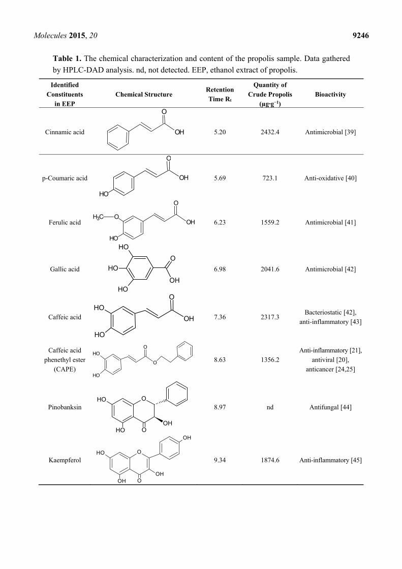
The identification of chromatographic peaks was accomplished by the information obtained from
HPLC-DAD analysis. Reference standards were used for p-coumaric acid, benzoic acid, ferulic acid,
gallic acid, caffeic acid, cinnamic acid, apigenin, pinobanksin, kaempferol, kaempferide, acacetin,
pinocembrin, galangin, chrysin, quercetin and caffeic acid phenethyl ester. The identification was
confirmed by direct comparison of the retention times and spectra acquired in the same analytical
conditions. The content of phenolic acids and flavonoid compounds of an ethanolic propolis sample is
reported in Table 1. In general, phenolic acids and their esters were the predominant class of
substances in ethanol extract of propolis (EEP), followed by flavones and flavonols. Qualitative and
quantitative analysis of selected flavonoids and phenolic acids identified pinocembrin, kaempferol,
galangin, chrysin, apigenin, quercetin, acacetin, gallic acid, ferulic acid, caffeic acid, caffeic acid
phenethyl ester (CAPE), p-coumaric acid and cinnamic acid. The flavonoid compounds identified in
this study included flavones, flavanones, flavanols, flavonols and chalcones. It is demonstrated that the
chemical content of ethanolic extracts of propolis is highly various. The main detected ingredients
belonging to phenolic compounds and flavonoids are cinnamic acid and quercetin, respectively. The
increasing sequence of quantitative EEP content for selected constituents is presented as follow:
flavones < flavonols < aromatic carboxylic acid < phenolic acid.
The complex composition of propolis can be responsible for its multi-directional antiproliferative
activity; however, a mixture of different polyphenols in EEP may exhibit the attenuating action
between selected constituents, with the partial reduction of their biological activity.
Molecules 2015, 20 9246
Table 1. The chemical characterization and content of the propolis sample. Data gathered
by HPLC-DAD analysis. nd, not detected. EEP, ethanol extract of propolis.
Identified
Constituents
in EEP
Chemical Structure Retention
Time Rt
Quantity of
Crude Propolis
(µg·g−1)
Bioactivity
Cinnamic acid OH
O
5.20 2432.4 Antimicrobial [39]
p-Coumaric acid OH
O
OH
5.69 723.1 Anti-oxidative [40]
Ferulic acid OH
O
OH
OCH36.23 1559.2 Antimicrobial [41]
Gallic acid OH
OH
OH
OH
O
6.98 2041.6 Antimicrobial [42]
Caffeic acid OH
O
OH
OH
7.36 2317.3 Bacteriostatic [42],
anti-inflammatory [43]
Caffeic acid
phenethyl ester
(CAPE) O
OOH
OH
8.63 1356.2
Anti-inflammatory [21],
antiviral [20],
anticancer [24,25]
Pinobanksin OOH
OH OOH
8.97 nd Antifungal [44]
Kaempferol OOH
OH O
OH
OH
9.34 1874.6 Anti-inflammatory [45]
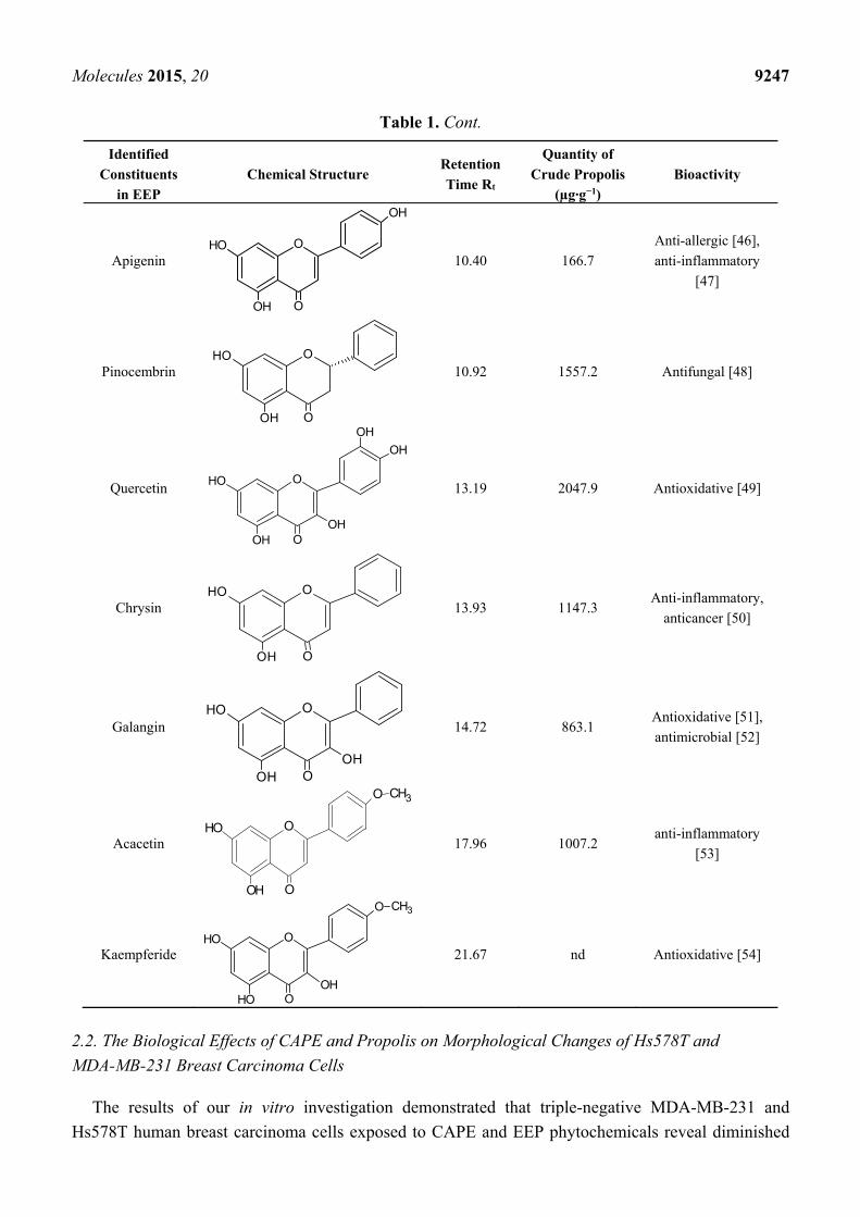
Molecules 2015, 20 9247
Table 1. Cont.
Identified
Constituents
in EEP
Chemical Structure Retention
Time Rt
Quantity of
Crude Propolis
(µg·g−1)
Bioactivity
Apigenin OOH
OH O
OH
10.40 166.7
Anti-allergic [46],
anti-inflammatory
[47]
Pinocembrin OOH
OH O
10.92 1557.2 Antifungal [48]
Quercetin O
OH
OH
OH O
OH
OH
13.19 2047.9 Antioxidative [49]
Chrysin OOH
OH O
13.93 1147.3 Anti-inflammatory,
anticancer [50]
Galangin O
OH
OH
OH O
14.72 863.1 Antioxidative [51],
antimicrobial [52]
Acacetin OOH
OH O
O CH3
17.96 1007.2 anti-inflammatory
[53]
Kaempferide OOH
OH O
O CH3
OH
21.67 nd Antioxidative [54]
2.2. The Biological Effects of CAPE and Propolis on Morphological Changes of Hs578T and
MDA-MB-231 Breast Carcinoma Cells
The results of our in vitro investigation demonstrated that triple-negative MDA-MB-231 and
Hs578T human breast carcinoma cells exposed to CAPE and EEP phytochemicals reveal diminished
Molecules 2015, 20 9248
metabolic activity and viability in a dose-dependent and time-dependent manner. Microscopic
assessment demonstrated numerous changes in cellular morphology of examined breast carcinoma
cells, including a decreased number of affected cells, cell shrinkage and cytoplasmic condensation.
These data support the hypothesis that the exposure to some phytochemicals, components of propolis,
including derivatives of caffeic acid, may hypothetically reduce the growth of breast cancer cells,
compared to the non-cancerous IMR-90 fibroblast control line.
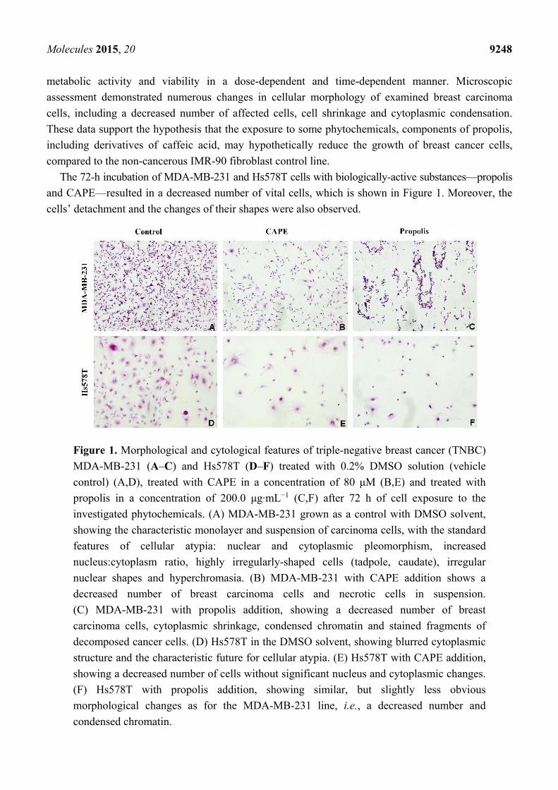
The 72-h incubation of MDA-MB-231 and Hs578T cells with biologically-active substances—propolis
and CAPE—resulted in a decreased number of vital cells, which is shown in Figure 1. Moreover, the
cells’ detachment and the changes of their shapes were also observed.
Figure 1. Morphological and cytological features of triple-negative breast cancer (TNBC)
MDA-MB-231 (A–C) and Hs578T (D–F) treated with 0.2% DMSO solution (vehicle
control) (A,D), treated with CAPE in a concentration of 80 μM (B,E) and treated with
propolis in a concentration of 200.0 μg·mL−1 (C,F) after 72 h of cell exposure to the
investigated phytochemicals. (A) MDA-MB-231 grown as a control with DMSO solvent,
showing the characteristic monolayer and suspension of carcinoma cells, with the standard
features of cellular atypia: nuclear and cytoplasmic pleomorphism, increased
nucleus:cytoplasm ratio, highly irregularly-shaped cells (tadpole, caudate), irregular
nuclear shapes and hyperchromasia. (B) MDA-MB-231 with CAPE addition shows a
decreased number of breast carcinoma cells and necrotic cells in suspension.
(C) MDA-MB-231 with propolis addition, showing a decreased number of breast
carcinoma cells, cytoplasmic shrinkage, condensed chromatin and stained fragments of
decomposed cancer cells. (D) Hs578T in the DMSO solvent, showing blurred cytoplasmic
structure and the characteristic future for cellular atypia. (E) Hs578T with CAPE addition,
showing a decreased number of cells without significant nucleus and cytoplasmic changes.
(F) Hs578T with propolis addition, showing similar, but slightly less obvious
morphological changes as for the MDA-MB-231 line, i.e., a decreased number and
condensed chromatin.
Molecules 2015, 20 9249
2.3. The Assessment of Viability of MDA-MB-231 and Hs578T Cells Exposed to CAPE and EEP with
the MTT Assay
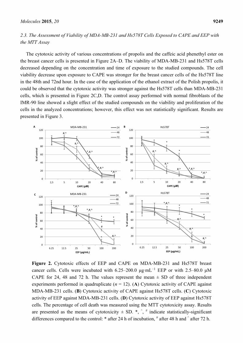
The cytotoxic activity of various concentrations of propolis and the caffeic acid phenethyl ester on
the breast cancer cells is presented in Figure 2A–D. The viability of MDA-MB-231 and Hs578T cells
decreased depending on the concentration and time of exposure to the studied compounds. The cell
viability decrease upon exposure to CAPE was stronger for the breast cancer cells of the Hs578T line
in the 48th and 72nd hour. In the case of the application of the ethanol extract of the Polish propolis, it
could be observed that the cytotoxic activity was stronger against the Hs578T cells than MDA-MB-231
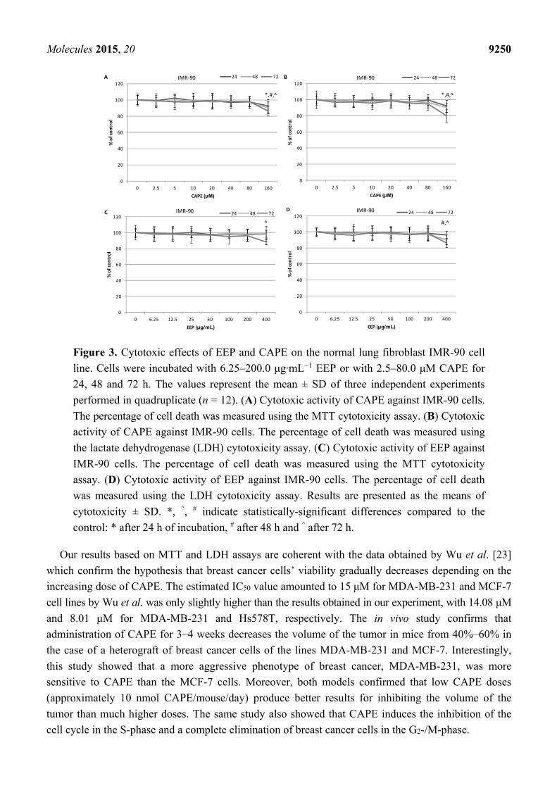
cells, which is presented in Figure 2C,D. The control assay performed with normal fibroblasts of the
IMR-90 line showed a slight effect of the studied compounds on the viability and proliferation of the
cells in the analyzed concentrations; however, this effect was not statistically significant. Results are
presented in Figure 3.
Figure 2. Cytotoxic effects of EEP and CAPE on MDA-MB-231 and Hs578T breast
cancer cells. Cells were incubated with 6.25–200.0 μg·mL−1 EEP or with 2.5–80.0 μM
CAPE for 24, 48 and 72 h. The values represent the mean ± SD of three independent
experiments performed in quadruplicate (n = 12). (A) Cytotoxic activity of CAPE against
MDA-MB-231 cells. (B) Cytotoxic activity of CAPE against Hs578T cells. (C) Cytotoxic
activity of EEP against MDA-MB-231 cells. (D) Cytotoxic activity of EEP against Hs578T
cells. The percentage of cell death was measured using the MTT cytotoxicity assay. Results
are presented as the means of cytotoxicity ± SD. *, ^, # indicate statistically-significant
differences compared to the control: * after 24 h of incubation, # after 48 h and ^ after 72 h.
0
20
40
60
80
100
120
2,5 5 10 20 40 80
% of control
CAPE (µM)
24
48
72
MDA‐MB‐231A
#,^
#,^
*,#,^
*,#,^
*,#,^
0
20
40
60
80
100
120
2,5 5 10 20 40 80
% of control
CAPE (µM)
24
48
72
Hs578TB
*,#,^
*,#,^
*
*
#,^
#,^
#,^
0
20
40
60
80
100
120
6.25 12.5 25 50 100 200
% of control
EEP (µg/mL)
24
48
72
MDA‐MB‐231C
*,#,^*,#,^
* *
#
#,^^
0
20
40
60
80
100
120
6.25 12.5 25 50 100 200
% of control
EEP (µg/mL)
24
48
72
Hs578TD
#,^
#,^
#,^
*,#,^*
*
*
Molecules 2015, 20 9250
Figure 3. Cytotoxic effects of EEP and CAPE on the normal lung fibroblast IMR-90 cell
line. Cells were incubated with 6.25–200.0 μg·mL−1 EEP or with 2.5–80.0 μM CAPE for
24, 48 and 72 h. The values represent the mean ± SD of three independent experiments
performed in quadruplicate (n = 12). (A) Cytotoxic activity of CAPE against IMR-90 cells.
The percentage of cell death was measured using the MTT cytotoxicity assay. (B) Cytotoxic
activity of CAPE against IMR-90 cells. The percentage of cell death was measured using
the lactate dehydrogenase (LDH) cytotoxicity assay. (C) Cytotoxic activity of EEP against
IMR-90 cells. The percentage of cell death was measured using the MTT cytotoxicity
assay. (D) Cytotoxic activity of EEP against IMR-90 cells. The percentage of cell death
was measured using the LDH cytotoxicity assay. Results are presented as the means of
cytotoxicity ± SD. *, ^, # indicate statistically-significant differences compared to the
control: * after 24 h of incubation, # after 48 h and ^ after 72 h.
Our results based on MTT and LDH assays are coherent with the data obtained by Wu et al. [23]
which confirm the hypothesis that breast cancer cells’ viability gradually decreases depending on the
increasing dose of CAPE. The estimated IC50 value amounted to 15 μM for MDA-MB-231 and MCF-7
cell lines by Wu et al. was only slightly higher than the results obtained in our experiment, with 14.08 μM
and 8.01 μM for MDA-MB-231 and Hs578T, respectively. The in vivo study confirms that
administration of CAPE for 3–4 weeks decreases the volume of the tumor in mice from 40%–60% in
the case of a heterograft of breast cancer cells of the lines MDA-MB-231 and MCF-7. Interestingly,
this study showed that a more aggressive phenotype of breast cancer, MDA-MB-231, was more
sensitive to CAPE than the MCF-7 cells. Moreover, both models confirmed that low CAPE doses
(approximately 10 nmol CAPE/mouse/day) produce better results for inhibiting the volume of the
tumor than much higher doses. The same study also showed that CAPE induces the inhibition of the
cell cycle in the S-phase and a complete elimination of breast cancer cells in the G2-/M-phase.
0
20
40
60
80
100
120
0 2.5 5 10 20 40 80 160
% of control
CAPE (µM)
24 48 72IMR‐90A
*,#,̂
0
20
40
60
80
100
120
0 2.5 5 10 20 40 80 160
% of control
CAPE (µM)
24 48 72IMR‐90B
*,#,̂
0
20
40
60
80
100
120
0 6.25 12.5 25 50 100 200 400
% of control
EEP (µg/mL)
24 48 72IMR‐90C
^
0
20
40
60
80
100
120
0 6.25 12.5 25 50 100 200 400
% of control
EEP (µg/mL)
24 48 72IMR‐90D
#,^
Molecules 2015, 20 9251
2.4. The Assessment of the Cytotoxic Activity of CAPE and EEP against MDA-MB-231 and Hs578T
Cells with the LDH Assay.
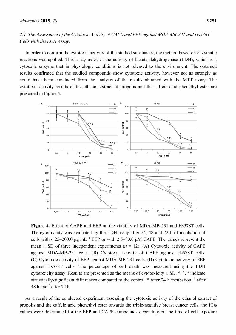
In order to confirm the cytotoxic activity of the studied substances, the method based on enzymatic
reactions was applied. This assay assesses the activity of lactate dehydrogenase (LDH), which is a
cytosolic enzyme that in physiologic conditions is not released to the environment. The obtained
results confirmed that the studied compounds show cytotoxic activity, however not as strongly as
could have been concluded from the analysis of the results obtained with the MTT assay. The
cytotoxic activity results of the ethanol extract of propolis and the caffeic acid phenethyl ester are
presented in Figure 4.
Figure 4. Effect of CAPE and EEP on the viability of MDA-MB-231 and Hs578T cells.
The cytotoxicity was evaluated by the LDH assay after 24, 48 and 72 h of incubation of
cells with 6.25–200.0 μg·mL−1 EEP or with 2.5–80.0 μM CAPE. The values represent the
mean ± SD of three independent experiments (n = 12). (A) Cytotoxic activity of CAPE
against MDA-MB-231 cells. (B) Cytotoxic activity of CAPE against Hs578T cells.
(C) Cytotoxic activity of EEP against MDA-MB-231 cells. (D) Cytotoxic activity of EEP
against Hs578T cells. The percentage of cell death was measured using the LDH
cytotoxicity assay. Results are presented as the means of cytotoxicity ± SD. *, ^, # indicate
statistically-significant differences compared to the control: * after 24 h incubation, # after
48 h and ^ after 72 h.
As a result of the conducted experiment assessing the cytotoxic activity of the ethanol extract of
propolis and the caffeic acid phenethyl ester towards the triple-negative breast cancer cells, the IC50
values were determined for the EEP and CAPE compounds depending on the time of cell exposure
0
20
40
60
80
100
120
2,5 5 10 20 40 80
% of control
CAPE (µM)
24
48
72
Hs578TB
*
*
*
*
^,#^,#
^,#
^,#
^
#
0
20
40
60
80
100
120
6,25 12,5 25 50 100 200
% of control
EEP (µg/mL)
24
48
72
MDA‐MB‐231C
*
*
*,#
^,#
^#
#
^
^0
20
40
60
80
100
120
6,25 12,5 25 50 100 200
% of control
EEP (µg/mL)
24
48
72
Hs578TD
^,#
^,#
^,#
^,#^
**
0
20
40
60
80
100
120
2,5 5 10 20 40 80
% of control
CAPE (µM)
24
48
72
MDA‐MB‐231A
*,^,#
*,^,#
^
*,^,#
Molecules 2015, 20 9252
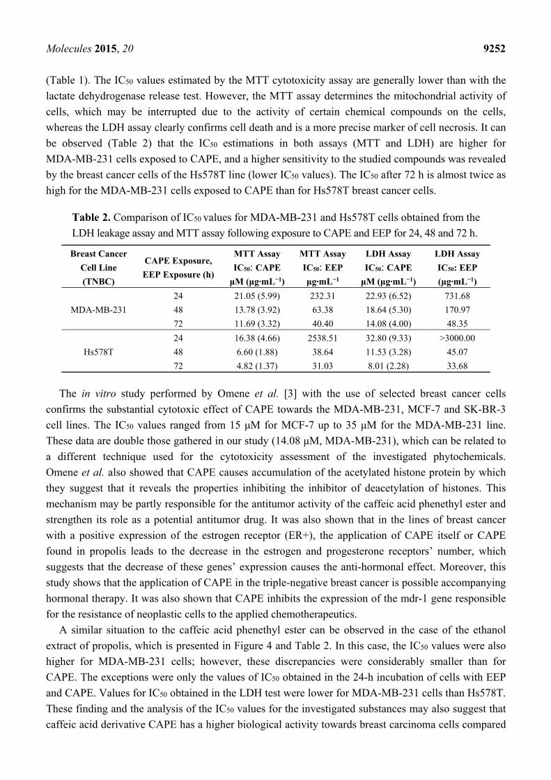
(Table 1). The IC50 values estimated by the MTT cytotoxicity assay are generally lower than with the
lactate dehydrogenase release test. However, the MTT assay determines the mitochondrial activity of
cells, which may be interrupted due to the activity of certain chemical compounds on the cells,
whereas the LDH assay clearly confirms cell death and is a more precise marker of cell necrosis. It can
be observed (Table 2) that the IC50 estimations in both assays (MTT and LDH) are higher for
MDA-MB-231 cells exposed to CAPE, and a higher sensitivity to the studied compounds was revealed
by the breast cancer cells of the Hs578T line (lower IC50 values). The IC50 after 72 h is almost twice as
high for the MDA-MB-231 cells exposed to CAPE than for Hs578T breast cancer cells.
Table 2. Comparison of IC50 values for MDA-MB-231 and Hs578T cells obtained from the
LDH leakage assay and MTT assay following exposure to CAPE and EEP for 24, 48 and 72 h.
Breast Cancer
Cell Line
(TNBC)
CAPE Exposure,
EEP Exposure (h)
MTT Assay
IC50: CAPE
μM (μg·mL−1)
MTT Assay
IC50: EEP
μg·mL−1
LDH Assay
IC50: CAPE
μM (μg·mL−1)
LDH Assay
IC50: EEP
(μg·mL−1)
MDA-MB-231
24 21.05 (5.99) 232.31 22.93 (6.52) 731.68
48 13.78 (3.92) 63.38 18.64 (5.30) 170.97
72 11.69 (3.32) 40.40 14.08 (4.00) 48.35
Hs578T
24 16.38 (4.66) 2538.51 32.80 (9.33) >3000.00
48 6.60 (1.88) 38.64 11.53 (3.28) 45.07
72 4.82 (1.37) 31.03 8.01 (2.28) 33.68
The in vitro study performed by Omene et al. [3] with the use of selected breast cancer cells
confirms the substantial cytotoxic effect of CAPE towards the MDA-MB-231, MCF-7 and SK-BR-3
cell lines. The IC50 values ranged from 15 μM for MCF-7 up to 35 μM for the MDA-MB-231 line.
These data are double those gathered in our study (14.08 μM, MDA-MB-231), which can be related to
a different technique used for the cytotoxicity assessment of the investigated phytochemicals.
Omene et al. also showed that CAPE causes accumulation of the acetylated histone protein by which
they suggest that it reveals the properties inhibiting the inhibitor of deacetylation of histones. This
mechanism may be partly responsible for the antitumor activity of the caffeic acid phenethyl ester and
strengthen its role as a potential antitumor drug. It was also shown that in the lines of breast cancer
with a positive expression of the estrogen receptor (ER+), the application of CAPE itself or CAPE
found in propolis leads to the decrease in the estrogen and progesterone receptors’ number, which
suggests that the decrease of these genes’ expression causes the anti-hormonal effect. Moreover, this
study shows that the application of CAPE in the triple-negative breast cancer is possible accompanying
hormonal therapy. It was also shown that CAPE inhibits the expression of the mdr-1 gene responsible
for the resistance of neoplastic cells to the applied chemotherapeutics.
A similar situation to the caffeic acid phenethyl ester can be observed in the case of the ethanol
extract of propolis, which is presented in Figure 4 and Table 2. In this case, the IC50 values were also
higher for MDA-MB-231 cells; however, these discrepancies were considerably smaller than for
CAPE. The exceptions were only the values of IC50 obtained in the 24-h incubation of cells with EEP
and CAPE. Values for IC50 obtained in the LDH test were lower for MDA-MB-231 cells than Hs578T.
These finding and the analysis of the IC50 values for the investigated substances may also suggest that
caffeic acid derivative CAPE has a higher biological activity towards breast carcinoma cells compared
Molecules 2015, 20 9253
to ethanol extract of propolis with a complex chemical composition. Possibly, the “attenuation
phenomenon” can be responsible for a less effective action of propolis as a mixture of variable ingredients.
The quantitative composition of propolis is heterogeneous; however, it always contains chemical
compounds, such as polyphenols, terpenoids, steroids and amino acids. Propolis samples obtained
from various plants may therefore differ in terms of chemical composition. The Polish propolis is
mainly classified as the poplar-type propolis, and its dominating components are flavonoids and
phenolic compounds, which constitute between 35% and 50%.
There is more and more proof that the polyphenol compounds found, e.g., in propolis may serve as
a supplement to standard chemotherapy and radiotherapy.
Apoptosis is one of the most potent defenses against cancer, because this process eliminates
potentially deleterious, mutated cells. Many dietary cancer-preventive compounds, including propolis
and its active derivatives, induce apoptosis in cancer cells. The mechanism of evoking apoptosis by
propolis seems to be independent of the type of the studied neoplastic cell; however, it is directly
dependent on the concentration of the applied extract for the purpose of the study. Some studies
suggest that propolis induces apoptosis by releasing cytochrome c from mitochondria to cytosol by
means of the caspases’ cascade and with the pro-apoptotic proteins.
The studies of Watabe et al. [36] depict the mechanism of CAPE activity as dependent on the
nuclear transcription factor NF-κB. These factors play a significant role in the regulation of death and
survival of cells. It is known that the caffeic acid phenethyl ester strongly inhibits NF-κB activation.
The study also confirms that CAPE is responsible for the activation of apoptosis in neoplastic cells of
various types, but does not lead to the activation of this pathway in the case of the normal fibroblast
cells WI-38. It may be caused by the fact that the neoplastic cells with a high base of NF-κB activity
are more sensitive to CAPE than normal cells. The study also analyzes the pathways of apoptosis
activation in the neoplastic cells. It confirms the activation of apoptosis both in the receptor pathway
dependent on the Fas receptor and the mitochondrial pathway with the release of cytochrome c.
Additionally, this mechanism may be responsible for the difference in the results regarding the assessment
of the cytotoxic activity in the MTT and LDH assays.
Therefore, the results of the experiment conducted by Lee et al. [54] are of special interest, since
they defined the effect of the caffeic acid phenethyl ester on the activity of proteins p53 and p38 in the C6
glioma cells. This study confirmed that propolis has cytotoxic activity. The scientists proved that CAPE
leads to the release of cytochrome c from mitochondrion to cytosol and the activation of caspase-3. What
is most important, the expression of the p53 protein, Bax and Bak increased only after 3 h of
incubation with CAPE, simultaneously causing a decrease of the expression of the anti-apoptotic
protein Bcl-2 upon a 36-h incubation. Similar results were also obtained by Jin et al. [55], who carried
out an experiment on the line of human myeloid leukemia U937. They proved, similarly to Lee et al.,
that the caffeic acid phenethyl ester (propolis component) has cytotoxic properties dependent on the
concentration and exposure time of the substance on the neoplastic cells. With DAPI staining, they
observed in the fluorescent microscope some changes characteristic for apoptosis in the cellular nuclei.
They did not confirm the expression of the Fas protein on the surface of the studied cells; however,
they observed the release of cytochrome c to cytosol, inhibition of the expression of the anti-apoptotic
protein Bcl-2 and an increase of the pro-apoptotic protein Bax. Our results confirm the observations of
Jin et al., since, similarly to the Korean team, we revealed that the caffeic acid phenethyl ester and the
Molecules 2015, 20 9254
ethanol extract of the Polish propolis show cytotoxic activity dependent on the concentration of the
studied compound and the time of cell exposure to these compounds.
Szliszka et al. [56] in their experiment showed that artepillin C found in the Brazilian propolis
causes an increase in neoplastic cells’ sensitivity to the TRAIL protein. This protein is a strong
stimulator of apoptosis in neoplastic cells and an important factor responsible for the elimination of
developing tumors. However, a number of cells undergoing oncogenesis is resistant to the apoptotic
death with the participation of the TRAIL protein. Therefore, Szliszka et al. decided to sensitize the
neoplastic cells to the TRAIL protein by adding substances of a natural origin, such as flavonoids and
phenolic acids. In order to confirm the advanced thesis, they used the cytotoxic assays (MTT, LDH),
fluorescent staining (to define apoptosis), flow cytometry (to assess the death receptors) and the
immune-enzymatic tests. It was proven that the application of the propolis component with the TRAIL
protein led to an increase of the number of dying cells though apoptosis by an increase of caspase-3
and -8 activity and inhibition of the nuclear factor NF-κB.
It should be noted that the obtained differences in the MTT and LDH assays suggest the changes at
the level of mitochondrial metabolism in the studied compounds. The possible mechanism of the
anticancer property of propolis and one of its active derivate, CAPE, seems to be the augmentation of
apoptosis phenomenon in human breast cancer cells. The detected changes of carcinoma cells’
morphology following EEP exposure may suggest and indicate the carcinoma cells’ death due to the
apoptosis pathway. Therefore, the authors’ next research aim shall be the determination of a
hypothetical way for the breast cancer cells’ metabolic death, after exposure to these highly-active
phytochemicals inhibiting malignant cells’ metabolism.
3. Experimental Section
3.1. Compounds and Reagents
CAPE, MTT, trypan blue, DMEM medium, Leibovitz’s medium, fetal bovine serum (FBS) and
tryptic soy broth were purchased from Sigma-Aldrich (St. Louis, MO, USA). Antibiotic/antimycotic,
trypsin and phosphate-buffered saline (PBS) were purchased from GE healthcare company (Waukesha,
WI, USA). Mayer’s hematoxylin, eosin, xylene and ethanol were obtained from Avantor Performance
Materials (Gliwice, Poland).
3.2. Propolis Sample Collection and Ethanol Extract of Propolis Preparation
Propolis obtained from the bee Apis mellifera was collected in Kamianna in the south of Poland in
the Małopolskie Province. It was stored at 4 °C out of the light until the extraction. In order to obtain
the ethanol extract, 10 g of raw propolis were minced, and 100 g of 75% ethanol were added. The
tightly closed flask was placed in a shaker for two weeks at room temperature, out of the light. After
this time, the extract was cooled at 4 °C for 24 h in order to remove all of the substances that are
insoluble in ethanol. Next, it was filtered under lower pressure. The obtained filtrate was vaporized with
a vacuum evaporator at 40 °C. In the next stage, the obtained extract was placed in an incubator for
7 days in order to vaporize the ethanol residues. The content of the flask with the known extract mass
was diluted in a pure dimethyl sulfoxide until the working concentration of 100 mg·mL−1 was obtained.
Molecules 2015, 20 9255
3.3. High-Performance Liquid Chromatography with Diode Array Detector Analysis
To determine the chemical composition of EEP, a high performance liquid chromatography method
was applied with the use of a Varian 920-LC HPLC (Harbor City, CA, USA), equipped with a 900-LC
model autosampler, gradient pump, 330 model DAD and the Galaxie software (Version 1.9 SP3,
Agilent Technologies, Santa Clara, CA, USA) for data acquisition and processing. The analysis was
carried out according to a protocol described in the study [5]. Reference substances in ethanol solution
(0.1 mg·mL−1) containing acacetin, apigenin, chrysin, galangin, kaempferide, kaempferol, pinobanksin,
pinocembrin, quercetin, caffeic acid, cinnamic acid, o-coumaric acid, m-coumaric acid, p-coumaric
acid, gallic acid, ferulic acid and caffeic acid phenylethyl ester (CAPE) were prepared from standard
stock solutions. Phenolic compounds were purchased from Carl Roth GmbH (Karlsruhe, Germany)
and Sigma Chemical Company (St. Louis, MO, USA).
3.4. Triple-Negative Breast Cancer Cell Cultures
Cell lines of a human breast (mammary gland) cancer were cultured in accordance with the
manufacturer’s recommendations. The metastatic human breast cancer line MDA-MB-231
(adenocarcinoma) derived from metastatic site pleural effusion (Catalogue No. ATCC HTB-26,
American Type Culture Collection, Manassas, VA, USA) was cultured with Leibovitz’s L-15 medium
with 10% of inactivated fetal bovine serum (FBS, Sigma-Aldrich) at 37 °C without CO2. Breast cancer
cells Hs578T (Catalogue No. ATCC HTB-126 American Type Culture Collection, Manassas, VA,
USA) were cultured on Dulbecco’s modified Eagle’s medium with 10% inactivated fetal bovine serum
with bovine insulin at a final concentration of 0.01 mg·mL−1. The cells were incubated in a CO2
incubator at 37 °C and in the air atmosphere containing 5% CO2. The studied cell lines were
supplemented with antibiotics with the following final concentrations: penicillin 100 U·mL−1,
streptomycin 100 µg·mL−1 and a fungistatic amphotericin B with a concentration of 0.25 µg·mL−1. The
medium was changed every 2–3 days, and the passage was carried out with a confluence of 80%–90%.
3.5. Microscopic Evaluation of Carcinoma Cells Morphology
The cells of the studied lines were inoculated onto 2-chamber microscopic culture vessels (Lab-TekTM,
Waltham, MA, USA) in the amount of 1000 cells per a well and were left for 24 h in order to obtain
the logarithmic growth. After this time, the studied compounds were added to the media in appropriate
concentrations and were left for 24, 48 and 72 h, respectively, depending on the time of the
experiment. After a defined time period, they were fixed for 12 h in 96% ethanol. Then, the cells were
hydrated in the following series of dilutions: 99.6%, 96%, 90%, 80%, 70% and 50% and stained with
hematoxylin for 12 min (standard H&E staining protocol). Next, the plates were washed with the PBS
solution for approximately 30 min in order to blue up and then were incubated for 30 s with eosin. The
plates were washed with PBS solution again and dehydrated with ethanol of increasing concentrations
of 50%, 70%, 80%, 90%, 96% and 99.6%. In the next stage, the plates were immersed in the ethanol
and xylene mixture (50:50) for 1 min and then in pure xylene. The plates were mounted and analyzed
under a microscope.
Molecules 2015, 20 9256
3.6. The Initial Evaluation of Viability of Triple-Negative Breast Cancer Cells
The trypan blue viability assay was applied in order to determine the efficient and optimal ranges of
concentrations of CAPE and EEP. The cells were inoculated on the 96-well plates with a density of
5 × 10³ per 100 µL of medium and were left to stick for 12 h. Then, the cells were treated with the
solvent or the active substances for 24 h and later with the use of maximum ranges of concentrations
from 0–320 µM and 0–400 µg·mL−1 for CAPE and propolis, respectively. The tested and chosen
concentrations for CAPE were 2.5, 5, 10, 20, 40 and 80 µM and for propolis were: 6.25, 12.5, 25, 50,
100 and 200 µg·mL−1. Investigated cells were trypsinized, centrifuged and stained with the trypan blue
solution 0.04% (w/v, Sigma-Aldrich) and counted with the automatic cell counter (TC20TM Automated
Cell Counter Bio-Rad, Hercules, CA, USA), in accordance with the manufacturer’s protocol. Cell
viability was presented as a percent mean ± SEM with the three-times independently repeated
experiments from three internal measurements in each.
3.7. MTT Cell Proliferation and Cytotoxicity Assay
The MTT assay is a colorimetric method, which enables assessing the cell metabolic activity. It
consists of a colorimetric determination of a colored product, which originates after bromide is added
(3(4,5-dimethyltiazol-2-yl)-2,5-diphenyl tetrazolium (MTT) to the cell culture in the presence of the
tested substances. The amount of the originating formazan is proportional to the amount of living cells.
In order to determine the cytotoxicity of the studied compound, the cells were inoculated on the
96-well plates in the amount of 104 cell/well, and fresh medium was added and left for 72 h to obtain
the logarithmic cell growth. At this time, the medium was decanted, and a culture medium containing
EEP and CAPE was added in the analyzed concentrations prepared in the course of a series of dilutions
in the culture medium. Zero-point-one milliliters of medium with a defined concentration of the
studied substance were added to each well and left for 24 h in the CO2 incubator at 37 °C in a steam
atmosphere with 5% of CO2 or in the thermostat in the case of the MDA-MB-231 cells.
At this time, the medium was decanted, and 100 μL of 1% MTT solution in the culture media were
added. The cells were incubated with a reagent for 4 h in the incubator at 37 °C. At this time, the MTT
solution was decanted from the crystals, and the obtained formazan crystals were diluted by adding to
each well 200 μL of DMSO. The whole was shaken evenly to dilute the obtained crystals. To read the
absorbance, the ELISA plate reader by BioTek was used with a wavelength of 570 nm.
The solvent control was the absorbance readings obtained from the plates where the cell lines were
cultured without the studied substances containing the additionally-applied solvents (dimethyl
sulfoxide with a concentration of 0.2%). This solvent control protocol was applied to both the MTT
and the LDH assay.
3.8. Lactate Dehydrogenase Release Assay
Upon reaching the almost confluent state, the cells were washed twice with the DPBS and removed
from the medium with 0.25% solution of trypsin in EDTA. The detached cells were centrifuged at 1000× g
for 3 min at room temperature (25 °C). The obtained cell sediment was suspended in the appropriate
amount of assay medium with 1% of the fetal bovine serum in such a way that the cell concentration
Molecules 2015, 20 9257
was 1 × 10⁵ cells/ml. One hundred microliters of cell suspension were added to each well of the
medium plate (96 wells), except the wells containing control I (background) and 100 μL of the assay
medium. To assess the degree of cytotoxicity, three types of controls were performed:
(i) Background control (Control I): 200 μL assay medium per well;
(ii) Low control (Control II = spontaneous LDH release): 100 μL assay medium and 100 μL
cell suspension;
(iii) High control (Control III = maximum LDH release): 100 μL Triton-X solution (final
concentration 2%) in assay medium and 100 μL cell suspension per well.
Additionally, controls of the tested substances were performed: 100 μL of assay medium and 100 μL
of the tested substance at the highest possible concentration without cells (separately for propolis and
CAPE). Then, the cells were incubated for 48 h in the incubator at 37 °C in a steam atmosphere with
5% CO2. Directly prior to use, dilutions were prepared of the tested substances in the assay medium in
such a way that the final volume of all of the ingredients in the well was 200 μL. The assay medium
was removed from above the cells attached to the bottom of the wells, and 100 μL of fresh assay
medium were added to each well, as well as 100 μL of the tested substance in appropriate
concentrations. The cells were incubated at 37 °C in a steam atmosphere with 5% CO2 depending on
the experimental needs for 24 and 48 h. At the defined incubation time, 100 μL of the supernatant were
collected from each plate and moved to the micro test plates (96 wells), and 100 μL of reaction mixture
were added. The micro test plates were incubated at room temperature for 30 min without light. At the
defined time, the absorbance measurement of each sample was performed with the ELISA reader with
a wavelength 490 nm. The length of the reference wave was 600 nm. All of the samples were
performed in triplicate. The value of background control absorbance must be subtracted from each
absorbance value, and the obtained values should be used in the following formula:
Cytotoxicity% = (absorbance value of the studied sample − absorbance value of Control II)/absorbance
value of Control III − absorbance value of Control II
3.9. Statistical Analysis
The statistical analysis was performed with the Statistica software (Version 8.0, StatSoft Poland).
The obtained results were shown in the form of the mean and standard variations. The obtained results
were repeated in 4 independent experiments (n = 96 for each studied concentration for the MTT assay
and n = 12 for each studied concentration for the LDH assay). The normality of the distribution was
defined with the Shapiro-Wilk test. In order to define the statistical significance, the Student t-test was
used, as well as the one-parameter ANOVA. Statistical significance was accepted at p < 0.05.
4. Conclusions
Based on our findings, propolis and the caffeic acid phenethyl ester substantially inhibit the growth
of the cells of triple-negative breast cancer of the lines MDA-MB-231 and Hs578T. The cytotoxic
activity of the studied compounds depends on the time of exposure and the concentration of the caffeic
acid phenethyl ester and ethanol extract of propolis. A more detailed study should reveal whether
Molecules 2015, 20 9258
CAPE, as a component of propolis, might potentially be applied as a therapeutic medium or as an
adjuvant for conventional chemotherapeutics, potentiating their biological, antiproliferative effects.
Acknowledgments
This study was supported by the research grant from Medical University of Silesia
no. KNW-1-064/N/4/0.
Author Contributions
Anna Rzepecka-Stojko and Agata Kabała-Dzik conceived the study idea, designed the experiments,
analyzed the data, and wrote the manuscript. Aleksandra Moździerz, Robert Kubina and
Magdalena Jurzak designed the experiments, and critically revised the manuscript.
Robert D. Wojtyczka, Rafał Stojko and Żaneta Jastrzębska-Stojko organized the data.
Arkadiusz Dziedzic organized the data and revised the manuscript. Jerzy Stojko and Ewa Buszman
critically revised the manuscript. All authors read and approved the final manuscript.
Conflicts of Interest
The authors declare no conflict of interest.
References
1. Brewster, A.M.; Chavez-MacGregor, M.; Brown, P. Epidemiology, biology, and treatment of
triple-negative breast cancer in women of African ancestry. Lancet Oncol. 2014, 15, e625–e634.
2. Murtaza, G.; Karim, S.; Akram, M.R.; Khan, S.A.; Azhar, S.; Mumtaz, A.; Bin Asad, M.H.
Caffeic acid phenethyl ester and therapeutic potentials. Biomed. Res. Int. 2014, 2014, 145342–145350.
3. Omene, C.; Kalac, M.; Wu. J.; Marchi, E.; Frenkel, K.; O’Connor, O.A. Propolis and its active
component, caffeic acid phenethyl ester (CAPE), modulate breast cancer therapeutic targets via an
epigenetically mediated mechanism of action. J. Cancer Sci. Ther. 2013, 5, 334–342.
4. Madrigal-Santillán, E.; Madrigal-Bujaidar, E.; Alvarez-González, I.; Sumaya-Martínez, M.T.;
Gutiérrez-Salinas, J.; Bautista, M.; Morales-González, A.; García-Luna, Y.; González-Rubio, M.;
Aguilar-Faisal, J.L.; et al. Review of natural products with hepatoprotective effects. World J.
Gastroentero. 2014, 20, 14787–14804.
5. Wojtyczka, R.D.; Dziedzic, A.; Idzik, D.; Kępa, M.; Kubina, R.; Kabała-Dzik, A.; Smoleń-Dzirba, J.;
Stojko, J.; Sajewicz, M.; Wąsik, T.J. Susceptibility of Staphylococcus aureus clinical isolates to
propolis extract alone or in combination with antimicrobial drugs. Molecules 2013, 18, 9623–9640.
6. Dziedzic, A.; Kubina, R.; Wojtyczka, R.D.; Kabała-Dzik, A.; Tanasiewicz, M.; Morawiec, T. The
antibacterial effect of ethanol extract of polish propolis on mutans streptococci and lactobacilli
isolated from saliva. Evid. -Based Complement. Alternat. Med. 2013, 2013, 681891–681902.
7. Wang, K.; Zhang, J.; Ping, S.; Ma, Q.; Chen, X.; Xuan, H.; Shi, J.; Zhang, C.; Hu, F.
Anti-inflammatory effects of ethanol extracts of Chinese propolis and buds from poplar
(Populus × canadensis). J. Ethnopharmacol. 2014, 155, 300–311.
Molecules 2015, 20 9259
8. Búfalo, M.C.; Bordon-Graciani, A.P.; Conti, B.J.; de Assis Golim, M.; Sforcin, J.M. The
immunomodulatory effect of propolis on receptors expression, cytokine production and fungicidal
activity of human monocytes. J. Pharm. Pharmacol. 2014, 66, 1497–1504.
9. Jastrzębska-Stojko, Z.; Stojko, R.; Rzepecka-Stojko, A.; Kabała-Dzik, A.; Stojko, J. Biological
activity of propolis-honey balm in the treatment of experimentally-evoked burn wounds.
Molecules 2013, 18, 14397–14413.
10. Kurek-Górecka, A.; Rzepecka-Stojko, A.; Górecki, M.; Stojko, J.; Sosada, M.; Swierczek-Zieba, G.
Structure and antioxidant activity of polyphenols derived from propolis. Molecules 2013, 19, 78–101.
11. Su, K.Y.; Hsieh, C.Y.; Chen, Y.W.; Chuang, C.T.; Chen, C.T.; Chen, Y.L. Taiwanese green propolis
and propolin G protect the liver from the pathogenesis of fibrosis via eliminating TGF-β-induced
smad2/3 phosphorylation. J. Agric. Food Chem. 2014, 62, 3192–3201.
12. Socha, R.; Gałkowska, D.; Bugaj, M.; Juszczak, L. Phenolic composition and antioxidant activity
of propolis from various regions of Poland. Nat. Prod. Res. 2014, 29, 416–422.
13. Chen, Y.J.; Shiao, M.S.; Hsu, M.L.; Tsai, T.H.; Wang, S.Y. Effect of caffeic acid phenethyl ester,
an antioxidant from propolis, on inducing apoptosis in human leukemic HL-60 cells. J. Agric.
Food Chem. 2001, 49, 5615–5619.
14. Kustiawan, P.M.; Puthong, S.; Arung, E.T.; Chanchao, C. In vitro cytotoxicity of indonesian
stingless bee products against human cancer cell lines. Asian Pac. J. Trop. Biomed. 2014, 4, 549–556.
15. Xuan, H.; Li, Z.; Yan, H.; Sang, Q.; Wang, K.; He, Q.; Wang, Y.; Hu, F. Antitumor activity of
Chinese propolis in human breast cancer MCF-7 and MDA-MB-231 cells. Evid. -Based
Complement. Altern. Med. 2014, 2014, doi:10.1155/2014/280120.
16. Kamiya, T.; Nishihara, H.; Hara, H.; Adachi, T. Ethanol extract of Brazilian red propolis induces
apoptosis in human breast cancer MCF-7 cells through endoplasmic reticulum stress. J. Agric.
Food Chem. 2012, 60, 11065–11070.
17. Kubina, R.; Kabała-Dzik, A.; Dziedzic, A.; Bielec, B.; Wojtyczka, R.D. Bułdak, R.J.; Wyszyńska, M.;
Stawiarska-Pięta, B.; Szaflarska-Stojko, E. The ethanol extract of Polish propolis exhibits
anti-proliferative and/or pro-apoptotic effect on HTC 116 colon cancer and Me45 malignant
melanoma cells in vitro conditions. Adv. Clin. Exp. Med. 2015, 24, 203–212.
18. Grunberger, D.; Banerjee, R.; Eisinger, K.; Oltz, E.M.; Efros, L.; Caldwell, M.; Estevez, V.;
Nakanishi, K. Preferential cytotoxicity on tumor cells by caffeic acid phenethyl ester isolated from
propolis. Experientia 1988, 44, 230–232.
19. Yildirim, O.; Yilmaz, A.; Oz, O.; Vatansever, H.; Cinel, L.; Aslan, G.; Tamer, L.; Adigüzel, U.;
Arpaci, R.; Kanik, A.; Emekdaş, G. Effect of caffeic acid phenethyl ester on treatment of
experimentally induced methicillin-resistant Staphylococcus epidermidis endophthalmitis in a
rabbit model. Cell Biochem. Funct. 2007, 25, 693–700.
20. Wang, P.; Liu, C.; Sanches, T.; Zhong, Y.; Liu, B.; Xiong, J.; Neamati, N.; Zhao, G. Design and
synthesis of novel nitrogen-containing polyhydroxylated aromatics as HIV-1 integrase inhibitors
from caffeic acid phenethyl ester. Bioorg. Med. Chem. Lett. 2009, 19, 4574–4578.
21. Cho, M.S.; Park, W.S.; Jung, W.K.; Qian, Z.J.; Lee, D.S.; Choi, J.S.; Lee, D.Y.; Park, S.G.; Seo, S.K.;
Kim, H.J., et al. Caffeic acid phenethyl ester promotes anti-inflammatory effects by inhibiting
MAPK and NF-κB signaling in activated HMC-1 human mast cells. Pharm. Biol. 2014, 52, 926–932.
Molecules 2015, 20 9260
22. Altuntaş, A.; Yılmaz, H.R.; Altuntaş, A.; Uz, E.; Demir, M.; Gökçimen, A.; Aksu, O.; Bayram, D.Ş.;
Sezer, M.T. Caffeic acid phenethyl ester protects against amphotericin B induced nephrotoxicity
in rat model. Biomed. Res. Int. 2014, 2014, 702981–702988.
23. Zhou, K.; Li, X.; Du, Q.; Li, D.; Hu, M.; Yang, X.; Jiang, Q.; Li, Z. A CAPE analogue as novel
antiplatelet agent efficiently inhibits collagen-induced platelet aggregation. Pharmazie 2014, 69,
615–620.
24. Wu, J.; Omene, C.; Karkoszka, J.; Bosland, M.; Eckard, J.; Klein, C.B.; Frenkel, K. Caffeic acid
phenethyl ester (CAPE), derived from a honeybee product propolis, exhibits a diversity of
anti-tumor effects in pre-clinical models of human breast cancer. Cancer Lett. 2011, 308, 43–53.
25. Akyol, S.; Ozturk, G.; Ginis, Z.; Armutcu, F.; Yigitoglu, M.R.; Akyol, O. In vivo and in vitro
antıneoplastic actions of caffeic acid phenethyl ester (CAPE): Therapeutic perspectives. Nutr. Cancer
2013, 65, 515–526.
26. Borrelli, F.; Izzo, A.A.; Di Carlo, G.; Maffia, P.; Russo, A.; Maiello, F.M.; Capasso, F.; Mascolo, N.
Effect of a propolis extract and caffeic acid phenethyl ester on formation of aberrant crypt foci
and tumors in the rat colon. Fitoterapia 2002, 73, S38–S43.
27. Xiang, D.; Wang, D.; He, Y.; Xie, J.; Zhong, Z.; Li, Z.; Xie, J. Caffeic acid phenethyl ester
induces growth arrest and apoptosis of colon cancer cells via the beta-catenin/T-cell factor
signaling. Anti-Cancer Drugs 2006, 17, 753–762.
28. Chen, M.F.; Wu, C.T.; Chen, Y.J.; Keng, P.C.; Chen, W.C. Cell killing and radiosensitization by
caffeic acid phenethyl ester (CAPE) in lung cancer cells. J. Radiat. Res. 2004, 45, 253–260.
29. Kudugunti, S.K.; Vad, N.M.; Ekogbo, E.; Moridani, M.Y. Efficacy of caffeic acid phenethyl ester
(CAPE) in skin B16-F0 melanoma tumor bearing C57BL/6 mice. Investig. New Drugs 2011, 29,
52–62.
30. Wu, C.S.; Chen, M.F.; Lee, I.L.; Tung, S.Y. Predictive role of nuclear factor-κB activity in gastric
cancer: A promising adjuvant approach with caffeic acid phenethyl ester. J. Clin. Gastroenterol.
2007, 41, 894–900.
31. Chen, M.J.; Chang, W.H.; Lin, C.C.; Liu, C.Y.; Wang, T.E.; Chu, C.H.; Shih, S.C.; Chen, Y.J.
Caffeic acid phenethyl ester induces apoptosis of human pancreatic cancer cells involving caspase
and mitochondrial dysfunction. Pancreatology 2008, 8, 566–576.
32. Lee, K.; Kang, N.; Kim, J.; Lee, K.; Lee, D.; Hur, H.J.; Lee, H.J. Caffeic acid phenethyl ester
inhibits invasion and expression of matrix metalloproteinase in SK-Hep1 human hepatocellular
carcinoma cells by targeting nuclear factor κB. Genes Nutr. 2008, 2, 319–322.
33. Huang, M.T.; Ma, W.; Yen, P.; Xie, J.G.; Han, J.; Frenkel, K.; Grunberger, D.; Conney, A.H.
Inhibitory effects of caffeic acid phenethyl ester (CAPE) on 12-O-tetradecanoylphorbol-13-
acetate-induced tumor promotion in mouse skin and the synthesis of DNA, RNA and protein in
HeLa cells. Carcinogenesis 1996, 17, 761–765.
34. Onori, P.; DeMorrow, S.; Gaudio, E.; Franchitto, A.; Mancinelli, R.; Venter, J.; Kopriva, S.; Ueno, Y.;
Alvaro, D.; Savage, J.; et al. Caffeic acid phenethyl ester decreases cholangiocarcinoma growth
by inhibition of NFκB and induction of apoptosis. Int. J. Cancer 2009, 125, 565–576.
35. Kuo, H.C.; Kuo, W.H.; Lee, Y.J.; Lin, W.L.; Chou, F.P.; Tseng, T.H. Inhibitory effect of caffeic
acid phenethyl ester on the growth of C6 glioma cells in vitro and in vivo. Cancer Lett. 2006, 234,
199–208.
Molecules 2015, 20 9261
36. Watabe, M.; Hishikawa, K.; Takayanagi, A.; Shimizu, N.; Nakaki, T. Caffeic acid phenethyl ester
induces apoptosis by inhibition of NFκB and activation of Fas in human breast cancer MCF-7
cells. J. Biol. Chem. 2004, 279, 6017–6026.
37. Eng-Wong, J.; Isaacs, C. Prediction of benefit from adjuwant treatment in patients with breast
cancer. Clin. Breast Cancer 2010, 10, 32–37.
38. Omene, C.O.; Wu, J.; Frenkel, K. Caffeic acid phenethyl ester (CAPE) derived from propolis,
a honeybee product, inhibits growth of breast cancer stem cells. Investig. New Drugs 2012, 30,
1279–1288.
39. Guzman, J.D. Natural cinnamic acids, synthetic derivatives and hybrids with antimicrobial
activity. Molecules 2014, 19, 19292–19349.
40. Etoh, H.; Murakami, K.; Yogoh, T.; Ishikawa, H.; Fukuyama, Y.; Tanaka, H. Anti-oxidative
compounds in barley tea. Biosci. Biotechnol. Biochem. 2004, 68, 2616–2618.
41. Borges, A.; Ferreira, C.; Saavedra, M.J.; Simões, M. Antioxidant and antimicrobial activities of
ferulic acid esters from ochrosia oppositifolia. Microb. Drug Resist. 2013, 19, 256–265.
42. Stojković, D.; Petrović, J.; Soković, M.; Glamočlija, J.; Kukić-Marković, J.; Petrović, S. In situ
antioxidant and antimicrobial activities of naturally occurring caffeic acid, p-coumaric acid and
rutin, using food systems. J. Sci. Food Agric. 2013, 93, 3205–3208
43. Fernández, M.A.; Sáenz, M.T.; García, M.D. Anti-inflammatory activity in rats and mice of
phenolic acids isolated from Scrophularia frutescens. J. Pharm. Pharmacol. 1998, 50, 1183–1186.
44. Boisard, S.; Le Ray, A.-M.; Landreau, A.; Kempf, M.; Cassisa, V.; Flurin, C.; Richomme, P.
Antifungal and antibacterial metabolites from a French poplar type propolis. Evid. -Based
Complement. Altern. Med. 2015, 2015, doi:10.1155/2015/319240.
45. Hämäläinen, M.; Nieminen, R.; Vuorela, P.; Heinonen, M.; Moilanen, E. Anti-inflammatory
effects of flavonoids: Genistein, kaempferol, quercetin, and daidzein inhibit STAT-1 and NF-κB
activations, whereas flavone, isorhamnetin, naringenin, and pelargonidin inhibit only NF-κB
activation along with their inhibitory effect on iNOS expression and NO production in activated
macrophages. Mediat. Inflamm. 2007, 2007, 45673, doi:10.1155/2007/45673.
46. Li, R.R.; Pang, L.L.; Du, Q.; Shi, Y.; Dai, W.J.; Yin, K.S. Apigenin inhibits allergen-induced
airway inflammation and switches immune response in a murine model of asthma. Immunopharm.
Immunot. 2010, 32, 364–370.
47. Lee, J.H.; Zhou, H.Y.; Cho, S.Y.; Kim, Y.S.; Lee, Y.S.; Jeong, C.S. Anti-inflammatory mechanisms
of apigenin: Inhibition of cyclooxygenase-2 expression, adhesion of monocytes to human
umbilical vein endothelial cells, and expression of cellular adhesion molecules. Arch. Pharm. Res.
2007, 30, 1318–1327.
48. Peng, L.; Yang, S.; Cheng, Y.J.; Chen, F.; Pan, S.; Fan, G. Antifungal activity and action mode of
pinocembrin from propolis against Penicillium italicum. Food Sci. Biotechnol. 2012, 21, 1533–1539.
49. Zhang, M.; Swarts, S.G.; Yin, L.; Liu, C.; Tian, Y.; Cao, Y.; Swarts, M.; Yang, S.; Zhang, S.B.;
Zhang, K., et al. Antioxidant properties of quercetin. Adv. Exp. Med. Biol. 2011, 701, 283–289.
50. Liu, Y.; Song, X.; He, J.; Zheng, X.; Wu, H. Synthetic derivatives of chrysin and their biological
activities. Med. Chem. Res. 2014, 23, 555–563.
51. Paulíková, H.; Berczeliová, E. The effect of quercetin and galangin on glutathione reductase.
Biomed. Pap. Med. Fac. Univ. Palacky. Olomouc. Czech Repub. 2005, 149, 497–500.
Molecules 2015, 20 9262
52. Lee, Y.S.; Kang, O.H.; Choi, J.G.; Oh, Y.C.; Chae, H.S.; Kim, J.H.; Park, H.; Sohn, D.H.;
Wang, Z.T.; Kwon, D.Y. Synergistic effects of the combination of galangin with gentamicin
against methicillin-resistant Staphylococcus aureus. J. Microbiol. 2008, 46, 283–288.
53. Carballo-Villalobos, A.I.; González-Trujano, M.E.; López-Muñoz, F.J. Evidence of mechanism of
action of anti-inflammatory/antinociceptive activities of acacetin. Eur. J. Pain 2014, 18, 396–405.
54. Lee, Y.J.; Kuo, H.C.; Chu, C.Y.; Wang, C.J.; Lin, W.C.; Tseng, T.H. Involvement of tumor
suppressor protein p53 and p38 MAPK in caffeic acid phenethyl ester-induced apoptosis of C6
glioma cells. Biochem. Pharmacol. 2003, 66, 2281–2289.
55. Jin, U.H.; Song, K.H.; Motomura, M.; Suzuki, I.; Gu, Y.H.; Kang, Y.J.; Moon, T.C.; Kim, C.H.
Caffeic acid phenethyl ester induces mitochondriamediated apoptosis in human myeloid leukemia
U937 cells. Mol. Cell Biochem. 2008, 310, 43–48.
56. Szliszka, E.; Zydowicz, G.; Janoszka, B.; Dobosz, C.; Kowalczyk-Ziomek, G.; Król, W. Ethanolic
extract of Brazilian green propolis sensitizes prostate cancer cells to TRAIL induced apoptosis.
Int. J. Oncol. 2011, 38, 941–953.
Sample Availability: Samples of the compounds are available from the authors.
© 2015 by the authors; licensee MDPI, Basel, Switzerland. This article is an open access article
distributed under the terms and conditions of the Creative Commons Attribution license
(http://creativecommons.org/licenses/by/4.0/).


![Subanesthetic isoflurane abates ROS-activated MAPK/NF-κB ......cells [9]. OGD-activated microglia upregulate the expression of inflammatory factors via nuclear factor (NF)-κB, the](https://img.pdfslide.us/doc/110x75/60bd0d2bb544f344d8358881/subanesthetic-isoflurane-abates-ros-activated-mapknf-b-cells-9-ogd-activated.jpg)