Embed Size (px)
Citation preview
Please onlyANNOTATEthe proof.Do not editthe PDF.If multipleauthors willreview this PDF,please returnone filecontaining allcorrections.
Submitted 20 February 2016Accepted 3 August 2016Published 11 October 2016
Corresponding authorNahla Ayoub, [email protected]
Academic editorRenate Scheibe
Additional Information andDeclarations can be found onpage 20
DOI 10.7717/peerj.2404
Copyright2016 Sobeh et al.
Distributed underCreative Commons CC-BY 4.0
OPEN ACCESS
Identification of phenolic secondarymetabolites from Schotia brachypetalaSond. (Fabaceae) and demonstrationof their antioxidant activities inCaenorhabditis elegans
Q1Mansour Sobeh1,*, Esraa ElHawary2,*, Herbenya Peixoto1,*, Rola M. Labib2,Heba Handoussa3, Noha Swilam4, Ahmed A. El-Khatib5,6, Farukh Sharapov1,Tamer Mohamed1, Sonja Krstin1, Michael W. Linscheid5, Abdel Nasser Singab2,Michael Wink1 and Nahla Ayoub2,7
1 Institute of Pharmacy and Molecular Biotechnology, Heidelberg University, Heidelberg, Germany2Department of Pharmacognosy, Faculty of Pharmacy, Ain Shams University, Cairo, Egypt3Department of Pharmaceutical Biology, Faculty of Pharmacy and Biotechnology, German University in Cairo,Cairo, Egypt
4Department of Pharmacognosy, Faculty of Pharmacy, British University in Egypt, Cairo, Egypt5Department of Chemistry, Humboldt Universität Berlin, Berlin, Germany6Pharmaceutical Analytical Chemistry Department, Faculty of Pharmacy, Ain Shams University, Cairo, Egypt7Department of Pharmacology and Toxicology, Faculty of Medicine, Umm Al-Qura University, Makkah,Saudi Arabia
*These authors contributed equally to this work.
ABSTRACTBackground. Schotia brachypetala Sond. (Fabaceae) is an endemic tree of SouthernAfrica whose phytochemistry and pharmacology were slightly studied. The presentwork aimed at profiling the major phenolics compounds present in the hydro-alcoholextract from S. brachypetala leaves (SBE) using LC/HRESI/MS/MS andNMR and provetheir antioxidant capabilities using novel methods.Methods. In vitro assays; DPPH, TEACpersulfate decolorizing kinetic and FRAP assays,and in vivo assays: Caenorhabditis elegans strains maintenance, Intracellular ROS inC. elegans, Survival assay, GFP expression and Subcellular DAF-16 localization wereemployed to evaluate the antioxidant activity.Results. More than forty polyphenols, including flavonoid glycosides, galloylatedflavonoid glycosides, isoflavones, dihydrochalcones, procyanidins, anthocyanins, hy-droxy benzoic acid derivatives, hydrolysable tannins, and traces of methylated andacetylated flavonoid derivatives were identified. Three compounds were isolated andidentified from the genus Schotia for the first time, namely gallic acid, myricetin-3-O-α-L-1C4-rhamnoside and quercetin-3-O-L-1C4-rhamnoside. The total phenolics contentof SBE was (376 mg CAE/g), followed by flavonoids (67.87 QE/g). In vitro antioxidantactivity of SBE was evidenced by DPPH radical scavenging activity (IC50 of 9 µg/ml),FRAP ferric reducing activity (5,000 mol Fe2+E/mg) and ABTS peroxide inhibitingactivity (1,054 mM TroloxE/mg). The tested extract was able to protect the wormsagainst juglone induced oxidative stress, an increased survival rate (up to 41%) wasrecorded, when compared with the control group (11%) and attenuate the reactive
How to cite this article Sobeh et al. (2016), Identification of phenolic secondary metabolites from Schotia brachypetala Sond. (Fabaceae)and demonstration of their antioxidant activities in Caenorhabditis elegans. PeerJ 4:e2404; DOI 10.7717/peerj.2404
oxygen species (ROS) accumulation in dose-dependent and reached up to 72% for thehighest tested concentration. SBE was also able to attenuate the levels of heat shockprotein (HSP) expression in dose-dependent up to 60% in the 150 µg SBE/mL group.In DAF-16 Subcellular localization SBE treated worms showed nuclear localizationpattern up to 78%, while it was only 5% in the untreated control group.Discussion. A pronounced antioxidant activity in vivo, which can be attributed to itsability to promote the nuclear translocation of DAF-16/FOXO, the main transcriptionfactor regulating the expression of stress response genes. The remarkable antioxidantactivity in vitro and in vivo correlates to SBE rich phenolic profile.
Subjects Plant Science, PharmacologyKeywords Polyphenolics, LC/HRESI/MS/MS, Caenorhabditis elegans, Antioxidant activity.,Schotia brachypetala
INTRODUCTIONPlants produce a wide diversity of secondary metabolites, which have evolved as defencecompounds against herbivores and microbes. Most secondary metabolites exhibit an
Q2interesting pharmacological activity. Therefore, many plants have been used in traditionalmedicine and phytomedicine for the treatment of health disorders all over the world (Wyk&Wink, 2004). In modern medicine, plants still have a special participation; anticancercompounds such as vinblastine, paclitaxel and camptothecin can be cited as enthusiasticexamples of the pharmaceutical potential of the natural products (Efferth & Wink, 2010)Anti-aging, antioxidants and anti-inflammatories are also currently found in naturalsources (Angerhofer, Maes & Giacomoni, 2008; Debnath, Kin & Lim, 2013; Kim et al., 2004;Yuan et al., 2006).
Antioxidant compounds are being studied extensively studied; they are supposed toplay a role on aging and aging related diseases due to their ability to attenuate the cellularoxidative damage which are caused essentially by the reactive oxygen species (ROS)(Barja, 2004; Shaw, Werstuck & Chen, 2014).
The production of ROS is an inevitable result of the cell metabolism which can beenhanced by endogenous and exogenous stress. High concentrations of ROS causeoxidative damage on DNA, lipids and proteins; as a consequence, quite a number ofhealth disorders are related to ROS intracellular imbalance, including arteriosclerosis andother cardio-vascular conditions, inflammation, cataract as well as Alzheimer’s disease(Dumont & Beal, 2011; Pendergrass et al., 2006) and even cancer (Valko et al., 2004; Valkoet al., 2007).
The cellular defence system against radicals include antioxidant enzymes, like superoxidedismutase, glutathione and catalase and compounds with antioxidant activity like proteins,vitamins, minerals and polyphenols (Sies & Stahl, 1995). ECGC and resveratrol areexamples of polyphenols with potent antioxidant activity and demonstrated health benefits(Fujiki et al., 1999; Patel et al., 2010; Rossi et al., 2008; Widlansky et al., 2007; Wolfram,2007).
Sobeh et al. (2016), PeerJ, DOI 10.7717/peerj.2404 2/25
Schotia brachypetala Sond. (Fabaceae), commonly named weeping boer-bean andhuilboer-bean (Afrikaans), is a tree endemic to southern Africa (Brenan, 1867; Watt
Q3& Breyer-Brandwijk, 1932). Polyhydroxystilbenes were isolated from the heartwood ofthe tree (Drewes & Fletcher, 1974) and two antibacterial fatty acids [methyl-5,11,14,17-eicosatetraenoate and 9,12,15-octadecatrienoic (δ-linolenic acid)] have been describedfrom the leaves (McGaw, Jäger & Staden, 2002). Flavonolacyl glucosides were recentlyreported from aerial parts of S. brachypetala (Du et al., 2014). A recent report indicatesthe identification of polyphenols from the stalks of S. Brachypetala for the first timenamely, daidzein, naringin, procyanidin isomers, procyanidin dimer gallate, quercetin3-O-glucuronide, quercetin hexose gallic acid, quercetin hexose protocatechuic acid, andellagic acid. The results of this study provide evidence that the phenolic rich extracts of S.brachypetala, Camellia sinensis,Markhamia platycalyx, and piceatannol have high potentialto be anti-Alzheimer’s disease drug leads (Hassaan et al., 2014). In addition, catechin andepicatechin have been isolated from the bark of Schotia latifolia Jacq (Masika, Sultana &Afolayan, 2004).
Traditional healers applied a decoction of the bark to strengthen the body and to treatdysentery and diarrhoea, nervous and heart conditions, flu symptoms and as an emetic.The roots are also used to treat diarrhoea and heartburn. The seeds can be roasted andeaten (Du et al., 2014). Extracts from various parts of S. brachypetala were active againstbacteria that cause gastrointestinal infections; this would explain the use of this plantin the traditional treatment of diarrhoea (Paiva et al., 2010). Furthermore, these extractsshowed anti-oxidant, anti-bacterial and anti-malarial activities (Du et al., 2014), and wereactive against Alzheimer’s disease, which was correlated to their anti-oxidant and probablyanti-inflammatory properties (Hassaan et al., 2014).
The current work aimed to characterize the phenolic secondary metabolites ofS. brachypetala leaves using LC/HRESI/MS/MS and NMR. To evaluate its antioxidantactivity in vivo, the nematode Caenorhabditis elegans was used, since it is a well-establishedmodel suitable to study stress resistance, aging, and longevity.
MATERIALS AND METHODSPlant materialDuring the spring season (April–May 2012) S. brachypetala leaves were collected fromtrees grown in Orman Botanical Garden, Dokki, Giza, (Arab Republic of Egypt). Theauthenticity of the species was confirmed by Professor Dr. Mohamed El Gebaly (Professorof Taxonomy at the National Research Centre, Egypt). The identity was further confirmedby DNA barcoding which was carried in our laboratory using rbcL as a marker geneto confirm the Schotia species. A voucher specimen was deposited at the herbarium ofdepartment of pharmacognosy, Faculty of Pharmacy, Ain Shams University, Egypt. Leavessample was kept under voucher number P856/12 at IPMB drug store. Specific permissionwas not required for research purpose because the plant was grown as an ornamentaltree in the Botanical Garden. The authors confirm that the field studies did not involveendangered or protected species.
Sobeh et al. (2016), PeerJ, DOI 10.7717/peerj.2404 3/25
Plant material, extraction and isolationS. brachypetala leaves (1 kg) were exhaustively extracted with distilled water (5 Lx3). At lowtemperature, the extract was dried under vacuum followed by alcohol extraction. Similarly,the soluble alcohol extract was dried under vacuum. SBE dried powder of the aqueousalcohol (30 g) was fractionated by column chromatography using polyamide S6 column.Gradient elution was carried out to obtain four main fractions. Fraction II showed onlyone major spot and was compared to reference gallic acid, Fraction III was applied on topof Sephadex LH-20 column and eluted using the solvent butanol saturated with water toyield chramatgraphically pure samples of compounds 1 and 2; Fraction IV was purifiedusing PPC (preparative paper chromatography). Both Fraction III and IV were subjectedto further analysis by LC/ESI/MSn. Compounds isolated from fractions III were analyzedusing 1H-NMR spectroscopy.
Solvents, chemicals and apparatusHPLC analysis was performed using HPLC grade solvents. All other chemicals used in thecurrent work in the isolation of the compounds and in the biological assays were purchasedfrom Sigma-Aldrich Chemicals with analytical grade. Spectrophotometer (Tecan GroupLtd., Männedorf, Switzerland) and Fluorescence microscope BIOREVO BZ-9000 (KeyenceDeutschland GmbH, Neu-Isenburg, Germany) were used in biological assay.
LC–HRESI-MS–MSThe chromatographic analysis was performed on an HPLC Agilent 1200 series instrument,the column was Gemini 3 µmC18 110A◦ from Phenomenex with dimensions 100× 1 mmi.d., protected with RP C18 100 A◦ guard column with dimensions (5 mm × 300 µm i.d.,5 µm). The mobile phase was consisted of two solvents (A) 2% acetic acid and (B) 90%MeOH, 2% acetic acid at a flow rate of 50 µL/min. The sample was dissolved in 5%MeOHand 2% acetic acid while the sample injection volume was 10 µl. A Fourier transform ioncyclotron resonance mass analyzer was used equipped with an electrospray ionization (ESI)system. X-calibur R© software was used to control the system. Detection was performed inthe negative ion mode applying a capillary voltage of 36 V and a temperature of 275 ◦C.The API source voltage was adjusted to 5 kV, and the desolvation temperature to 275 ◦C.Nitrogen was used as a nebulizing gas with a flow adjusted to 15 L/min. The analytical runtime was 89 min and the full mass scan covered the mass range from 150 to 2,000m/z withresolution up to 100,000 (Shaw, Werstuck & Chen, 2014).
NMRFor 1H-NMR experiments, samples were dissolved in deuterated DMSO-d6 and measuredin 5 mm tubes at 25 ◦C on a BRUKER 400 MHz NMR spectrometer.
HPLC standardization of SBEThe hydro-alcohol extract (SBE) was standardized using an Agilent 1200 series HPLCinstrument equipped with an Agilent quaternary pump connected to a photodiode arraydetector (PDA) with variable wavelengths. The separation was performed on a RP-C18
column with the following dimensions: 150 mm, 4.6 mm, 5 µm. The standard used was
Sobeh et al. (2016), PeerJ, DOI 10.7717/peerj.2404 4/25
gallic acid (Sigma-Aldrich Chemicals) prepared in a dilution of 1.296 mg/mL in HPLCgrade methanol to give a stock solution from which serial dilutions were prepared (0.001,0.002, 0.003 and 0.004mg/mL). All samples were tested using 4% acetic acid/ water (solventA) and methanol (solvent B) in gradient program. The gradient program was 0–4 min100% A, 4.01–10 min 50% A in 50% B, 10–20 min 20% A in 80% B, 20–22 min 50% Ain 50% B, 22–26 min 100% B, with flow rate 0.6 mL/min. 20 µL was injected onto thechromatograph, the detection was carried out at 280 nm wavelength (Mradu et al., 2012).Different concentrations of the reference standard were plotted against the peak area toestablish the calibration curve.
Antioxidant activity in vitroDPPH• assayThe radical scavenging activity of SBE was assessed using the stable free radical DPPH• (2,2-diphenyl-1-picrylhydrazyl). The assay was performed according to the standard techniquedescribed by Blois (1958) with some modifications to a 96-well microplate. In brief, 100 µLof DPPH solution (200 µM) were added to 100 µL of the SBE with concentrations rangesbetween (50–1.25µg/mL). In the dark at room temperature, the samples were incubated for30 min. The absorbance was measured at 517 nm. The ability of the samples to scavengethe DPPH radicals was calculated according to the following equation:
DPPH scavenging effect (%)= [(A0−A1)/A0]×100
where A0 represents the control absorbance, and A1 the absorbance of SBE. Allmeasurements were performed in triplicate. The EC50 value (µg SBE/mL) was estimatedby sigmoid non-linear regression using adequate software.
TEAC persulfate decolorizing kinetic assayTrolox equivalent antioxidant capacity (TEAC) assay uses green-coloured cation radicalsof ABTS [2,2′-azinobis-(3-ethylbenzothiazoline-6-sulfonic acid)]. The assay was carriedout to assess the quenching ability of the compounds in relation to the reactivity of Trolox,a water-soluble vitamin E analogue. TEAC assay was performed as described by Re et al.(1999) adapted to a 96-well microplate. Initially, the reaction between 7 mM ABTS•+
and 2.45 mM potassium persulfate in water (final concentration) was used to generateABTS•+ radical. The reaction was kept for 12–16 h (stock solution) in the dark and atroom temperature. The ABTS•+ working solution was prepared in water. The absorbanceof the working solution was (A734 = 0.7±0.02). Trolox stock solution (11.5 mM) wasprepared in ethanol and then diluted in water to give the working solution. 50 µL of Troloxor SBE were added in each individual well. Consequently, 250 µL of ABTS•+ workingsolution was added. The samples were kept for 6 min at room temperature, and then theabsorbance was measured at 734 nm using a spectrophotometer plate reader. All measureswere performed in triplicate and repeated at least three times. The results were expressedin Trolox equivalent/mg of sample.
Sobeh et al. (2016), PeerJ, DOI 10.7717/peerj.2404 5/25
FRAP assayFRAP assay, Ferric Reducing Antioxidant Power, was performed as previously reported byBenzie & Strain (1996) adapted to a 96-well microplate. The assay depends on the abilityof the extract to reduce the ferric complex (2,4,6-tripyridyl-s-triazine-Fe3+-TPTZ) to itsferrous form (Fe2+-TPTZ) at low pH. 300 mM acetate buffer at pH 3.6, 10 mM TPTZ(2,4,6-tripyridyl-s-triazine) in 40 mMHCl and 20 mM FeCl3.6H2O were used to preparethe FRAP working solution by mixing them in the ratio 10:1:1 prior to analysis. The freshFRAP working solution was warmed to 37 ◦C for 30 min prior to the assay. FeSO4.7H2Owas used as standard.
A freshly prepared FRAP working solution (175 µL) was added to the samples (25 µL),the reaction was kept for 7 min at 37 ◦C. All measurements performed in triplicate andrepeated three times. As a colorimetric assay, the reduction is indicated by development ofan intense blue colour measured at 595 nm using a spectrophotometer microplate reader.FRAP values were showed as molFe(II)/mg of SBE sample.
Antioxidant activity in vivoCaenorhabditis elegans strains and maintenanceNematodes were maintained under standard conditions (on nematode growth medium—NGM—inoculated with living E. coli OP50, and incubated at 20 ◦C). Age synchronizedcultures were obtained by sodium hypochlorite treatment of gravid adults; the eggs were al-lowed to hatch inM9buffer and larvae obtainedwere subsequently transferred to S-mediuminoculated with living E. coli OP50 (D.O600= 1.0) (Stiernagle, 2009). In the current workthe following C. elegans strains were used: Wild type (N2), TJ375 [hsp-16.2::GFP(gpls1)]
Q4and TJ356. All of them provided by the Caenorhabditis Genetic Center (CGC).
Survival assay under juglone-induced oxidative stressSynchronized worms (L1 larvae stage, N2 strain grown at 20 ◦C in S-media inoculatedwith living E. coli OP50−D.O600 = 1.0) were treated with 50 µg, 100 µg and 150 µgSBE/mL for 48 h, except the control group. Then, juglone 80 µM was added as a singledose to the medium. 24 h after of the juglone treatment, the survivors were counted(Abbas & Wink, 2014). The result is presented as percentage of live worms, compared byone-way ANOVA followed by Bonferroni (post-hoc) correction.
Intracellular ROS in C. elegansSynchronized worms (L1 larvae stage, N2 strain grown at 20 ◦C in S-media inoculated withliving E. coli OP50−D.O600= 1.0) were treated with 50 µg, 100 µg and 150 µg SBE/mLfor 48 h, except the control group. After treatment, the worms were carefully washed inM9 buffer and then transferred to 1 mL of CM-H2DCF-DA 20 µM and incubated for 30min at 20 ◦C. To remove the excess of dye, the worms were washed once more with M9buffer and finally analysed by fluorescence microscopy (λEx 480/20 nm; λEm 510/38 nm).The worms were paralyzed with sodium azide 10 mM and placed on a glass slide. Imageswere taken from at least 30 worms at constant exposure time. The relative fluorescence ofthe whole body was determined densitometrically using Image J software. The results are
Sobeh et al. (2016), PeerJ, DOI 10.7717/peerj.2404 6/25
shown as mean pixel intensity (mean± SEM) and compared by one-way ANOVA followedby Bonferroni (post-hoc) correction.
Quantification of hsp-16.2::GFP expressionSynchronized transgenic C. elegans TJ375 [expressing hsp-16.2::GFP(gpls1)] were grownat 20 ◦C in S media with living E. coli OP50 (D.O600 nm = 1.0). L4 worms were treatedfor 48 h with 50, 100 and 150 µg SBE/mL, except the control group. Then they wereexposed to juglone 20 µM for 24 h and finally analysed by fluorescence microscopy(λEx 480/20 nm; λEm 510/38 nm). The mutant strain contains hsp-12.6 promoter coupledto the gene encoding GFP (green fluorescence protein), whose expression is directlyquantified by observing the fluorescence intensity of the GFP reporter in the pharynx ofthe worm. The worms were paralyzed with sodium azide 10 mM and placed on a glassslide. Images were taken from at least 30 nematodes using 20X objective lens at constantexposure time. The relative fluorescence of the pharynx was determined densitometricallyusing imageJ software. The results are shown as mean pixel intensity (mean ± SEM) andthen compared by one-way ANOVA followed by Bonferroni (post-hoc) correction.
Subcellular DAF-16 l ocalizationSynchronized transgenic TJ356 worms (L1 larvae grown in S media at 20 ◦C with livingE. coli OP50−D.O600 nm = 1.0), which have a DAF-16::GFP fusion protein as reporter,were treated for 72 h with 50, 100 and 150 µg SBE/mL, except the control group. In M9buffer, the worms were paralyzed with sodium azide 10 mM and placed on a glass slide.Images were taken from at least 30 worms using 10X objective lens at constant exposuretime. According to DAF-16::GFP fusion protein major location, the worms were sorted inthree categories: cytosolic, intermediate and nuclear. The results are shown as percentage(mean ± SEM) and compared by one-way ANOVA followed by Bonferroni (post-hoc)correction.
RESULTS AND DISCUSSIONIdentification of the isolated flavonoid glycosides by NMRTwo flavonoid glycosides (myrecitin-3-O-α-L-1C4-rhamnoside) and (quercetin-3-O-α-L-1C4-rhamnoside), were isolated and identified from SBE for the first time.
Compound 1 (23 mg) was isolated as yellow crystalline powder. On PC, it showed adark purple spot under short UV light. Rf values: 24.5 (BAW) and 13.5 (6% AcOH). Itgave a dirty green colour with FeCl3 spray reagent which is specific for phenolics. Also,its UV spectrum showed two bands at λmaxMeOH (350 nm band I and 206 nm bandII), which are indicative the flavone nucleus. It showed a bathochromic shift (19 nm) onaddition of sodium methoxide and (66 nm) in band II with sodium acetate to prove thatthe 3′, 4′, 5′ and 7 OH positions are free. The 1H-NMR spectra indicated the absence ofthe signal for H-3, the presence of aromatic proton signals at δ= 6.15 ppm (1H, s, H-8)and δ= 6.31 ppm (1H, s, H-6), presence of O-glycosidic anomeric signal at δ= 5.2 ppm(1H, s, H-1′′) and signal for methyl of rhamnose at δ= 1.51 ppm (3H, S, CH3 rhamnose).UV as well as 1H-NMR chemical shifts were found to be similar to those previously reported
Sobeh et al. (2016), PeerJ, DOI 10.7717/peerj.2404 7/25
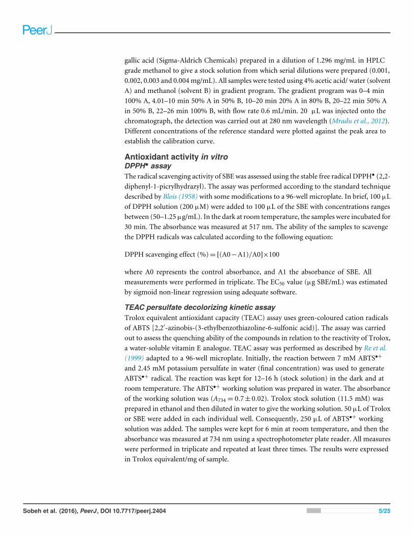
Figure 1 Negative LC/ESI/mass spectrum of phenolics from hydro-alcoholic extract of Schotia brachy-petalea.
for myrecitin-3-O-α-L-1C4-rhamnoside. Consequently, compound 1 was confirmed to bemyrecitin-3-O-α-L-1C4-rhamnoside (Hayder et al., 2008).
Compound 2 (39 mg) was obtained as yellow crystalline powder. On PC, it showed adark purple spot under short UV light. Rf values: 22.5 (BAW) and 7.5 (6% AcOH). It gavea dirty green colour with the FeCl3spray reagent. Also, its UV spectrum showed two bandsat λmaxMeOH (350 nm band I and 206 nm band II) which indicated the presence of aflavone nucleus. It showed a bathochromic shift (30 nm) on addition of sodiummethoxideand (20 nm) in band II with sodium acetate indicating that the 3′, 4′′ and 7 OH positionsare free. From these data we conclude that compound 2 corresponds to quercetin-3-O-α-L-1C4-rhamnoside.
The 1H-NMR spectrum of compound 2 indicated the absence of the signal for H-3,the presence of aromatic proton signals at δ= 7.199 (1H, d , J = 2.5 Hz, H-2′), δ= 6.909(1H, dd, J = 2.5Hz, 8 Hz, H-6′), δ= 6.882 (1H, d , J = 8Hz, H-5′), presence ofO-glycosidicanomeric signal at δ= 5.214 ppm (1H, S, H-1′′) and a signal for methyl of rhamnose atδ = 1.242 ppm (3H, s, CH3rhamnose). UV as well as 1H-NMR chemical shifts werefound to be similar to those previously reported for quercetin-3-O-α-L-1C4-rhamnoside.Consequently, compound 2 was identified as quercetin-3-O-α-L-1C4-rhamnoside (Ma etal., 2005).
Identification of constituents by LC/HRESI/MS/MSHPLC-MS plays an important role in the separation and identification of complex plantmixtures. Among its main advantages is the high sensitivity and specificity which can beused both for volatile and non-volatile compounds (Dumont & Beal, 2011).
A total of 43 secondary metabolites were identified from SBE, its fractions and sub-Q5Q6 fractions using LC/ESI/MS/MS (Table 1). LC/HRESI/MS/MS profiles of SBE, its fractions
and sub-fractions are shown in Figs. 1–5. Different classes of phenolics were discovered,which will be discussed in the following:
Q7
Sobeh et al. (2016), PeerJ, DOI 10.7717/peerj.2404 8/25
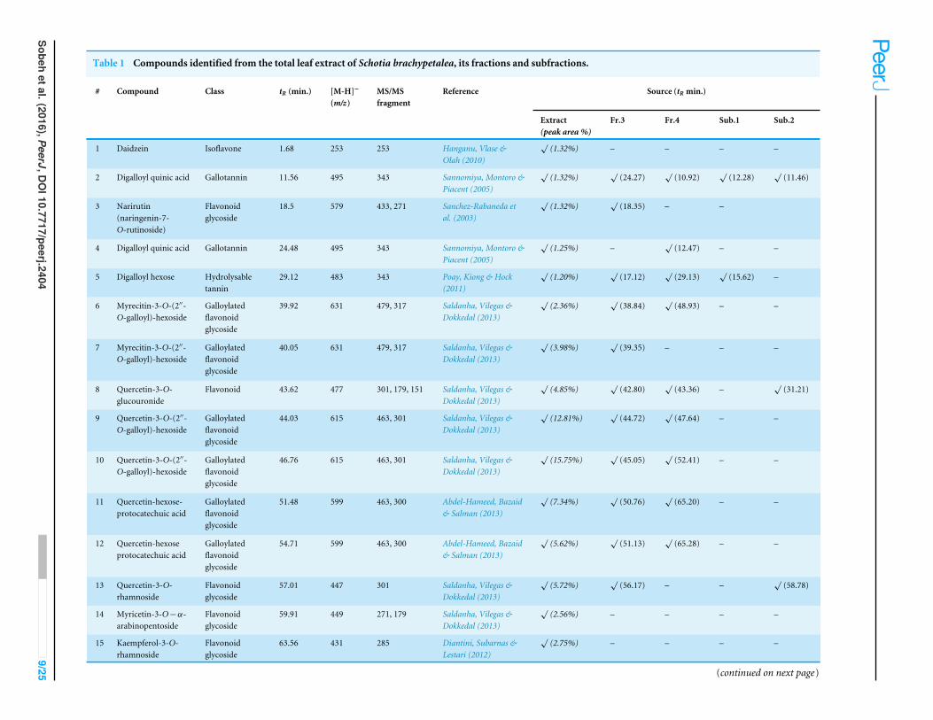
Table 1 Compounds identified from the total leaf extract of Schotia brachypetalea, its fractions and subfractions.
# Compound Class tR (min.) [M-H]−
(m/z)MS/MSfragment
Reference Source (tR min.)
Extract(peak area %)
Fr.3 Fr.4 Sub.1 Sub.2
1 Daidzein Isoflavone 1.68 253 253 Hanganu, Vlase &Olah (2010)
√(1.32%) – – – –
2 Digalloyl quinic acid Gallotannin 11.56 495 343 Sannomiya, Montoro &Piacent (2005)
√(1.32%)
√(24.27)
√(10.92)
√(12.28)
√(11.46)
3 Narirutin(naringenin-7-O-rutinoside)
Flavonoidglycoside
18.5 579 433, 271 Sanchez-Rabaneda etal. (2003)
√(1.32%)
√(18.35) – –
4 Digalloyl quinic acid Gallotannin 24.48 495 343 Sannomiya, Montoro &Piacent (2005)
√(1.25%) –
√(12.47) – –
5 Digalloyl hexose Hydrolysabletannin
29.12 483 343 Poay, Kiong & Hock(2011)
√(1.20%)
√(17.12)
√(29.13)
√(15.62) –
6 Myrecitin-3-O-(2′′-O-galloyl)-hexoside
Galloylatedflavonoidglycoside
39.92 631 479, 317 Saldanha, Vilegas &Dokkedal (2013)
√(2.36%)
√(38.84)
√(48.93) – –
7 Myrecitin-3-O-(2′′-O-galloyl)-hexoside
Galloylatedflavonoidglycoside
40.05 631 479, 317 Saldanha, Vilegas &Dokkedal (2013)
√(3.98%)
√(39.35) – – –
8 Quercetin-3-O-glucouronide
Flavonoid 43.62 477 301, 179, 151 Saldanha, Vilegas &Dokkedal (2013)
√(4.85%)
√(42.80)
√(43.36) –
√(31.21)
9 Quercetin-3-O-(2′′-O-galloyl)-hexoside
Galloylatedflavonoidglycoside
44.03 615 463, 301 Saldanha, Vilegas &Dokkedal (2013)
√(12.81%)
√(44.72)
√(47.64) – –
10 Quercetin-3-O-(2′′-O-galloyl)-hexoside
Galloylatedflavonoidglycoside
46.76 615 463, 301 Saldanha, Vilegas &Dokkedal (2013)
√(15.75%)
√(45.05)
√(52.41) – –
11 Quercetin-hexose-protocatechuic acid
Galloylatedflavonoidglycoside
51.48 599 463, 300 Abdel-Hameed, Bazaid& Salman (2013)
√(7.34%)
√(50.76)
√(65.20) – –
12 Quercetin-hexoseprotocatechuic acid
Galloylatedflavonoidglycoside
54.71 599 463, 300 Abdel-Hameed, Bazaid& Salman (2013)
√(5.62%)
√(51.13)
√(65.28) – –
13 Quercetin-3-O-rhamnoside
Flavonoidglycoside
57.01 447 301 Saldanha, Vilegas &Dokkedal (2013)
√(5.72%)
√(56.17) – –
√(58.78)
14 Myricetin-3-O−α-arabinopentoside
Flavonoidglycoside
59.91 449 271, 179 Saldanha, Vilegas &Dokkedal (2013)
√(2.56%) – – – –
15 Kaempferol-3-O-rhamnoside
Flavonoidglycoside
63.56 431 285 Diantini, Subarnas &Lestari (2012)
√(2.75%) – – – –
(continued on next page)
Sobehetal.(2016),PeerJ,D
OI10.7717/peerj.2404
9/25
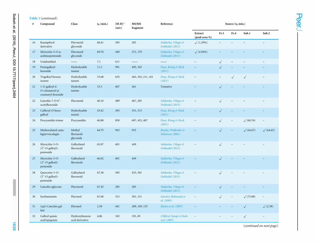
Table 1 (continued)# Compound Class tR (min.) [M-H]−
(m/z)MS/MSfragment
Reference Source (tR min.)
Extract(peak area %)
Fr.3 Fr.4 Sub.1 Sub.2
16 Kaempferolderivative
Flavonoidglycoside
68.61 583 285 Saldanha, Vilegas &Dokkedal (2013)
√(1.29%) – – – –
17 Myricetin-3-O-α-arabinopentoside
Flavonoidglycoside
69.70 449 271, 179 Saldanha, Vilegas &Dokkedal (2013)
√(4.94%) – – – –
18 Unidentified —— 7.1 611 —— —— –√
– – –
19 Pentagalloyl-hexoside
Hydrolysabletannin
11.2 991 495, 343 Poay, Kiong & Hock(2011)
–√
– – –
20 Trigalloyl hexoseisomer
Hydrolysabletannin
33.68 635 463, 343, 211, 161 Poay, Kiong & Hock(2011)
– –√ √
–
21 1-O-galloyl-6-O-cinnamoyl-p-coumaryl-hexoside
Hydrolysabletannin
33.3 607 461 Tentative –√
– – –
22 Luteolin-7-O-6′′-acetylhexoside
Flavonoid 40.10 489 467, 285 Saldanha, Vilegas &Dokkedal (2013)
–√
– – –
23 Caffeoyl-O-hexo-galloyl
Hydrolysabletannin
43.62 493 331, 313 Poay, Kiong & Hock(2011)
–√
– – –
24 Procyanidin trimer Procyanidin 60.88 850 697, 425, 407 Poay, Kiong & Hock(2011)
–√
–√
(60.76) –
25 Methoxylated casta-lagin/vescalagin
Methylflavonoidglycoside
64.75 963 933 Rauha, Wolfender &Salminen (2001)
–√
–√
(64.67)√
(64.65)
26 Myrecitin-3-O-(2′′-O-galloyl)-pentoside
Galloylatedflavonoid
65.07 601 449 Saldanha, Vilegas &Dokkedal (2013)
–√
– – –
27 Myrecitin-3-O-(2′′-O-galloyl)-pentoside
Galloylatedflavonoid
66.02 601 449 Saldanha, Vilegas &Dokkedal (2013)
–√
– – –
28 Quercetin-3-O-(2′′-O-galloyl)-pentoside
Galloylatedflavonoid
67.38 585 433, 301 Saldanha, Vilegas &Dokkedal (2013)
–√
– – –
29 Luteolin aglycone Flavonoid 67.45 285 285 Saldanha, Vilegas &Dokkedal (2013)
–√
– – –
30 Isorhamnetin Flavonol 67.68 315 301, 151 Sanchez-Rabaneda etal. (2003)
–√
–√
(75.88) –
31 (epi) Catechin gal-late
Flavanol 2.58 441 289, 169, 135 Bastos et al. (2007) – – –√ √
(2.58)
32 Galloyl quinicacid/epiquinic
Hydroxybenzoicacid derivative
4.86 343 191, 85 Clifford, Stoupi & Kuh-nert (2007)
– – –√
–
(continued on next page)
Sobehetal.(2016),PeerJ,D
OI10.7717/peerj.2404
10/25
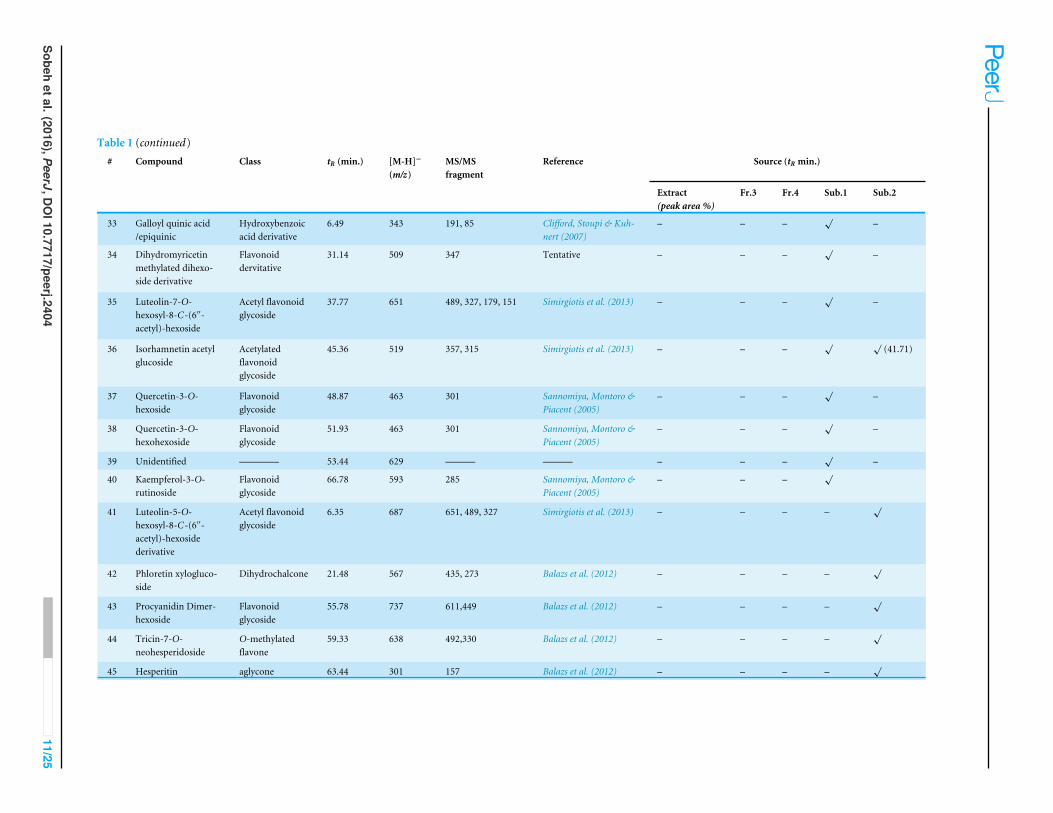
Table 1 (continued)# Compound Class tR (min.) [M-H]−
(m/z)MS/MSfragment
Reference Source (tR min.)
Extract(peak area %)
Fr.3 Fr.4 Sub.1 Sub.2
33 Galloyl quinic acid/epiquinic
Hydroxybenzoicacid derivative
6.49 343 191, 85 Clifford, Stoupi & Kuh-nert (2007)
– – –√
–
34 Dihydromyricetinmethylated dihexo-side derivative
Flavonoiddervitative
31.14 509 347 Tentative – – –√
–
35 Luteolin-7-O-hexosyl-8-C-(6′′-acetyl)-hexoside
Acetyl flavonoidglycoside
37.77 651 489, 327, 179, 151 Simirgiotis et al. (2013) – – –√
–
36 Isorhamnetin acetylglucoside
Acetylatedflavonoidglycoside
45.36 519 357, 315 Simirgiotis et al. (2013) – – –√ √
(41.71)
37 Quercetin-3-O-hexoside
Flavonoidglycoside
48.87 463 301 Sannomiya, Montoro &Piacent (2005)
– – –√
–
38 Quercetin-3-O-hexohexoside
Flavonoidglycoside
51.93 463 301 Sannomiya, Montoro &Piacent (2005)
– – –√
–
39 Unidentified ———— 53.44 629 ——— ——— – – –√
–
40 Kaempferol-3-O-rutinoside
Flavonoidglycoside
66.78 593 285 Sannomiya, Montoro &Piacent (2005)
– – –√
41 Luteolin-5-O-hexosyl-8-C-(6′′-acetyl)-hexosidederivative
Acetyl flavonoidglycoside
6.35 687 651, 489, 327 Simirgiotis et al. (2013) – – – –√
42 Phloretin xylogluco-side
Dihydrochalcone 21.48 567 435, 273 Balazs et al. (2012) – – – –√
43 Procyanidin Dimer-hexoside
Flavonoidglycoside
55.78 737 611,449 Balazs et al. (2012) – – – –√
44 Tricin-7-O-neohesperidoside
O-methylatedflavone
59.33 638 492,330 Balazs et al. (2012) – – – –√
45 Hesperitin aglycone 63.44 301 157 Balazs et al. (2012) – – – –√
Sobehetal.(2016),PeerJ,D
OI10.7717/peerj.2404
11/25
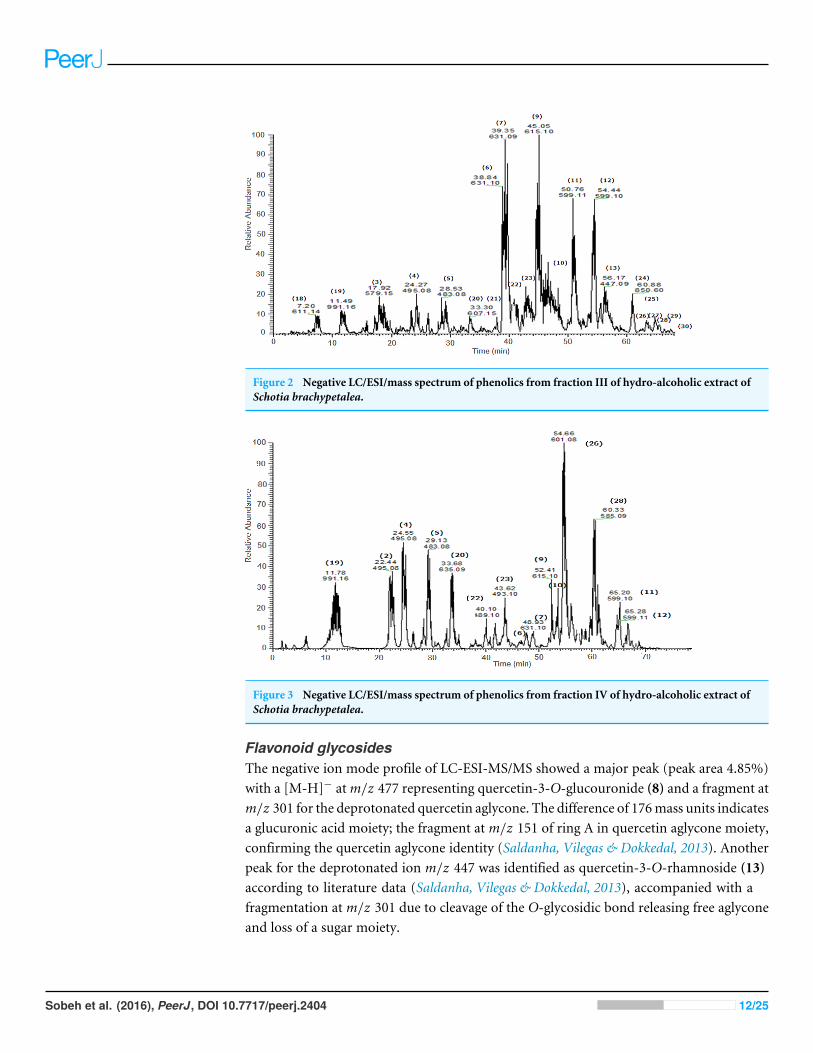
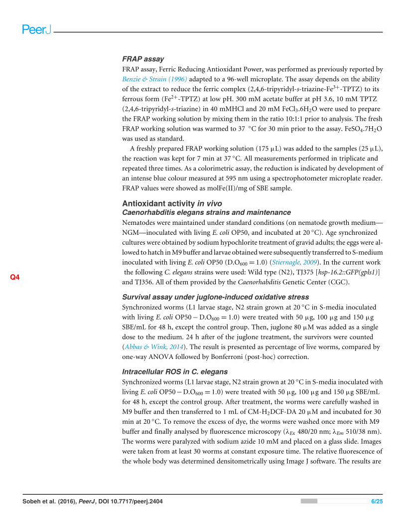
Figure 2 Negative LC/ESI/mass spectrum of phenolics from fraction III of hydro-alcoholic extract ofSchotia brachypetalea.
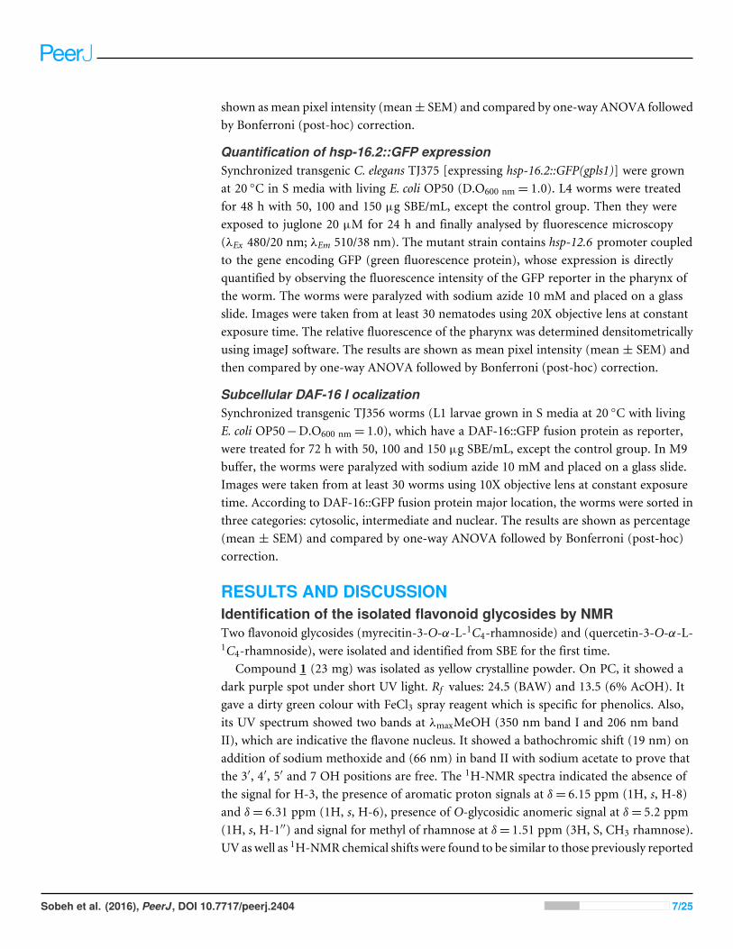
Figure 3 Negative LC/ESI/mass spectrum of phenolics from fraction IV of hydro-alcoholic extract ofSchotia brachypetalea.
Flavonoid glycosidesThe negative ion mode profile of LC-ESI-MS/MS showed a major peak (peak area 4.85%)with a [M-H]− atm/z 477 representing quercetin-3-O-glucouronide (8) and a fragment atm/z 301 for the deprotonated quercetin aglycone. The difference of 176mass units indicatesa glucuronic acid moiety; the fragment at m/z 151 of ring A in quercetin aglycone moiety,confirming the quercetin aglycone identity (Saldanha, Vilegas & Dokkedal, 2013). Anotherpeak for the deprotonated ion m/z 447 was identified as quercetin-3-O-rhamnoside (13)according to literature data (Saldanha, Vilegas & Dokkedal, 2013), accompanied with afragmentation at m/z 301 due to cleavage of the O-glycosidic bond releasing free aglyconeand loss of a sugar moiety.
Sobeh et al. (2016), PeerJ, DOI 10.7717/peerj.2404 12/25
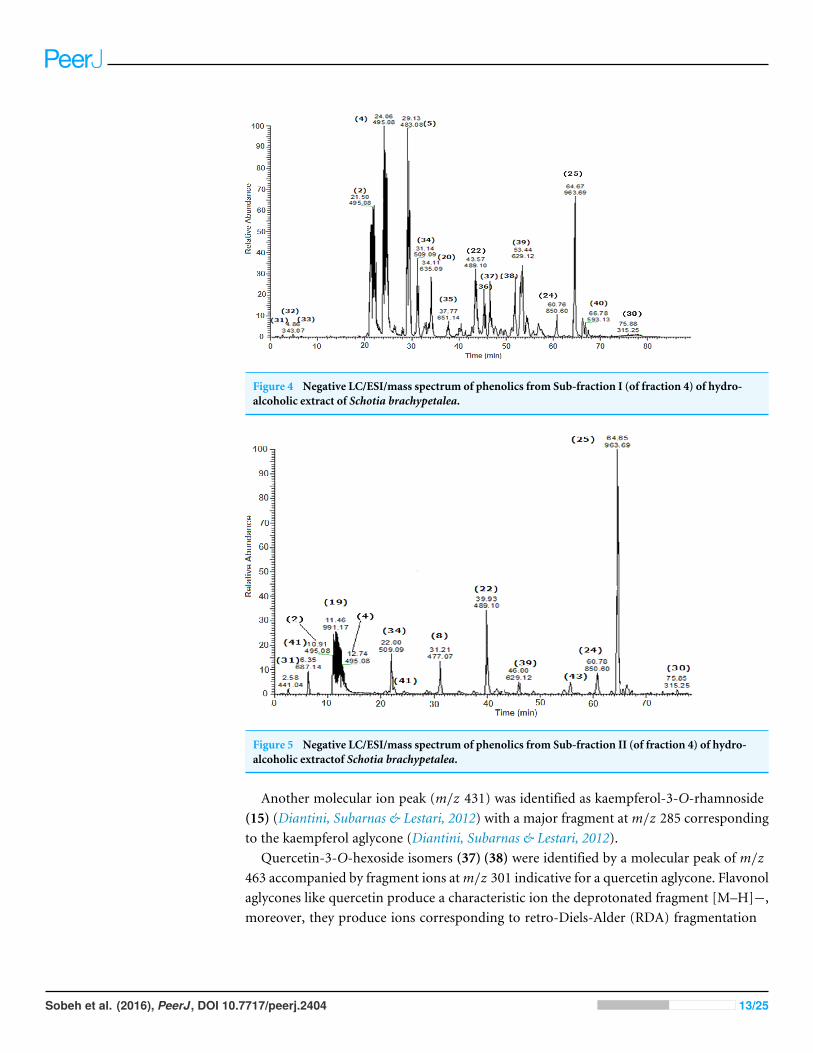
Figure 4 Negative LC/ESI/mass spectrum of phenolics from Sub-fraction I (of fraction 4) of hydro-alcoholic extract of Schotia brachypetalea.
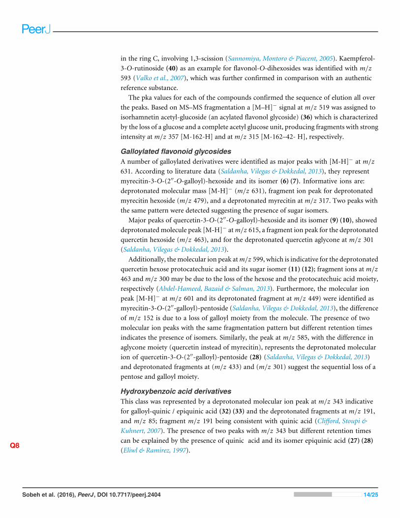
Figure 5 Negative LC/ESI/mass spectrum of phenolics from Sub-fraction II (of fraction 4) of hydro-alcoholic extractof Schotia brachypetalea.
Another molecular ion peak (m/z 431) was identified as kaempferol-3-O-rhamnoside(15) (Diantini, Subarnas & Lestari, 2012) with a major fragment atm/z 285 correspondingto the kaempferol aglycone (Diantini, Subarnas & Lestari, 2012).
Quercetin-3-O-hexoside isomers (37) (38) were identified by a molecular peak of m/z463 accompanied by fragment ions atm/z 301 indicative for a quercetin aglycone. Flavonolaglycones like quercetin produce a characteristic ion the deprotonated fragment [M–H]−,moreover, they produce ions corresponding to retro-Diels-Alder (RDA) fragmentation
Sobeh et al. (2016), PeerJ, DOI 10.7717/peerj.2404 13/25
in the ring C, involving 1,3-scission (Sannomiya, Montoro & Piacent, 2005). Kaempferol-3-O-rutinoside (40) as an example for flavonol-O-dihexosides was identified with m/z593 (Valko et al., 2007), which was further confirmed in comparison with an authenticreference substance.
The pka values for each of the compounds confirmed the sequence of elution all overthe peaks. Based on MS–MS fragmentation a [M–H]− signal at m/z 519 was assigned toisorhamnetin acetyl-glucoside (an acylated flavonol glycoside) (36) which is characterizedby the loss of a glucose and a complete acetyl glucose unit, producing fragments with strongintensity at m/z 357 [M-162-H] and at m/z 315 [M-162–42- H], respectively.
Galloylated flavonoid glycosidesA number of galloylated derivatives were identified as major peaks with [M-H]− at m/z631. According to literature data (Saldanha, Vilegas & Dokkedal, 2013), they representmyrecitin-3-O-(2′′-O-galloyl)-hexoside and its isomer (6) (7). Informative ions are:deprotonated molecular mass [M-H]− (m/z 631), fragment ion peak for deprotonatedmyrecitin hexoside (m/z 479), and a deprotonated myrecitin at m/z 317. Two peaks withthe same pattern were detected suggesting the presence of sugar isomers.
Major peaks of quercetin-3-O-(2′′-O-galloyl)-hexoside and its isomer (9) (10), showeddeprotonatedmolecule peak [M-H]− atm/z 615, a fragment ion peak for the deprotonatedquercetin hexoside (m/z 463), and for the deprotonated quercetin aglycone at m/z 301(Saldanha, Vilegas & Dokkedal, 2013).
Additionally, themolecular ion peak atm/z 599, which is indicative for the deprotonatedquercetin hexose protocatechuic acid and its sugar isomer (11) (12); fragment ions at m/z463 and m/z 300 may be due to the loss of the hexose and the protocatechuic acid moiety,respectively (Abdel-Hameed, Bazaid & Salman, 2013). Furthermore, the molecular ionpeak [M-H]− at m/z 601 and its deprotonated fragment at m/z 449) were identified asmyrecitin-3-O-(2′′-galloyl)-pentoside (Saldanha, Vilegas & Dokkedal, 2013), the differenceof m/z 152 is due to a loss of galloyl moiety from the molecule. The presence of twomolecular ion peaks with the same fragmentation pattern but different retention timesindicates the presence of isomers. Similarly, the peak at m/z 585, with the difference inaglycone moiety (quercetin instead of myrecitin), represents the deprotonated molecularion of quercetin-3-O-(2′′-galloyl)-pentoside (28) (Saldanha, Vilegas & Dokkedal, 2013)and deprotonated fragments at (m/z 433) and (m/z 301) suggest the sequential loss of apentose and galloyl moiety.
Hydroxybenzoic acid derivativesThis class was represented by a deprotonated molecular ion peak at m/z 343 indicativefor galloyl-quinic / epiquinic acid (32) (33) and the deprotonated fragments at m/z 191,and m/z 85; fragment m/z 191 being consistent with quinic acid (Clifford, Stoupi &Kuhnert, 2007). The presence of two peaks with m/z 343 but different retention timescan be explained by the presence of quinic acid and its isomer epiquinic acid (27) (28)
Q8(Eliwl & Ramirez, 1997).
Sobeh et al. (2016), PeerJ, DOI 10.7717/peerj.2404 14/25
IsoflavonesA minor peak of daidzein aglycone (1) was recognized as a deprotonated peak at m/z 253.
DihydrochalconesA hexoside derivative of phloretin, a characteristic and quite common aglycone previouslyreported in apple, was identified in SBE as phloretin-3-O-xyloglucoside (42) withm/z 567 and a major ion peak at m/z 273 corresponding to the aglycone of phloretin(Balazs et al., 2012).
ProcyanidinsA procyanidin dimer-hexoside (43) was identified and recognized at m/z 737 withfragmentation pattern as follows: A product ion ofm/z 611 containing the galactoside wasformed by the loss of gallic acid (126 Da). However, the second product ion with m/z 449was detected in the spectrum indicates the loss of both the gallic acid and the sugar moiety(Sies & Stahl, 1995). A procyanidin trimer (24)was identified according to its deprotonatedbase peak at m/z 850 and its deprotonated fragments at m/z 697, 425 and 407, which areproduced by a cleavage of the inter-flavan bond through a quinine-methide (QM) cleavage(Passos, Cardoso & Domingues, 2007) to give (m/z 425) then a loss of water moleculeto yield m/z 407 in agreement with a procyanidin trimer MS fragmentation pathway(Passos, Cardoso & Domingues, 2007).
Hydrolysable tanninsFor tri-galloyl hexose isomer (20) a [M-H]− was identified withm/z 635. The contributionof the major peak (m/z 483) is due to the presence of a digalloyl-hexose moiety. Besides,two intermediate ions were detected atm/z 271 andm/z 211. They are indicative formono-and di-galloyl-hexose; the elimination of a hexose moiety from mono galloyl-hexose wasdetected which subsequently lead to the formation of the deprotonated gallic acid at m/z169 (Poay, Kiong & Hock, 2011).
Represented by a deprotonated parent ion peak atm/z 495 for di-galloyl quinic acid (2)(4), different positional isomers arise from the difference in hydroxyl attachment site givingrise to peaks of same m/z value. The identification was done according to the identity ofthe obtained peaks as follows: a [M–H]− at m/z 343 indicates the loss of a galloyl moietyfrom the parent peak and fragmentation showed fragments at m/z 191 and m/z 169,corresponding to quinic acid and gallic acid moieties, respectively (Sannomiya, Montoro &Piacent, 2005). Compound (5) with m/z 483, identified as digalloyl hexose, showed an ionpeak typical for the dimer analogue of m/z 169 produced by gallic acid.
Methyl and acetyl flavonoid glycosidesA peak at m/z 963 is typical for deprotonated methoxylated castalagin/vescalagin (25)showing a major peak atm/z 933, corresponding to the polyphenol castalagin or its isomervescalagin (Rauha, Wolfender & Salminen, 2001).
Two acetyl flavonoid glycosides were detected luteolin-7-O-hexosyl-8-C-(6′′-acetyl)—hexoside (35) with m/z 651. The detected fragments at m/z 179, 151 provide the evidencethat luteolin was the aglycone of compound (35) (Simirgiotis et al., 2013). Compound (41)
Sobeh et al. (2016), PeerJ, DOI 10.7717/peerj.2404 15/25
with a [M-H]− ion at m/z 687 showed fragments at m/z 651, 489, 327. These ions matchwith the MS data previously reported for compound (41) [luteolin-5-O-hexosyl-8-C-(6′′-acetyl)-hexoside derivative], full MS at (m/z 651) after the loss of 38 amu and thus wastentatively assigned to its analogue luteolin-7-O-hexosyl-8-C-(6′′-acetyl)-hexoside (35)(Masika, Sultana & Afolayan, 2004).
Methyl flavone, flavanol and flavonolA methyl-flavone was identified as tricin-7-O-neohesperidoside (44) from its exact mass(m/z 638) [M-H]−; by taking into consideration the additional mass of 30 for the extramethoxy group on the [M-H]− ion. The major fragments of (38) were atm/z 492 and 330corresponding, respectively, to ions [M-H-146]− and [M-H-146-162]. The losses of 146and 162 Da are characteristic for rhamnose and glucose moieties, respectively, and the ionat m/z 329 is characteristic of the aglycone tricin (Paiva et al., 2010).
A flavanol was represented by a deprotonated parent peak for (epi) catechin gallate atm/z 441(31) and its deprotonated fragments at m/z 289, 169 and 135 (Bastos et al., 2007).
Q9The fragment at m/z 289 for the deprotonated (epi) catechin (Ivanova et al., 2011), m/z169 for the galloyl moiety, and m/z 135 for ring (A) of flavones nucleus. As an example ofthe flavonol isorhamnetin (30), a deprotonated molecular ion peak was detected at m/z315 with deprotonated fragments at (m/z 301, m/z 151) (Sanchez-Rabaneda et al., 2003).
Standardization of SBE using HPLCThe SBE showed an intense peak at Rt 3.983 min corresponding to gallic acid (identifiedby peak matching with a gallic acid standard). Through the standardization experiment,it was shown that each mg SBE constitutes 0.0022 mg gallic acid. The calibration curveshowed good linearity for gallic acid (reference compound) in the range of 0.3 up to 1mg/mL with correlation coefficient (R2) 0.999.
Antioxidant activities in vitro and in vivo:Antioxidant activity in vitroTotal phenolic contents of SBE were 376 mg of caffeic acid equivalents (CAE)/g SBE whilethe total flavonoid content was 67.87 mg (quercetin equivalents)/g SBE. The antioxidantactivity of SBE was evaluated in vitro using three different assays, DPPH, ABTS andFRAP. These methods are widely employed for the antioxidant activity evaluation of purecompounds, plant extracts, as well as food items because long-lived radicals such as DPPH•
and ABTS•+ as well as FeSO4 are sensitive and reliable (Prior, Wu & Schaich, 2005). Allmethods revealed a strong antioxidant capacity of SBE (Table 2).
Antioxidant activity in vivo in C. elegansSurvival assayJuglone (5-hydroxy-1,4-naphthoquinone) is a natural quinine from Juglans regiawith toxicpro-oxidant activity (Saling et al., 2011). Exposure of C. elegans to a high concentrationof juglone kills the worms; however, antioxidant compounds can prevent such an effect.According to our results (Fig. 6), worms pre-treated with SBE showed an increased survivalrate (up to 41%), when compared with the control group (11%), which was treated
Sobeh et al. (2016), PeerJ, DOI 10.7717/peerj.2404 16/25
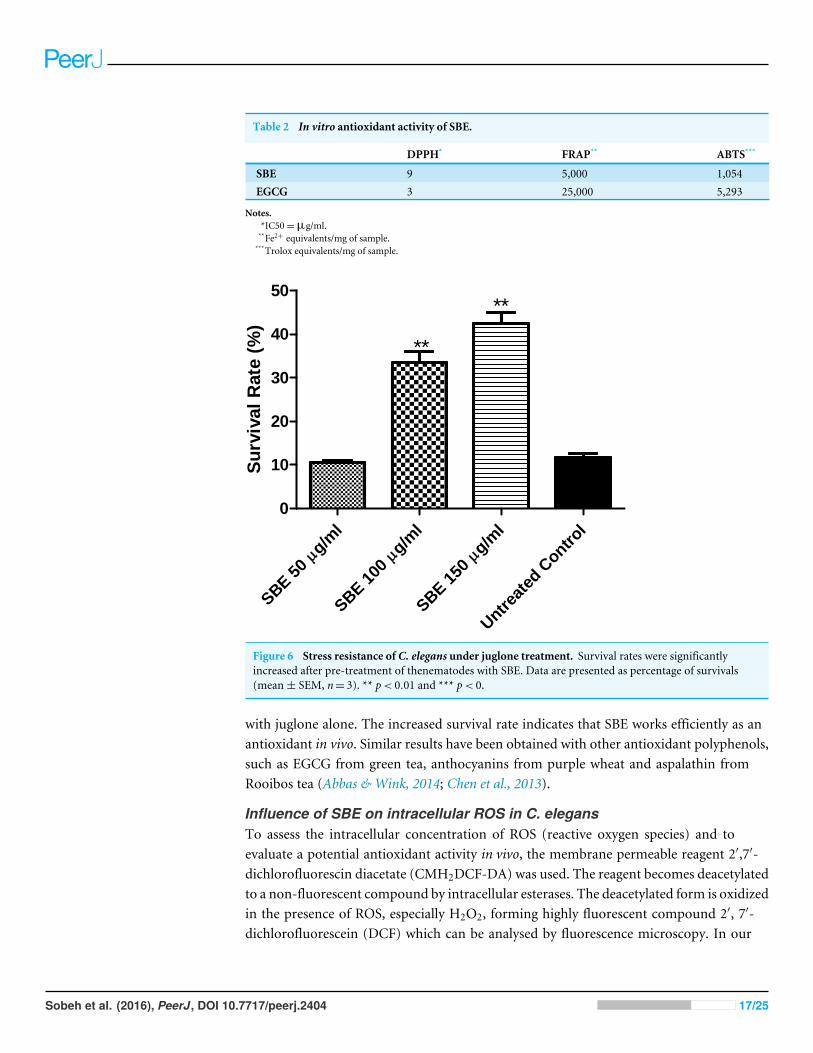
Table 2 In vitro antioxidant activity of SBE.
DPPH* FRAP** ABTS***
SBE 9 5,000 1,054EGCG 3 25,000 5,293
Notes.*IC50=µg/ml.**Fe2+ equivalents/mg of sample.***Trolox equivalents/mg of sample.
g/ml
SBE50
g/ml
SBE
100
g/ml
SBE
150
Untr
eate
dContr
ol0
10
20
30
40
50
**
**
Su
rviv
al
Rate
(%)
Figure 6 Stress resistance of C. elegans under juglone treatment. Survival rates were significantlyincreased after pre-treatment of thenematodes with SBE. Data are presented as percentage of survivals(mean± SEM, n= 3). ** p< 0.01 and *** p< 0.
with juglone alone. The increased survival rate indicates that SBE works efficiently as anantioxidant in vivo. Similar results have been obtained with other antioxidant polyphenols,such as EGCG from green tea, anthocyanins from purple wheat and aspalathin fromRooibos tea (Abbas & Wink, 2014; Chen et al., 2013).
Influence of SBE on intracellular ROS in C. elegansTo assess the intracellular concentration of ROS (reactive oxygen species) and toevaluate a potential antioxidant activity in vivo, the membrane permeable reagent 2′,7′-dichlorofluorescin diacetate (CMH2DCF-DA) was used. The reagent becomes deacetylatedto a non-fluorescent compound by intracellular esterases. The deacetylated form is oxidizedin the presence of ROS, especially H2O2, forming highly fluorescent compound 2′, 7′-dichlorofluorescein (DCF) which can be analysed by fluorescence microscopy. In our
Sobeh et al. (2016), PeerJ, DOI 10.7717/peerj.2404 17/25
g/ml
SBE50
g/ml
SBE10
0g/m
l
SBE15
0
Untrea
ted
Contr
ol0
500
1000
1500
***
***
***Pix
el
Inte
nsit
y(a
rbit
rary
un
its)
B C
D E
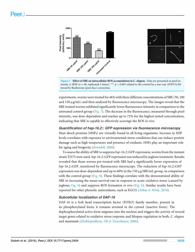
Figure 7 Effect of SBE on intracellular ROS accumulation in C. elegans. Data are presented as pixel in-tensity± SEM (n= 40, replicated 3 times). *** p< 0.001 related to the control by a one-way ANOVA fol-lowed by Bonferroni (post-hoc) correction.
experiments, wormswere treated for 48 hwith three different concentrations of SBE (50, 100and 150 µg/mL) and then analysed by fluorescence microscopy. The images reveal that theSBE treated worms exhibited significantly lower fluorescence intensity in comparison to theuntreated control group (Fig. 7). The decrease in the fluorescence, measured through pixelintensity, was dose-dependent and reaches up to 72% for the highest tested concentration,indicating that SBE is capable to effectively scavenge the ROS in vivo.
Quantification of hsp-16.2:: GFP expression via fluorescence microscopyHeat shock proteins (HSPs) are virtually found in all living organisms. Increase in HSPlevels correlates with exposure to environmental stress conditions that can induce proteindamage such as high temperature and presence of oxidants. HSPs play an important rolefor aging and longevity (Swindell, 2009).
To assess the ability of SBE to suppress hsp-16.2::GFP expression, worms from themutantstrain TJ375 were used. hsp-16.2::GFP expressionwas induced by juglone treatment. Resultsrevealed that those worms pre-treated with SBE had a significantly lower expression ofhsp-16.2::GFP, monitored by fluorescence microscopy. The reduction of hsp-16.2::GFPexpression was dose-dependent and up to 60% in the 150 µg SBE/mL group, in comparisonwith the control group (Fig. 8). These findings correlate with the demonstrated ability ofSBE in increasing the mean survival rate in response to acute oxidative stress (caused byjuglone; Fig. 6) and suppress ROS formation in vivo (Fig. 8). Similar results have beenreported for other phenolic antioxidants, such as EGCG (Abbas & Wink, 2014).
Subcellular localization of DAF-16DAF-16 is a fork head transcription factor (FOXO) family member, present inits phosphorylated form; it remains arrested in the cytosol (inactive form). Thedephosphorylated active form migrates into the nucleus and triggers the activity of severaltarget genes related to oxidative stress response and lifespan regulation in both, C. elegansand mammals (Mukhopadhyay, Oh & Tissenbaum, 2006).
Sobeh et al. (2016), PeerJ, DOI 10.7717/peerj.2404 18/25
g/ml
SBE50
g/ml
SBE10
0g/m
l
SBE15
0
Untr
eate
dContr
ol0
20000
40000
60000
*
***
***
Pix
el
Inte
nsit
y(a
rbit
rary
un
its)
C
ED
B
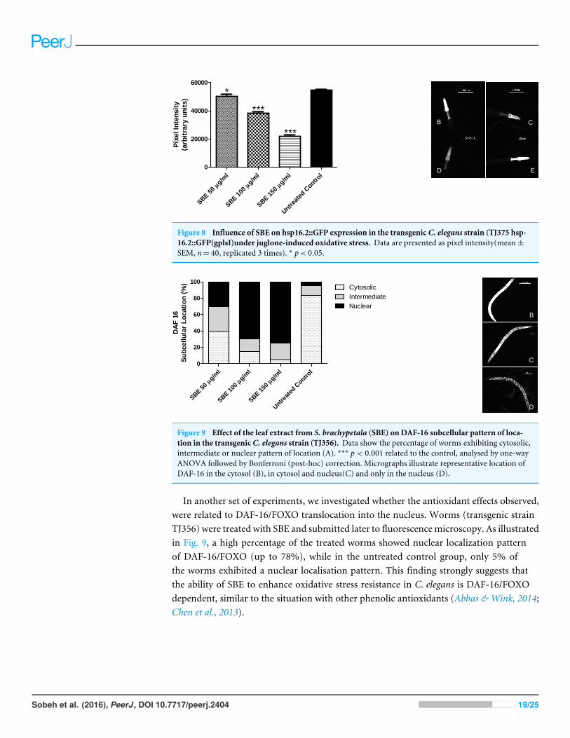
Figure 8 Influence of SBE on hsp16.2::GFP expression in the transgenic C. elegans strain (TJ375 hsp-16.2::GFP(gplsI)under juglone-induced oxidative stress. Data are presented as pixel intensity(mean±SEM, n= 40, replicated 3 times). * p< 0.05.
g/ml
SBE
50g/m
l
SBE
100
g/ml
SBE
150
Untr
eate
dContr
ol0
20
40
60
80
100Cytosolic
Intermediate
Nuclear
DA
F16
Su
bcell
ula
rL
ocati
on
(%)
B
C
D
Figure 9 Effect of the leaf extract from S. brachypetala (SBE) on DAF-16 subcellular pattern of loca-tion in the transgenic C. elegans strain (TJ356). Data show the percentage of worms exhibiting cytosolic,intermediate or nuclear pattern of location (A). *** p< 0.001 related to the control, analysed by one-wayANOVA followed by Bonferroni (post-hoc) correction. Micrographs illustrate representative location ofDAF-16 in the cytosol (B), in cytosol and nucleus(C) and only in the nucleus (D).
In another set of experiments, we investigated whether the antioxidant effects observed,were related to DAF-16/FOXO translocation into the nucleus. Worms (transgenic strainTJ356) were treated with SBE and submitted later to fluorescencemicroscopy. As illustratedin Fig. 9, a high percentage of the treated worms showed nuclear localization patternof DAF-16/FOXO (up to 78%), while in the untreated control group, only 5% ofthe worms exhibited a nuclear localisation pattern. This finding strongly suggests thatthe ability of SBE to enhance oxidative stress resistance in C. elegans is DAF-16/FOXOdependent, similar to the situation with other phenolic antioxidants (Abbas & Wink, 2014;Chen et al., 2013).
Sobeh et al. (2016), PeerJ, DOI 10.7717/peerj.2404 19/25
CONCLUSIONSThe current study resulted in the identification of different phenolic metabolite classesincluding flavonoid glycosides, procyanidins, anthocyanins, dihydrochalcones, andhydroxy benzoic acid derivatives. Myricetin-3-O-α-L-1C4-rhamnoside, quercetin-3-O-L-1C4-rhamnoside, and gallic acid were isolated and identified for the first time from theleaves of S. brachypetala.
SBE is rich in phenolics, especially flavonoid glycosides such as quercetin which areknown as powerful antioxidants in vitro (Boukatib, Atmani & Rolando, 2002). Potentialhealth effects of polyphenols have been discussed: several studies reported the abilityof quercetin to ameliorate pathological conditions linked to ROS such as oxidationof LDL-cholesterol, to counteract cardiovascular risks (Chopra et al., 2000), to protectprimary neurons against Aβ deposits (Ansari et al., 2009). Furthermore, antioxidants arebeneficial for chronic inflammation (Comalada et al., 2005; Shoskes et al., 1999) and canavoid Ca2+-dependent cell death (Sakanashi et al., 2008)
Our study showed that SBE exhibits a strong antioxidant activity in vitro as well as in vivo.It is able to decrease ROS production and attenuates hsp16.2 expression under oxidativestress conditions in C. elegans. We assume that a modulation of the DAF-16/FOXOtranscription factor by the phenolics is responsible for the observed antioxidant effects.The leaf extract can increase the nuclear location of DAF-16, thereby activating manyimportant biological processes including target genes related to stress resistance andlongevity.
Further in vivo experiments are needed to develop the polyphenols of S. brachypetalainto a useful nutraceutical compounds.
ADDITIONAL INFORMATION AND DECLARATIONS
FundingThe authors received no funding for this work.
Competing InterestsMichael Wink is an Academic Editor for PeerJ.
Author Contributions• Mansour Sobeh and Herbenya Peixoto performed the experiments, wrote the paper.• Esraa ElHawary performed the experiments, wrote the paper, reviewed drafts of thepaper.• Rola M. Labib wrote the paper.• Heba Handoussa and Noha Swilam analyzed the data, wrote the paper, reviewed draftsof the paper.• Ahmed A. El-Khatib, Farukh Sharapov, Tamer Mohamed and Sonja Krstin performedthe experiments, prepared figures and/or tables.• Michael W. Linscheid and Abdel Nasser Singab contributed reagents/materials/analysistools.
Sobeh et al. (2016), PeerJ, DOI 10.7717/peerj.2404 20/25
All supplementalinformationwill be madeavailable fordownload exactlyas they weresupplied.This link to theSIwill only workwhen the articleis published.
• Michael Wink and Nahla Ayoub conceived and designed the experiments, contributedreagents/materials/analysis tools, wrote the paper, reviewed drafts of the paper.
Data AvailabilityThe following information was supplied regarding data availability:
The raw data has been supplied as a Supplemental Information.
Supplemental InformationSupplemental information for this article can be found online at http://dx.doi.org/10.7717/peerj.2404#supplemental-information.
REFERENCESAbbas S, WinkM. 2014. Green tea extract induces the resistance of Caenorhabditis
elegans against oxidative stress. Antioxidants 3:129–143 DOI 10.3390/antiox3010129.Abdel-Hameed SS, Bazaid SA, SalmanMS. 2013. Characterization of the phytochemical
constituents of Taif rose and its antioxidant and anticancer activities. BioMedResearch International 345–465.
Q10Angerhofer CK, Maes D, Giacomoni P. 2008. The use of natural compounds and
botanicals in the development of anti-aging skin care products Skin Aging Handbook: anintegrated Approach to Biochemistry and Product Development . New York: WilliamAndrew Inc., 205–263.
Ansari MA, Abdul H, Joshi G, Opii W, Butterfield DA. 2009. Protective effect ofquercetin in primary neurons against Aβ (1–42): relevance to Alzheimer’s disease.The Journal of Nutritional Biochemistry 20:269–275DOI 10.1016/j.jnutbio.2008.03.002.
Balazs A, TothM, Blazics B, Hethelyi E, Szarka S. 2012. Investigation of dietary impor-tant components in selected red fleshed apples by GC-MS and LC-MS. Fitoterapia83:1356–1363 DOI 10.1016/j.fitote.2012.04.017.
Barja G. 2004. Free radicals and aging. Trends in Neurosciences 27:595–600DOI 10.1016/j.tins.2004.07.005.
Bastos LAS, Catharino R, Alexandra C, Sawaya H, Carvalho P, Eberlin M. 2007. Phe-nolic antioxidants identified by ESI-MS from Yerba Maté (Ilex paraguariensis) andgreen tee (Camelia sinensis) extracts.Molecules 12:423–432 DOI 10.3390/12030423.
Benzie IF, Strain J. 1996. The ferric reducing ability of plasma (FRAP) as a measure of‘‘antioxidant power’’: the FRAP assay. Analytical Biochemistry 239:70–76DOI 10.1006/abio.1996.0292.
Blois MS. 1958. Antioxidant determinations by the use of a stable free radical.Boukatib M, Atmani A, Ronaldo C. 2002. Regio-and stereoselective synthesis of the
major metabolite of quercetin, quercetin-3-O-β-d-glucuronide. Tetrahedron Letters43:6263–6266 DOI 10.1016/S0040-4039(02)01264-9.
Brenan J. 1867. Leguminosae Part 2, Caesalpinioideae. Flora of tropical East Africa.London: Crown Agents.
Sobeh et al. (2016), PeerJ, DOI 10.7717/peerj.2404 21/25
ChenW,Muller D, Richling E,WinkM. 2013. Anthocyanin-rich purple wheat prolongsthe life span of Caenorhabditis elegans probably by activating the DAF-16/FOXOtranscription factor. Journal of Agricultural and Food Chemistry 61:3047–3053DOI 10.1021/jf3054643.
ChopraM, Fitzsimons P, Strain J, ThurnhamD, Howard A. 2000. Nonalcoholic redwine extract and quercetin inhibit LDL oxidation without affecting plasma antioxi-dant vitamin and carotenoid concentrations. Clinical Chemistry 46:1162–1170.
CliffordMN, Stoupi S, Kuhnert N. 2007. Profiling and characterization by LC-MSnof the galloylquinic acids of green gea, tara tannin, and tannic acid. Journal ofAgricultural and Food Chemistry 55:2797–2807 DOI 10.1021/jf063533l.
ComaladaM, Camuesco D, Sierra S, Ballester I, Xaus J, Glvez J, Zarzuelo A. 2005. Invivo quercitrin anti-inflammatory effect involves release of quercetin, which inhibitsinflammation through down-regulation of the NF-κB pathway. European Journal ofImmunology 35:584–592 DOI 10.1002/eji.200425778.
Debnath T, Kin D, Lim B. 2013. Natural products as a source of anti-inflammatoryagents associated with inflammatory bowel disease.Molecules 18.
Diantini A, Subarnas A, Lestari K. 2012. Kaempferol-3-O-rhamnoside isolated fromthe leaves of Schima wallichii Korth. Inhibits MCF-7 breast cancer cell proliferationthrough activation of the caspase cascade pathway. Oncology Letters 3:1096–1072DOI 10.3892/ol.2012.596.
Drewes SE, Fletcher IP. 1974. Polyhydroxystilbenes from the heartwood of Schotiabrachypetala. Journal of the Chemical Society 1:961–962.
DuK,Marston A, Vuuren SV, Zyl RV, Coleman C, Zietsman P, Bonnet S, Ferreira D,Westhuizen JVD. 2014. Flavonolacyl glucosides from the aril of Schotia brachypetalaSond and their antioxidant, antibacterial and antimalarial activities. PhytochemistryLetters 124–129.
DumontM, Beal MF. 2011. Neuroprotective strategies involving ROS in Alzheimerdisease. Free Radical Biology and Medicine 51:1014–1026DOI 10.1016/j.freeradbiomed.2010.11.026.
Efferth T,WinkM. 2010. Chemical-biology of natural products from medicinal plants forcancer therapy, in alternative and complementary therapies for Cancer. Springer.
Eliwl EL, Ramirez MB. 1997. (-)-Quinic acid: configurational (stereochemical) descrip-tors. Tetrahedron Assymmetry 8:3551–3554 DOI 10.1016/S0957-4166(97)00454-0.
Fujiki H, SuganumaM, Okabe S, Sueoka E, Suga K, Imai K, Nakachi K, Kimura S. 1999.Mechanistic findings of green tea as cancer preventive for humans. ExperimentalBiology and Medicine 220:225–228 DOI 10.3181/00379727-220-44370.
Hanganu D, Vlase L, Olah N. 2010. LC/MS analysis of isoflavones from Fabaceae speciesextracts. Farmacia 2:177–183.
Hassaan Y, Handoussa H, El-Khatib A, LinscheidM, El-Sayed N, Ayoub N. 2014.Evaluation of plant phenolic metabolites as a source of alzheimer’s drug leads.BioMed Research International 22:567–581 DOI 10.1155/2014/843263.
Hayder N, Hayder N, Skandrani I, Kadri M, Steiman R, Mariotte A, Ghedira K,Dijoux-FrancaM, Chekir-Ghedira L. 2008. In vitro antioxidant and antigenotoxic
Sobeh et al. (2016), PeerJ, DOI 10.7717/peerj.2404 22/25
potentials of myricetin-3-O-galactoside and myricetin-3-O-rhamnoside fromMyrtuscommunis: modulation of expression of genes involved in cell defence system usingcDNA microarray. Toxicology.
Ivanova V, Dörnyei A, Márk L, Vojnoski B, Stafilov T, StefovaM, Kilár F. 2011.Polyphenolic content of Vranec wines produced by different vinification conditions.Food Chemistry 124:316–325 DOI 10.1016/j.foodchem.2010.06.039.
KimHP, Son K, Chang H, Kang S. 2004. Anti-inflamatory plant flavonoids and cellularaction mechanism. Journal of Pharmacological Sciences 96:229–245DOI 10.1254/jphs.CRJ04003X.
MaX, TianW,Wu L, Cao X, Ito Y. 2005. Isolation of quercetin-3-O-L-rhamnoside fromAcer truncatum Bunge by high-speed counter-current chromatography. Journal ofChromatography 1070:211–214 DOI 10.1016/j.chroma.2005.02.052.
Masika PJ, Sultana N, Afolayan AJ. 2004. Antibacterial activity of two flavonoids isolatedfrom Schotia latifolia. Pharmaceutical Biology 42:105–108DOI 10.1080/13880200490510856.
McGaw L, Jäger A, Staden JV. 2002. Isolation of antibacterial fatty acids from Schotiabrachypetala. Fitoterapia 73:431–433 DOI 10.1016/S0367-326X(02)00120-X.
Mradu G, Saumyakanti S, Sohini M, ArupM. 2012.HPLC profiles of standard phenoliccompounds present in medicinal plants. International Journal of Pharmacognosy andPhytochemical Research 4:162–167.
Mukhopadhyay A, Oh SW, TissenbaumHA. 2006.Worming pathways to and fromDAF-16/FOXO. Experimental Gerontology 41:928–934DOI 10.1016/j.exger.2006.05.020.
Paiva PMG, Gomes FS, Napoleão TH, Sá RA, Correia MTS, Coelho LC. 2010. Antimi-crobial activity of secondary metabolites and lectins from plants. Current research.Technology and Educationtopics in Applied Microbiology and Microbial Biotechnology.
Passos CP, Cardoso SM, Domingues MR. 2007. Evidence for galloylated type-Aprocyanidins in grape seeds. Food Chemistry 105:1457–1467DOI 10.1016/j.foodchem.2007.05.026.
Patel K, Brown V, Jones D, Britton R, Hemingway D, Miller A,West KP, Booth TD,Perloff M, Crowell JA, Brenner DE, StewardWP, Gescher AJ, Brown K. 2010.Clinical pharmacology of resveratrol and its metabolites in colorectal cancer patients.Cancer Research 70:7392–7399 DOI 10.1158/0008-5472.CAN-10-2027.
PendergrassWR, Penn P, Possin D,Wolf N. 2006. Cellular debris and ROS in age-related cortical cataract are caused by inappropriate involution of the surfaceepithelial cells into the lens cortex.Molecular Vision 12:712–724.
Poay T, Kiong L, Hock C. 2011. Characterisation of galloylated cyanogenic glucosidesand hydrolysable tannins from leaves of Phyllagathis rotundifolia by LC-ESI-MS/MS.Phytochemical Analysis 22:516–525 DOI 10.1002/pca.1312.
Prior RL,Wu X, Schaich K. 2005. Standardized methods for the determination ofantioxidant capacity and phenolics in foods and dietary supplements. Journal ofAgricultural and Food Chemistry 53:4290–4302 DOI 10.1021/jf0502698.
Sobeh et al. (2016), PeerJ, DOI 10.7717/peerj.2404 23/25
Rauha JP,Wolfender JL, Salminen JP. 2001. Characterization of the polyphenoliccomposition of purple loosestrife (Lythrum salicaria). Zeitschrift fur Naturforschung C56:13–20.
Re R, Rea R, Pellegrinia N, Proteggentea A, Pannalaa A, YangaM, Catherine E. 1999.Rice-, Antioxidant activity applying an improved ABTS radical cation decolorizationassay. Free Radical Biology and Medicine 26:1231–1237DOI 10.1016/S0891-5849(98)00315-3.
Rossi L, Mazzitelli S, Arciello M, Capo CR, Rotilio G. 2008. Benefits from dietarypolyphenols for brain aging and Alzheimer’s disease. Neurochemical Research33:2390–2400 DOI 10.1007/s11064-008-9696-7.
Sakanashi Y, Oyama K, Matsui H, Oyama T, Oyama T, Nishimura Y, Sakai H,Nishimura Y. 2008. Possible use of quercetin, an antioxidant, for protection of cellssuffering from overload of intracellular Ca2+: a model experiment. Life Sciences83:164–169 DOI 10.1016/j.lfs.2008.05.009.
Saldanha LL, VilegasW, Dokkedal A. 2013. Characterization of flavonoids and phenolicacids inMyrcia bella Cambess using FIA-ESI-IT-MS(n) and HPLC-PAD-ESI-IT-MScombined with NMR.Molecules 18:8402–8416 DOI 10.3390/molecules18078402.
Saling SC, Comar J, MitoM, Peralta R, Bracht A. 2011. Actions of juglone on energymetabolism in the rat liver. Toxicology and Applied Pharmacology 257:319–327DOI 10.1016/j.taap.2011.09.004.
Sanchez-Rabaneda F, Jáuregui O, Casals I, Andrés-Lacueva C, Izquierdo-PulidoM,Lamuela-Raventós R. 2003. Liquid chromatographic/electrospray ionization tandemmass spectrometric study of the phenolic composition of cocoa (Theobroma cacao).Journal of Mass Spectrometry 38.
SannomiyaM,Montoro P, Piacent S. 2005. Application of liquid chromatography/-electrospray ionization tandem mass spectrometry to the analysis of polyphenoliccompounds from an infusion of Byrsonima crassa Niedenzu. Rapid Communicationon Mass Spectrometry 19:2244–2250 DOI 10.1002/rcm.2053.
Shaw PX,Werstuck G, Chen Y. 2014. Oxidative stress and aging diseases. OxidativeMedicine and Cellular Longevity.
Shoskes DA, Zeitlin SI, Shahed A, Rajfer J. 1999. Quercetin in men with category IIIchronic prostatitis: a preliminary prospective, double-blind, placebo-controlled trial.Urology 54:960–963 DOI 10.1016/S0090-4295(99)00358-1.
Sies H, StahlW. 1995. Vitamins E and C, beta-carotene, and other carotenoids asantioxidants. The American Journal of Clinical Nutrition 62:1315S–1321S.
Simirgiotis MJ, Schmeda-Hirschmann G, Bórquez J, Kennelly E. 2013. The Passifloratripartita (Banana Passion) fruit: a source of bioactive flavonoid C-glycosides isolatedby HSCCC and characterized by HPLC-DAD-ESI/MS/MS.Molecules 18:1672–1692DOI 10.3390/molecules18021672.
Stiernagle T. 2009. Maintenance of C. elegans. In: The C. elegans Research Community.Wormbook edition. WormBook. Available at http://www.wormbook.org .
Swindell WR. 2009.Heat shock proteins in long-lived wormsand mice with insulin/insulin-like signaling mutations. Aging 1:573 DOI 10.18632/aging.100058.
Q11
Sobeh et al. (2016), PeerJ, DOI 10.7717/peerj.2404 24/25
ValkoM, Izakovic M, MazurM, Rhodes C, Telser J. 2004. Role of oxygen radicals inDNA damage and cancer incidence.Molecular and Cellular Biochemistry 266:37–56DOI 10.1023/B:MCBI.0000049134.69131.89.
ValkoM, Leibfritz D, Moncol J, CroninMT, MazurM, Telser J. 2007. Free radicals andantioxidants in normal physiological functions and human disease. The InternationalJournal of Biochemistry and Cell Biology 39:44–84 DOI 10.1016/j.biocel.2006.07.001.
Watt JM, Breyer-Brandwijk M. 1932. The medicinal and poisonous plants of SouthernAfrica.
WidlanskyME, Hamburg N, Anter E, HolbrookM, Kahn D, Elliott J, Keaney J, VitaJ. 2007. Acute EGCG supplementation reverses endothelial dysfunction in patientswith coronary artery disease. Journal of the American College of Nutrition 26:95–102DOI 10.1080/07315724.2007.10719590.
Wolfram S. 2007. Effects of green tea and EGCG on cardiovascular and metabolic health.Journal of The American College of Nutrition 26:373S–388SDOI 10.1080/07315724.2007.10719626.
Wyk BEV,WinkM. 2004.Medicinal plants of the world: an illustrated scientific guide toimportant medicinal plants and their uses. Timber Press.
Yuan G,Wahlqvist M, Guoqing H, YangM, Duo L. 2006. Natural products and anti-inflammatory activity. Asia Pacific Journal of Clinical Nutrition 15:143–152.
Zhang Q, Zhang J, Shen J, Silva A, Dennis D, Barrow C. 2006. A simple 96-wellmicroplate method for estimation of total polyphenolic content in seaweeds. Journalof Applied Phycology 18:445–450 DOI 10.1007/s10811-006-9048-4.
Sobeh et al. (2016), PeerJ, DOI 10.7717/peerj.2404 25/25
Author Queries
Journal: PEERJ
Article id: 2404
Author: Sobeh et al.
Title: Identification of phenolic secondary metabolites from Schotia brachypetala Sond. (Fabaceae) and demon-stration of their antioxidant activities in Caenorhabditis elegans
Q1 (Page 1)
Please check the author affiliations to confirm they are accurate.
Q2 (Page 2)
Please carefully check the section headings to confirm they are accurate.
Q3 (Page 3)
The in-text citation ‘‘Brenan (1967)’’ has been changed to ‘‘Brenan (1867)’’to match the reference list. Please confirm this iscorrect or provide the correct citation if necessary.
Q4 (Page 6)
The in-text citation ‘‘Stiernagle (2006)’’ has been changed to ‘‘Stiernagle (2009)’’to match the reference list. Please confirmthis is correct or provide the correct citation if necessary.
Q5 (Page 8)
Tables 1 and 2/Figures 1–5: Please confirm whether the text provided is a title or the legend body.
Q6 (Page 8)
The in-text citation ‘‘Sanchez-Rabaneda et al. (2004)’’ has been changed to ‘‘Sanchez-Rabaneda et al. (2003)’’ to match thereference list. Please confirm this is correct or provide the correct citation if necessary.
Q7 (Page 8)
The in-text citation ‘‘Balázs et al. (2012)’’ has been changed to ‘‘Balazs et al. (2012)’’ to match the reference list. Please con-firm this is correct or provide the correct citation if necessary.
Q8 (Page 14)
The in-text citation ‘‘Eliel & Ramirez (1997)’’ has been changed to ‘‘Eliwl & Ramirez (1997)’’ to match the reference list.Please confirm this is correct or provide the correct citation if necessary..
Q9 (Page 16)
The in-text citation ‘‘Markowicz Bastos et al. (2007)’’ has been changed to ‘‘Bastos et al. (2007)’’ to match the reference list.Please confirm this is correct or provide the correct citation if necessary.
Q10 (Page 21)
References Abdel-Hameed, Bazaid & Salman (2013), Blois (1958), Debnath, Kin & Lim (2013), Du et al. (2014), Efferth &Wink (2010), Hayder et al. (2008), Paiva et al. (2010), Sanchez-Rabaneda et al. (2003), Shaw, Werstuck & Chen (2014), Watt& Breyer-Brandwijk (1932), Wyk &Wink (2004) are incomplete. Please provide any of the relevant missing information: au-thor list with initials, title, publication year, volume, page range, website, location of publisher, publisher name.
Q11 (Page 24)
Reference Swindell (2009) appears with one page number instead of a page range. If this is not a single page reference, pleaseprovide the page range. If this is an abstract, please confirm that it is a single page abstract and we will insert ‘‘[Abstract]’’ inthe reference.
Uncited Reference(s)
This section includes references in the reference list but are not cited in the body of the text. Please indicate where eachreference should be cited in the text or, alternatively, indicate that it should be deleted. Any reference not cited in the text butrequired as a resource must instead be placed in a ‘‘Further Reading’’ section, please identify any such references in the proof.
Zhang et al. (2006)


![Lecture 31 - Hydrolysable Tannins [Compatibility Mode]](https://img.pdfslide.us/doc/110x75/577d230e1a28ab4e1e98dd9c/lecture-31-hydrolysable-tannins-compatibility-mode.jpg)