Embed Size (px)
Citation preview

T h e D e n t a l C o m p a n y
ORTHOPHOS XGPlus DS/Ceph – PERFECTION IN DIGITAL PANORAMIC X-RAY
The specialist for your diagnosis.
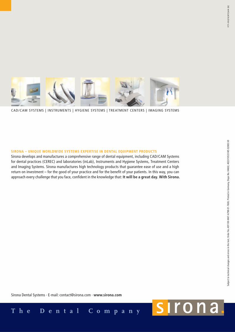
CAD/CAM SYSTEMS | INSTRUMENTS | HYGIENE SYSTEMS | TREATMENT CENTERS | IMAGING SYSTEMS

02 | 03
ORTHOPHOS XGPlus – PERFECT X-RAY IMAGING
Perfect results only come whenyou start with perfection.
Do you put great value in an accurate diagnosis? Do you expect simple and intuitive operation? And you want a secure investment for the future? ORTHOPHOS XGPlus, the specialist of the digital panoramic X-ray systems, always puts you on the safe side. The unique “Easypad“ user interface, the absolutely safe and reliable patient positioning, and the SIDEXIS XG image processing software – these are just a few advan-tages of our top-of-the-line system. And especially important for you, as a specialist yourself: You can retrofit ORTHOPHOS XGPlus at any time with ceph and TSA. It will be a great day. With Sirona.

04 | 05
ORTHOPHOS XGPlus – RELIABLE, ACCURATE X-RAY IMAGING
Be 3 times as sure.
Operational reliability! Optimum, exact patient
positioning! Intuitive control via Easypad
touchscreen! Prevention of errors through
interactive messages
Diagnostic accuracy! Logical program structure! Enhanced diagnostic options! Individual adaptation to the
patient
Investment security! Practice-oriented workflow! Retrofittable with ceph/TSA! Software upgrades! 5-year warranty on tube
assembly and sensor
06 | 07
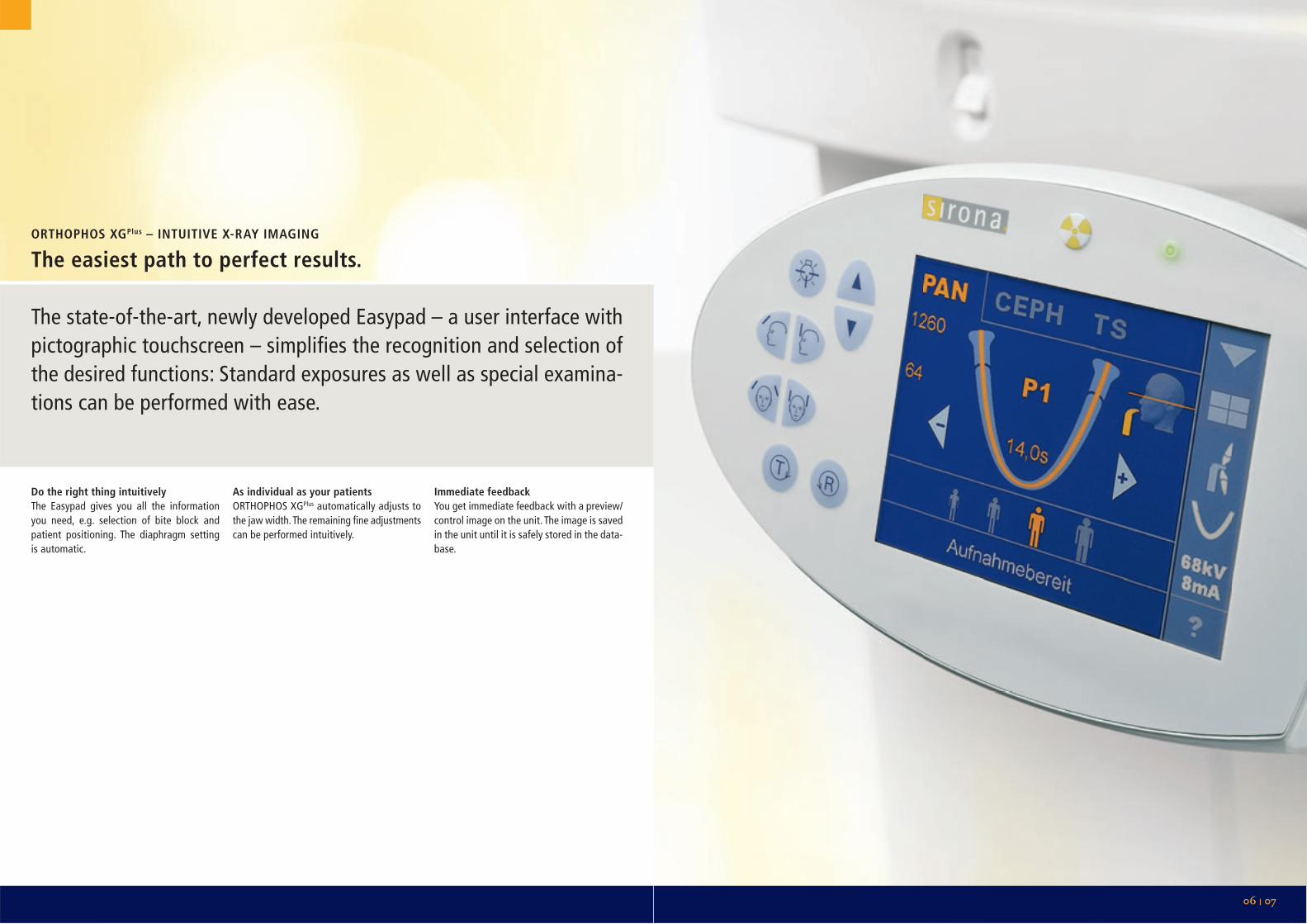
ORTHOPHOS XGPlus – INTUITIVE X-RAY IMAGING
The easiest path to perfect results.
The state-of-the-art, newly developed Easypad – a user interface with pictographic touchscreen – simplifies the recognition and selection of the desired functions: Standard exposures as well as special examina-tions can be performed with ease.
Do the right thing intuitivelyThe Easypad gives you all the information you need, e.g. selection of bite block and patient positioning. The diaphragm setting is automatic.
As individual as your patientsORTHOPHOS XGPlus automatically adjusts to the jaw width. The remaining fi ne adjustments can be performed intuitively.
Immediate feedbackYou get immediate feedback with a preview/control image on the unit. The image is saved in the unit until it is safely stored in the data-base.

Automatic adjustment to the jaw width
ORTHOPHOS XGPlus – HIGH-QUALITY X-RAY IMAGING
Diagnosis at its best.
ORTHOPHOS XGPlus opens up a variety of diagnostic possibilities and uniquely fulfills all the prerequisites of the “image quality chain“:
! Quick, precise positioning of the jaw in the sharp slice! Avoidance of motion blurring! Automatic radiation management! Diagnostic precision through specific orbits! Elimination of operator errors! Automatic image preprocessing
08 | 10
ORTHOPHOS XGPlus – THE RIGHT X-RAY IMAGING
The secret of image quality.
The jaw must be located exactly in the sharp slice!
1. Only 2 planes have to be adjusted for all Sirona panoramic systems: the mid-sagittal and the Frankfurt horizontal planes. As soon as the patient‘s anterior teeth are located in the indentation of the bite block, they are already in the slice (because the indentation is the reference point for the orbit) – immedi-ately, precisely, without the third light beam and without misinterpretation.
2. ORTHOPHOS automatically adapts the slice orientation to the width of the patient‘s jaw (acquired with the temple supports), so that the molars are in the middle of the slice, i.e. in the zone with maximum sharpness.
In special cases, it is possible to make a manual fi ne adjustment to the shape of the anterior tooth region.
If the patient is exactly positioned in the slice, motion blurring can be avoided.
! The patient looks in the direction of the unit in the mirror and not at the operator. He/she will not try to follow you with the eyes when you leave the X-ray room.
! The 3-point immobilization with bite block, forehead support and temple support prevent movement.
! The position of the patient is geo-metrically determined by the 3-point immobilization. The exact positioning data are saved so that the scan is reproducible.
Image quality is also infl uenced by management of the radiation.
! Uniform radiation due to the high-frequency generator.
! Automatic exposure control to adjust the radiation to variations in object thickness.
! Automatic kV increase in the spinal region (increase in the quality, not the quantity of the radiation) for differen-tiated imaging of the anterior teeth.
! Exposure parameters are available as meaningful value pairs (kV/mA) to avoid mistakes and uncertainty – based on the experience of more than 40,000 manufactured panoramic systems.
! “Quickshot“ is selectable for panoramic and ceph on the Easypad.
Logical positioning in the sharp slice Effective immobilization of the patient Optimized radiation Specifi c orbits Easy operation Image acquisition and preprocessing
The results in SIDEXIS XG (refer also to page 18/19)
1
2
The panoramic programs for ORTHOPHOS XGPlus are selectablein different versions for specifi c diagnostic requirements.
! “Standard“ (orthoradial): the basic overview to prevent teeth overlapping.
! Artifact-free: Offset projections in order to avoid double projections, e.g. metal bridges.
! With constant magnifi cation 1.25 of the entire mandibular arch, e.g. for implantology.
Additional examples:! Thick slice in anterior tooth region for
extreme anomalies (P12).! Program for lateral and axial temporo-
mandibular joints with specifi c beam direction and special slice orientation.
Save time, avoid uncertainty, eliminate misinterpretation!
! Patient height up to 2 m (panorama)! Immediate, exact positioning in the
anterior plane without the third light beam.
! Automatic adjustment of the orbit to the jaw width.
! Easy selection of exposure parameters and programs on the Easypad.
! Fast preview image on the unit.! Automatic diaphragm adjustment at
program selection.! Handling options Pan/ceph and TSA
mode without changing sensors available as an option.
! Time-savings with control exposure through stored positioning and exposure data.
ORTHOPHOS XGPlus uses sensors in CCD technology with a pixel size of 27 µm for panorama and ceph.
! A special wide sensor is used for transversal slices based on wide beam tomography.
! The image is acquired at 16 bits and then automatically preprocessed in order to obtain a maximum of informa-tion. An example: To ensure that fi ne details are recognizable, the image is automatically “stretched“ in the largest possible gray scale between black and white. Regardless of whether it was slightly over- or underexposed.
! Security is high: the image data of the last exposure remain stored in the system.
! Another advantage: ORTHOPHOS XGPlus is directly network-capable: It needs no allocated PC and is independent of hardware changes in the computer.
P 12

With the ORTHOPHOS XGPlus Easypad your choices are intuitively right. Simply structured, eliminating selection errors. You are guided pictori-ally and surely through the menu. Pan programs can be selected as standard, with constant magnification for implant planning and free of artifacts.
12 | 13
ORTHOPHOS XGPlus – SIMPLE X-RAY IMAGING
Intuitive program selectionfor individual diagnostics.
Simple program selection (example: PAN P1A)Easy selection of exposure parameters via icons –and you‘re done!
Possible fi ne adjustmentsExample: Shape of jaw in anterior tooth region
Simple switching to cephalometric X-rayThe diaphragm adjustment is made automatically.
08 | 11
TM1
* With closed or open occlusion, with only one slice position, or in a multi-slice view.
TM2
TM3
TM4
TM5
TM6
Standardimage
S1
S2
MS1
Sinus
Transversal multi-slice posterior teeth
Constantmagnifi cation
Artifact-free
P1
P2
P10
Additional programs: Transversal slices (TSA) in digital wide beam tomography: (see page 14/15)Cephalometric X-ray: (see page 16/17)
P12
Selection of radiation window possible
C
A
P1
S3
S4
TMJ lateral*
TMJ axial*
14 | 15
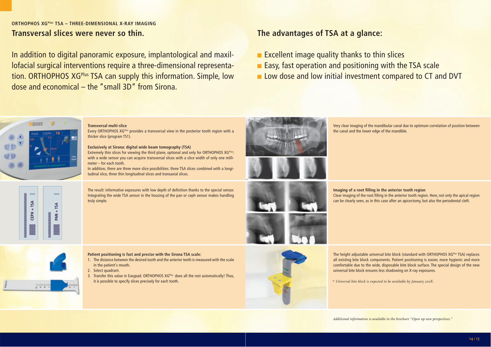
Transversal multi-sliceEvery ORTHOPHOS XGPlus provides a transversal view in the posterior tooth region with a thicker slice (program TS1).
Exclusively at Sirona: digital wide beam tomography (TSA)Extremely thin slices for viewing the third plane, optional and only for ORTHOPHOS XGPlus: with a wide sensor you can acquire transversal slices with a slice width of only one milli-meter – for each tooth.In addition, there are three more slice possibilities: three TSA slices combined with a longi-tudinal slice, three thin longitudinal slices and transaxial slices.
ORTHOPHOS XGPlus TSA – THREE-DIMENSIONAL X-RAY IMAGING
Transversal slices were never so thin.
In addition to digital panoramic exposure, implantological and maxil-lofacial surgical interventions require a three-dimensional representa-tion. ORTHOPHOS XGPlus TSA can supply this information. Simple, low dose and economical – the “small 3D“ from Sirona.
The advantages of TSA at a glance:
!!Excellent image quality thanks to thin slices!!Easy, fast operation and positioning with the TSA scale!!Low dose and low initial investment compared to CT and DVT
Additional information is available in the brochure “Open up new perspectives.”
The result: informative exposures with low depth of defi nition thanks to the special sensor. Integrating the wide TSA sensor in the housing of the pan or ceph sensor makes handling truly simple.
Patient positioning is fast and precise with the Sirona TSA scale:1. The distance between the desired tooth and the anterior teeth is measured with the scale
in the patient‘s mouth.2. Select quadrant.3. Transfer this value in Easypad. ORTHOPHOS XGPlus does all the rest automatically! Thus,
it is possible to specify slices precisely for each tooth.
Very clear imaging of the mandibular canal due to optimum correlation of position between the canal and the lower edge of the mandible.
Imaging of a root fi lling in the anterior tooth regionClear imaging of the root fi lling in the anterior tooth region. Here, not only the apical region can be clearly seen, as in this case after an apicectomy, but also the periodontal cleft.
The height adjustable universal bite block (standard with ORTHOPHOS XGPlus TSA) replaces all existing bite block components. Patient positioning is easier, more hygienic and more comfortable due to the wide, disposable bite block surface. The special design of the new universal bite block ensures less shadowing on X-ray exposures.
* Universal bite block is expected to be available by January 2008.

16 | 17
ORTHOPHOS XGPlus CEPH – SPECIALIZED X-RAY IMAGING
Cephalometric X-ray with vision.
ORTHOPHOS XGPlus Ceph is perfectly designed for orthodontists and maxillofacial surgeons. The scan technique combines high resolution with low dose. The image width is selectable: 30 cm or 18 cm. In addi-tion to the usual programs for lateral, symmetric (p.a. or a.p.) and car-pus images, a variety of special projections are possible, such as the half-axial or the Clementschitsch exposures. Patients up to approx. 1.90 min height can be scanned in standing position.
Perfectly adaptedORTHOPHOS XGPlus Ceph is perfectly adapted to the patients and workfl ow in the ortho-dontist’s practice.
For panoramic andcephalometric X-raysthrough the “Quickshot” option for shorten-ing the acquisition cycle, through the auto-matic diaphragm and through other workfl ow advantages; see also page 20/21 (in fi gure: lateral cephalometric exposure with acti-vated soft tissue fi lter in Quickshot mode).
For panoramic exposuresthrough specifi c programs such as the pedi-atric program 10 (with reduced radiation window) or program 12 (thick slice in the anterior tooth region for a sharp image with extreme anomalies), as well as through the automatic adjustment of the slice position to the jaw width.
Sensor
Shorter acquisition cyclesthrough the pan/ceph/carpus exposure func-tion as exposure series with shorter cool-down phase.
Principle of the horizontal scan
X-ray radiation
The ceph arm can also be retrofi tted at a later time. From pan to ceph without changing sensor: optionally, the system can be operated with 2 sensors.

18 | 19
SIDEXIS XG IMAGE PROCESSING SOFTWARE – INTELLIGENT X-RAY IMAGING
Ideal diagnosis for the digital practice.
The SIDEXIS XG image processing software lets you optimally diagnose your X-ray images with intuitive, simple procedures. Of course, SIDEXIS XGis compatible with practice management and special programs (such as orthodontic analysis software) DICOM environments are also sup-ported. SIDEXIS XG also connects all the other elements of the digital practice from Sirona: digital intraoral X-ray, digital panoramic and ceph X-ray, SIROCAM.
FastWith SIDEXIS XG, diagnostic fi ndings and treatment are clearlydocumented for quick retrieval. You can, for example, make several measurements, draw or write on a single image.
IndividualThe user interface can be customized to your needs. You can auto-matically pre-select fi lters, brightness and contrast. That means fewer clicks and saves time.
Tailored for treatmentWith the “Examination“ function you retrieve and update images, fi ndings and notes according to the desired case. Everything is sorted, clear and immediately available upon retrieval. The integratedImplantPlus plug-in makes it possible to visualize implants, allowing you to explain your treatment quickly and with graphic clarity.
FlexibleFewer PCs, more fl exibility: The ORTHOPHOS XGPlus in the X-ray room does not have to be connected to a PC, but can be directly con-nected to the network.
Intraoral X-ray stations, the connection points for intraoral sensors:– X-ray box, directly to the C+ treatment centers– Wall box, can also be integrated in the network without its own PC– USB box
Better patient consultationright at the treatment center with SIVISION and SIDEXIS XG.
20 | 21
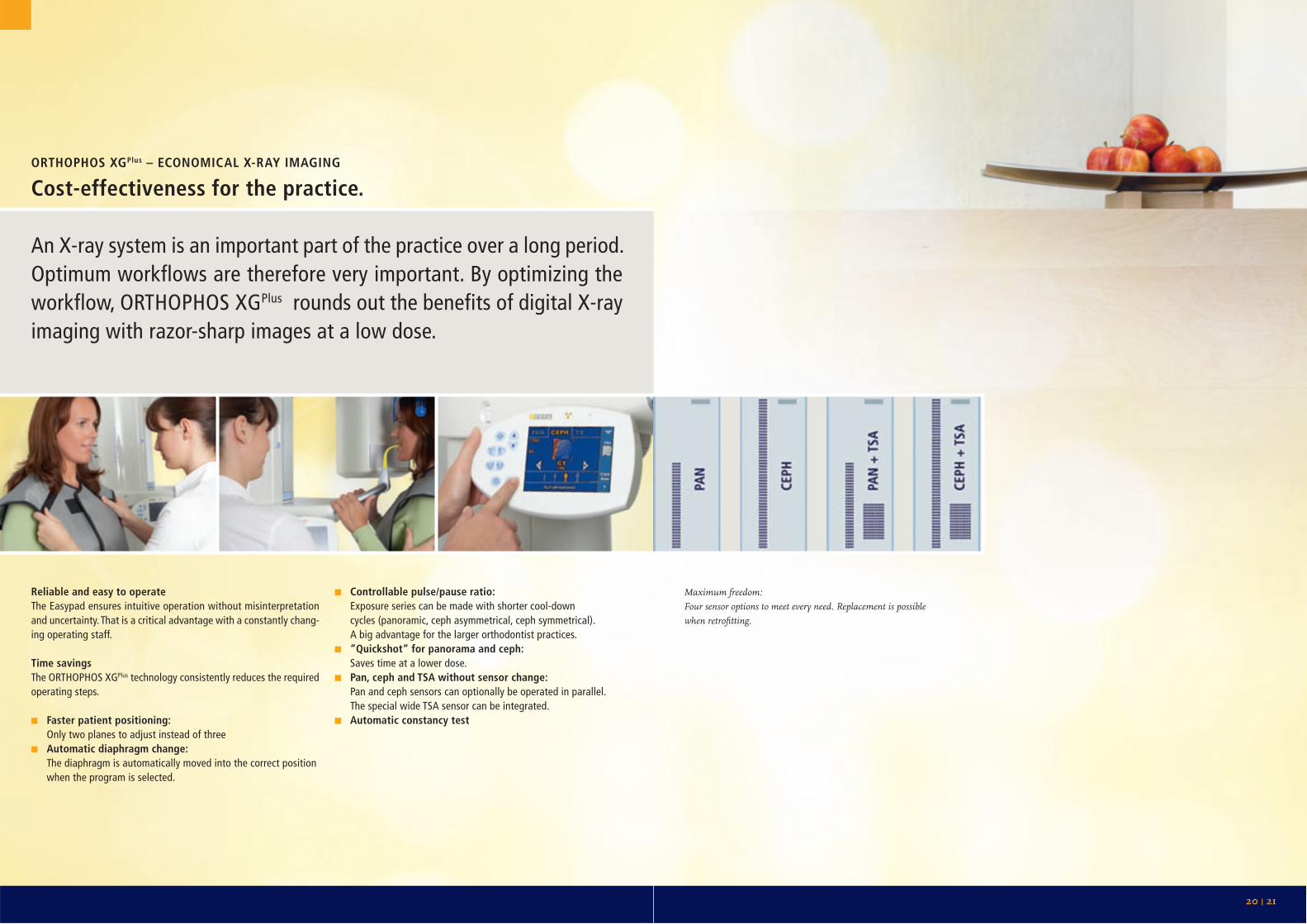
ORTHOPHOS XGPlus – ECONOMICAL X-RAY IMAGING
Cost-effectiveness for the practice.
An X-ray system is an important part of the practice over a long period. Optimum workflows are therefore very important. By optimizing the workflow, ORTHOPHOS XGPlus rounds out the benefits of digital X-ray imaging with razor-sharp images at a low dose.
Reliable and easy to operateThe Easypad ensures intuitive operation without misinterpretation and uncertainty. That is a critical advantage with a constantly chang-ing operating staff.
Time savingsThe ORTHOPHOS XGPlus technology consistently reduces the required operating steps.
! Faster patient positioning: Only two planes to adjust instead of three! Automatic diaphragm change: The diaphragm is automatically moved into the correct position
when the program is selected.
! Controllable pulse/pause ratio: Exposure series can be made with shorter cool-down
cycles (panoramic, ceph asymmetrical, ceph symmetrical).A big advantage for the larger orthodontist practices.
! “Quickshot“ for panorama and ceph: Saves time at a lower dose.! Pan, ceph and TSA without sensor change: Pan and ceph sensors can optionally be operated in parallel.
The special wide TSA sensor can be integrated.! Automatic constancy test
Maximum freedom:Four sensor options to meet every need. Replacement is possiblewhen retrofi tting.
22 | 23
ORTHOPHOS XGPlus – X-RAY IMAGING FOR A SECURE FUTURE
The future is all included.
The practice situation changes – and ORTHOPHOS XGPlus changesflexibly with it. With integrated technology for the future and open structures for new developments.
Technological power bundleWith state-of-the-art technology ORTHOPHOS XGPlus offers everything needed for ultra-fast data transmission: integrated power PC, 16-bit image acquisition, high-speed interface, CAN bus technology. The components‘ pow-er reserves will have no problem with the increased data fl ow of future applications.
Flexibility for tomorrowThe system software can be expanded or changed at any time. Additional programs or functionalities are loadable from data media. The Easypad touchscreen interface can be adapted in a variety of ways.
Uncomplicated retrofi ttingAt the moment you need only the pan-oramic system? Subsequent retrofi tting of cephalometric X-ray or transversal slices is no problem.
Award winningThe ORTHOPHOS XGPlus technology of the fu-ture hits the mark: The elegant, modern design has already won a design prize.
Ethernet technologyORTHOPHOS XGPlus is directly network-cap-able and therefore controllable from every PC in the network for image creation. You don‘t need an additional PC to operate the system. ORTHOPHOS XGPlus has an optical interface for future connection to fi ber optic networks.
More than a systemORTHOPHOS XGPlus comes from Sirona, the co-inventor of panoramic X-ray and a pioneer of digital panoramic and ceph X-ray. Production experience with over 40,000 systems, re-fl ected in reliability and longevity, are the marks of a market leader. Through the Sirona dealers and the Customer Service Center, you get the support you expect with advanced technology.
5-year warrantyFor all ORTHOPHOS XGPlus systems there is an additional warranty on the tube assembly and sensors. Over and above the legal warranty period of one year, Sirona provides the parts warranty for sensors and tube assembly for an additional 4 years.

Pan digitalCeph digital
ORTHOPHOS XGPlus DSORTHOPHOS XGPlus DS Ceph
Radiation generator Multipulse generator (max. 120 kHz)
X-ray tube SR 90/15 FN
Focal spot size according IEC 336/82 0.5 mm x 0.5 mm
Total fi lter 2.5 mm AL
Tube voltage 60–90 kV
Tube current 3–16 mA
Nominal voltage 230–240 V, 50–60 Hz
Nominal current 12 A
Line internal resistance max. 0.8 Ohm
Fuse 16 A slow blow
Power consumption 2.8 kW
Permissible line voltage fl uctuations ± 10 %
Panoramic exposure time (P1) 14.2 s
Panoramic exposure time (P1), “Quickshot” 9.1 s
Range of height of bite block 800–1850 mm
Ceph
Radiation time 9.4 s
Radiation time “Quickshot” 18 x 24 cm 4.7 s
Effective exposure time approx. 270 ms
24 | 25
ORTHOPHOS XGPlus – FLEXIBLE X-RAY IMAGING
The right choice in any situation.
Technical features
! Operation via Easypad! Intuitive program structure! Preview/control image on the system
user interface! Remote control optional! 90 kV high-frequency generator! Automatic adjustment of the panoramic
orbit to the individual jaw width of the patient; jaw shape in the anterior tooth region is selectable
! Spine compensation through automatic kV increase
! Quickshot mode for panorama and ceph is available
! CCD sensor technology with high-speed interface, 27-µm pixel size and image acquisition in 16-bit technology; data transfer 100 Mbit, Ethernet
! Pan/ceph and TSA sensors can becombined
! Investment security through integrated power PC and bus architecture
! System software is upgradable! Touchscreen with virtually unlimited
software enhancement options! Ceph is retrofi ttable! Transversal slices (TSA) are retrofi ttable! System versions: ORTHOPHOS XGPlus DS,
ORTHOPHOS XGPlus DS Ceph! SIDEXIS XG image processing software! Optional fl oor stand! Wheelchair compatible
Programs
Panoramic programs:! Standard panorama (P1)! Standard panorama without ascending
branches (P2)! Pediatric panorama (P10) available in
the following versions: – Standard orthoradial – or with constant magnifi cation (1.25) – or artifact-free with selectable image detail: – Thick slice, anterior tooth region (P12)
Lateral temporomandibular programs:! with selectable image detail! with open and closed occlusion! with one slice position! multi-slice Axial temporomandibular programs:! with open and closed occlusion! with one slice position! multi-slice Sinus programs:! Maxillary sinuses – 2 images! Maxillary sinuses – 2 images (linear)! Paranasal sinuses! Paranasal sinuses (linear)
Transversal multi-slice posteriorteeth Ceph programs:! Ceph asymmetric! Ceph symmetric p. a.! Ceph symmetric a. p.! Carpus (hand/wrist)Additional projections possible
Optional: Transversal slice acquisition in digital wide beam tomography (TSA):
! 10 programs with thin slices
Remote control with display of the exposure parameters.
If you do not have a mountable wall, we offer an extremely stable, wheelchair compatible fl oor stand.
ORTHOPHOS XGPlus DS space requirements:min. 1280 mm x min. 1411 mm
Technical data
ORTHOPHOS XGPlus DS Ceph space requirements: min. 2155 mm x min. 1411 mm
T h e D e n t a l C o m p a n y
CAD/CAM SYSTEMS | INSTRUMENTS | HYGIENE SYSTEMS | TREATMENT CENTERS | IMAGING SYSTEMS
SIRONA – UNIQUE WORLDWIDE SYSTEMS EXPERTISE IN DENTAL EQUIPMENT PRODUCTS Sirona develops and manufactures a comprehensive range of dental equipment, including CAD/CAM Systems for dental practices (CEREC) and laboratories (inLab), Instruments and Hygiene Systems, Treatment Centers and Imaging Systems. Sirona manufactures high technology products that guarantee ease of use and a high return on investment – for the good of your practice and for the benefi t of your patients. In this way, you can approach every challenge that you face, confi dent in the knowledge that: It will be a great day. With Sirona.
Sirona Dental Systems · E-mail: [email protected] · www.sirona.com
Subj
ect t
o te
chni
cal c
hang
es a
nd e
rrors
in th
e te
xt, O
rder
No.
A91
100-
M47
-A79
8-01
-760
0, P
rinte
d in
Ger
man
y, Di
spo
No. 0
4602
, 402
3/10
533
WS
0208
3.V0