Embed Size (px)
Citation preview
[Frontiers in Bioscience 14, 1851-1856, January 1, 2009]
1851
Ca2+-sensitive transcriptional regulation: direct DNA interaction by DREAM Sherry Sours-Brothers1, Rong Ma1, Peter Koulen2,3,4 1Department of Integrative Physiology, 2Department of Pharmacology and Neuroscience, 3North Texas Eye Research Institute, 4Institute for Aging and Alzheimer’s Disease Research, University of North Texas Health Science Center, Fort Worth, Texas TABLE OF CONTENTS 1. Abstract 2. Introduction 3. Influence of Ca2+-dependent kinases and phosphatases 4. Direct transcriptional regulation by Ca2+-sensitive downstream regulatory element antagonistic modulator 5. Ca2+binding to DREAM results in derepression of DRE 6. Downstream effects of DREAM regulated gene expression 7. Non-transcriptional activity of DREAM 8. Discussion 9. References 1. ABSTRACT
Calcium is a major regulator of cell function, acting as a second messenger to relay signals from the cell surface to other parts of the cell. It plays an integral role in contraction of muscle cells and it regulates cell growth and proliferation, as well as cell death (1). The present review will discuss how Ca2+ mediates these functions through the regulation of gene expression. This can be accomplished by Ca2+-sensitive protein kinases as well as phosphatases which activate transcription factors. It can also be mediated by direct interaction of Ca2+-sensitive proteins with regulatory elements within the DNA sequence itself. Special attention will be given to recent advances in research in the down-stream regulatory element (DRE) and its Ca2+-sensitive modulator DREAM (downstream regulatory element antagonist modulator; also named calsenilin or K+ channel interacting protein 3 (KChIP3)) (2).
2. INTRODUCTION
The Ca2+-sensitivity of transcriptional regulatory factors is mediated by EF-hand Ca2+-binding motifs (3). This allows Ca2+-binding proteins to act as both a Ca2+ buffers and Ca2+ sensors. The EF-hand domain is composed of two perpendicular α-helices connected by a loop, i.e. a helix-loop-helix motif, that may be repeated 2-12 times within a protein’s amino acid sequence (2, 4, 5). Upon Ca2+ binding, the EF-hand undergoes a conformational change from a closed to an open conformation resulting in activation of regulatory domains, as is the case with calmodulin (5). There are more than 600 EF-hand motif containing proteins (2), and a number of these proteins affect gene transcription in response to changes in intracellular Ca2+ concentration. For example, activation of the Ca2+/calmodulin-dependent protein kinase (CaMK) signaling cascade results in activation of cAMP response-element (CRE) binding protein (CREB) to
Ca2+-sensitive transcriptional regulation
1852
activate transcription (6). The Ca2+-dependent phosphatase calcineurin dephosphorylates nuclear factor of activated T-cells (NFAT), a transcription factor which is then transported to the nucleus to regulate transcription (7). More recently, the Ca2+-binding protein DREAM has been identified as a transcriptional repressor that is directly sensitive to nuclear Ca2+ concentrations (8). Together, these Ca2+-sensitive transcriptional regulators represent a significant role of Ca2+ in the regulation of cellular function.
3. INFLUENCE OF Ca2+-DEPENDENT KINASES AND PHOSPHATASES
A well-documented example of Ca2+-sensitive transcriptional regulation is the CaMK signaling cascade. This family of proteins, including CaMKI/IV and CaMKII, is activated by the binding of Ca2+ to calmodulin, followed by activating phosphorylations by CaMK kinase or by auto- or transphosphorylation (6). CaMKs exert their transcriptional influence via CREB. All three of the CaMKs can bind CREB and phosphorylate its activating site, Ser-133, in vitro, although CaMKII can phosphorylate an additional site, Ser-142 (9), while CaMKIV is the major activator of CREB (10). Once activated, CREB not only binds to CRE to induce transcription, but also interacts with numerous other transcription factors, including p53 (11), c-Jun (12), and c-fos (13). CREB also binds to the Ca2+ response element (CARE), which is structurally similar to CRE and is also found in c-fos. The effects of CREB binding are extremely widespread. A genome-wide search to identify putative CREB targets, followed by chromatin immunoprecipitation of over 16,000 human gene promoter regions (Hu19k promoter microarray) identified ≈3,000 promoter sites occupied by CREB in vivo (14).
Another transcription factor, NFAT is regulated
in a Ca2+-dependent manner by the protein phosphatase calcineurin. In this case, increase in intracellular Ca2+ by phospholipase C mediated store-operated Ca2+ entry and binding to calmodulin leads to activation of calcineurin (15). Calcineurin then dephosphorylates multiple serine residues of NFAT, which is then translocated to the nucleus and binds to kappa-B sites via Rel homology regions to regulate gene transcription (7). 4. DIRECT TRANSCRIPTIONAL REGULATION BY Ca2+-SENSITIVE DOWNSTREAM REGULATORY ELEMENT ANTAGONISTIC MODULATOR
While the transcriptional regulation described above is dependent on kinase or phosphatase activity, there is another mode by which Ca2+ regulates gene transcription more directly yet still mediated in the nucleus via a downstream regulatory element (DRE). The DRE site was first identified in the prodynorphin gene which is highly expressed in the brain (8). Prodynorphin is the precursor of dynorphin, which plays a role in memory acquisition and pain sensation (16, 17). Four cAMP response elements have been identified within the prodynorphin gene (18). CREB binding to the third CRE of the prodynorphin promoter represses gene expression, while binding of AP-1
to this site activates transcription (18). Through mutation analysis of the human prodynorphin promoter region, Carrión et al. identified a DRE consensus sequence (PuNGTCAPuPuG; see reference 8). Derepression of prodynorphin at the DRE site was required for protein kinase A- (PKA) dependent transcription (8). A 110-kD protein complex was also identified that specifically bound to the DRE site, and whose binding was reduced upon PKA stimulation (8). Therefore, DRE functions as a transcriptional silencer. In a subsequent study, Carrión et al. identified the DRE modulatory protein by screening a human caudate cDNA expression library using a double-stranded DRE oligonucleotide as a probe (19). A 284 amino acid protein with a predicted molecular weight of 31,800 was identified, which was named the DRE-binding protein DRE-antagonist modulator (DREAM). DREAM binds to DRE as a homotetramer (corresponding to the 110 kDa protein described above; reference 8) with high affinity, and as a monomer with low affinity. Four Ca2+-binding EF-hand motifs were identified within the amino acid sequence of DREAM, with high sequence homology to recoverin and recoverin-like binding proteins vilip1, and hippocalcin (20, 21). Functionality of these EF-hands was confirmed, as release of intracellular Ca2+ by caffeine resulted in derepression of DRE by DREAM. DREAM also repressed expression of the Ca2+-sensitive proto-oncogene c-fos (22), which also contains a DRE site (8).
5. Ca2+ BINDING TO DREAM RESULTS IN DEREPRESSION OF DRE
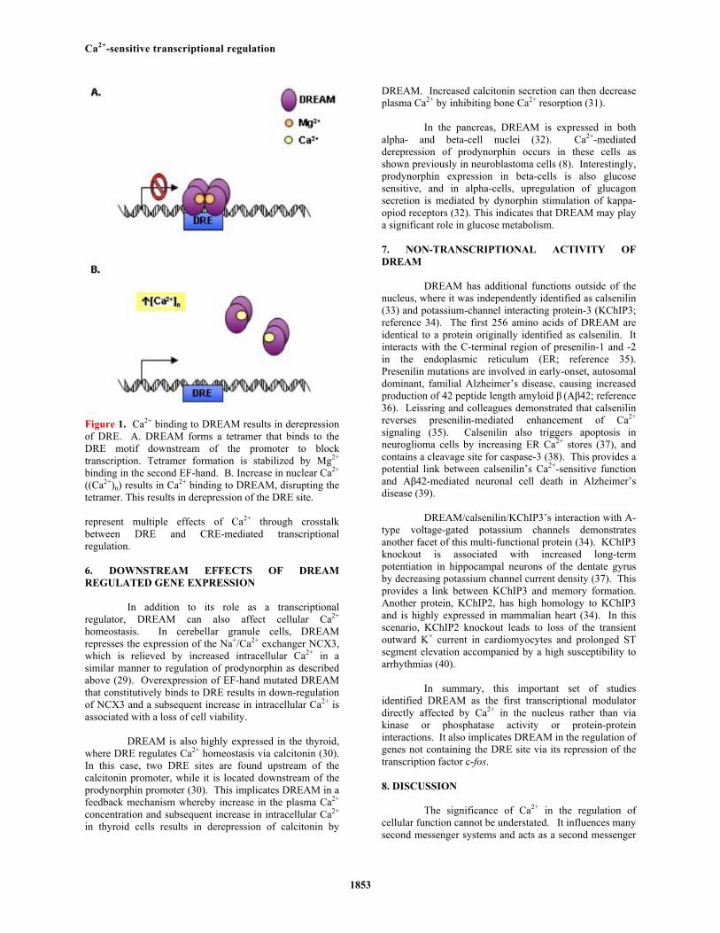
The DNA binding properties of DREAM have been further explored in order to elucidate its Ca2+-sensitive transcriptional properties. Osawa and colleagues demonstrated that DREAM binds three Ca2+ ions in its C-terminal EF-hand domains (23). This is consistent with previous reports that the first of DREAM’s four EF-hand motifs is non-functional (24). Ca2+ binding results in conformational changes in DREAM structure which disrupts tetramer formation (Figure 1) (23). Ca2+-bound DREAM dimers have a lower DNA binding affinity and do not bind to the DRE site, which accounts for the Ca2+-mediated derepression (Figure 1B) (23). Ca2+-free DREAM binds to DNA via residues in both its basic N-terminal and Ca2+-binding C-terminal regions (23). Interestingly, Mg2+ also binds DREAM in the nucleus, and is required for sequence-specific DNA binding (Figure 1A) (25, 26).
In addition to regulation by other ions, DREAM activity is sensitive to PKA activity via interaction with cAMP response element modulator (alpha CREM; reference 23). Two leucine-charged residue-rich domains (LCDs) within alpha CREM and DREAM result in binding of the two proteins, blocking interaction of DREAM with DRE and resulting in derepression of prodynorphin (27). DREAM also interacts with CRE function by blocking the binding of CREM and CREB via LCD domains in vitro (28). This effect is abolished by Ca2+-binding to DREAM. Additionally, the binding of DREAM to the CREB LCD domain normally occupied by CREB binding protein (CBP) results in lower CBP activity. These examples
Ca2+-sensitive transcriptional regulation
1853
Figure 1. Ca2+ binding to DREAM results in derepression of DRE. A. DREAM forms a tetramer that binds to the DRE motif downstream of the promoter to block transcription. Tetramer formation is stabilized by Mg2+ binding in the second EF-hand. B. Increase in nuclear Ca2+ ((Ca2+)n) results in Ca2+ binding to DREAM, disrupting the tetramer. This results in derepression of the DRE site.
represent multiple effects of Ca2+ through crosstalk between DRE and CRE-mediated transcriptional regulation.
6. DOWNSTREAM EFFECTS OF DREAM REGULATED GENE EXPRESSION
In addition to its role as a transcriptional regulator, DREAM can also affect cellular Ca2+ homeostasis. In cerebellar granule cells, DREAM represses the expression of the Na+/Ca2+ exchanger NCX3, which is relieved by increased intracellular Ca2+ in a similar manner to regulation of prodynorphin as described above (29). Overexpression of EF-hand mutated DREAM that constitutively binds to DRE results in down-regulation of NCX3 and a subsequent increase in intracellular Ca2+ is associated with a loss of cell viability.
DREAM is also highly expressed in the thyroid,
where DRE regulates Ca2+ homeostasis via calcitonin (30). In this case, two DRE sites are found upstream of the calcitonin promoter, while it is located downstream of the prodynorphin promoter (30). This implicates DREAM in a feedback mechanism whereby increase in the plasma Ca2+ concentration and subsequent increase in intracellular Ca2+ in thyroid cells results in derepression of calcitonin by
DREAM. Increased calcitonin secretion can then decrease plasma Ca2+ by inhibiting bone Ca2+ resorption (31).
In the pancreas, DREAM is expressed in both
alpha- and beta-cell nuclei (32). Ca2+-mediated derepression of prodynorphin occurs in these cells as shown previously in neuroblastoma cells (8). Interestingly, prodynorphin expression in beta-cells is also glucose sensitive, and in alpha-cells, upregulation of glucagon secretion is mediated by dynorphin stimulation of kappa-opiod receptors (32). This indicates that DREAM may play a significant role in glucose metabolism. 7. NON-TRANSCRIPTIONAL ACTIVITY OF DREAM
DREAM has additional functions outside of the nucleus, where it was independently identified as calsenilin (33) and potassium-channel interacting protein-3 (KChIP3; reference 34). The first 256 amino acids of DREAM are identical to a protein originally identified as calsenilin. It interacts with the C-terminal region of presenilin-1 and -2 in the endoplasmic reticulum (ER; reference 35). Presenilin mutations are involved in early-onset, autosomal dominant, familial Alzheimer’s disease, causing increased production of 42 peptide length amyloid β (Aβ42; reference 36). Leissring and colleagues demonstrated that calsenilin reverses presenilin-mediated enhancement of Ca2+ signaling (35). Calsenilin also triggers apoptosis in neuroglioma cells by increasing ER Ca2+ stores (37), and contains a cleavage site for caspase-3 (38). This provides a potential link between calsenilin’s Ca2+-sensitive function and Aβ42-mediated neuronal cell death in Alzheimer’s disease (39).
DREAM/calsenilin/KChIP3’s interaction with A-
type voltage-gated potassium channels demonstrates another facet of this multi-functional protein (34). KChIP3 knockout is associated with increased long-term potentiation in hippocampal neurons of the dentate gyrus by decreasing potassium channel current density (37). This provides a link between KChIP3 and memory formation. Another protein, KChIP2, has high homology to KChIP3 and is highly expressed in mammalian heart (34). In this scenario, KChIP2 knockout leads to loss of the transient outward K+ current in cardiomyocytes and prolonged ST segment elevation accompanied by a high susceptibility to arrhythmias (40).
In summary, this important set of studies
identified DREAM as the first transcriptional modulator directly affected by Ca2+ in the nucleus rather than via kinase or phosphatase activity or protein-protein interactions. It also implicates DREAM in the regulation of genes not containing the DRE site via its repression of the transcription factor c-fos.
8. DISCUSSION
The significance of Ca2+ in the regulation of cellular function cannot be understated. It influences many second messenger systems and acts as a second messenger
Ca2+-sensitive transcriptional regulation
1854
itself. Ca2+ also regulates the life and death of cells, influencing growth and proliferation as well as apoptosis (1). Many of these important functions are carried out by Ca2+-binding proteins that influence the transcriptional regulation of a host of genes. The ability of Ca2+-sensitive protein kinases and phosphatases to affect activity of transcription factors has long been established (15). More recently, Ca2+ has been shown to directly bind to and regulate transcription factors in the nucleus, as exemplified here by the DRE modulatory protein DREAM (2). This unique mode of transcriptional regulation may serve as a treatment target in a number of cell types. For example, DREAM regulation of the endogenous opiod dynorphin may play a role in central sensitization of nociceptors (41). This may make it a target in pain management, as evidenced by analgesia described in DREAM KO mice (42, 43). DREAM-mediated regulation of dynorphin may also be a target for modulation of glucagon secretion in diabetes (32). In addition, DREAM regulation of genes that effect Ca2+ handling presents a novel mechanism by which cells maintain Ca2+ homeostasis. This presents a new level of significance for Ca2+-sensitive transcriptional regulation. 9. ACKNOWLEDGEMENTS
This work was supported in part by NIH grants EY14227, AG22550, AG10485, and by Texas Higher Education Coordinating Board Advanced Research Program (ARP) Research Grant 000130-0007-2006 (P.K.).
10. REFERENCES 1. E. Carafoli: Calcium - a universal carrier of biological signals. Delivered on 3 July, 2003 at the Special FEBS Meeting in Brussels. FEBS Journal 272(5), 1073-1089 (2005) 2. E. Carafoli: Calcium signaling: A tale for all seasons. Proceedings of the National Academy of Sciences 99(3), 1115-1122 (2002) 3. I. Niki, H. Yokokura, T. Sudo, M. Kato, and H. Hidaka: Ca2+ Signaling and Intracellular Ca2+ Binding Proteins. J Biochem (Tokyo) 120(4), 685-698 (1996) 4. R. M. Tufty and R. H. Kretsinger: Troponin and parvalbumin calcium binding regions predicted in myosin light chain and T4 lysozyme. Science 187(4172), 167-169 (1975) 5. M. Ikura: Calcium binding and conformational response in EF-hand proteins. Trends in Biochemical Sciences 21(1), 14-17 (1996) 6. E. E. Corcoran and A. R. Means: Defining Ca2+/Calmodulin-dependent Protein Kinase Cascades in Transcriptional Regulation. J Biol Chem 276(5), 2975-2978 (2001) 7. G. Ghosh, G. V. Duyne, S. Ghosh, and P. B. Sigler: Structure of NF-(kappa)B p50 homodimer bound to a (kappa)B site. Nature 373(6512), 303-310 (1995)
8. A. M. Carrion, B. Mellstrom, and J. R. Naranjo: Protein Kinase A-Dependent Derepression of the Human Prodynorphin Gene via Differential Binding to an Intragenic Silencer Element. Mol Cell Biol 18(12), 6921-6929 (1998) 9. P. Sun, H. Enslen, P. S. Myung, and R. A. Maurer: Differential activation of CREB by Ca2+/calmodulin-dependent protein kinases type II and type IV involves phosphorylation of a site that negatively regulates activity. Genes Dev 8(21), 2527-2539 (1994) 10. D. Tardito, J. Perez, E. Tiraboschi, L. Musazzi, G. Racagni, and M. Popoli: Signaling Pathways Regulating Gene Expression, Neuroplasticity, and Neurotrophic Mechanisms in the Action of Antidepressants: A Critical Overview. Pharmacol Rev 58(1), 115-134 (2006) 11. M. L. Avantaggiati, V. Ogryzko, K. Gardner, A. Giordano, A. S. Levine, and K. Kelly: Recruitment of p300/CBP in p53-dependent signal pathways. Cell 89(7), 1175-1184 (1997) 12. A. J. Bannister, T. Oehler, D. Wilhelm, P. Angel, and T. Kouzarides: Stimulation of c-Jun activity by CBP: c-Jun residues Ser63/73 are required for CBP induced stimulation in vivo and CBP binding in vitro. Oncogene 11(12), 2509-2514 (1995) 13. R. Janknecht and A. Nordheim: Regulation of the c-fos promoter by the ternary complex factor Sap-1a and its coactivator CBP. Oncogene 12(9), 1961-1969 (1996) 14. X. Zhang, D. T. Odom, S. H. Koo, M. D. Conkright, G. Canettieri, J. Best, H. Chen, R. Jenner, E. Herbolsheimer, E. Jacobsen, S. Kadam, J. R. Ecker, B. Emerson, J. B. Hogenesch, T. Unterman, R. A. Young, and M. Montminy: Genome-wide analysis of cAMP-response element binding protein occupancy, phosphorylation, and target gene activation in human tissues. Proceedings of the National Academy of Sciences 102(12), 4459-4464 (2005) 15. P. G. Hogan, L. Chen, J. Nardone, and A. Rao: Transcriptional regulation by calcium, calcineurin, and NFAT. Genes Dev 17(18), 2205-2232 (2003) 16. M. G. Weisskopf, R. A. Zalutsky, and R. A. Nicoll: The opioid peptide dynorphin mediates heterosynaptic depression of hippocampal mossy fibre synapses and modulates long-term potentiation. Nature 362(6419), 423-427 (1993) 17. J. Naranjo, B. Mellstrom, M. Achaval, and P. Sassone-Corsit: Molecular pathways of pain: Fos/Jun-mediated activation of a noncanonical AP-1 site in the prodynorphin gene. Neuron 6(4), 607-617 (1991) 18. J. Collins-Hicok, L. Lin, C. Spiro, P. J. Laybourn, R. Tschumper, B. Rapacz, and C. T. McMurray: Induction of the rat prodynorphin gene through Gs-coupled receptors
Ca2+-sensitive transcriptional regulation
1855
may involve phosphorylation-dependent derepression and activation. Mol Cell Biol 14(5), 2837-2848 (1994) 19. A. M. Carrion, W. A. Link, F. Ledo, B. Mellstrom, and J. R. Naranjo: DREAM is a Ca2+-regulated transcriptional repressor. Nature 398(6722), 80-84 (1999) 20. A. M. Dizhoor, S. Ray, S. Kumar, G. Niemi, M. Spencer, D. Brolley, K. A. Walsh, P. P. Philipov, J. B. Hurley, and L. Stryer: Recoverin: a calcium sensitive activator of retinal rod guanylate cyclase. Science 251(4996), 915-918 (1991) 21. S. Nakayama and R. H. Kretsinger: Evolution of the EF-Hand Family of Proteins. Annual Review of Biophysics and Biomolecular Structure 23(1), 473-507 (1994) 22. J. I. Morgan and T. Curran: Role of ion flux in the control of c-fos expression. Nature 322(6079), 552-555 (1986) 23. M. Osawa, K. I. Tong, C. Lilliehook, W. Wasco, J. D. Buxbaum, H.-Y. M. Cheng, J. M. Penninger, M. Ikura, and J. B. Ames: Calcium-regulated DNA Binding and Oligomerization of the Neuronal Calcium-sensing Protein, Calsenilin/DREAM/KChIP3. J Biol Chem 276(44), 41005-41013 (2001) 24. F. Ledo, W. A. Link, A. M. Carrion, V. Echeverria, B. Mellstrom, and J. R. Naranjo: The DREAM-DRE interaction: key nucleotides and dominant negative mutants. Biochimica et Biophysica Acta (BBA) - Molecular Cell Research 1498(2-3), 162-168 (2000) 25. T. A. Craig, L. M. Benson, S. Y. Venyaminov, E. S. Klimtchuk, Z. Bajzer, F. G. Prendergast, S. Naylor, and R. Kumar: The Metal-binding Properties of DREAM. Evidence for calcium-mediated changes in dream structure. J Biol Chem 277(13), 10955-10966 (2002) 26. M. Osawa, A. Dace, K. I. Tong, A. Valiveti, M. Ikura, and J. B. Ames: Mg2+ and Ca2+ Differentially Regulate DNA Binding and Dimerization of DREAM. J Biol Chem 280(18), 18008-18014 (2005) 27. F. Ledo, A. M. Carrion, W. A. Link, B. Mellstrom, and J. R. Naranjo: DREAM-alpha CREM Interaction via Leucine-Charged Domains Derepresses Downstream Regulatory Element-Dependent Transcription. Mol Cell Biol 20(24), 9120-9126 (2000) 28. F. Ledo, L. Kremer, B. Mellstrom, and J. R. Naranjo: Ca2+-dependent block of CREB-CBP transcription by repressor DREAM. EMBO J 21(17), 4583-4592 (2002) 29. R. Gomez-Villafuertes, B. Torres, J. Barrio, M. Savignac, N. Gabellini, F. Rizzato, B. Pintado, A. Gutierrez-Adan, B. Mellstrom, E. Carafoli, and J. R. Naranjo: Downstream Regulatory Element Antagonist Modulator Regulates Ca2+ Homeostasis and Viability in Cerebellar Neurons. J Neurosci 25(47), 10822-10830 (2005)
30. M. Matsuda, T. a. Yamamoto, and M. Hirata: Ca2+-Dependent Regulation of Calcitonin Gene Expression by the Transcriptional Repressor DREAM. Endocrinology 147(10), 4608-4617 (2006) 31. M. Zaidi, A. M. Inzerillo, B. S. Moonga, P. J. R. Bevis, and C. L. Huang: Forty years of calcitonin--where are we now? A tribute to the work of Iain Macintyre, FRS. Bone 30(5), 655-663 (2002) 32. D. A. Jacobson, J. Cho, L. R. Landa, Jr., N. A. Tamarina, M. W. Roe, J. D. Buxbaum, and L. H. Philipson: Downstream regulatory element antagonistic modulator regulates islet prodynorphin expression. Am J Physiol Endocrinol Metab 291(3), E587-E595 (2006) 33. J. D. Buxbaum, E. K. Choi, Y. Luo, C. Lilliehook, A. C. Crowley, D. E. Merriam, and W. Wasco: Calsenilin: A calcium-binding protein that interacts with the presenilins and regulates the levels of a presenilin fragment. Nat Med 4(10), 1177-1181 (1998) 34. W. F. An, M. R. Bowlby, M. Betty, J. Cao, H. P. Ling, G. Mendoza, J. W. Hinson, K. I. Mattsson, B. W. Strassle, J. S. Trimmer, and K. J. Rhodes: Modulation of A-type potassium channels by a family of calcium sensors. Nature 403(6769), 553-556 (2000) 35. M. A. Leissring, T. R. Yamasaki, W. Wasco, J. D. Buxbaum, I. Parker, and F. M. LaFerla: Calsenilin reverses presenilin-mediated enhancement of calcium signaling. Proceedings of the National Academy of Sciences 97(15), 8590-8593 (2000) 36. D. Scheuner, C. Eckman, M. Jensen, X. Song, M. Citron, N. Suzuki, T. D. Bird, J. Hardy, M. Hutton, W. Kukull, E. Larson, L. Levy-Lahad, M. Viitanen, E. Peskind, P. Poorkaj, G. Schellenberg, R. Tanzi, W. Wasco, L. Lannfelt, D. Selkoe, and S. Younkin: Secreted amyloid (beta)-protein similar to that in the senile plaques of Alzheimer's disease is increased in vivo by the presenilin 1 and 2 and APP mutations linked to familial Alzheimer's disease. Nat Med 2(8), 864-870 (1996) 37. C. Lilliehook, S. Chan, E. K. Choi, N. F. Zaidi, W. Wasco, M. P. Mattson, and J. D. Buxbaum: Calsenilin Enhances Apoptosis by Altering Endoplasmic Reticulum Calcium Signaling. Molecular and Cellular Neuroscience 19(4), 552-559 (2002) 38. E. K. Choi, N. F. Zaidi, J. S. Miller, A. C. Crowley, D. E. Merriam, C. Lilliehook, J. D. Buxbaum, and W. Wasco: Calsenilin Is a Substrate for Caspase-3 That Preferentially Interacts with the Familial Alzheimer's Disease-associated C-terminal Fragment of Presenilin 2. J Biol Chem 276(22), 19197-19204 (2001) 39. D. G. Jo, J. W. Chang, H. S. Hong, I. Mook-Jung, and Y. K. Jung: Contribution of presenilin/(gamma)-secretase to calsenilin-mediated apoptosis. Biochemical and Biophysical Research Communications 305(1), 62-66 (2003)

Ca2+-sensitive transcriptional regulation
1856
40. H. C. Kuo, C. F. Cheng, R. B. Clark, J. J. Lin, J. L. Lin, M. Hoshijima, V. T. B. Nguyen-Tran, Y. Gu, Y. Ikeda, P. H. Chu, J. Ross, W. R. Giles, and K. R. Chien: A Defect in the Kv Channel-Interacting Protein 2 (KChIP2) Gene Leads to a Complete Loss of Ito and Confers Susceptibility to Ventricular Tachycardia. Cell 107(6), 801-813 (2001) 41. C. J. Woolf: Pain: Moving from Symptom Control toward Mechanism-Specific Pharmacologic Management. Ann Intern Med 140(6), 441-451 (2004) 42. H. Y. Cheng, G. M. Pitcher, S. R. Laviolette, I. Q. Whishaw, K. I. Tong, L. K. Kockeritz, T. Wada, N. A. Joza, M. Crackower, J. Goncalves, I. Sarosi, J. R. Woodgett, A. J. Oliveira-dos-Santos, M. Ikura, D. van der Kooy, M. W. Salter, and J. M. Penninger: DREAM Is a Critical Transcriptional Repressor for Pain Modulation. Cell 108(1), 31-43 (2002) 43. M. Costigan and C. J. Woolf: No DREAM, No Pain: Closing the Spinal Gate. Cell 108(3), 297-300 (2002) Abbreviations: CaM: calmodulin; CaMK: Ca2+/calmodulin-dependent protein kinase; KChIP: potassium channel interacting protein; CRE: cAMP response element; CREB: CRE binding protein; CBP: CREB binding protein; DRE: downstream regulatory element; DREAM; downstream regulatory element antagonistic modulator Key Words: Calcium, Transcription, Downstream Regulatory Element, Downstream Regulatory Element Antagonistic Modulator, Review Send correspondence to: Peter Koulen, Department of Pharmacology and Neuroscience, University of North Texas Health Science Center, 3500 Camp Bowie Blvd, Fort Worth, TX 76107, Tel: 817-735-2068, Fax: 817-735-0408, E-mail: [email protected] http://www.bioscience.org/current/vol14.htm


![Evidence of Ca2+-Dependent Carbohydrate Association ... · Ca2+I2+ and [2Lex + Ca2+]2+. The CID experiments of the [2Lex-LacCer + Ca2+I2+ dimers resulted in a neutral loss covalently](https://img.pdfslide.us/doc/110x75/5f8af1f17b5f935beb015692/evidence-of-ca2-dependent-carbohydrate-association-ca2i2-and-2lex-ca22.jpg)