Embed Size (px)
Citation preview

Ca2+ Binding to EF Hands 1 and 3 Is Essential for the Interaction ofApoptosis-Linked Gene-2 with Alix/AIP1 in Ocular Melanoma†
Lalita Subramanian,‡ John W. Crabb,§ Jos Cox,| Isabelle Durussel,| Teresa M. Walker,⊥ Paul R. van Ginkel,⊥
Saswati Bhattacharya,⊥ Julia M. Dellaria,⊥ Krzysztof Palczewski,∇ and Arthur S. Polans*,‡,⊥
Department of Biomolecular Chemistry and the Department of Ophthalmology and Visual Sciences, UniVersity of Wisconsin,Madison, Wisconsin 53792, Cole Eye Institute and Lerner Research Institute, CleVeland Clinic Foundation,CleVeland, Ohio 44195, Department of Biochemistry, UniVersity of GeneVa, GeneVa 1211, Switzerland, and
Departments of Ophthalmology, Pharmacology, and Chemistry, UniVersity of Washington, Seattle, Washington 98195
ReceiVed June 4, 2004; ReVised Manuscript ReceiVed June 29, 2004
ABSTRACT: Apoptosis-linked gene-2 (ALG-2) encodes a 22 kDa Ca2+-binding protein of the penta EF-hand family that is required for programmed cell death in response to various apoptotic agents. Here,we demonstrate thatALG-2 mRNA and protein are down-regulated in human uveal melanoma cellscompared to their progenitor cells, normal melanocytes. The down regulation of ALG-2 mayprovide melanoma cells with a selective advantage. ALG-2 and its putative target molecule, Alix/AIP1,are localized primarily in the cytoplasm of melanocytes and melanoma cells independent of the intracellularCa2+ concentration or the activation of apoptosis. Cross-linking and analytical centrifugation studiessupport a single-species dimer conformation of ALG-2, also independent of Ca2+ concentration. However,binding of Ca2+ to both EF-1 and EF-3 is necessary for ALG-2 interaction with Alix/AIP1 as demonstratedusing surface plasmon resonance spectroscopy. Mutations in EF-5 result in reduced target inter-action without alteration in Ca2+ affinity. The addition of N-terminal ALG-2 peptides, residues 1-22or residues 7-17, does not alter the interaction of ALG-2 or an N-terminal deletion mutant of ALG-2with Alix/AIP1, as might be expected from a model derived from the crystal structure of ALG-2.Fluorescence studies of ALG-2 demonstrate that an increase in surface hydrophobicity is primarily dueto Ca2+ binding to EF-3, while Ca2+ binding to EF-1 has little effect on surface exposure of hydrophobicresidues. Together, these data indicate that gross surface hydrophobicity changes are insufficient for targetrecognition.
Apoptosis-linked gene-2 (ALG-2) was first identified ina death-trap assay using a mouse T cell hybridoma model(1). These cells when transfected with an antisense ALG-2expression vector were resistant to T cell receptor-mediatedcell death and were partially protected from other agents thatnormally initiate programmed cell death such as Fas,staurosporine, actinomycin D, and C2-ceramide (1). Morerecent work suggests that ALG-2 might be involved in theapaf1-independent intrinsic apoptotic pathway activated asa result of endoplasmic reticulum (ER) stress (2).
ALG-2 contains five putative EF-hand sequences and is amember of the family of PEF1 (penta-EF-hand) proteins thatincludes the calpain small subunit, sorcin, and grancalcin(3). In ALG-2, EF-1 and EF-3 are considered the high-affinity Ca2+-binding sites, while one low-affinity site wasalso detected, which may correspond to EF-5 (4). Studiesusing recombinant mouse ALG-2 indicated that the solubilityof the protein in the presence of Ca2+ is enhanced when theN-terminal hydrophobic non-PEF region was deleted, sug-gesting that this region might be exposed as a result of Ca2+
binding, leading to protein aggregation (3-5). Cross-linkingstudies using the recombinant protein and deletion mutants(3) also showed that at elevated protein concentrations,ALG-2 formed a weak homodimer even in the absence ofCa2+. The crystal structure of a truncated form of ALG-2identified the C-terminal domain, consisting of the fifth EF-
† This work was supported by NIH Grants EY12768 and EY13705(A.S.P.), EY08061 (K.P.), and EY06603 and EY14239 (J.W.C.) andthe Swiss National Science Foundation 31-65071.01 (J.C.), as well asgrants from the Retina Research Foundation, Research to PreventBlindness Inc. (RPB), the University of Wisconsin ComprehensiveCancer Center, and the E. K. Bishop Foundation. L.S. is a recipient ofthe Cremer Scholarship. A.S.P. is a Jules and Doris Stein RPBProfessor.
* To whom correspondence should be addressed. Tel: 608-265-4423.Fax: 608-265-6021. E-mail: [email protected].
‡ Department of Biomolecular Chemistry, University of Wisconsin.⊥ Department of Ophthalmology and Visual Sciences, University of
Wisconsin.§ Cleveland Clinic Foundation.| University of Geneva.∇ University of Washington.
1 Abbreviations: ER, endoplasmic reticulum; PEF, penta-EF-hand;GAPDH, glyceraldehyde-3-phosphate dehydrogenase; IPTG, isopropyl-â-D-thiogalactopyranoside; GST, glutathione S-transferase; CNBr,cyanogen bromide; TBST, Tris-buffered saline with Tween-20;HRP, horse-radish peroxidase; FITC, fluorescein isothiocyanate con-jugate; DSP, dithiobis[succinimidylpropionate]; SPR, surface plasmonresonance; TNS, 2-(p-toluidino)naphthalene-6-sulfonate; UM, uvealmelanocytes; CD, circular dichroism.
11175Biochemistry2004,43, 11175-11186
10.1021/bi048848d CCC: $27.50 © 2004 American Chemical SocietyPublished on Web 08/12/2004

hand, as the region involved in the formation of the dimercomplex (Figure 1) (4). The crystal structure was obtainedfor Ca2+-loaded ALG-2 following digestion with elastase toremove residues 1-20. A decapeptide, consisting of multipleglycine and proline residues and likely originating from aportion of the digested N-terminus of ALG-2, was foundbound to a hydrophobic cleft between the N- and C-terminalhalves of ALG-2. Binding of Ca2+ purportedly exposes thehydrophobic cleft, allowing binding of its N-terminal regionto the newly formed pocket; this then supposedly induces asubstantial conformational change in ALG-2, triggering targetrecognition and binding.
Two groups (6, 7) have independently identified the targetprotein for ALG-2, Alix or AIP1. Alix/AIP1 is a cytoplasmicprotein∼98 kDa in size, containing a proline-rich C-terminalregion encompassing several Src homology domain 3 (SH3)binding motifs. This region was shown to interact with SETA(SH3 domain expressed in tumorigenic astrocytes) (8),whereby Alix/AIP1 could mediate UV light-induced celldeath in astrocytes. Alix/AIP1 has also been implicated inmultivesicular sorting mediated by the interactions of theN-terminal region of the protein with CHMP4b (9), whilethe C-terminal region is associated with ALG-2 interactions.It has been proposed that Alix/AIP1 could form a molecularlink between endosomal sorting and cell death pathways (10).
The coimmunoprecipitation of ALG-2 with Alix/AIP1requires Ca2+ concentration in the submicromolar range (7).Two isoforms of ALG-2 have been reported in mouse tissues;one form lacks two amino acids (G121 and F122), has
different Ca2+-binding properties from the longer version,and does not interact with Alix/AIP1 (11). In Dictyostelium,two isoforms of ALG-2 also were identified, displayingdifferences in the length of their N-terminal non-PEF regions.Only the isoform with the shorter N-terminal region couldinteract with Alix/AIP1 (12).
Here, we demonstrate that in uveal melanoma, the mostcommon primary tumor originating in the adult eye, mela-noma cells down-regulateALG-2, thereby imbuing someselective advantage upon these cells. In an attempt tounderstand the effect of Ca2+ on the structure and interactionsof ALG-2, we have determined the conditions for dimerformation, the cellular localization of ALG-2 and Alix/AIP1,the roles of the N-terminal region and the functional EFhands of ALG-2 on Ca2+-induced conformational changes,and their effect on subsequent interactions with Alix/AIP1.These studies are critical for understanding how Ca2+ initiatesrecognition between a member of the family of PEF proteinsand its target.
MATERIALS AND METHODS
Cell Culture.Cells were cultured as previously described(13). Briefly, Mel290 cells were established from a biopsyof human uveal melanoma and maintained in RPMI-1640medium supplemented with 10% fetal bovine serum, 10 mMHEPES, penicillin (100 U/ml), streptomycin (100µg/mL),and 0.1% amphotericin B. Normal uveal melanocytes wereobtained from human donor eyes and kept in short-termcultures according to published procedures (14).
RNase Protection Assay. ALG-2 transcript levels werecompared in uveal melanocytes and Mel290 cells by RNaseprotection assay using 250 base antisense ALG-2 riboprobesand a 177 base antisense glyceraldehyde-3-phosphate dehy-drogenase (GAPDH) riboprobe as an internal standard.Methods were described previously (13).
Expression and Purification of Full-Length ALG-2 anddNALG-2. ALG-2 exons were amplified using the primers5′-CCG CGA ACA TAT GGC CGC CTA CTC TTA CCG-3′ and 5′-GCG GCG GAT CCG TTG TGC TGC TCT TCACGA GA-3′ from cDNA derived from uveal melanocytesand cloned into pCR2.1 using a TA cloning kit (Invitrogen).The insert containing the correct sequence was isolated andpurified after restriction digestion withNdeI andBamHI andthen cloned into the pET3a expression system. Recombinantprotein was expressed inEscherichia colistrain BL21(DE3)cells. Cells in LB media containing 100µg/mL ampicillinwere grown to an OD600 of 0.6 and then induced with 0.4mM IPTG and grown overnight at 25°C. ALG-2 proteinwas purified from bacterial pellets using methods previouslydescribed (3). ALG-2 partitioned to the soluble fraction afterextraction of the post-French press bacterial pellet with10 mM Tris, pH 7.5, containing 1 mM EDTA. ALG-2 wasfurther enriched and purified using an ion-exchange FPLCcolumn (Mono Q, Pharmacia) and NaCl gradient for elution.Protein was quantified spectrophotometrically. The N-terminal deletion mutant construct of ALG-2 (dNAlg-2),corresponding to residues 21-191, was obtained by PCRamplification of purified pET3aAlg-2 plasmid using theprimers 5′-GCC CAT ATG GCG CTG CCG GAC CAG AGC-3′ and 5′-GCG GCG GAT CCG TTG TGC TGC TCT TCACGA GA-3′. The PCR product was cloned, expressed,
FIGURE 1: Structural features of ALG-2. The model represents thestructure of ALG-2 monomer (colored) with the C-terminalfragment from the second monomer (gray, C′), which in the crystalstructure forms a part of the dimer interface between two monomers(Protein Data Bank: 1HQV). The helices are depicted in therainbow colors from the first helix in blue and the last in dark red.Treatment with elastase eliminates residues 1-7 and 18-20;however, decapeptide 8-17 (PGPGGGPGPA) remains bound to ahydrophobic cleft formed by EF-4 and the C-terminal helix. ThreeCa2+ ions are shown as spheres in light blue color coordinated toEF-1, EF-3, and EF-5. To reduce affinity for Ca2+, critical residuesin position 12 for the coordination of this metal ion were mutated(E47D, E114D, and D169A). To eliminate interactions with thetarget protein, F122 deletion mutant was generated. These sites areshown as ball-and-stick residues in chartreuse color. The figurewas generated using the Molscript program.
11176 Biochemistry, Vol. 43, No. 35, 2004 Subramanian et al.

and purified using the same methods as for full-lengthALG-2.
Expression and Purification of Alix/AIP1.The N-terminalGST (glutathione S-transferase)-tagged Alix/AIP1 clone wasobtained from Dr. R. Sadoul (Universite´ Joseph-Fourier,Pavillon de Neurologie, Centre Hospitalier Universitaire deGrenoble). The protein was expressed in bacteria, the GST-tag was cleaved using the Pre-scission protease (Amersham)according to the manufacturer’s recommendations, and Alix/AIP1 was purified by affinity chromatography as describedpreviously (12).
Site-Directed Mutagenesis of ALG-2.The ALG-2 mutantsE114D, E47D, D169A, and dF122 were made using theQuickchange site-directed mutagenesis kit (Stratagene) ac-cording to the manufacturer’s instructions. Complementaryprimers used were as follows: for E114D, 5′-ATGATCGA-TAAGAACGACCTGAAGCAGGCCCTC-3′ and 5′-GAGG-GCCTGCTTCAGGTCGTTCTTATCGATCAT-3′; for E47D,5′-GTGATATCAGACA CCGACCTTCAGCAAGCTCTC-3′ and 5′-GAGAGCTTGCTGAAGGTCGGTGTCTGATA-TCAC-3′; for D169A, 5′-ATATTCAGACGTTACGCCAC-GGATCAGGACGGC-3′ and 5′-GCCGTCCTGATCCGTG-GCGTAAC GTCTGAATAT-3′; for dF122, 5′-CTCTCAG-GTGGCTACCGGCTCTCTGACCAGTTC-3′ and 5′-GA AC-TGGTCAGAGAGCCGGTAGCCACCTGAGAG-3′. The twocomplementary primers used were extended during temper-ature cycling usingPfu Turbo DNA polymerase withpET3aALG-2 as the template. FollowingDpnI digestion ofthe parental cDNA, the preparation was transformed intoXL1-blue supercompetent cells. Cycling parameters usedwere 30 s at 95°C and 12 cycles of 30 s at 95°C, 1 min at55 °C, 10 min at 68°C. Plasmids from sequenced cloneswere then transformed into BL21pLysS(DE3) and expressedand purified using the same methods used for full-lengthALG-2.
Preparation of Affinity-Purified Antibodies.Antibodies torecombinant full-length ALG-2 were raised in rabbits fol-lowing published procedures (15). The immunoreactiveserum was purified on an affinity column whereby ALG-2was immobilized on CNBr-activated Sepharose beads (Phar-macia). Polyclonal rabbit antibodies to a peptide derived fromthe Alix/AIP1 sequence, LDEEATDNDLRAK, were purifiedsimilarly. Antibodies to ALG-2 protein were also raised inrats, and the antiserum was used directly for the detectionof ALG-2.
Immunoblotting Analysis.Proteins were resolved using15% SDS-polyacrylamide gel electrophoresis and transferredto poly(vinylidene difluoride) membranes (Immobilon-P,Millipore). Blots were blocked with 5% BSA and 5% nonfatdry milk in TBST, pH 8.0. Primary antibody incubationswere carried out for 1 h atroom temperature followed by 1h incubation with an HRP-conjugated anti-immunoglobulin(Jackson Immunoresearch Lab.). Bands were visualized usingthe enhanced chemiluminescence system (ECL, Amersham)and exposure to X-ray film.
Immunocytochemistry.Staining of fixed cells was carriedout as previously described (16). Rat anti-ALG-2 and rabbitanti-Alix/AIP1 were used as primary antibodies followed byTexas red conjugated anti-mouse (Amersham) and FITCconjugated anti-rabbit (Vector Lab.) secondary antibodies.Cells were viewed with a Zeiss Axiophot microscope.
Cellular Cross-Linking of ALG-2.Mel290 cells (5× 10⁶)were incubated for 30 min at room temperature in Hank’sbalanced salt solution containing the amino-reactive, cleav-able cross-linker dithiobis[succinimidylpropionate] (1 mMDSP, Pierce) after 15 min pretreatment with either 10µMthapsigargin or 1 mM EDTA at room temperature. Cells werescraped and lysed in 10 mM Tris, 10 mM NaCl, pH 7.5,containing protease inhibitors. Lysates were analyzed byimmunoblotting under reducing and nonreducing conditionsand probed with anti-ALG-2 antibodies.
Sedimentation Equilibrium of ALG-2 and dNALG-2.Protein samples at 5, 10, and 20µM prepared in Chelex-treated 10 mM Tris, pH 7.5 containing 150 mM NaCl (withor without 1 mM EDTA or containing 5µM CaCl2) wereanalyzed in a Beckman Optima XL-A analytical centrifuge.Double sector charcoal-filled Epon centerpieces were usedwith path lengths of 12 mm. Buffer was used as referencein one sector, and 105µL of sample was in the other. Theconcentration gradients were recorded at 280 nm every 2-4h until the gradients became superimposable (after∼12-16 h). Initial absorbance values were recorded at 3000 rpm.Equilibrium data were collected at 9000, 12 000, 16 000, and22 000 rpm at 20°C. Reversibility was checked at 12 000rpm by reducing the speed after equilibrium had been reachedat 22 000 rpm. At the end of the run, sample depletion wascarried out at 42 000 rpm to obtain baseline absorbancevalues for each cell. The molecular weight and partial specificvolume were calculated from the amino acid sequence to be21 454 and 0.727, respectively, for full-length ALG-2(determined to be residues 3-189 by mass spectrometricanalysis) and 20 067 and 0.727 for dNALG-2 (residues 21-191). The extinction coefficient for ALG-2 was determinedand found to match the reported value of 39 200 M-1 cm-1.The buffer density was measured with an Anton PaarDMA5000 density meter and found to be 1.003 g/mL at 20°C.
A program written for Igor Pro (Wavemetrics Inc., LakeOswego, OR) by Darrell R. McCaslin (Biophysics Instru-mentation Facility, UW Madison) was used for the analysisof the sedimentation equilibrium data. Prior to analysis, alldata sets were corrected for baseline absorbance as deter-mined from the values obtained after protein depletion. Alldata sets (three concentrations each at four speeds) were fitsimultaneously to a single species (monomer) and anoligomeric model.
Flow Dialysis.Metal-free protein samples were preparedby incubating the concentrated proteins with 100µM EGTAfollowed by Sephadex G-25 gel filtration in a buffercontaining 50 mM Tris-HCl, pH 7.5, and 150 mM KCl. Theprotein concentration was measured spectrophotometricallyusing a molar extinction coefficient at 278 nm of 39 670M-1 cm-1. Ca2+ binding was measured at 25°C by flowdialysis using 20-25 µM protein (17). Experiments werecarried out in the presence of 0.5% Tween 20 to preventprotein precipitation as previously published (18). Detergentwas present in both the stationary protein compartment andthe perfusate. Treatment of the raw data and evaluation ofthe intrinsic metal-binding constants have been describedelsewhere (19).
Surface Plasmon Resonance.Real-time binding analysiswas performed at 25°C using an SPR biosensor, BIAcoresystem (Biacore, Uppsala, Sweden), and methods previously
ALG-2 Dimerization and Interactions in Ocular Melanoma Biochemistry, Vol. 43, No. 35, 200411177
described (12, 20). The amount of immobilized Alix/AIP1corresponded to 14 000 resonance units. Running buffer forprotein interaction contained 20 mM Tris, 150 mM NaCl,pH 7.5, and 0.005% surfactant P-20 (BIACORE). Flow ratewas maintained at 20µL/min. Reference sensorgrams forsubtraction of bulk refractive index background were ob-tained by running protein over an unimmobilized flow cell.Surfaces were regenerated by injecting 100µL of 1 mMEDTA. Association studies were carried out with 60µL ofsample followed by 120 s of dissociation either with buffercontaining Ca2+ (COINJECT program) or with runningbuffer (KINJECT program).
TNS Fluorescence Measurements.Fluorescence measure-ments were carried out using 2-(p-toluidino)naphthalene-6-sulfonate (TNS) fluorescence enhancement. All fluorescencemeasurements were recorded at room temperature with aShimadzu RF-5301PC spectrofluorometer in 20 mM Tris,150 mM NaCl, pH 7.5. Typically 500µL of buffer contain-ing 2 µM protein and 10µM TNS with or without addedCa2+ or EDTA was used. Excitation wavelength was setat 340 nm and emission spectra were measured from 350to 550 nm. Fluorescence intensity values were measured at436 nm.
RESULTS
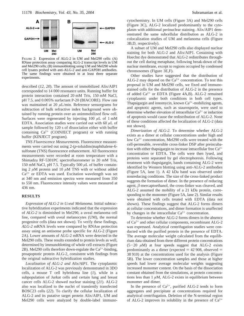
Expression of ALG-2 in UVeal Melanoma.Initial subtrac-tive hybridization experiments indicated that the expressionof ALG-2 is diminished in Mel290, a uveal melanoma cellline, compared with uveal melanocytes (UM), the normalprogenitor cells (data not shown). To verify these findings,ALG-2 mRNA levels were compared by RNAse protectionassay using an antisense probe specific for ALG-2 (Figure2A). Lower amounts of ALG-2 mRNA were detected in theMel290 cells. These results extended to protein levels as well,determined by immunoblotting of whole cell extracts (Figure2B). Mel290 cells therefore down-regulate the Ca2+-binding,proapoptotic protein ALG-2, consistent with findings fromthe original subtractive hybridization studies.
Localization of ALG-2 and Alix/AlP1.The cytoplasmiclocalization of ALG-2 was previously demonstrated in 3DOcells, a mouse T cell hybridoma line (1), while in asubpopulation of Jurkat cells and certain lung and breastcancer cells ALG-2 showed nuclear staining (21). ALG-2also was localized to the nuclei of transiently transfectedBOSC23 cells (22). To clarify the cellular localization ofALG-2 and its putative target protein Alix/AIP1, UM andMel290 cells were analyzed by double-label immuno-
cytochemistry. In UM cells (Figure 3A) and Mel290 cells(Figure 3C), ALG-2 localized predominantly to the cyto-plasm with additional perinuclear staining. Alix/AIP1 dem-onstrated the same subcellular distribution as ALG-2 incolocalization studies of UM and melanoma cells (Figure3B,D, respectively).
A subset of UM and Mel290 cells also displayed nuclearstaining for both ALG-2 and Alix/AIP1. Costaining withHoechst dye demonstrated that ALG-2 redistributes through-out the cell during metaphase, following break-down of thenuclear membrane, except to regions occupied by condensedchromosomes (Figure 3E,F).
Other studies have suggested that the distribution ofALG-2 may depend on the Ca2+ concentration. To test thisproposal in UM and Mel290 cells, we fixed and immuno-stained cells for the distribution of ALG-2 in the presenceof added Ca2+ or EDTA (Figure 4A,B). ALG-2 remainedcytoplasmic under both conditions in both cell types.Thapsigargin and ionomycin, known Ca2+-mobilizing agents,and apoptotic agents, such as staurosporin, were used todetermine whether elevation of intracellular Ca2+ or inductionof apoptosis would cause the redistribution of ALG-2. Noneof these conditions affected the localization of ALG-2 (datanot shown).
Dimerization of ALG-2.To determine whether ALG-2exists as a dimer at cellular concentrations under high andlow Ca2+ concentration, Mel290 cells were treated with thecell-permeable, reversible cross-linker DSP after preincuba-tion with either thapsigargin to increase intracellular free Ca2+
concentration or EDTA. The cells were then lysed, andproteins were separated by gel electrophoresis. Followingtreatment with thapsigargin, bands containing ALG-2 wereidentified by Western blotting using anti-ALG-2 antibodies(Figure 5A, lane 1). A 42 kDa band was observed undernonreducing conditions. The size of the cross-linked productsuggests the formation of dimer. In the presence of reducingagent,â-mercaptoethanol, the cross-linker was cleaved, andALG-2 assumed the mobility of a 21 kDa protein, corre-sponding to the monomer (Figure 5A, lane 2). Similar resultswere obtained with cells treated with EDTA (data notshown). These findings suggest that ALG-2 forms dimersat cellular concentrations, and dimer formation is unaffectedby changes in the intracellular Ca2+ concentration.
To determine whether ALG-2 forms dimers in the absenceof Ca2+ and other cellular components, recombinant ALG-2was expressed. Analytical centrifugation studies were con-ducted with the purified protein in the presence of EDTA.The average molecular weight calculated from the equilib-rium data obtained from three different protein concentrations(5-20 µM) at four speeds suggest that ALG-2 existspredominantly as a dimer (expected) 42 908, observed)38 910) at the concentrations used for the analysis (Figure5B). The lower concentration samples and those at higherspeeds had lower average molecular weights suggestingincreased monomer content. On the basis of the dissociationconstant obtained from the simulations, at protein concentra-tions less than 1µM, ALG-2 exists in equilibrium betweenmonomer and dimer.
In the presence of Ca2+, purified ALG-2 tends to formaggregates and precipitate at concentrations required foranalytical centrifugation. Deletion of the N-terminal regionof ALG-2 improves its solubility in the presence of Ca2+
FIGURE 2: Expression of ALG-2 in UM and Mel290 cells: (A)RNase protection assay comparing ALG-2 transcript levels in UMand Mel290 cells; (B) immunoblotting using UM and Mel290 wholecell lysates probed with anti-ALG-2 and anti-GAPDH antibodies.The same findings were obtained in at least three separateexperiments.
11178 Biochemistry, Vol. 43, No. 35, 2004 Subramanian et al.

(5). dNALG-2, the truncated form of ALG-2 comprised ofamino acids 21-191, therefore, was cloned and expressed.Analytical centrifugation experiments both in the presence(5 µM Ca2+) and absence (1 mM EDTA) of Ca2+ were thenconducted. Observed average molecular weights were 40 505and 39 626, respectively (expected dimer weight) 40 128).Results from these experiments support a single-speciesdimer model independent of Ca2+ (Figure 5C).
Interaction with Alix/AIP1.Surface plasmon resonance(SPR) has been used successfully to look at the interactionbetweenDictyostelium discoideumALG-2 and murine Alix/AIP1 (12). We examined the in vitro interaction betweenhuman ALG-2 and Alix/AIP1 to determine regions of
interaction and the Ca2+ dependency of their interaction.Purified Alix/AIP1 was immobilized on the surface of a CM5chip (Biacore). ALG-2 interaction with Alix/AIP1 wascompared in the presence and absence of Ca2+ (Figure 6A).No interaction was observed in the presence of EDTA, andthe response was similar to the control flow cell containingno immobilized Alix/AIP1. However, in the presence ofCa2+, there was an increased response to ALG-2 in the flowcell containing Alix/AIP1. Removal of both protein and Ca2+
resulted in the rapid dissociation of the complex as can beseen in Figure 6A. The relative response, measured at theplateau of the sensorgrams, increased as a function of bothprotein concentration (Figure 6B) and Ca2+ concentration
FIGURE 3: Cellular localization of ALG-2 and Alix/AIP1. UM (A,B) and Mel290 cells (C,D) were immunostained with anti-ALG-2 antibodiesfollowed by Texas red-conjugated secondary antibody (A,C) and with anti-Alix/AIP1 antibodies followed by FITC-conjugated secondaryantibodies (B,D). Mel290 cells were immunostained with anti-ALG-2 antibodies (E) and Hoechst dye (F). Intense labeling of ALG-2 wasobserved throughout metaphase cells except in the region of condensed chromosomes, indicated by a white arrow. Results are shown fromone of five similar experiments.
ALG-2 Dimerization and Interactions in Ocular Melanoma Biochemistry, Vol. 43, No. 35, 200411179

(Figure 6C) until saturation was reached at∼1000 nMALG-2 in the presence of 50µM Ca2+ or 400 nM ALG-2 inthe presence of 100µM Ca2+.
Deletion of F122, a residue absent in a noninteractingALG-2 isoform identified in mice (18), markedly reducedthe affinity for Alix/AIP1 and served as a negative control(Figure 8A).
These data collectively demonstrate that the interactionbetween ALG-2 and Alix/AIP1 is specific and depends onCa2+ concentration.
Effect of Ca2+ Binding to Functional EF Hands.Todetermine which Ca2+-binding sites are essential for theinteraction of ALG-2 with Alix/AIP1, site-directed mutagen-esis was performed to replace critical Ca2+-coordinatingresidues within the reported functional EF hands of ALG-2(4, 5, 18). Mutants were generated with single amino acidsubstitutions at glutamate residues in the consensus sequenceswithin EF-1 and EF-3, converting them to aspartate residues(E47D and E114D, respectively). As shown in other EF-hand-type calcium-binding proteins, replacement of glutamate,contributing two oxygens to the coordination of Ca2, with ashorter aspartate increases the rate of Ca2+ dissociation andlowers overall affinity without affecting electrostatic chargerequirements for loop stability (23, 24). In addition to thesereported high-affinity sites, the aspartate in EF-5, thepurported low-affinity site, was replaced with an alanine(D169A). CD measurements of mutants when compared toALG-2 show superimposable molar ellipticity spectra indi-cating that mutants retained their secondary structure (datanot shown).
Flow dialysis confirmed that wild-type ALG-2 possessestwo high-affinity Ca2+-binding sites and one low-affinity site(Figure 7), as published previously (18). In contrast, the
E47D mutant possesses only one high-affinity site, suggestingthat EF-1 is a normal high-affinity site in ALG-2. Thesubstitution of glutamate for aspartate in position 12 of thefirst Ca2+-chelating loop corresponding to EF-1 does noteliminate but lowers the affinity for Ca2+ by a factor of 30(K2′(ALG-2) ) 4.0 × 10⁵; K2′(E47D) ) 1.3 × 10⁴). Theaffinities of the remaining two sites are not significantlyaltered by this mutation. The mutation E114D leads to moreprofound alterations in the Ca2+-binding properties of ALG-
FIGURE 4: Effect of Ca2+ on ALG-2 Localization. Mel290 cellswere immunostained with anti-ALG-2 antibodies in the presenceof EDTA (A) and CaCl2 (B). Duplicate experiments were performedfor each condition with the same findings.
FIGURE 5: Dimerization of ALG-2. In panel A, The cell-permeablecross-linker DSP was added to Mel290 cells pretreated withthapsigargin. Cells were lysed and analyzed by immunoblottingusing anti-ALG-2 antibodies. Lane 1 contains cross-linked celllysate; lane 2 contains cross-linked cell lysate solubilized in thepresence ofâ-mercaptoethanol. In panel B, the absorbance at 280nm was recorded for three different concentrations of ALG-2 duringcentrifugation in the presence of 1 mM EDTA. The natural log ofthe absorbance values at equilibrium were plotted against the radialposition in the three cells during centrifugation at 9000, 12 000,16 000, and 22 000 rpm. In panel C, a similar plot was generatedfrom data obtained for dNALG-2 (5µM Ca2+). Bold lines showthe overlay of recovery values measured at 12 000 rpm aftercentrifugation at higher speeds with initial 12 000 rpm values,indicating little loss in protein due to irreversible precipitation. Thesedimentation equilibrium curves were fitted assuming the presenceof a single thermodynamically ideal species. Data were fitted asoutlined in the Materials and Methods. Cross-linking data representone of four independent experiments, while analytical centrifugationdata were gathered from duplicate experiments.
11180 Biochemistry, Vol. 43, No. 35, 2004 Subramanian et al.

2. Only two binding sites can be titrated (Figure 7), and theaffinities of these sites are lower than the high-affinity sitesin the wild-type protein (K1′(ALG-2) ) 5.3× 10⁵, K2′(ALG-2) ) 4.0 × 10⁵; K1′(E114D) ) 8.0 × 10⁴, K2′(E114D) )3.0× 10⁴). Thus it seems that this mutation simultaneously
weakens Ca2+ binding to EF-1 and EF-3 and abolishes thelow-affinity site. The binding curve of E114D shows negativecooperativity, and we suppose that EF-3 displays the lowerintrinsic affinity constant of the two (3.0× 10⁴). At theconcentrations of calcium (10-50 µM) used in subsequent
FIGURE 6: Interaction of ALG-2 with Alix/AIP1. In panel A, Alix/AIP1 was immobilized to the sensor chip, and 400 nM ALG-2 ordNALG-2 in buffer containing either 50µM CaCl2 or 20µM EDTA was injected using the KINJECT program as outlined in the Materialsand Methods. In panel B, different concentrations of ALG-2 (0-2 µM) in the presence of 50µM CaCl2 were reacted with an Alix/AIP1immobilized sensor chip. Average response values from duplicate runs at equilibrium plateau for each curve were plotted as a function ofALG-2 concentration. In panel C, samples containing 400 nM ALG-2 in the presence of varying amounts of CaCl2 (0-200 µM) wereinjected over the Alix/AIP1 immobilized sensor chip. Average response values from duplicate runs at equilibrium plateau for each curvewere plotted as a function of Ca2+ concentration. Response for buffer salts and Ca2+ were subtracted using an unimmobilized control flowcell in all cases.
ALG-2 Dimerization and Interactions in Ocular Melanoma Biochemistry, Vol. 43, No. 35, 200411181

experiments, the curves in Figure 7 demonstrate that wild-type ALG-2 binds 2 mol of Ca2+ per mole of protein, whilethe E47D and E114D mutants bind only 1 mol of Ca2+ permole of protein. The Ca2+-binding properties of the mutantD169A are quite similar to that of the wild-type ALG-2(Figure 7).
These mutants were then compared with ALG-2 for theirability to interact with Alix/AIP1 (Figure 8B). At Ca2+
concentration concentrations of 50µM, corresponding to thetwo high-affinity sites being occupied in ALG-2 and onesite in E47D and E114D (Figure 7), the mutants lackedsignificant interaction with Alix/AIP1, comparable to re-sponses using the unimmobilized flow cell. D169A had alower response in the presence of Ca2+ than full-lengthALG-2 but more than the baseline signal obtained for theEF-1 and EF-3 mutants. Experiments could not be conductedat higher concentrations of calcium in an attempt to occupyadditional calcium-binding sites in mutant forms of ALG-2owing to protein precipitation that occurs at elevated levelsof calcium in the absence of detergent.
Effect of the N-Terminal Non-PEF Region on Interactionswith Alix/AIP1.Studies of the crystal structure of a truncatedform of ALG-2 (residues 21-191) suggested that the bindingof the N-terminal region of ALG-2 to a hydrophobic pocketoccurs upon Ca2+ binding to EF-1 and EF-3 (Figure 1) (4).The folding of the N-terminal region into the hydrophobicpocket then causes a hinge effect, opening the molecule fortarget recognition. This model would suggest that the absenceof the N-terminal region might prevent or attenuate theinteraction of ALG-2 with the target molecule. To furthertest the model, the interaction of the N-terminal deletionmutant (dNALG-2) with Alix/AIP1 was studied using SPR.In the presence of Ca2+, the response obtained with dN-ALG-2 was comparable to full-length ALG-2 for im-mobilized Alix/AIP1. Further, the addition of peptidescorresponding to residues 7-17 (Figure 8C) and the deletedN-terminal portion, residues 1-22 (data not shown), to eitherdNALG-2 or ALG-2 had no effect on the interaction withAlix/AIP1. These results suggest that regions other than theN-terminal portion of ALG-2 interact with Alix/AIP1, and
the N-terminal region does not enhance the binding of ALG-2to Alix/AIP1.
Ca2+-Induced Conformational Changes in ALG-2.TheCa2+-induced conformational changes in ALG-2 and mutantswere studied by examining fluorescence emission spectra inthe presence of TNS. These experiments were carried out toinvestigate whether changes in surface hydrophobicity cor-related with affinity for the target molecule, Alix/AIP1, asdetermined by SPR. In the presence of Ca2+, ALG-2 showeda marked increase in fluorescence, similar to results obtainedwith mouse ALG-2 (5). Deletion of the N-terminal regionhad little effect on this Ca2+-induced fluorescence change(Figure 9A). Both full-length and truncated forms of ALG-2showed maximum fluorescence intensity at 40µM Ca2+ andsaturation at higher Ca2+ concentration. Based on the flowdata, this concentration is suggestive of only the high-affinitysites being occupied. As with the interaction with Alix/AIP1,there was no consequence of addition of the N-terminalpeptides (residues 7-17 or 1-21) to the Ca2+-induced TNSfluorescence changes of dNALG-2 or full-length ALG-2(data not shown). These results correlate with the results fromSPR studies where deletion of the N-terminal region had littleeffect on the interaction with Alix/AIP1.
SPR results clearly indicate that interference with Ca2+
binding at both EF-1 and EF-3 significantly compromisesthe interaction with Alix/AIP1 and that the D169A mutationalso has a moderate effect. TNS fluorescence experimentswere carried out with the mutants of ALG-2, E47D (EF-1),E114D (EF-3), and D169A (EF-5) in the absence (1 mMEDTA) and presence of 60µM Ca2+. Mutation in EF-3completely abrogated the Ca2+-induced hydrophobicity changesdetectable by TNS fluorescence (Figure 9B), correlating withthe lack of interaction of this mutant with Alix/AIP1. Incontrast, the EF-1 mutation only slightly reduced the TNSfluorescence signal upon the addition of Ca2+. The D169Amutation resulted in an intermediate Ca2+-induced fluores-cence change. Results suggest that the most significantconformational change, detectable by TNS fluorescencechanges, arises from Ca2+ binding to EF-3.
DISCUSSION
In addition to unregulated cell growth, disruption ofapoptosis may be important during tumor growth andmetastasis. More malignant tumor cells may be selectedowing to their higher expression of antiapoptotic factors ortheir down regulation of proapoptotic genes (25, 26). Ca2+
plays a significant role in apoptosis and mediates at least aportion of its function through the action of Ca2+-bindingproteins (27). In uveal melanoma, we demonstrate thedifferential expression ofALG-2, a gene identified as a Ca2+-dependent regulator of apoptosis. Mel290 cells derived froma human tumor were found to express lower levels of ALG-2mRNA and protein than normal uveal melanocytes. Thedown regulation of ALG-2 suggests one way by which thesecells may achieve a selective advantage, by interfering withCa2+-mediated apoptotic signals and thereby enhancing cellsurvival.
Krebs et al. (28) compared the expression of ALG-2 incell extracts obtained from biopsies of normal and malignanttissues from the same patient. Unlike uveal melanoma, asignificant increase in ALG-2 expression in the cancerous
FIGURE 7: Ca2+ binding by recombinant ALG-2 and three pointmutants. Ca2+ binding was measured by flow dialysis at 25°C in50 mM Tris-HCl buffer, pH 7.5, 150 mM KCl containing 0.5%Tween 20. Duplicate experiments are indicated with rectangles andcircles. Intrinsic binding constants were derived from the stoichio-metric constants, which were obtained by fitting of the experimentaldata to the Adair equation for two (E114D) or three sites (ALG-2,E47D, D169A).
11182 Biochemistry, Vol. 43, No. 35, 2004 Subramanian et al.

tissues from different lung and breast tumors was reported.Immunohistochemistry of the lung carcinomas in thosestudies showed intense ALG-2 staining largely localized tothe nuclei of tumor cells. However, some commercial anti-ALG-2 antibodies have been found by this and otherlaboratories (29, 30) to recognize a protein of the same sizeas ALG-2 but to be unable to recognize the recombinantprotein. We therefore generated polyclonal antibodies topurified and sequenced human ALG-2 and used them forimmunoblotting and immunohistochemical analyses. ALG-2
was found to localize to the cytoplasm of UM and melanomacells independent of Ca2+ concentration or activation ofprogrammed cell death. Treatment of these cells withthapsigargin, a drug known to release calcium from intra-cellular stores and cause cell death through ER stress withpotential ALG-2 involvement (2), did not result in anychange in localization of ALG-2. The contrast in findingsbetween our studies and others (21, 28, 29) may be due tothe different cancer cell types being examined or the fidelityof the antibodies used in the studies (30).
FIGURE 8: Interactions of ALG-2 and mutants with Alix/AIP1. In panel A, ALG-2 and dF122 (400 nM each) were injected over theimmobilized Alix/AIP1 surface in the presence of 50µM CaCl2 using the COINJECT program outlined in the Materials and Methods. Inpanel B, similarly, ALG-2 and the three EF-hand mutants, E47D, E114D, and D169A were compared using the COINJECT method. Inpanel C, ALG-2 and dNALG-2 (500 nM each) in the presence of 50µM CaCl2 with or without 200µM decapeptide were analyzed bysurface plasmon resonance for interaction with immobilized Alix/AIP1 using the KINJECT program. Flat plateaus as seen with KINJECTwere not seen with COINJECT due to methodological differences between the two programs; however, slower dissociation rates wereobservable with COINJECT by retaining Ca2+ concentration in the dissociation phase. Experiments were conducted at least twice usingduplicate samples in each experiment.
ALG-2 Dimerization and Interactions in Ocular Melanoma Biochemistry, Vol. 43, No. 35, 200411183

Various biochemical and structural approaches have beenused to characterize different ALG-2 analogues (3, 4, 12,18, 20, 31) . As with other members of this family of Ca2+-binding proteins, ALG-2 exists as a dimer in solution (32,33). From our studies, dimer formation is independent ofCa2+ and is unaffected by the deletion of the N-terminalregion. The role of the N-terminal non-EF-hand region ofALG-2 might vary between species. The ALG-2 isoformsin Dictyosteliumwere found to require the N-terminal regionfor both dimer formation and interaction (12). Both ALG-2forms had longer N-terminal regions than mouse or humanALG-2. In the case of human ALG-2, deletion of theN-terminal region improved the solubility of the protein inthe presence of Ca2+. The deletion, however, did not affectdimer formation, consistent with the involvement of theC-terminal region in this process based on the crystalstructure (Figure 1) (4). Deletion of the N-terminal regionalso did not significantly affect the Ca2+-induced conforma-tional change as observed by TNS fluorescence measure-ments. Ca2+-dependent interaction with Alix/AIP1 for full-length ALG-2 and dNALG-2 also were comparable. TheN-terminal region of ALG-2 therefore might have more
significant consequences in terms of its interactions withtarget molecules other than Alix/AIP1 in other cell types.
On the basis of the crystal structure of Ca2+-bound,elastase-digested ALG-2, it was suggested that the bindingof Ca2+ resulted in the formation of a hydrophobic pocket,occupied by the N-terminal region of ALG-2 and causing ahinge-like effect, opening the molecule for target binding(4). TNS fluorescence and Alix/AIP1 interaction studiespresented here show no difference between ALG-2 anddNALG-2 with or without the addition of exogenous peptidescorresponding to the cocrystallized decapeptide derived fromelastase treatment of ALG-2, as well as the entire N-terminalpeptide, suggesting that at least in the case of Alix/AIP1,the interaction mechanism does not involve the N-terminalregion of human ALG-2.
Of the five EF-hands in ALG-2, EF-1 and EF-3 have beenidentified as the high-affinity Ca2+-binding sites (4, 5, 18).The location of the low-affinity site is less well established.In the crystal structure, a third Ca2+ is bound to EF-5 (4),potentially due to artificially high concentrations of proteinand Ca2+. Site-directed mutagenesis was used in the studiespresented here to specifically alter a critical residue required
FIGURE 9: Ca2+-induced conformational change in ALG-2 and mutants measured by TNS fluorescence. In panel A, fluorescence intensityvalues at 436 nm were measured for ALG-2 ([) and dNALG-2 (9) with varying amounts of added CaCl2 (0-200 µM). Average valuesfrom duplicate measurements were plotted as a function of Ca2+ concentration. In panel B, emission fluorescence spectra were measuredfor solutions containing 2µM ALG-2, E47D, E114D, or D169A in the presence of 60µM CaCl2 and 10µM TNS as described in theMaterials and Methods. ALG-2 (2µM) with no added Ca2+ in the presence of 10µM TNS indicates basal fluorescence.
11184 Biochemistry, Vol. 43, No. 35, 2004 Subramanian et al.

for Ca2+ coordination in each of these EF hands. Flowdialysis data of the E47D mutant confirm that EF-1comprises one of the high-affinity Ca2+-binding sites andthe mutation effectively eliminates Ca2+ binding. In contrast,the concomitant decrease of both high-affinity sites in theE114D mutant is unexpected and points to a structural roleof this residue. Flow dialysis data of E114D indicate a directeffect on EF-3, the second high-affinity Ca2+-binding site,and an indirect effect on EF-1. The relatively small differencein the binding curves of D169A and wild-type ALG-2,together with the fact that the low-affinity site is notabolished, suggest that the precise location of the latter siteremains unresolved. The crystal structure of ALG-2 showstwo Ca2+ ions bound to EF-3; perhaps the E114D mutationdisables the binding of one ion but not the other. If the extraCa2+ in EF-3 corresponds to the low-affinity site rather thanEF-5, then this might explain the ineffectiveness of theD169A mutation in abolishing the low-affinity Ca2+-bindingsite. The physiological relevance of the low-affinity site,however, is unclear since occupancy requires millimolarCa2+, unexpected in the cytosol.
Interestingly, the E47D and E114D mutations both resultedin loss of interaction with Alix/AIP1. The D169A mutation,despite little change in Ca2+ affinity, also affected theinteraction though not as significantly as the EF-1 and EF-3mutants. The crystal structure (Figure 1) and studies on othermembers of the PEF family (34-38) indicate that this regionis important for dimer formation. Residues from the F-helixof EF-5 also contribute to the hydrophobic pocket, potentiallyinvolved in target recognition. The reduced interaction mighttherefore result from the mutation in EF-5 impacting on otherstructural aspects important to the interaction of ALG-2 withAlix/AIP1 rather than influencing Ca2+-binding. This furthersupports a model whereby EF-1 and EF-3 are the functionalCa2+-binding sites in ALG-2 and both sites have to beoccupied for substantive interaction with Alix/AIP1.
The shorter aspartate in the E114D mutant may reorientthe C-terminalR-helix of EF-3, reshape the microenviron-ment around F122, and reduce hydrophobic exposure. TNSfluorescence results support this conclusion, whereby Ca2+
binding to EF-3 is essential for the conformational changeresulting in the increased exposure of hydrophobic surfaces.Since both EF-1 and EF-3 are required for the interaction ofALG-2 with Alix/AIP1, Ca2+ binding to EF-1 might resultin angle changes within the protein that are necessary fortarget recognition but do not significantly alter surfacehydrophobicity.
In summary, we demonstrate that ALG-2 exists as a dimerin the cytoplasm of cells, independent of Ca2+ concentration,and undergoes conformational changes, including the in-creased exposure of hydrophobic surfaces through Ca2+
binding to EF-1 and EF-3. These conformational changesallow for target recognition and binding of Alix/AIP1. Basedon the expression of putative target molecules in differentcell types and physiological conditions, ALG-2 can mediatepathways involved in apoptosis (1), cell proliferation (28,29), or both.
In uveal melanoma cells, reduced expression of ALG-2likely favors cell survival through diminished apoptosis. Mostcurrent therapies are nonspecific and can result in consider-able damage to normal tissue and have little impact onmortality, reaching more than 50% in high-risk melanoma
patients (39-41). The characterization of biochemical path-ways that can modulate cell death may lead to moleculartherapies selectively targeting tumor cells.
ACKNOWLEDGMENT
We would like to thank Dr. R. Stenkamp for his help withFigure 1 and Dr. D. R. McCaslin for his help with theanalysis of the analytical centrifugation data.
REFERENCES
1. Vito, P., Lacana, E., and D′Adamio, L. (1996) Interfering withapoptosis: Ca(2+)-binding protein ALG-2 and Alzheimer’sdisease gene ALG-3,Science 271, 521-525.
2. Rao, R. V., Poksay K. S., Castro-Obregon S., Schilling B., RowR. H., del Rio G., Gibson B. W., Ellerby H. M., and Bredesen,D. E. (2004) Molecular components of a cell death pathwayactivated by endoplasmic reticulum stress,J. Biol. Chem. 279,177-187.
3. Lo, K. W. H., Zhang, Q., Li, M., and Zhang, M. J. (1999)Apoptosis-linked gene product ALG-2 is a new member of thecalpain small subunit subfamily of Ca2+-binding proteins,Bio-chemistry 38, 7498-7508.
4. Jia, J., Tarabykina, S., Hansen, C., Berchtold, M., and Cygler, M.(2001) Structure of apoptosis-linked protein ALG-2: insights intoCa2+-induced changes in penta-EF-hand proteins,Structure(Cambridge) 9, 267-275.
5. Maki, M., Yamaguchi, K., Kitaura, Y., Satoh, H., and Hitomi, K.(1998) Calcium-induced exposure of a hydrophobic surface ofmouse ALG-2, which is a member of the penta-EF-hand proteinfamily, J. Biochem. (Tokyo) 124, 1170-1177.
6. Missotten, M., Nichols, A., Rieger, K., and Sadoul, R. (1999) Alix,a novel mouse protein undergoing calcium-dependent interactionwith the apoptosis-linked-gene 2 (ALG-2) protein,Cell DeathDiffer. 6, 124-129.
7. Vito, P., Pellegrini, L., Guiet, C., and D′Adamio, L. (1999) Cloningof AIP1, a novel protein that associates with the apoptosis-linkedgene ALG-2 in a Ca2+-dependent reaction,J. Biol. Chem. 274,1533-1540.
8. Chen, B., Borinstein, S. C., Gillis, J., Sykes, V. W., and Bogler,O. (2000) The glioma-associated protein SETA interacts withAIP1/Alix and ALG-2 and modulates apoptosis in astrocytes,J.Biol. Chem. 275, 19275-19281.
9. Katoh, K., Shibata, H., Suzuki, H., Nara, A., Ishidoh, K.,Kominami, E., Yoshimori, T., and Maki, M. (2003) The ALG-2-interacting protein Alix associates with CHMP4b, a humanhomologue of yeast Snf7 that is involved in multivesicular bodysorting,J. Biol. Chem. 278, 39104-39113.
10. Trioulier, Y., Torch, S., Blot, B., Cristina, N., Chatellard-Causse,C., Verna, J. M., and Sadoul, R. (2004) Alix, a protein regulatingendosomal trafficking, is involved in neuronal death,J. Biol.Chem. 279, 2046-2052.
11. Tarabykina, S., Moller, A. L., Durussel, I., Cox, J., and Berchtold,M. W. (2000) Two forms of the apoptosis-linked protein ALG-2with different Ca2+ affinities and target recognition,J. Biol. Chem.275, 10514-10518.
12. Aubry, L., Mattei, S., Blot, B., Sadoul, R., Satre, M., and Klein,G. (2002) Biochemical characterization of two analogues of theapoptosis-linked gene 2 protein in Dictyostelium discoideum andinteraction with a physiological partner in mammals, murine Alix,J. Biol. Chem. 277, 21947-21954.
13. Walker, T. M., Van Ginkel, P. R., Gee, R. L., Ahmadi, H.,Subramanian, L., Ksander, B. R., Meisner, L. F., Albert, D. M.,and Polans, A. S. (2002) Expression of angiogenic factors Cyr61and tissue factor in uveal melanoma,Arch. Ophthalmol. 120,1719-1725.
14. Hu, D. N., McCormick, S. A., Orlow, S. J., Rosemblat, S., andLin, A. Y. (1997) Regulation of melanogenesis by human uvealmelanocytes in vitro,Exp. Eye Res. 64, 397-404.
15. Polans, A. S., Palczewski, K., Asson-Batres, M. A., Ohguro, H.,Witkowska, D., Haley, T. L., Baizer, L., and Crabb, J. W. (1994)Purification and primary structure of Capl, an S-100-relatedcalcium-binding protein isolated from bovine retina,J. Biol. Chem.269, 6233-6240.
ALG-2 Dimerization and Interactions in Ocular Melanoma Biochemistry, Vol. 43, No. 35, 200411185

16. van Ginkel, P. R., Gee, R. L., Walker, T. M., Hu, D. N., Heizmann,C. W., and Polans, A. S. (1998) The identification and differentialexpression of calcium-binding proteins associated with ocularmelanoma,Biochim. Biophys. Acta Mol. Cell Res. 1448, 290-297.
17. Colowick, S. P., and Womack, F. C. (1969) Binding of diffusiblemolecules by macromolecules: rapid measurement by rate ofdialysis,J. Biol. Chem. 244, 774-777.
18. Tarabykina, S., Moller, A. L., Durussel, I., Cox, J., and Berchtold,M. W. (2000) Two forms of the apoptosis-linked protein ALG-2with different Ca(2+) affinities and target recognition,J. Biol.Chem. 275, 10514-10518.
19. Cox, J. A. (1996) inGuidebook to the Calcium-binding Proteins(Celio, M. R., Paul, S. T., and Schwaller, B., Eds.) pp 1-12,Oxford University Press, Oxford, U.K.
20. Satoh, H., Shibata, H., Nakano, Y., Kitaura, Y., and Maki, M.(2002) ALG-2 interacts with the amino-terminal domain ofannexin XI in a Ca2+-dependent manner,Biochem. Biophys. Res.Commun. 291, 1166-1172.
21. Krebs, J., and Klemenz, R. (2000) The ALG-2/AIP-complex, amodulator at the interface between cell proliferation and cell death?A hypothesis,Biochim. Biophys. Acta 1498, 153-161.
22. Hwang, I. S., Jung, Y. S., and Kim, E. (2002) Interaction of ALG-2with ASK1 influences ASK1 localization and subsequent JNKactivation,FEBS Lett. 529, 183-187.
23. Palczewski, K., Polans, A. S., Baehr, W., and Ames, J. B. (2000)Ca2+-binding proteins in the retina: structure, function, and theetiology of human visual diseases,BioEssays 22, 337-350.
24. Babu, A., Su, H., Ryu, Y., and Gulati, J. (1992) Determination ofresidue specificity in the EF-hand of troponin C for Ca2+
coordination, by genetic engineering,J. Biol. Chem. 267, 15469-15474.
25. Green, D. R., and Evan, G. I. (2002) A matter of life and death,Cancer Cell 1, 19-30.
26. Soengas, M. S., Capodieci P., Polsky D., Mora J., Esteller M.,Opitz-Araya X., McCombie R., Herman J. G., Gerald W. L.,Lazebnik Y. A., Cordon-Cardo C., and Lowe S. W. (2001)Inactivation of the apoptosis effector Apaf-1 in malignantmelanoma,Nature 409, 207-211.
27. Hajnoczky, G., Davies, E., and Madesh, M. (2003) Calciumsignaling and apoptosis,Biochem. Biophys. Res. Commun. 304,445-454.
28. Krebs, J., Saremaslani, P., and Caduff, R. (2002) ALG-2: a Ca2+-binding modulator protein involved in cell proliferation and incell death,Biochim. Biophys. Acta 1600, 68-73.
29. la Cour, J. M., Mollerup, J., Winding, P., Tarabykina, S., Sehested,M., and Berchtold, M. W. (2003) Up-regulation of ALG-2 inhepatomas and lung cancer tissue,Am. J. Pathol. 163, 81-89.
30. Mollerup, J., Krogh, T. N., Nielsen, P. F., and Berchtold, M. W.(2003) Properties of the co-chaperone protein p23 erroneously
attributed to ALG-2 (apoptosis-linked gene 2),FEBS Lett. 555,478-482.
31. Kitaura, Y., Matsumoto, S., Satoh, H., Hitomi, K., and Maki, M.(2001) Peflin and ALG-2, members of the penta-EF-hand proteinfamily, form a heterodimer that dissociates in a Ca2+-dependentmanner,J. Biol. Chem. 276, 14053-14058.
32. Teahan, C. G., Totty, N. F., and Segal, A. W. (1992) Isolationand characterization of grancalcin, a novel 28 kDa EF-handcalcium-binding protein from human neutrophils,Biochem. J. 286(Part 2), 549-554.
33. Hamada, H., Okochi, E., Oh-hara, T., and Tsuruo, T. (1988)Purification of the Mr 22,000 calcium-binding protein (sorcin)associated with multidrug resistance and its detection withmonoclonal antibodies,Cancer Res. 48, 3173-3178.
34. Jia, J., Han Q., Borregaard N., Lollike K., and Cygler M. (2000)Crystal structure of human grancalcin, a member of the penta-EF-hand protein family,J. Mol. Biol. 300, 1271-1281.
35. Kitaura, Y., Satoh, H., Takahashi, H., Shibata, H., and Maki, M.(2002) Both ALG-2 and peflin, penta-EF-hand (PEF) proteins,are stabilized by dimerization through their fifth EF-hand regions,Arch. Biochem. Biophys. 399, 12-18.
36. Hansen, C., Tarabykina, S., la Cour, J. M., Lollike, K., andBerchtold, M. W. (2003) The PEF family proteins sorcin andgrancalcin interact in vivo and in vitro,FEBS Lett. 545, 151-154.
37. Maki, M., Kitaura, Y., Satoh, H., Ohkouchi, S., and Shibata, H.(2002) Structures, functions and molecular evolution of the penta-EF-hand Ca2+-binding proteins,Biochim. Biophys. Acta 1600,51-60.
38. Todd, B., Moore, D., Deivanayagam, C. C., Lin, G. D., Chatto-padhyay, D., Maki, M., Wang, K. K., and Narayana, S. V. (2003)A structural model for the inhibition of calpain by calpastatin:crystal structures of the native domain VI of calpain and itscomplexes with calpastatin peptide and a small molecule inhibitor,J. Mol. Biol. 328, 131-146.
39. Seddon, J. M., Gragoudas, E. S., Albert, D. M., Hsieh, C. C.,Polivogianis, L., and Friedenberg, G. R. (1985) Comparison ofsurvival rates for patients with uveal melanoma after treatmentwith proton beam irradiation or enucleation,Am. J. Ophthalmol99, 282-290.
40. Kath, R., Hayungs J., Bornfeld N., Sauerwein W., Hoffken K.,and Seeber S. (1993) Prognosis and treatment of disseminateduveal melanoma,Cancer 72, 2219-2223.
41. Bedikian, A. Y., Legha, S. S., Mavligit, G., Carrasco, C. H.,Khorana, S., Plager, C., Papadopoulos, N., and Benjamin, R. S.(1995) Treatment of uveal melanoma metastatic to the liver- Areview of the M D Anderson Cancer Center experience andprognostic factors,Cancer 76, 1665-1670.
BI048848D
11186 Biochemistry, Vol. 43, No. 35, 2004 Subramanian et al.