Embed Size (px)
Citation preview
DISCLOSURE OF RELATIONSHIPS WITH INDUSTRY
Carolyn S. Lee, MD, PhD, FAAD [email protected] @CarolynLeeMDPhD
C001 Structure and Function of the Epidermis
DISCLOSURES
Idonothaveanyrelevantrela6onshipswithindustry.
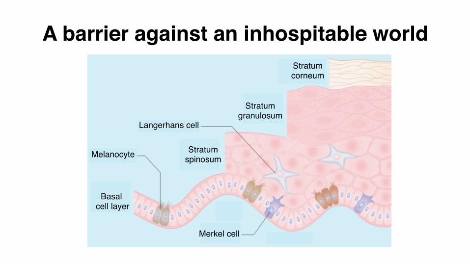
Langerhans cell
Melanocyte Stratum spinosum
Stratum granulosum
Stratum corneum
Basal cell layer
Merkel cell
A barrier against an inhospitable world
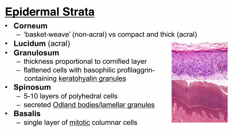
Epidermal Strata• Corneum
– ‘basket-weave’ (non-acral) vs compact and thick (acral) • Lucidum (acral) • Granulosum
– thickness proportional to cornified layer – flattened cells with basophilic profilaggrin- containing keratohyalin granules
• Spinosum – 5-10 layers of polyhedral cells – secreted Odland bodies/lamellar granules
• Basalis – single layer of mitotic columnar cells
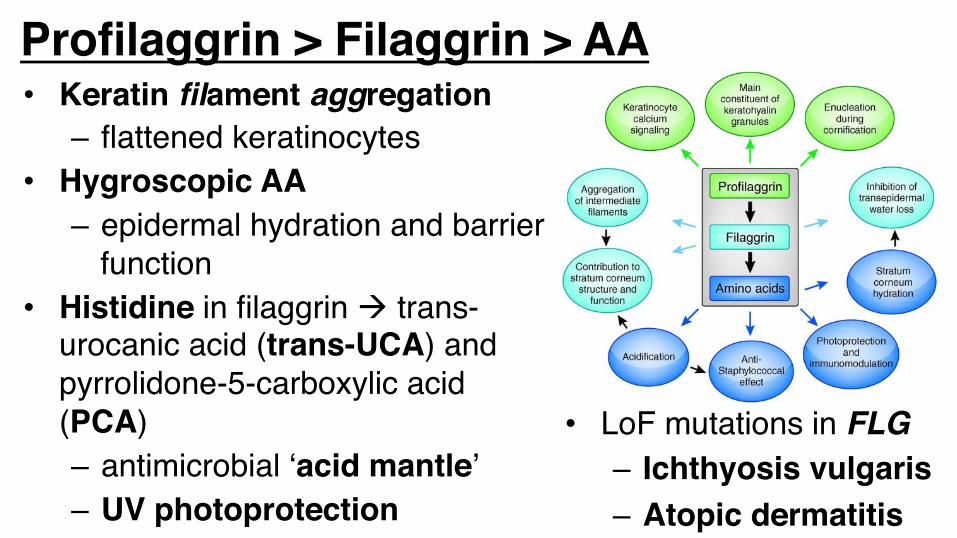
Profilaggrin > Filaggrin > AA
• LoF mutations in FLG– Ichthyosis vulgaris– Atopic dermatitis
• Keratin filament aggregation– flattened keratinocytes
• Hygroscopic AA– epidermal hydration and barrier
function• Histidine in filaggrin à trans-
urocanic acid (trans-UCA) and pyrrolidone-5-carboxylic acid (PCA)– antimicrobial ‘acid mantle’– UV photoprotection
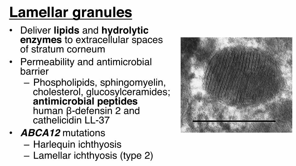
Lamellar granules• Deliver lipids and hydrolytic
enzymes to extracellular spaces of stratum corneum
• Permeability and antimicrobial barrier– Phospholipids, sphingomyelin,
cholesterol, glucosylceramides; antimicrobial peptides human β-defensin 2 and cathelicidin LL-37
• ABCA12 mutations– Harlequin ichthyosis– Lamellar ichthyosis (type 2)
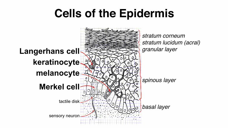
Cells of the Epidermis
basal layer
spinous layer
granular layer
stratum corneumstratum lucidum (acral)
sensory neuron
tactile disk
Merkel cell melanocyte
keratinocyte Langerhans cell
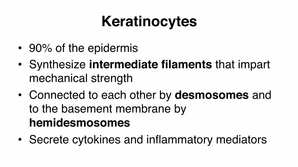
Keratinocytes• 90% of the epidermis• Synthesize intermediate filaments that impart
mechanical strength• Connected to each other by desmosomes and
to the basement membrane by hemidesmosomes
• Secrete cytokines and inflammatory mediators
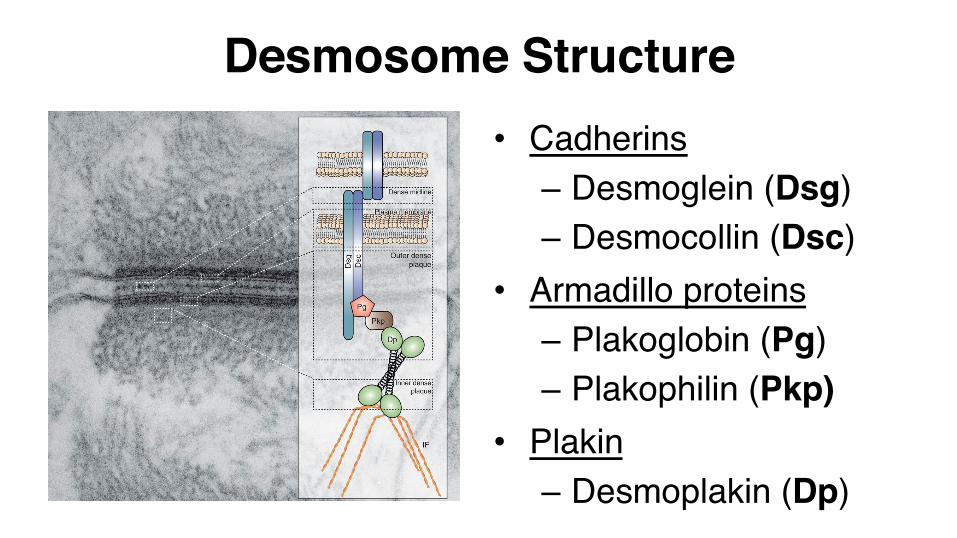
Desmosome Structure• Cadherins
– Desmoglein (Dsg)– Desmocollin (Dsc)
• Armadillo proteins– Plakoglobin (Pg)– Plakophilin (Pkp)
• Plakin– Desmoplakin (Dp)
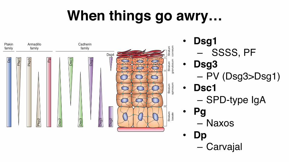
When things go awry…• Dsg1
– SSSS, PF • Dsg3
– PV (Dsg3>Dsg1)• Dsc1
– SPD-type IgA • Pg
– Naxos• Dp
– Carvajal
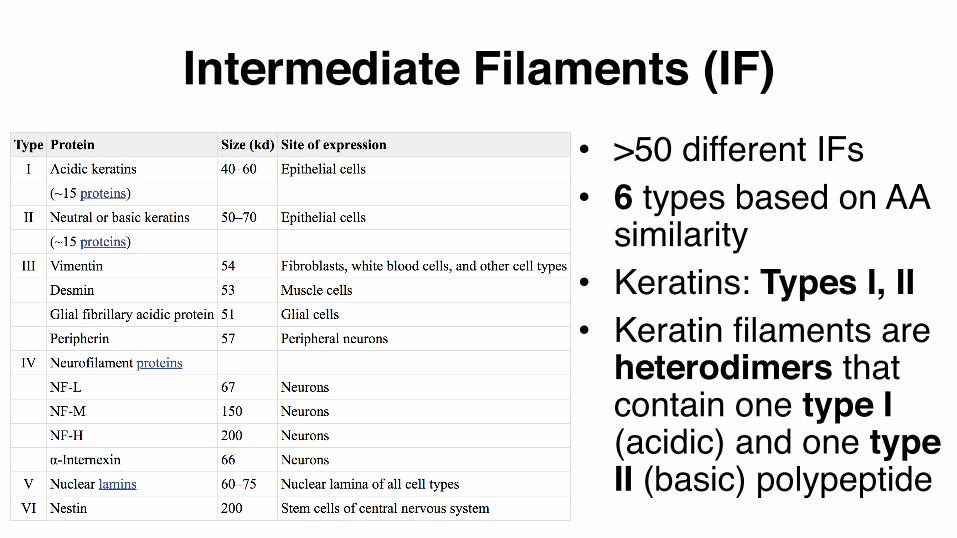
• >50 different IFs• 6 types based on AA
similarity• Keratins: Types I, II• Keratin filaments are
heterodimers that contain one type I (acidic) and one type II (basic) polypeptide
Intermediate Filaments (IF)
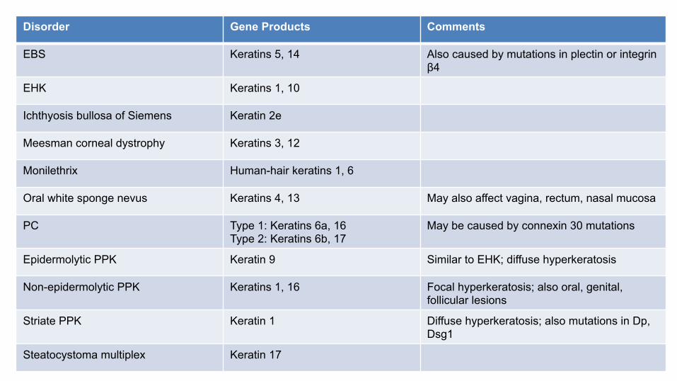
Disorder Gene Products Comments
EBS Keratins 5, 14 Also caused by mutations in plectin or integrin β4
EHK Keratins 1, 10
Ichthyosis bullosa of Siemens Keratin 2e
Meesman corneal dystrophy Keratins 3, 12
Monilethrix Human-hair keratins 1, 6
Oral white sponge nevus Keratins 4, 13 May also affect vagina, rectum, nasal mucosa
PC Type 1: Keratins 6a, 16 Type 2: Keratins 6b, 17
May be caused by connexin 30 mutations
Epidermolytic PPK Keratin 9 Similar to EHK; diffuse hyperkeratosis
Non-epidermolytic PPK Keratins 1, 16 Focal hyperkeratosis; also oral, genital, follicular lesions
Striate PPK Keratin 1 Diffuse hyperkeratosis; also mutations in Dp, Dsg1
Steatocystoma multiplex Keratin 17
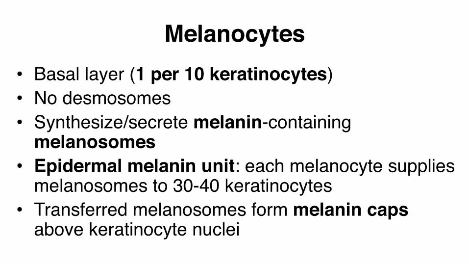
Melanocytes• Basal layer (1 per 10 keratinocytes)• No desmosomes• Synthesize/secrete melanin-containing
melanosomes• Epidermal melanin unit: each melanocyte supplies
melanosomes to 30-40 keratinocytes • Transferred melanosomes form melanin caps
above keratinocyte nuclei
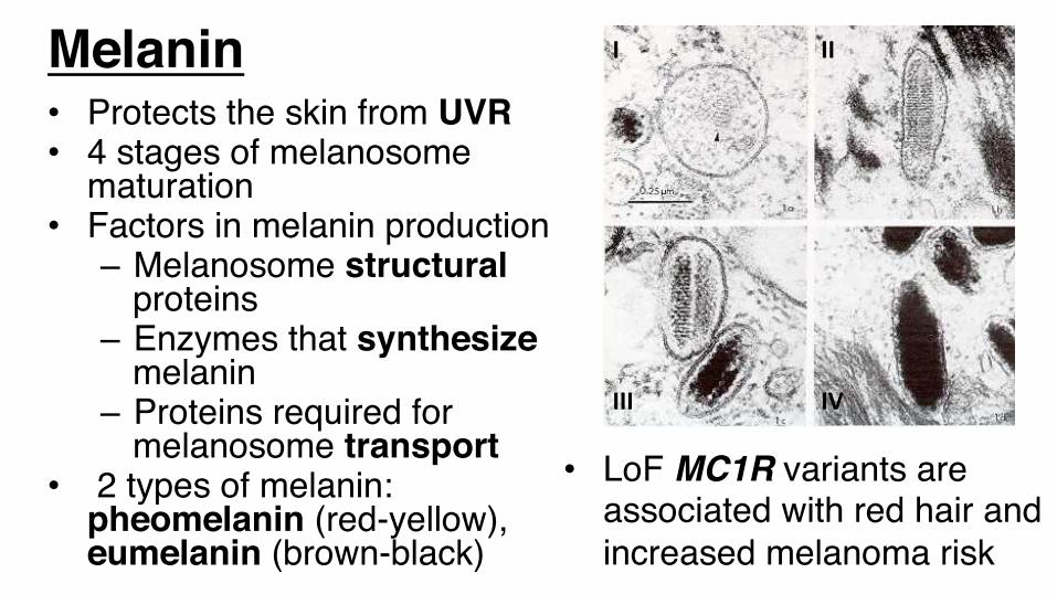
Melanin• Protects the skin from UVR• 4 stages of melanosome
maturation• Factors in melanin production
– Melanosome structural proteins
– Enzymes that synthesize melanin
– Proteins required for melanosome transport
• 2 types of melanin: pheomelanin (red-yellow), eumelanin (brown-black)
I II
III IV
• LoF MC1R variants are associated with red hair and increased melanoma risk
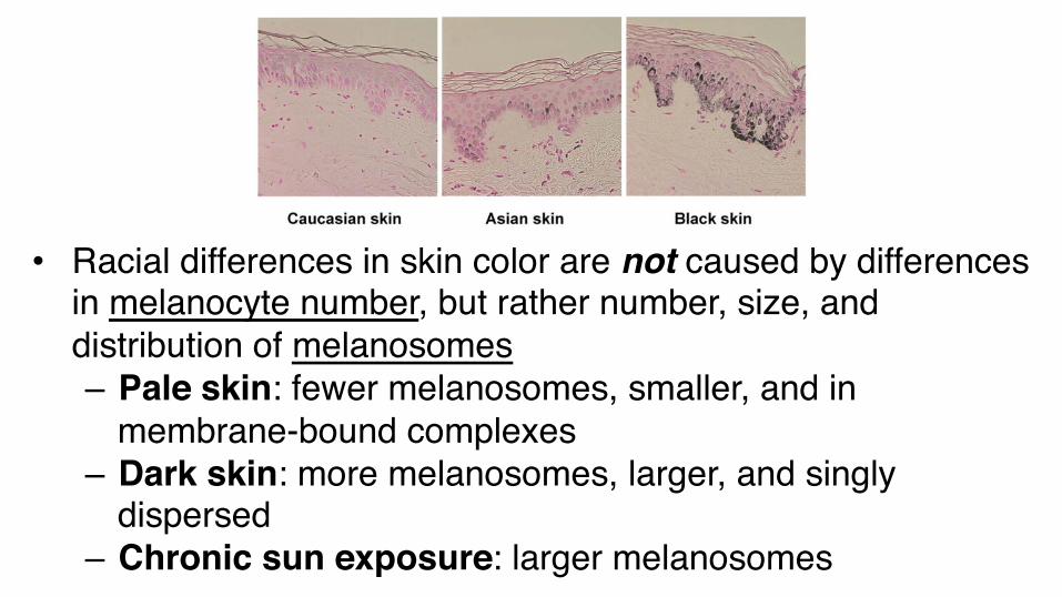
• Racial differences in skin color are not caused by differences in melanocyte number, but rather number, size, and distribution of melanosomes– Pale skin: fewer melanosomes, smaller, and in
membrane-bound complexes– Dark skin: more melanosomes, larger, and singly
dispersed– Chronic sun exposure: larger melanosomes
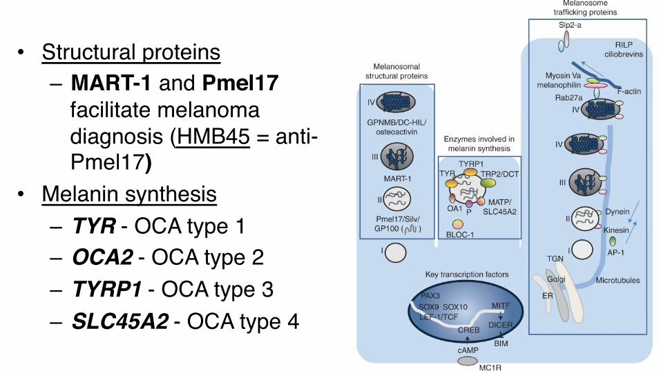
• Structural proteins– MART-1 and Pmel17
facilitate melanoma diagnosis (HMB45 = anti-Pmel17)
• Melanin synthesis– TYR - OCA type 1– OCA2 - OCA type 2– TYRP1 - OCA type 3– SLC45A2 - OCA type 4
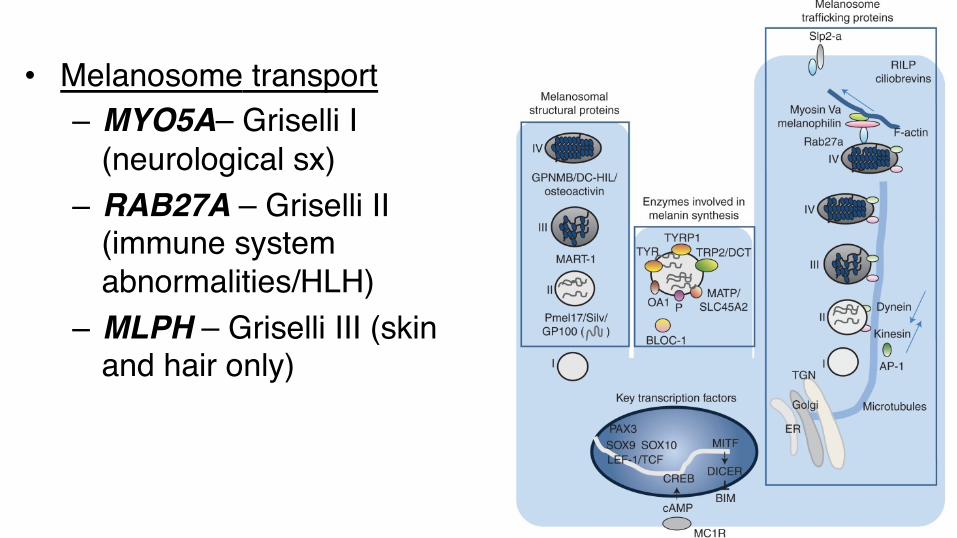
• Melanosome transport– MYO5A– Griselli I
(neurological sx)– RAB27A – Griselli II
(immune system abnormalities/HLH)
– MLPH – Griselli III (skin and hair only)
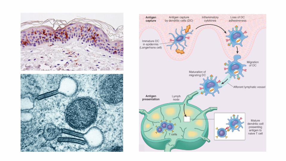
Langerhans cells• Predominantly spinous layer (3-5%)• No desmosomes• Bone marrow-derived dendritic cells that function
as skin-resident APC• Constitutively express MHC class II (HLA-DR)• Stain with gold chloride, CD1a, S-100• Contain reniform nuclei and Langerhans/Birbeck
granules by EM
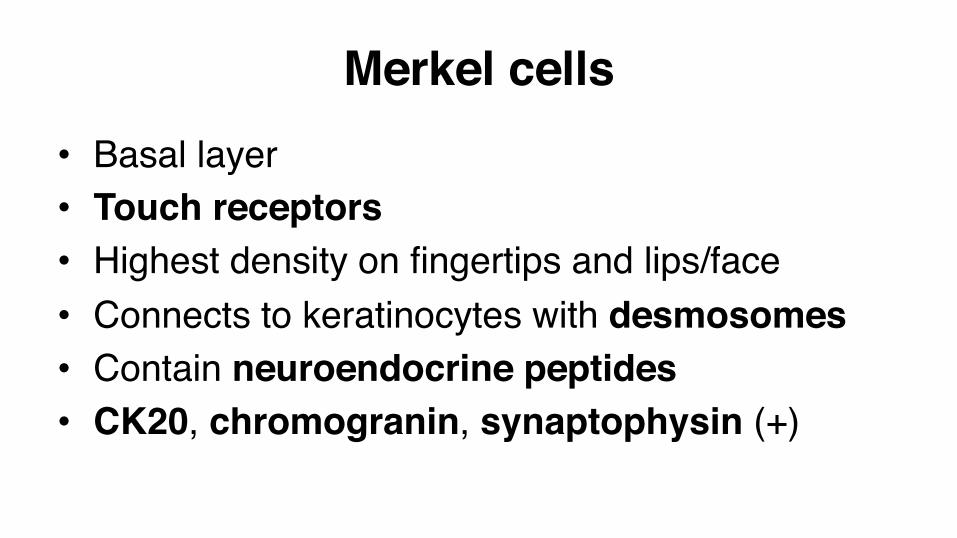
Merkel cells• Basal layer• Touch receptors• Highest density on fingertips and lips/face• Connects to keratinocytes with desmosomes• Contain neuroendocrine peptides• CK20, chromogranin, synaptophysin (+)
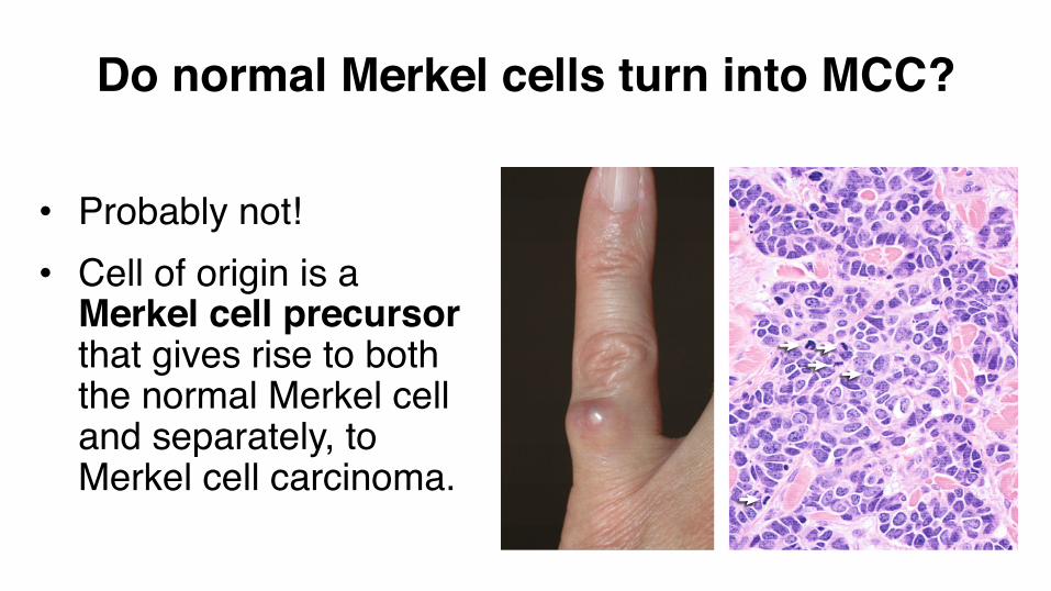
Do normal Merkel cells turn into MCC?
• Probably not!• Cell of origin is a
Merkel cell precursor that gives rise to both the normal Merkel cell and separately, to Merkel cell carcinoma.
Key points1. Keratinocytes are the main cell of the epidermis2. Layers in ascending order: basal, spinous, granular,
cornified3. Basal cells are undifferentiated, proliferating cells4. Keratinocytes are connected to each other by
desmosomes
5. Profilaggrin is an important component of keratohyalin granules
6. The cornified layer is the major physical barrier7. Number and size of melanosomes, not
melanocytes, determine skin color8. Langerhans cells are derived from bone
marrow and function as a vital part of the skin’s immunologic defense
Key points


![Differentiation-Dependent Expression of Keratins in Human Oral … · 2017. 2. 1. · [ 16, 17]. The expression of specific keratins appears to depend on the type of tissue, as well](https://img.pdfslide.us/doc/110x75/5ff979cead588c6cd35f8d9b/differentiation-dependent-expression-of-keratins-in-human-oral-2017-2-1-16.jpg)