Embed Size (px)
Citation preview
8/16/2019 C V M - JULY.pptx
http://slidepdf.com/reader/full/c-v-m-julypptx 1/46

8/16/2019 C V M - JULY.pptx
http://slidepdf.com/reader/full/c-v-m-julypptx 2/46
DEFINITION
• Cerebrovascular malforma!o"s #CVMs$ area %eero&e"eous &rou's of (!sor(ers %are'rese" mor'%o&e"e!c errors a)ec!"&
arer!es* ca'!llar!es* ve!"s or var!ouscomb!"a!o" of vessels+
• Develo'me" of %e %uma" feal vasculars,sem occurs v!a o relae( 'rocesses.
/+ Vasculo&e"es!s+
0+ A"&!o&e"es!s+

8/16/2019 C V M - JULY.pptx
http://slidepdf.com/reader/full/c-v-m-julypptx 3/46
CLASSIFICATION
HISTO1ATHOLO2ICAL CLASSIFICATION
#/$ arer!ove"ous malforma!o"s#AVMs$#0$ ve"ous a"&!omas #(evelo'me"al
ve"ous a"omal!es$
#3$ ca'!llar, ela"&!ecas!as+#4$ caver"ous malforma!o"s+
FUNCTIONAL CLASSIFICATION
#/$CVMs %a (!s'la, arer!ove"ouss%u"!"&+
#0$CVMs !%ou AV s%u"!"& .
8/16/2019 C V M - JULY.pptx
http://slidepdf.com/reader/full/c-v-m-julypptx 4/46
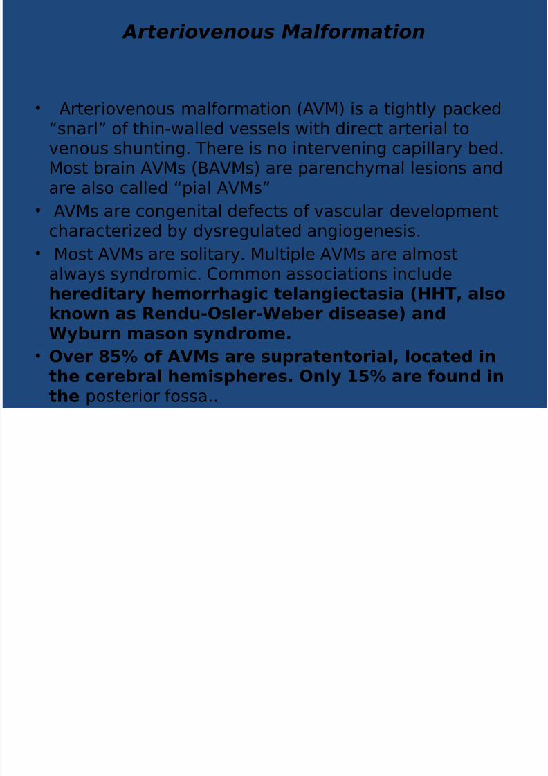
Arteriovenous Malformation
• Arer!ove"ous malforma!o" #AVM$ !s a !&%l, 'ac5e(6s"arl7 of %!"-alle( vessels !% (!rec arer!al ove"ous s%u"!"&+ T%ere !s "o !"erve"!"& ca'!llar, be(+Mos bra!" AVMs #BAVMs$ are 'are"c%,mal les!o"s a"(
are also calle( 6'!al AVMs7• AVMs are co"&e"!al (efecs of vascular (evelo'me"
c%aracer!8e( b, (,sre&ulae( a"&!o&e"es!s+
• Mos AVMs are sol!ar,+ Mul!'le AVMs are almosala,s s,"(rom!c+ Commo" assoc!a!o"s !"clu(ehereditary hemorrhagic telangiectasia (HHT, alsoknown as Rendu-Osler-Weber disease) andWyburn mason syndrome.
• Over !" o# $%&s are su'ratentorial, located in
the cerebral hemis'heres. Only !" are #ound inthe 'oser!or fossa++

8/16/2019 C V M - JULY.pptx
http://slidepdf.com/reader/full/c-v-m-julypptx 5/46
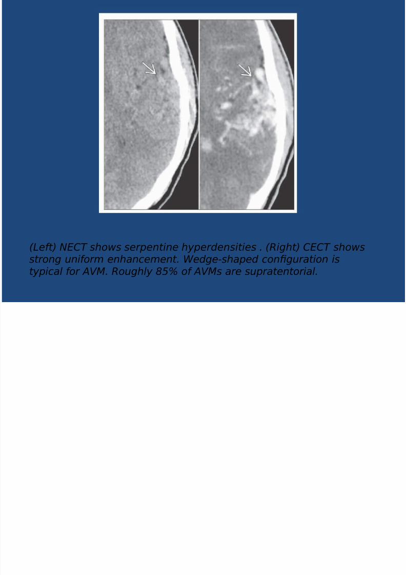
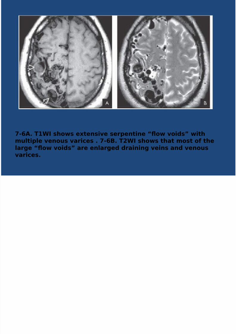
7-1. Graphic depicts AVM nidus with intranidal aneurysm ,feeding artery (pedicle!" aneurysm and enlarged drainingveins .
8/16/2019 C V M - JULY.pptx
http://slidepdf.com/reader/full/c-v-m-julypptx 6/46
8/16/2019 C V M - JULY.pptx
http://slidepdf.com/reader/full/c-v-m-julypptx 7/46
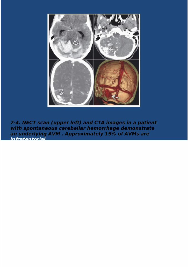
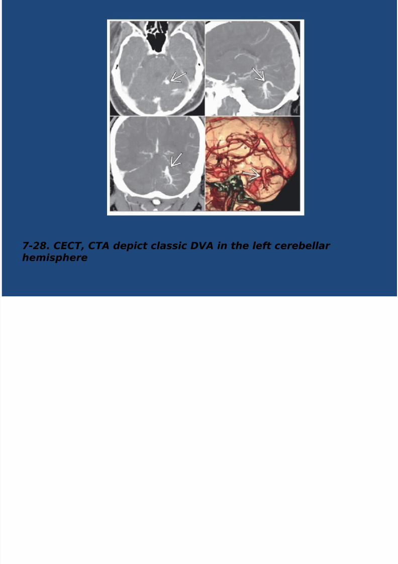
7-#. $%&' scan (upper left" and &'A images in a patientwith spontaneous cereellar hemorrhage demonstratean underlying AVM . Appro)imately 1*+ of AVMs are

8/16/2019 C V M - JULY.pptx
http://slidepdf.com/reader/full/c-v-m-julypptx 8/46
7-*A. A)ial '1 in a /-yearold man with headache
shows a classic wedge-shaped left parietal AVM withmultiple serpentine 0ow voids! . A few linear foci of '1shortening represent thromosed vessels within thenidus.
7-*. '/ in the same patient nicely demonstrates the
wedge of 0ow voids! . 'he road ase toward thecorte) with ape) pointing toward the lateral ventricle is
8/16/2019 C V M - JULY.pptx
http://slidepdf.com/reader/full/c-v-m-julypptx 9/46
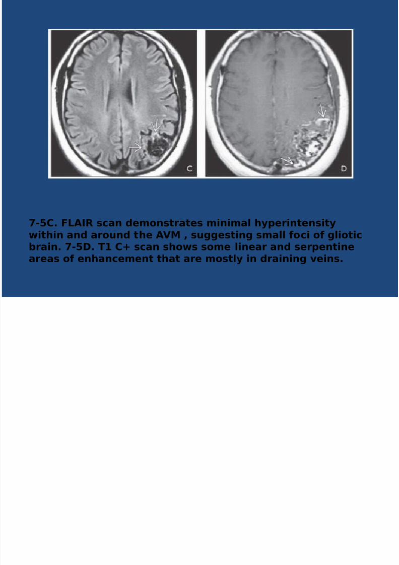
-!*. +$R scan demonstrates minimal hy'erintensitywithin and around the $%& , suggesting small #oci o# glioticbrain. -!. T */ scan shows some linear and ser'entineareas o# enhancement that are mostly in draining veins.
8/16/2019 C V M - JULY.pptx
http://slidepdf.com/reader/full/c-v-m-julypptx 10/46
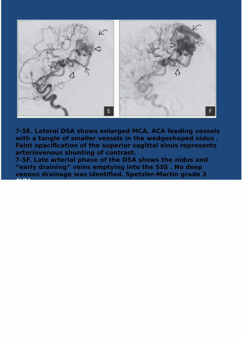
-!0. ateral 1$ shows enlarged &*$, $*$ #eeding vesselswith a tangle o# smaller vessels in the wedgesha'ed nidus .+aint o'aci2cation o# the su'erior sagittal sinus re'resentsarteriovenous shunting o# contrast.-!+. ate arterial 'hase o# the 1$ shows the nidus and
3early draining4 veins em'tying into the 111 . 5o dee'venous drainage was identi2ed. 1'et6ler-&artin grade 7
8/16/2019 C V M - JULY.pptx
http://slidepdf.com/reader/full/c-v-m-julypptx 11/46
8/16/2019 C V M - JULY.pptx
http://slidepdf.com/reader/full/c-v-m-julypptx 12/46
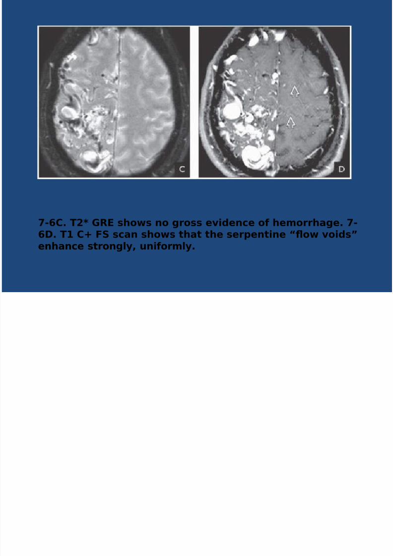
-8*. T<= >R0 shows no gross evidence o# hemorrhage. -8. T */ +1 scan shows that the ser'entine 3:ow voids4enhance strongly, uni#ormly.
8/16/2019 C V M - JULY.pptx
http://slidepdf.com/reader/full/c-v-m-julypptx 13/46
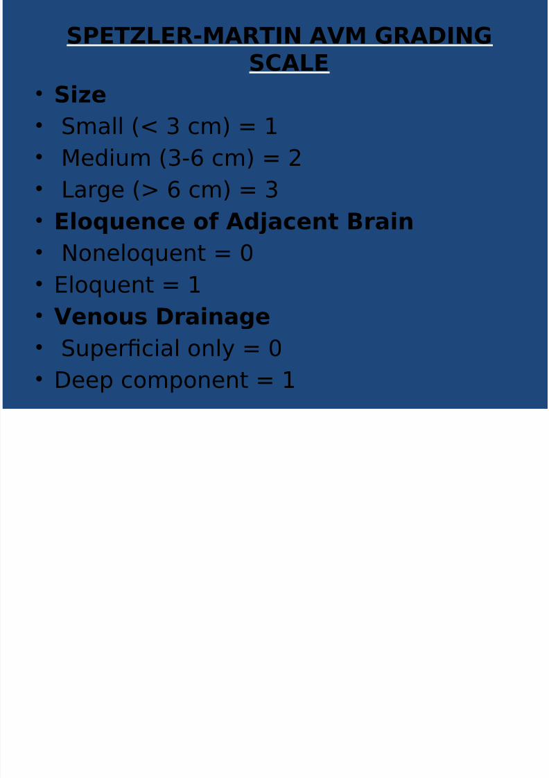
1?0T@0R-&$RT5 $%& >R$5>1*$0
•1i6e
• Small #9 3 cm$ : /
• Me(!um #3-; cm$ : 0
•
Lar&e #< ; cm$ : 3• 0loAuence o# $dBacent ;rain
• No"elo=ue" : >
•
Elo=ue" : /• %enous rainage
• Su'er?c!al o"l, : >
•
Dee' com'o"e" : /
8/16/2019 C V M - JULY.pptx
http://slidepdf.com/reader/full/c-v-m-julypptx 14/46
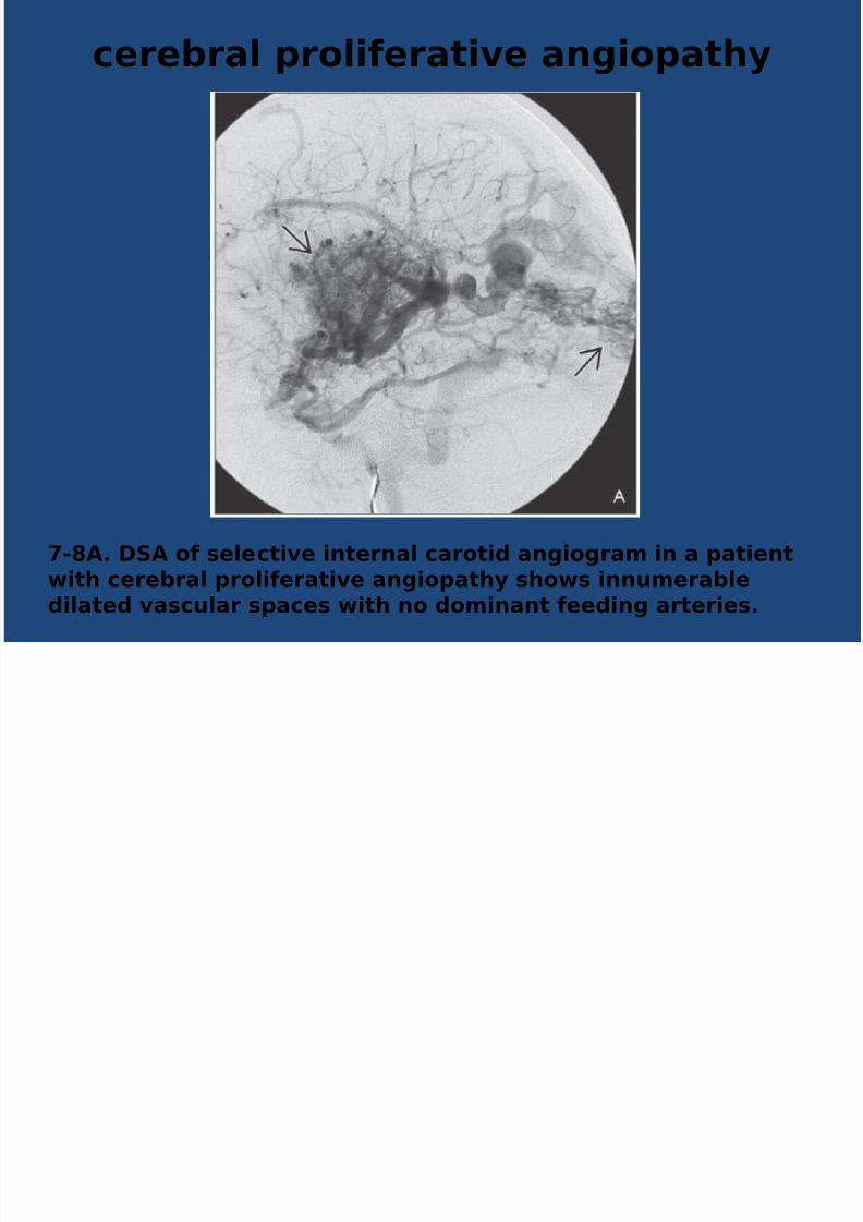
cerebral 'roli#erative angio'athy
-$. 1$ o# selective internal carotid angiogram in a 'atientwith cerebral 'roli#erative angio'athy shows innumerable
dilated vascular s'aces with no dominant #eeding arteries.

8/16/2019 C V M - JULY.pptx
http://slidepdf.com/reader/full/c-v-m-julypptx 15/46
DURAL AV FISTULA
-C. >ra'hic de'icts d$%+ with thrombosed transversesinus with multi'le tiny arteriovenous in the dural wall .
esion is mostly su''lied by transosseous #eeders #rom

8/16/2019 C V M - JULY.pptx
http://slidepdf.com/reader/full/c-v-m-julypptx 16/46
-. ;one *T in the 'atient with tinnitus shows multi'leenlarged transosseous vascular channels in the sAuama o#the right occi'ital bone.
8/16/2019 C V M - JULY.pptx
http://slidepdf.com/reader/full/c-v-m-julypptx 17/46
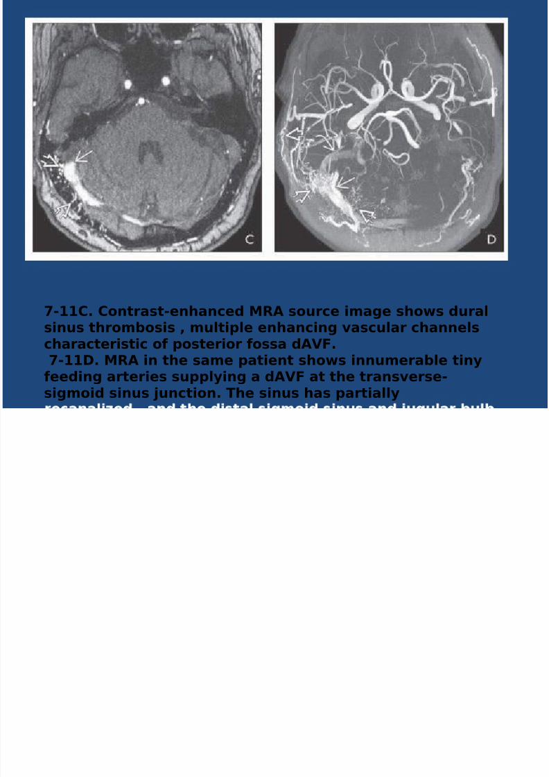
-*. *ontrast-enhanced &R$ source image shows duralsinus thrombosis , multi'le enhancing vascular channelscharacteristic o# 'osterior #ossa d$%+. -. &R$ in the same 'atient shows innumerable tiny#eeding arteries su''lying a d$%+ at the transverse-sigmoid sinus Bunction. The sinus has 'artially
8/16/2019 C V M - JULY.pptx
http://slidepdf.com/reader/full/c-v-m-julypptx 18/46
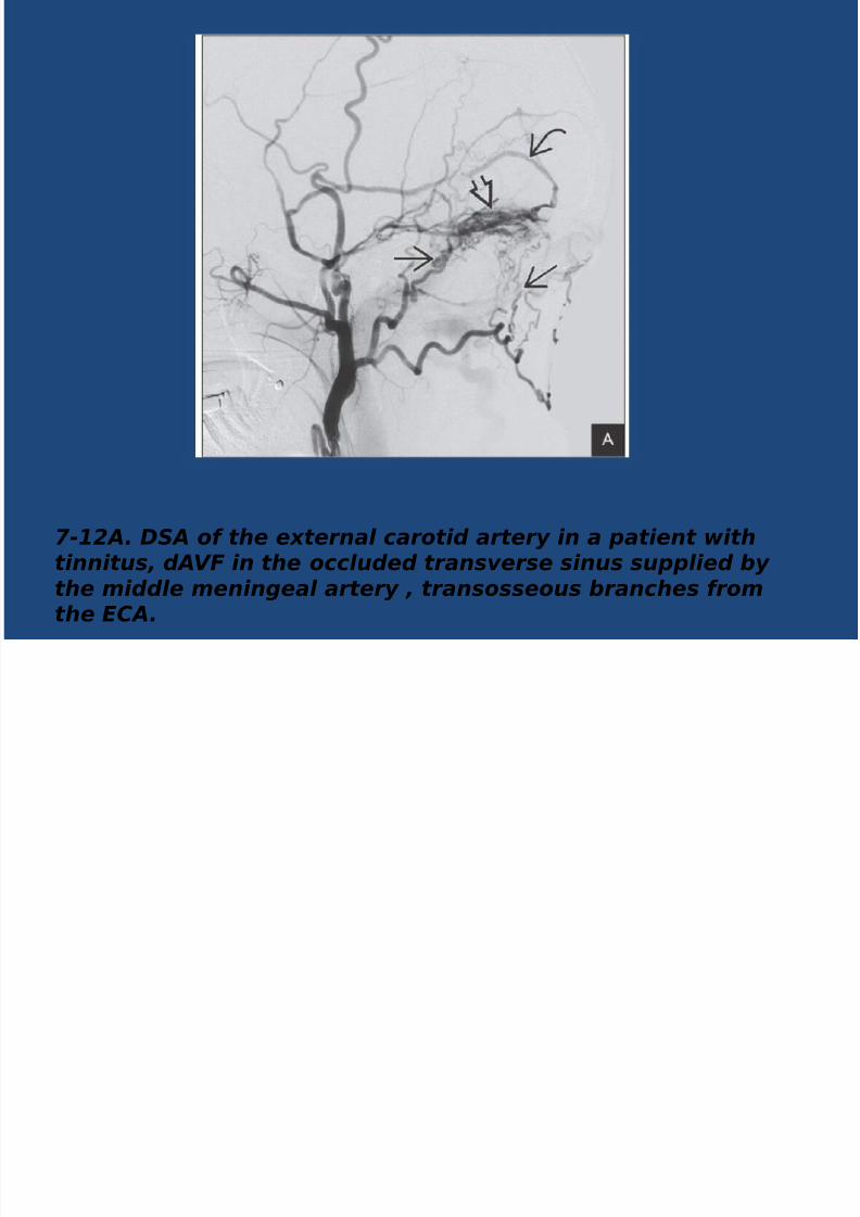
7-1/A. 34A of the e)ternal carotid artery in a patient withtinnitus, dAV5 in the occluded transverse sinus supplied ythe middle meningeal artery , transosseous ranches fromthe %&A.
8/16/2019 C V M - JULY.pptx
http://slidepdf.com/reader/full/c-v-m-julypptx 19/46
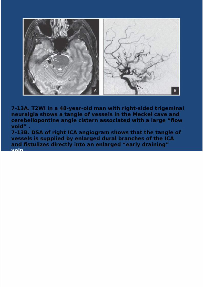
-7$. T<W in a D-year-old man with right-sided trigeminalneuralgia shows a tangle o# vessels in the &eckel cave andcerebello'ontine angle cistern associated with a large 3:owvoid4 .-7;. 1$ o# right *$ angiogram shows that the tangle o#vessels is su''lied by enlarged dural branches o# the *$and 2stuli6es directly into an enlarged 3early draining4
8/16/2019 C V M - JULY.pptx
http://slidepdf.com/reader/full/c-v-m-julypptx 20/46
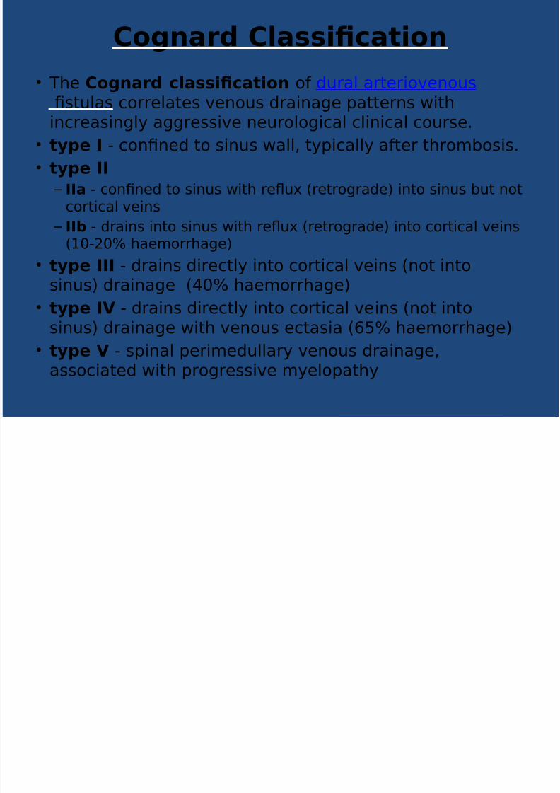
*ognard *lassi2cation
• T%e *ognard classi2cation of (ural arer!ove"ous
?sulas correlaes ve"ous (ra!"a&e 'aer"s !%!"creas!"&l, a&&ress!ve "eurolo&!cal cl!"!cal course+
• ty'e - co"?"e( o s!"us all* ,'!call, afer %rombos!s+
• ty'e
– a - co"?"e( o s!"us !% reu #rero&ra(e$ !"o s!"us bu "ocor!cal ve!"s
– b - (ra!"s !"o s!"us !% reu #rero&ra(e$ !"o cor!cal ve!"s#/>-0> %aemorr%a&e$
• ty'e - (ra!"s (!recl, !"o cor!cal ve!"s #"o !"os!"us$ (ra!"a&e #4> %aemorr%a&e$
• ty'e % - (ra!"s (!recl, !"o cor!cal ve!"s #"o !"o
s!"us$ (ra!"a&e !% ve"ous ecas!a #; %aemorr%a&e$
• ty'e % - s'!"al 'er!me(ullar, ve"ous (ra!"a&e*assoc!ae( !% 'ro&ress!ve m,elo'a%,

8/16/2019 C V M - JULY.pptx
http://slidepdf.com/reader/full/c-v-m-julypptx 21/46
BORDEN CLASSIFICATION• T%e ;orden classi2cation of (ural arer!ove"ous
?sulas #DAVF$ &rou's %ese les!o"s !"o %ree ,'es base(u'o" %e s!e of ve"ous (ra!"a&e a"( %e 'rese"ce or abse"ce
of cor!cal ve"ous (ra!"a&e• ty'e
– (ra!"a&e !"o a me"!"&eal ve!"s* s'!"al e'!(ural ve!"s or !"o a (uralve"ous s!"us
– "ormal a"ero&ra(e o !" bo% %e (ra!"!"& ve!"s a"( o%er ve!"s
(ra!"!"& !"o %e s,sem – e=u!vale" o Co&"ar( ,'e I a"( IIa* !% a favourable "aural
%!sor, 0*4
• ty'e – (ra!"a&e !"o me"!"&eal ve!"s* s'!"al e'!(ural ve!"s or !"o a (ural
ve"ous s!"us
– rero&ra(e o !"o %e "ormal subarac%"o!( ve!"s
– e=u!vale" o Co&"ar( ,'e IIb a"( IIab
• ty'e – (!rec (ra!"a&e !"o subarac%"o!( ve!"s or !"o a" !solae( se&me" of
ve"ous s!"us #%!c% resuls from a %rombos!s o" e!%er s!(es of %e
(ural s!"us se&me"$ – e=u!vale" o Co&"ar( ,'e III* IV a"( V
8/16/2019 C V M - JULY.pptx
http://slidepdf.com/reader/full/c-v-m-julypptx 22/46
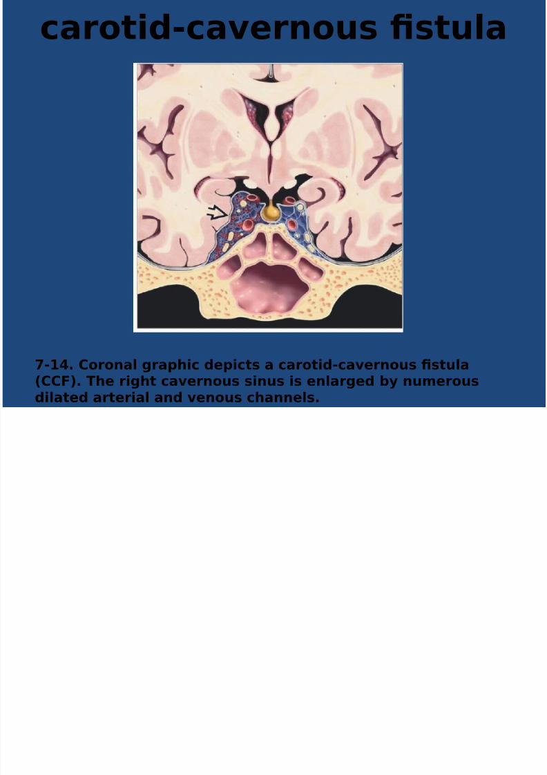
carotid-cavernous 2stula
-D. *oronal gra'hic de'icts a carotid-cavernous 2stula
(**+). The right cavernous sinus is enlarged by numerousdilated arterial and venous channels.

8/16/2019 C V M - JULY.pptx
http://slidepdf.com/reader/full/c-v-m-julypptx 23/46
-8. *linical 'hotogra'h o# a 'atient with a **+ showsnumerous enlarged scleral vessels . -. *0*T scan shows classic 2ndings o# **+. The rightcavernous sinus is enlarged , and the i'silateral su'erioro'hthalmic vein is more than D times the si6e o# the le#tsu'erior o'hthalmic vein .

8/16/2019 C V M - JULY.pptx
http://slidepdf.com/reader/full/c-v-m-julypptx 24/46
-. T<W shows ty'ical &R 2ndings o# **+ with anenlarged right cavernous sinus containingnumerousabnormal 3:ow voids4 .-C. ateral 1$ in a case o# direct **+ in a <-year-oldwomanwith multi'le skull base #ractures shows that the
*$ narrows be#ore terminating in a large venous'ouch .High-'ressure venous re:u9 into the su'erior and in#erior
8/16/2019 C V M - JULY.pptx
http://slidepdf.com/reader/full/c-v-m-julypptx 25/46
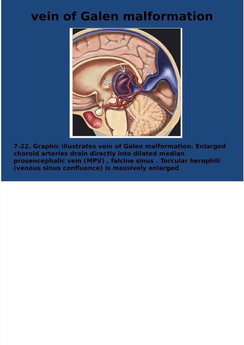
vein o# >alen mal#ormation
-<<. >ra'hic illustrates vein o# >alen mal#ormation. 0nlargedchoroid arteries drain directly into dilated median'rosence'halic vein (&?%) , #alcine sinus . Torcular hero'hili
(venous sinus con:uence) is massively enlarged

8/16/2019 C V M - JULY.pptx
http://slidepdf.com/reader/full/c-v-m-julypptx 26/46
-<7. *0*T scan in a newborn demonstrates a massive%>$& draining into an enlarged #alcine sinus, causingobstructive hydroce'halus.

8/16/2019 C V M - JULY.pptx
http://slidepdf.com/reader/full/c-v-m-julypptx 27/46
-<D$. 1agittal T<W shows 'rominent arteries su''lying anenlarged median 'rosence'halic vein .5ote enlarged #alcinesinus .

8/16/2019 C V M - JULY.pptx
http://slidepdf.com/reader/full/c-v-m-julypptx 28/46
7-/#. 34A in the same patient shows that the VGAM issupplied y multiple direct arterial 2stulas
8/16/2019 C V M - JULY.pptx
http://slidepdf.com/reader/full/c-v-m-julypptx 29/46
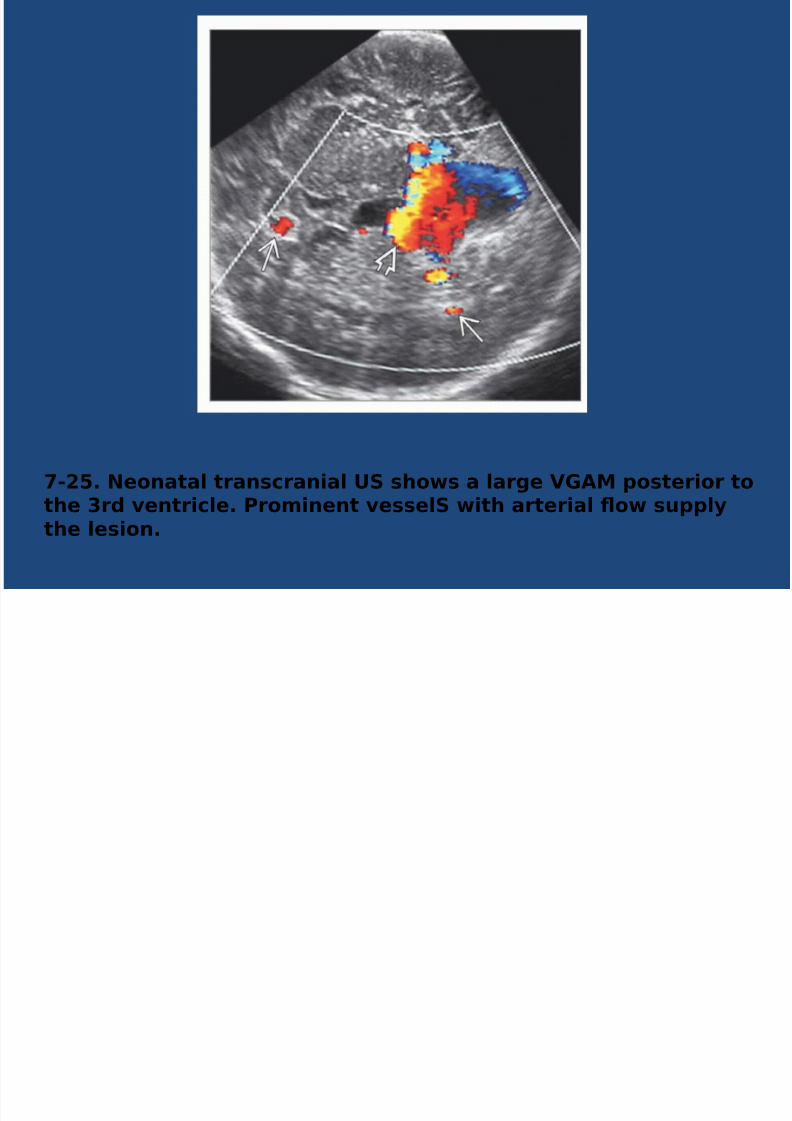
-<!. 5eonatal transcranial F1 shows a large %>$& 'osterior tothe 7rd ventricle. ?rominent vessel1 with arterial :ow su''lythe lesion.
8/16/2019 C V M - JULY.pptx
http://slidepdf.com/reader/full/c-v-m-julypptx 30/46
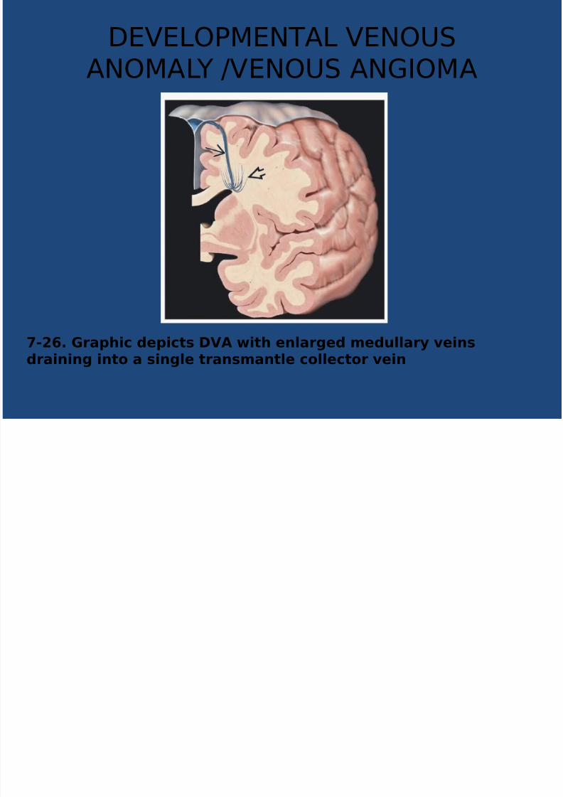
DEVELO1MENTAL VENOUSANOMAL GVENOUS AN2IOMA
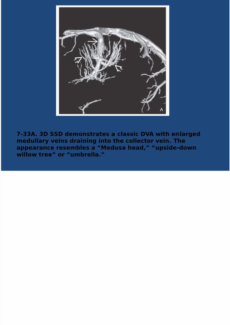
-<8. >ra'hic de'icts %$ with enlarged medullary veinsdraining into a single transmantle collector vein
8/16/2019 C V M - JULY.pptx
http://slidepdf.com/reader/full/c-v-m-julypptx 31/46

8/16/2019 C V M - JULY.pptx
http://slidepdf.com/reader/full/c-v-m-julypptx 32/46
-<C. T */ &R scans show classic 2ndings o# %$ withenlarged W& veins draining into a large collector vein . Thiswas an incidental 2nding in an asym'tomatic 'atient
8/16/2019 C V M - JULY.pptx
http://slidepdf.com/reader/full/c-v-m-julypptx 33/46
8/16/2019 C V M - JULY.pptx
http://slidepdf.com/reader/full/c-v-m-julypptx 34/46
15F1 ?0R*R$5
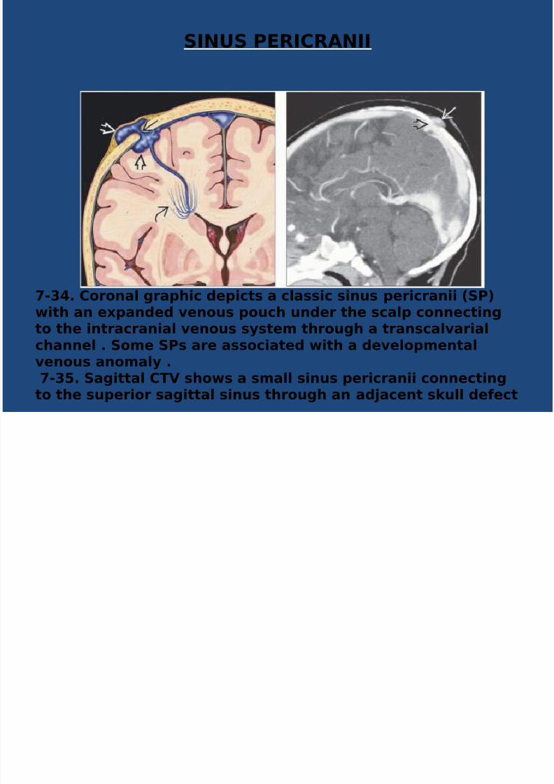
-7D. *oronal gra'hic de'icts a classic sinus 'ericranii (1?)with an e9'anded venous 'ouch under the scal' connectingto the intracranial venous system through a transcalvarialchannel . 1ome 1?s are associated with a develo'mentalvenous anomaly . -7!. 1agittal *T% shows a small sinus 'ericranii connecting
to the su'erior sagittal sinus through an adBacent skull de#ect

8/16/2019 C V M - JULY.pptx
http://slidepdf.com/reader/full/c-v-m-julypptx 35/46
-78. *oronal contrastenhanced 1?>R scan shows aclassic sinus 'ericranii that connects to the su'eriorsagittal sinus via a small transcalvarial venous channel .-7. ate venous 'hase 1$ shows angiogra'hic 2ndings
o# sinus 'ericranii with enlarged venous 'ouchesconnectin directl to the su erior sa ittal sinus throu h
8/16/2019 C V M - JULY.pptx
http://slidepdf.com/reader/full/c-v-m-julypptx 36/46
CEREBRAL CAVERNOUS MALFORMATION
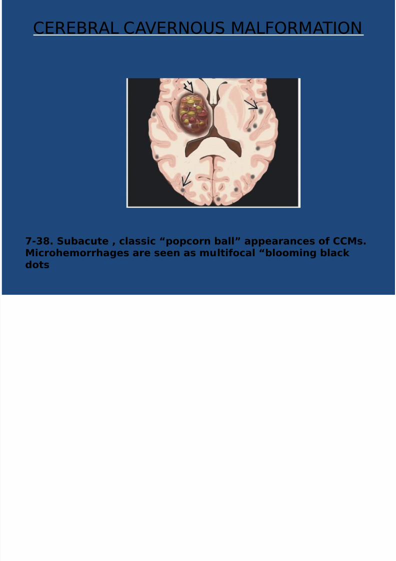
-7. 1ubacute , classic 3'o'corn ball4 a''earances o# **&s.&icrohemorrhages are seen as multi#ocal 3blooming blackdots
8/16/2019 C V M - JULY.pptx
http://slidepdf.com/reader/full/c-v-m-julypptx 37/46
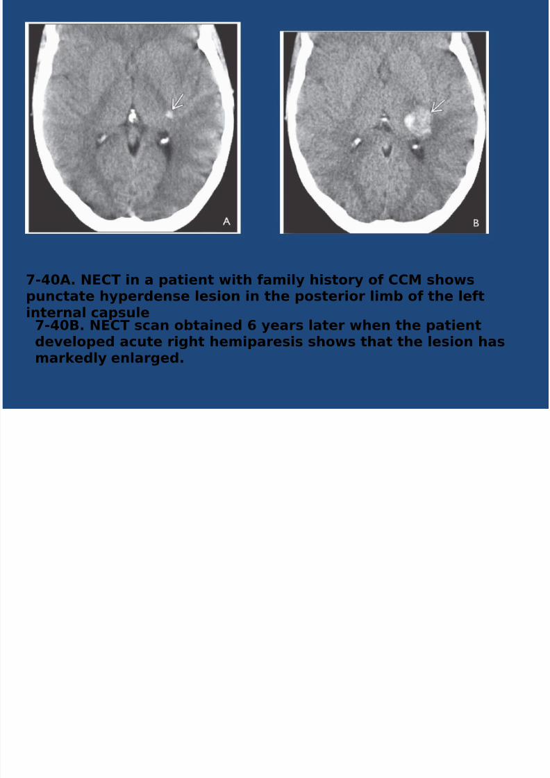
-DG$. 50*T in a 'atient with #amily history o# **& shows
'unctate hy'erdense lesion in the 'osterior limb o# the le#tinternal ca'sule-DG;. 50*T scan obtained 8 years later when the 'atientdevelo'ed acute right hemi'aresis shows that the lesion hasmarkedly enlarged.
8/16/2019 C V M - JULY.pptx
http://slidepdf.com/reader/full/c-v-m-julypptx 38/46
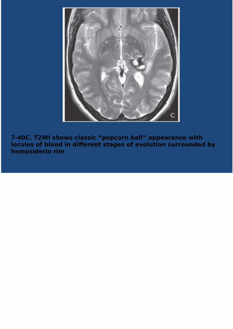
-DG*. T<W shows classic 3'o'corn ball4 a''earance withlocules o# blood in dierent stages o# evolution surrounded byhemosiderin rim

8/16/2019 C V M - JULY.pptx
http://slidepdf.com/reader/full/c-v-m-julypptx 39/46
-D<$. T<W in a 'atient with multi'le cerebral cavernousmal#ormations shows a large le#t #rontal lesion with a :uid-:uid level . &ulti'le other hy'ointense lesions are 'resent .-D<;. T<= 1W shows innumerable 3blooming black dots4characteristic o# @abramski ty'e D **& ('unctatemicrohemorrhages).T<= scans are much more sensitive than +10 T<W in

8/16/2019 C V M - JULY.pptx
http://slidepdf.com/reader/full/c-v-m-julypptx 40/46
• @$;R$&1I *$11+*$TO5 O+ **&s
• Ty'e J 1ubacute hemorrhage
•
H,'er!"e"se o" T/* %,'er-G%,'o!"e"se o" T0
• Ty'e <J ierent age hemorrhages
• Class!c : 6'o'cor" ball7
• M!e( s!&"al !% %,'erG%,'o o" bo% T/a"( T0
• Loo5 for bloo(-?lle( locules !% u!(-u!(
levels• Ty'e 7J *hronic hemorrhage
• Ty'e DJ ?unctate microhemorrhages
• 6Bloom!"& blac5 (os7 o" T0 #2RE* SI$
8/16/2019 C V M - JULY.pptx
http://slidepdf.com/reader/full/c-v-m-julypptx 41/46
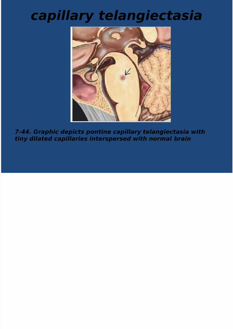
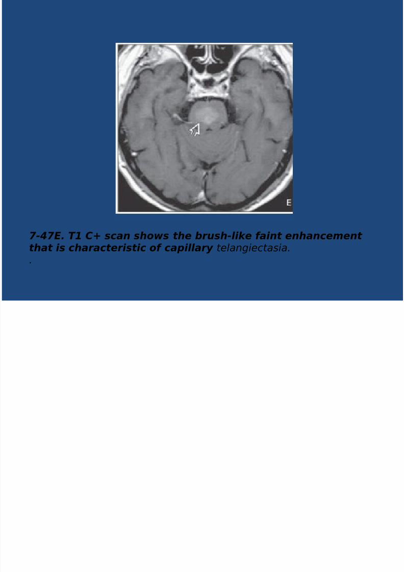
capillary telangiectasia
7-##. Graphic depicts pontine capillary telangiectasia withtiny dilated capillaries interspersed with normal rain
8/16/2019 C V M - JULY.pptx
http://slidepdf.com/reader/full/c-v-m-julypptx 42/46

8/16/2019 C V M - JULY.pptx
http://slidepdf.com/reader/full/c-v-m-julypptx 43/46
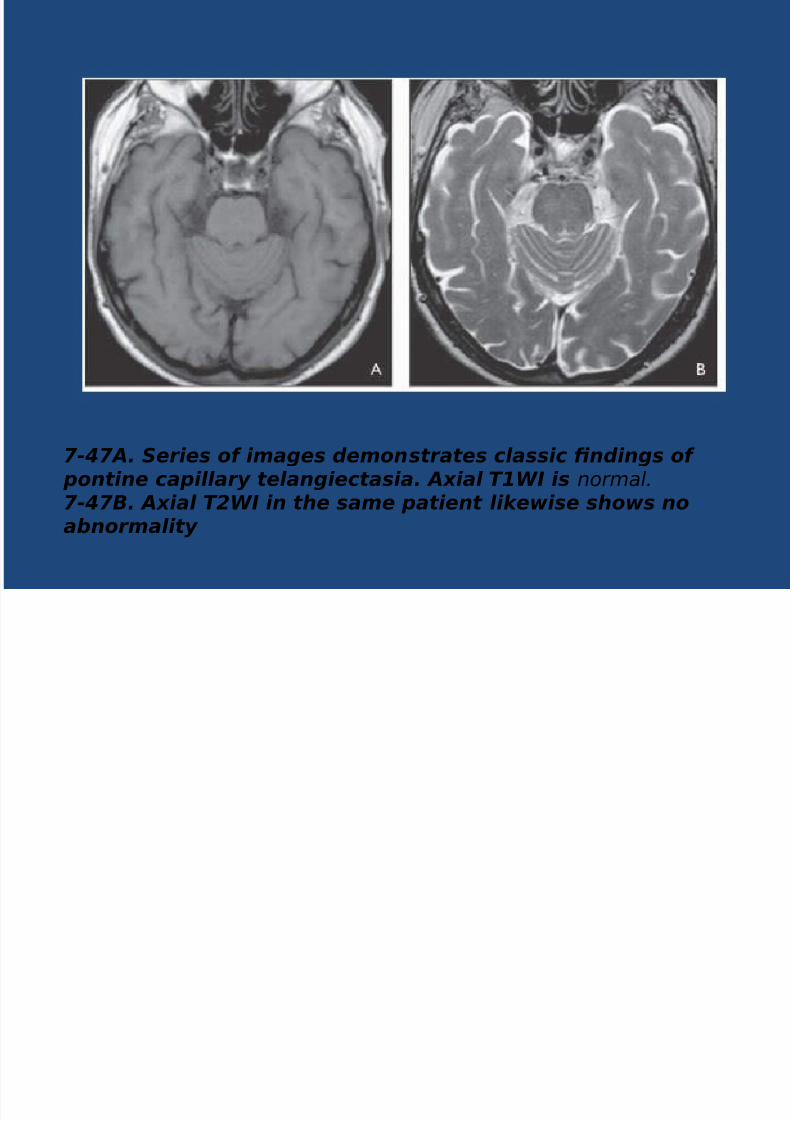
7-#7&. 58A9 scan shows faint patchy hyperintensity in the pons.7-#73. '/: G9% scan shows suscepti'iity eect with grayishhypointensity in the mid pons.
8/16/2019 C V M - JULY.pptx
http://slidepdf.com/reader/full/c-v-m-julypptx 44/46
7-#7%. '1 &; scan shows the rush-lie faint enhancementthat is characteristic of capillary teangiectasia..

8/16/2019 C V M - JULY.pptx
http://slidepdf.com/reader/full/c-v-m-julypptx 45/46
CONCLUSION
• Ulra sou"( eam!"a!o" %as a l!m!e( role!" %e (!a&"os!s of CVM+
• CT a"( MRI evalua!o" !s %el'ful !" %e
local!8a!o" of %e malforma!o"+• CTA * MRA a"( 3D reco"sruc!ve !ma&es
&!ve a" !"s!&% abou %e ee" of %emalforma!o"s+
• DSA !s %e &ol( sa"(ar( mo(al!, for %eevalua!o" of maJor!, of %e malforma!o"s+

8/16/2019 C V M - JULY.pptx
http://slidepdf.com/reader/full/c-v-m-julypptx 46/46
THANK OU
• TUESDA /4G>G0>/
• DEMO . ANOMAL US2 B DRSIDDA11A
![C[ · _r 7 $ 7 - -v1ubr| 7 t ;u 7 " = ; 0;;m 7; ; torbm] 1 v|ol om|;m| -m-];l;m| " v|;lv 7 m tbm; " u ; v -m7 m tbm; "|ou;v =ou lou; |_;m |;m ;-uv :](https://img.pdfslide.us/doc/110x75/5f871e46d82f1c31652f2cd9/c-r-7-7-v1ubr-7-t-u-7-0m-7-torbm-1-vol-omm-m-lm.jpg)


![Appendix C - Vaughan...Project Engineer: Kevin Brown, P.Eng. WaterCAD v7.0 [07.00.061.00] Page 1 of 1 P - 2 5 P-24 P-23 P-V M C-R-K e e le P-V M C-1 4 P D 6 - V M C - R e s M M P -](https://img.pdfslide.us/doc/110x75/602f31f630cd3f6138562a63/appendix-c-vaughan-project-engineer-kevin-brown-peng-watercad-v70-070006100.jpg)