Embed Size (px)
Citation preview
1
Joint Pathology Center
Veterinary Pathology Services
WEDNESDAY SLIDE CONFERENCE 2018-2019
C o n f e r e n c e 14 9 January 2018
CASE I: NE 17-176 (JPC 4101745).
Signalment: 6-year-old neutered male
brindle Mastiff dog, Canis familiaris
History: This dog had a 10-minute seizure
at home and afterwards he was blind. He
had had a similar, but less severe episode of
altered mental status several weeks prior.
Physical exam findings included dull
mentation, circling to the right, and negative
menace response. After premedication for
imaging, the dog developed ventricular
tachycardia and was euthanized.
Gross Pathology: The dog was in obese
nutritional condition (body condition score
4.5/5) and the tail was alopecic. The thyroid
glands were small (right 3x2x32mm; left
5x3x30mm). The walls of many vessels
including the coronary arteries, prostatic
vessels, and meningeal vessels had white
opaque firm walls. The myocardium
adjacent to affected coronary vessels was
often red.
Laboratory results: None.
Microscopic Description:
2
Thyroid gland, dog. The thyroid glands were small.
Vessels within the gland and fibroadipose tissue are
occasional opaque and whitish-yellow prominent due to
mural accumulation of cholesterol and inflammatory
cells. (Photo courtesy of: University of Tennessee College
of Veterinary Medicine, Department of Biomedical and
Diagnostic Sciences, 2407 River Drive, Room A205,
Knoxville, TN 37996,
https://vetmed.tennessee.edu/departments/Pages/biomedic
al_diagnostic_sciences.aspx)
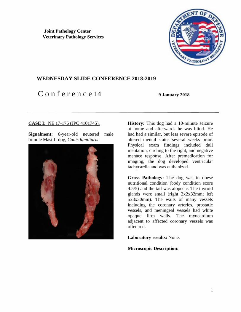
Heart, dog. Atherosclerotic plaques are prominent
within coronary arteries. (Photo courtesy of: University
of Tennessee College of Veterinary Medicine, Department
of Biomedical and Diagnostic Sciences, 2407 River Drive,
Room A205, Knoxville, TN 37996,
https://vetmed.tennessee.edu/departments/Pages/biomedic
al_diagnostic_sciences.aspx)
Prostate gland. Atherosclerotic plaques are prominent
within prostatic and paraprostatic arterioles. (Photo
courtesy of: University of Tennessee College of
Veterinary Medicine, Department of Biomedical and
Diagnostic Sciences, 2407 River Drive, Room A205,
Knoxville, TN 37996,
https://vetmed.tennessee.edu/departments/Pages/biomedic
al_diagnostic_sciences.aspx)
Thyroid: The thyroid glands are small and
lack follicles. The follicles are replaced by
dense infiltrates of small lymphocytes and
plasma cells. The remaining C
(parafollicular) cells are prominent. The
vessels within and around the gland have
foamy macrophages and acicular
(cholesterol) clefts replacing and expanding
their walls, often resulting in occlusion of
the lumen. The parathyroid gland is
unremarkable.
Artery and surrounding vessels: The
muscular artery at the center of the section
(taken from the hilus of the liver) has mild
multifocal expansion of the tunica
muscularis by foamy macrophages and
small acicular clefts (not present in all
sections). The surrounding smaller vessels
have severe replacement and expansion of
their walls by foamy macrophages and
acicular (cholesterol) clefts. The deposits
often narrow or occlude the lumens, which
occasionally contain thrombi. There is mild
lymphoplasmacytic inflammation within the
adventitia of the affected vessels.
Contributor’s Morphologic Diagnoses:
Severe chronic diffuse lymphocytic
thyroiditis with follicular atrophy
Severe multifocal chronic vascular
atherosclerosis with thrombosis.
Contributor’s Comment: Lymphocytic
thyroiditis in dogs is similar to Hashimoto’s
disease of humans. Both are thought to have
a polygenic inheritance. Predisposed dog
breeds include: boxers, bulldogs,
dachshunds, great Danes, Doberman
pinschers, golden and Labrador retrievers.
Circulating autoantibodies to thyroglobulin
are present in affected humans and dogs.
Interestingly, the infiltrating lymphocytes
and plasma cells are thought to mechanically
dislodge the thyroid follicular epithelial cells
from their basement membranes resulting in
destruction of the follicle.3

3
Thyroid gland (left) and hepatic hilar vessels: The
thyroid gland (left) is largely effaced by an inflammatory
infiltrate. At right, hepatic hilar arterioles are tortuous
and markedly expanded. (HE, 5X)
Thyroid gland: Follicular architecture is lost and
replaced by marked lymphoplasmacytic inflammation.
Large nests of interfollicular cells remain (arrows). (HE
154X)
The vessel (and nerve) bundle on the
submitted slides was taken from the hilus of
the liver. The central vessel is presumed to
be the hepatic artery (although pretty small
for a mastiff). The surrounding more
severely affected vessels were presumed to
be veins, but veins are reportedly unaffected
in canine atherosclerosis.4 Grossly, they
resembled lymphatic vessels, but those are
also presumably unaffected by
atherosclerosis.
This dog had a history of hypothyroidism
and had been recently restarted (too late) on
his thyroid supplementation in response to
his clinical signs. The seizures were likely
due to ischemic brain lesions; meningeal and
cerebral vessels were occluded by
atherosclerosis. Coronary artery athero-
sclerosis and the resulting myocardial
ischemia likely caused the arrhythmia.
Externally the findings consistent with
hypothyroidism were obesity and tail
alopecia.
In summary, this was a case of lymphocytic
thyroiditis which led to hypothyroidism
which led to hyperlipidemia which led to
widespread atherosclerosis which led to
cerebral and myocardial ischemia which led
to seizures and arrhythmia, respectively.
Contributing Institution: University of Tennessee College of
Veterinary Medicine
Department of Biomedical and Diagnostic
Sciences
2407 River Drive, Room A205
Knoxville, TN 37996
https://vetmed.tennessee.edu/departments/Pa
ges/biomedical_diagnostic_sciences.aspx
JPC Diagnosis: 1. Thyroid gland:
Thyroiditis, lymphoplasmacytic, diffuse,
severe with severe follicular atrophy and
moderate parafollicular hyperplasia.
2. Thyroid gland, adjacent perivascular
fibroadipose tissue, arterioles:
Atherosclerosis, diffuse, severe with
thrombosis and recanalization.
JPC Comment: As discussed above, this
case is an excellent example of primary
hypothyroidism due to canine lymphocytic
thyroiditis (CLT) in the dog. According to
various studies, 65-80% of cases are the
result of autoimmune disease, with resulting
lymphoplasmacytic thyroiditis and
destruction of the thyroid gland.2 A number
of breeds show a breed predisposition for
hypothyroidism, including dobermans, Great
Danes, poodles, Irish Setters, miniature and
giant schnauzers, boxers, golden retrievers,
dachshunds, Shetland Sheepdogs,
Pomeranians, Cocker Spaniels and Airedales
and Hovawarts.1 Both cellular and humoral
mechanisms of immunity are involved in
this pathogenesis. Affected animals
generally exhibit decreased thyroxine and
4
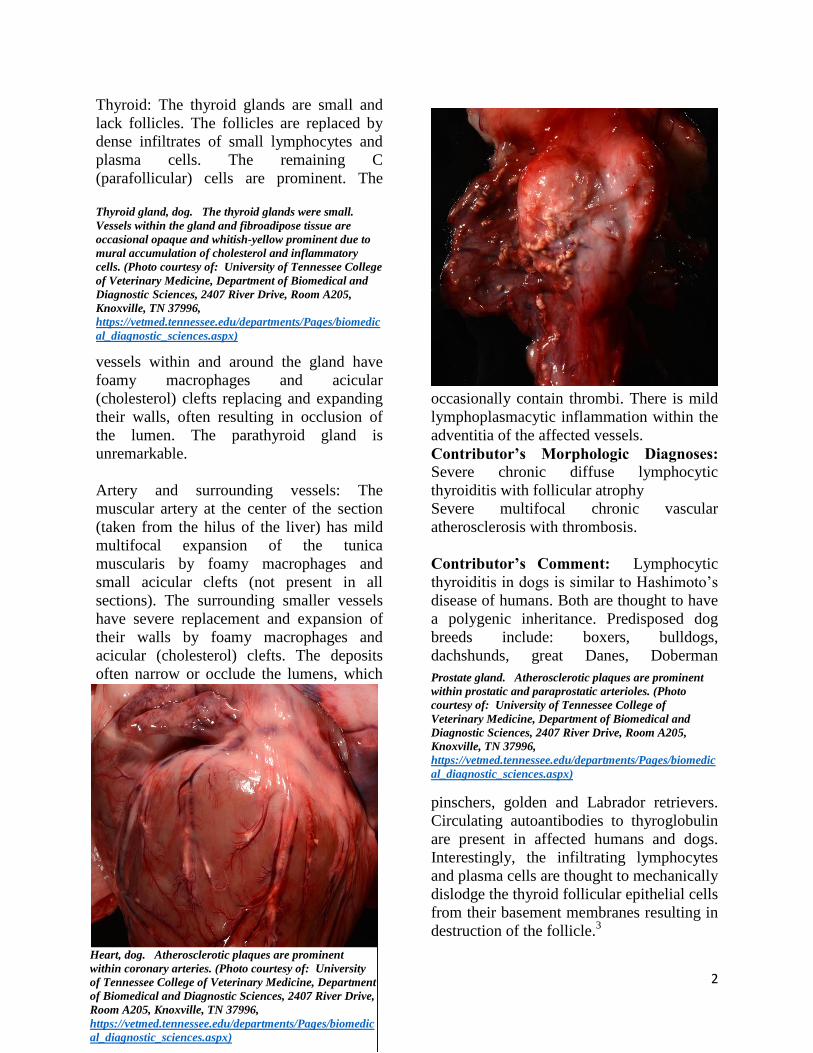
Thyroid gland: Remaining follicles are lined by cuboidal,
often detached epithelium and do not contain colloid. A
cross section through a Kursteiner’s cyst is present at
right. (HE, 190X)
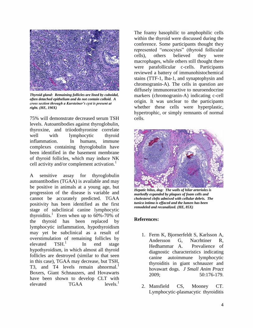
Hepatic hilus, dog: The walls of hilar arterioles is
markedly expanded by plaques of foam cells and
cholesterol clefts admixed with cellular debris. The
tunica intima is effaced and the lumen has been
remodeled and recanalized. (HE, 85X)
75% will demonstrate decreased serum TSH
levels. Autoantibodies against thyroglobulin,
thyroxine, and triiodothyronine correlate
well with lymphocytic thyroid
inflammation. In humans, immune
complexes containing thyroglobulin have
been identified in the basement membrane
of thyroid follicles, which may induce NK
cell activity and/or complement activation.1
A sensitive assay for thyroglobulin
autoantibodies (TGAA) is available and may
be positive in animals at a young age, but
progression of the disease is variable and
cannot be accurately predicted. TGAA
positivity has been identified as the first
stage of subclinical canine lymphocytic
thyroiditis.1 Even when up to 60%-70% of
the thyroid has been replaced by
lymphocytic inflammation, hypothyroidism
may yet be subclinical as a result of
overstimulation of remaining follicles by
elevated TSH.1 In end stage
hypothyroidism, in which almost all thyroid
follicles are destroyed (similar to that seen
in this case), TGAA may decrease, but TSH,
T3, and T4 levels remain abnormal.1
Boxers, Giant Schnauzers, and Hovawarts
have been shown to develop CLT with
elevated TGAA levels.1
The foamy basophilic to amphophilic cells
within the thyroid were discussed during the
conference. Some participants thought they
represented “oncocytes” (thyroid follicular
cells), others believed they were
macrophages, while others still thought there
were parafollicular c-cells. Participants
reviewed a battery of immunohistochemical
stains (TTF-1, Iba-1, and synaptophysin and
chromogranin-A). The cells in question are
diffusely immunoreactive to neuroendocrine
markers (chromogranin-A) indicating c-cell
origin. It was unclear to the participants
whether these cells were hyperplastic,
hypertrophic, or simply remnants of normal
cells.
References:
1. Ferm K, Bjornerfeldt S, Karlsson A,
Andersson G, Nacrhtiner R,
Hedhammar A. Prevalience of
diagnostic characteristics indicating
canine autoimmune lymphocytic
thyroiditis in giant schnauzer and
hovawart dogs. J Small Anim Pract
2009; 50:176-179.
2. Mansfield CS, Mooney CT.
Lymphocytic-plasmacytic thyroiditis

5
Lung, horse. At subgross magnification, areas of a dense
cellular infiltrate as well as foci of hemorrhage are
scattered randomly throughout the section. (HE, 4X)
and glomerulonephritis in a boxer. J
Small Anim Pract 2006: 47:396-399.
3. Rosol TJ, Grone A. Endocrine
Glands. In: Maxie MG, ed. Jubb,
Kennedy, and Palmer’s Pathology of
Domestic Animals. Vol 3, 6th
ed. St.
Louis Elsevier; 2016:315-319.
4. Robinson WF, Robinson NA.
Cardiovascular System. In: Maxie
MG, ed. Jubb, Kennedy, and
Palmer’s Pathology of Domestic
Animals. Vol 3, 6th
ed. St. Louis
Elsevier; 2016:57-59.
CASE II: S1603556 (JPC 4084299).
Signalment: A 1-year-old Quarter horse
filly
History: The filly had a history of 10-day
lethargy. The owner was suspicious of a
rattlesnake bite.
Gross Pathology: The lip was diffusely
thickened with edema. Both submandibular
lymph node were enlarged, firm and dark
red. Bilaterally, the parietal pleura had
multiple petechiae and ecchymoses.
Multifocally in both lungs there were
numerous white, firm nodules,
approximately 2 to 3mm in diameter,
surrounded by a dark red halo. The
parenchyma between these nodules was dark
red and firm. The pericardium, mainly in
proximity to the coronary vessels and
paragonal septum, also showed several
multiple petechiae and ecchymoses. The
kidneys had several multifocal white, firm
cortical spots of approximately 1 to 3mm in
diameter. The cortex was diffusely pale. The
mesentery had multifocal random white firm
nodules of approximately 3 to 5 mm in
diameter surrounded by a dark red halo.
Laboratory results: No bacterial pathogens
were isolated form liver, lung, spleen and
submandibular lymph node on aerobic
culture.
Microscopic Description:
Lung: centering in small caliber vessels and
extending into the adjacent alveolar spaces
and septa, there is a multifocal and random
inflammatory infiltrate composed by large
numbers of viable and degenerated
neutrophils, fewer histiocytes and occasional
multinucleated giant cells that are admixed
with basophilic cellular debris and several
rounded eosinophilic protozoa of
approximately 15 to 20 µm in diameter, with
an eccentric single round nucleus. In
addition, there are numerous fibrin thrombi
and segments of alveolar necrosis. A few
blood vessels display transmural necrosis,
with fibrinoid degeneration and the presence
of pyknotic debris. The remaining
parenchyma shows moderate congestion,
occasional small hemorrhages and multiple
focal areas of intra-alveolar edema.
Contributor’s Morphologic Diagnosis:
Embolic pneumonia, necrosuppurative,
6
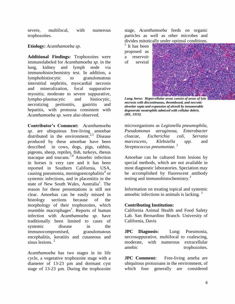
Lung, horse: Hypercellular areas consist of areas of lytic
necrosis with discontinuous, thrombosed, and necrotic
alveolar septa and expansion of alveoli by innumerable
degenerate neutrophils admixed with cellular debris.
(HE, 335X)
severe, multifocal, with numerous
trophozoites.
Etiology: Acanthamoeba sp.
Additional Findings: Trophozoites were
immunolabeled for Acanthamoeba sp. in the
lung, kidney and lymph node via
immunohistochemistry test. In addition, a
lymphohistiocytic to granulomatous
interstitial nephritis, myocardial necrosis
and mineralization, focal suppurative
myositis; moderate to severe suppurative,
lympho-plasmacytic and histiocytic,
necrotizing peritonitis, gastritis and
hepatitis, with protozoa consistent with
Acanthamoeba sp. were also observed.
Contributor’s Comment: Acanthamoeba
sp. are ubiquitous free-living amoebae
distributed in the environment.2,3
Disease
produced by these amoebae have been
described in cows, dogs, pigs, rabbits,
pigeons, sheep, reptiles, fish, turkeys, rhesus
macaque and toucans.7,8
Amoebic infection
in horses is very rare and it has been
reported in Southern California, USA,
causing pneumonia, meningoencephalitis4 or
systemic infections, and in placentitis in the
state of New South Wales, Australia1. The
reason for these presentations is still not
clear. Amoebas can be easily missed in
histology sections because of the
morphology of their trophozoites, which
resemble macrophages1. Reports of human
infection with Acanthamoeba sp. have
traditionally been limited to cases of
systemic disease in the
immunocompromised, granulomatous
encephalitis, keratitis and cutaneous and
sinus lesions. 2
Acanthamoeba has two stages in its life
cycle, a vegetative trophozoite stage with a
diameter of 13-23 µm and dormant cyst
stage of 13-23 µm. During the trophozoite
stage, Acanthamoeba feeds on organic
particles as well as other microbes and
divides mitotically under optimal conditions. 5 It has been
proposed as
a reservoir
of several
microorganisms as Legionella pneumophila,
Pseudomonas aeruginosa, Enterobacter
cloacae, Escherichia coli, Serratia
marcescens, Klebsiella spp. and
Streptococcus pneumoniae. 2
Amoebae can be cultured from lesions by
special methods, which are not available in
most diagnostic laboratories. Speciation may
be accomplished by fluorescent antibody
testing and immunohistochemistry.3
Information on treating topical and systemic
amoebic infections in animals is lacking. 3
Contributing Institution: California Animal Health and Food Safety
Lab. San Bernardino Branch. University of
California, Davis
JPC Diagnosis: Lung: Pneumonia,
necrosuppurative, multifocal to coalescing,
moderate, with numerous extracellular
amebic trophozoites.
JPC Comment: Free-living ameba are
ubiquitous protozoans in the environment, of
which four generally are considered
7
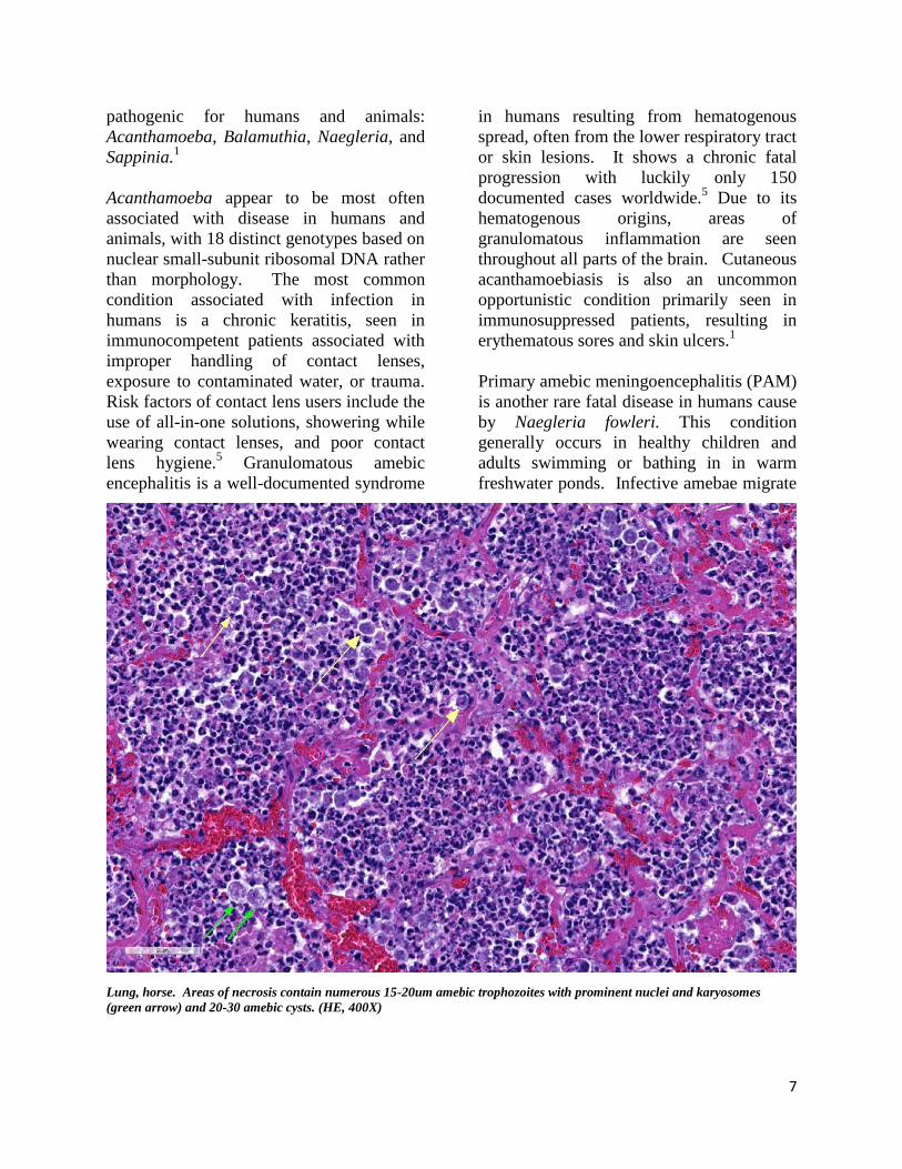
Lung, horse. Areas of necrosis contain numerous 15-20um amebic trophozoites with prominent nuclei and karyosomes
(green arrow) and 20-30 amebic cysts. (HE, 400X)
pathogenic for humans and animals:
Acanthamoeba, Balamuthia, Naegleria, and
Sappinia.1
Acanthamoeba appear to be most often
associated with disease in humans and
animals, with 18 distinct genotypes based on
nuclear small-subunit ribosomal DNA rather
than morphology. The most common
condition associated with infection in
humans is a chronic keratitis, seen in
immunocompetent patients associated with
improper handling of contact lenses,
exposure to contaminated water, or trauma.
Risk factors of contact lens users include the
use of all-in-one solutions, showering while
wearing contact lenses, and poor contact
lens hygiene.5 Granulomatous amebic
encephalitis is a well-documented syndrome
in humans resulting from hematogenous
spread, often from the lower respiratory tract
or skin lesions. It shows a chronic fatal
progression with luckily only 150
documented cases worldwide.5 Due to its
hematogenous origins, areas of
granulomatous inflammation are seen
throughout all parts of the brain. Cutaneous
acanthamoebiasis is also an uncommon
opportunistic condition primarily seen in
immunosuppressed patients, resulting in
erythematous sores and skin ulcers.1
Primary amebic meningoencephalitis (PAM)
is another rare fatal disease in humans cause
by Naegleria fowleri. This condition
generally occurs in healthy children and
adults swimming or bathing in in warm
freshwater ponds. Infective amebae migrate

8
along olfactory nerves from the nose to the
brain; fatal infection proceeds rapidly and is
almost always fatal. Due to this unique entry
portal, areas of lytic necrosis are clustered at
the base of the brain; hypothalamus, pons,
and occasionally seen in posterior areas such
as the medulla oblongata.
Balamuthia mandrillaris is also a cause of
granulomatous amebic encephalitis which
ranges in duration between Acanthamoeba
and Naegleria, which usually results from
hematogenous spread from soil-
contaminated wounds.
Other species of free-living amoeba which
may have been identified in cases of
keratitis include Hartmanella,
Vahlkamphiae, and Allovahlkampfiae
spelae.1
As the contributor mentioned above, free-
living amebae may act as vectors for a wide
range of pathogenic bacilli. They may also
act as hosts for a range of viruses, including
coxsackieviruses and adenoviridae
pathogenic for humans. Other viruses, the
so-called giant viruses, may act as
endocytobionts, including representatives of
the Mimi-, Moumo- and Megaviridae, as
well as Pandoviridae. Co-cultivation in
amebae has been of great benefit in the
eventual isolation of these putative human
pathogens.
The moderator and participants could not
definitively identify cysts in tissues although
PAS and GMS were run, they were of no
help in identifying cysts. Megakaryocytes
were identified in the alveolar capillaries.
The moderator noted that they can normally
be found there as the lungs are a site of
thrombopoiesis and as such can serve as
reservoirs for platelet production and release
them in response to various stimuli.9 Finally,
the moderator reviewed the various types of
free-living amebae, and participants
discussed the associated syndromes in
humans and animals.
References:
1. Balczun C, Scheid PL. Free-living
amoebae as hosts for and vectors of
intracellular microorganisms with
public health significance. Viruses
2017; 9:^%, doi 10-3390/v9040065.
2. Begg A., Todhunter, K., Donahoe, S,
Krockenberger, M., Slapeta J. Severe
amoebic placentitis in a horse caused
by an Acanthamoeba hatchetti
isolate identified using next
generation sequencing. Journal of
clinical microbiology. 3101-3104.
2014
3. Bradbury R S, French L.P., Blizzard
L. Prevalence of Acanthamoeba spp
in Tasmanian intensive care clinical
specimen. Journal of Hospital
Infection 86, 178-181. 2014.
4. Greene C.E. Infectious diseases of
the dog and cat. 802-804. Fourth
edition. 2012
5. Krol-Turminska K, Olendar A.
Human infections caused by free-
living amoebae. Ann Agric Env Med
2016 24(2):240-260
6. Kinde, H., Read D.H.,Daft, B.,
Manzer, M., Nordhausen R., Kelly
D., Fuerst P.A., Booton G.,
Visvesvara G.S. Infections caused by
pathogenic free-living amebas
(Balamuthia mandrillaris and
Acanthamoeba sp.) in horses. J Vet
Diagn Invest 19:317-322. 2007.
7. Siddiqui R and Khan N A. Biology
and pathogenesis of Acanthamoeba.
Review. Parasites and vectors. 5:6.
2012.
8. Westmoreland S.V., Rosen J.,
MacKey J., Romsey C., Xia D.L.,
Visvesvera G.S., Mansfield K.G..

9
Viscera, goat: The lungs and liver had innumerable
disseminated, soft to semi-firm, pale yellow, 0.3-0.6 cm
diameter, occasionally raised nodular foci scattered
throughout the parenchyma. (Photo courtesy of:
University of Connecticut, Connecticut Veterinary
Medical Diagnostic Laboratory, Department of
Pathobiology and Veterinary Science, College of
Agriculture, Health and Natural Resources
http://patho.uconn.edu/)
Lung, goat. The lungs had innumerable disseminated,
soft to semi-firm, pale yellow, occasionally raised nodular
foci scattered throughout the parenchyma, which
frequently oozed pale yellow, thick liquid upon sectioning.
(Photo courtesy of: University of Connecticut,
Connecticut Veterinary Medical Diagnostic Laboratory,
Department of Pathobiology and Veterinary Science,
College of Agriculture, Health and Natural Resources
http://patho.uconn.edu/)
Necrotizing Meningoencephalitis
and Pneumonitis in a Simian
Immunodeficiency Virus-Infected
Rhesus Macaque due to
Acanthamoeba. Veterinary
Pathology. July 2004. vol 41 398-
404.
9. Weyrich AS, Zimmerman GA.
Platelets in lung biology. Annu Rev
Physiol. 2013;75:569-591.
CASE III: S1603556 (JPC 4084299).
Signalment: A 14-month-old, 5 kg, female,
Nigerian dwarf goat (Capra hircus)
History: A total of 4 goats in an original
herd of 8, died in a period of 8 months with
a history of limping, progressive loss of
body condition, and difficulty in getting up.
Three goats died naturally and one was
euthanized. This goat died naturally.
Gross Pathology: The thoracic cavity
contained approximately 25 mL of thin, pink
fluid. The lungs and liver had innumerable
disseminated, soft to semi-firm, pale yellow,
0.3-0.6 cm diameter, occasionally raised
nodular foci scattered throughout the
parenchyma, which frequently oozed pale
yellow, thick liquid upon sectioning. The
spleen had a few similar foci.
Laboratory results: Bacteriology: aerobic
bacterial culture of the liver yielded heavy
growth of Rhodococcus equi, Streptococcus
sp. and Staphylococcus sp
Microscopic Description:
Lung: centering in small caliber vessels and
extending into the adjacent alveolar spaces
and septa, there is a multifocal and random
inflammatory infiltrate composed by large
numbers of viable and degenerated
neutrophils, fewer histiocytes and occasional
multinucleated giant cells that are admixed
with basophilic cellular debris and several
rounded eosinophilic protozoa of
approximately 15 to 20 µm in diameter, with
an eccentric single round nucleus. In
addition, there are numerous fibrin thrombi
10
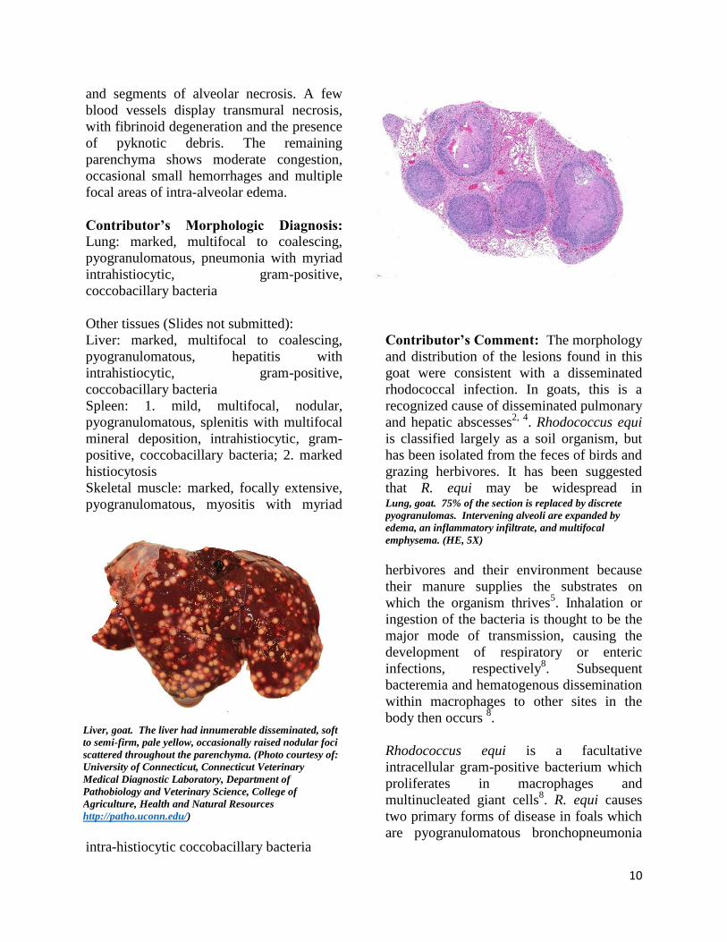
Liver, goat. The liver had innumerable disseminated, soft
to semi-firm, pale yellow, occasionally raised nodular foci
scattered throughout the parenchyma. (Photo courtesy of:
University of Connecticut, Connecticut Veterinary
Medical Diagnostic Laboratory, Department of
Pathobiology and Veterinary Science, College of
Agriculture, Health and Natural Resources
http://patho.uconn.edu/)
Lung, goat. 75% of the section is replaced by discrete
pyogranulomas. Intervening alveoli are expanded by
edema, an inflammatory infiltrate, and multifocal
emphysema. (HE, 5X)
and segments of alveolar necrosis. A few
blood vessels display transmural necrosis,
with fibrinoid degeneration and the presence
of pyknotic debris. The remaining
parenchyma shows moderate congestion,
occasional small hemorrhages and multiple
focal areas of intra-alveolar edema.
Contributor’s Morphologic Diagnosis:
Lung: marked, multifocal to coalescing,
pyogranulomatous, pneumonia with myriad
intrahistiocytic, gram-positive,
coccobacillary bacteria
Other tissues (Slides not submitted):
Liver: marked, multifocal to coalescing,
pyogranulomatous, hepatitis with
intrahistiocytic, gram-positive,
coccobacillary bacteria
Spleen: 1. mild, multifocal, nodular,
pyogranulomatous, splenitis with multifocal
mineral deposition, intrahistiocytic, gram-
positive, coccobacillary bacteria; 2. marked
histiocytosis
Skeletal muscle: marked, focally extensive,
pyogranulomatous, myositis with myriad
intra-histiocytic coccobacillary bacteria
Contributor’s Comment: The morphology
and distribution of the lesions found in this
goat were consistent with a disseminated
rhodococcal infection. In goats, this is a
recognized cause of disseminated pulmonary
and hepatic abscesses2, 4
. Rhodococcus equi
is classified largely as a soil organism, but
has been isolated from the feces of birds and
grazing herbivores. It has been suggested
that R. equi may be widespread in
herbivores and their environment because
their manure supplies the substrates on
which the organism thrives5. Inhalation or
ingestion of the bacteria is thought to be the
major mode of transmission, causing the
development of respiratory or enteric
infections, respectively8. Subsequent
bacteremia and hematogenous dissemination
within macrophages to other sites in the
body then occurs 8.
Rhodococcus equi is a facultative
intracellular gram-positive bacterium which
proliferates in macrophages and
multinucleated giant cells8. R. equi causes
two primary forms of disease in foals which
are pyogranulomatous bronchopneumonia
11
Lung, goat: Higher magnification of a pyogranuloma
with a lamellated center of eosinophilic and basophilic
cellular debris. Viable inflammatory cells populate the
periphery, enmeshed in collagen and dilated capillaries.
(HE, 34X)
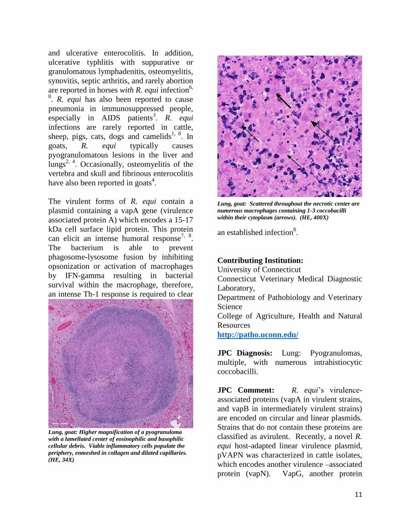
Lung, goat: Scattered throughout the necrotic center are
numerous macrophages containing 1-3 coccobacilli
within their cytoplasm (arrows). (HE, 400X)
and ulcerative enterocolitis. In addition,
ulcerative typhlitis with suppurative or
granulomatous lymphadenitis, osteomyelitis,
synovitis, septic arthritis, and rarely abortion
are reported in horses with R. equi infection6,
8. R. equi has also been reported to cause
pneumonia in immunosuppressed people,
especially in AIDS patients3. R. equi
infections are rarely reported in cattle,
sheep, pigs, cats, dogs and camelids1, 8
. In
goats, R. equi typically causes
pyogranulomatous lesions in the liver and
lungs2, 4
. Occasionally, osteomyelitis of the
vertebra and skull and fibrinous enterocolitis
have also been reported in goats4.
The virulent forms of R. equi contain a
plasmid containing a vapA gene (virulence
associated protein A) which encodes a 15-17
kDa cell surface lipid protein. This protein
can elicit an intense humoral response7, 8
.
The bacterium is able to prevent
phagosome-lysosome fusion by inhibiting
opsonization or activation of macrophages
by IFN-gamma resulting in bacterial
survival within the macrophage, therefore,
an intense Th-1 response is required to clear
an established infection8.
Contributing Institution: University of Connecticut
Connecticut Veterinary Medical Diagnostic
Laboratory,
Department of Pathobiology and Veterinary
Science
College of Agriculture, Health and Natural
Resources
http://patho.uconn.edu/
JPC Diagnosis: Lung: Pyogranulomas,
multiple, with numerous intrahistiocytic
coccobacilli.
JPC Comment: R. equi’s virulence-
associated proteins (vapA in virulent strains,
and vapB in intermediately virulent strains)
are encoded on circular and linear plasmids.
Strains that do not contain these proteins are
classified as avirulent. Recently, a novel R.
equi host-adapted linear virulence plasmid,
pVAPN was characterized in cattle isolates,
which encodes another virulence –associated
protein (vapN). VapG, another protein

12
Lung, goat: The bacteria within the macrophages
(arrow) stain positively with a tissue Gram stain (Twort’s,
600X)
identified in virulent strains has recently
been identified as a potential vaccinal
protein which has shown efficacy in the
partial protection of R. equi in mice (in
addition to current vaccinal work with
vapA). In addition, a highly conserved
conjugal transfer protein gene (traA) is
common to all strains which carry these
plasmids and may now be used to identify
pathogenic strains.1 In previous studies, the
virulent and intermediately virulent strains
are primarily identified in foals and pigs,
while most reports of R. equi isolated from
cattle and dogs appear to be of avirulent
(non-vapA, non-vapB) strains.1
A recent article by Bryan et al1 describes the
clinical syndrome associated with R equi
infection in five dogs. Two of the isolated
strains were avirulent, one was vap-A
positive, and one carried the vapN
virulence-associated plasmid. Four dogs
were on immunosuppressive drugs or had
endocrinopathies. The varied presentation
and distribution of disease in the five dogs
reflects the systemic and almost random
nature of R. equi infection of the dog, with
granulomatous dermatitis, aortic vegetative
valvular endocarditis, pulmonary
abscessation, and lymphadenitis the most
common presentation, but each only being
present in two out of five cases.
Considered an emerging pathogen in
humans, over 100 cases of R. equi infection
have been reported since the first description
of its occurrence in 1967, likely as a result
of improvement in isolation techniques and
recognition of this bacterium as a human
pathogen.9 The vast majority of cases have
occurred in immunosuppressed patients with
over 66% in cases in AIDS patients, and an
additional 10% in transplant recipients as a
late complication (mean 49 months post-
transplant) of immunomodulation. 80% of
cases involved pulmonary infection, most
commonly abscesses with necrotic centers,
although pulmonary disease characterized
by microabscesses is also common. As in
other species, pyogranulomatous infection
may occur in a wide range of organs.
Treatment involves regimes of multiple
antibiotics (often including rifampin) and is
complicated by the typical presence of
immuno-suppression in affected
individuals.9
The
moderator reviewed the differences
between Corynebacterium pseudo-
tuberculosis and Rhodococcus equi.
Additionally, he identified several recent
review articles discussing rhodococcal
infection in several atypical species such as
dogs1 and goats
2,4.
References:
1. Bryan LK, Clark SD, Diaz-Delgado J.
Rhodococcus equi Infections in Dogs. Vet
Pathol. 2017; 54(1):159-163.
2. Davis WP, Steficek BA, Watson GL.
Disseminated Rhodococcus equi infection in
two goats. Vet Pathol. 1999;36(4):336-339.
3. Ferretti F, Boschini A, Iabichino C.
Disseminated Rhodococcus equi infection in
HIV infection despite highly active
antiretroviral therapy. BMC Infect Dis 2011;
11:343-2334-11-343.

13
Lung, NCTR Sprague Dawley (CD) rat. Subgross image
of an expansile neoplasm involving the lung, pleura, and
adjacent rib. (HE, 6X) (Photo courtesy of: EPL, Inc.,
P.O. Box 12766, Research Triangle Park, NC 27709
http://epl-inc.com/)
4. Jeckel S, Holmes P, King S.
Disseminated Rhodococcus equi infection in
goats in the UK. Vet Rec 2011; 169(2):56.
5. Muscatello G. Rhodococcus equi
pneumonia in the foal--part 1: pathogenesis
and epidemiology. Vet J 2012; 192(1):20-
26.
6. Szeredi L, Molnar T, Glavits R. Two
cases of equine abortion caused by
Rhodococcus equi. Vet Pathol 2006;
43(2):208-211.
7. Trevisani MM, Hanna ES, Oliveira AF, et
al. Vaccination of Mice with Virulence-
Associated Protein G (VapG) Antigen
Confers Partial Protection against
Rhodococcus equi Infection through
Induced Humoral Immunity. Front
Microbiol 2017; 8:857.
8. Vazquez-Boland JA, Giguere S, Hapeshi
A, et al. Rhodococcus equi: the many facets
of a pathogenic actinomycete. Vet Microbiol
2013; 167(1-2):9-33.
9. Weinstock DM, Brown AE.
Rhodococcus equi: an emerging pathogen.
Clin Inf Dis 2002: 34:1379-85.
CASE IV: Case 1 (JPC 4103659).
Signalment: 2-year old, male, NCTR
Sprague Dawley (CD) rat, (Rattus rattus)
History: This male rat was part of a 2-year
carcinogenesis study that included prenatal
and perinatal exposure to the test article.
The animal survived to terminal sacrifice
and was euthanized via CO2 inhalation.
Gross Pathology: At necropsy, an extensive
tumor was identified within the thoracic
cavity that involved the lung, pleura,
pericardium, diaphragm, and rib.
Laboratory results: None.
Microscopic Description:

14
Lung, NCTR Sprague Dawley (CD) rat. The neoplasm is
composed of cuboidal to columnar cells arranged in
tubules and cords as well as papillary and alveolar-like
structures. (HE, 100X) (Photo courtesy of: EPL, Inc.,
P.O. Box 12766, Research Triangle Park, NC 27709
http://epl-inc.com/ )
Lung, NCTR Sprague Dawley (CD) rat. Areas of
abundant fibrous connective tissue (schirrous response)
with large numbers of hemosiderin-laden macrophages
are also observed. (HE, 200X) (Photo courtesy of: EPL,
Inc., P.O. Box 12766, Research Triangle Park, NC 27709
http://epl-inc.com/ )
Lung: a multifocal, unencapsulated,
expansile, neoplasm is diffusely expanding
the pleural surface, compressing the adjacent
lung parenchyma and adhering to the costal
pleura of the adjacent rib. The neoplasm is
composed of epithelial cells, arranged in
tubules and cords, as well as papillary and
alveolar-like structures that are supported by
small amounts of fine, fibrovascular stroma.
Neoplastic cells are cuboidal to columnar,
with moderate amounts of finely fibrillar,
eosinophilic cytoplasm and a single, round
to oval, central to basilar nucleus, with 1-3
variably distinct nucleoli and stippled
chromatin. Anisocytosis and anisokaryosis
are minimal and the mitotic rate is low with
0-2 mitotic figures per 40X field. Some
areas within the neoplasm contain abundant
fibrous connective tissue (schirrous
response) infiltrated by large numbers of
hemosiderin-laden macrophages. Large
influxes of foamy, alveolar macrophages are
frequently observed within the neoplasm.
Multifocal areas of hemorrhage, emphysema
and edema are also present.
Immunohistochemistry: neoplastic cells
exhibit a similar staining pattern to that of
alveolar-bronchiolar (AB carcinoma) with
positive cytoplasmic staining with SPC and
cytokeratin 18 antibodies and negative
staining with vimentin and CC10.
Contributor’s Morphologic Diagnosis:
Lung: Alveolar-bronchiolar carcinoma
(bronchiolo-alveolar carcinoma)
Contributor’s Comment: Alveolar-
bronchiolar carcinomas (AB carcinomas),
are the most commonly induced pulmonary
malignancies in rodent studies conducted by
the National Toxicology Program (NTP).2
However, the current literature now refers to
these types of tumors as bronchiolo-alveolar
carcinomas.2 Although the cell of origin for
bronchiolo-alveolar tumors remains
somewhat controversial, the International
Harmonization of Nomenclature and
Diagnostic Criteria (INHAND) guidance
document on rodent respiratory lesions
indicates that bronchiolo-alveolar carcinoma
may originate from club cells (formerly
known as Clara cells) or type II alveolar
cells, but more commonly arise from type II
alveolar cells.2,5
These types of tumors
typically invade the lung parenchyma as
poorly circumscribed, nodular masses that
may occupy an entire lung lobe and infiltrate
adjacent tissues.1 However, extra-
pulmonary AB carcinomas that are largely
mediastinal have been reported.4
Several growth patterns for AB carcinoma
have been described in the rat, these include:
Alveolar (glandular) patterns - consisting of
cuboidal to columnar cells forming
glandular structures, papillary growth -
cuboidal to columnar cells forming papillary
structures supported by a connective tissue
core, tubular patterns - prominent elongated

15
Lung, NCTR Sprague Dawley (CD) rat. Lung, NCTR
Sprague Dawley (CD) rat. Large influxes of foamy
alveolar macrophages are frequently found within
neoplastic structures. (HE, 400X) (Photo courtesy of:
EPL, Inc., P.O. Box 12766, Research Triangle Park, NC
27709 http://epl-inc.com/ )
tubules, solid- composed of tightly arranged
round cells with no visible space in-between
or mixed patterns where multiple patterns
are apparent. Marked pleomorphism, areas
of squamous metaplasia or abundant fibrosis
(schirrous response) are additional features
that have been described in rats and mice as
well as large influxes of macrophages into
the tumor and adjacent alveoli, as observed
in this case.1,5
This case was presented to a pathology
working group (PWG) because the study
and reviewing pathologists were torn
between a diagnosis of AB carcinoma or
malignant mesothelioma.
Immunohistochemistry was applied to help
determine the cell of origin. The neoplasm
exhibited IHC staining characteristics
similar to a positive AB Carcinoma control
that was pulled from the NCTR archives.
The majority of neoplastic cells
demonstrated positive cytoplasmic staining
for SPC (surfactant protein C, alveolar type
II cells) and CK18 (cytokeratin 18, epithelial
cells) markers, confirming that the neoplasm
was most likely an epithelial tumor arising
from type II alveolar cells. The cells lining
the pleural surface of the neoplasm appeared
to represent a layer of reactive mesothelium,
as these cells stained positive with both
CK18 and vimentin stains, similar to a
malignant mesothelioma control also pulled
from the NCTR archives; most of the
neoplasm was negative for vimentin. Based
on the IHC profile, the tumor was diagnosed
an AB carcinoma. However, it should be
noted, that not every pathologist that
reviewed these findings were convinced of
the diagnosis.
Contributing Institution:
EPL, Inc., P.O. Box 12766, Research
Triangle Park, NC 27709 http://epl-
inc.com/
JPC Diagnosis: Lung: Alveolar-
bronchiolar carcinoma, papillary type.
JPC Comment: A review of the archives
of the National Toxicology Program (NTP)
almost a decade ago identified the lung as
the organ that is third most common target
site of chemical carcinogens in the female
rat (with liver and mammary gland
preceding it) and as an uncommon site in
male rats.2 Inhalation and gavage studies
account for 70% of tumors at approximately
35% each, with water-dosing, IP, topical,
and in utero exposure accounting for the
remainder. Spontaneous A/B neoplasms of
the lung occur at incidences of less than 4%
in both genders of F344 rats, but over 90%
of chemically induced pulmonary neoplasm.
The most common site of pulmonary
metastases in these rats, interestingly, the
skin.2
Alveolar-bronchiolar adenocarcinoma is
considered one of the best models for non-
small cell lung cancer (NSCLC) in humans.
K-RAS, EGFR, and TP53 are the three most

16
Lung, NCTR Sprague Dawley (CD) rat. Neoplastic cells
exhibit positive cytoplasmic staining with SPC (surfactant
protein C) and CK18 (cytokeratin 18) consistent with AB
carcinoma. (HE, 200X) (Photo courtesy of: EPL, Inc.,
P.O. Box 12766, Research Triangle Park, NC 27709
http://epl-inc.com/ )
commonly altered genes “driver mutations”
in NSCLC, with TP53 genes exhibit the
highest frequency of mutations in lung
tumors (both squamous cell and NSCLC) in
smokers.
Differentials for alveolar-bronchiolar
adenocarcinoma in the rat should include
AB adenomas, acinar carcinomas,
adenosquamous carcinoma, mesothelioma,
and neoplasms metastatic to the lung.5 AB
adenomas, which may represent a
continuum with AB carcinoma, are often
diagnose based on size, as well as a lack of
cellular pleomorphism or atypia as well as a
lack of invasive growth.5 Acinar carcinoma
grow in a glandular pattern utilizing pre-
existent alveolar walls, and may contained
mixed populations of pleomorphic cuboidal
cells, mucous cells, or ciliated cells.
Adenosquamous carcinoma has areas of
malignant squamous cells or is composed
entirely of squamous cell carcinoma.
Metastatic foci of primary adenocarcinoma
from other organs may be multifocal and
often present within vessels or in
perivascular locations. Finally,
mesothelioma may be the most difficult of
these tumors to differentiate from A/B
carcinoma due to its similar predilection to
grow along pleural surfaces or within the
mediastinum, as well as the variability of
cellular morphology.5
References:
1. Boorman GA, Eustis SL. Lung. In:
Boorman GA, ed. Pathology of the
Fisher Rat. San Diego, CA:
Academic Press, Inc., 1990: 351-
357.
2. Dixon D, Herbert RA, Kissling GE,
Brix AE, Miller RA, Maronpot R.
Summary of chemically induced
pulmonary lesions in the National
Toxicology Program (NTP)
toxicology and carcinogenesis
studies. Toxicolog Pathol, 36: 428-
439, 2008.
3. Hong, H, Hoenerhoff MJ, Ton T,
Herbert R, Kissling GE, Hooth MJ,
Behl M, Witt KL, Smith-Roe SL,
Sills RC, Pandiri AR. Kras, Egfr,
and Tp53 mutations in B6C3F1/N
Mouse and F344/NTac rat
alveolar/bronchiolar carcinomas
resulting from chronic inhalation
exposure to cobalt metal. Toxicol
Pathol 2015; 43(6):872-882.
4. Howroyd P, Allison N, Foley JF,
Hardisty J. Apparent Alveolar
Bronchiolar Tumors Arising in the
Mediastinum of F344 Rats.
Toxicologic Pathology, 37: 351-358,
2009.

17
5. Renne R, Brix A, Harkema J,
Herbert R, Kittel B, Lewis D, March
T, Nagano K, Pino M, Rittinhausen
S, Rosenbruch M, Tellier P,
Wohrmann T. Proliferative and
Nonproliferative Lesions of the Rat
and Mouse Respiratory Tract.
Toxicologic Pathology, 5S-73S,
2009.