Embed Size (px)
Citation preview
©2006 American Society for Clinical Laboratory Science All rights reserved.
1
CCLLIINNIICCAALL LLAABB IINNVVEESSTTIIGGAATTIIOONNSS::
CCAASSEE SSTTUUDDIIEESS FFOORR TTHHEE
LLAABBOORRAATTOORRYY PPRROOFFEESSSSIIOONNAALL
CCAASSEE SSEETT ##22 SSpprriinngg 22000066
Immunohematology and Hemostasis
Case Studies:
An Unusual Case of Hemolytic Disease of the Newborn
And
Protein C Deficiency
This set of case studies is approved for 1.0 contact hour of P.A.C.E.®
credit.
P.A.C.E.®
credits are accepted for continuing education requirements for maintaining
certification by NCA & ASCP-BOR and for maintaining the licensure of laboratory professionals
in the states of CA, FL, LA, MT, NV, RI and TN.
©2006 American Society for Clinical Laboratory Science All rights reserved.
2
Copyright © 2006 American Society for Clinical Laboratory Science Clinical Laboratory Investigations No part of this publication may be reproduced, stored in a retrieval system, or transmitted in any form or by any means, electronic, mechanical, photocopying, recording or otherwise, without prior written permission from the American Society for clinical Laboratory Science.
American Society for Clinical Laboratory Science
6701 Democracy Blvd., Suite 300
Bethesda, MD 20817 USA
www.ascls.org
301-657-2768
©2006 American Society for Clinical Laboratory Science All rights reserved.
3
CLINICAL LAB INVESTIGATIONS: CASE STUDIES FOR THE LABORATORY PROFESSIONAL
CASE SET #2
Immunohematology and Hemostasis
Case Studies: An Unusual Case of Hemolytic Disease of the Newborn
Protein C Deficiency
Welcome to this ASCLS continuing education offering! To obtain P.A.C.E.® credit for this learning activity, you must read both case studies and complete the 15 question multiple choice quiz that accompanies this learning activity. Simply circle the single best answer for each question on the answer sheet. Upon completion, mail or fax your answer sheet along with payment -- $15 for ASCLS members and $25 for nonmembers -- to the address listed on the answer sheet. You must score a 70% or better to obtain P.A.C.E.® credit; quizzes may only be taken once. Be sure to print your name. mailing address and email address legibly on your answer sheet for receipt of your P.A.C.E.® certificate with 1.0 contact hour. Please return the P.A.C.E.® evaluation form with your completed quiz. Your evaluation of this learning experience will help us to develop future learning activities. Best wishes for continued success!
American Society for Clinical Laboratory Science 6701 Democracy Blvd., Suite 300
Bethesda, MD 20817 USA www.ascls.org 301-657-2768
©2006 American Society for Clinical Laboratory Science All rights reserved.
4
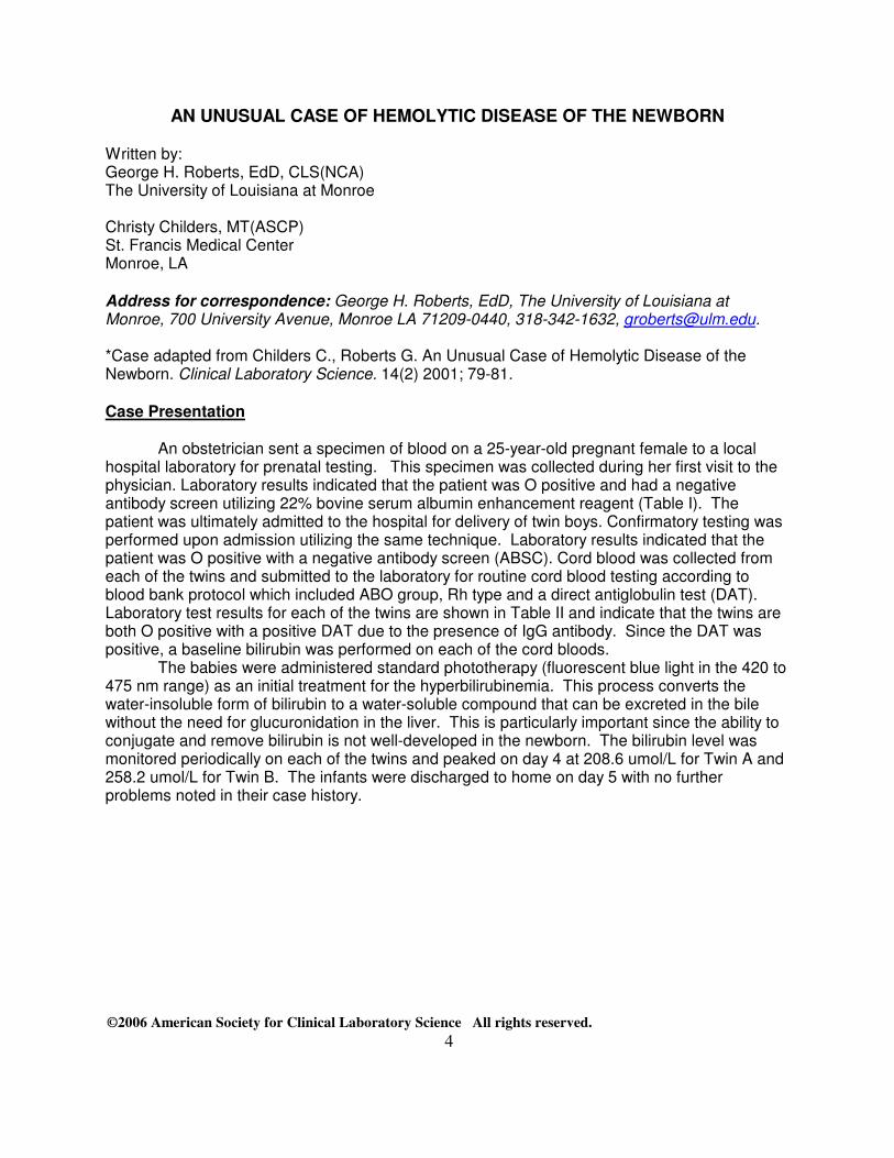
AN UNUSUAL CASE OF HEMOLYTIC DISEASE OF THE NEWBORN Written by: George H. Roberts, EdD, CLS(NCA) The University of Louisiana at Monroe Christy Childers, MT(ASCP) St. Francis Medical Center Monroe, LA Address for correspondence: George H. Roberts, EdD, The University of Louisiana at Monroe, 700 University Avenue, Monroe LA 71209-0440, 318-342-1632, [email protected]. *Case adapted from Childers C., Roberts G. An Unusual Case of Hemolytic Disease of the Newborn. Clinical Laboratory Science. 14(2) 2001; 79-81. Case Presentation
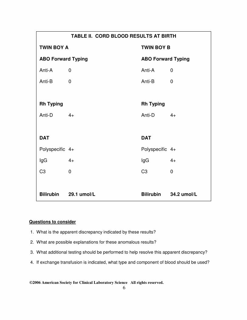
An obstetrician sent a specimen of blood on a 25-year-old pregnant female to a local hospital laboratory for prenatal testing. This specimen was collected during her first visit to the physician. Laboratory results indicated that the patient was O positive and had a negative antibody screen utilizing 22% bovine serum albumin enhancement reagent (Table I). The patient was ultimately admitted to the hospital for delivery of twin boys. Confirmatory testing was performed upon admission utilizing the same technique. Laboratory results indicated that the patient was O positive with a negative antibody screen (ABSC). Cord blood was collected from each of the twins and submitted to the laboratory for routine cord blood testing according to blood bank protocol which included ABO group, Rh type and a direct antiglobulin test (DAT). Laboratory test results for each of the twins are shown in Table II and indicate that the twins are both O positive with a positive DAT due to the presence of IgG antibody. Since the DAT was positive, a baseline bilirubin was performed on each of the cord bloods.
The babies were administered standard phototherapy (fluorescent blue light in the 420 to 475 nm range) as an initial treatment for the hyperbilirubinemia. This process converts the water-insoluble form of bilirubin to a water-soluble compound that can be excreted in the bile without the need for glucuronidation in the liver. This is particularly important since the ability to conjugate and remove bilirubin is not well-developed in the newborn. The bilirubin level was monitored periodically on each of the twins and peaked on day 4 at 208.6 umol/L for Twin A and 258.2 umol/L for Twin B. The infants were discharged to home on day 5 with no further problems noted in their case history.
©2006 American Society for Clinical Laboratory Science All rights reserved.
5
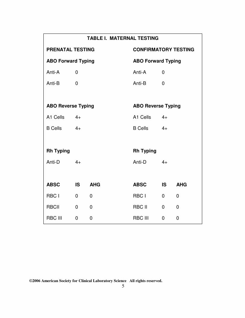
TABLE I. MATERNAL TESTING
PRENATAL TESTING CONFIRMATORY TESTING
ABO Forward Typing ABO Forward Typing
Anti-A 0 Anti-A 0
Anti-B 0 Anti-B 0
ABO Reverse Typing ABO Reverse Typing
A1 Cells 4+ A1 Cells 4+
B Cells 4+ B Cells 4+
Rh Typing Rh Typing
Anti-D 4+ Anti-D 4+
ABSC IS AHG ABSC IS AHG
RBC I 0 0 RBC I 0 0
RBCII 0 0 RBC II 0 0
RBC III 0 0 RBC III 0 0
©2006 American Society for Clinical Laboratory Science All rights reserved.
6
Questions to consider
1. What is the apparent discrepancy indicated by these results?
2. What are possible explanations for these anomalous results?
3. What additional testing should be performed to help resolve this apparent discrepancy?
4. If exchange transfusion is indicated, what type and component of blood should be used?
TABLE II. CORD BLOOD RESULTS AT BIRTH
TWIN BOY A TWIN BOY B
ABO Forward Typing ABO Forward Typing
Anti-A 0 Anti-A 0
Anti-B 0 Anti-B 0
Rh Typing Rh Typing
Anti-D 4+ Anti-D 4+
DAT DAT
Polyspecific 4+ Polyspecific 4+
IgG 4+ IgG 4+
C3 0 C3 0
Bilirubin 29.1 umol/L Bilirubin 34.2 umol/L
©2006 American Society for Clinical Laboratory Science All rights reserved.
7
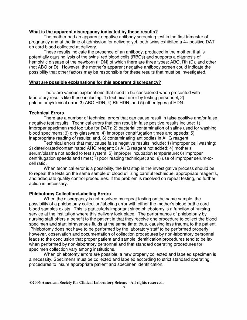
What is the apparent discrepancy indicated by these results? The mother had an apparent negative antibody screening test in the first trimester of
pregnancy and at the time of admission for delivery; yet, both twins exhibited a 4+ positive DAT on cord blood collected at delivery.
These results indicate the presence of an antibody, produced in the mother, that is potentially causing lysis of the twins’ red blood cells (RBCs) and supports a diagnosis of hemolytic disease of the newborn (HDN) of which there are three types: ABO, Rh (D), and other (not ABO or D). However, the mother’s apparent negative antibody screen could indicate the possibility that other factors may be responsible for these results that must be investigated.
What are possible explanations for this apparent discrepancy?
There are various explanations that need to be considered when presented with laboratory results like these including: 1) technical error by testing personnel, 2) phlebotomy/clerical error, 3) ABO HDN, 4) Rh HDN, and 5) other types of HDN.
Technical Errors
There are a number of technical errors that can cause result in false positive and/or false negative test results. Technical errors that can result in false positive results include: 1) improper specimen (red top tube for DAT); 2) bacterial contamination of saline used for washing blood specimens; 3) dirty glassware; 4) improper centrifugation times and speeds; 5) inappropriate reading of results; and, 6) contaminating antibodies in AHG reagent.
Technical errors that may cause false negative results include: 1) improper cell washing; 2) deteriorated/contaminated AHG reagent; 3) AHG reagent not added; 4) mother’s serum/plasma not added to test system; 5) improper incubation temperature; 6) improper centrifugation speeds and times; 7) poor reading technique; and, 8) use of improper serum-to-cell ratio.
When technical error is a possibility, the first step in the investigative process should be to repeat the tests on the same sample of blood utilizing careful technique, appropriate reagents, and adequate quality control procedures. If the problem is resolved on repeat testing, no further action is necessary.
Phlebotomy Collection/Labeling Errors
When the discrepancy is not resolved by repeat testing on the same sample, the possibility of a phlebotomy collection/labeling error with either the mother’s blood or the cord blood samples exists. This is particularly important since phlebotomy is a function of nursing service at the institution where this delivery took place. The performance of phlebotomy by nursing staff offers a benefit to the patient in that they receive one procedure to collect the blood specimen and start intravenous fluids at the same time; thus, causing less trauma to the patient. Phlebotomy does not have to be performed by the laboratory staff to be performed properly; however, observation and documentation of collection procedures by non-laboratory personnel leads to the conclusion that proper patient and sample identification procedures tend to be lax when performed by non-laboratory personnel and that standard operating procedures for specimen collection vary among institutions.
When phlebotomy errors are possible, a new properly collected and labeled specimen is a necessity. Specimens must be collected and labeled according to strict standard operating procedures to insure appropriate patient and specimen identification.
©2006 American Society for Clinical Laboratory Science All rights reserved.
8
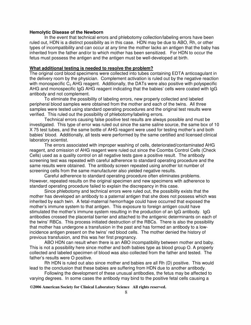
Hemolytic Disease of the Newborn In the event that technical errors and phlebotomy collection/labeling errors have been
ruled out, HDN is a distinct possibility as in this case. HDN may be due to ABO, Rh, or other types of incompatibility and can occur at any time the mother lacks an antigen that the baby has inherited from the father and/or to which mother has been sensitized. For HDN to occur the fetus must possess the antigen and the antigen must be well-developed at birth.
What additional testing is needed to resolve the problem? The original cord blood specimens were collected into tubes containing EDTA anticoagulant in the delivery room by the physician. Complement activation is ruled out by the negative reaction with monospecific C3 AHG reagent. Additionally, the DATs were also positive with polyspecific AHG and monospecific IgG AHG reagent indicating that the babies’ cells were coated with IgG antibody and not complement.
To eliminate the possibility of labeling errors, new properly collected and labeled peripheral blood samples were obtained from the mother and each of the twins. All three samples were tested using standard operating procedures and the original test results were verified. This ruled out the possibility of phlebotomy/labeling errors.
Technical errors causing false positive test results are always possible and must be investigated. This type of error was ruled out since the same saline source, the same box of 10 X 75 test tubes, and the same bottle of AHG reagent were used for testing mother’s and both babies’ blood. Additionally, all tests were performed by the same certified and licensed clinical laboratory scientist.
The errors associated with improper washing of cells, deteriorated/contaminated AHG reagent, and omission of AHG reagent were ruled out since the Coombs Control Cells (Check Cells) used as a quality control on all negative tests gave a positive result. The antibody screening test was repeated with careful adherence to standard operating procedure and the same results were obtained. The antibody screen repeated using another lot number of screening cells from the same manufacturer also yielded negative results.
Careful adherence to standard operating procedure often eliminates problems. However, repeated results on the original specimen and new specimens with adherence to standard operating procedure failed to explain the discrepancy in this case.
Since phlebotomy and technical errors were ruled out, the possibility exists that the mother has developed an antibody to a paternal antigen that she does not possess which was inherited by each twin. A fetal-maternal hemorrhage could have occurred that exposed the mother’s immune system to that antigen. This exposure to foreign antigen could have stimulated the mother’s immune system resulting in the production of an IgG antibody. IgG antibodies crossed the placental barrier and attached to the antigenic determinants on each of the twins’ RBCs. This process initiated destruction of the RBCs. There is also the possibility that mother has undergone a transfusion in the past and has formed an antibody to a low-incidence antigen present on the twins’ red blood cells. The mother denied the history of previous transfusion, and this was her first pregnancy.
ABO HDN can result when there is an ABO incompatibility between mother and baby. This is not a possibility here since mother and both babies type as blood group O. A properly collected and labeled specimen of blood was also collected from the father and tested. The father’s results were O positive.
Rh HDN is ruled out also since mother and babies are all Rh (D) positive. This would lead to the conclusion that these babies are suffering from HDN due to another antibody.
Following the development of these unusual antibodies, the fetus may be affected to varying degrees. In some cases the antibody may bind to the positive fetal cells causing a
©2006 American Society for Clinical Laboratory Science All rights reserved.
9
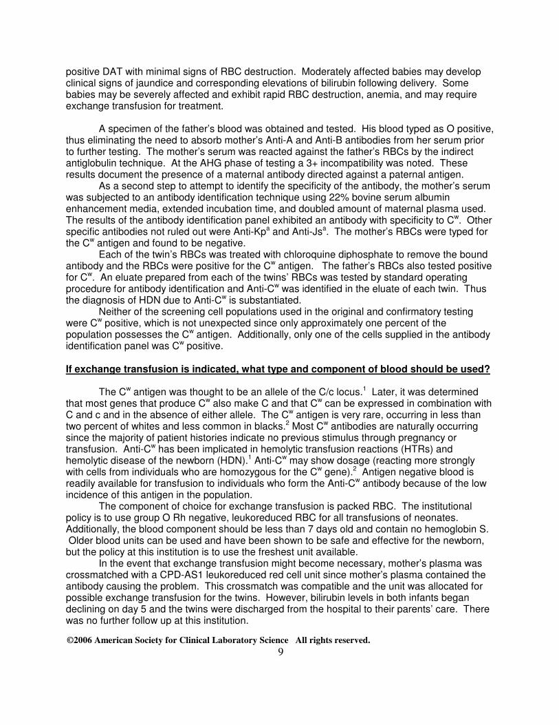
positive DAT with minimal signs of RBC destruction. Moderately affected babies may develop clinical signs of jaundice and corresponding elevations of bilirubin following delivery. Some babies may be severely affected and exhibit rapid RBC destruction, anemia, and may require exchange transfusion for treatment.
A specimen of the father’s blood was obtained and tested. His blood typed as O positive, thus eliminating the need to absorb mother’s Anti-A and Anti-B antibodies from her serum prior to further testing. The mother’s serum was reacted against the father’s RBCs by the indirect antiglobulin technique. At the AHG phase of testing a 3+ incompatibility was noted. These results document the presence of a maternal antibody directed against a paternal antigen.
As a second step to attempt to identify the specificity of the antibody, the mother’s serum was subjected to an antibody identification technique using 22% bovine serum albumin enhancement media, extended incubation time, and doubled amount of maternal plasma used. The results of the antibody identification panel exhibited an antibody with specificity to Cw. Other specific antibodies not ruled out were Anti-Kpa and Anti-Jsa. The mother’s RBCs were typed for the Cw antigen and found to be negative.
Each of the twin’s RBCs was treated with chloroquine diphosphate to remove the bound antibody and the RBCs were positive for the Cw antigen. The father’s RBCs also tested positive for Cw. An eluate prepared from each of the twins’ RBCs was tested by standard operating procedure for antibody identification and Anti-Cw was identified in the eluate of each twin. Thus the diagnosis of HDN due to Anti-Cw is substantiated.
Neither of the screening cell populations used in the original and confirmatory testing were Cw positive, which is not unexpected since only approximately one percent of the population possesses the Cw antigen. Additionally, only one of the cells supplied in the antibody identification panel was Cw positive. If exchange transfusion is indicated, what type and component of blood should be used?
The Cw antigen was thought to be an allele of the C/c locus.1 Later, it was determined that most genes that produce Cw also make C and that Cw can be expressed in combination with C and c and in the absence of either allele. The Cw antigen is very rare, occurring in less than two percent of whites and less common in blacks.2 Most Cw antibodies are naturally occurring since the majority of patient histories indicate no previous stimulus through pregnancy or transfusion. Anti-Cw has been implicated in hemolytic transfusion reactions (HTRs) and hemolytic disease of the newborn (HDN).1 Anti-Cw may show dosage (reacting more strongly with cells from individuals who are homozygous for the Cw gene).2 Antigen negative blood is readily available for transfusion to individuals who form the Anti-Cw antibody because of the low incidence of this antigen in the population.
The component of choice for exchange transfusion is packed RBC. The institutional policy is to use group O Rh negative, leukoreduced RBC for all transfusions of neonates. Additionally, the blood component should be less than 7 days old and contain no hemoglobin S. Older blood units can be used and have been shown to be safe and effective for the newborn, but the policy at this institution is to use the freshest unit available.
In the event that exchange transfusion might become necessary, mother’s plasma was crossmatched with a CPD-AS1 leukoreduced red cell unit since mother’s plasma contained the antibody causing the problem. This crossmatch was compatible and the unit was allocated for possible exchange transfusion for the twins. However, bilirubin levels in both infants began declining on day 5 and the twins were discharged from the hospital to their parents’ care. There was no further follow up at this institution.
©2006 American Society for Clinical Laboratory Science All rights reserved.
10
References
1. Blaney KD, Howard PR. Basic and Applied Concepts of Immunohematology. St. Louis:The C. V. Mosby Company; 2000. p283-302.
2. Harmening DM. Modern Blood Banking and Transfusion Practices 4th ed.
Philadelphia:F. A. Davis Company; 1999. p71-89, 421-435. 3. Quinley ED. Immunohematology: Principles and Practice 2nd ed.
Philadelphia:Lippincott-Raven Publishers; 1998. p79-88, 282-292. 4. Ruddman SV. Textbook of Blood Banking and Transfusion Medicine.
Philadelphia:W.B.Saunders Company; 1995. p436-495. 5. Turgeon ML. Fundamentals of Immunohematology: Theory and Technique.
Philadelphia:Lea and Febiger; 1989. p321-343. 6. Widman FK. Technical Manual 13th ed. Bethesda, MD:American Association of Blood
Banks; 1999. 495-508.
©2006 American Society for Clinical Laboratory Science All rights reserved.
11
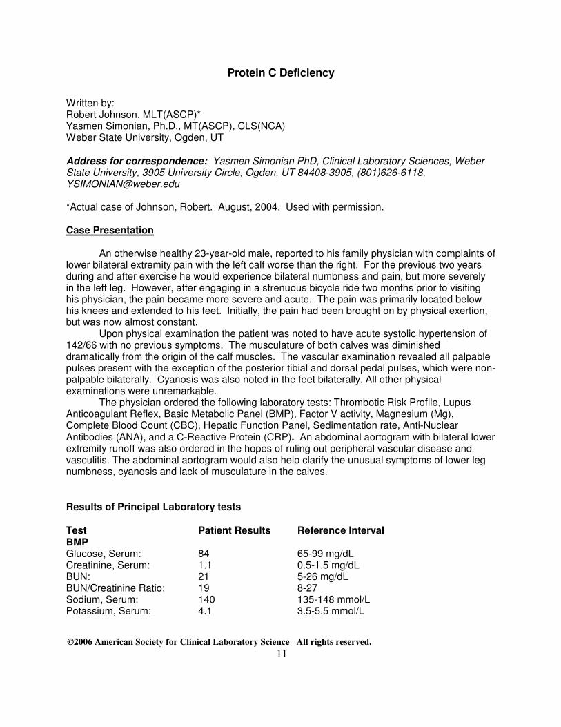
Protein C Deficiency
Written by: Robert Johnson, MLT(ASCP)* Yasmen Simonian, Ph.D., MT(ASCP), CLS(NCA) Weber State University, Ogden, UT Address for correspondence: Yasmen Simonian PhD, Clinical Laboratory Sciences, Weber State University, 3905 University Circle, Ogden, UT 84408-3905, (801)626-6118, [email protected] *Actual case of Johnson, Robert. August, 2004. Used with permission. Case Presentation
An otherwise healthy 23-year-old male, reported to his family physician with complaints of lower bilateral extremity pain with the left calf worse than the right. For the previous two years during and after exercise he would experience bilateral numbness and pain, but more severely in the left leg. However, after engaging in a strenuous bicycle ride two months prior to visiting his physician, the pain became more severe and acute. The pain was primarily located below his knees and extended to his feet. Initially, the pain had been brought on by physical exertion, but was now almost constant.
Upon physical examination the patient was noted to have acute systolic hypertension of 142/66 with no previous symptoms. The musculature of both calves was diminished dramatically from the origin of the calf muscles. The vascular examination revealed all palpable pulses present with the exception of the posterior tibial and dorsal pedal pulses, which were non-palpable bilaterally. Cyanosis was also noted in the feet bilaterally. All other physical examinations were unremarkable.
The physician ordered the following laboratory tests: Thrombotic Risk Profile, Lupus Anticoagulant Reflex, Basic Metabolic Panel (BMP), Factor V activity, Magnesium (Mg), Complete Blood Count (CBC), Hepatic Function Panel, Sedimentation rate, Anti-Nuclear Antibodies (ANA), and a C-Reactive Protein (CRP). An abdominal aortogram with bilateral lower extremity runoff was also ordered in the hopes of ruling out peripheral vascular disease and vasculitis. The abdominal aortogram would also help clarify the unusual symptoms of lower leg numbness, cyanosis and lack of musculature in the calves.
Results of Principal Laboratory tests
Test Patient Results Reference Interval BMP Glucose, Serum: 84 65-99 mg/dL Creatinine, Serum: 1.1 0.5-1.5 mg/dL BUN: 21 5-26 mg/dL BUN/Creatinine Ratio: 19 8-27 Sodium, Serum: 140 135-148 mmol/L Potassium, Serum: 4.1 3.5-5.5 mmol/L
©2006 American Society for Clinical Laboratory Science All rights reserved.
12
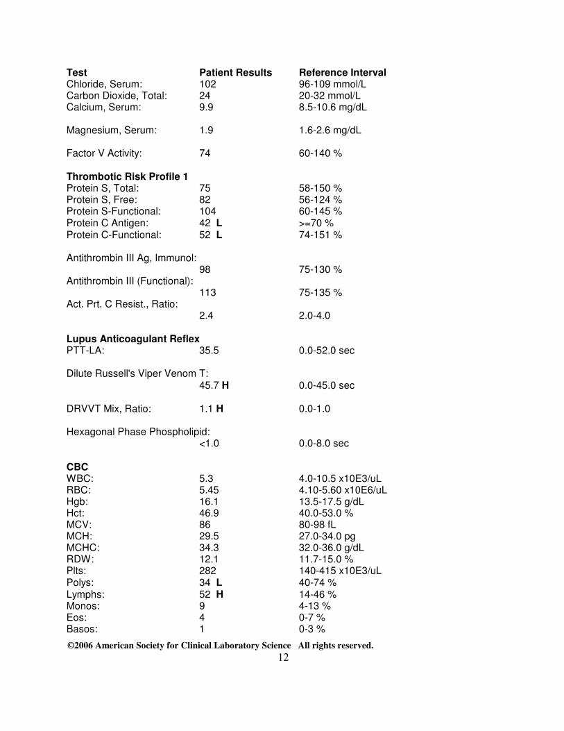
Test Patient Results Reference Interval Chloride, Serum: 102 96-109 mmol/L Carbon Dioxide, Total: 24 20-32 mmol/L Calcium, Serum: 9.9 8.5-10.6 mg/dL Magnesium, Serum: 1.9 1.6-2.6 mg/dL Factor V Activity: 74 60-140 % Thrombotic Risk Profile 1 Protein S, Total: 75 58-150 % Protein S, Free: 82 56-124 % Protein S-Functional: 104 60-145 % Protein C Antigen: 42 L >=70 % Protein C-Functional: 52 L 74-151 % Antithrombin III Ag, Immunol: 98 75-130 % Antithrombin III (Functional): 113 75-135 % Act. Prt. C Resist., Ratio:
2.4 2.0-4.0
Lupus Anticoagulant Reflex PTT-LA: 35.5 0.0-52.0 sec Dilute Russell's Viper Venom T: 45.7 H 0.0-45.0 sec DRVVT Mix, Ratio: 1.1 H 0.0-1.0 Hexagonal Phase Phospholipid: <1.0 0.0-8.0 sec CBC WBC: 5.3 4.0-10.5 x10E3/uL RBC: 5.45 4.10-5.60 x10E6/uL Hgb: 16.1 13.5-17.5 g/dL Hct: 46.9 40.0-53.0 % MCV: 86 80-98 fL MCH: 29.5 27.0-34.0 pg MCHC: 34.3 32.0-36.0 g/dL RDW: 12.1 11.7-15.0 % Plts: 282 140-415 x10E3/uL Polys: 34 L 40-74 % Lymphs: 52 H 14-46 % Monos: 9 4-13 % Eos: 4 0-7 % Basos: 1 0-3 %
©2006 American Society for Clinical Laboratory Science All rights reserved.
13
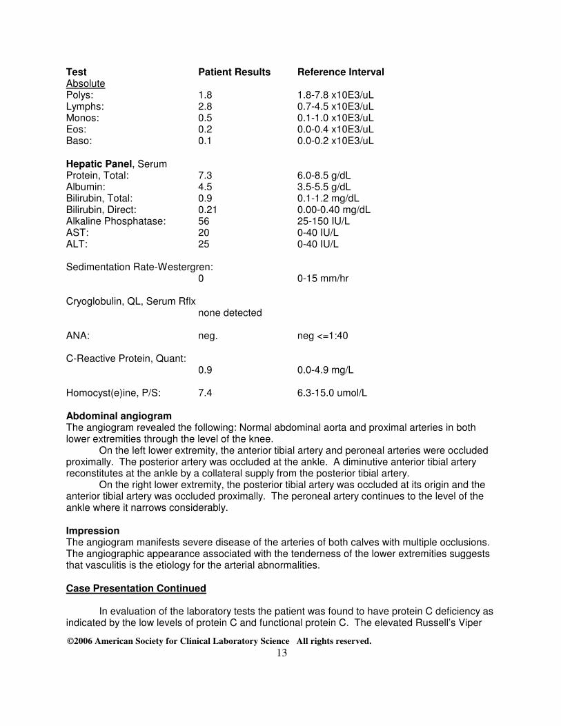
Test Patient Results Reference Interval Absolute Polys: 1.8 1.8-7.8 x10E3/uL Lymphs: 2.8 0.7-4.5 x10E3/uL Monos: 0.5 0.1-1.0 x10E3/uL Eos: 0.2 0.0-0.4 x10E3/uL Baso: 0.1 0.0-0.2 x10E3/uL Hepatic Panel, Serum Protein, Total: 7.3 6.0-8.5 g/dL Albumin: 4.5 3.5-5.5 g/dL Bilirubin, Total: 0.9 0.1-1.2 mg/dL Bilirubin, Direct: 0.21 0.00-0.40 mg/dL Alkaline Phosphatase: 56 25-150 IU/L AST: 20 0-40 IU/L ALT: 25 0-40 IU/L Sedimentation Rate-Westergren:
0 0-15 mm/hr Cryoglobulin, QL, Serum Rflx
none detected ANA: neg. neg <=1:40 C-Reactive Protein, Quant:
0.9 0.0-4.9 mg/L Homocyst(e)ine, P/S: 7.4 6.3-15.0 umol/L Abdominal angiogram The angiogram revealed the following: Normal abdominal aorta and proximal arteries in both lower extremities through the level of the knee.
On the left lower extremity, the anterior tibial artery and peroneal arteries were occluded proximally. The posterior artery was occluded at the ankle. A diminutive anterior tibial artery reconstitutes at the ankle by a collateral supply from the posterior tibial artery.
On the right lower extremity, the posterior tibial artery was occluded at its origin and the anterior tibial artery was occluded proximally. The peroneal artery continues to the level of the ankle where it narrows considerably. Impression The angiogram manifests severe disease of the arteries of both calves with multiple occlusions. The angiographic appearance associated with the tenderness of the lower extremities suggests that vasculitis is the etiology for the arterial abnormalities. Case Presentation Continued
In evaluation of the laboratory tests the patient was found to have protein C deficiency as indicated by the low levels of protein C and functional protein C. The elevated Russell’s Viper
©2006 American Society for Clinical Laboratory Science All rights reserved.
14
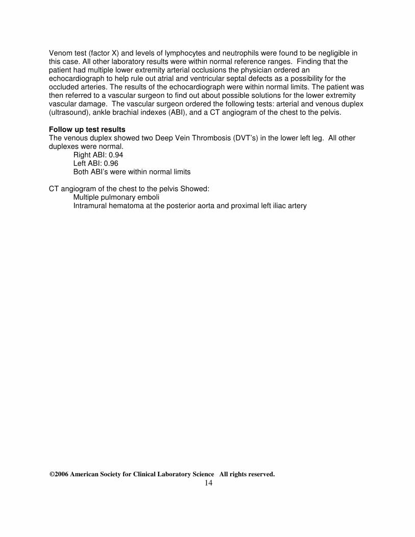
Venom test (factor X) and levels of lymphocytes and neutrophils were found to be negligible in this case. All other laboratory results were within normal reference ranges. Finding that the patient had multiple lower extremity arterial occlusions the physician ordered an echocardiograph to help rule out atrial and ventricular septal defects as a possibility for the occluded arteries. The results of the echocardiograph were within normal limits. The patient was then referred to a vascular surgeon to find out about possible solutions for the lower extremity vascular damage. The vascular surgeon ordered the following tests: arterial and venous duplex (ultrasound), ankle brachial indexes (ABI), and a CT angiogram of the chest to the pelvis. Follow up test results The venous duplex showed two Deep Vein Thrombosis (DVT’s) in the lower left leg. All other duplexes were normal.
Right ABI: 0.94 Left ABI: 0.96 Both ABI’s were within normal limits
CT angiogram of the chest to the pelvis Showed:
Multiple pulmonary emboli Intramural hematoma at the posterior aorta and proximal left iliac artery
©2006 American Society for Clinical Laboratory Science All rights reserved.
15
Discussion
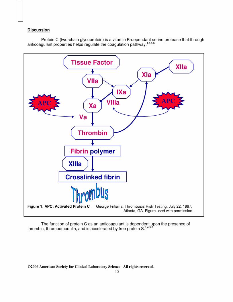
Protein C (two-chain glycoprotein) is a vitamin K-dependant serine protease that through anticoagulant properties helps regulate the coagulation pathway.1,4,5,6
Figure 1: APC: Activated Protein C George Fritsma, Thrombosis Risk Testing, July 22, 1997,
Atlanta, GA. Figure used with permission.
The function of protein C as an anticoagulant is dependent upon the presence of
thrombin, thrombomodulin, and is accelerated by free protein S.1,4,5,6
VIIa
Xa
Thrombin
Fibrin polymer
Crosslinked fibrin
Va
XIIIa
IXa
VIIIa
XIa XIIa
Tissue Factor
APC APC
©2006 American Society for Clinical Laboratory Science All rights reserved.
16
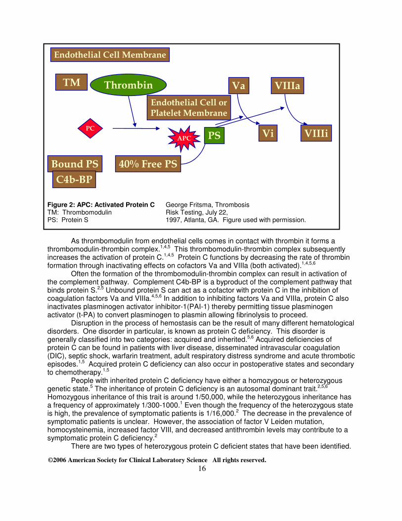
Figure 2: APC: Activated Protein C George Fritsma, Thrombosis TM: Thrombomodulin Risk Testing, July 22, PS: Protein S 1997, Atlanta, GA. Figure used with permission.
As thrombomodulin from endothelial cells comes in contact with thrombin it forms a thrombomodulin-thrombin complex.1,4,5 This thrombomodulin-thrombin complex subsequently increases the activation of protein C.1,4,5 Protein C functions by decreasing the rate of thrombin formation through inactivating effects on cofactors Va and VIIIa (both activated).1,4,5,6
Often the formation of the thrombomodulin-thrombin complex can result in activation of the complement pathway. Complement C4b-BP is a byproduct of the complement pathway that binds protein S.2,5 Unbound protein S can act as a cofactor with protein C in the inhibition of coagulation factors Va and VIIIa.4,5,6 In addition to inhibiting factors Va and VIIIa, protein C also inactivates plasminogen activator inhibitor-1(PAI-1) thereby permitting tissue plasminogen activator (t-PA) to convert plasminogen to plasmin allowing fibrinolysis to proceed. Disruption in the process of hemostasis can be the result of many different hematological disorders. One disorder in particular, is known as protein C deficiency. This disorder is generally classified into two categories: acquired and inherited.5,6 Acquired deficiencies of protein C can be found in patients with liver disease, disseminated intravascular coagulation (DIC), septic shock, warfarin treatment, adult respiratory distress syndrome and acute thrombotic episodes.1,5 Acquired protein C deficiency can also occur in postoperative states and secondary to chemotherapy.1,5
People with inherited protein C deficiency have either a homozygous or heterozygous genetic state.5 The inheritance of protein C deficiency is an autosomal dominant trait.2,5,6 Homozygous inheritance of this trait is around 1/50,000, while the heterozygous inheritance has a frequency of approximately 1/300-1000.1 Even though the frequency of the heterozygous state is high, the prevalence of symptomatic patients is 1/16,000.2 The decrease in the prevalence of symptomatic patients is unclear. However, the association of factor V Leiden mutation, homocysteinemia, increased factor VIII, and decreased antithrombin levels may contribute to a symptomatic protein C deficiency.2
There are two types of heterozygous protein C deficient states that have been identified.
TM Thrombin
Endothelial Cell Membrane
Bound PS
C4b-BP
40% Free PS
Va
Vi
VIIIa
VIIIi PS
Endothelial Cell or
Platelet Membrane
PC
APC
©2006 American Society for Clinical Laboratory Science All rights reserved.
17
Type I deficiency is the most prevalent. It is characterized by a decrease in both the functional and the antigenic levels of protein C.1,5,6 Type II is less common. Patients with type II deficiency typically have normal antigenic levels, but reduced functional protein C.1,5,6 Patients with type I or type II can exhibit venous thromboses with provocation.6 In rare cases, patients with protein C deficiency can exhibit spontaneous arterial and venous thrombosis.
Unlike the heterozygous state, patients with homozygous inheritance have a significantly higher prevalence of symptoms. Infants born with homozygous protein C deficiency usually develop a fatal condition known as neonatal purpura fulminans.1,5 Only with many difficulties do patients with this condition normally live longer than infancy.1,5
Treatment
Patients with provoked thrombotic episodes are usually placed on acute anticoagulation therapy. This therapy typically indicates that patients be placed on either heparin or enoxaparin (Lovenox) in conjunction with warfarin until therapeutic drug ranges are reached.3,4 Once therapeutic drug ranges have been reached the patient is taken off heparin or enoxaparin.4 Warfarin treatment then continues for a period of three to six months.4 Significant disease without predisposing risk factors commonly requires life-long therapy with warfarin. If care is not taken the use of warfarin can be counter productive. In some cases the misuse of warfarin has led to complications in patients, ranging from skin necrosis to gastrointestinal bleeding.1,4,5 One important factor that influences the function of warfarin is the dietary intake of vitamin K.4 Depending on the dietary intake of the individual, the vitamin K levels in the blood can fluctuate. This fluctuation can alter the levels of the vitamin K-dependent coagulation factors in circulation, therefore increasing or decreasing the effectiveness of warfarin .4 Warfarin functions by inhibiting the enzymatic recycling of vitamin K in the liver.1,4 Vitamin K is a cofactor in the carboxylation of not only protein C and S but also the coagulation factors II, VII, IX and X.1,4,5,7 The half life of protein C is 8-10hrs and is shorter than that of the vitamin K-dependent coagulation proteins.3,4,5 If a patient is not already in an anticoagulant state, warfarin will induce a hypercoagulable condition by reducing the function of protein C before that of the vitamin K-dependent coagulation proteins.1,3,5
An alternative to the use of anticoagulant drugs for acute treatment of protein C deficiency can be achieved by the use of fresh frozen plasma (FFP) and/or protein C concentrate. Both FFP and protein C concentrate can be administered by subcutaneous methods. In recent years, there has been an increase in the use of FFP and protein C concentrate. However, due to their cost, availability and administration they are typically limited to acute treatments of homozygous protein C deficient patients. Diagnostic Testing
The testing for protein C deficiency is divided into two areas: the immunologic (antigen) and the functional (activity) assays.1,4,5,6 Testing for the antigen is typically done by ELISA or EIA, but another commonly used method is Laurell rocket immuno-electrophoresis.1,5 Functional protein C is typically measured by clot-based or by using synthetic chromogenic substrate methods.1,5,6 Once the patient has been placed on anticoagulation therapy, prothrombin time (PT) tests and international normalized ratio’s (INR) are used to monitor intravenous and/or oral anticoagulation.4
©2006 American Society for Clinical Laboratory Science All rights reserved.
18
Case Conclusion
The patient was diagnosed with inherited type I protein C deficiency resulting in lower leg DVT’s and multiple pulmonary emboli. The arterial occlusions were thought to be a result of the intraluminal thrombus (hematoma). As to the cause of the intraluminal thrombus, it is still unknown. The vascular surgeon, after consulting with a hematologist, hospitalized the patient for anticoagulation therapy. The anticoagulation therapy consisted of a heparin drip of 100 units/ml by IV bag with the addition of Warfarin 7.5 mg tab PO QD (one tablet once a day) as the PT/INR became therapeutic.
The therapeutic levels of anticoagulation for this patient were determined to be an INR of 2.5-3.5. The patient was released from the hospital a week later with an INR of 2.4 and instructed to continue life long anticoagulation therapy by means of warfarin and continued PT/INR monitoring.
References
1. Harmening, Denise M. Clinical Hematology and Fundamentals of Hemostasis. 4th ed. Philadelphia, PA: F.A. Davis, 2002; 461:490:540-541:556-557:672-673.
2. Fritsma, George A. “Thrombosis Risk Testing”. Powerpoint Presentation. Intermountain
States Seminar; 1997 July 22; Atlanta, GA. 3. Tierney L, McPhee S, Papadakis M, editors. Current Medical Diagnosis & Treatment.
39th ed. San Francisco, CA:Lange medical books/McGraw-Hill, 2000;326-329:548. 4. Stevens, Marcella L. Fundamentals of Clinical Hematology. Philadelphia:W.B.
Saunders, 1997;246-247p:252-255:271-274:333dp:337dp. 5. McKenzie, Shirlyn B. Clinical Laboratory Hematology. New Jersey:Pearson,
2004;681:696-697:767-768:796. 6. Turgeon, Mary L. Clinical Hematology Theory and Procedures. 3rd ed.
Philadelphia:Lippincott W&W, 1999;415-416:436:440-441. 7. Harmening, Denise M. Modern Blood Banking and Transfusion Practices. 4th ed.
Philadelphia, PA:F.A. Davis, 1999;350:355.