Embed Size (px)
Citation preview

Volume 2 • Issue 3 • 1000123J Stem Cell Res TherISSN:2157-7633 JSCRT, an open access journal
Open AccessLetter
Sharma et al., J Stem Cell Res Ther 2012, 2:3 DOI: 10.4172/2157-7633.1000123
Overexpression of C-Terminal Domain of Talin-1 Enhances Survival, Migration and Differentiation of Human Cardiac Stem CellsUmesh C Sharma*, Duc M Vu, Jia-Qiang He, Xian-Liang Tang, Gregg Rokosh and Roberto Bolli
Institute of Molecular Cardiology, University of Louisville, Louisville, Kentucky, USA
*Corresponding author: Umesh C Sharma, Institute of Molecular Cardiology, University of Louisville, Louisville, Kentucky, USA, E-Mail: [email protected]
Received May 21, 2012; Accepted June 13, 2012; Published June 15, 2012
Citation: Sharma UC, Vu DM, He JQ, Tang XL, Rokosh G, et al. (2012) Overexpression of C-Terminal Domain of Talin-1 Enhances Survival, Migration and Differentiation of Human Cardiac Stem Cells. J Stem Cell Res Ther 2:123. doi:10.4172/2157-7633.1000123
Copyright: © 2012 Sharma UC, et al. This is an open-access article distributed under the terms of the Creative Commons Attribution License, which permits unrestricted use, distribution, and reproduction in any medium, provided the original author and source are credited.
To The EditorCardiovascular Diseases (CVD) constitute the single leading cause
of death in the United States. According to the heart disease and stroke statistics released by American Heart Association in 2010, 1 out of every 2.9 deaths is due to cardiovascular disease. Even with early diagnosis and timely reperfusion of ischemic myocardium, up to 30% patients develop Left Ventricular (LV) remodeling and loss of cardiac function [1,2]. Current treatment modality of cardiac failure is mainly focused to relieve symptoms. Although cardiac transplantation has offered some hope, organ shortage, donor-recipient organ mismatch, and transplant rejection are the major challenges limiting its widespread use.
Unlike many other tissues in our body, adult cardiomyocytes have limited ability to self-regenerate. This means, an ischemic injury will lead to irreversible myocardial damage leading to loss of cardiac function. Cardiac Stem Cell (CSC) therapy has emerged as a promising strategy for cardiac repair after acute Myocardial Infarction (MI). We have recently reported that intracoronary infusion of autologous CSCs is effective in improving LV systolic function and reducing infarct size in patients with heart failure after myocardial infarction [3].
There are several molecules that control the survival, migration and differentiation of stem cells. Integrins are family heterodimer proteins that regulate the interaction between circulating cells and endothelial surface. Actin filaments are the molecular motors that propel the migrating cells. Talin-1 is an important intracellular molecule that serves as a latch between integrins and actin filaments. Talin-1 is also a component of Focal Adhesions Assembly (FAA), along with vinculin and paxillin [4]. Previous studies have reported a strong and direct relationship between the level of Talin-1 expressions and risk factors that contribute to the development of cardiomyopathy. For example, Talin-1 expression is inversely related to the extent of diabetes control [5]. Previous studies have also reported an increased Talin-1 expression both in left and right ventricular dysfunction. It has been reported that treating heart failure patients with Angiotension Converting Enzyme (ACE)-inhibitors or Angiotensin Receptor Blockers (ARBs) can preserve Talin-1 function by inhibiting its proteolytic partner calpain-II [6]. Furthermore, as it relates to the use of CSCs for myocardial repair following acute MI, selective knockout of Talin from embryonic cells has shown loss of differentiation of embryonic cells to cardiomyocyte-committed embryoid bodies [7]. However, it is still unclear whether gain of Talin-1 function can be a therapeutic target for survival, migration and differentiation CSCs.
The C-terminal part of talin rod, TalinC, contains binding sites and regulatory elements that convey essential aspects of talin function in cells. In this study, we aimed to investigate the role of TalinC for the survival and migration CSCs and their differentiation to adult cardiomyocytes. In this study, we have examined the effect of TalinC overexpression for human CSC survival, adhesion, migration, and lineage commitment to adult cardiomyocytes.
We used the right atrial appendages to isolate c-kit-positive CSCs. For CSC isolation, we minced right atrial appendages into small pieces and incubated with collagenase II (Figure 1A). Undigested tissue was then filtered out (Figure 1B and 1C). The cells suspension was plated in culture flask and cultured in incubator supplied with 5% CO2 at 37°C to confluence (Figure 1D). C-kit positive cells were selected using a c-kit MACS kit (Miltenyi Biotec, Germany) (Figure 1E). The purification of c-kit positive cell population was confirmed by immunostaining (Figure 1F).
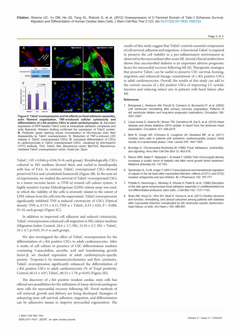
Utilizing a novel Baculovirus-mediated protein expression system in mammals (BacMam, Invitrogen), we overexpressed Red Fluorescent Protein (RFP)-tagged TalinC in c-Kit-positive CSCs isolated from human right atrial appendages. A robust expression of TalinC was noted in CSC cytoskeleton and intracellular FAA. We also confirmed TalinC expression Western blotting by using a TalinC-specific monoclonal antibody (EMD Millipore) (Figure 2A).
We then examined the adhesion of CSCs to a fibronectin coated cell culture disks in a normal or High-Glucose (HG) culture system. TalinC overexpression normalized the HG-induced loss of cell adhesion (Optical density: Control, 1.04 ± 0.02; Glucose, 0.92 ± 0.07; Glucose +
Figure 1: Isolation and retrieval of c-Kit positive CSCs from human right atrial appendages (RAA). A. RAA was minced into small pieces, B. enzymatically digestion tissue with collagenase solution, C. undigested debris filtered out, D. cells grown in vitro, E. sorting of c-kit +ve cells by magnetic beads, F. staining of c-kit protein expressed by human CSCs.
Journal ofStem Cell Research & TherapyJo
urna
l of S
temCell Research&
Therapy
ISSN: 2157-7633
Citation: Sharma UC, Vu DM, He JQ, Tang XL, Rokosh G, et al. (2012) Overexpression of C-Terminal Domain of Talin-1 Enhances Survival, Migration and Differentiation of Human Cardiac Stem Cells. J Stem Cell Res Ther 2:123. doi:10.4172/2157-7633.1000123
Page 2 of 2
Volume 2 • Issue 3 • 1000123J Stem Cell Res TherISSN:2157-7633 JSCRT, an open access journal
TalinC, 1.05 ± 0.046; p=0.04, N=8, each group). Morphologically, CSCs cultured in HG medium showed thick and curled-in lamellopodia with loss of FAA. In contrast, TalinC overexpressed CSCs showed preserved FAA and cytoskeletal framework (Figure 2B). In the next set of experiments, we studied the survival of TalinC-overexpressed CSCs in a tumor necrosis factor- α (TNF-α)-treated cell culture system. A highly sensitive Lactate Dehydrogenase (LDH)-release assay was used, in which the viability of the cells is inversely related to the extent of LDH-release from the cells into the supernatant. TalinC overexpression significantly inhibited TNF-α-induced cytotoxicity of CSCs (Optical density: TNF-α, 0.713 ± 0.11; TNF-α + TalinC, 0.33 ± 0.02, P = 0.008, N=10, each group) (Figure 2C).
In addition to improved cell adhesion and reduced cytotoxicity, TalinC overexpression enhanced cell migration in HG culture medium (Migration Index: Control, 24.6 ± 2.7; HG, 15.34 ± 2.7; HG + TalinC, 34 ± 4.7; p=0.03, N=3-4, each group).
We also investigated the effect of TalinC overexpression for the differentiation of c-Kit positive CSCs to adult cardiomyocytes. After 4 weeks of cell culture in presence of CSC differentiation medium containing 5-azacytidine, ascorbic acid and transforming growth factor-β, we checked expression of adult cardiomyocyte-specific protein- Troponin-I, by immunocytochemistry and flow cytometry. TalinC overexpression significantly enhanced the differentiation of c-Kit positive CSCs to adult cardiomyocytes (% of TropI positivity:Control, 66.13 ± 4.07; TalinC, 80.53 ± 1.79; p=0.05) (Figure 2D).
The discovery of c-Kit positive resident cardiac stem cells has offered new possibilities for the utilization of tissue-derived autologous stem cells for myocardial recovery following MI. Novel methods of cell retrieval, growth and delivery are being developed. Strategies of enhancing stem cell survival, adhesion, migration, and differentiation can be adjunctive means to improve myocardial regeneration. The
results of this study suggest that TalinC controls essential components of cell survival, adhesion and migration. A functional TalinC is required to preserve the cell viability in a pro-inflammatory environment as observed in the myocardium after acute MI. Several clinical studies have shown that uncontrolled diabetes is an important adverse prognostic factor for myocardial recovery following MI [8]. Therapeutic strategies that preserve TalinC can be useful to preserve CSC survival, homing, migration and enhanced lineage commitment of c-Kit positive CSCs to adult cardiomyocytes. Overall, the results of this study can add to the current success of c-Kit positive CSCs of improving LV systolic function and reducing infarct size in patients with heart failure after MI.References
1. Bolognese L, Neskovic AN, Parodi G, Cerisano G, Buonamici P, et al. (2002) Left ventricular remodeling after primary coronary angioplasty: Patterns of left ventricular dilation and long-term prognostic implications. Circulation 106: 2351-2357.
2. Lloyd-Jones D, Adams RJ, Brown TM, Carnethon M, Dai S, et al. (2010) Heart disease and stroke statistics--2010 update: A report from the american heart association. Circulation 121: e46-e215.
3. Bolli R, Chugh AR, D’Amario D, Loughran JH, Stoddard MF, et al. (2011) Cardiac stem cells in patients with ischaemic cardiomyopathy (scipio): Initial results of a randomised phase 1 trial. Lancet 378: 1847-1857.
4. Burridge K, Chrzanowska-Wodnicka M (1996) Focal adhesions, contractility, and signaling. Annu Rev Cell Dev Biol 12: 463-518.
5. Renno WM, Saleh F, Klepacek I, Al-Awadi F (2006) Talin immunogold density increases in sciatic nerve of diabetic rats after nerve growth factor treatment. Medicina (Kaunas) 42: 147-163.
6. Sandmann S, Yu M, Unger T (2001) Transcriptional and translational regulation of calpain in the rat heart after myocardial infarction--effects of AT(1) and AT(2) receptor antagonists and ace inhibitor. Br J Pharmacol 132: 767-777.
7. Priddle H, Hemmings L, Monkley S, Woods A, Patel B, et al. (1998) Disruption of the talin gene compromises focal adhesion assembly in undifferentiated but not differentiated embryonic stem cells. J Cell Biol 142: 1121-1133.
8. Shah AM, Hung CL, Shin SH, Skali H, Verma A, et al. (2011) Cardiac structure and function, remodeling, and clinical outcomes among patients with diabetes after myocardial infarction complicated by left ventricular systolic dysfunction, heart failure, or both. Am Heart J 162: 685-691.
Figure 2: TalinC overexpression and its effects on focal adhesion assembly, actin filament organization, TNF-α-induced cellular cytotoxicity and differentiation of c-Kit positive CSCs to adult cardiomyocytes. A. Abundant expression of RFP-labeled TalinC (red) at intracellular adhesion complexes and actin filaments; Western blotting confirmed the expression of TalinC protein. B. Phalloidin green staining shows normalization of HG-induced actin fiber disassembly by TalinC overexpression; C. Reduction of TNF-α-induced LDH release in TalinC overexpressed CSCs; D. Increased differentiation of CSCs to cardiomyocytes in TalinC overexpressed CSCs visualized by anti-troponin I-FITC antibody. TlnC, TalinC; Bac, Baculovirus vector; BacTlnC, Baculovirus-mediated TalinC overexpression vector. Scale bar: 20µm