Embed Size (px)
Citation preview

C-5 Propyne-Modi®ed Oligonucleotides Penetrate theEpidermis in Psoriatic and Not Normal Human Skin AfterTopical Application
Paul J. White,*² Andrew C. Gray,*² Rhys D. Fogarty,² Rodney D. Sinclair,³ Susan P. Thumiger,²George A. Werther,² and Christopher J. Wraight²*Department of Pharmaceutical Biology & Pharmacology, Victorian College of Pharmacy, Monash University, Parkville, Victoria, Australia;
²Centre for Hormone Research, Murdoch Children's Research Institute, Royal Children's Hospital, Parkville, Victoria, Australia;
³University of Melbourne, Department of Medicine (Dermatology), St Vincent's Hospital, Fitzroy, Victoria, Australia
We have previously shown that antisense oligo-nucleotides effectively reduced insulin-like growthfactor I receptor expression in human psoriatic skingrafted on to nude mice when injected intradermally.We therefore investigated the penetration of C-5 pro-pyne modi®ed antisense oligonucleotides into humannormal and psoriatic skin after topical administration.Oligonucleotide (37.5 mg; 250 mM) was applied inaqueous solution or 5% methylcellulose gel for 24 h,prior to live confocal microscopy and ¯uorescencemicroscopy of ®xed sections. We found that oligo-nucleotide could penetrate through the stratum cor-neum of psoriatic but not normal human skin overlarge regions of the epidermis. The oligonucleotide
was localized to the nucleus of large parakeratoticcells in the psoriatic skin as well as smaller basal andsuprabasal keratinocytes. In normal human skin,oligonucleotide was con®ned to the stratum corneum,with little or no oligonucleotide apparent in theviable epidermis. Electrophoresis of oligonucleotiderecovered from treated psoriatic and normal skinrevealed that the oligonucleotide remained intact overthe 24 h period. In summary, we found that C-5 pro-pyne modi®ed antisense oligonucleotides could reachthe target cells (in this case basal keratinocytes) aftertopical administration to psoriatic but not normalskin. Key words: antisense oligonucleotide/psoriasis/topicaldelivery. J Invest Dermatol 118:1003±1007, 2002
Anti-sense oligonucleotides are increasingly beingconsidered as therapeutic options for a range ofdisease states, particularly as the development ofsecond generation oligonucleotides have reducedconcerns regarding toxicity. We have recently shown
that antisense oligonucleotides targeting the insulin-like growthfactor-I receptor could be used to normalize the psoriatic epidermisafter repeated intradermal injection into grafted human skin(Wraight et al, 2000). A desired feature of any cutaneous therapyis a user-friendly topical formulation, and thus further investigationof the ability of these agents to be effective, topically-applied drugsis warranted.
The use of antisense oligonucleotides in dermatology has beenthe subject of considerable interest due to the attractiveness of theconcept of antisense therapy and the ease of access to the targetorgan for these large, charged therapeutic agents. The stratumcorneum, however, is well equipped to prevent the access of largecharged substances due to its physical, chemical, and enzymaticbarrier properties. We have previously found that oligonucleotidedoes not penetrate normal human skin grafts on athymic mice to asigni®cant degree (White et al, 1999). When grafts were tape
stripped, however, there was extensive nuclear localization ofoligonucleotide throughout the epidermis, indicating that thestratum corneum does act as a signi®cant barrier to oligonucleotidedelivery across the skin.
A number of studies have demonstrated antisense penetrationand or ef®cacy using different methods to overcome the skinbarrier. These methods included intradermal injection (White et al,1999; Wraight et al, 2000), iontophoresis (Vlassov et al, 1994;Oldenburg et al, 1995; Regnier and Preat, 1998), and electropor-ation (Regnier and Preat, 1998). None of these methods, however,are currently well suited to use in the delivery of therapeutic agentsdue to considerations of cost or discomfort.
One skin condition known to involve impaired differentiation ofkeratinocytes and reduced stratum corneum barrier function is thecommon autoimmune disease psoriasis. The reduction in barrierfunction is attributed to a number of characteristics of psoriatic skinthat are a result of the impairment of corneocyte differentiation,including impaired formation and secretion of lamellar bodycontents and processing of lamellar body contents into lamellarbilayers (Ghadially et al, 1996). Conditions in which there is animpaired ability to prevent oligonucleotide penetration maytherefore be far more amenable to antisense therapy than those inwhich the barrier function remains intact.
The aim of this study was therefore to compare the penetrationof oligonucleotides in human psoriatic skin with that in normalhuman skin. We found that oligonucleotide penetration in psoriasisis indeed markedly greater than that in normal skin, apparentlyinvolving the entry of oligonucleotide through areas of severe
Manuscript received August 3, 2001; October 24, 2001; accepted forpublication October 31, 2001.
Reprint requests to: Dr. Paul White, Lecturer, Department ofPharmaceutical Biology & Pharmacology, Victorian College ofPharmacy, Monash University 381 Royal Parade, Parkville 3052,Victoria, Australia. Email: [email protected]
0022-202X/02/$15.00 ´ Copyright # 2002 by The Society for Investigative Dermatology, Inc.
1003
barrier function impairment followed by lateral oligonucleotidespread throughout the viable epidermis.
MATERIALS AND METHODS
Ex vivo maintenance of normal and psoriatic skin biopsies Psoriaticskin was obtained from volunteers under protocol 94012A of the RoyalChildren's Hospital Ethics in Human Research Committee. Two 8 mmpunch biopsies were obtained from affected areas on the abdomen.Normal human skin was obtained from discards of plastic surgery. Themethod of skin maintenance for 24 h was adapted from that of Russoet al (1994). Speci®cally, on receipt of psoriatic or normal skin,subcutaneous tissue was removed and the skin was placed on a stainlesssteel mesh coated in 2% agar, with the dermis in contact with the agar.The agar was itself in contact with Dulbecco's modi®ed Eagle's medium(Gibco BRL, Melbourne, Australia) with 10% fetal bovine serum. Areservoir containing phosphate-buffered saline (PBS) surrounded theculture medium reservoir, providing humidity.
Application of ¯uorescein isothiocyanate (FITC)-oligonucleo-tide A total of 14 normal skin samples and 13 psoriatic skin sampleswere used in the study. Six normal skin samples and six psoriatic skinsamples received 37.5 mg (250 mM) FITC-R451 in PBS. The aqueousapplications involved 3 3 10 ml applications over a period of 2 h, andimaging was performed at 24 h. Alternatively, six normal and ®vepsoriatic samples received 10±37.5 mg FITC-oligonucleotide in 5%methylcellulose gel in a single application, with the nature of the gelimposing some variability on the ®nal dose applied. Finally, two normaland two psoriatic samples received an alternative oligonucleotide, FITC-DT1064, in PBS as above.
Live confocal microscopy (LCM) LCM of skin samples wasperformed as previously described (White et al, 1999). The samples wereimaged 24 h after oligonucleotide application for all samples. Imagingwas performed using an Optiscan f900e laser scanning confocalmicroscope (Optiscan Ltd, Melbourne, Australia) equipped with anargon ion laser providing 488 nm (blue) excitation and with ¯uorescencedetection above 515 nm (green). For each sample, a series of horizontalsections were taken at increasing depths into the epidermis. Pilotexperiments demonstrated that ¯uorescence could be detected up to100 mm below the skin surface.
Assessment of oligonucleotide penetration in ®xed sections ofhuman skin After live confocal imaging, the skin was ®xed in 4%paraformaldehyde for 24 h followed by 24±72 h in 0.5 M sucrose. Thesamples were then embedded in paraf®n wax, and 5 mm sections werecut and mounted on silanized slides. Sections were dewaxed in histolene,rehydrated in graded ethanols, rinsed, and mounted with Mowiol
(Calbiochem, Clayton, Victoria, Australia) with DABCO anti-fade(Sigma, St Louis, MO).
Analysis of oligonucleotide stability The treated skin samples werehomogenized with a Kinematica PT 1200 Polytron in 20 mM Tris±HClpH 8.0; 150 mM NaCl; 10% glycerol buffer containing 1% Triton X-100, and centrifuged at 20,000 3 g for 10 min at 4°C. The pellet wasresuspended in distilled water containing 1 mg per ml proteinase K andincubated for 1 h at 56°C. Formamide (25 ml) was added and the sampleheated to 55°C for 5 min The samples were analyzed on a 19%polyacrylamide 90 mM Tris borate, 20 mM ethylenediamine tetraaceticacid gel, containing 7 M urea and transferred on to a Zeta-Probemembrane (Bio-Rad, Sydney, Australia). The membrane was blockedwith 10 mM Tris±HCl, pH 7.4, 150 mM NaCl, 0.1% Tween 20containing 5% skim milk powder for 1 h at room temperature, thenincubated with alkaline phosphatase-conjugated anti-¯uorescein immuno-globulin (Vistra ECF Western blotting kit; Amersham, Buckinghamshire,U.K.); 1:2500 dilution for 1 h at room temperature. Bands werevisualized by chemi¯uorescence.
Oligonucleotides Two different-3¢-FITC-conjugated C-5 propynyldC dU phosphorothioate oligonucleotide 15mers were used, with thefollowing sequences: R451ÐUAACACGAUACGCGA; DT1064ÐCACAGUUGCUGCAAG.
RESULTS
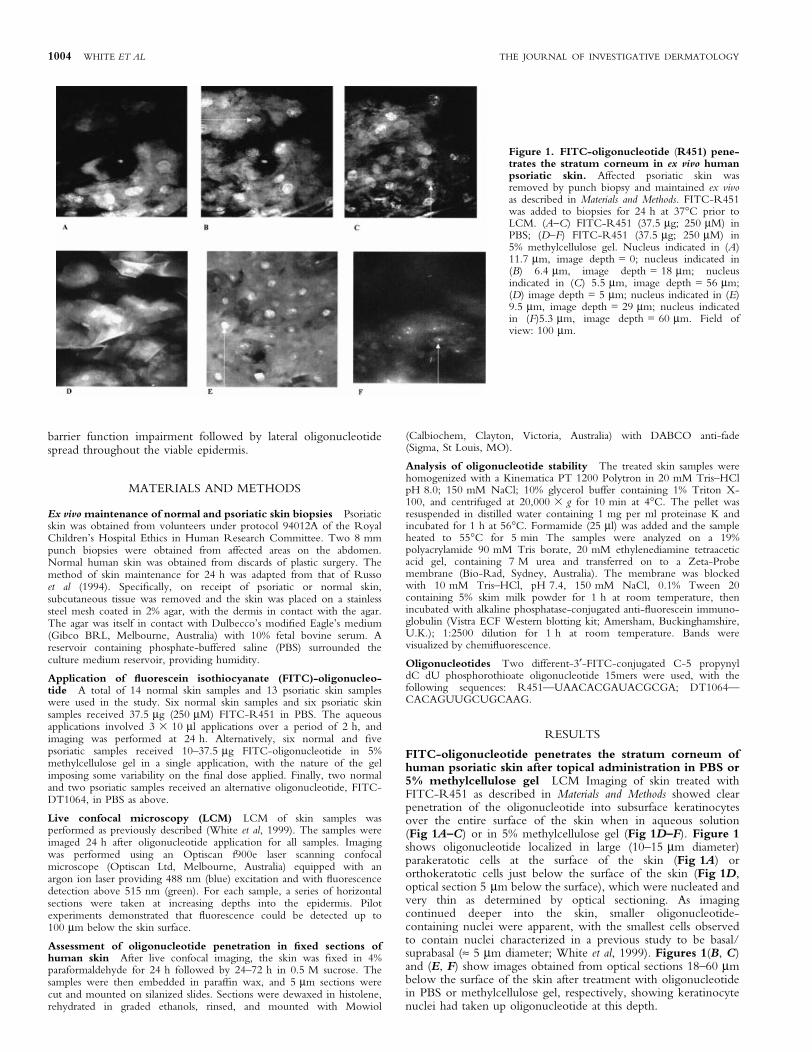
FITC-oligonucleotide penetrates the stratum corneum ofhuman psoriatic skin after topical administration in PBS or5% methylcellulose gel LCM Imaging of skin treated withFITC-R451 as described in Materials and Methods showed clearpenetration of the oligonucleotide into subsurface keratinocytesover the entire surface of the skin when in aqueous solution(Fig 1A±C) or in 5% methylcellulose gel (Fig 1D±F). Figure 1shows oligonucleotide localized in large (10±15 mm diameter)parakeratotic cells at the surface of the skin (Fig 1A) ororthokeratotic cells just below the surface of the skin (Fig 1D,optical section 5 mm below the surface), which were nucleated andvery thin as determined by optical sectioning. As imagingcontinued deeper into the skin, smaller oligonucleotide-containing nuclei were apparent, with the smallest cells observedto contain nuclei characterized in a previous study to be basal/suprabasal (» 5 mm diameter; White et al, 1999). Figures 1(B, C)and (E, F) show images obtained from optical sections 18±60 mmbelow the surface of the skin after treatment with oligonucleotidein PBS or methylcellulose gel, respectively, showing keratinocytenuclei had taken up oligonucleotide at this depth.
Figure 1. FITC-oligonucleotide (R451) pene-trates the stratum corneum in ex vivo humanpsoriatic skin. Affected psoriatic skin wasremoved by punch biopsy and maintained ex vivoas described in Materials and Methods. FITC-R451was added to biopsies for 24 h at 37°C prior toLCM. (A±C) FITC-R451 (37.5 mg; 250 mM) inPBS; (D±F) FITC-R451 (37.5 mg; 250 mM) in5% methylcellulose gel. Nucleus indicated in (A)11.7 mm, image depth = 0; nucleus indicated in(B) 6.4 mm, image depth = 18 mm; nucleusindicated in (C) 5.5 mm, image depth = 56 mm;(D) image depth = 5 mm; nucleus indicated in (E)9.5 mm, image depth = 29 mm; nucleus indicatedin (F)5.3 mm, image depth = 60 mm. Field ofview: 100 mm.
1004 WHITE ET AL THE JOURNAL OF INVESTIGATIVE DERMATOLOGY

Fluorescence microscopy of FITC-oligonucleotidedistribution Topical oligonucleotide-treated psoriatic skinsamples were also imaged using ¯uorescent microscopy after®xation and sectioning. Figure 2 shows representative¯uorescence imaging of sections treated with FITC-R451 in PBS(Fig 2A) or 5% methylcellulose (Fig 2B). Oligonucleotideappeared to penetrate to the basal epidermis over a wide area ofthe biopsy for each of the donors. Arrows in Fig 2(A) showapparent points of entry through the stratum corneum, from whichthe oligonucleotide may have gained access to a wide area of theepidermis.
The pattern of widespread distribution and nuclear localizationwas consistent with minor variability across the biopsies from thedifferent patients. Also, an alternative FITC-labeled oligonucleo-tide (DT1064) exhibited similar distribution characteristics to theoligonucleotide used for the majority of the experiments (R451).
FITC-oligonucleotide does not penetrate the stratumcorneum of normal human skin after topical adminis-tration in 5% methylcellulose gel or PBS Both LCM and¯uorescence microscopy of ®xed sections demonstrated thatoligonucleotide does not penetrate through the stratum corneumof normal skin samples. Figure 3(A, B) show LCM images of skinthat received FITC-R451 in 5% methylcellulose and PBS,respectively. Consistent images of ®elds of large squamous cellswere obtained from all normal skin samples imaged after topicalapplication. The diameter (often 20±30 mm), lack of thickness(often < 10 mm) of the cells and the lack of apparent nuclei wereindicative of fully differentiated stratum corneum acting as a barrierfor the oligonucleotide. Optical sections taken deeper into theepidermis revealed no ¯uorescence (data not shown), indicating alack of penetration of the oligonucleotide into normal skin. Thiswas con®rmed by ¯uorescence microscopy of ®xed sections of thesamples imaged using LCM. Figure 3(C) shows a ¯uorescencemicroscopy image of oligonucleotide appearing at the surface of theepidermis and not any deeper into the skin. This was consistent foreach of the samples examined and was con®rmed by immuno-histochemical analysis of the FITC-oligonucleotide location (datanot shown).
Figure 3(D) shows an image of normal human skin not treatedwith oligonucleotide, demonstrating that no auto¯uorescence wasdetected using the same confocal settings as images in Figs 3(A, B).
FITC-oligonucleotide remains intact after topical adminis-tration in both normal and psoriatic human skin There wasno apparent degradation of the oligonucleotide after topicalapplication for either normal or psoriatic skin. Figure 4 shows awestern immunoblot of homogenized samples of psoriatic skinfrom four individual donors treated with FITC-R451 in 5%
Figure 3. LCM and ¯uorescence microscopy of ®xed sectionsdemonstrate that oligonucleotide does not penetrate the stratumcorneum of normal human skin. Normal human skin was maintainedex vivo as described in Materials and Methods. FITC-R451 was added tobiopsies for 24 h at 37°C prior to LCM and ®nally ¯uorescence imagingof ®xed sections. (A, B) LCM of FITC-R451 (37.5 mg; 250 mM) in 5%methylcellulose gel; (B) FITC-R451 (37.5 mg; 250 mM) in PBS; (C)¯uorescence microscopy of FITC-R451 (37.5 mg; 250 mM) in 5%methylcellulose gel (arrow indicates stratum corneum); (D) image ofcontrol (no FITC-oligonucleotide) confocal image of normal humanskin. Scale bars: (A, B) 20 mm; (C, D) 100 mm.
Figure 2. Fluorescence microscopy of ®xedsections con®rms that FITC-oligonucleotide(R451) penetrates the stratum corneum in exvivo human psoriatic skin. Affected psoriaticskin was removed by punch biopsy andmaintained ex vivo as described in Materials andMethods. FITC-R451 was added to biopsies for24 h at 37°C prior to ®xation (4% paraform-aldehyde), embedding and sectioning. (A) FITC-R451 (37.5 mg; 250 mM) in PBS; (B) FITC-R451(37.5 mg; 250 mM) in 5% methylcellulose gel; (C)control no FITC-oligonucleotide sections for¯uorescence. Scale bars: (A, B) 100 mm; (C)50 mm.
VOL. 118, NO. 6 JUNE 2002 C-5 PROPYNE-MODIFIED OLIGONUCLEOTIDES IN THE EPIDERMIS OF PSORIATIC AND NORMAL HUMAN SKIN 1005

methylcellulose. Analysis of oligonucleotide size was performed asdescribed in Materials and Methods. Each of the samples appeared tocontain full length (15mer) oligonucleotide as evidenced by a singleband that appears at the same position as the 15mer control sample.
DISCUSSION
The results of this study indicate that oligonucleotide in aqueoussolution or methylcellulose gel can penetrate the stratum corneumof psoriatic skin maintained ex vivo from a range of individualdonors. LCM indicated that oligonucleotide can be detected in thenuclei of small, undifferentiated keratinocytes, over the entire areaof application of the oligonucleotide. The pattern of uptake wassimilar in skin receiving oligonucleotide in aqueous solution ormethylcellulose gel, although brighter ¯uorescence was detected inthe skin that received oligonucleotide in aqueous solution.Fluorescence imaging of ®xed sections con®rmed the LCM ®ndingthat oligonucleotide does penetrate throughout the epidermis.Signi®cantly, local areas of severely impaired stratum corneumformation appeared to allow extensive oligonucleotide entry to thebasal epidermis, from which lateral spread of the oligonucleotideappeared to occur. Oligonucleotide did not appear to penetratethrough regions of the stratum corneum that are relatively intactand properly formed. These ®ndings were not dependent onoligonucleotide sequence, as an alternative FITC-labeled oligo-nucleotide exhibited similar distribution characteristics to theoligonucleotide used for the majority of the experiments. Normalskin maintained in the same ex vivo apparatus demonstrated its
ability to prevent the penetration of oligonucleotide through thestratum corneum. No ¯uorescence was found below the stratumcorneum in 14 skin samples maintained ex vivo with oligonucleo-tide in aqueous solution or methylcellulose. The lack of penetrationof oligonucleotide across normal stratum corneum was in agree-ment with the ®ndings of Butler et al (1997) but in contrast to thoseof Wingens et al (1999) and Mehta et al (2000). Wingens et al (1999)used chimeric oligonucleotides that could have different pharmaco-kinetic properties to those oligonucleotides used in this study.Mehta and colleagues found that phosphorothioate oligonucleotidecould cross the stratum corneum of human skin grafts afterapplication of a 2% oligonucleotide cream. The variance in resultscould be as a result of the oligonucleotide dose (we used » 1/20thof the oligonucleotide dose of that used by Mehta et al, 2000) orvarying limits of detection. The problem with the use of largeamounts of oligonucleotide is the increased likelihood ofnonsequence-speci®c effects of the oligonucleotide. It was foundpreviously that repeated intradermal injection of 2.5 mg oligo-nucleotide produced a reliable antisense effect but also a smallnonsequence-speci®c effect (Wraight et al, 2000. The use of greateroligonucleotide dose may allow for some penetration of oligonu-cleotide into the viable epidermis of human skin, but may result insigni®cant nonsequence-speci®c antisense effects. In any case, at thedoses used in this study, oligonucleotide was found to penetrate thestratum corneum of psoriatic human skin and not normal humanskin.
The oligonucleotide used in this study was intact after topicaldelivery to both normal and psoriatic human skin. A clear singleband was apparent after electrophoresis of the recovered oligo-nucleotide, and the band was at the same position as a 15mercontrol sample. This result is somewhat surprising as oligonucleo-tide systemically delivered is quickly broken down by nucleases(Crooke et al, 1996) and the epidermis has a large amount ofenzymatic activity. These results are, however, consistent withthose of Vlassov et al (1994), who found intact oligonucleotide inmouse tumors after topical administration.
C-5 propyne-modi®ed oligonucleotides were used in this study.These modi®ed oligonucleotides were selected because theirincreased af®nity for target mRNA allows inhibition with lowerconcentrations (Wagner et al, 1993) and shorter oligonucleotidelength (Flanagan et al, 1996) than unmodi®ed phosphorothioates,theoretically reducing the incidence of aptameric effects on targetcells. One of the aims of this study was to deliver theseoligonucleotides topically into psoriatic skin, and therefore thesame chemical modi®cations were used in this study.
The results of this study indicate that oligonucleotides can betopically applied to psoriatic skin in simple topical formulations andef®ciently reach the basal epidermis fully intact. This suggests thatantisense therapy is particularly suited to the treatment of psoriasisand that complex topical formulations may not be required. Theissue of penetration of oligonucleotide in normal skin is somewhatmore complex. This study indicates that phosphorothioateoligonucleotide does not penetrate a wide range of normalhuman skin samples to any signi®cant degree.
REFERENCES
Butler M, Stecker K, Bennett CF: Cellular distribution of phosphorothioateoligodeoxynucleotides in normal rodent tissues. Lab Invest 77:379±388, 1997
Crooke ST, Graham MJ, Zuckerman JE, et al: Pharmacokinetic properties of severalnovel oligonucleotide analogs in mice. J Pharmacol Exp Ther 277:923±937, 1996
Flanagan WM, Su LL, Wagner RW: Elucidation of gene function using C-5 propyneantisense oligonucleotides. Nature Biotech 14:1139±1145, 1996
Ghadially R, Reed JT, Elias PM: Stratum corneum structure and function correlateswith phenotype in psoriasis. J Invest Dermatol 107:558±564, 1996
Mehta RC, Stecker KK, Cooper SR, et al: Intercellular adhesion molecule-1suppression in skin by topical delivery of antisense oligonucleotides. J InvestDermatol 115:805±812, 2000
Oldenburg KR, Vo KT, Smith GA, Selick HE: Iontophoretic delivery ofoligonucleotides across full thickness hairless mouse skin. J Pharm Sci 84:915±921, 1995
Regnier V, Preat V: Localization of a FITC-labeled phosphorothioateoligodeoxynucleotide in the skin after topical delivery by iontophoresis andelectroporation. Pharm Res 15:1596±1602, 1998
Figure 4. Oligonucleotide stability after topical application toskin maintained under organ culture conditions. Samples of normaland psoriatic skin treated with 37.5 mg (250 mM) FITC-DT1064 in 5%methylcellulose were homogenized and analysis of oligonucleotide sizewas performed as described in Materials and Methods. Homogenatesamples were analyzed by western immunoblot following denaturingpolyacrylamide gel electrophoresis. Each of the samples from psoriaticskin (samples A±D) appeared to contain full length (15mer)oligonucleotides as evidenced by a single band that appears at the sameposition as the 15mer control sample.
1006 WHITE ET AL THE JOURNAL OF INVESTIGATIVE DERMATOLOGY

Russo VC, Edmondson SR, Mercuri FA, Buchanan CR, Werther GA:Identi®cation, localization, and regulation of insulin-like growth factorbinding proteins and their messenger ribonucleic acids in the newborn ratolfactory bulb. Endocrinology 135:1437±1446, 1994
Vlassov VV, Nechaeva MV, Karamyshev VN, Yakubov LA: Iontophoretic deliveryof oligonucleotide derivatives into mouse tumor. Antisense Res Dev 4:291±293,1994
Wagner RW, Matteucci MD, Lewis JG, Gutierrez AJ, Moulds C, Froehler BC:Antisense gene inhibition by oligonucleotides containing C-5 propynepyrimidines. Science 260:1510±1513, 1993
White PJ, Fogarty RD, Liepe IJ, Delaney PM, Werther GA, Wraight CJ: Liveconfocal microscopy of oligonucleotide uptake by keratinocytes in human skingrafts on nude mice. J Invest Dermatol 112:887±892, 1999
Wingens M, Pfundt R, van Vlijmen-Willems IM, van Hooijdonk CA, van Erp PE,Schalkwijk J: Sequence-speci®c inhibition of gene expression in intact humanskin by epicutaneous application of chimeric antisense oligodeoxynucleotides.Lab Invest 79:1415±1424, 1999
Wraight CJ, White PJ, McKean SC, et al: Reversal of epidermal hyperproliferation inpsoriasis by insulin-like growth factor I receptor antisense oligonucleotides. NatBiotechnol 18:521±526, 2000
VOL. 118, NO. 6 JUNE 2002 C-5 PROPYNE-MODIFIED OLIGONUCLEOTIDES IN THE EPIDERMIS OF PSORIATIC AND NORMAL HUMAN SKIN 1007