Embed Size (px)
Citation preview
UNIVERSITY OF HAWNI LIBRARY
A SURVEY OF HAWAIIAN MARINE FUNGI AND YEAST
A THESIS SUBMIITED TO THE GRADUATE DIVISION OF THE UNIVERSITY OF HAWAI'IIN PARTIAL FULFILLMENT OF THE
REQUIREMENTS FOR THE DEGREE OF MASTER OF SCIENCE
IN MICROBIOLOGY (MARINE BIOLOGy)
DECEMBER 2008
By Leena Emiko Mahdl
Thesis Committee:
Stuart P. Donachie, Chairperson Paul Patek
Thomas Hemscheidt
412. b
We certify that we have read this thesis and that, in our opinion, it is satisfactory in scope and quality as a thesis for the degree of Master of Science in Microbiology.
ii
© 2006, Leena E. Mahdi
iii

ACKNO~EDGEMENTS
I would like to thank Dr. Stuart Donachle for being my mentor and for the opportunity to
study In his laboratory. His gUidance, support, and direction helped bring this project to
completion. I would like to thank Dr. Paul Patek, and Dr. Thomas Hemscheldt for serving
on my committee, and for their support, and guidance In this project.
I would also like to extend my appreciation to the Individuals in the lab that provided
advice, assistance, and friendships during this project: Dr. Mark Brown for assistance
with phylogenetic analyses, Mark Speck for general guidance, advice, and support,
Keall'lmanauluokeahl Taylor for her help with statistics and general assistance In the lab,
and Doug Gebhart for assisting around the lab. I would also like to thank Dr. Andy Taylor
for his guidance and advice with statistics.
Finally, I would like express my sincere gratitude to my family who, through their
unconditional love and belief In me, have allowed me to achieve my goals and continue
to make new ones. I would also like to thank my close friends who have provided love
and support throughout the duration of this research.
This research was made possible through funds awarded under a Pilot Project program
to Stuart Donachie, by the University of Hawal'l Pacffic Research Center for Marine
Biomedicine, part of the Centers for Oceans and Human Health (COHH) program of the
National Institute of Environmental Health Sciences (P50ES012740), National Institutes
of Health, and the National Science Foundation (OCE04-32479). Travel to outer Islands
was primarily funded by a grant from the Lewis and Clark Exploration Award (The
American Philosophical Society).
iv
LIST OF TABLES
Table Page
1. Sample sites and materials collected at each location.................. 66
2. Water samples collected at Station ALOHA.. ............ ...... ............ 69
3. Assimilation of single carbon sources by LM418.......................... 76
4. Physiological properties of LM418, type species
Sympodiomycopsis mahdil and related species.............................. 88
5. Number of different fungi and yeast grouped by location......... ..... 104
6. Krukal-Wallis Statistical Analysis for Hawai'i and Palmyra
Atoll-Fungi Data..... ..... ..................... ........................ ... ......... 110
7. Krukal-Wallis Statistical Analysis for Station ALOHA Data. ........... 111
v
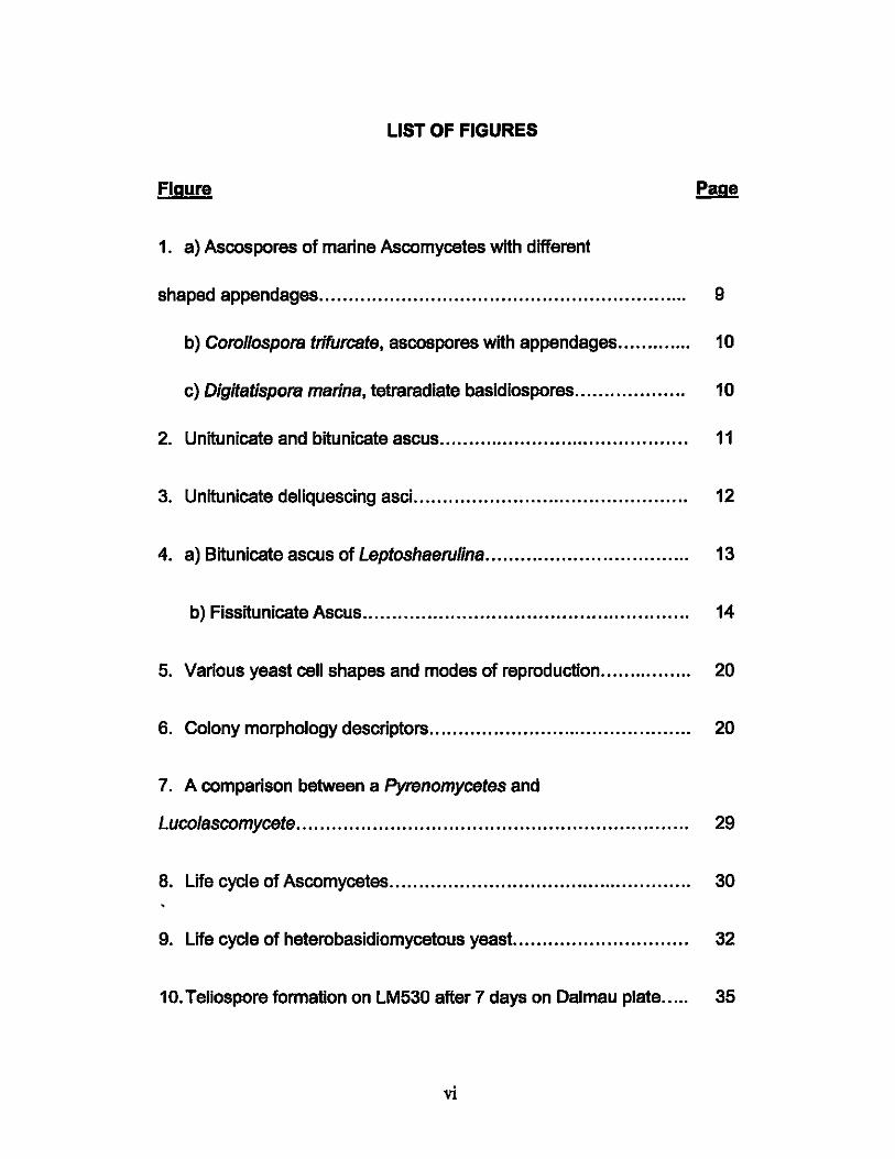
LIST OF FIGURES
Figure
1. a) Ascospores of marine Ascomycetes with different
shaped appendages............................................................... 9
b) Corollaspora trifurcate, ascospores with appendages............. 10
c) Digitatispora marina, tetraradiate basidiospores................... 10
2. Unitunicate and bitunicate ascus........................... ................ 11
3. Unitunicate deliquescing asci............................................... 12
4. a) Bitunicate ascus of Leptoshaerulina................................... 13
b) Fissitunicate Ascus........................................ ........ ........ 14
5. Various yeast cell shapes and modes of reproduction.... ............ 20
6. Colony morphology descriptors.................... ......................... 20
7. A comparison between a Pyrenomycetes and
Luco/ascomycete. ... ......... .......... ......... .. .......... ............... ..... ... 29
8. Life cycle of Ascomycetes.................................................... 30
9. Life cycle of heterobasidiomycetous yeast.............................. 32
10. Teliospore formation on LM530 after 7 days on Oalmau plate..... 35
vi
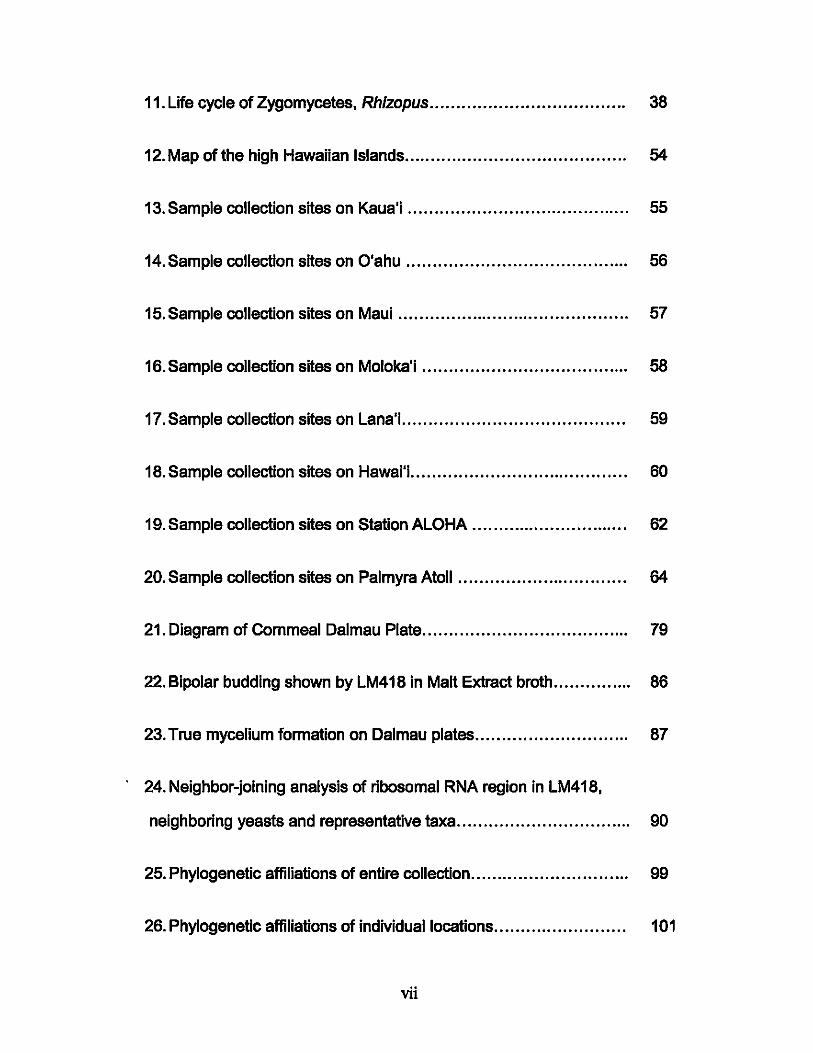
11. Life cycle of Zygomycetes. Rhizopus..................................... 38
12. Map of the high Hawaiian Islands.......................................... 54
13. Sample collection sites on Kaua'i .......................................... 55
14. Sample collection sites on O'ahu .......... ............................ .... 56
15. Sample collection sites on Maui .............. ....... . ..... .. ............... 57
16. Sample collection sites on Moloka'i ................................... .... 58
17. Sample collection sites on Lana'i.......................................... 59
18. Sample collection sites on Hawai'i.......................... ............... 60
19. Sample collection sites on Station ALOHA....... ....... ......... ....... 62
20. Sample collection sites on Palmyra Atoll ................. ........ ... .... 64
21. Diagram of Commeal Dalmau Plate................ ......... .......... .... 79
22. Bipolar budding shown by LM418 in Malt Extract broth........... .... 86
23. True mycelium formation on Dalmau plates.......................... ... 87
24. Neighbor-joining analysis of ribosomal RNA region in LM418,
neighboring yeasts and representative taxa............................. .... 90
25. Phylogenetic affiliations of entire collection..... ...................... ... 99
26. Phylogenetic affiliations of individual locations. ........................ 101
vii
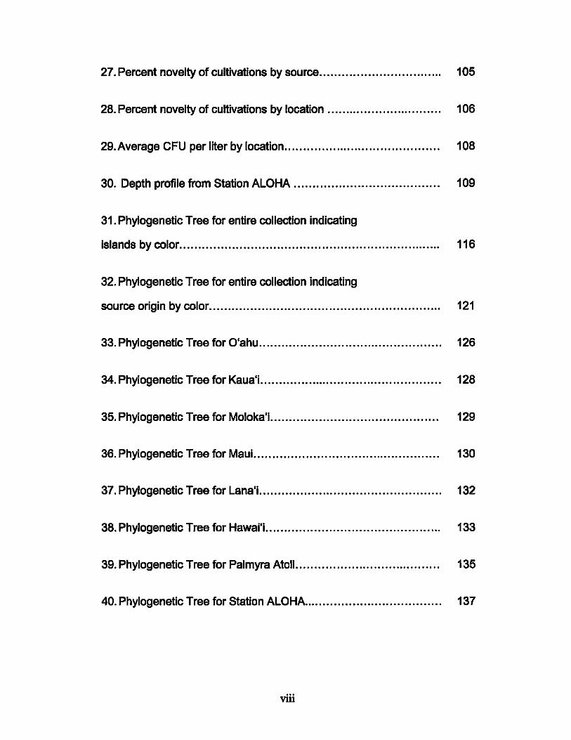
27. Percent novelty of cultivations by source........ .................. ... .... 105
28. Percent novelty of cultivations by location ..... ....... ............... .... 106
29. Average CFU per liter by location.............. ..... ....................... 108
30. Depth profile from Station ALOHA....... ............... ............. .... 109
31. Phylogenetic Tree for entire collection indicating
islands by color...................................... .. . .. . .. . .. . .. . .. ... . ... .... .... 116
32. Phylogenetic Tree for entire collection indicating
source origin by color............. ......... ............ ............ ....... ......... 121
33. Phylogenetic Tree for O·ahu.. .. ....... .. .......... ..... .......... ........... 126
34. Phylogenetic Tree for Kaua·i.............. .............. ........ .. ........... 128
35. Phylogenetic Tree for Moloka·i...................................... ..... .. 129
36. Phylogenetic Tree for Maui..... ........... .................................. 130
37. Phylogenetic Tree for Lana·i..... ........ .... ...... .......................... 132
38. Phylogenetic Tree for Hawai·i........... .............................. ... ... 133
39. Phylogenetic Tree for Palmyra Atoll................ ........... ............ 135
40. Phylogenetic Tree for Station ALOHA..................................... 137
viii
ABSTRACT
A fundamental gap exists in our understanding of the phylogenetic diversity and
distribution of marine fungi and yeasts off the Hawaiian Islands and Palmyra Atoll
(Line Islands). During this 2 year study, 689 pure cultures of fungi and yeasts
were prepared from seawater, sand, algae, wood, and other marine samples
collected from 118 Hawaiian coastal sites and Palmyra Atoll, and water from the
-5000 m water column at the open ocean Station ALOHA north of O'ahu. The
approach was Innovative because it combined traditional cultivation techniques
with 'new' molecular methods to facilitate rapid identification of cultured isolates,
including those which may represent new species. Cultivated fungi and yeast
abundance did not vary significantly in seawater collected off six of the high
Hawaiian islands. Phylogenetic diversity was high, however, and Included
previously uncultivated strains. Potentially novel LM418T isolated from wood on a
Lana'i beach is tentatively described here as Sympodiomycopsis mahdli.
ix
TABLE OF CONTENTS
COPYRIGHT PAGE.................................................................... iii
ACKNOWLEDGMENTS............................................................... iv
LIST OF TABLES.... ............ ............ ....... .............................. ....... v
LIST OF FIGURES...................................................................... vi
ABSTRACT............................. ................... .......... .. .................... ix
CHAPTER I: LITERATURE REVIEW.............................................. 1
A. Introduction to Marine Microbiology................................... 1
1. History. ............. ....... .. .......... ...................... 1
2. Nature ofthe Ocean...................................... 4
B. Marine Mycology........................................................... 4
1. Definition.......... ................... ........ .......... ....... 4
2. Geographic Distribution and Abundance.... ...... .. 5
3. Environmental Role....................................... 7
4. Physiological Adaptations............................... 8
C. Marine Fungi............................................................... 14
1. Colony Morphology........................................ 15
2. Ultrastructure.. ........................ ...................... 15
3. Reproduction ................................................ 17
D. Marine yeast................................................................ 18
1. Cell and Colony Morphology.............. .. ............ 19
2. Ultrastructure.............. .................................. 21
x

3. Reproduction................................................ 24
E. Ascomycetes ................................................................ 26
F. Basidiomycetes.............................................................. 31
G. Deuteromycetes............................................................ 36
H. Zygomycetes................................................................ 37
I. Fungi in the environment.................................. ................ 38
1. SandIFoam........................................... .............. 40
2. Algae ................................................................ 42
3. Wood................................................................. 42
4. Animals........... ............................................... .... 43
5. Avian Guano.................. .............. ...... ................. 44
6. Human Impact..................................................... 45
J. Deep Sea Fungi and Yeast.............................................. 46
CHAPTER II: BACKGROUND OF THIS STUDy............................... 48
A. Motivation for the study........ ............ ........ ....................... 48
B. Defining the problem and proposed solution.............. .......... 49
C. Thesis goals and objectives........................................ ..... 50
D. Experimental Design................ ..................................... 52
CHAPTER III: MATERIALS AND METHODS........ ............................ 53
A. Sample sites....................... ......... ............ ............. ..... ... 53
Kaua'i .................................................................... 55
O'ahu .................................................................... 56
Maui....................................................................... 57
xi
Moloka'i.......... ........................ ......... .... .............. ..... 58
Lana'i............................. ..... ........................ ........... 59
Big Island of Hawai'i..... ......... ............ .. ............. ........ 60
Station ALOHA.... ....... ... ............ ............. ............ ..... 61
Palmyra Atoll.. .. .............................................. ......... 63
B. Sample Collection......................................................... 65
C. Sample Processing and Cultivations................... ..... ....... .... 70
D. Isolation Media............................. .................... ............. 71
E. Enumeration of colonies ........ .......... ......... .......... ......... ... 72
F. Preservation and Archiving Samples.................................. 72
G. Identification............................................ ............ .......... 73
H. Describing a novel species of marine Yeast-Sympodiomycopsis mahdii................. ......... ........... ....... ... ... 75
1. Materials and Methods.. ........... ............................. 75
2. FEMS Yeast Research paper.......... ..... .......... ... ...... 80
I. Statistical Analysis.......... ......... ........................ .... .......... 95
CHAPTER IV: RESULTS........... .................................... .............. 96
CHAPTER V: SUMMARY AND CONCLUSiON.......... ............... ... ..... 138
A. Summary of results.......... ............................. .................. 138
B. Conclusions and Recommendations.................................. 139
REFERENCES................... ............ ......... ............. .................. ... 140
xii
CHAPTER I. LITERATURE REVIEW
A. Introduction to Marine Microbiology
1. History
Marine microbiology is generally considered to be the study of organisms
smaller than 1 mm in the ocean. While these organisms are now known to be
abundant, they were historically Ignored sinca they were assumed to have
little or no Impact on marine biogeochemical cycles. Although marine biology
as a field did grow rapidly during the nineteenth century, marine microbiology
was still largely overlooked because methods to cultivate and even count
marine microbes were still in their Infancy. It wasn't until the Challenger
expedition (1873-1876) completed the first deep sea studies, however, that
the existence of bacteria in the sea was confirmed (Jannash, 1984). In
addition to the widely used cultivation approaches, microscopy also became a
method through which bacteria in aquatic samples were enumerated. The
advent of microscopy methods that coupled DNA stains with flat
polycarbonate filters led to the conclusion that bacteria were much more
abundant In the ocean than anyone had believed possible (Hobbie, 1977).
Such observations also began a debate on the usefulness of cultivation
1
methods to count bacterial cells in environmental samples (e.g., Staley &
Konopka, 1985). For example, it was argued that cultivation approaches
yielded such a small percentage of cells compared to the number determined
by microscopy and DNA stains that they had little value In environmental
mIcrobIology. The realization that such abundant bacteria In the ocean mIght
actually play important roles In nutrient cycling and thus global
biogeochemIcal cycles, however, Is at the heart of what became known as the
mIcrobIal loop (Pomeroy, 1974; Azam at a/., 1983). Soon after, what are now
termed 'molecular methods' redefined what we knew of bacterial diversity in
the ocean by allowing us to determIne the nucleotide sequence of 16S rRNA
genes In DNA extracted from an environmental sample (Stackebrandt &
Weese, 1981). ThIs approach revealed unexpected and largely uncultivated
phylogenetic dIversity among the marine bacteria (GIovannonI, 1990). Little
attention was paid to the presence or dIversity of pIcoeukaryotes In the ocean,
and work on fungi and yeasts continued to focus largely on those that could
be cultivated on enrichment medIa, especIally from coastal areas.
Marine mycology has been relatively overlooked, despite early findIngs of the
pathogenIc marine Ascomycete Sphaaria posidonia dating to 1846 by C.
Durleu de Maisonneuve and Montagne. It wasn't until the early 1900s that
2
studies focusing on marine fungi or yeast were conducted (Hyde, 2000).
Zobell and Feltham (1934) observed yeasts in the open ocean and from other
marine materials, while several other workers observed similar findings
elsewhere (Johnson, 1961). Initial findings of marine fungi and yeast primarily
described them as pathogens or parasites of fish, clams or oysters, but soon
after, reports of parasitic ralatlonshlps between algae were also described
(Reed, 1902). By the late 1950's scientists began to describe different
relationships between marine fungi and marine animals. Nevertheless, some
scientists stili believe marine fungi and yeast are neither Important nor worthy
of further discussion (Sherr et ai, 2001). The first published HawaIIan
mycology reports were of freshwater semples (Anastaslou 1964; Sparrow
1965), but the first to describe the mycoflora In marine semples was
Kohlmeyer (1969). Thus, HawaIIan marine mycology has a rather short history
compared with marine microbiology In general, but also of marine mycology.
The 1970s brought about a 'social awareness' of the ocean and its potential
source of products for human use beyond fishing. The 'biosaline concept' was
promoted. suggesting that material needs could be met by using the ocean In
a variety of ways which benefit the environment and man (Aller & Zaborsky,
1979). Such views of the ocean led to significant developments In what
3
became the field of marine biotechnology and the search for new antibiotics
and anti-tumor compounds from marine sources. In recent years marine
mycology has attracted some Interest as researchers consider the potential of
marine fungi and yeasts as sources of novel byproducts.
2. Nature of the Ocean
The ocean covers -70% of the Earth's surface. The Hawaiian Islands are
located in the center of the Pacific Ocean, stretching from 18° 55' N (tropical)
to 29° N (sub tropical), and longitude 154° 40' W to 162° W. The entire
archipelago covers some one thousend five hundred twenty miles. Seawater
surface temperature ranges from 71°F to 81 OF, with an average of 78.3 OF
(O'ahu -NOAA), with a salinity of 34 to 35. The average pH of Hawaiian
surface waters is 8.2. Continental shelves to a depth of 200 m are considered
part of the island topography and are part of the littoral zone, while depths
between 200 and 400 m are considered the sublittoral zone.
B. Marine Mycology
1. Definition
As recently as 40 years ago, fungi Isolated from marine habitats were not
always recognized as marine, sensu strictu, despite several marine species
4

having bean described in the previous one hundred years. It was only in the
last 50 years that marine mycology was established as a distinct field
(Johnson & Sparrow, 1961). Unlike other taxonomic groups, however, marine
fungi cannot be defined exclusively by physiological or nutrltlonai
requirements. In this respect, Kohlmeyer (1965) defined marine Fungi as
those that can reproduce or grow submerged in seawater or on intertidal
substrates such as wood, sand or algae and are permanently or intermittently
submerged. Facultative marine fungi are those that typically occupy
freshwater or terrestrial milieus but are able to grow and sporulate in the
marine environment (Kohlmeyer, 1974).
Marine fungi living part of their life cycle as single cells, primarily reproducing
by fission or budding are considered marine yeasts. They are dMded into two
categories, obligate and facultative marine yeast. Obligate marine yeasts are
those that have been collected exclusively from the marine environment, while
facultative marine yeasts have also been collected from terrestrial sites
(Kohlmeyer, 1979).
2. Geographic Distribution and Abundance
5
Marine Fungi and yeast are ubiquitous in the world's oceans, so to define a
pattem of distribution the marine environment must be divided into habitats.
The open ocean comprises benthic and pelagic regions, while coastal waters
Include 'littoral regions', those spanning the low tide mark toward the
continental shelf, which typically reaches an average depth of 200-400 m
(Wood, 1963). Within the littoral biotic zone three subdMsions exist: 1) the
Intertidal zone, 2) the eullttoral zone, and 3) the sublittoral zone. The pelagic
zone can be divided horizontally into the oceanic and neritic regions. The
oceanic region consists of open ocean over depths greater than 200 m and
the latter Includes open water from the shoreline down to a depth of 200 m.
Vertically, the pelagic zone can be dMded into the epipelagic zone from the
surface to 200 m, and the mesopelaglc zone extends down to the
bathypelagic zone which receives no light penetration. These depths vary
greatiy depending on location. The abyssal pelagic zone refers to depths
greater than 2,000 m.
The distribution of fungal habitats is determined by several factors such as the
location of hosts, availability of nutrients, competition or availability of
substrates. The most abundant marine populations occur on substrates In the
Intertidal zone, but Indigenous fungi and yeast do occur In the deep sea.
6
Indeed, fungi have been collected from the Mariana Trench at 10,897 meters
(Takamia, 1997). The availability of dissolved oxygen is an important factor In
the distribution of fungi In marine habHats. Fungal growth is inhibited by low
levels of dissolved oxygen In the water column or sediment. Dissolved oxygen
concentrations of -0.30 mllliter or less typify an "oxygen minimum zone", and
can inhibH growth of fungi, while an oxygen concentration of 1.26 mllliter
allows "ample fungal growth" (Kohlmeyer, 1969a).
3. Environmental Role
The role of fungi in marine food webs might on the one hand be considered
significant, given that they take up dissolved organic substances, act as a
food source for some benthic animals, can be a source of C02 to
phytoplankton, and mediate decomposition of plant material such as leaves,
wood and algae due to their ability to depolymerize cellulose, xylans, and
pectin (Rhelnhelmer, 1992). In addition, the mycelia of Ascomycetous and
Deuteromycetous fungi growing on cellulosic debris play an important role in
the reproductive success of a species of nematode, since the fungi provide
sufficient nutritional needs for the nematode by attracting animal predators. In
another case, the fungus attracts pregnant females of another nematode
species (Sleburth, 1979).
7
4. Physiological Adaptations
There have been several adaptations to life in the sea, many of which include
the utilization of different nutrients. Barghoom (1944) carried out a series of
experiments that determined the most important adaptations were the ability
to survive on media with sodium chloride concantratlons three times those in
seawater, as well as an elevated pH. Ascomycates have adapted to the
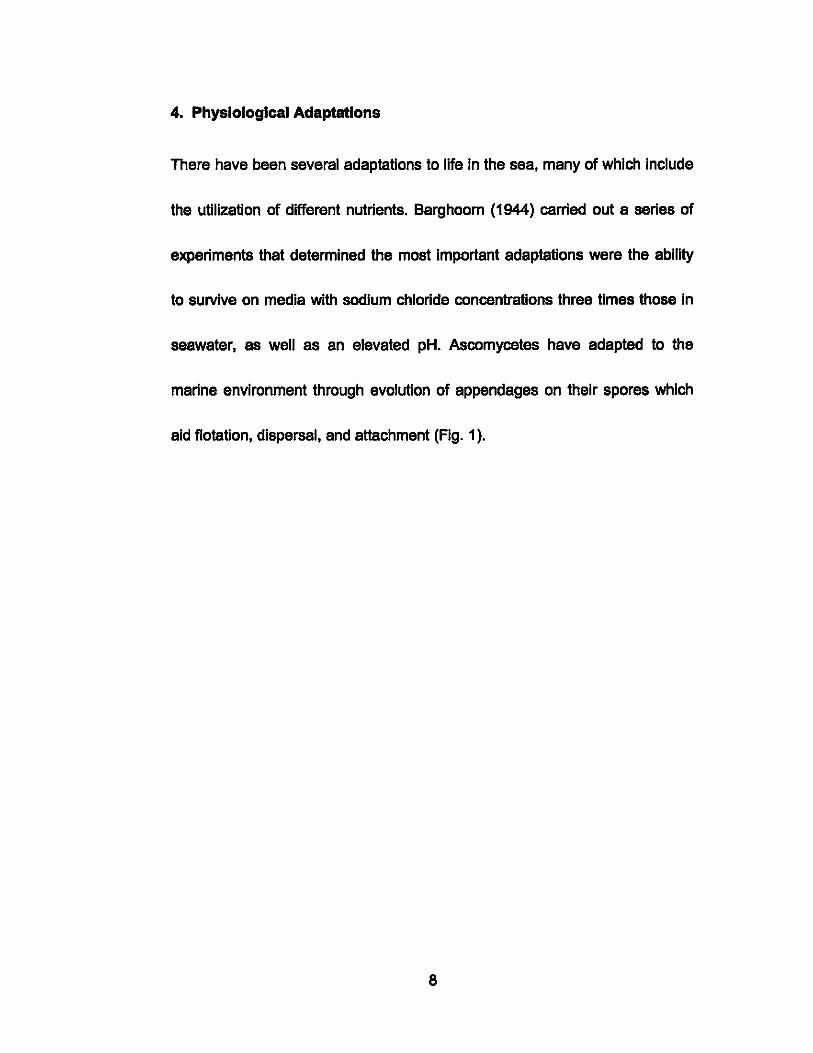
marine environment through evolution of appendages on their spores which
aid flotation, dispersal, and attachment (Fig. 1).
8
a b c
,\'( ?",~;:~. ,~
d
h
FIgure 1a. Ascospores of marine Ascomycetes with different shaped
appendages (except 1.d). a) Amylocerpus encepha/oides Currey. b)
Torpedospora radiate Meyers, c) Ceriosporopsis ca/yptrate Kohlm., d)
Mlcrothelia mart/ma (Under) Kohlm., e) Ha/osphaeria medlosetigera Cribb et
Cribb, f) Remispora maritma Under, g) Peritrlchospora integra Under, h)
Ha/osphaeria torquata Kohl. Magnification -2000x. Figures by E. Kohlmeyer
(Kohlmeyer, 1963).
9
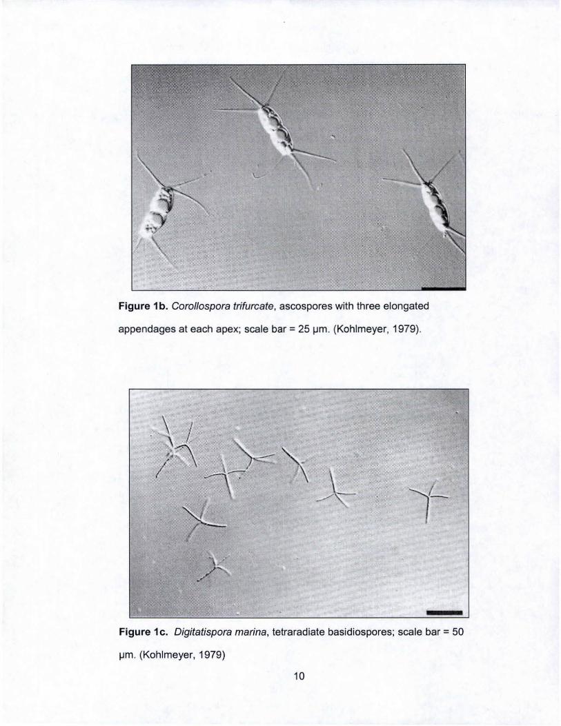
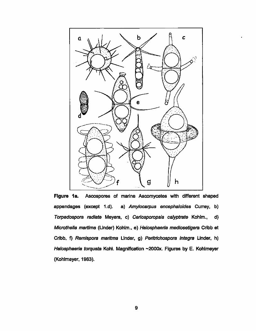
Figure 1 b. Corol/ospora trifurcate , ascospores with three elongated
appendages at each apex; scale bar = 25 ~m . (Kohlmeyer, 1979).
Figure 1 c. Digitatispora marina, tetra radiate basidiospores; scale bar = 50
~m . (Kohlmeyer, 1979)
10
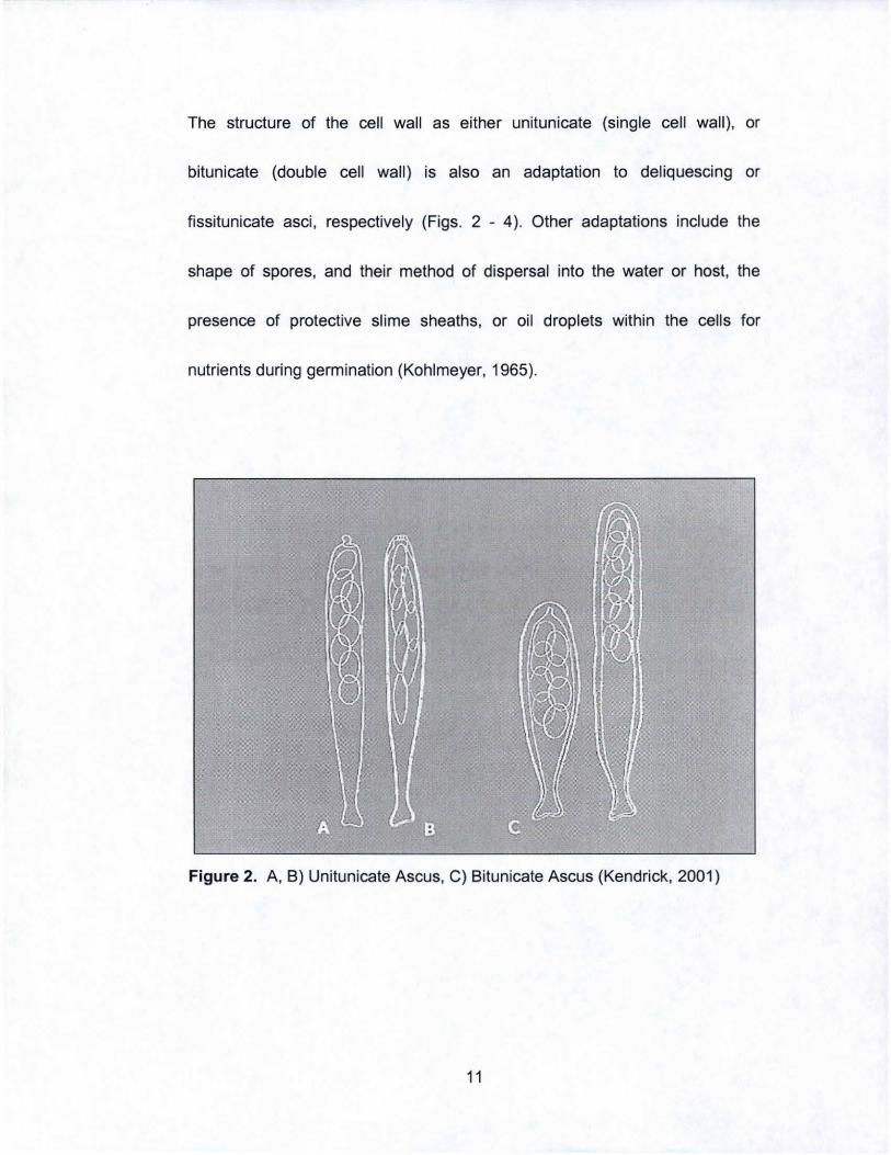
The structure of the cell wall as either unitunicate (single cell wall), or
bitunicate (double cell wall) is also an adaptation to deliquescing or
fissitunicate asci , respectively (Figs. 2 - 4). Other adaptations include the
shape of spores, and their method of dispersal into the water or host, the
presence of protective slime sheaths, or oil droplets within the cells for
nutrients during germination (Kohlmeyer, 1965).
Figure 2. A, B) Unitunicate Ascus, C) Bitunicate Ascus (Kendrick, 2001)
11
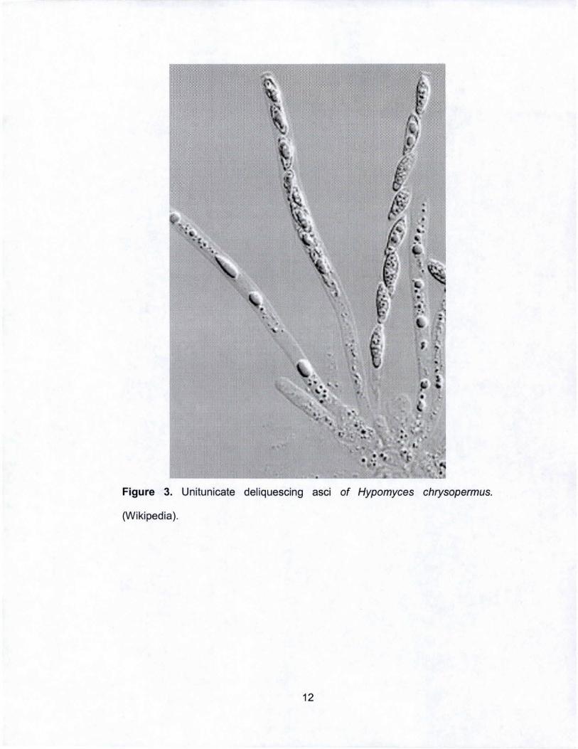
I ,
•
• • ~.
•
. . , \ I
' .
. .. '. ~ . . .. .. . , . " •• ••
Figure 3. Unilunicale deliquescing asci of Hypomyces chrysopermus.
(Wikipedia).
12
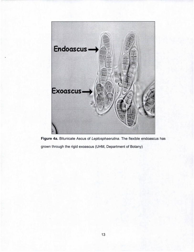
Endoascus-+
Figure 4a. Bitunicate Ascus of Leptosphaerulina. The flexible endoascus has
grown through the rigid exoascus (UHM, Department of Botany)
13
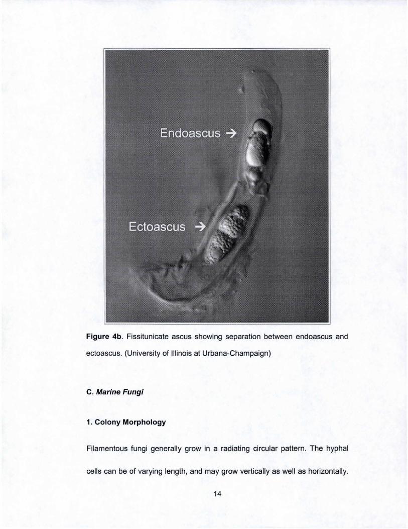
Figure 4b. Fissitunicate ascus showing separation between endoascus and
ectoascus. (University of Illinois at Urbana-Champaign)
C. Marine Fungi
1. Colony Morphology
Filamentous fungi generally grow in a radiating circular pattern. The hyphal
cells can be of varying length, and may grow vertically as well as horizontally.
14
The colony can have one or more colors In rings, and visible spores. Spore
density can vary from single spores on hyphal tips to a powdery layer of
spores across the entire colony surface.
2. Ultrastructure
Filamentous fungi are eukaryotlc cells composed of long, thread like filaments
called hyphae. Connected end to end, these hyphae compose the body of the
fungus known as the mycelia. Typically, each mycelium acquires nutrients by
absorption or penetration of a substrate. Hyphae that penetrate host cells for
nutrient absorption are known as haustoria. The hyphae have rigid walls that
are responsible for the stability of their vegetative and reproductive structures.
The rigid cell wall is comprised of a matrix that includes mannans, glucans,
and polyuronides which also embeds microflbrils. The microflbrils are made
either of chitin or cellulose, and provide structure and rigidity. Chitin is a long
carbohydrate polymer that is also found in the exoskeleton of arthropods,
insects, and spiders. The plasmalemma is the space between the cell wall
and the cell membrane, and is where lomasomes characteristic of fungal
structures are found. Lomasomes are membrane-bound tubules that can
occur singly or in groups, and while their function is not entirely known, they
15
may be involved in excretion as well as increasing the surface area at the
periphery of the cell.
The cytoplasmic organelles are similar to those in other eukaryotlc cells and
include the nucleus, mitochondria, storage vacuoles, and ribosomes. Golgi
bodies are rare in fungi, and the endoplasmic ratlculum forms a bubble like
vesicle in young cells but is generally infrequent in mature cells. The nuclei
are well defined and often multiple nuclei are present. When there is no
distinction between indMdual cells they are referred to as coanocytlc, but
otherwise they are normally separated by septa with or without pores.
Typically Ascomycetes and Basidiomycetes have septa, while Zygomycetes
are coanocyUc.
Most fungi lack flagella, with the exception of the chytrlds. The chytrlds are the
oldest fungal lineage known and contain flagellated gametes. Since chytrlds
are aquatic in nature it is believed that fungi evolved in water. The absence of
flagella is then a synapomorphy which unites the remaining groups of fungi.
The lack of flagella means two organisms must come into direct contact for
reproduction. In fungi, there is a division of labor where some mycelia
participate in conjugation with specific structures, and/or the dissemination of
spores and others with the assimilation of nutrients.
16
3. Reproduction
Fungi are able to reproduce both sexually and asexually. The biological act of
spore formation is a direct response at the DNA level to a physiochemical
environmental change. This results In the alteration from a vegetative state to
sphericai reproductive state by an internal stimulus that regulates cellular
activity.
The formation of asexual spores in large numbers occurs under stressful
conditions by fission, fragmentation, cieavage and extrusion (Smith, 1977).
Asexual spores are important for a build up in biomass, and normally occur at
the end of a growth cycle. It is a means of survival, dispersel, and
propagation. Fission occurs when cells within the mycelium separate at
double walled septa forming two genetically identical cells; formation via
fragmentation occurs when the cytoplasm becomes concentrated in certain
cells within a filament, leaving the remaining cells without cytoplasmic
contents. Formation by cleavage is when the protoplasmic material of a cell
divides into fragments, each of which becomes surrounded by a new cell wall,
and finally, spore formation by extrusion describes the process of spores
being produced as extensions from the sides or ends of sporogenous cells.
The requirements of spore formation are a mixture of biological events that
17
are distinguished by low water content, a lack of cytoplasmic movement, and
a low metabolic turnover.
Spores formed by sexual reproduction In fungi and yeasts are often resistant
to environmental stressors; they are thick-wailed structures that develop
through the simple union of hyphae between haploid nuclei, or the conjugation
of differentiated multinucleated female and male gametangia. The specific
details of spore formation and methods of spore dispersal will be detailed In
the following chapters. In general however, spore dispersal Is by sacrifice of a
supporting cell, fracture, or fission of a double septum.
D. Marine Yeast
Yeasts are considered a polyphyletic group of Ascomycetous and
BaSidiomycetous fungi characterized by unicellular growth phases, and
reproduction by budding, fiSSion, or fragmentation. They can build up self
perpetuating populations in marine environments (Uden, 1968). They are also
able to sexually produce spores and are In fact ubiquitous in the ocean. They
occur on substrates such as sediment, wood, and algae. Many yeasts are
obligate aerobes, occurring no deeper than the upper few centimeters of
marine sediments (Uden, 1968; Kurtzman, 2004). Approximately 100 genera
18
and more than 700 specles have been described from the ocean (Kurtzman &
Fell, 2004). Yeast populations decrease with distance from land, but they
remain the dominant fungi In the open ocean (Fell, 1986).
1. Cell and Colony Morphology
The morphology of yeast cells can be described as spherical, ogival,
triangular, elongated, flask shaped, ovoid, apiculate, globose, or cYlindrical
and are often specialized to a specific species (Fig. 5). It is possible for a
single species to be polymorphic during its ontogenetic development. A typical
yeast cell size Is between 2 and 50 IJm in length, and up to 10 IJm across
(Phaff, 1966). Colony morphology has taxonomic significance when
describing a species, whether novel or extant, and Is described in a similar
manner as for bacteria. For example, elevations are described as slightly
raised, crateriform, umbonate, flat, convex, or subaerial. It Is also imporlant to
note the form of the colony, as rhizoid, filamentous, irregular, or circular.
Finally, the colony margin is described as curled, lobate, filamentous,
unduiate, or entire. Any pigmentation as well as surface texture of the colony,
e.g., glistening, dry, dull, curled further are also noted and aid identification
(Fig. 6).
19
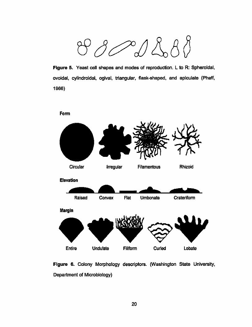
Figure 5. Yeast cell shapes and modes of reproduction. L to R: Spheroidal,
ovoidal, cylindroidal, ogival, triangular, flask-shaped, and apiculate (Phaff,
1966)
Form
Circular Irregular Filamentous Rhizoid
Elevation
Raised Convex Rat Umbonate Crateriform
Margin
~ Entire Undulate Filiform Curled Lobate
Figure 6. Colony Morphology descriptors. (Washington State University,
Department of Microbiology)
20

Yeast morphology is generally determined by the vegetative reproduction of a
cell, or in some ceses the formation of spores. The vegetative body of yeast Is
known as the thallus or soma, and can also be a determining characteristic of
a species. For example, the thallus may be seen alone, or with one or more
budding cells stili attached to It. This may result in the formation of chains or
small clusters which form a structure called a pseduomycelium (Phaff, 1966).
2. Ultrastructure
The rigid cell wall accounts for 25% of the dry weight of the yeast cell and is
approximately 25 nm thick (Berry, 1982). It comprises a matrix that Includes
mannans, glucans, microfibrils, and polyuronldes which also embeds
microfibrils. Mannans provide an upper layer of rigidity, and are water-soluble
polysaccharides of the sugar D-mannose that are linked by a-1, 6, a-1, 2 or
sometimes a-1, 3 bonds between residues. Some yeasts such as
Sporobo/omyces and Rhodotorula do not contain this type of mannan In their
cell walls. The highly insoluble polysaccharide, glucan. is what gives the cell
its basic shape and rigidity (Phaff, 1966). Glucan has been shown to contain
varying proportions of ~1, 3 linkages of glucose as well as ~1, 6 linkages
between units, providing a stable branched structure (Rose, 1969). The
microfibrils are either chitin or cellulose, and provide structure and rigidity.
21
Chitin is a long carbohydrate polymer of 13-1.4 linked N. acetyl-glucosamine
that is assoclated with the production of bud scars. The concentration of chitin
can vary with species. e.g., Sporob%myces and Rhodotoru/a both have a
much higher content than other genera, while in some species in the same
genus chitin may be absent (Phaff, 1966; Berry, 1982).
The plasmalemma is the space between the cell wall and the cell membrane.
It contains receptors for mating hormones which trigger a chain reaction
leading to the production of a diploid cell (Spencer & Spencer, 1997). During
budding, the plasmalemma forms a closing aperture to separate mother and
daughter cells during the 'pinching otr process, resulting in a bud scar. Bud
scars are the most distinct feature visible on the cell wall, and can be
indicative of the cell's age. The birth scar also remains on the cell surface, but
is usually less visible than bud scers (Rose, 1969). Other functions of the
plasmalemma include the intake of nutrients from the media into the cell, as
well as the release of compounds from fermentation such as ethanol out of
the cell into the medium (Rose. 1969; Berry, 1982).
Certain genera of yeast such as Hansenu/a, Rhodortoru/a, Cryptococcus,
candida, and Trichosporon may excrete capsular materials or other
extracelluar substances that surround the cells. These substances may be
22
phosphomannans, which form a viscous slimy layer on the surface of cells,
heteropolysaccharides, or hydrophobic substances which allows the formation
of a pellicle in liquid media (Phaff, 1966). The function of these compounds is
not yet known (Sieburth, 1979).
Similar to the cytoplasmic organelles found In other eukaryotic cells, yeast
also have a membrane bound nucleus, mitochondria, endoplasmic raticulum,
Goigi bodies, perixosomes, storage vacuoles, lipid granules (sphaerosomes),
and ribosomes. The nucleus of yeast Is protected In a double membrane
which contains several nuclear pores. Mitochondria are found throughout the
cell, have a double layered outer wall and are important In the respiratory
activity of the yeast-aerobic energy conversion, the synthesis of proteins and
RNA (Rose 1969; Phaff, 1966). The endoplasmic reticulum Is Important in
proteins synthesis and secretion, budding yeast cells, and the formation of
distinct organelles (Rose, 1969). Goigi bodies are controversial In yeast, but
sort and package proteins Into secretory vesicles, and carry these vesicles to
the plasmalemma for cell wall synthesis (Rose, 1966; Spencer & Spencer,
1997). Storage vacuoles are easily Identified usually by as single large sac
under the microscope. They are surrounded by a single membrane and hold
small particles such as ribosomes, lipids, volutin, enzymes, and esterases.
23
The vacuoles act as a reservoir for the parent cell, and fragments Into smaller
vacuoles during budding such that the daughter cell ends up with several
small vacuoles (Rose, 1969; Berry, 1982).
3. Reproduction
Yeast are capable of asexual and sexual reproduction. Species that are
considered 'perfect' form spores, and alternate between the dlplophase and
haplophase, while species referred to as 'Imperfect' may be haploid, and the
dlplophase is suppressed (Spencer & Spencer, 1997). Asexual reproduction
occurs primarily In the formation of spores by budding, and fission. Multilateral
budding is the most common form of reproduction in most genera, and Is
characterized by buds forming on the ends of long axes and shoulders of
vegetative cells. Yeast that are spherical can form buds anywhere on the
surface, while in some aplculate yeast, budding Is restricted to opposite poles
of the cell and Is known as bipolar budding. The latter results In a lemon
shaped yeast cell (Phaff, 1966; Rose, 1969).
The average number of daughter cells produced by a single cell Is twenty four
but can range from nine to forty three (Phaff, 1966). Depending on the genus,
these asexual spores are called conidia, blastospores, arthrospores,
24
balllstospore, or chlamydospores, and are taxonomically important (Phaff,
1966). Reproduction by fission is another distinguishing characteristic of some
genera, and occurs when one call forms cross-walls, or septa without any
constriction of the original call wall, and essentially splits forming two separate
cells (Phaff, 1966). Bud scers are not produced as a result of fission (Rose,
1969; Phaff 1966).
Sexual reproduction cen occur by conjugation, a process initiated by the
production of a-mating factor from a haploid cell. This chemical induces other
potential mates to release a-mating hormone to signal availability. The cell
that produces the highest concentration of the hormone is selected as the
mate. Both cells then begin to form copulation tubes known as
·protuberances· towards each other, which, upon contact fuse and become
contiguous. Karyogamy takes place causing transition from a haploid stage to
a diploid one, eventually leading to the production of buds or promycellum
(Spencer, 1997) Yeasts can produce sexual spores of various shapes such as
"fat ovals·, bean or kidney bean shaped, round, needle-shaped, or crescent
(Sieburth, 1979). One adaptation to yeast sporulation was discovered in the
Injection of needle-shaped ascospores into its brine shrimp host by the
25
pathogenic yeast Metschnikowia bicusp/dates var. australis (Kohlmeyer,
1979).
E. Ascomycetes
Of the at least 400,000 filamentous fungal species believed to exist, the
largest group is the Ascomycetes, yet fewer than one thousand are marine in
origin. Ascomycetes cen produce both asexual spores called conidia, and
sexual spores termed ascospores. Ascospores occur within an ascus, a highly
specialized structure, as a result of karyogamy and meiosis. The ultrastructure
of the spores and ascus vary depending on the Ascomycetes order in
question (Sieburth, 1979). Unlike their terrestrial counterparts who are often
darkly pigmented to protect against desiccation and UV radiation, marine
ascospores are generally hyaline (clear) in appearance. Spore dispersal in
marine species is aiso often unlike that in terrestrial species which show a
"jack-in-the-box" mechanism for effective dispersal. The spores or ascus of
marine species are passively released into the water and quickly dispersed
without the nead for active expulsion. Active sporulation can take place in
species that may be exposed at low tide, and which exploit the wind to
disperse the spores.
26
Adaptations In marine species for spore dispersal Include a unitunlcate (single
cell wall), or bitunicate (double cell wall) that either deliquesce or flssltunlcate
asci (Sieburth, 1979). In unitunlcate and bltunicate asci, maturity of the cells
causes an increase in osmotic pressure which elongates the ascus
(Kohlmeyer, 1979). Unitunicate asci are enclosed In a single wall and readily
deliquesce the asci, which Is the release of spores through the breakdown or
liquefaction of the cell wall upon maturity. Flssltunicate asci describe a
method of active spore discharge that requires the presence of two clearly
identifiable cell walls known as the endoascus, and the ectoascus. The outer
ectoascus breaks open liberating the Inner endoascus, which then releases
the spores through a single pore called the ostlole (Kohlmeyer, 1979;
Kohlmeyer, 1986). Another method of passive spore dispersal Is by oozing of
the spores from the perltheclum (Sleburth, 1979). Marine Ascomycetes have
adapted to life in the sea with appendages on the spores, which aid In
flotation, dispersal and settlement on suitable substrates (Wood, 1963). Such
appendages may be mucilaginous, gelatinous, or tough veil-, thom-, tube-,
capo, spine- or fiber-like structures protruding from the body of the cell. These
are often dry, and sticky. A germ tube penetrates the substrate once
attachment is successful. These appendages can appear identical In different
27
genera, but are often produced In varying manners during ontogeny, and thus
have some value during taxonomic descriptions (Kohlmeyer, 1979).
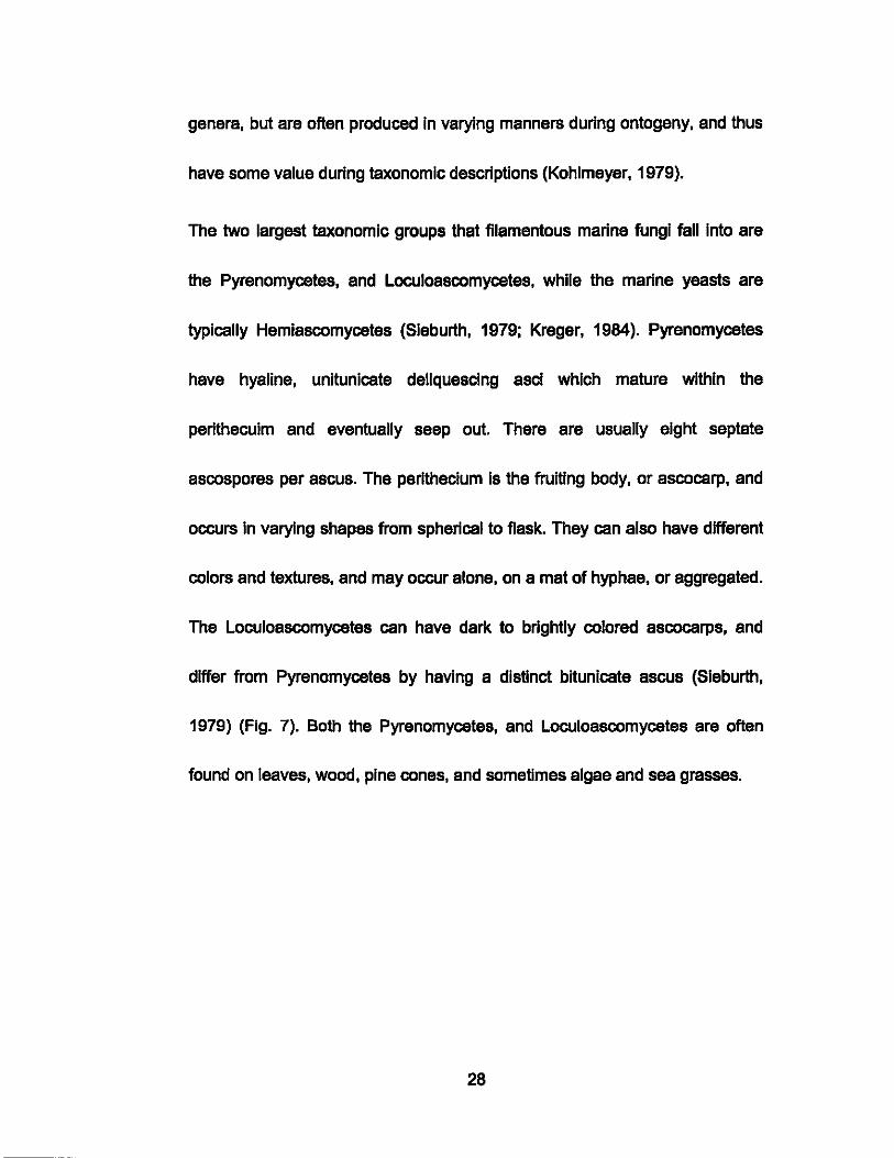
The two largest taxonomic groups that filamentous marine fungi fall Into are
the Pyrenomycetes, and Loculoascomycetes, while the marine yeasts are
typically Hemiascomycetes (Sleburth, 1979; Kreger, 1984). pyrenomycetes
have hyaline, unltunlcate deliquescing asci which mature within the
perithecuim and eventually seep out. There are usually eight septate
ascospores per ascus. The peritheclum Is the fruiting body, or ascocarp, and
occurs In varying shapes from spherical to flask. They can also have differant
colors and textures, and may occur alone, on a mat of hyphae, or aggregated.
The Loculoascomycetes can have dark to brightiy colored ascocarps, and
differ from pyrenomycetes by having a distinct bitunicate ascus (Sleburth,
1979) (Fig. 7). Both the Pyrenomycetes, and Loculoascomycetes are often
found on leaves, wood, pine cones, and sometimes algae and sea grasses.
28
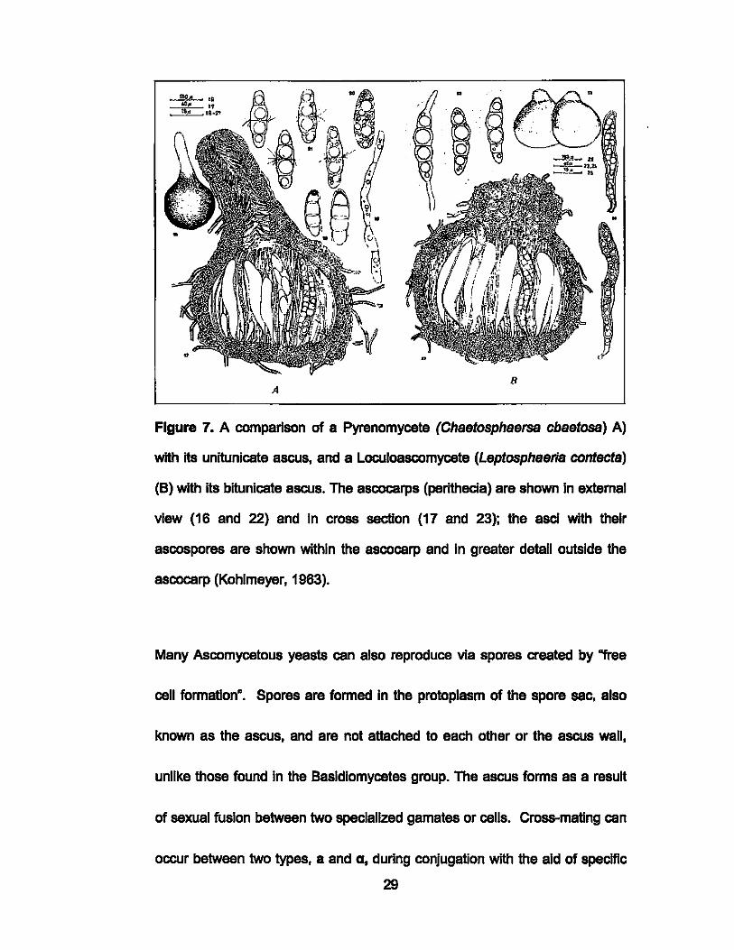
B A
Figure 7. A comparison of a Pyrenomycete (Chsetosphsersa cbsetosa) A)
with ItS unitunicate ascus, and a Loculoascomycete (Leptosphserls contects)
(8) with its bitunlcate ascus. The ascocarps (perlthecia) are shown In extemal
view (16 and 22) and In cross section (17 and 23); the asci with their
ascospores are shown within the ascocarp and In greater detail outside the
ascocarp (Kohlmeyer, 1963).
Many Ascomycetous yeasts can also reproduce via spores created by "free
cell formation". Spores are formed In the protoplasm of the spore sac, also
known as the ascus, and are not attached to each other or the ascus wall,
unlike those found In the Basidiomycetes group. The ascus forms as a result
of sexual fusion between two specialized gamates or cells. Cross-mating cen
occur between two types, a and a, during conjugation with the aid of speclflc
29
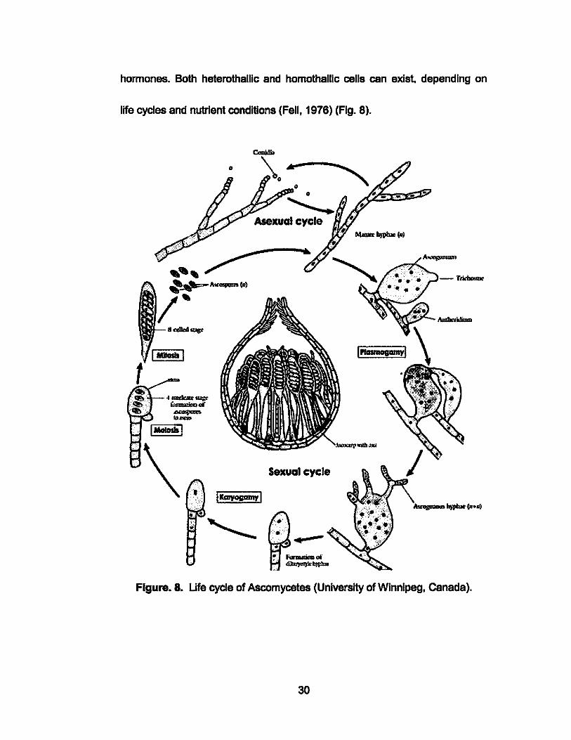
hormones. Both heterothallic and homothallic cells can exist. depending on
life cycles and nutrient conditions (Fell, 1976) (Fig. 8).
Sexual cycle
Figure. 8. Life cycle of Ascomycetes (University of Winnipeg, Canada).
30
Ascomycetous yeast are part of the class Hemiascomycetes, whose members
characteristically lack ascocarps and ascogenous hyphae. Marine yeast
genera fall into two major families, the Spermopthora and
Saccharomycetaceae. Needle- or spindle-like ascospores are what
determines the Spermophthoraceae family, while the Saccharomycetaceae
family have asci that vary in shape from oval to hat shaped, but they also
reproduce by fission or budding of single cells (Sieburth, 1979). Early studies
revealed that most Ascomycetous yeast belonged In the class
Cryptococcaceae with the perfect stages of the species Rhodotoru/a,
Candida, and Cryptococcus. It was later discovered, however, that the most
common species in the ocean belonged in the genus Metschnikowia
(Sleburth, 1979).
F. Basidiomycetes
Of the at least 30,000 Basidiomycetes believed to exist, less than two dozen
are obligately marine Basidiomycetes, although they have been found In all
three classes, the Uredinomycotina, Ustilaglnomycetes, and Hymenomycetes.
The Basidiomycetes are the most highly evolved class of fungi and are
distinguished by external basldlospores on structures called the basidium,
which is where karyogamy and meiosis take place as part of the life cycle
31
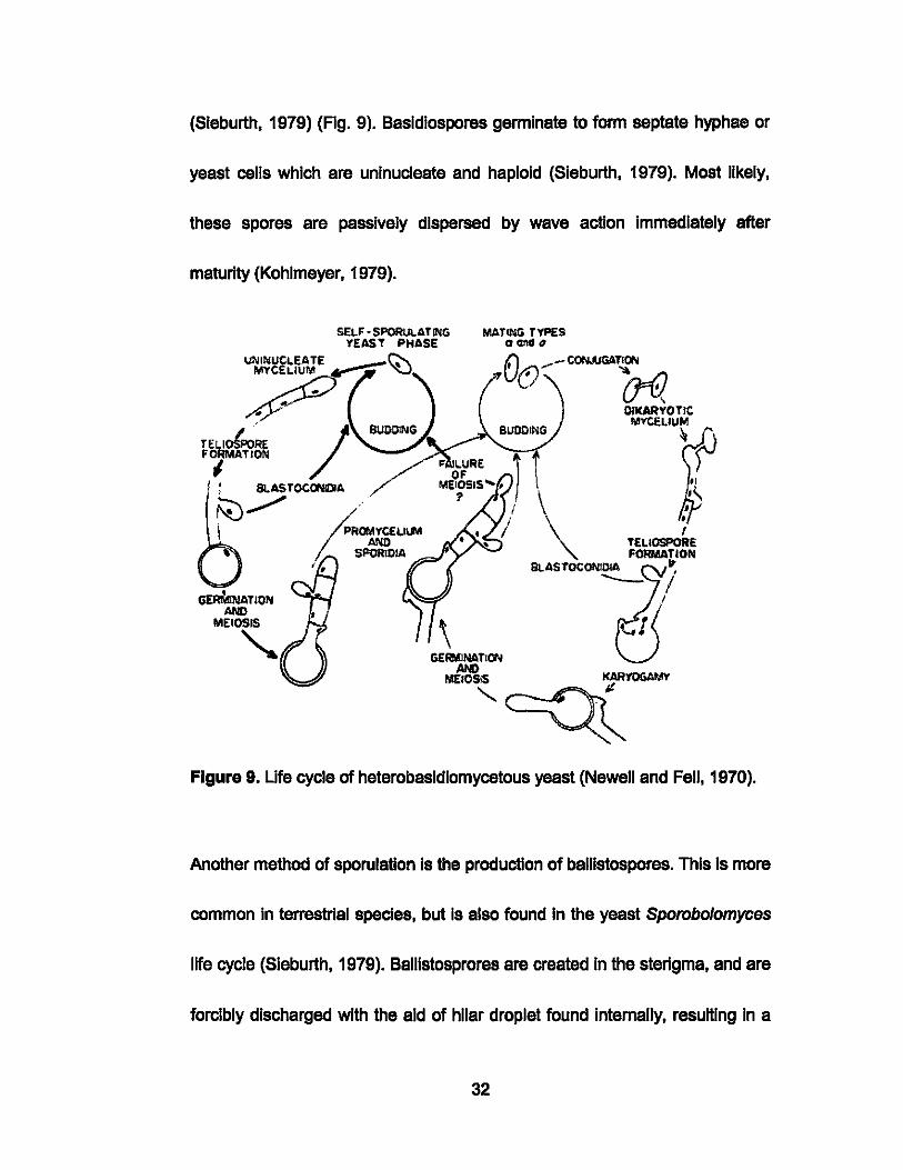
(Sleburth, 1979) (Fig. 9). Basldlospores germinate to form septate hyphae or
yeast cells which are uninucleate and haploid (Sleburth, 1979). Most likely,
these spores are passively dispersed by wave action immediately after
maturity (Kohlmeyer, 1979).
TELI~R£ FORMATION
I I , BLASTOCON[lA
~ ...........
GERMlNATIOI'l AND
MEIOSIS
'-
• I
TELIOSPORE FORMATION
"
Figure 9. ute cycle of heterobasldlomycetous yeast (Newell and Fell, 1970).
Another method of sporulation Is the production of ballistospores. This Is more
common In terrestrial species, but Is also found In the yeast Sporobolomyces
life cycle (Sieburth, 1979). Ballistosprores are created In the sterigma, and are
forcibly discharged with the aid of hllar droplet found internally, resulting in a
32
surface tension cetapult. They are asexual, and created by a number of
different cells such as yeast cells, hyphae, baSidia, or even other
ballistospores (Kurtzman, 2000). Many marine species have lost this ability
over evolutionary time. Terrestrial species In this the Basidiomycetes include
mushrooms, rusts, and smuts. The few recognized (obllgately) marine fungal
species Include Me/anotaen/um rupp/ae, Digitatlsporea marina, and Nia·
vibrissa. Digitatlsporea marina has an adaptation to the marine envlronment
with the presence of tetraradlate basldlospores which Is believed to Increase
the efficiency of spore Impectlon and rate of attachment to substrate
(Webster, 1959). Nia vibrissa has also become uniquely adapted to optimize
maximum spore dispersal distance. It traps air within several superficial hairs
on Its exterior allowing It to float on the surface of the water for up to seven
days, after detachment from the substrate (Kohlmeyer, 1979).
Basidiomycetous marine yeast were discovered In 1949 after evldence of a
heterobasldlomycete-like sexual cycle for Sporidobo/us, and soon after the
sexual life cycle of certain strains of Rhodotoru/a was discovered. The new
genus was named Rhodosporidlum and placed in the Basidiomycetes. This
red yeast Is the most common type of yeast found In the open ocean
(Sieburth, 1979). Sexual reproduction In Basidiomycetous yeasts is either
33

heterothallic or homothallic, although neither are well understood (Kurtzman,
2000). Conjugation of opposite mating types of haploid uninucleate cells leads
to the formation of binucleate mycelial formation with clamp connections, and
the development of tellospores. The clamp connections act as a link between
cells during cell dMsion, allowing the movement of nuclear products to
adjacent cells, maintaining dlkaryotic conditions (Sieburth, 1979; Kurtzman,
2000). The mycelium then forms tellospores which are thick walled spores
much larger than the vegetative cells (Fig. 10). They eventually germinate to
form a promycelium after karyogamy has taken place (Sleburth, 1979;
Kurtzman, 2000). The result Is a four-celled promycellum with transverse
septa. Uninucleate sporidia then bud from the tip of the cell as well as at the
septa. From the sides of the mycelium, chains of blastoconldla are formed
which are also uninucleate (Sleburth, 1979). All fungi and yeast that produce
clamp connections are Basidiomycetes, but not all Basidiomycetes produce
clamp connections (Kurtzman, 2000).
34
Figure 21. Teliospore formation in LM530 after 7 days on Dalmau plate
Several genera within the Basidiomycetes share similar life cycles, but have
been given different names based on their ability to produce pigments. This is
the case between the red to orange colored Rhodotorula , and the cream or
white colored Candida, who have nearly indistinguishable life cycles. Few of
the 1,000 known yeast species are obligately marine, but at least 50% of
those cultivated from marine samples today are probably novel (Jack Fell ,
pers. comm.).
35
G. Deuteromycetes
The 25,000 species classified as Deuteromycetes are also known as Fungi
Imperfecti, a subdivision that hosts fungi which produce only conidial states,
meaning they are unable to form sexual spores of any type. This subdivision
includes the imperfect, asexual, or conidial states of Ascomycetes,
Basidiomycetes, and even some Zygomycetes. Deuteromyces produce
asexual spores that are known as conidia. If the conidia arise on hyphae
(conidiophores) they belong to the class Hyphomycetes and are usually
passively released . If the spores are inside fruiting bodies, the species
belongs in the class Coelomycetes. Spore release has not yet been observed
in marine Coelomycetes (Kohlmeyer, 1979; Sieburth, 1979). The
conidiophores can be highly differentiated or simple, but function to assist in
positioning the developing conidium away from the parent mycelium. They are
not developed by free cell formations or by cytoplasmic cleavage. Other forms
of reproduction include hyphal fragmentation resulting in arthrospores,
budding, formation of true mycelium by fission, or the formation of
pseudomycelium. The pseudo-mycelium is often accompanied by
blastospores created by budding, or pseudo-hyphae (Sieburth, 1979).
36
Deuteromycetous yeast from marine environments are found in the
Cryptococcaceae which lack ballistospores, or In the Sporobolomycetaceae,
which produce ballistospores. Bailistospores are actively projected upon
maturity (Sleburth, 1979).
H. Zygomycetes
Marine-related species of the Zygomycetes are rarely mentioned In the
literature and will be considered only briefly here. The Zygomycetes consists
of roughly one percent of all described fungi and yeast species, amounting to
less than one thousand species (Kirk, 2001). Zygomycetes species range
from being pathogens In humans, plants, or animals to being saprophytes, or
mutualists IMng on plants.
The terrestrial Zygomycetes are characterized by asexual reproductive
structures called zygomycota, chlamydoconldla, conidia, or sporanglospores
contained on simple or branched hyphae known as sporanglophores. They
can be uni-to multi-spored sporengla. They are also distinguished by the thick
walled sexual reproductive structure that results from gametes fusing, called
zygospores. When the zygosporanglum germinates it produces a
mitosporanglum (Fig. 11). The spores are often actively dispersed (Kendrick,
2001). The hyphae of Zygomycetes are coenocytlc, thin-wailed, and wide.
37
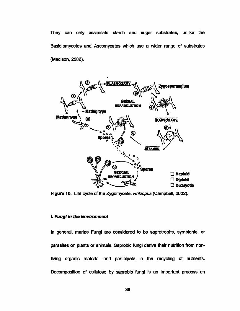
They can only assimilate starch and sugar substrates, unlike the
Basidiomycetes and Ascomycetes which use a wider range of substrates
(Madison, 2006).
o HaploId o Diploid
o DIIaUyotJo
FIgure 10. life cycle of the Zygomycete, Rhlzopus (Campbell, 2002).
I. Fungi In the Environment
In general, marine Fungi are considered to be saprotrophs, symbionts, or
parasites on plants or animals. Saprobic fungi derive their nutrition from non-
IMng organic materiai and participate in the racycllng of nutrients.
Decomposition of cellulose by saproblc fungi Is an important process on
38
driftwood, mangrove roots, algae, sea grass, and leaves. Symbiotic
relationships have been determined between fungi and algae, snails and
tubeworms. Parasitic relationships exist between fungi and fish, invertebrates,
plants (mangroves), and marine mammals, but primarily with algae (Polglase,
1986; Porter, 1986; Rheinhelmer, 1992; Kohlmeyer, 2004). Most of the 40
species of parasitic fungi belong in the Ascomycetes. Fungi as pathogens are
known to infect fish (eggs and larvae), crustaceans, and shellfish, among
other marine animals. These ralationships were determined through reports of
fungi on economically important stocks such as oysters, herring, and
mackerel.
Ascomycetes, Basidiomycetes, and Deuteromycetes are often associated
with plants, animals, guano and sand in saprobic, symbiotic, or parasitic
relationships. Saprobes use dead organic material as a source of nutrients,
such as that trapped in the interstitial species of the sediments. Symbiotic,
commensal and mutualistic relationships on the other hand involve differing
degrees of dependence or interdependence between fungi and/or yeasts and
another organism; a parasitic fungus or yeast is usually an ectoparasite, IMng
externally on the host and causing damage to the host in the form of
discoloration or tissue damage.
39

1. Sandi Foam
Arenlcolous fungi are those living among or on grains of sand. They do not
obtain nutrients from the sand, but Instead break down organic material In the
Interstitial space. Organic material here can be derived from algae, sea
grasses, leaves, animal remains, feces, or driftwood. Arenicolous fungi are
able to break down cellulosa, alginate, lamlnarln, or agar from algae (Koch,
1974; Kohlmeyer, 1979). The ability to degrade cellulose likely makes
arenlcolous fungi Important In the marine nutrient cycling, at least in coastal
waters. Sand grains are commonly covered with bacteria, diatoms, algae and
sometimes fungi, but studies have primarily been taxonomic and
morphological, with a few exceptions (Kohlmeyer, 1966). Fungi can usually be
found in the upper few centimeters of sand, but up to seven species of fungi,
Including Corol/ospora and Csrbosphaerella has been found below 15 em on
untreated burled wood panels (Fell, 1960; Koch, 1974; Johnson, 1974) Yeast
were reported In the upper 2 cm of sediments at 540 m depth In the Gulf
Stream (Fell & Uden 1963; Fell, 1968) but up to 9 em In areas with higher
wave impact. These observations led to the conclusion that distribution of
yeast populations in sand Is limited by the availability of oxygen (Kohlmeyer,
1979).
40
During sporulation events, spores released by fungi can become trapped by
air bubbles in the foam. Foam is the accumulation of marine microorganisms
generated by wave action that traps bubbles, and washes ashore. Foam Is an
excellent source of marine adapted conidia, ascospores, and basidlospores;
all of which are non-germinated due to constant wave action. Upon removal
from foam, or cessation of wave action, these spores germinate within hours.
Ascospores have adapted to survive in a dried state for several days, and if
left at the high tide line will survive until the next high tide. Fruiting bodies of
Ascomycetes can also be found attached to shell fragments or grains of sand
(Kohlmeyer, 1979).
There is no method to accurately quantify fungi or yeast in sand or foam, or
the frequency of a species within this habitat. The presence and abundance of
foam depends on tides, precipitation, wind direction and velocity. The fact that
foam Is present, however, is an unreliable indicator of both fungi and yeast
abundance, and their phylogeny. Studies have confirmed that on a dally basis
the ability of foam to trap spores varies even over a period of hours. Probably
the only accurate study of fungi and yeast associated with foam is that
conducted by Kohlmeyer (1979), who reported significant differences in
abundance of thee taxa in foam over periods of just hours.
41
2. Algae
Fungi and yeast IMng on algae are known as alglcolous, and have been
found In all groups except Basidiomycetes (Kohlmeyer, 1979). Most marine
fungi found on algae are Ascomycetes, while several are Deuteromycetes.
These species are parasitic, symbiotic, or saprobes but are not consistently
found on all types of algae. Growth Is often Inhibited by antibiotic substances
produced by healthy algae or competing bacteria (Sleburth, 1968). The
relationship betwean parasitic fungi and their algal host Is not well understood
as some of them have little or no effect on the hosrs appearance, while others
cen cause light or dark patches to appear. There does appear to be a
correlation betwean specific hosts and colonizers (Kohlmeyer, 1979).
3. Plant Material-Wood, Leaves, Mangroves
Fungi or yeast on wood or other cellulosic materials are termed Iignlcolous.
Untreated wood Is the easiest substrate to test for fungal colonization
although It should be borne In mind that low levels of dissolved oxygen limited
colonization degradation of wood by higher marine fungi (Kohlmeyer,
1969a).Such a phenomenon may be observed In buried or partially buried
wood. 'Driftwood', defined as pieces of wood that are found fioating along the
shore, and 'intertidal wood' (fragments of wood or structures partially buried in
42
the sand or wedged between rocks) are generally excellent sources of fungi
and yeasts. The most common colonizers on wood are the Ascomycetes,
followed by the Deuteromycetes, and finally the Basidiomycetes. It appears
that the fungi are not host specific. Marine fungi or yeast associated with
leaves are termed foliicolous. They are also considered saprobes on dead
loose leaves.
4. Animals
Associations between animals and marine fungi are generally limited to
saproblc relationships with exoskeletons, protective tubes, or shells of
crustaceans or invertebrates. There are also well defined relationships
between wood boring mollusks, crustaceans, amphipods, and nematodes
(Johnson & Sparrow, 1961) Marine yeasts are more frequentiy isolated from
surfaces or within animals, Including Invertebrates, fish and marine mammals
(Fell & Uden, 1963). This association Is likely due to the incidental Ingestion of
yeast since they are ubiquitous In the ocean. Yeasts have been isolated from
surface swabs and gut samples of shellfish, lobsters, conch, fiddler crabs,
amphlpods, copepods, mollusks, and oysters (Johnson & Sparrow, 1961).
Yeast may also be associated with the guts, skin, gills, mouth, and feces of
fish. Few studies have been conducted on yeast collected from marine
43
mammals. On two occasions, however, yeasts were Isolated from the
stomach and Intestines of a dolphin and porpoises (Fell, 1970; MOri, 1973). A
true relationship has never been established, however, and it was thought the
yeast were associated with recentiy consumed food. For example, intestinal
samples taken from eight California Sea Lions lacked yeasts, most likely
because conditions in the gut were unsuitable for survival of the yeast (Fell &
Uden, 1963).
5. Avian Guano
Marine yeasts are common in the guts of invertebrates, fishes, marine
mammals and seabirds, and are therefore present in these animals' feces or
guano. The intestines and rectal regions of free-IMng gulls and terns from
Baja, California hosted high densities of yeasts. The most common species
isolated from birds in the Pacific and Atlantic oceans were Candida troplca/ls
and Toru/opsis g/abrata (Fell & Uden, 1968). It has bean suggested that yeast
cells proliferate in birds who then disperse them into bodies of water
woridwide (Kawakita & Uden, 1965). This theory has been disputed by
evidence that yeast found in gulls were not aiways found in the surrounding
seawater (Uden & Castelo-Branco, 1963). This cannot be conclusive
evidence that seabirds do not disperse these cells, however, since rapid
44
dispersion of guano In seawater might require an Inordinately large water
sample be procassed to actually detect these yeasts.
6. Impact on Humans
The Impact on humans of Fungi and yeast ranges from diseases, to beneficial
applications In the food Industry, e.g., brewing and baking. A positive example
Is of course the discovery and subsequent application of Penidllln as an
antibiotic by Alexander Fleming In the early 1900's. On the other hand,
several species of fungi and yeast can be detrimental to human health.
Infections can be minor such as Athletes Foot, to severe such as Aspergillosis
which can be fatal. The use of fungi and yeast spedes In the production of
beverages and food Is a common practice, most commonly In the production
of alcohol, bread and cheeses.
Marine fungi and yeast are also slowly making their mark on humans. In 1999
H was reported that two species, Corol/ospara lacera and Corol/aspara
maritima were being used In the In bloremedlation of all spills (Cooney, 1993).
Negative publicity has come from marine fungi and yeast contaminating
seafood for human consumption, such as oysters Infected with
Dermocystidlum marlnum on the Gulf coast and southeastem states
(Sleburth, 1979). Marine fungi are also known to colonize marine
45
Infrastructure. damage to which may be costly to repair. Marine fungi have
been Implicated In attracting wood boring organisms such as the crustacaan
Ummoria, and larvae of the boring mollusk, Toredo pedlcallata to
preferentially colonize pre-digested wood rather than fresh wood. Colonization
of terrestrial wood by many Ascomycates and Deuteromycetes results In 'soft
rot', or decomposition of the wood.
J. Deep Sea Fungi and Yeast
In the context of this research the deep sea will be considered as that below
500 m, where hydrostatic pressure exceeds 50 atm. This Is based upon the
observation that fungi found below 500 m differ from those found In the
eplpelaglc zone. Adaptations In deep sea fungi to high pressure and low
temperatures are expected (Kohlmeyer. 1979) although none have thus far
been proven.
Meyers et a/. (1967) noted that the Black Sea contained the largest yeast
populations In Its upper 1000 m, while below this point only 25% of the cells
cultivated were yeast. This distribution Is consistent with the distribution of
dissolved oxygen In the Black Sea, currents, and high concentrations of
hydrogen sulfide at greater depths (Meyers et a/. 1967). It was also noted that
46
a seasonal variation of yeast populations existed, with greatest abundances
during the summer likely being related to a bloom of a large marine
dinoflagellate, Noct/luca millar/s.
47
CHAPTER II. BACKGROUND OF THIS STUDY
A. Motivation for the study
The Hawaiian Archipelago Is a 'biodiversity hotspor and home to thousands
of unique plant and animal species, but one In which novel microorganisms
are rarely reported (Amadon, 1947; Kohlmeyer, 1969, 1985; Dring et a/.,
1971; Barr & Hodges, 1987; Carr et a/., 1989; Kohlmeyer & Volkmann
Kohlmeyer, 1989; Myers et a/., 2000; Donachie et a/., 2003; 2004a, b, 2005).
It is reasonable to assume, however, that many microorganisms are yet to be
discovered In Hawai'i's diverse and Isolated habHats (Donachie et aI., 2004a).
Each of the six major Hawaiian Islands is unique in age and human
population. Spatially constrained habHats over smali vertical scales and
across well defined microclimates on each Island can host unique plant and
animal species (cf. Carr et a/., 1989).
The marine mycoflora of the six main Hawaiian Islands, Station ALOHA, and
Palmyra Atoll has never been extensively studied. The few studies that have
been conducted in marine waters of the Hawaiian Islands primarily utilized
tradHional culture techniques rather than new molecular methods, were of
limited duration, and also of limited geographic extent. In the study described
48
here I exploited advances In methods in the last 20 years, specifically that
while using 'traditional' techniques to bring fungi and yeasts into culture, I also
employed molecular methods to rapidly sort cultures phylogenetically. This
combined approach has not bean used In any study of the HawaIIan marine
mycoflora to date.
B. Defining the problem and proposed solution
Since no region wide study of Hawai'i's marine fungi and yeast has bean
conducted, the primary goal of this research was to establish the first locally
based collection of fungi and yeast from Hawaiian coastal waters and station
ALOHA; The opportunity to collect samples from Palmyra Atoll some 950
miles south of Hawai'i came late In the course of this research, but enabled
the project's geographic coverage to be significantly expanded. Other goals
included determining phylogenetic relationships among the fungi and yeasts
isolated from diverse habitats, isolating and describing novel species.
The value of cultivating microorganisms versus using a solely molecular
approach to describe phylogenetic diversity by amplifying and cloning
ribosomal genes from community DNA is that cultures in vitro provide a
collection that can be accassed by for years to come; researchers thus have
accass to cultures that can be screened for secondary metabolite production
49
including antibiotics, cytotoxlns and nutraceuticals. Previous workers have
approached marine fungi and yeast by only one of cultivation, molecular or
chemical methods. Studies of the Hawaiian marine mycoflora, however, have
to date only employed cultivation techniques. The significant novelty and
value of the work described here, is that I combined traditional cultivation on
different enrichment media with molecular methods, specifically DNA
sequencing which facilitated rapid phylogenetic placement of the isolates. I
thus applied this combined approach to conduct the most extensive
investigation to date of marine fungi and yeast seawater and other marine
associated samples around the six main Hawaiian Islands, Station ALOHA.
and Palmyra Atoll. This is in fact the first such work in Hawai'i since
Kohlmeyer's studies 30 years ago, during which he collected relatively few
samples that were sub"sequently incubated on cultivation media. Moreover,
Kohlmeyer's descriptive work was to some extent limited by the lack of rapid
DNA sequenclng technologies and absence of ribosomal DNA nucleotide
sequence databases.
C. Thesis goals and objectives
The specific aim of the research described here was to determine the
abundance and phyiogenetic diversity of fungi and yeast in marine samples
50
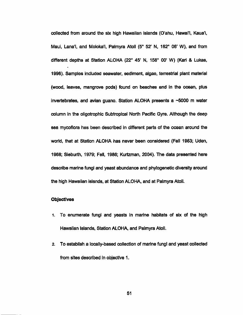
collected from around the six high Hawaiian Islands (O'ahu, Hawsl'l, Kaua'l,
Maul, Lana'i, and Moloka'l, Palmyra Atoll (50 52' N, 1620 06' W), and from
different depths at Station ALOHA (220 45' N, 1580 ~O' W) (Karl & Lukas,
1996). Samples Included seawater, sediment, algae, terrestrial plant material
(wood, leaves, mangrove pods) found on beaches and In the ocean, plus
Invertebrates, and avian guano. Station ALOHA presents a -5000 m water
column In the oligotrophic Subtropical North Pacific Gyre. Although the deep
sea mycoflora has been described In different parts of the ocean around the
world, that at Station ALOHA has never been considered (Fell 1963; Uden,
1968; Sieburth, 1979; Fell, 1986; Kurtzman, 2004). The data presented here
describe marine fungi and yeast abundance and phylogenetic diversity around
the high Hawaiian islands, at Station ALOHA, and at Palmyra Atoll.
Objectives
1. To enumerate fungi and yeasts In marine habitats of six of the high
Hawaiian Islands, Station ALOHA, and Palmyra Atoll.
2. To establish a locally-based collection of marine fungi and yeast collected
from sites described In objective 1.
51
3. To assign cultivated strains to taxonomic groups using a combined
molecular and physiological approach.
4. To describe at least one novel species.
5. To determine which material or sites host the most abundant novel
species, I.e., Island and sourca.
6. To compare the contribution of phylogenetic Classes to the fungal
community at each location.
7. To determine whether fungi and yeast abundance varies in water
samples collected around each Island.
8. To determine if the abundance of fungi and yeast differ with depth at
Station ALOHA.
9. To determine if phylogenetic differences exist In the mycoflore Isolated
from different depths at Station ALOHA.
D. Experimental Design
Fungi and yeasts were enumerated on different enrichment media Inoculated
with a range of samples collected from the coasts of each of the six high
HawaIIan Islands and Palmyra Atoll. The number of visits to each Island
52
varied as a function of site accessibility. Station ALOHA collections were
based on ship availability. Standard microbiological procedures described
below were used to prepare pure cultures of representative strains from each
sample and medium, after which DNA sequencing and in selected cases
other descriptive criteria were used to assign isolates to taxonomic groups.
CHAPTER III. MATERIALS AND METHODS
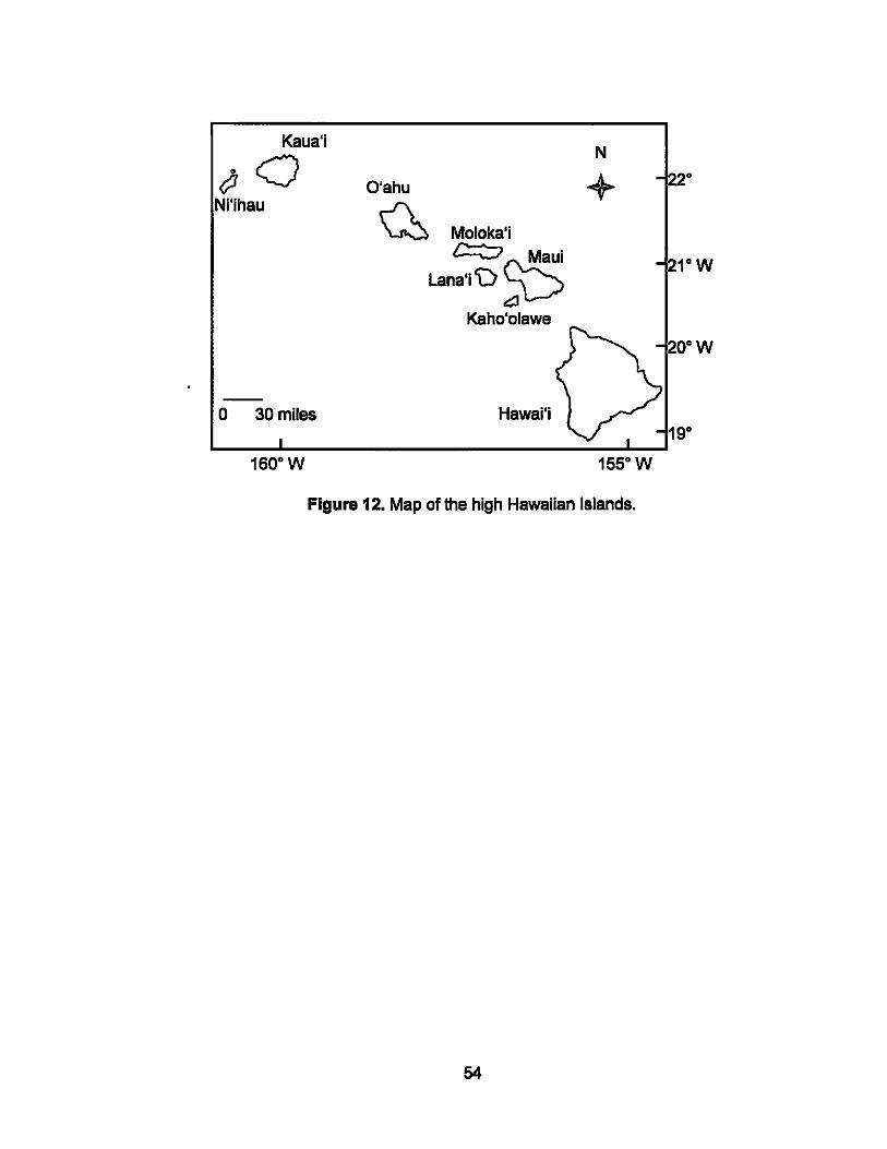
A. Sample sites
The high Hawaiian Islands are those in which the original volcanic feature
remains above sea level (Fig. 12), as opposed to the low islands in the north
of the archipelago which comprise limestone caps (atolls) on eroded or
subsided volcanic bases.
53
Kaua'j
tfJ O Nj'jhau
N
O'ahu + Q Moloka'j
c:::::::; Mauj
Lana'i'\)~ Kaho'olawe
20·W
o 30 miles Hawaj'j 19·
160·W 155·W
Figure 12. Map of the high Hawaiian Islands.
54
Kaua'i
The fourth largest and the northemmost of the populated high islands, Kaua'i
covers 550 sq. miles and has 111 miles of coastline. The island is geologically
the most mature of the main Hawaiian Islands and there are abundant fringing
coraValgal reefs and sandy beaches (Fig. 13).
l:::"
<> 0
0
Lehua Rock o o
Au~ust 11, 2005
October 24, 2005
February 14, 2006
February 15, 2006
Figure 13. Sample collection sites on Kaua'i.
55
1590 30'
N
+ Kaua'i
220
o 10 miles
O'ahu
The third largest of the Hawaiian islands, covering 608 sq. miles, with 112
miles of coastline. The most populous island with almost one million residents,
O'ahu also hosts approximately five million tourists annually. The island has
abundant fringing coraUalgal reefs and sandy beaches (Fig. 14).
N
+ O'ahu
21° 3~' N
0 5 miles
b, August 20, 2004
<> September 3, 2004
0 September 6, 2004
0 July 1, 2005
• August 17, 2005
A February 6, 2005
• February 13, 2006
Figure 14. Sample collection sites on O'ahu
56
Maul
The second largest Hawaiian island, covering 728 sq. miles and with 120
miles of coastline. Maui hosts over 117,000 residents and some 2.2 million
visitors annually (Fig. 15).
1560 30'
o Molok!ni
Maui
~ October 22, 2005
o October 23, 2005
o January 31, 2006
D February 2, 2006
N
+
o 10 miles
Figure 15. Sample collection sites on Maui
57
21°
Moloka"
The fourth largest island, covering 260 sq. and with 88 miles of coastline, this
island hosts 7,400 residents (Fig. 16).
157°W
N
+
Moloka'i
21°
o 5 miles
f::j. September 3. 2005
0 September 4. 2005
0 Mav21.2006
0 Mav22.2006
Figure 16. Sample collection sites on Moloka'i
58
Lana'i
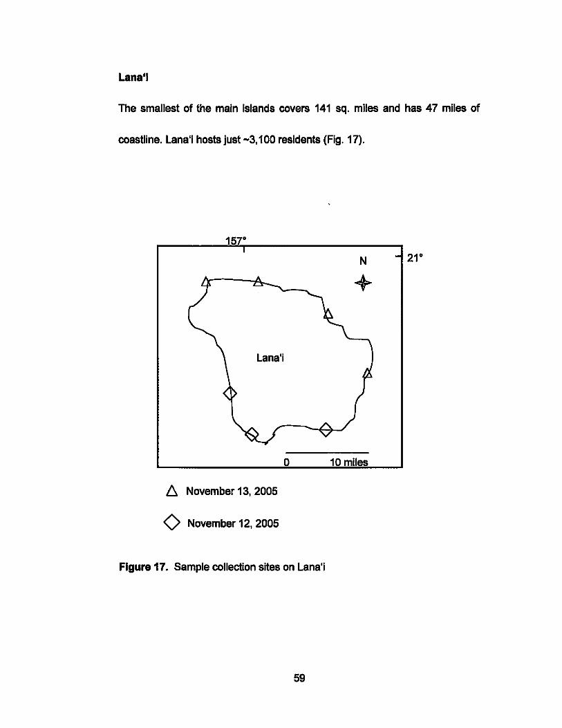
The smallest of the main islands covers 141 sq. miles and has 47 miles of
coastline. Lana'i hosts just -3,100 residents (Fig. 17).
1570
Lana'i
!:::. November 13, 2005
o November 12, 2005
N 21 0
+
o 10 miles
Figure 17. Sample collection sites on Lana'i
59
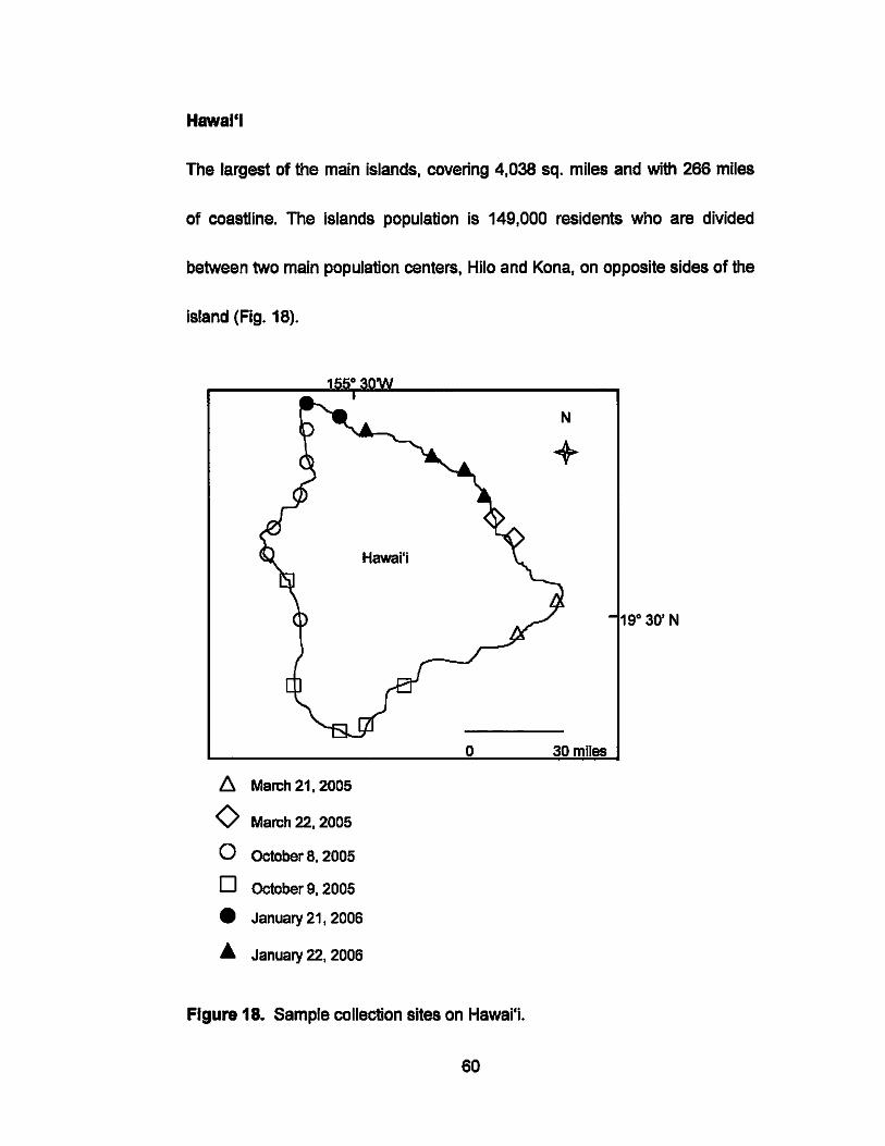
Hawal'l
The largest of the main Islands, covering 4,038 sq. miles and wlth 266 miles
of coastline. The islands population is 149,000 reSidents who are divided
between two main population centers, Hllo and Kona, on opposite sides of the
island (Fig. 18).
N
+
Hawaj'j
19° 30' N
o 30 miles
6- March 21, 2005
0 March 22, 2005
0 October 8, 2005
0 October9,2005
• January 21, 2006
• January 22, 2006
Figure 18. Sample collection sites on Hawai'i.
60
Station ALOHA
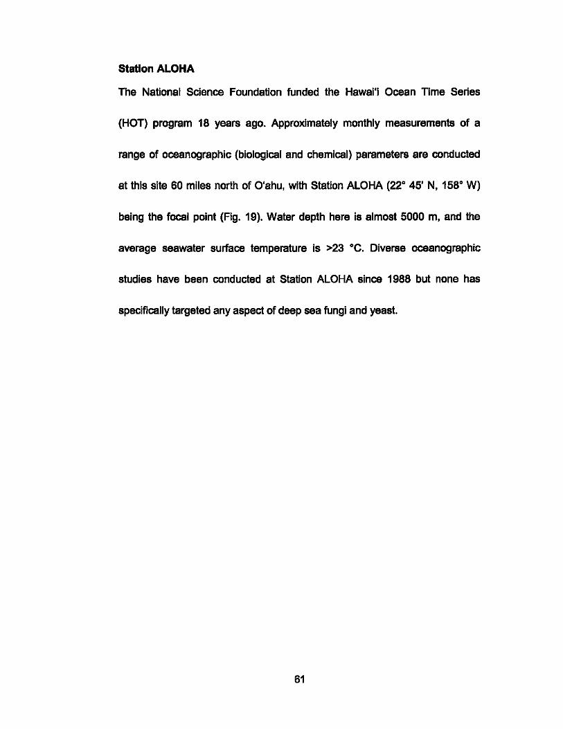
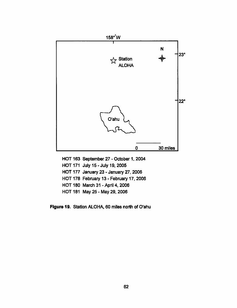
The National Science Foundation funded the Hawal'i Ocean TIme Series
(HOT) program 18 years ago. Approximately monthly measurements of a
range of oceanographic (biological and chemical) parameters are conducted
at this site 60 miles north of O'ahu, with Station ALOHA (22° 45' N, 158° W)
being the focal point (Fig. 19). Water depth here is almost 5000 m, and the
average seawater surface temperature is >23 °C. Diverse oceanographic
studies have been conducted at Station ALOHA since 1988 but none has
specifically targeted any aspect of deep sea fungi and yeast.
61
'* Station ALOHA
N
+
o 30 miles
HOT 163 September 27 - October 1, 2004
HOT 171 July 15 - July 19, 2005
HOT 177 January 23 - January 27,2006
HOT 178 February 13 - February 17, 2006
HOT 180 March 31 - April 4, 2006
HOT 181 May 25 - May 29,2006
Figure 19. Station ALOHA, 60 miles north of O'ahu
62
23°
22°
Palmyra Atoll
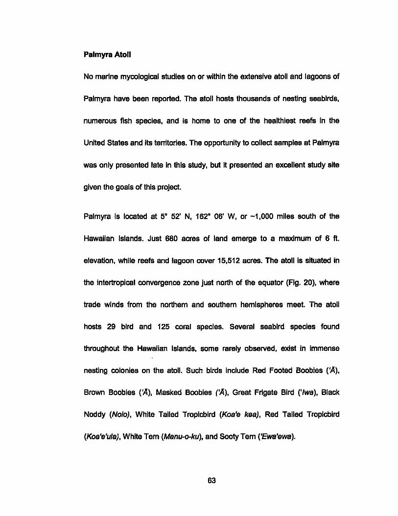
No marine mycological studies on or within the extensive atoll and lagoons of
Palmyra have been reported. The atoll hosts thousands of nesting seabirds,
numerous fish species, and is home to one of the healthiest reefs In the
United States and Its territories. The opportunity to collect samples at Palmyra
was only presented late in this study, but it presented an excellent study site
given the goals of this project.
Palmyra Is located at 50 52' N, 1620 06' W, or -1,000 miles south of the
Hawaiian Islands. Just 680 acres of land emerge to a maximum of 6 ft.
elevation, while reefs and lagoon cover 15,512 acres. The atoll is situated in
the intertropical convergence zone just north of the equator (Fig. 20), where
trade winds from the northern and southern hemispheres meet. The atoll
hosts 29 bird and 125 coral species. Several seebird species found
throughout the Hawaiian Islands, some rarely observed, exist In Immense
nesting colonies on the atoll. Such birds include Red Footed Boobies rAl,
Brown Boobies rA), Masked Boobies (~), Great Frigate Bird ('Iwal, Black
Noddy (Nolo), White Tailed Troplcblrd (Koa'e kaa), Red Tailed Tropicbird
(Koa'e'ula), White Tern (Manu-o-kul, and Sooty Tern ('Ewe'ewe).
63
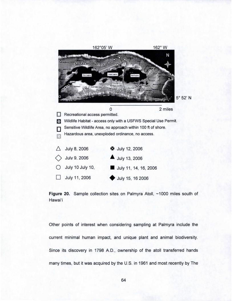
162°05' W 162° W
5° 52' N
o 2 miles o Recreational access permitted.
1::3 Wildlife Habitat - access only with a USFWS Special Use Permit.
o lSI
6.
<> 0
D
Sensitive Wildlife Area, no approach within 100 ft of shore.
Hazardous area, unexploded ordinance, no access.
July 8, 2006 .:. July 12, 2006
July 9. 2006 A July 13, 2006
July 10 July 10, • July 11 , 14, 16, 2006
July 11 , 2006 • July 15, 162006
Figure 20. Sample collection sites on Palmyra Atoll , -1000 miles south of
Hawai'i
Other points of interest when considering sampling at Palmyra include the
current minimal human impact, and unique plant and animal biodiversity.
Since its discovery in 1798 A.D., ownership of the atoll transferred hands
many times, but it was acquired by the U.S. in 1961 and most recently by The
64
Nature Conservancy In 2000. The atoll's human population varies between 8
and 25, but the greatest population of -5,000 was reached during the 1940's
when the atoll was 'occupied' by the United States Navy. The Navy dredged
channels in the lagoon and dumped the 'spoil' to form causeways for vehicles
and landing strips for aircraft. Several Islets have seen little human Impact
since the 1940's because of the presence of unexploded ordinance, but they
have become popular nesting sites for seabirds. The high density of seabirds
likely impacts the abundance and phylogenetic diversity of culturable fungi
and yeast In the surrounding waters.
B. Sample collection
Sampling began on August 20, 2004 on the island of O'ahu and ended In July
2006 at Palmyra Atoll (Tabs. 1 - 2). Each of the six high Hawaiian Islands was
sampled, covering as much of the circumference as geographically possible in
the time available (Fig. 12). Samples were also collected at various depths
during six Hawal'l Ocean TIme (HOT) series cruises to Station ALOHA.
beginning with HOT-163 (September 27 - October 1, 2004), and ending with
HOT-181 (May 25 - May 29, 2006). Palmyra Atoll samples were collected In
July 2006.
65
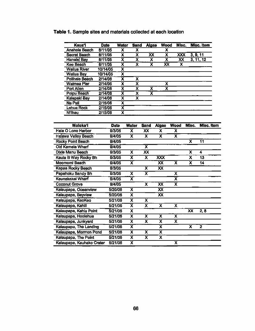
Table 1. Sample sites and materials collected at each location
Kaua'i Data Watar Sand Algae Wood Misc. MJsc.ltem Anahola Beach B/11/05 X X X Secret Beach 8111105 X X XX X XXX 3,9,11 Hanalei Bay 8111/05 X X X X XX 3,11,12 KeeBeach B/l1/05 X X X XX X WalluaRiver 10/14/05 X WailuaBa~ 10/14/05 X Pollhale Beach 2114106 X X Waimea Pier 2114106 X X X Port Allen 2114/06 X X X X Pol!!u Beach 2114/06 X X X Kale!!ak1 Ba;t 2114106 X X NaPall 2115/06 X Lehua Rock 2115/06 X NI'lhau 2115/06 X
Moloka'i Data Water Sand Algae Wood Mise. MJsc.ltem Hale 0 Lono Harbor 9/3/05 X XX X X Halawa Valley Beach 9/4/05 X X X X Rocky Point Beach 9/4105 X 11 Old Kamela Wharf 9/4105 X Dlxla Manu Beach 9/3/05 X XX X 4 Kaula III Way Rocky Bh 9/3/05 X X XXX X 13 Moomoml Beach 9/4105 X XX X X 14 Kapaa Rocky Beach 9/3105 X XX Papahoku Sendy Bh 9/3/05 X X X Kaunakakai Wharf 9/4/05 X X Coconut Grove 9/4/05 X XX X Kalaupapa, Oceanvlaw 5/20/06 X XX Kalaupapa, Bayvlaw 5/20/06 X XX Kalaupapa, KeoKee 5/21/06 X X Kalaupapa, Kahlll 5121106 X X X X Kalaupapa, Kahlu Point 5121/06 X XX 2,B Kalaupapa, Hoolehua 5/21/06 X X X X Kalaupapa, Junkyard 5/21/06 X X X X Kalaupape, The landing 5/21/06 X X X 2 Kalaupapa, Mormon Pond 5/21/06 X X X Kalaupapa, The Point 5/21/06 X X X Kalaupapa, Kauhako Crater 5/21/06 X X
66
Table 1. (Continued) ~Almnll"sites and materials collected at each location
iO iOiiilv iOiiilv
I BaiiCiiP8ik rBaacl1 .Baad1
PiiiikiiBiiSch iBiiV
MllolII eech Kallue [one
DiiiiI VViiiiir Siiiid Algae Wood Mise. Misc. Item
X X X ) x XXX x XXX
x X X X X X X X X X X X X
x X
X 3:
Green land Baach X XX
~o:aCh ~~---!!....-X .....,,-----_
POIOIU'~~:~V ~ X 2 Bav
X
67
Table 1. ,~v" ..... "I'''''' sites and materials collected at each locati on Maul Date Water sand A1gaa Wood Misc. Misc. Item
X X X
~ X XX 9,11
II X X X X 11 )C X X 11
XXX X X 5,10 XX
X X
1 BeaCh X X 1 Klnel X XX 1 Big X X
X X X X 8 1 Bey X
I ArCh X I Grave X
;01 Beae lee ,Sand BeaCh X
'1'06 X 11 m06 X m06
Miscellaneous sample key: 1) Crab Exoskeleton; 2) Foam; 3) Guano; 4) Hemil Crab;
5) Mucus; 6) Black Sponge (Axlny888 sp.); 7) 'Oily' Film on seawater; 8)
Cyanobacteria Mat; 9) Fecal Pellets (origins unknown, and from Calc/nus e/egans
(Hemil crab»; 10) 'Scum' (on rocks); 11) Leaf; 12) Flower (unidentified); 13) Sea
urchin shell (species unknown); 14) Pencil urchin spine.
68
Table 2. Water samples collected at Station ALOHA.
HOT cruise no. Depth (m) 163 171 1n 178 180 181
5 X X X X X X 75 X X X X X 125 X X X X X 250 X X X X X X 520 X X X X X X 1000 X X X X X X 2000 X X X X X 3000 X X X X X 4000 X X X X X 4740 X X X X X
Dates: HOT-163, 9/27 -10/1/04; 171,7/15 -7/19/05; 1n, 1/23 -1/27/06;
178,2113 - 2117/06; 180, 3/31 - 4/4/06; 181,5/25 - 5/29/06
Seawater samples were collected from the shore or tidal pools directly into
sterile Nalgene bottles or polypropylene tubes. Subsurface water samples at
Station ALOHA were collected into 20 L Go-Flo bottles on a 24 bottle rosette.
Upon return to the surface, sub-samples were transferred from each Go-Flo to
sterile 1 L or 4 L bottles. Sediments in tidal pools were collected with sterile
plastic scoops or by 'corlng' with sterile 50 rnl centrifuge tubes. Marine
invertebrates, driftwood, macroalgae, or other plant material were collected
directly into sterile 50 ml centrifuge tubes or 500 ml Nalgene bottles,
depending on their size. Seabird guano was collected opportunistically into
69
sterile 50 ml centrifuge tubes. Sample sites on each Island were selected on
the basis of distance between them and their accessibility, such that an
island's entire circumference could be sampled at approximately regular
spatial intervals. Among the species of algae collected during this study are
Dictyota aeuti/oba, Dletyosphaerla eavemosa, D/ctyopterls p/ag/rgramma,
Curstose coralline, Entermorpha flexuosa, Graci/aria eoronopifo/la, Ha/lmeda
Inerassate, Sargassum eehinoearpum, Spyrld/um fI/imentosa, Turblnarla
ornata, U/va faseiata and Padlna sp.
C. Sample Processing and Cultivations
Upon retum to the lab (Iand- or ship-based), 150 ml of each water sample was
filtered (0.2 IJm Mllllpore HA) and placed onto media designed to enrich fungi
and yeasts (§ III. D). Homogenized or sectioned invertebrate and plant
tissues, and guano were serially diluted in 0.2 I1Ill filtered and autoclaved
seawater and spread (200 IJI each dilution) on the same enrichment media.
Plates were incubated at 25 ·C for up to four weeks, and inspected dally for
growth. A duplicate set of media inoculated with seawater from below 1.000 m
collected at Station ALOHA was also Incubated at 4 ·C to cater to potentially
psychrophlllc strains. Hyphal growth and yeast colonies considered
representative of growth on a medium in terms of morphology, including
70
shape, color, opacity, etc., were streaked for purification on the same isolation
medium.
Each Isolate was assigned an Identification number that began wlth the prefix
"lM", commencing wlth lM1. Each culture was assigned a unique descriptor,
which describes the isolation medIum, the Island of origIn, and type of sample.
For example, colony number 4 from a plate of filtered water collected from
Helawa Valley on Moloka'i grown on Potato Dextrose would be called: PO Mo
HV W4. O'ahu was represanted by "0", Kaua'i = "K", Maui = "Ma", Moloka'i =
"Mo", lana'i = "l", Big Island="Bi", Station ALOHA HOT cruise = "HOT#",
Palmyra Atoll = "PAD. In the event an island was visited multiple times, the
number of the visit was listed after the location code, for example the second
trip to Maul is recorded as "Ma2", or the third trip to the Big Island is recorded
as "BI3". The sample source was Identified wlth a similar code, water was
recorded with a "W", and sand, algae, wood were assigned numerical values.
D. Isolation Media
Three media were used for primary cultivation of organisms, specifically
Sabouraud Dextrose (SO), Potato Dextrose (PO), and Wickerham's YM. The
first two are manufactured by Oifco, while the latter Is made in housa. YM, or
Yeast Extract-Malt Extract, comprises by weight 1% glucose, 0.5% peptone,
71
0.3% yeast extract, and 0.3% malt extract. Each medium was supplemented
with 20 grams of sodium chloride per liter. Prior to pouring to Petri plates, 300
mg of Penicillin G, and 250 mg of Streptomycin Sulfate per liter were added to
inhibit bacterial growth.
E. Enumel8tion of colonies
Fungi and yeast colonies were counted manually using a colony counter pen
(Bel-Art Products) and the average of three counts per plate was considered
the result. Filters that contained too many colonies to count were recorded
thus and assigned a numerical value of 500. Fungi were distinguished from
yeast by colony morphology, i.e., the presence of hyphal cells indicated fungi,
while a lack thereof was considered to Indicate the presence of yeast.
Colonies were counted after a minimum of four days of Incubation, and a
maximum of twenty one days based on the rate of overgrowth of neighboring
colonies, or time. Plates that did not yield any colonies after one week of
Incubation at 25°C were transferred to 4°C for one week to encourage slow
growing organisms or psychrophiles.
F. Preservation and Archiving Samples
Colony morphology, size, color and texture were recorded along with digital
photographs of the Original sample, original culture plates, and isolated
72
culture. Pure cultures were transferred to agar slants for storage at 4 ·C, and
into 10 ml of liquid medium in a 15 ml Falcon tube to an OD600 >1.5. After 48
hours, -1 ml of culture was transferred to three 1.8 ml Nunc Cl)'ovial tubes
containing sterile glycerol (60% final concentration) for long term storage at -
20 ·C. Approximately 1 ml of culture was also transferred to a microfuge tube
and spun down (5000 rpm, 30 seconds) to create a pellet of cells for DNA
extraction; pellets were stored at -20°C, as was as the remainder of the liquid
culture.
G. Identification
Each culture was provisionally identified through sequencing the Internal
transcribed spacer (ITS) 1 and 2 regions between the 18S and 28S rDNA
genes (Fell, 2001). No single DNA extraction technique suited all the cultures,
so a range of methods was utilized, including the classic phenol-chloroforrn
extraction, and modified commercial systems (Marmur, 1961; e.g., Fell, 2001).
Initial phylogenetic assignment was based on a polymerase chain reaction
(PCR) with -1 III of the loose pellet, and 1 III of each of the forward NS7 (5'-
GAGGCAATAACAGGTCTGTGATGC-3') and reverse LR6 (5'-
CGCCAGTTCTGCTTACC-3') primers. PCR products were cleaned in the
MoBio Ultraclean PCR Clean-Up KIt and sequenced in a core laboratory at
73
the University of Hawal'!. Sequence chromatograms were manually checked
for quality and edited where required. A consensus sequence was compared
with other sequences In the public domain through a BLASTn search of the
GenBank database (Altschul et a/., 1997) through which the nearest
neighbors were determined. This was also used as a selection process for
potentially novel species, with novelty defined as there being <97% nucleotide
Identity with the nearest validly described neighbor. For phylogenetic
placement, sequencing provided approximately 320 base pairs of high quality
sequence from the 3' end of the 18S rRNA along with full ITS 1, 5S rRNA, and
partial ITS2 sequence. The 188 rRNA gene portion was subsequentiy used
in phylogenetic analyses. Sequences were aligned In BioEdlt (Hall, 1997)
initially using Clustal W followed by manual editing. Trees Inferring sequence
relationships were generated in the PHYLIP software package (Felsensteln,
2004) using the program DNAdist with Jukes-Cantor correction to construct
distance matrices and the program Neighbor to carry out Nelghbor-Jolning
analysis. Phylogenetic trees were visualized and annotated In the program
Adobe Illustrator 9.0 (Adobe Systems Incorporated).
I collaborated with Dr. Jack W. Fell at the Rosenstiel School of Marine and
Atmospheric Science at the University of Miami in order to describe novel
74
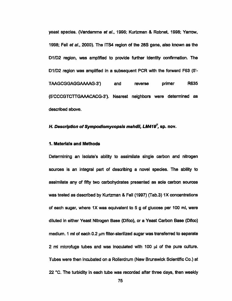
yeast species. (Vandamme at s/., 1996; Kurtzman & Robnet, 1998; Yarrow,
1998; Fell at s/., 2000). The ITS4 region of the 28S gene, also known as the
01/02 region, was amplified to provide further Identity confirmation. The
01/02 region was amplified In a subsequent PCR with the forward F63 (5'-
TAAGCGGAGGAAAAG-3') and reverse primer R635
(5'CCCGTCTTGAAACACG-3'). Nearest neighbors were determined as
described above.
H. Description of SympodJomycopsJs mahdll, LM418T, sp. nov.
1. Materials and Methods
Oetermlnlng an Isolate's ability to assimilate single carbon and nitrogen
sources Is an integral part of describing a novel species. The ability to
assimilate any of fifty two carbohydrates presented as sole carbon sources
was tested as described by Kurtzman & Fell (1997) (Tab.3) 1X concentrations
of each sugar, where 1X was equivalent to 5 g of glucose per 100 ml, were
diluted In either Yeast Nitrogen Base (Olfco), or a Yeast Carbon Base (Oifco)
medium. 1 ml of each 0.2 J.LITI filter-sterilized sugar was transferred to separate
2 ml mlcrofuge tubes and was inoculated with 100 J.LI of the pure culture.
Tubes were then Incubated on a Rollerdrum (New Brunswick Scientific Co.) at
22 ·C. The turbidity In each tube was recorded after three days, then weekly
75
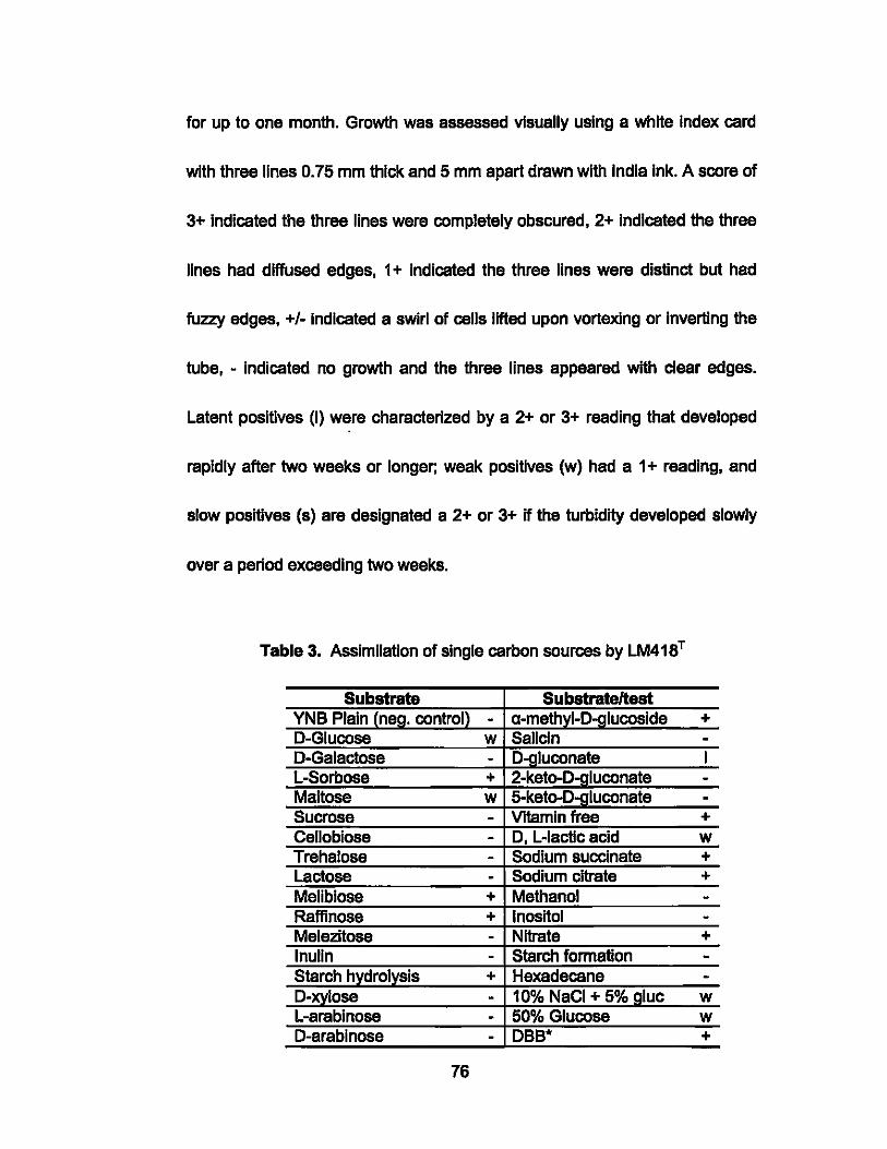
for up to one month. Growth was assessed visually using a white Index card
with three lines 0.75 mm thick and 5 mm apart drawn with India Ink. A score of
3+ Indicated the three lines were completely obscured, 2+ Indicated the three
lines had diffused edges, 1 + indicated the three lines were distinct but had
fuzzy edges, +1- Indicated a swirl of cells lifted upon vortexlng or inverting the
tube, - indicated no growth and the three lines appaared with cleer edges.
Latent positives (I) were characterized by a 2+ or 3+ reading that developed
rapidly after two weeks or longer; weak positives (w) had a 1 + reading, and
slow positives (s) are designated a 2+ or 3+ if the turbidity developed slowly
over a period exceeding two weeks.
Table 3. Assimilation of single carbon sources by LM41ST
Substrate SubstrateJtest YNB Plain (neg. control) - a-methyl-D-glucoside + D-Glucose w Salicin -D-Galactose - D-gluconate I L-Sorbose + 2-keto-D-gluconate -Maltose w 5-keto-D-Qluconate -Sucrose - Vitamin free + Cellobiose - D, L-Iactic acid w Trehalose - Sodium succinate + Lactose - Sodium citrate + Melibiose + Methanol -Raffinose + Inositol -Melezitose - Nitrate + Inulin - Starch formation -Starch hydrolysis + Hexadecane -D-xylose - 10% NaCI + 5% gluc w L-arabinose - 50% Glucose w D-arabinose - DBB" +
76
+, positive
-, negative
D-rlbose L-rhamnose D-glucosamine Ethanol Glycerol Erythritol Adonitol Dulcitol D-mannltol D-glucltol
I, latent positive
w, weak positive
- Gelatin liQuefaction - Growth at 37 'c - K D saccharate - Sodium D-glucouronate w D-gluconate lactone - Creatinine - Urease - Creatine + Glucose fermentation +
., DBB distinguishes Ascomycetes from Basidiomycetes
----+ -+ --
Fermentation tests were conducted In Durham tubes containing 2 - 4% of a
single sugar in a yeast extract, peptone base. Durham tubes consisted of a
screw top 16 mm test tube with an inverted Durham Insert. Fermentation
media also contained a pH indicator dye, Brom Cresol purple. Fermentation
was indicated by accumulation of gas In the Inner test tube, and a color
change in the medium which Indicates acidification. Results were read after 3
days, and then weekly for up to one month. Results were read as 'positive' If
gas filled the tube within seven days, 'delayed positive' If the tube was filled
rapidly after seven days, 'slow positive' If the tube was filled slowly after seven
days, 'weakly positive' If the tube filled to less than one third full, or negative if
77
no gas accumulated In the tube. Glucose Is often used as an indicator of the
ability to ferment, and if this test is negative no further fermentation tests are
considered necassary. Literature also states that other members of
Sympodiomycopsls do not ferment sugars (Kurtzman & Fell, 2000). Other
tests included starch formation, growth at 37 ·C, gelatin liquefaction (for
proteolytic activity), Dlazonlum Blue B color reaction (DBB) to determine
whether anasexual yeast is an Ascomycete or Basldiomycete, high osmotic
pressure, hydrolysis of urea, and cyclohexamide resistance.
Colony morphology was described on Malt Extract plates with and without
NaCI supplement for three days at 22 ·C. The elevation, shape, size, color,
texture, and margin of the colony were all recorded. Variations or preference
in growth between Malt Extract with 2% NaCl, and without NaCI were also
recorded. Cell shapes and dimensions were observed on a microscope slide
in a three day culture in liquid Malt Extract medium without salt. The lengths
and widths of twenty cells were measured and averaged. Observations of cell
shape and if they occurred singly, in pairs, or in clusters were conducted.
The presence or absence of sexual structures or mycelial formation was
determined on cornmeal Dalmau plates after seven days and thirty days
(Kurtzman & Fell, 1998). Dalmau plates are created by taking a loop or needle
78
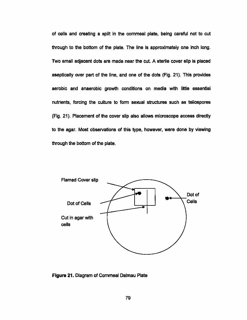
of cells and creating a split in the cornmeal plate, being careful not to cut
through to the bottom of the plate. The line is approximately one Inch long.
Two small adjacent dots are made near the cut. A sterile cover slip is placed
aseptically over part of the line, and one of the dots (Fig. 21). This provides
aerobic and anaerobic growth conditions on media with little essential
nutrients, forcing the culture to form sexual structures such as teliospores
(Fig. 21). Placement of the cover slip also allows microscope access directly
to the agar. Most observations of this type, however, were done by viewing
through the bottom of the plate.
Flamed Cover slip
Dot of Cells
Cut in agar with cells
Figure 21. Diagram of Cornmeal Dalmau Plate
79
Dot of .... - ........ -Cells
2. Sympod/omycopsls mahdll sp. nov., a basidiomycetous yeast from
the HawaIIan Islands. To be submitted to FEMS Yeast Research
Abstract
A new basidiomycetous yeast is described for which the name
Sympodiomycops/s mahdii is proposed. The type strain LM418T, Isolated from
driftwood collected on a beach In Lana'i (Hawal'l), shares prominent
characteristics of each of the genera Sympod/omycopsis, Rhodotorula,
Mlcrostroma, and Quambalaria, which make up three different families In the
Microstromatales. The speCies was determined based on morphologicel,
physiological, and molecular data. Sexual reproduction was not observed.
GenBank accession numbers for nucleotide sequences of regions of the 18S
and 28S ribosomal RNA genes in LM418T are OQ990016, OQ990017 and
OQ990018.
Introduction
The Hawaiian Archipelago hosts thousands of unique plant and animal
species (Amadon, 1947; Carr et al., 1989). Although the region is widely
acknowledged as a 'biodiversity hotspot', few novel cultivated microorganisms
from the archipelago have been described (Kohlmeyer, 1969, 1985; Oring et
al., 1971; Barr & Hodges, 1987; Kohlmeyer & Volkmann-Kohlmeyer, 1989;
80
Myers st a/., 2000;). In this respect, one-third of Bacteria species (each
'species' shared :S97% 16S rONA nucleotide sequence identity) cultivated
from Hawaiian lakes and at the submarine LO'ihi volcano were potentially new
species or genera, just four of which have thus far been described (Oonachle
et a/., 2003; 2004a, 2005, 2006). Since just a fraction of the effort devoted
globally to cultivating marine bacteria has been applied to cultivating marine
fungi and yeasts, the latter two are poorly represented In culture collections
and more so In the literature. Although fungi and yeasts are ubiquitous In the
ocean, those off Hawal'l have rarely been considered (Fell, 1976, 2001;
Oonachle & Zdanowskl, 1998; Nagahama et a/., 2001; Liu et a/., 2003;
Tsukamoto et a/., 2004). OVer thirty years ago, however, Kohlmeyer (1969)
stated that, •... the marine fungal flora of Hawal'l Is rich In species and
deserves further thorough Investigation." Kohlmeyer (1985) also made an
excellent case for novelty in Hawal'l by Isolating Nlmbospora octonae during
two collections In the state 15 years apart, but nowhere else In the world In
4000 collections over a 25 year period. Considering Kohlmeyer's work here,
and that phylogenetlcally novel taxa appears abundant among cultivated
Hawaiian Bacteria, we believe that a dedicated cultivation effort directed
towards Hawaii's marine fungi and yeasts wlll yield new species. In this
81
respect. fewer than one thousand of the known -400,000 Ascomycetes are
marine in origin, less than thirty obllgately marine Basidiomycetes have been
described, and few of the 1000 known yeast species originate from the ocean.
Thus, a large fraction of the marine fungi and yeasts cultivated from marine
samples today are likely to be novel species. As part of a project to describe
phylogenetic diversity and secondary metabolite production In Hawaiian
marine fungi and yeasts, we sample diverse marine habitats throughout the
Hawaiian Archipelago, supporting cultivation techniques with DNA sequencing
methods that quickly enable tentative phylogenetic assignment of cultures.
Molecular evidence of phylogenetic novelty is then tested through
morphological and physiological observations. We presant here a description
of Sympodiomycopsis mahdli LM418T, a new basidiomycetous yeast isolated
from driftwood collected on a beach In Lana'i, Hawai'i.
Materials and Methods
Sample col/ection. In November 2005 we collected a piece of driftwood in
the Intertidal zone of 'ShipwreCk Beach' on the island of Lana'i, Hawai'i (20'
55' 15" N, 156' 54' 30 W). The sample was collected directly Into a 50 ml
sterile polypropylene tube with a few milliliters of seawater, and processed In
the laboratory at the University of Hawai'i at Manoa as described below for
82
cultivation of yeast and fungi (Fig.17).
Yeast Isolation. The sample was vortexed vigorously for 30 seconds, after
which 200 J.LI sub-samples of the 'dlluenf were spread on Potato Dextrose
Agar (PDA) (Ditco) containing sodium chloride (20 gIL), 300 mg/L of penicillin
G, and 250 mg/L of streptomycin sulfate. Plates were incubated at 25 ·C.
Representatives of colonies that arose after seven days of incubation were
streaked for isolation on PDA and incubatad at 25 ·C. The purity of isolated
strains was checked by consistency of colony morphology and uniformity of
cells in wet mount preparations. Strain LM418T was characterized by standard
methods (yarrow, 1998). Cells were archived on Potato Dextrose slants, and
in Potato Dextrose broth containing 2% NaCI and 60% glycerol, and stored at
-20 ·C. Non-frozen cultures were retained for preliminary phylogenetic
assignment through DNA sequencing.
Identification. Cells of LM418T grown in 10 ml Potato Dextrose broth (with
2% NaCI) for 72 hr in an orbital shaker at 120 rpm and room temperature
were collected by genUe centrifugation (30 sec). Initial phylogenetic
assignment was based on a polymerase chain reaction (PCR) with 1 J.LI of the
loose pellet, and 1 J.LI of each of the forward NS7 (5'-
83
GAGGCAATAACAGGTCTGTGATGC-3') and reverse LR6 (5'-
CGCCAGTTCTGCTTACC-3') primers. The 01102 region of the 28S large
sub-unit rONA gene was amplified In a subsequent PCR with the forward F63
(5'-TAAGCGGAGGAAAAG-3') and reverse R635
(5'CCCGTCTTGAAACACG-3') primers. PCR products were cleaned In the
MoBio Ultraclean PCR Clean-Up Kitand sequenced In a core laboratory at the
University of Hawal'l. Sequence chromatograms were manually checked for
quality and edited. Consensus sequences were compared with other
sequences In the public domain (GenBank) through BLASTn searches
(Altschul et s/., 1997). Sequencing provided apprOximately 545 base pairs
from the 3' end of the 28S rRNA gene, including the 01/02 region (Fell,
2001). Sequences were aligned In BioEdit followed by manual editing, and
phylogenetic trees were generated In the PHYLIP software package
(Felsensteln, 2004) using the program ONAdlst with Jukes-Cantor correction
to construct distance matrices and the program 'Neighbour' to cerry out
Nelghbour-Joinlng analysis. Maximum likelihood analysiS was conducted In
the PHYLIP software package (Felsensteln, ibid) using the program ONAmI.
Phylogenetic trees were annotated and refined for publication In Adobe
Illustrator 9.0 (Adoba Systems Incorporated).
84
Morphological and physiological analyses. The morphology of LM418 T
colonies was determined on Malt Extract (ME) plates containing NaCI (2%
w/w) after incubation at 22 ·C for three days and one month. Cell size and
shape were recorded after seven days and one month In ME broth with NaCI
(2% w/v). The assimilation of single carbon and nitrogen sources by LM418T
was determined in liquid media In 2 ml mlcrofuge tubes after Kurtzman & Fell
(2000). Fermentation of glucose was investigated in Durham tubes with a 2%
sugar mixture and Brom Cresol purple, read after 3 days and then weekly for
up to one month. The presenca or absenca of sexual structures was
determined on commeal Dalmau plates after seven days and thirty days. The
presence or absence of ballistospores was determined on Inverted malt
extract plates observed for one month (Kurtzman & Fell, 2000).
Results
Colony morphology
LM418 colonies on ME after three days at 22 ·C are opaque white with a
slight pink hue. They are also smooth, convex, and have entire margins.
Surfaca texture Is creamy with a sticky conSistency. A preference for 2% NaCI
in ME was determined as colonies on this medium were larger than those
85
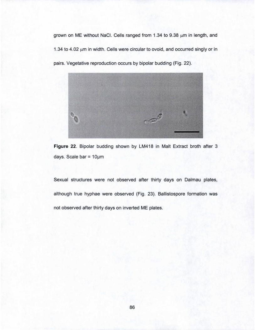
grown on ME without NaCI. Cells ranged from 1.34 to 9.38 ~m in length, and
1.34 to 4.02 ~m in width . Cells were circular to ovoid, and occurred singly or in
pairs. Vegetative reproduction occurs by bipolar budding (Fig. 22).
Figure 22. Bipolar budding shown by LM418 in Malt Extract broth after 3
days. Scale bar = 10jJm
Sexual structures were not observed after thirty days on Dalmau plates,
although true hyphae were observed (Fig. 23). Ballistospore formation was
not observed after thirty days on inverted ME plates.
86

Figure 23. True mycelium formation on Oalmau plates after seven days.
Scale bar = 10~m
A range of carbohydrates presented as single carbon sources was
assimilated by LM418 T (Tab. 4). Since glucose was not fermented , other
fermentation tests were deemed unnecessary. Neighbor-joining and
maximum parsimony analyses show that LM418 T comprises part of the
Microstromatales (Fig. 24) however the use of various alignment methods
indicated differing nearest neighbors. Consensus sequences generated in
PCRs with the NS7/LR6, 18S forward/reverse, and F63/R635 primer pairs
were submitted to GenBank and assigned the accession numbers 00990017.
00990018 and 00990016, respectively.
87
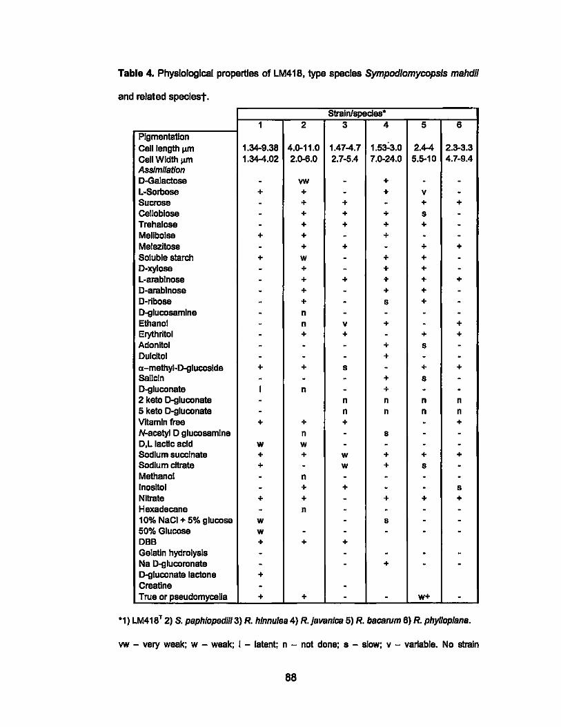
Table 4. Physiological properties of LM418, type species Sympodlomycopsls mahdll
and related speclest.
StraIn/species' 1 2 3 4 5 6
Pigmentation Cell length J.IIl1 1.34-9.38 4.0-11.0 1.47-4.7 1.5a:3.0 2.4-4 2.3-3.3 Cell Width J.IIl1 1.34-4.02 2.0-6.0 2.7-5.4 7.0-24.0 5.5-10 4.7-9.4 Assimilation D-Galactose - vw - + - -L-80rbose + + - + v -Sucrose - + + - + + Cellobiose - + + + s -Trehalose - + + + + -Mellbolse + + - + - -Melezitose - + + - + + Soluble starch + w - + + -O-xylose - + - + + -L-arablnose - + + + + + D-erablnose - + - + + -D-rlbose - + - s + -[).glucosamlne - n - - - -Ethanol - n v + - + Erythritol - + + - + + Adonitol - - - + s -Dulcitol - - - + - -a-methyl-[).glucoslde + + s - + + Sellcln - - - + s -D-gluconata I n - + - -2 keto [).gluconata - n n n n 5 keto D-gluconata - n n n n VItamin free + + + - + N-acetyl D glucosemlne n - s - -D,L lactic acid w w - - - -Sodium succinate + + w + + + Sodium citrate + - w + s -Methanol - n - - - -Inositol - + + - - s Nltrata + + - + + + Hexadecane - n - - - -10% NaCI + 5% glucose w - s - -50% Glucose w - - - - -DBB + + + Gelelln hydrolysis - - - - -Na [).glucoronata - - + - -D-gluconata lactone + Creatine - -True or pseudomyceUa + + - - w+ -
'1) LM418T 2) S. paphlopedll/3) R. hlnnulea 4) R.}avanlca 5) R. bacarum 6) R. phylloplana.
vw - very weak; W - weak; I - latent; n - not done; s - slow; v - variable. No strain
88

assimilates L-rhamnose, forms starch, ferments glucose, or grows at 37 ·C; only S.
paphlopedUI assimilates lactose and Inulin, and In both cases It Is weak. LM41ST does not
assimilate K O-saccharate or creatinine. All atralns assimilate raffinose, [).glucltol and urea;
all assimilate [).glucose, maltose and glycerol, but thasa are weak In LM41S T. All strains
assimilate O-mannltol, but It Is weak In R. bacarum.
89
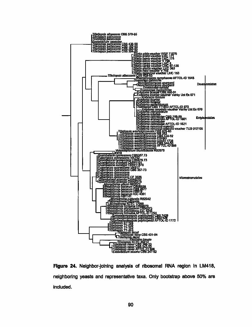
1Il/1!JZ-" AfTOL·ID 1645
Figure 24. Neighbor-joining analysis of ribosomal RNA region In LM418.
neighboring yeasts and representative taxa. Only bootstrap above 50% are
included.
90
Discussion
Characterization of yeast strain LM418T isolated from driftwood collected on
Shipwreck Beach, Lana'i showed it could not be assigned to an extant
species. Although molecular data place LM418 T firmly In the
Mlcrostromatales, morphological and physiological data support
establishment of the straln as a new species within the Sympod/omycops/s,
for which the name S. mahdif is proposed. S. mahdii LM418T appears to be
the anamorphic haploid yeast phase of an unknown telomorph. Different
alignment methods of the 01102 and ITS1 and 2 regions of the 18S and 288
rRNA genes show that LM418 T is part of the Microstromatales but is not
specifically associated with an exlstlng genus (Fig.24). Genus placament
could not be determined through molecular data alone, and creating a new
genus was rejected on the basis of the lack of definitive tests used to define
such terms, the lack of relevant data for species In neighboring genera, and
the fact that we have only a single strain.
Placing LM418T in the Sympodiomycopsis was governed by other factors,
specifically the results of assimilation and fermentation tests. This course of
action was determined by. the fact that DNA sequence analysis could not
consistently placa the strain in just one genus. For example, the nucleotide
91
sequence of the 01102 region shared 96.0% identity with that in Quambalaria
cyanescens (OQ317623), although taxa also related on the basis of this
region include Sympodlomycposls paphlopedlli (94.8%), Rhodotorula
bacarum (94.4%), Rhodotorula hinnulea, Rhodotorula javanlca, and
Rhodotorula phytloplana. The inclusion of Rhodotorula javanlca as a related
species was determined by keying the species based on assimilation results
(Kurtzman, & Fell, 2000).The nucleotide sequence of the ITS1 and ITS4
regions indicated that Sympodlomycposis paphiopedlli (OQ317631) Is the
nearest described strain in this respect, sharing 96.9% sequence identity,
while Mlcrostroma juglandls (OQ317632) is the nearest neighbor (95.6%
identity) on the basis of 188 DNA sequence comparison. Assimilation data for
LM418T distinguishes It from the Rhodotorula and Sympodlomycopsis,
although limited data on Sympodlomycopsls are available since only one
species in this genus has been described (Sugiyama et al., 1991).
Furthermore, insufficient physiological data have been published for
Quamba/aria spp. and Fugomyces cyanescans (Bagerow et aI, 2001; De Beer
at ai, 2006). It Is known, however. that Quambalaria spp. and Fugomyces
cyanescens both belong in the ascomycota, while LM418 T was determined to
belong in the basldiomycota by the Olazonlum Blue B test (Kurtzman & Fell.
92
2000).
Phylogenetic placement of LM41ST is also complicated by the fact that some
of the species with which It aligns are likely synonyms, e.g., Fugomyees spp.,
and the apparent relationship with Volvoe/spor/um Is probably due to long
branch attraction (Flg.24). This group comprises mostly plant-associated
filamentous basidiomycetes, with a handful of Rhodotorula and
Sympodiomyeopsis yeasts, and this is the most likely reason that
Quamba/ar/a and Volvoe/spor/um species do not occur in the genus.
Describing LM41ST as a new species in the group will validate the strain until
the teleomophic state is found, and preclude subsequent loss of an
anamorphic generic name should we opt to describe a new genus at this
point. Addition of a new anamorphic genus based on a single strain is also
unwarranted until the sexual state Is determined.
Rhodotorula species do not form pseudomycelia or true mycelia on Dalmau
plates, whereas this is a characteristic of Sympodiomyeopsis. In this respect,
LM41ST did form true mycelia after seven days on Dalmau plates, and also
displayed sympodial budding as seen In Sympod/omyeopsis paph/oped/I/.
LM41ST also exhibited a slight pink pigmentation when grown on ME, another
shared characteristic with Rhodotorula species, while the lack of pigmentation
93
in Sympodiomycops/s paphiopedi/i distinguishes it from LM418T (Sugiyama et
a/., 1991).
We thus propose that LM418Tls the type strain of a novel species, termed
Sympodlomycopsis mahdii based on both molecular and morphological data.
Although the nearest neighbors of S. mahdll on the basis of nucleotide
sequences are Quamba/aria cyanescens, Sympodlomycopsis paphiopedlli,
and Mlcrostroma jug/andis, LM418T is morphologically similar to
Sympodlomycopsis paphiopedill. Both LM418 T and Sympodiomycopsis
paphlopedlll assimilate glucose, maltose, raffinose, O-mannltol, O-glucitol, and
Na Succinate, and do not assimilate L-rhamnose, hexadecane, nor produce
starch. Neither ferments glucose. It should be reiterated that comparative data
for Q. cyancescens and M. juglandls are unavailable, while the OBB test ,
Indicated LM418T Is a basldlomycota. In addition to differences observed In
assimilation results, S. mahdli differs from S. paphiopedlll on the basis of 32
nucieotldes In the 01/02 regions of the 28S gene and the production of a pale
pink pigment, while both exhibit sympodiaJ budding In their formation of true
mycelia.
94
I. Statistical Analyses
The average number of CFU from water samples collected from the same
islands and Palmyra Atoll were compared with the non-parametric Kruskal-
Wallis test in Minitab (Minitab, 2004). This test was also used to compare the
CFU of fungi and yeast at different depths at Station ALOHA This non-
parametric test is ideal for samples which are not normally distributed, and
where sample sizes may be unequal. The calculation converts observations to
ranks, and uses the following formula:
n1(n1 +1) ~= n1n2+ -R1
2
Where n = number of observations, R = sum of ranks of observations in each
sample
95
CHAPTER IV. RESULTS
Between August 20, 2004 and July 17, 2006, 379 samples were collected
from coastal weters and other marine-associated materials around six of the
high Hawaiian Islands and Palmyra Atoll. Water samples (n = 169) only were
collected at standard depths between the surface and -5000 m during six
cruises to Station ALOHA. This is the most extensive study of marine
mycology ever conducted in the HawaIIan Islands, and the first of such at
Station ALOHA. Fungi and yeasts in all water samples were enumerated on
three differant enrichment media, and rapresentative colonies and hyphal
growth (based on colony characteristics) transferrad to the same enrichment
media for purification. Enumerating fungi and yeests in other samples wes a
sacondary consideration since the diversity of materials tested makes
comparisons difficult and likely unreliable, at least on the basis of CFU per
gram of material; the number of CFU arising from the different dlluents used
to wash and serially dilute sand cannot be compared with the number of CFU
arising from a piece of driftwood settled on the same media (Kohlmeyer,
1979). A significant product of this work is the first locally housed collection of
fungi and yeasts isolated from Hawaiian marine habitats. In addition to using
traditional cultivation methods to enumerate and bring these taxa into culture,
96
DNA-based molecular methods enabled the tentative identification of isolated
strains by relatively rapid sequencing of fragments of their ribosomal genes. A
second advantage of this combined 'traditional' and molecular approach is
that it minimizes redundancy In the collection, an Important consideration
given the effort required to maintain a large collection, and difficulties
microbiologists have historically had in differentiating taxonomically distant
microorganisms that may share similar physiological profiles, pigmentation,
and even colony size and shape.
By August 2006, 689 pure fungi and yeast cultures had been prepared from
all samples. The majority of these cultures were sequencad In terms of their
ITS1 and 2 or D11D2 regions. Indeed, of the 592 cultures In which these
sequences were determined, 272 (46%) share less than 97% nucleotide
Identity over one or both these regions with the nearest described species,
I.e., one for which a type strain has been published in the peer reviewed
literature. The Implication of these data Is that Significant phylogenetic novelty
exists In both the collection established here, and by definition In the HawaIIan
marine environment. Several non-fungal taxa were also cultivated, Including
five viridiplantae (algae), and two antibiotic resistant bacteria determined to be
Serratia marcesens and Pseudomonas put/da. The former Is commonly
97
cultivated from environmental samples. Ps. put/da has a more cosmopolitan
distribution, having been Implicated in nosocomial infections and showing
promisa In bloremediation through its ability to degrade organic solvents, and
even to convert styrofoam to biodegradable polyhydroxyalkanoate plastics
(Ward et a/., 2006). Two protozoans and one stramenoplle were also brought
into pure culture. These non-fungal taxa will not be discussad further here.
Taken as a whole, the phylogenetic affiliations of ali cultures In the collection
reflect that which would be expected for samples of marine origin. On the
basis of only BLASTn results, for which nearest neighbors of the test
sequence are listed, 398 Ascomycete cultures were defined, and these
comprise 68% the collection, the 175 Basidiomycetes are 30%, and the
remainder comprises just 10 (2%) Zygomycetes (Fig. 25).
98
30%
Figure 25. Phylogenetic affiliations of entire collection
o Ascor!¥celeS
m BasldlomyceleS
• ZygomyceleS
This ratio of major taxa Is consistent with previously published reports of such
work (Kohlmeyer 1979; Sieburth, 1979). In terms of each Island's
contributions to the collection, the majority of isolates sequenced to date were
isolated from O'ahu (141 pure cultures), of which 82"10 were Ascomycetes
(116), 16% Basidiomycetes (23), and 2% Zygomycetes (2) (Fig. 26). 59
isolated cultures came from Kaua'l, 73% Ascomycetes (43), 22%
Basidiomycetes (13), and 5% Zygomycetes (3) (Fig. 26). Moloka'i yielded 66
sequenced isolated cultures with 78% Ascomycetes (52), 17%
Basidiomycetes (22), and 5% Zygomycetes (3) (Fig. 26); Maul had 55
sequenced cultures which were 73% Ascomycetes (40). 25% Basidiomycetes
(14), and 2% Zygomycetes (1) (Fig. 26); Lana'i represented 16 of the entire
99
collection and was comprised of 63% Ascomycetes (10), and 37%
Basidiomycetes (Fig. 26); Hawai'i had 84 sequenced cultures, 74%
Ascomycetes (62), and 26% Basidiomycetes (22) (Fig. 26).
100
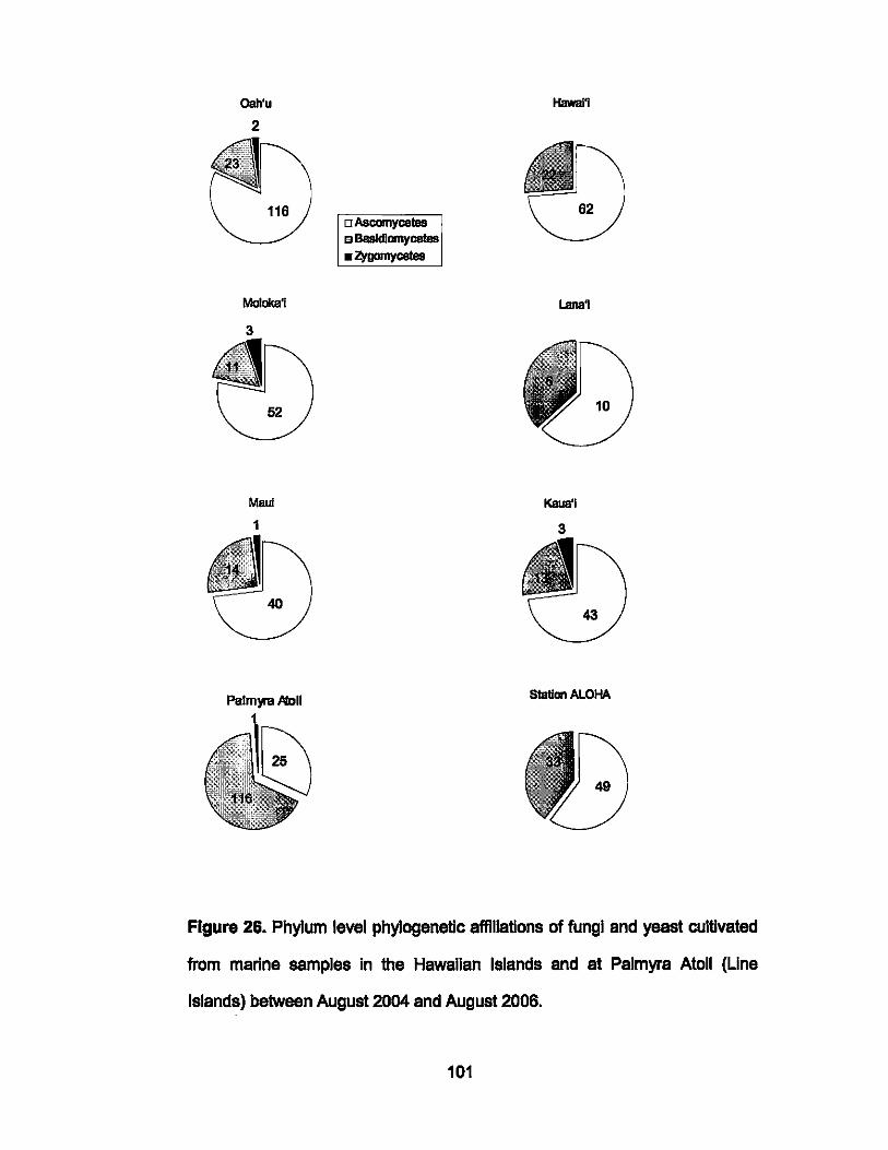
Qah'u
2
MoIoka1
3
Maul
1
Palmyra ,Il1011
o Ascomycetes " Basidiomycetes • Z¥gomycetes
Hawal1
Lana1
Kaua'i
3
10
SIatIan ALOHA
Figure 26. Phylum level phylogenetic affiliations of fungi and yeast cultivated
from marine samples In the HawaIIan Islands and at Palmyra Atoll (Line
Islands) betwaen August 2004 and August 2006.
101
The phylogenetic composition of the mycoflora cultivated from Palmyra Atoll
differs markedly from that of the six high HawaIIan Islands. For example, of
the 79 cultures from Palmyra sequenced to date, just 32% were Ascomycetes
(25), 67% Basidiomycetes (53), and 1% Zygomycetes (1) (Fig. 26) No
Zygomycetes were cultivated from Station ALOHA, but 81 other sequenced
isolates comprised 60% Ascomycetes (49) and 40% Basidiomycetes (Fig.
26).
Zygomycetes were cultivated from all sites except Lana'l, Hawal'l and Station
ALOHA. The latter In particular Is consistent with the fact that currently
described Zygomycetes are primarily of terrestrial origin. One would not
expect predominantly, if not exclusively terrestrial taxa to be represented at an
open-ocean location such as ALOHA and more so below the mixed layer at
this location. That Zygomycetes were actually present in any sample is of
interest given that this was a marine-centric study, but the fact Is that with the
exception of collections at Station ALOHA, all samples here were taken from
coastal sites. Their relatively poor representation Is thus consistent with
reports that Zygomycetes in marine samples are rare (Kohlmeyer 1979;
Sieburth, 1979; Kurtzman & Fell, 2000; Maddison, 2006). Only ten
102
Zygomycetes were cultivated overall, and two of these share a -95% (which
gene) sequence Identity wlth their nearest described neighbor In the public
domain. These collections and Isolations represent the first of marine
Zygomycetes around Hawal'l and Palmyra Atoll, and have the potential basis
to establish marine Zygomycetes as representative class of marine fungI. The
data described here for fungi and yeast abundance and phylogeny at Palmyra
Atoll are significant because the marine mycoflora here has never been
examined. Indeed, studies of fungi and yeasts In any isolated atoll lagoon are
noticeably absent from the IHereture.
The diversity of species cultivated at Palmyra varies from those found In the
Hawaiian Islands and at Station ALOHA. wlth the majority belonging In the
Basidiomycetes (Fig. 26). According to BLAST results, most species were
recorded In semples taken around O'ahu, while only 4 distinct species were
collected around Lana'i.
The greatest number of distinct species, described here as a function of the
different taxa listed In the BLASTn outputs, was derived from water semples,
while animal sources provided the lowest number of taxa (Tab. 5).
103
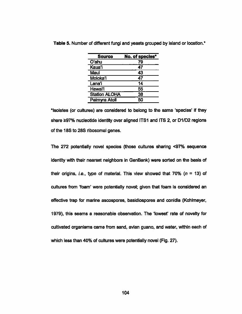
Table 5. Number of different fungi and yeasts grouped by Island or location.'
Source No. of specles* O'ahu 79 Kaua'i 47 Maui 43 Moloks'i 47 Lana'i 14 Hawai'i 55 Station ALOHA 38 Palmyra Atoll 50
*Isolates (or cultures) are considered to belong to the same 'species' If they
share l!;97% nucleotide identity over aligned ITS1 and ITS 2, or 01/02 regions
of the 18S to 28S ribosomal genes.
The 272 potentially novel species (those cultures sharing <97% sequence
identity with their nearest neighbors in GenBank) were sorted on the basis of
their origins, i.e., type of material. This view showed that 70% (n = 13) of
cultures from 'foam' were potentially novel; given that foam Is considered an
effective trap for marine ascospores, besidlospores and conidia (Kohlmeyer,
1979), this seems a reasonable observation. The 'lowest' rate of novelty for
cultivated organisms ceme from sand, avian guano, and water, within each of
which less than 40% of cultures were potentially novel (Fig. 27).
104
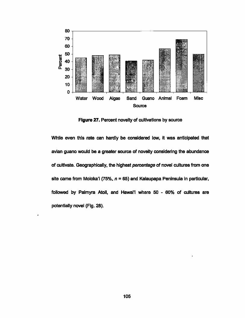
oo,-------------------------------------~
70
60
cOO ~40 0..30
20
10
o Water Wood Algae Sand Guano Animal Foam Mise
Source
Figure 27. Percent novelty of cultivations by source
While even this rate can hardly be considered low, It was anticipated that
avian guano would be a greater source of novelty considering the abundance
of cultivate. Geographically, the highest percentage of novel cultures from one
site came from Moloka'i (75%, n = 65) and Kalaupapa Peninsula In particular,
followed by Palmyra Atoll, and Hawal'l where 50 - 60% of cultures are
potentially novel (Fig. 28).
105
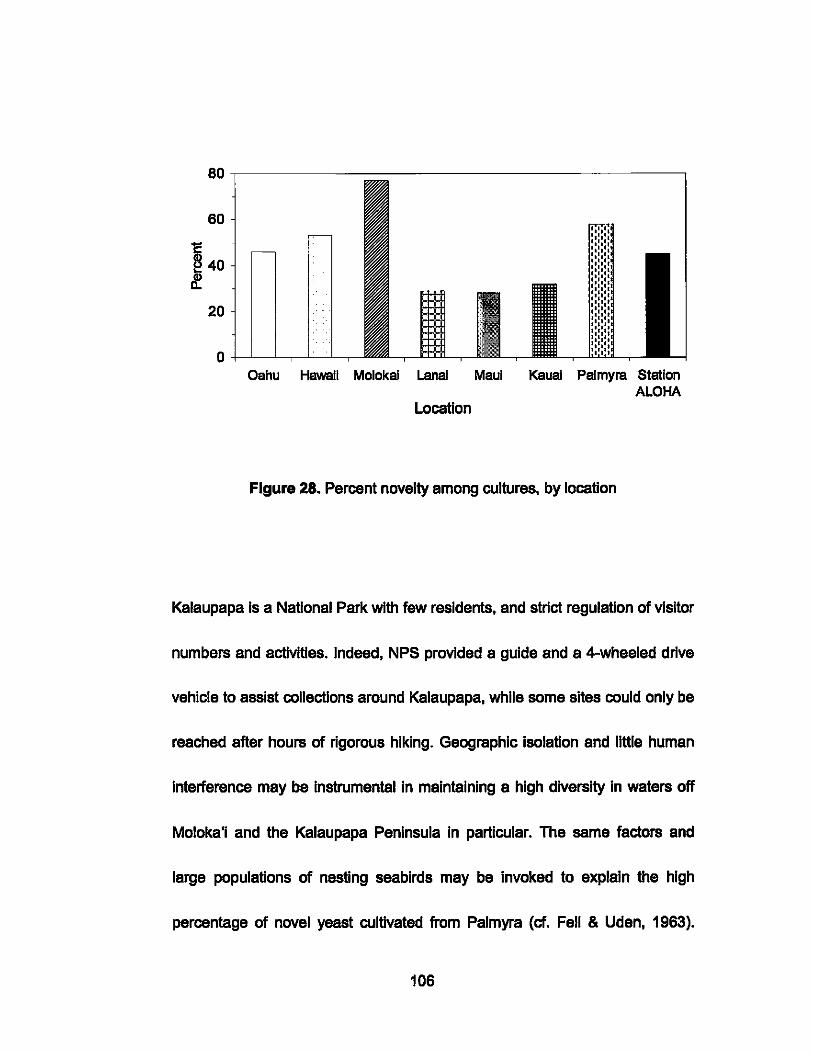
80,----------------------------------------,
60
c ~ 40 If
20
Oahu Hawaii Molokal lanai Maul Kaual Palmyra Station ALOHA
Location
Figure 28. Percent novelty among cultures, by location
Kalaupapa is a National Park wlth few residents, and strict regulation of visitor
numbers and activities. Indeed, NPS provided a guide and a 4-wheeled drive
vehicle to assist collections around Kalaupapa, while some sites could only be
reached after hours of rigorous hiking. Geographic Isolation and little human
Interference may be Instrumental In maintaining a high diversity In waters off
Moloka'i and the Kalaupapa Peninsula In particular. The same factors and
large populations of nesting seabirds may be Invoked to explain the high
percentage of novel yeast cultivated from Palmyra (cf. Fell & Uden, 1963).
106
Hawai'i is also rugged, and a 4-wheel drive vehicle and considerable hiking
were required to access the shoreline. By the same token. Palmyra Atoll is an
isolated small island surrounded by deep ocean; most samples were easily
collected along the shore or by accessing the lagoons by boat. Almost one
third (88) of potentially novel species from all habitats were Basidiomycetes. a
Significant finding given that only -24 obligate marine Basidiomycetes species
have been described (Kohlmeyer 1979; Sieburth. 1979; Kurtzman & Fell.
2000; Maddison. 2006). A reevaluation of this opinion in light of the data
presanted here could have significant repercussions for how Basidiomycetes
are viewed in terms of their role In marine nutrient cycling.
A total of 581 represantative ITS1 and ITS2. and 01/02 nucleotide saquences
submitted to GenBank at the NCBI have been assigned accession numbers
EF060395 to EF060975.
The number of CFU recorded per liter of seawater from each site ranged from
55 to 356; the mean CFU for the Hawaiian Islands and Palmyra was 2251L
(Fig. 29). CFU in the upper 500 m at Station ALOHA numbered 22IL. and 261L
below 500 m (Fig. 30).
107
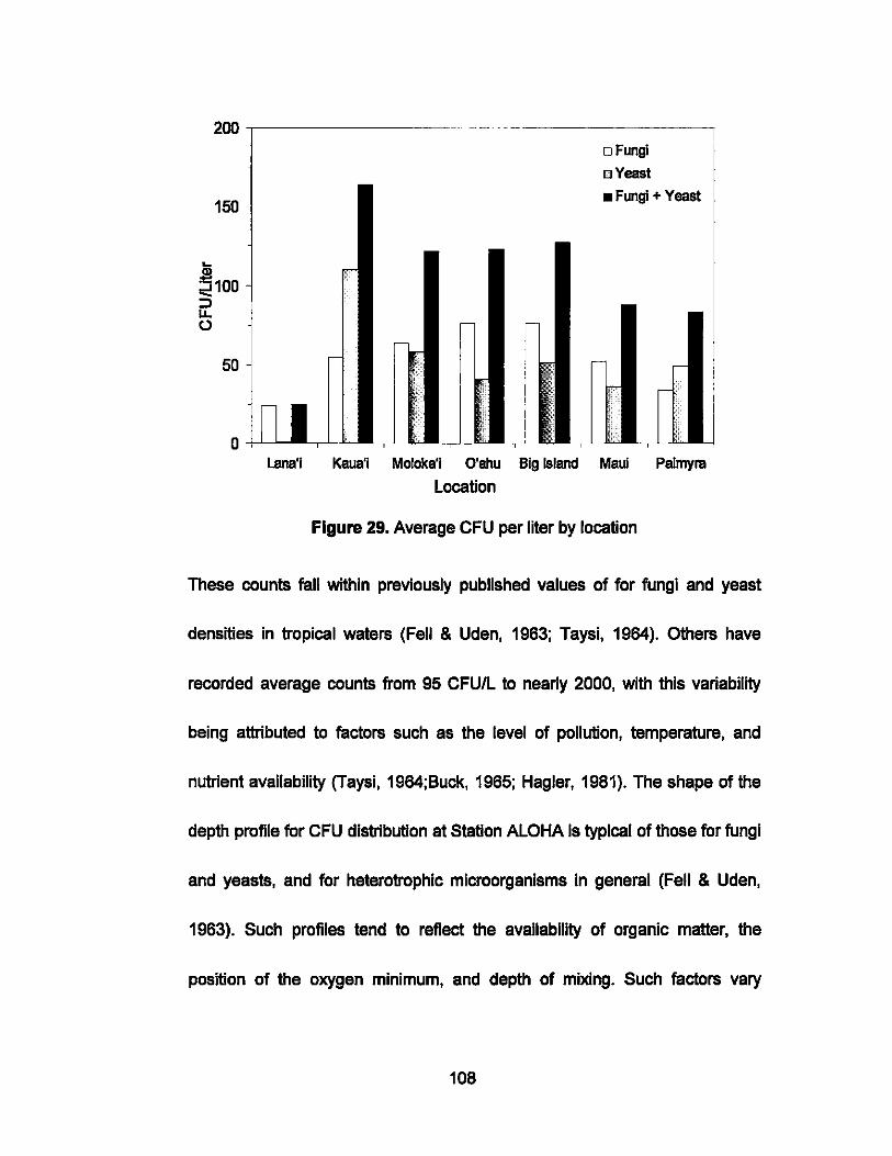
.... CD
200
150
~100 ::l u. o
50
o
--~- --
o Fungi
o Yeast
• Fungi + Yeast
if
,- -
r-- ,- -
-
n lana'j Kaua'j Moloka'j O'ahu Big Island Maui Palmyra
Location
Figure 29. Average CFU per liter by location
These counts fall within previously published values of for fungi and yeast
densities in tropical waters (Fell & Uden, 1963; Taysi, 1964). others have
recorded average counts from 95 CFUIL to nearly 2000, with this variability
being attributed to factors such as the level of pollution, temperature, and
nutrient availability (Taysi, 1964;Buck, 1965; Hagler, 1981). The shape of the
depth profile for CFU distribution at Station ALOHA Is typical of those for fungi
and yeasts, and for heterotrophic microorganisms in general (Fell & Uden,
1963). Such profiles tend to reflect the availability of organic matter, the
position of the oxygen minimum, and depth of mixing. Such factors vary
108
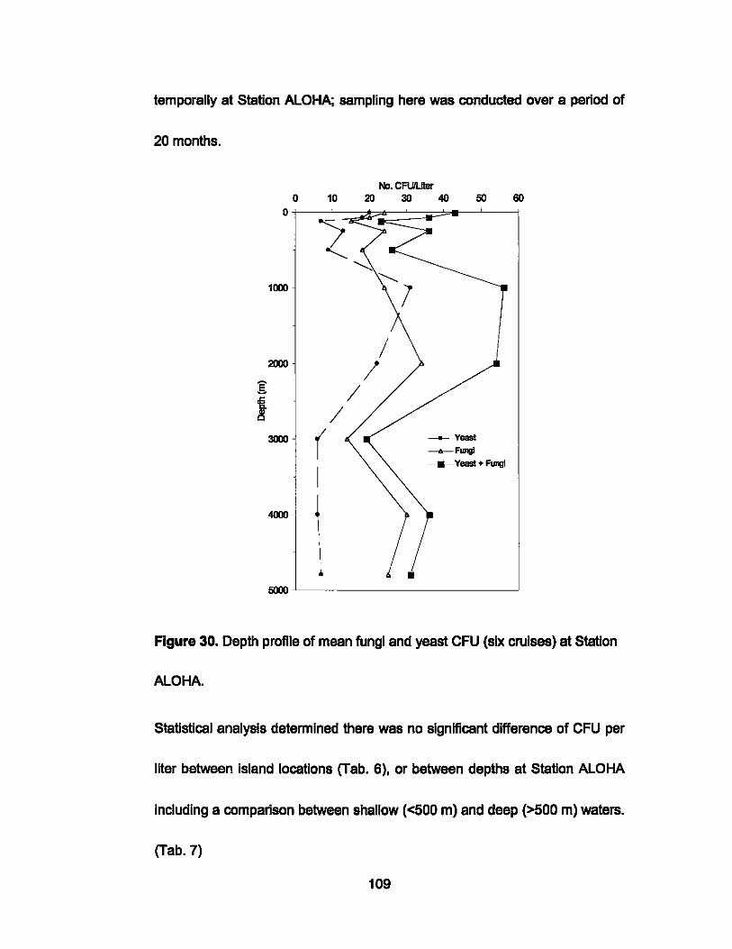
temporally at Station ALOHA; sampling here was conducted over a period of
20 months.
No. CFU'LJter 0 10 20 30 40 50 60
0
< ~ "'-1000 1
/ 2000 I g /
I / 3000 ( ___ yeast
-6-Fungi
I ______ Yeast + Fungi
4000 ~ \ ..
5000
Figure 30. Depth profile of mean fungi and yeast CFU (six cruises) at Station
ALOHA.
Statistical analysis determined there was no significant differenca of CFU per
liter between island locations (Tab. 6). or between depths at Station ALOHA
including a comparison between shallow «500 m) and deep (>500 m) waters.
(Tab. 7)
109
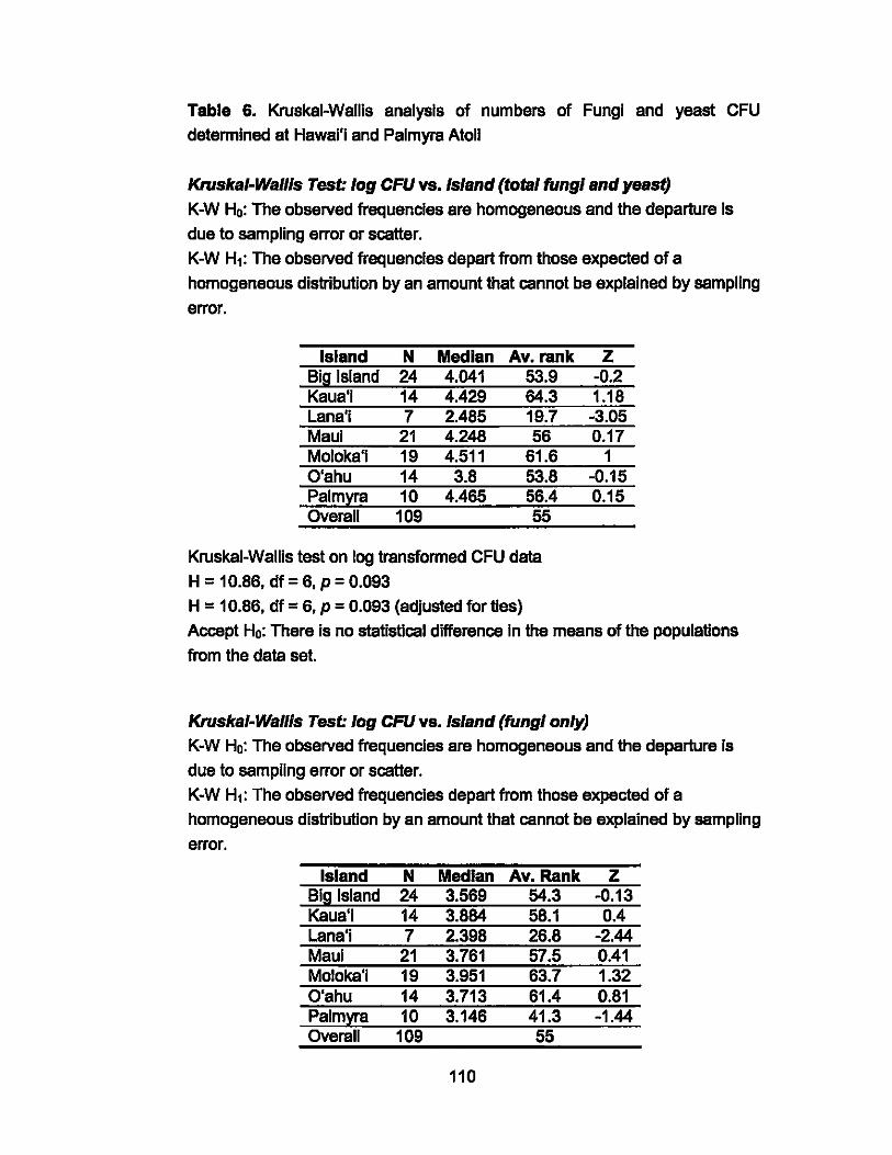
Table 6. Kruskal-Wallls analysis of numbers of Fungi and yeast CFU
determined at Hawal'l and Palmyra Atoll
Kruskal-Wailis Test: log CFU vs. Island (total fungi and yeast)
K-W Ho: The observed frequencies are homogeneous and the departure Is
due to sampling error or scatter.
K-W H1: The observed frequencies depart from those expected of a
homogeneous distribution by an amount that cannot be explained by sampling
error.
Island N Median Av. rank Big Island 24 4.041 53.9 Kaua'i 14 4.429 64.3 Lana'i 7 2.485 19.7 Maul 21 4.248 56 Moloka'i 19 4.511 61.6 O'ahu 14 3.8 53.8 Palmyra 10 4.465 56.4 Overell 109 55
Kruskal-Wallis test on log transformed CFU data
H = 10.86, df = 6, P = 0.093 H = 10.86, df = 6, P = 0.093 (adjusted for ties)
z -0.2 1.18 -3.05 0.17
1 -0.15 0.15
Accept Ho: There Is no statistical differenca In the means of the populations
from the data set.
Kruska/-Wailis Test: log CFU vs. Island (fungi only)
K-W Ho: The observed frequencies are homogeneous and the departure Is
due to sampling error or scatter.
K-W Hl: The observed frequencIes depart from those expected of a
homogeneous distribution by an amount that cannot be explained by sampling
error.
Island N Median Av. Rank Z Big Island 24 3.569 54.3 -0.13 Kaua'i 14 3.864 58.1 0.4 Lana'i 7 2.398 26.8 -2.44 Maul 21 3.761 57.5 0.41 Moloka'i 19 3.951 63.7 1.32 O'ahu 14 3.713 61.4 0.81 Palmyra 10 3.146 41.3 -1.44 Overell 109 55
110

Kruskal-Wallls test on log transformed CFU data H = 9.76, df= 6, P = 0.135 H = 9.76, df = 6, P = 0.135 (adjusted for ties) Accept Ho: There Is no statistical dlfferenca In the means of the populations from the data set.
Kruska/·Wallls Test log CFU vs. Island (yeast only) K-W Ho: The observed frequencies are homogeneous and the departure is merely due to sampling error or scatter. K-W H1: The observed frequencies depart from those expected of a homogeneous distribution by an amount that cannot be explained by sampling error. 104 cases were used 5 cases contained miSSing values
Island N Median Av. Rank Big Island 23 2.773 54.3 Kaua'i 14 2.850 59.9 Lana'i 5 0.000 11.4 Maul 20 2.835 55.5 Moloks'l 19 2.944 54.6 O'ahu 13 1.792 42.9 Palmyra 10 3.598 61.3 Overall 104 52.5
Kruskal-Wallls Test on log transformed CFU data H = 12.65, df = 6, P = 0.049 H = 12.67, df = 6, P = 0.048 (adjusted forties)
z 0.32 0.99 -3.12 0.49 0.33 -1.23 0.96
Accept Ho: There Is no statistical dlfferenca In the means of the populations from the data set
Table 7. Krusksl-Wallis analysis of numbers of Fungi and yeast CFU determined at different depths at Station ALOHA.
Kruska/·Wallis Test log10 (CFU) vs. depth (m) (total fungi and yeast) K-W Ho: The observed frequencies are homogeneous and the departure Is merely due to sampling error or scatter. K-W H1: the observed frequencies depart from those expected of a homogeneous distribution by an amount that cannot be explained by sampling error. 149 cases were used
111
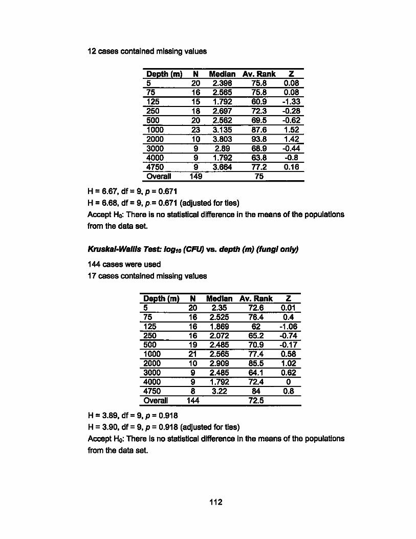
12 cases contained missing values
Depth (m) N Median Av.Rank z 5 20 2.398 75.8 0.08 75 16 2.565 75.8 0.08 125 15 1.792 60.9 -1.33 250 18 2.697 72.3 -0.28 500 20 2.562 69.5 -0.62 1000 23 3.135 87.6 1.52 2000 10 3.803 93.8 1.42 3000 9 2.89 68.9 -0.44 4000 9 1.792 63.8 -0.8 4750 9 3.664 n.2 0.16 Overall 149 75
H = 6.67, df = 9, P = 0.671
H = 6.68, df = 9, p.= 0.671 (adjusted for ties)
Accept Ho: There Is no statistical dlfferanca In the means of the populations
from the data set.
Kruska/·Wallls Test: 10910 (CFU) VB. depth (m) (fungi only)
144 cases were used
17 cases contained miSSing values
Depth (m) N Median Av. Rank z 5 20 2.35 72.6 0.01 75 16 2.525 76.4 0.4 125 16 1.869 62 -1.06 250 16 2.072 65.2 -0.74 500 19 2.485 70.9 -0.17 1000 21 2.565 n.4 0.58 2000 10 2.909 85.5 1.02 3000 9 2.485 64.1 0.62 4000 9 1.792 72.4 o 4750 8 3.22 84 0.8 Overall 144 72.5
H = 3.89, df = 9, P = 0.918
H = 3.90, df = 9, P = 0.918 (adjusted for ties)
Accept Ho: There Is no statistical dlfferenca In the means of the populations
from the data set.
112
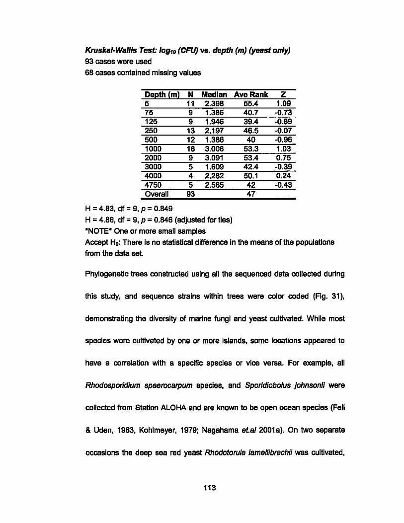
Kruska/-Wallls Test: 10910 (CFU) vs. depth (m) (yeast only)
93 cases were used
68 cases contained missing values
Depth (m) N Median Ave Rank z 5 11 2.398 55.4 1.09 75 9 1.386 40.7 -0.73 125 9 1.946 39.4 -0.89 250 13 2,197 46.5 -0.07 500 12 1.386 40 -0.96 1000 16 3.006 53.3 1.03 2000 9 3.091 53.4 0.75 3000 5 1.609 42.4 -0.39 4000 4 2.282 50.1 0.24 4750 5 2.565 42 -0.43 Overall 93 47
H = 4.83, dt = 9, P = 0.849
H = 4.86, dt = 9, P = 0.846 (adjusted for ties)
·NOTE· One or more small samples
Accept Ho: There is no statistical differenca in the means of the populations
from the data set.
Phylogenetic trees constructed using all the saquenced data collected during
this study, and sequence strains within trees were color coded (Fig. 31),
demonstrating the diversity of marine fungi and yeast cultivated. While most
species were cultivated by one or more islands, some locations appeared to
have a correlation with a specific species or vice versa. For example, all
Rhodosporidium spaerocarpum species, and Sporidiobofus johnsonll were
collected from Station ALOHA and are known to be open ocean species (Fell
& Uden, 1963, Kohlmeyer, 1979; Nagahama et.af 2001 a). On two saparate
occasions the deep sea red yeast Rhodotorufa fameffibrachil was cultivated,
113
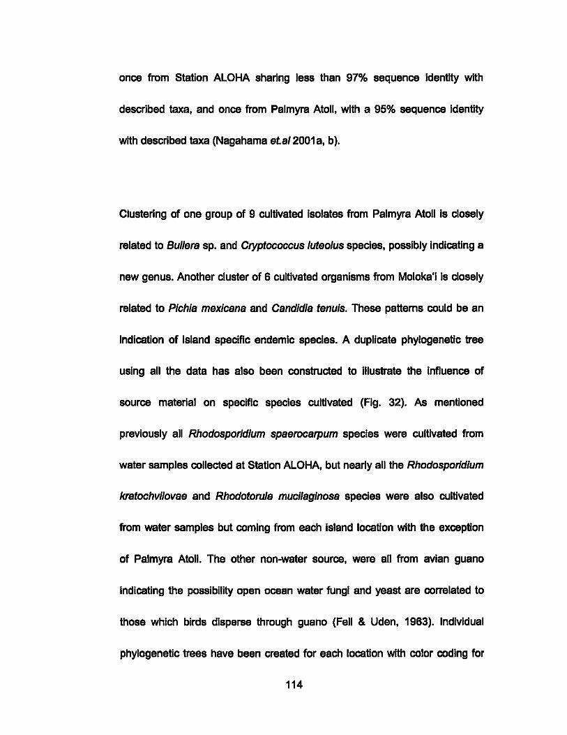
once from Station ALOHA sharing less than 97% sequence Identity with
described taxa, and once from Palmyra Atoll, with a 95% sequence Identity
with described taxa (Nagahama et.sl 2001 a, b).
Clustering of one group of 9 cultivated Isolates from Palmyra Atoll Is closely
related to Bul/era sp. and Cryptococcus luteolus species, possibly Indlceting a
new genus. Another cluster of 6 cultivated organisms from Moloka'i Is closely
related to Pichia mexlcans and Csndidla tenuls. These patterns could be an
Indication of island specific endemic species. A duplicate phylogenetic tree
using all the data has also been constructed to illustrate the Influence of
source material on specific species cultivated (Fig. 32). As mentioned
previously all Rhodosporldlum spaerocarpum species were cultivated from
water semples collected at Station ALOHA, but nearly all the Rhodosporldlum
kratochvilovae and Rhodotorula mucllaglnosa species were also cultivated
from water samples but coming from each island location with the exception
of Palmyra Atoll. The other non-water source, were all from avian guano
Indicating the possibility open ocean water fungi and yeast are correlated to
those which birds disperse through guano (Fell & Uden, 1963). Individual
phylogenetic trees have been created for each location with color coding for
114
source material (Figs. 33-40). Among IndMdual locations. there are no large
groupings based on source material of any particular species indicating that
most species are ubiquitous in the marine environment (Fell & Uden. 1963.
Fell 1967. Kohlmeyer. 1979).
115
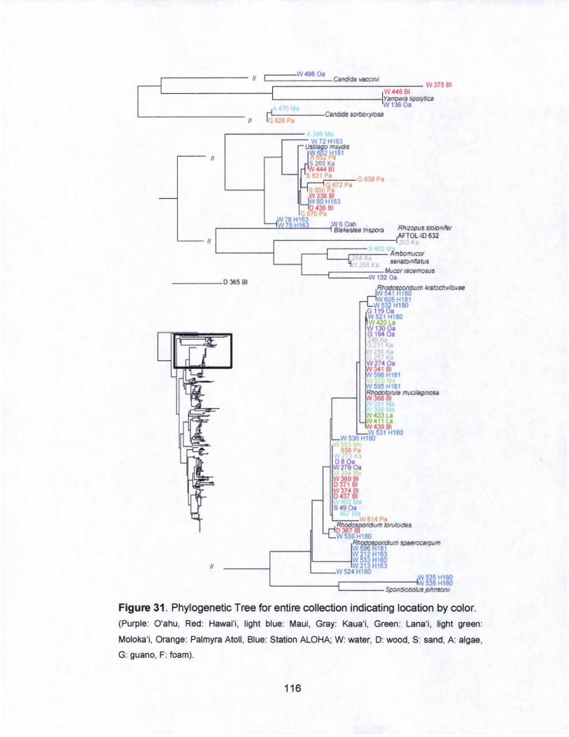
[================-~~=c=====W=4~~~00~==========~~~~ [
/I Cafldida vlICCm/f W37581
I W44<i 81 I y~",,",. 'po/yI<ca W l3600
r--- II
II
/I .-4 Candela sOltx)'(ylOS8 1- 2 Pa
i-----;:==- W72 HI63 USIl'!i!1 ma,os '!t II!it Iii 81
269Ka 44481
S:.' _"pa,,==-G 638 Pe
S G672P.
ItiO Pa W338 81
80 HI63 438 81
670P, W76H163
W60"" .------= =""'''----1 81akes.~ mspora RhJzopus s:O/ollJMr .-___________ IIAFTOL,IO 632
r--{--;:==--- Ambomucor SfJn8.'omf!atus
_ ____ 036581 L __ -C=== :W1iMtlCOr mcemosus
W 132 o.
II
Rf;ooos~:um /(ratocllvl/o'J8B W541 HI80 W605 H181
W532 Hl80 Gl1900 W521 HI80 W42081 W l 30 G 194
W274 00 34181 598 H181
rV~9'HI81 RIlOdOlOtu/a muc;:tsgmoss
36881
tH23La !N411l.
43981 W 531 Hl80
W530 Hl80
658 P,
080. W2790.
W36981 037181 W314BI 043781
S4900 '-,;,=~W61" Pa
Rhodosoondtum ~C1es 036781 W539Hl80
RlfOdOSDOndIum SDBerocarpum W596 R181 W 212 H163
IL-----1W 533 Hl80 W 213 H163
W52' Hl80 W535Hl80
L ______ ~========S;~~~.538Hl80 $poOOtOtJoIus p hn!lOftN
Figure 31 , Phylogenetic Tree for entire collection indicating location by coloe
(Purple: O'ahu, Red: Hawai'i, light blue: Maui, Gray: Kaua'i, Green: Lana'i, light green:
Moloka'i, Orange: Palmyra Atoll , Blue: Station ALOHA; W: water, D: wood, S: sand, A: algae,
G: guano, F: foam) .
116
1020.
Tnchosporon montel"ldeenS8 L - i-t 170.
Figure 31 . (Continued) Phylogenetic Tree for entire collection indicating location by color. (Purple: O'ahu, Red: Hawai'i, light blue: Maui, Gray: Kaua'i, Green:
Lana'i, light green: Moloka'i, Orange: Palmyra Atoll , Blue: Station ALOHA; W: water, D: wood,
S: sand, A algae, G: guano, F: foam)
117
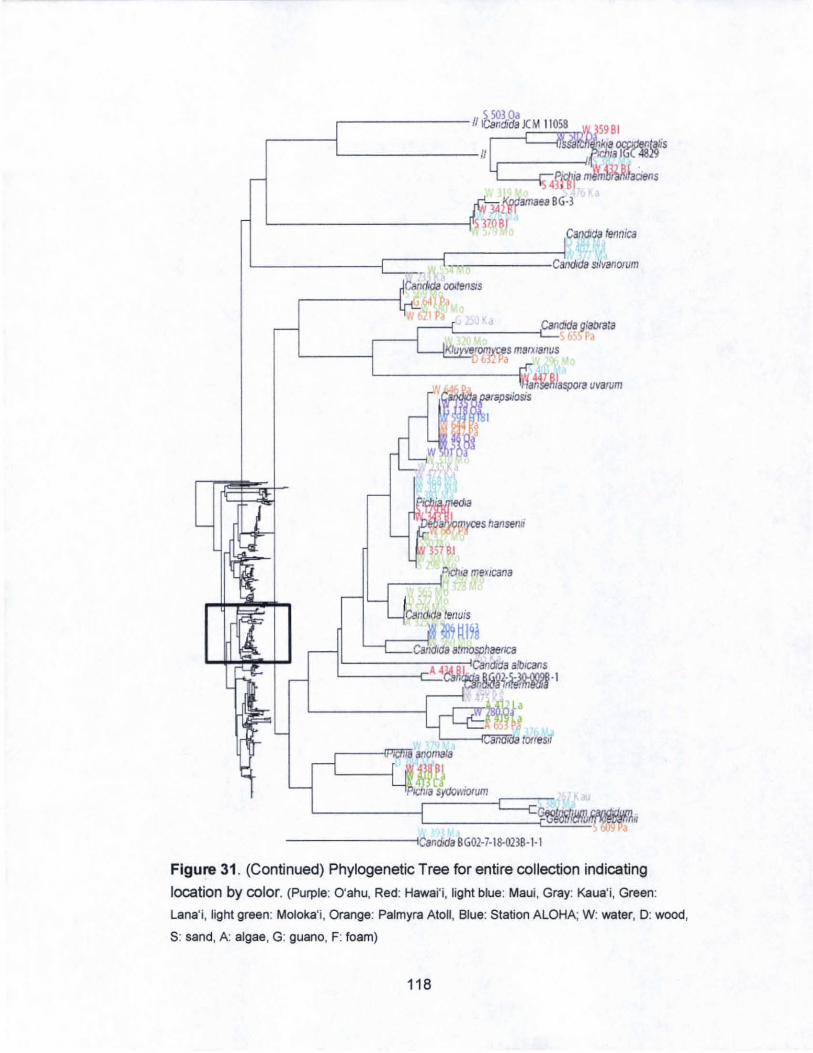
L--- -----1/
Y )A fgfiamaea BG-l
'------------;~ HOB! Candldfl fennica
L ______ --r----r:::======c;iCandida silvanOl1lm -CandIda OOItenSis
r------1 41 p , -L-____ -t::Ca=ndJda g:abtala
.---1
Candida alfflOSIlhaenca 44MR~~~daa~~s
'-------;:::..::cann'h!~~~-I
<--" ",.t:
~~. --------tICandKla BG01-J- I8-021B-I-I
Figure 31 _ (Continued) Phylogenetic Tree for entire collection indicating
location by color. (Purple: O'ahu, Red: Hawai'i, light blue: Maui, Gray: Kaua'i, Green:
Lana'i, light green: Moloka'i, Orange: Palmyra Atoll, Blue: Station ALOHA; W: water, D: wood,
S: sand, A: algae, G: guano, F: foam)
118
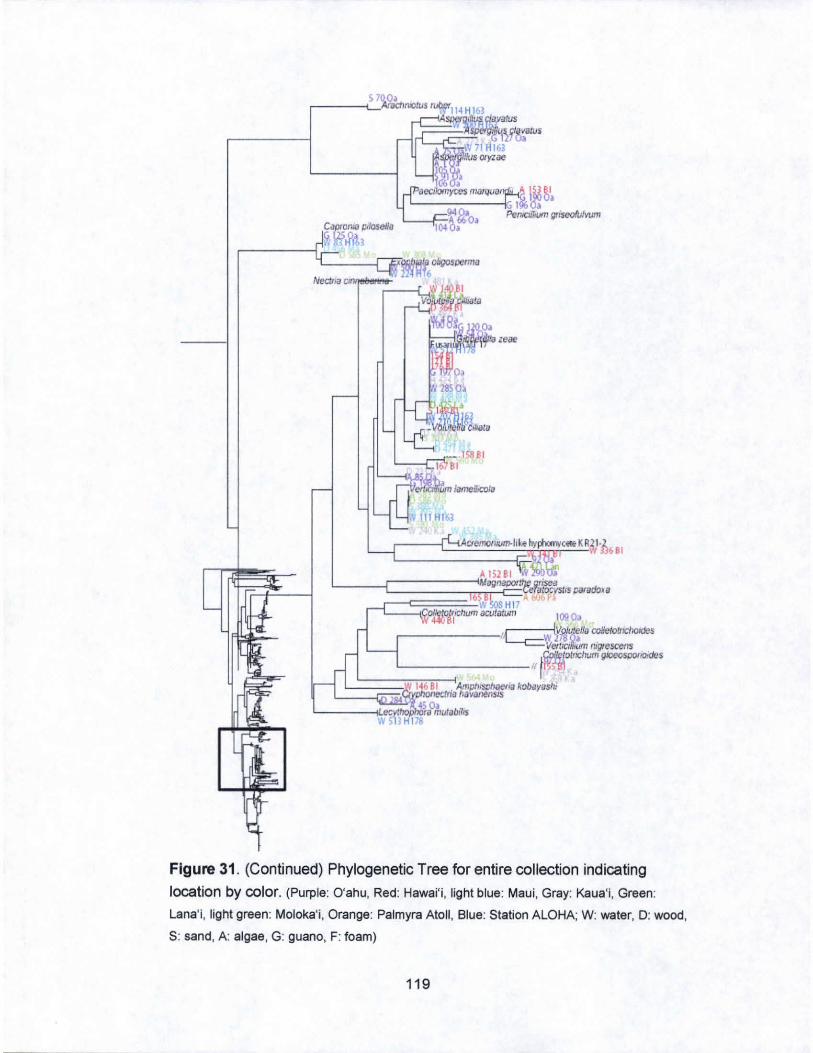
• • e<J~'yc';, m.lI'Q<IO" I ~ ~1 '-------1:; 1133'
L_--F=94 Oa PenICIllium gllseolufVIJm A 660. 1040.
L_-f~_,1S881 16 81
~J1~ lameS""".
A 15281 ,,' MlY,loolX>,"'orisea 'cera:ocystls parodo~iI
b ~ '" ~l W HI 7
~°Kt/cllumaQJ!il!Um \090" .
,--------r,c:c=W rt#~lIa coUclo~lCIlOJdos Verf,~,~JUm ntgIt!scens
}Wk!otncJrum Qk>cospooOiclcS '--------- 11 1 ~;'S1
j ~~::ij~~I~~66~BB~II ;;;; " Amph'SDlu!eriahobayas!U L pIK>neClfii) haVC1nM5JS
'-- ---iLecyI' 45 0,tulabillS I'IIH HI 78
Figure 31 . (Continued) Phylogenetic Tree for entire collection indicating
location by color. (Purple: O'ahu, Red: Hawai'i, light blue: Maui, Gray: Kaua'i, Green:
Lana'i, light green: Moloka'i, Orange: Palmyra Atoll , Blue: Station ALOHA; W: water, D: wood ,
S: sand, A: algae , G: guano, F: foam)
119
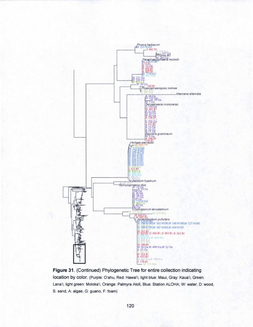
W).48Bt 56 O~ W 499 Oa.W 52 oa 210.
"mIl 117881
Figure 31. (Continued) Phylogenetic Tree for entire collection indicating
location by color. (Purple: O'ahu, Red: Hawai' i, light blue: Maui, Gray: Kaua'i, Green:
Lana';, light green: Moloka'i, Orange: Palmyra Atoll , Blue: Station ALOHA; W: water, 0 : wood,
S: sand, A: algae, G: guano, F: foam)
120

11 ~ .. 70MI G 826 P.
r------;:==!." 38a lUI nH163
~-- II u~i;;r.s
1/
>r 44481
_--,PC!'~-=-G 638 P. ~ 672 P.
w"3fe B1 SOHI63
GS90'fo 81
W78HI63 Weooh . , ____ ---.:~:IIIW117i5 l!Hll!i6"_3 --+1 Bt,/l.e5fea tnspora RhizopIJ$ stoIonrfer
f--:=:r=:;;';;~~i,AFTOL.'0 632 263 KII
L-----l
-----036581 L_---1===::;"',Mucor racemosus W 1320a
1/
Figure 32. Phylogenetic Tree for entire collection indicating source origin by
color. (Blue: water, Green: algae, Orange: sand, Red: wood, Purple: guano, light
blue: foam; O'ahu : Oa, Kaua'i: Ka, Maui: Ma, Moloka'i: Mo, Hawai'i:BI , Lana'i: La,
Station ALOHA: H#, Palmyra Atoll :Pa)
121
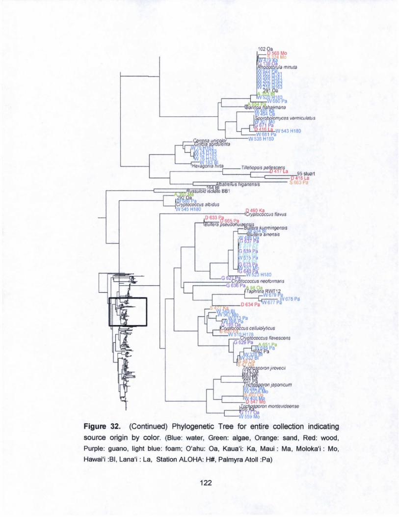
vel1mwlotus
Tit:etIOPSJS
I----n;;:;:;-o.---it:~ijjfo(:licc."' II.vos
~--r---~~~~~~1618P.
Figure 32. (Continued) Phylogenetic Tree for entire collection indicating
source origin by color. (Blue: water, Green: algae, Orange: sand, Red: wood,
Purple: guano, light blue: foam; O'ahu: Oa, Kaua'i: Ka, Maui : Ma, Moloka'i : Mo,
Hawai'i :BI , Lana'i : La, Station ALOHA: H#, Palmyra Atoll :Pa)
122
~ '0' r-------II~andida)( M 11 058 \ lS9BI
.------1 II ~~WiS
lIn 0 4gPftl11a ~ , adens I amaea [.3
~----_____ ~I ' ,
~ )9 0 5Ji~ lennica
L-------[=j~~:o:====Ca~~1!l SlIVanOlum OOIteflSls
.v ~W~o DO Ka 1--::!:;;;:::'-'_-E.Car.~,dida g:alxata
r------i IV 110 Mo ~-----1 ~" 8'C'Masma~i~~o
~ )f.~~):~ra uvarum ,~rapSliOsiS
~~
~7Kau
G~.:&mffi~ 39 y~ ,
-------II~ :';'rl.Kia BGOl-7-I8-0llB-I-1
Figure 32" (Continued) Phylogenetic Tree for entire collection indicating
source origin by color. (Blue: water, Green: algae, Orange: sand , Red: wood,
Purple: guano, light blue: foam; O'ahu : Oa, Kaua"i : Ka, Maui : Ma, Moloka'i : Mo,
Hawai'i :BI , Lana'i : La, Station ALOHA : H#, Palmyra Atoll :Pa)
123
Figure 32. (Continued) Phylogenetic Tree for entire collection indicating
source origin by color. (Blue: water. Green: algae, Orange: sand, Red: wood,
Purple: guano, light blue: foam; O'ahu : Oa, Kaua'i : Ka, Maui : Ma, Moloka'i : Mo,
Hawai'i :BI. Lana'i : La. Station AlOHA : H#, Palmyra Atoll :Pa)
124
r--......:......,,..,;.~----IIII.,n.f1. alrtNnBra
,------I?l !iifi;.!P<"'lum fenurSSrmum
C~~~=: 570 \'.0
542 H 180W 540 Hl80W 527 Hl80 587H1811V604H181
Figure 32. (Continued) Phylogenetic Tree for entire collection indicating
source origin by color. (Blue: water, Green: algae. Orange: sand, Red: wood ,
Purple: guano, light blue: foam; O'ahu : Oa, Kaua'i : Ka, Maui : Ma, Moloka'i : Mo,
Hawai'i :BI, Lana'i : La, Station ALOHA : H#, Palmyra Atoll :Pa)
125

'iC la
r ____ c::=====H~'Ca::!ld~id~a JC~'J 11058 W5020ah W 280 Oa~ Issa!cilenkia ocCJdenlahs
~------C=i~:'fr'da torresii JCM 1845 :N 53 nah ~ ... canai'Ja parapsr~
¥5010a, Gl190ah Candida a/mosp/laenca JCM 9549 '~ 192 8~"
"274 ah ofUla ml&/Jagll1O"
. ~AMH3S43
r£;;;W~.,tiS cilndJdtJ>
Figure 33. Phylogenetic Tree for O'ahu (Blue: water, Green: algae, Orange:
sand, Red: wood, Purple: guano, light blue: foam)
126
_==~======H~Us~~~~~S ::fi( If T/rilllSi'oclMriumKK17 J :: Ii CadixiIy,l .. n ~naospailm
Figure 33. (Continued) Phylogenetic Tree for O'ahu (Blue: water, Green: algae,
Orange: sand , Red : wood, Purple: guano, light blue: foam)
127
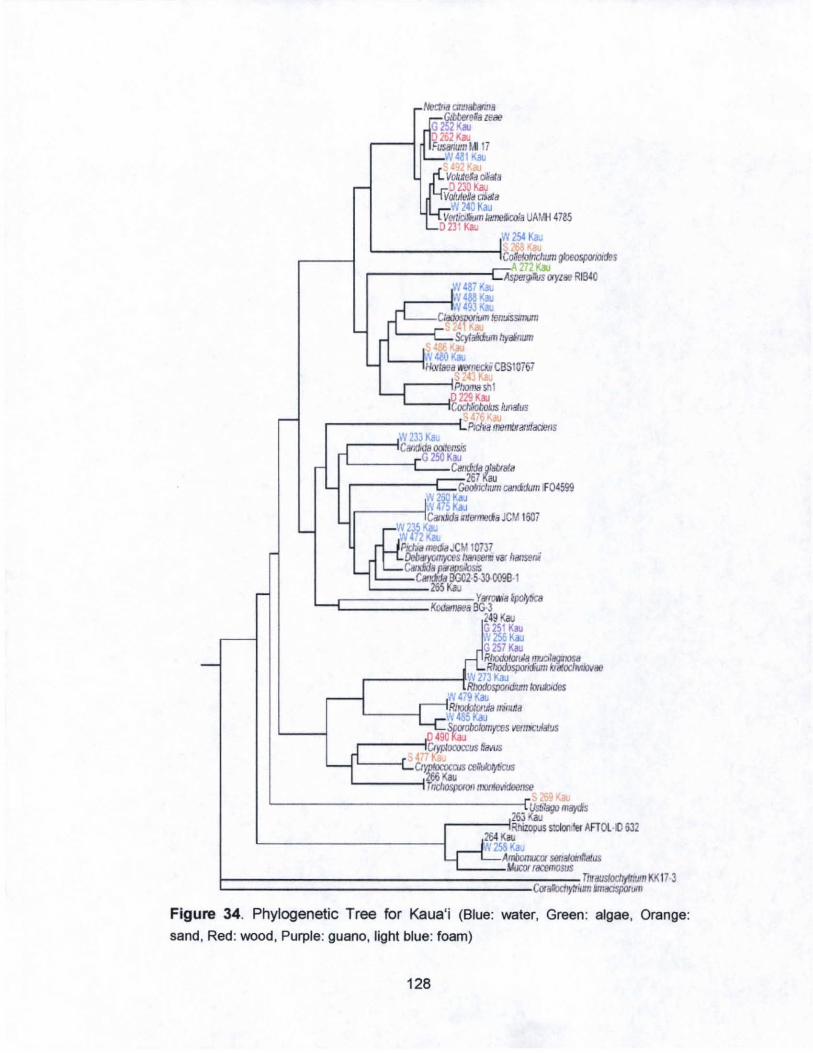
..-
L....
.--
-
-
L...
L-
r-r taatm n Jff~~·ze~ G22Kao
0202 Kao FusWlum t.l17
I L...-\V4.l1 Kao
fi. V~$a ciaI. I4vP 230 Kao VoMeJa oilla
Lrv: 24OKaJ Vettiau 1aI,,,""'I, UM'H 4785
_ D231Kao IW?54 Ka\I
-. CoIeIoIncIIw1 :::.i27U",
~Aspe1gJl1s ayz"" R W4a7 1(;..
gbcospotlOldeS
1840
~4as Kau t-----~ . IV 493 O(a; ~ lfl1!lSSt+fUn ~ ,t
Scytali<itim hyairum,
~,~4cIOKao HOtIaea ... m.ct.i CBSIOi67 ,4
L..j Pt,omashl
~ IJtJatus
'::Pma'ioonnillrfaae11S W 233 Ka,
-,CilIlCkI8 OOOCfrsis _G 250K",
C8Iid:da (:'ata 267
.,...,..,GooInciwm c",,<fllum IFD4599 l~mKa N4 Ka~
- IC",>ida "Cfma JCI,l I607 ~W'>fK&J ~4 2oU!L Prchal110GaJCM 10737 4 Qeb<ryoIr.)<eS h/iI1""~ vat /I.,,..,. '--CBlxJriJa fIi*I!Il.s.los:s
C8I~ BGIl2 5 30 OO'!B·I -SKau
I I
Lf
Y_a/rXl¥lca J(od""~8 BG-3
249 ,(au G 251 Kau ,~ 256Kau
Rt_nwlil(lll1OS8 d G2S7Kau ~RllOOOspOtjdiJJm ~it
IV 273 KaJ ~Rlxxbspor.d;"", _
ilf 479 KaIJ Ri1OOOlOflia nilWl' W~KaJ
.0 ~oboIon:ytes "'''>CUiatus 490 u t;?PIoroa:us favus . ~ ~~ '- Kau
TI1choSjJ/Jlon " .. 1IcvKJccIrse
26J~IiM}On oa'jd's
I Rhizopus ,:olen
-G-e!tu ... AFTOl IO 632
N 258 Kau AI1w,wx SCf,IlfOll rllatus
Ali.ICVf f8cemoSllS Tlwaustoch/frllJl11 KK 17 3
iiRn JnldC1$pOftJm CaaibdryIt'
Figure 34. Phylogenetic Tree for Kaua'i (Blue: water, Green: algae, Orange:
sand , Red: wood, Purple: guano, light blue: foam)
128
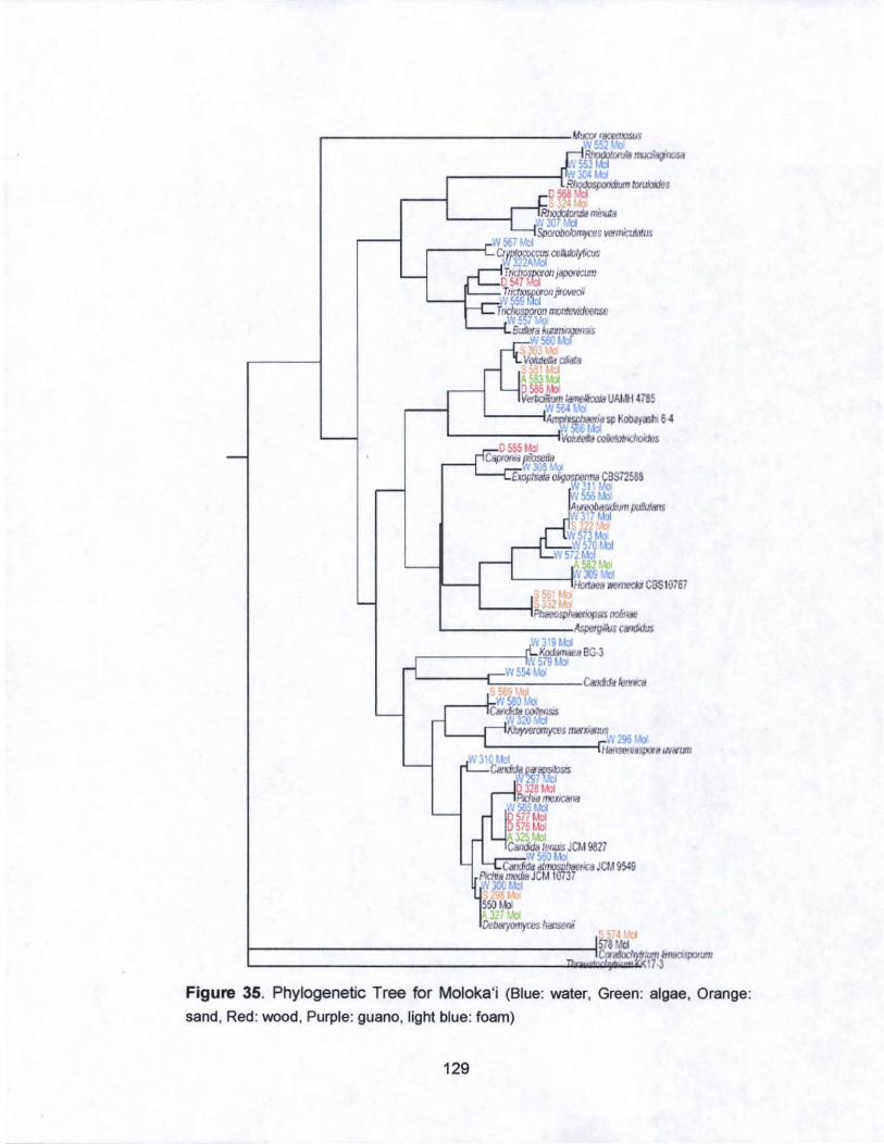
L------1PI~IOflS/S ,JOlt ... L-______ Asperg.i!l'c_
l'/3'9 Mol r-_____ ~~HB~3
,'w 579 M~ VI 554 L10
'-----1c;:::;~~-Cat_ fe,r>CII
4 1-________________ ~578Md
L __________________ ---l:z... ..... C:li"',;;;;;jiliiUtn ~IJ8CISPOItm1 173
Figure 35. Phylogenetic Tree for Moloka'j (Blue: water, Green: algae, Orange:
sand , Red : wood , Purple: guano, light blue: foam)
129
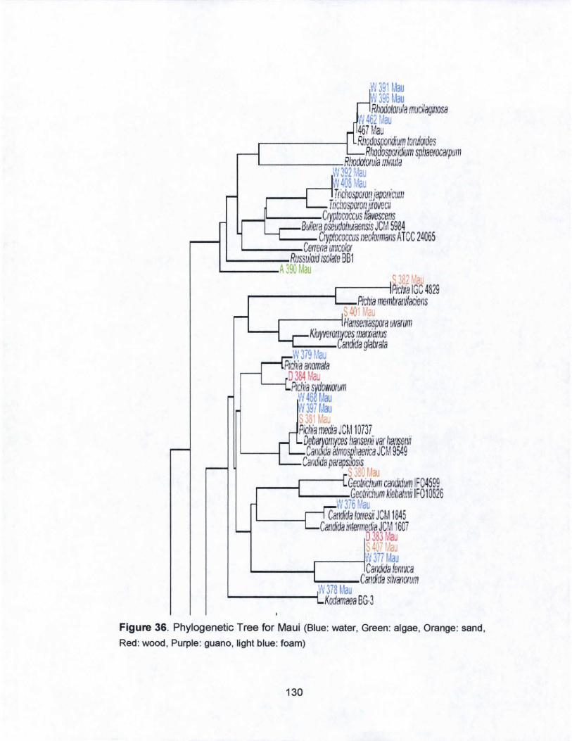
Vi 391 Mau (1396 Mau Rlloooloruia muciJagmosa
tV 462 '\~au 467 Mau Rllodospondium lorufoldes
RllOdospOIidiulII spliaerocillpum L-___ -.,.:-:Rliodo!oruliJ millula
VI 392 :AJU N408 '~'a T ric/lOsporo(! japollicUIII
T ncliospo!fJIUffovecll _....JL..----:o-.,.Cryptococcus IfavescetlS
Buflera.pseudoilliaetlsis Jer., 5984 '-----:-- Cryp!ococcus lleoformans ATec 24065
'--__ -=-Ceftooa un:ClJhJr '----~Russu!Oid ISo/ate BBl
'--___ A 390 Mau veL t?
.-----~PiCINa IGC 4829 ,...-------1 Picilia memtxalli/aciens
S 401 'a, ...--------iHalisemaspora INil'UflI
'----I KIu}WroliItces lI)(l1xiatlUS L-,-__ - Candida glalxata Vl3i9 f.'a
Ptchia allOlllaia OJ84 ~Jau Ptchia s oWtOfum
W4 Mau ',V 397 Mau 338' Ma. Pichia media CM 10737 DebillYOII~ceS Ilan5elui var hallsetltt Candida allllospliaetica JCM 9549
Candida parapsilosis S J.v Mau
r __ -[==~GeotJiClium ciifldlduIII IF04599 Geottic/wm kiebalmj IF010826
W 376 riaL Candida lorresii JCM 1845
CamMa iniermedia JC ,1 1607 0383 Mau S ~O; ''<1u W377 Ma~
'--__ --I.-------iCandida ienl!lca ~::,:,,:"~Ca!ldida Sl,valiorum
iN 378 Mau L..-____ ---! KodamaeaBG.3
Figure 36. Phylogenetic Tree for Maui (Blue: water, Green: algae, Orange: sand,
Red: wood, Purple: guano, light blue: foam)
130
'--_____ ya71l1!!a IpoiMa a SOIboIyb18
Figure 36. (Continued) Phylogenetic Tree for Maui (Blue: water, Green: algae,
Orange: sand , Red: wood , Purple: guano, light blue: foam)
131
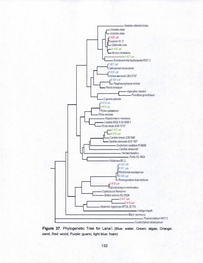
.-------VolJie,ia ro'IeIotrdJOidcs Vo/uieffa a,la/a Volute/a ci5ala
0425LM FuswillmMI 17
GtiJbefeila leaa A 414..al Nectna C/lrlaba,,/a
.--------~ 421LM AaetI"'~JlII·I'e hyohomytl!:e KR21 ·2
'11 427l"" ~---I
C/IldoSjJOlliJllI wlhiSSIJmJlII
W430LM L--..jHooac8 W&'IJeOO CBSI0757
W431l ... .---1 P",ap/Jaoosp/laena IIlc1JObi
PIKIma he!bwwll AS/Iflrg:iI1JS Cli/latus
r----L-= PeniCJllumgriseofuMnIl C8IIfDllla ",Iosena
W 41e la, A413lan 1'Ic/,. sydowlofUm
Pld,a alKlmaia
.----- XIuyieromyces """xi8l1US Candida BG02 5·30-0099·1
PicllBmedJaJCM 10737 ~412l"" A419l
CIllKida lonesi JCt.! 1845 CIIIJ6diJ /lie/media JCM IW7
L-_____ Geolllcoomc8IKildum IF04599 L---____ C8lItida SllVaflorum
L-________ Y8ITOvn81po1ytJca
.---------- Pod .. IGC 4829 "L-____ Kodamaea 00·3
I~ 423lan N411lan RlIodotOfui811liJCiJIlIJIK1S8
.--____ ---1 .~ 420 La, RhodoSjJOlldumlcraloctM/ovae
0416lan L-___ --1 ~es """"CJJatus
CtypfCJalCaJS Rawsams L..._--{==-= Bu,m ~""",s AS 21524 0417lan r------C=::..:.:.o 418lan
L-__ AlI8kelus higatl6l1~s AFTOl·l0 714 1....------------Usf,lagomaytis
L...----------------MOCOf,£emosus 1----------------------Tlv8USloc/JyloomKK173 L-------------------u.alloc/Jy/liulllhIlI8l:lSjJOl""
Figure 37. Phylogenetic Tree for Lana'i (Blue: water, Green : algae, Orange:
sand, Red: wood, Purple: guano, light blue: foam)
132
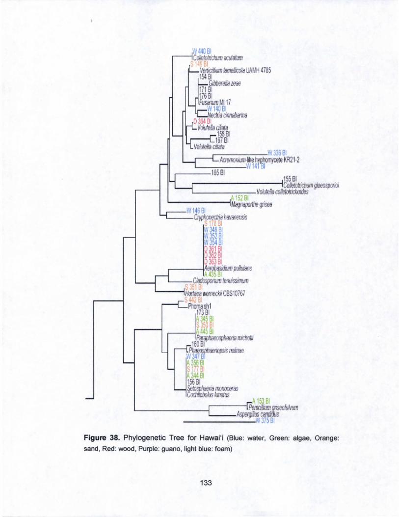
W 440 81 r-----iColietoirictun OCtJatum
. ( Verl.a&vm JameIXoIa W.ti 4785 15481
Glbbere!!a zeae 17181 17681 FuslrlUm t.U 17
WI40 81 rona cinliahililia D 364 0:
Voluleffa Clkala 158 8t
.----i 167 BI VoMe~ ClNaia ...--_ ___ w 336B
ACtetOOlllRll-il:e h ohoolvcete KR21·2 . ~ 14 'BI •
r--~::""16:::-5 8""1- -< 15581
....----------ICOf:elolrich<Jm gk>eospCXIoi '--------..,.,...,--VCJliJfe!!8 cor,gtoilicrores
A 152 Bt L..-----~Magnapal/Jre grisea .---- w 146 81 '----CIyp/Jotrectria hil'/ilIe/lsis
5 78 31 :13488 N3528 N3548 D 3618 1
D36281 :J 363 81
.------1AefObasid:um piJlu!ans ~4'58
L---C/adosiMlm letilissimum
L....---lHol1aea wernecki CBS10767 4-
.-------Phoola sh 1 17381
'14581
.----i .-_---;A 4d, Pifii{Jiraeosp!Jaena tOChotJ
160 BI P11ilI!OSIJIlilfJ1/o/lSIS liO!:I:ae
VI 347 81 A 356 81
A J44 81 1568 Selose!r!lna Ir/oltoceras CocIl,IObollls IUliaius
A 153 81 L------C=:k~Pelllci/1uJm f1ISeQluWII AspergHus caJKiIilus - ______ W375B
Figure 38. Phylogenetic Tree for Hawai'i (Blue: water, Green: algae, Orange:
sand. Red: wood. Purple: guano, light blue: foam)
133
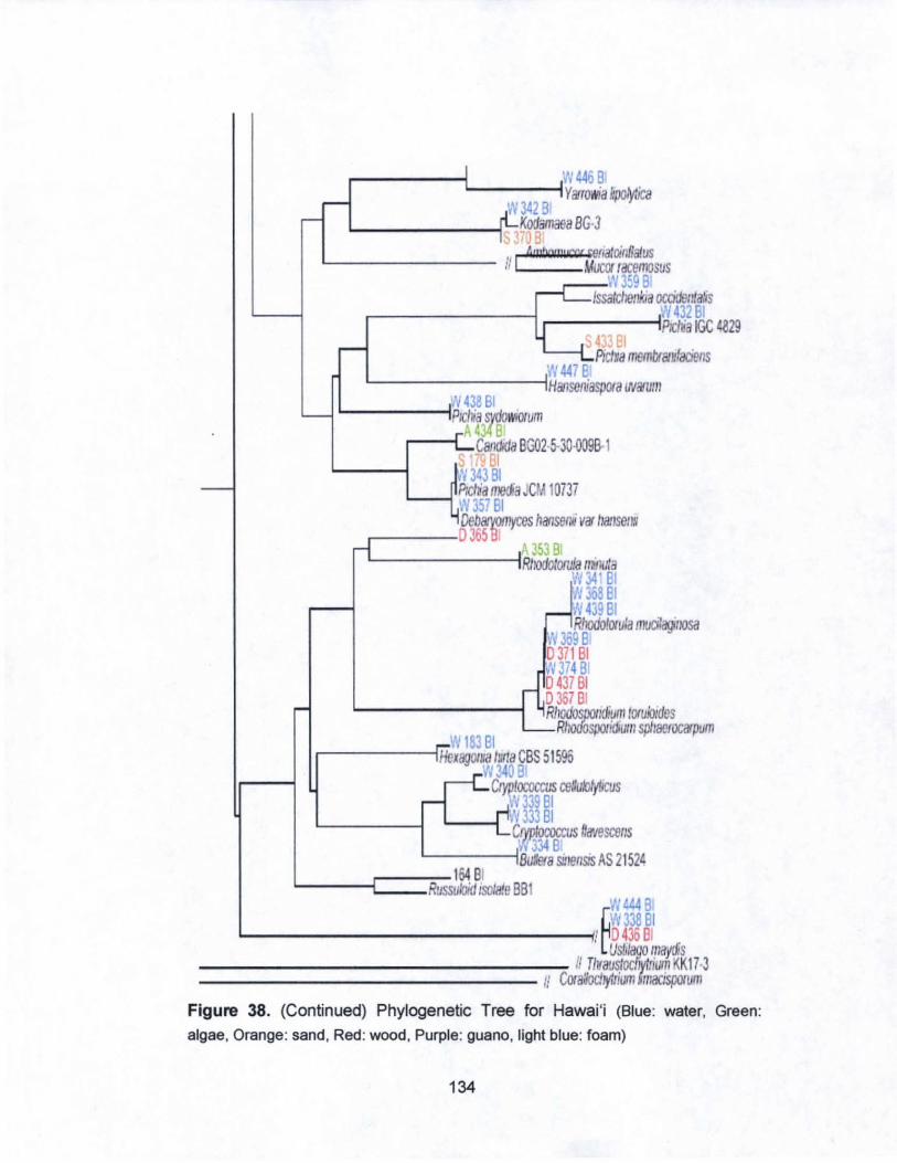
r-----t. ___ --1W446 B YfifoNia l:poIytica
\'/342 B Kodamaea BG3 '"J B
4mbow, U'cr renaJcjnflatus '--------- II I Mucor lacemosus
,..--'1'1359 Bi IssatchellYJa occklluda'/s
r-- ---- - L ___ --;~V432BI .- PIcha IGC 4829
s ~ .81 ----PIcIua memlYatufaciens W447 B'
'---------iHatiseniaspora lNarum W438BI
L-----iPlChia sWolI\O/um A 4J4BI
r----I Calldida BG02·5-30-OO9B-1 _ 61 .'I 343 BI
L--_r Piclia fOOIia JCM 10737 W357BI DebaryOmyces hanserni Vir IIIl!Jsel1ll
.-----0365 B! A 353 B
L.--- -----lRtlOdolow!a ninula II{ 34' BI 11'36881 N439BI RhodoIO/u/a mucitagfJOSd
W359B 0371 BI .'1374 B: D 437 BI D 357 BI RIK)(loSDOlldium Irxulo:des
RhoOOsporidlUm spll8efOCirPum W 183 BI
.------IHeXaglJIII8 Iw/a CBS 51595 W343Bi
CrvIilocoa:us ceNukJ!fl/cus · ~V 3J9 BI ~ 333 BI C tococcus na'lescells
334 B '-----iiBU"..eta sinensis AS 2 1 52~
L---C=J ussl64 B: Russukiid Isolate BB W~BI
~W338BI L-____________ ~! D436BI
Uslilago maydis ________________ --::: II Tlvaustocllv/rium KK17 3
--------------- Ii Ccrallocliylrium ' milOspolum
Figure 38. (Continued) Phylogenetic Tree for Hawai 'i (Blue: water, Green:
algae, Orange: sand, Red: wood, Purple: guano, light blue: foam)
134
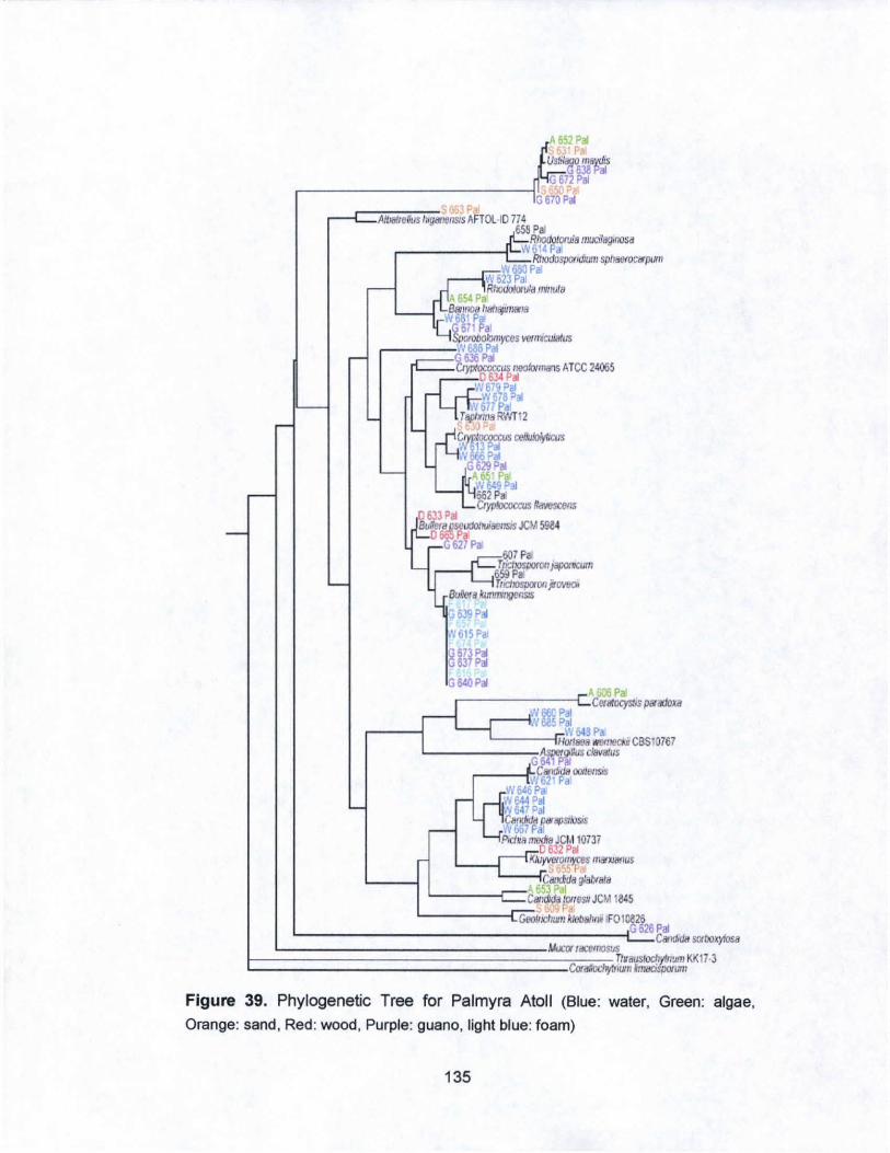
~ 65;1 Pal
uS~'"'~' G672~
~------------~GI 6i't:.1 '<t, P
AlIJolre/1J. l.fIIIt"'ISIS AFTOL 10 m 658P~
RhodotOflJJa mucifagmosa ~ ____ ~I'-'W';.1' P ..
RllOdospotldJum SpfltteroCWpum W680P.
W623Pa' RllOdoIow:a mmuttl
M51Pa __ ., Sal/lOb 1~1I8"a
WG~7r~al Slaoboomyces ~micWaflJS
,----..... W 686 Pal ---==G 636 Pal ,,- Ctyploroccus ,lOOi:I<mans Alec 24065
0634 Pal
0633 Pal
W679 Pal W6/SPa!
WinP. T8plfW·~12
~orocclJS cel*Jlo/yOaJs
:, 13 Pal N Pal
G 629PII AOhP VI 90al 66 Pal Ctyp- ".."""""'
BUIent~'aC"sis JC>.I 5984 o P~ 627Pa
601 Pal T ridpsporoo J""",flc"m 659P~ rrlCl:ospaofJ ~f11tJ01
BuIIoralwtm,1gCllSS
G 6..'9 Pal
N615Pa
". Pal r---------c:C.,~Sp8IiIIoxa _---lIN 66C Pal
r- .v 6a5 P3l W 54! Pa
L---mHotI .. a """",,*, cas 10767 L-----_ASl>9f9tius dBvsrus
G541PJJ ___ .r:.CBtKlda """",/Sis
r Wo21 Pa W6'OPa
'N~g:m ar/dJcJa PWi4JSI!ogs
\ 661 pal PId,a ,,_ JCt.llom
0632 Pal L--L-,,::K!Jf,yvorOI/~$ mCW'Xla1lus
:,t ~ P Callada g:a/JI,.8
A Pal L----r-C8I a"",e~,JCMI645 ,
GeoIIIcI,u'rll lel/aht" IFOI0826 .G 626 Pal
Ci>1!I<Ia SCI IJOl'j.osa
Figure 39. Phylogenetic Tree for Palmyra Atoll (Blue: water. Green: algae.
Orange: sand, Red: wood, Purple: guano, light blue: foam)
135
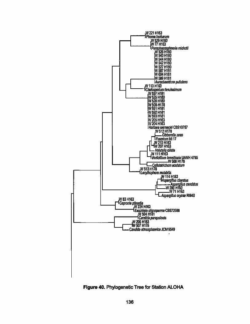
.-__ -IW221 HI63
Phoma I!erbIwm ....---1 W 529 H18D
nHI63 L-----l~~~nichaIii
54DHl8D
~~l~ 527HI8D 587 H181 004 H181 589 H181
L---IW113Hl63 .. pliIJJJJB1IS - C/adospoJium tenuissinwm
W597H181 '---- 525 Hl8D
528HI8D 509-HI78 591 H1S1 592 H181 593 H181 205Hl63 204Hl63
HIlI1ae8 wemecJdi CBS10767 W512H178
G1bbereI1a zeas Fusarium Ml17
W21DHl63 W2D7Hl63
wY,~ciIIaIa VIIIIIdIIium Iam8IIicoJa UAMH 4785
.----:-----'W 508 H178 --..-CoIietoIJichum IlCII1B/Um W513H178 LIq/hDpIIIIfa muJabIIjs
114 Hl63 AspeIgi1Ius cIIMlIus
~ __________ ~~.~--~W~2~~ ~~W71Hl63
W83HI63 Aspergi'lus O/)'Zse RIB4D
C8ptonia ~ H163 ~~CBS72588
....-----!CBndida parapsiIosIs L..-____ --I _--IWW206 HI83
r 5D7H178 CBndida 8ImospJ/BeIicB JCM 9549
Figure 40. Phylogenetic Tree for Station ALOHA
136
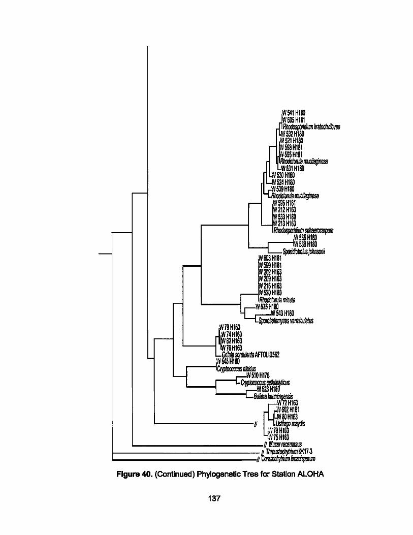
-
-
-
-'--
W79HI63
82 HI 76HI63
r--
JlH 605H
ISO 181
R/JodCspD!IlDum kratocIIii!ova8 80 W532Hl
~FHl 598 HI ~,5S5Hl
80 81 81
=\'1531 HI ~FH1SO
RIiOODIotuIa I1llIIiIIJ;JimI 80
524H18O
Lfr539HI80 =1 212HI63 533 HISO
- 213 HI63 ~~ocarpum ,W
,'" W803H181
538H1SO Spo/itfDbo/lJs john&lnU
599H181 IHI63
HI63 216 HI63 S20H1SO
RhDdtJ!orufa minuIa W536HI81l
W543HI8O ~
~r4Hlft ",.SIlO!iIe1!taAFTOUD562 ,W545HI80
'~~OHI78 : .. ~sfHr
Bu6eta~63
~~.' WaoHI63
U W78V 15HI63
n IJJiaJI /BCIl I1IDSLIS KK17·3
DmadspoIum U~ Figure 40. (Continued) Phylogenetic Tree for Station ALOHA
137
CHAPTER V, SUMMARY AND CONCLUSION
A, Summary of Results
A total of 689 Isolates were purified from over 250 samples of water, sand,
macroalgae, wood, and avlan guano from the six high Hawaiian Islands,
Palmyra Atoll, and Station ALOHA. Yeasts cultivated from Station ALOHA and
Palmyra Atoll differs phylogenetically from those cultivated from Hawaiian
littoral and sub-littoral sites. Those from the latter two habitats tend to be most
closely related to, or appear to comprise, genera widely considered terrestrial
in origin. Open ocean strains are more often related to those reported from
the deep sea and marine sediments around the world, such as Rhodotoru/a
spp.
The greatest number of potentially novel species was derived from Moloka'i,
followed by Palmyra. The Islands with the fewest potentially novel cultivated
species were Lana'l, Maui, and Kaua'l.
The number of CFU Isolated from water samples did not vary significantly
across the six high Hawaiian Islands. Only the numbers of yeasts cultivated
from Lana'i showed some Indication of being lower than at any other Island
site, but there were Insufficient data to support such a conclusion (Tab. 6).
138
The numbers of yeast only, fungi only, and yeast and fungi combined did not
vary significantly over the -5000 m water column at Station ALOHA; data
were compared between indMdual depths, and shallow versus deep water
samples. The Kruskall-Wallls test did suggest there was a higher number of
CFU In the shallow «500 m) water versus those in deep water (>500 m), but
again, insufficient data based on just six cruises were unable to resolve this
{Tab. 7).
One culture, LM418 Isolated from wood on Shipwreck Beach, Lana'ils being
described as a new Sympodlomycopsis, specifically Sympodiomycopsls
mahdli. A manuscript Is being prepared for submission to FEMS Yeast
Research, but Is summarized here In §H2.
B. Conclusions and Recommendations
this study Indicates Significant phylogenetic diversity among the marine
mycoflora exists around the high HawaIIan Islands, at Station ALOHA and
Palmyra Atoll, both In water per se and on many other materials. Cultures
generated In this study will serve as a foundation for future research on
secondary metabolites In Hawaiian marine fungi and yeast.
139
REFERENCES
1. Abbanat D., Leighton M., Malse W., Jones E.B.G., Pierce C. &
Greenstein M. (1998) Cell wall active antifungal compounds produced
by the marine fungus Hypoxylon oceanicum LL-15G256. I. Taxonomy
and fermentation. J Antibiotics 51 :296-302
2. Ainsworth, G.C., and Sussman, A.S. (1968) The Fungi: An advanced
treatise. Acedemlc Press, New York.
3. Aller, J.C., Zaborsky, O.R. (1979) The Blosaline Concept.
Environmental Science Research, 14:1-16.
4. Altschul S.F., Madden T.L., SchlUfer A.A., Zhang J., Zhang Z., Miller
W., Lipman D.J. (1997) Gapped BLAST and PSI-BLAST: a new
generation of protein database search programs. Nucleic Acids Res
25:3389-3402
5. Amadon D. (1947) Ecology and the evolution of some HawaIIan birds.
Evolution 1 :63-68
6. Anastasiou, C. J. (1964) Some aquatic Fungllmperfecti from Hawal'l.
Pacific Sci. 18: 202-206.
7. Azam F., Fenchel T., Field J.G .. Gray J.S., Meyer-Reil R.A. &
Thingstad F. (1983) The ecologlcel role of water column microbes In
the sea. Marine Ecology Progress Series, 10:257-263
8. Azam, F. (1998) Microbial control of oceanic carbon flux: the plot
thickens. Science 280 (5364): 694-696
9. Barghoom, E.S., Linder, D. H. (1944) Marine Fungi: their taxonomy
and biology. Far/owla, 1:395467.
10. Barr M.E. & Hodges Jr. C.S. (1987) HawaIIan forest fungi. VIII. New
species In Gnomoniella and Stigmochore. Mycologia 79: 782-786
140
11. Berry, D.R. (1982) The Biology of Yeast. Edward Arnold Publishers
Umited, London.
12. Buck, J.D., and Meyers, S.P. (1965) Antiyeast ActMty in the Marine
Environment. 1. Ecological Considerations. Umnology and
Ocaanography, 10: 3, 385-391
13. Bull A.T., Ward A.C. & Goodfellow M. (2000) Search and discovery
strategies for biotechnology: The paradigm shift. Mlcroblol Mol Bioi Rev
64:573-606
14. Campbell, N.A, Reece, J.B, Mitchell, L.G., Taylor, M.R. (2002) Biology:
concepts and connections. 4th Edition, Benjamin Cummings Publishing
Co.
15. Carmichael, J. W. (1971) Blastospores, aleurospores, chlamydospores.
Taxonomy of fungi Imperfectl University of Toronto Press
16. Carr G.D.R., Robichaux R.H., Witter M.S. & Kyhos D.W. (1989)
Adaptive radiation of the Hawaiian sllversword alliance (Compositae
Madiinae): A comparison with Hawaiian picture-winged Drosophila. In:
Giddings LV, Kaneshiro KY & Anderson WW (eds.). Genetics,
SpeCiation and the Founder Principle. Oxford University Press, New
York, pp. 79-97
17. Colwell R.R. (1997) Microbial diversity: the importance of exploration
and conservation. J. Ind Mlcroblol. Blotechnol. 18:302-307
18. Colwell R.R. (2000) Biocomplexity and the ocaans. Sea TechnoI41:22-
23
19. Cooney J.J., S. Wuertz, M.M. Doolittle, M.E. Miller, K. Henry, and D.
Ricca (1993). Marine fungi: novel catalysts for bloremediation of 011
141
spills. In: Trends in Microbial Ecology (Guerrero R. and Pedros-Allo C.,
eds.), p. 639-642
20. Denner E.B .• Mark B .• Busse H.J., Turkiewicz M. & Lubitz W. (2001)
Psychrobacter proteolytlcus sp. nov., a psychrotrophic, halotolerant
bacterium isolated from the Antarctic krill Euphausia superba Dana,
excreting a cold-adapted metalloprotease. Syst Appl MlcrobloI24:44-
53
21. Dring D.M., Meeker J. & Goes R. (1971) Clathrus oahuensis, a new
species from Hawai'l. Mycologla 63:893-897
22. Donachie, S.P. & M.K. Zdanowski (1998) Potential digestive function of
bacteria In krill, Euphausia superba stomach. Aquatic Microbial
Ecology 14:129-136
23. Donachie S.P., Hou S., Gregory T.S., Malahoff A. & Alam M. (2003)
Idlomarina loihlensis, sp. nov., a new halophilic OProteobacterium
isolated from the LO'ihi submarine volcano, Hawai'!. Int J Syst Evol
Mlcroblol53:1873-1879
24. Donachie S.P .. , Hou S., Lee, K.S., Riley C.W., Pikina A., Belisle C.,
Kempe S., Gregory T.S., Bossuyt A., Boerema J., Uu J., Freitas T.A.,
Malahoff A. & Alam M. (2004a) The Hawaiian Archipelago: A Microbial
Diversity Hotspot. Mlcrob Eco/, 48:509-520
25. Donachie S.P, Bowman J.P., Alam M. (2004b) Psychroflexus traplcus,
sp. nov., a novel, obligately halophilic Cytophaga-Flavobacterlum
Bacteroides group bacterium isolated from an Hawaiian hypersaline
lake. Int J Syst Evol M/crobiol 54:935-940
142
26 .. Donachle S.P., Bowman J.P., On S.L.W. & Aiam M. (2005) Arcobacter
ha/ophllus sp. nov., the first obllgately halophilic species In the genus
Arcobacter. Int J Syst Evol Mlcroblolln press
27. Fell, J.W., Adheam, D.G., Meyers, S.P., and Roth, F.J. (1960) Isolation
of yeasts from Biscayne Bay" Florida, and adjacant benthic areas.
Ummno/. Oceanogr. 5: 336-371
28. Fell, J.W., Uden, N. Van (1963) Yeasts In Marine Envlronment In:
Symposium on Marine Microbiology. 32:329-340
29. Fell, J.W., Phaff, H.J., Newell, S.Y. (1970) Rhodosporldium Benno.ln:
The Yeasts. ( J. Lodder ed.) p. 803-814. North Holland Publ.,
Amsterdam.
30. Fell, J.W. (1976) Yeasts In oceanic regions. In: Jones E .. BG. (ed)
Recent Advances in Aquatic Mycology. Elek Science, London, pp. 93-
124
31. Fell, J.W., Boekhout T, Fonseca A, Scorzettl G & Statzell-Tallman A
(2000) Biodiversity and systematics of baslodlomycatous yeasts as
determined by large subunit rD11D2 domain sequenca analysis. Int J
Syst Evol MicrobloI50:1351-1371
32. Fell, J.W. (2001) Collection and Identification of marine yeasts. In: Paul
J.H. (ed) Marine Microbiology, Methods in Microbiology, 30:347-356
33. Felsensteln J. (2004) PHYLIP (Phylogeny Inferenca Package) version
3.6.
Distributed by the author. Department of Genome Sciences, University
of
Washington, Seattle.
143
34. Fowler, J., Cohen, L., Jarvis, P. (2003) Practical Statistics for Field
Biology. Second Edition. John Wiley & Sons, New York.
35. Giovannoni. S. J .• Brltschgl. T. B •• Moyer, C. L., and Field, K. G. (1990)
Genetic diversity In Sargasso Sea bacterioplankton. Nature 345:60-63.
36. Guerriero A. D'Ambrosio M, Cuomo V, Vanzanella F & Pletra F (1988)
Oendryphiellin A, the first fungal trinor-eremophilane. Isolation from the
marine deuteromycete Dendryphiel/a salina (Sutherland) Puch et Nlcot.
Helvet Chim Acta 71:57-61
37. Hall, T. (1997) BloEdlt Software: Biological sequenca alignment for
Windows XP. Ibis Theraputlcs, Carlsbad, CA
http://www.mblo.ncsu.edutBloEdltlbloedlt.html
38. Hagler, A.N., Mendoca-Hagler, L.C. (1981) Yeasts from Marine and
Estuarine Waters with different levels of pollution In the state of Rio de
Janerio, Brazil. Appl Environ Microbiol. 1981 January; 41(1): 173-178.
39. Hobbie, J.E., Daley, R.J., Jasper, B. (1977) Use of nuclepore filters for
counting bacteria by fluorascanca microscopy. Applied and
Environmental Microbiology 33 (5) 1225-1228
40. Hyde, K.O., Venkateswara Sarma. V. Gareth Jones, E. B. (2000)
Morphology and taxonomy of higher marine fungI. Marine Mycology - A
Practical Approac Fungal Diversity Press: p 172-204
41. Hyde K.O. & Pointing S.B. (eds) (2000) Marine Mycology - A Practical
Approach. Fungal Diversity Press
42.Jannasch, H.W. and Taylor, C.O. (1984) Deep-sea microbiology. Annu.
Rev. Microbial. 38, 487-514.
144
43.Jensen P.R. & Fenical W. (2000) Marine microorganisms and drug
discovery: Current status and future potential. In Fusetanl N. (ed)
Drugs from the sea. Basel, Karger, pp. 6-29
44. Jensen P.R. & Fenical W. (2002) Secondary metabolHes from marine
fungI. In: Hyde KD (ed) Fungi In Marine Environments. Fungal Diversity
Research Series 7:293-315
45.Johnson, R.G. (1974) Particulate matter at the sediment-water
Interface In coastal environments. J. Mar. Res. 32: 313-330
46. Johnson, T. W., Sparrow F. K. (1961) Fungi In Oceans and Estuaries.
Hafner Publishing Co, New York
47. Karl D.M., Lukas, R. (1996) The Hawal'i Ocean Time-series (HOT)
Program: Background, rationale and field Implementation. Deep-Sea
Res. 11,43, 129-156
48. Kawaldta, S, and van Uden, N. (1965) Occurrance and population
densities of yeast species In the digestive traelsof gulls and tems.
J.Gen. Mlcroblo/. 39:125-129
49. Kendrick, B. (1971) Ed. Taxonomy of fungi imperfect}. Toronto and
Buffalo, UnlversHy of Toronto.
50. Kendrick, B. (2001) The FIfth KIngdom. Focus Publlshlng/R. Pullins
Company; 3rd ad.
51. KIrk, P. M., P. F. Cannon, J. C. David, and J. Stalpers. (2001)
Ainsworth and Blsby's Dictionary of the FungI. 9th ad. CAB
Intemational, Wallingford, UK.
52. Koch, J. (1974) Marine fungi on driftwood from the wast coast of
Jutland, Denmark. Friasla: 10, 209-250
145
53. Kohlmeyer, J. (1965) The Importance of Fungi In the Sea. Symposium
on Marine Microbiology edited by Carl H. Oppenheimer. 30:300-314.
54. Kohlmeyer J. (1966) Ecological observations on arenicolous marine
fungi. Z Allg Mikroblo/6:94-1 05
55. Kohlmeyer J. (1969a) Deterioration of wood by marine fungi in the
deep sea. In: Materials performance and the Deep Sea, Am Soc. Test.
Mater.,Spec. Tech.Publ. 455:20-29
56. Kohlmeyer J. (1969b) Marine fungi of Hawai'i including the new genus
Hel/cascus. Can J Bot 47:1469-1487
57. Kohlmeyer, J. (1974) On the definition of and taxonomy of higher
marine fungi. Veroeff. InstMeereshorsch. Bremerhaven, Suppl. 5: 263-
268
58. Kohlmeyer J. & Kohlmeyer E. (1979) Marine Mycology. The Higher
Fungi. Academic Press, New York, NY.
59. Kohlmeyer J. (1985) Nlmbospore octonae, a new marine ascomycete
from Hawsi'i. Can J Bot63:1122-1125 Kohlmeyer, J. (1986).
Taxonomic Studies of the Marine Ascomycetina. In: The Biology of
Marine Fungi. Ed: Moss, S. T. P .199-210
60. Kohlmeyer J. & Volkmann-Kohlmeyer B. (1989) Hawaiian marine fungi,
including two new genera of Ascomycotina. Mycol Res 92:410-421
61. Kohlmeyer J. & Volkmann-Kohlmeyer B., Newell S. (2004) Marine and
Estuarine Mycelial Eumyceta and Oomycota. Biodiversity of Fungi,
Inventory and Monitoring Methods.
62. Krager-van Rij, N.J.W., Kurtzman, C.P. (1984) Classification of the
ascoporogenous yeasts. The Yeasts: A Taxonomic study, ~ Edltfon,
Elsevier P. 25-32
146
63. Kurtzman, C.P., and Fell, J.W. (2000) The Yeasts: A Taxonomic Study.
4th Edition, Elsevier
54. Kurtzman, C.P., and Fell, J.W. (2004) Yeasts. Biodiversity of Fungi,
Inventory and Monitoring Methods. 16:337-342
65. Kurtzman C.P. & Robnet C.J. (1998) Identification and phylogeny of
ascomycetous yeasts from analysis of large subunit (26S) ribosomal
DNA parHal sequences. Anton van Leeuwen 73:331-371
66. Untott W.H. & Untott E.A. (2002) Marine fungi from New Zealand. In:
Hyde KD (ed) Fungi In Marine Environments. Fungal Diversity
Research Series 7:285-292
67. Maddison, D. R. and Schulz, K.-S. (eds.) (1996-2006) The Tree of life
Web Project. Intemet address: http://tolweb.org
68. Meyers, S. P., Ahearn D. G., and Roth, F.J. Jr. (1967) Yeasts from the
North Sea. Marine Biology, 1: 2, 118-123
69. Meyers, S. P., Ahearn D. G., and Roth, F.J. Jr. (1976) Mycological
Investigations of the Black Sea. Bull. Mar. Sci. 17:576-596
70. Marmur J. (1961) A procadure for the Isolation of deoxyribonucleic acid
from microorganisms. J Mol Bioi 3:208-218
71.Mlnltab Statistical Software. (2004) Mlnitab Inc., State College, PA
72. Moril, H. (1973) Yeasts predominating In the stomach of marine little
toothed whales. Bull. Jpn. Soc. Sci. Fish 39:333
73. Myers N., Mittermeler R.A., Mlttermeler C.G., da Fonseca G.A.B. &
Kent J. (2000) Biodiversity hotspots for conservation priorities. Nature
403:853-858
74. Nagahama T., Hamamoto M., Nakase T., Takaml H. & Horikoshl K.
(2001 a) Distribution and Identification of red yeasts In deep-sea
147
environments around the northwest Pacific Ocean. Anton van Leeuwen
80:101-110
75. Nagahama T., Hamamoto M., Nakase T., Takaml H. & Horlkoshi K.
(2001b) Rhodotorula lamellibrachii sp. nov., a new yeast species from
a tubeworm collected at the deep-sea floor in Sagami Bay and its
phylogenetic analysis. Antonle van Leeuwenhoek, 80:3-4, 317-323
76. Olsen, G.J., Lane, E.J., Giovannoni, S.J., Pace, N.R., Stahl, D.A.
(1986) Microbial ecology and evolution: A ribosomal RNA approach.
Annual Review of Microbiology 40: 337-365
77. Phaff, H.J., Miller M.W., Mrak E.M. (1966) The Life of Yeasts. Harvard
University Press
78. Pointing S.B., Fryar S.C., Hyde K.D. & Mouzouras R. (2000)
Assessment of wood dacey, fungal Interactions and colonization. In:
Hyde K.D. & Pointing S.B. (eds), Marine Mycology - A Prectical
Approach. Fungal Diversity Research Series 1, Fungal Diversity Press,
Hong Kong, pp. 113-136
79. Polgase, J.L., Alderman D.J. & Richards R.H. (1986) Aspects of the
Progress of Mycotic Infections In Marine Animals. The Biology of
Marine Fungi, edited by S. T. Moss. 14:155-164.
80. Pomeroy L.R. (1974) The ocean's food web, a changing paradigm.
Bioscience 24:499-504
81. Porter, D. (1986) Mycoses of Marine Organisms: An Overvlewof
Pathogenic Fungi. The Biology of Marine Fungi, edited by S. T. Moss.
13:141-154
82. Raghukumar C., Raghukumar 5., Sheelu G., Gupta S.M., Nath B.N. &
Rao B.R. (2004) Burled In time: culturable fungi In a deep-sea
148
sediment core from the Chagos Trench, Indian Ocean. Deep-Sea Res I
51:1759-1768
83. Reed, M. (1902) Two new Ascomycetous fungi parasitic on marine
algae. Unlv. Calif. Pulb. Bot. 1: 141-164
84. Rhelnhelmer, G. 1992. Aquatic Biology. 4th Edition, John Wiley & Sons,
New York.
85. Rose, A. H., Harrison, J. S. (1969) The Yeasts. Biology of Yeasts
v1:102-107, 220-303, 461-470
86. Seshadri R. & Sieburth J.McN. (1971) Cultural estimation of yeasts on
seaweeds. Appl MicrobloI22:507-512
87. Sherr, B. F., Sherr, E. B., and del Giorgio, P. (2001) Enumeration of
total and highly active bacteria. In J. Paul (edJ, Methods in
Microbiology, Volume 30: Marine Microbiology. Academic Press,
London, p. 129-159.
88. Sieburth, J. M. (1968) The Influence of algae antibiosis of the ecology
of marine microorganisms. Adv. Mlcrobol. Sea 1: 63-94
89.Sieburth, J.M. (1979) Sea Microbes. University of Rhode Island,
Oxford University Press.
90.Smlth, J.E., Deans, S.G., Anderson, J.G., and Davis, B. (1977) The
nature of fungal sporulation. Biotechnology and Fungal Different/at/on,
FEMS Symposium #4. Academic Press, New York. P. 17-43.
91. Sparrow, F.K. (1965) Occurrence of physoderrna In Hawai'l, with notes
on other Hawaiian phycomycetes. Mycopathologla 25: 119-143
92. Spencer, J. F. T., Spencer D. M. (1997) Yeasts In Natural and ArtifIcial
Habitats. Springer-Verlag, New York.
149
93. Stackebrandt, E., & Woese, C.R. (1981) Towards a phylogany of
actinomycetes and related organisms. Cun: Mlcroblo/o. 5: 131-136
94. Stackebrandt E. & Goebel B. M. (1994) Taxonomic note: A place for
DNA:DNA reassociation and 16S rRNA sequence analysis In the
present species definition In bacteriology. Int J Syst Bacteriol 44:846-
849
95. Staley, J.T., Konopka, A. (1985) Measurement of in situ activltles of
non-photosynthetic microorganisms In aquatic and terrestrial habitats.
Annu. Rev. Microbiol. 39, 321-346.
96. Takamla, H., Inouea, A., Fujla, F., Horlkoshla, K. (1997) Microbial flora
In the deepest sea mud of the Mariana Trench. FEMS Microbiology
Letters v.152(2) 279-285
97. Taysl, I., van Uden, N. (1964) Occurrence and Population Densities of
Yeast Species in an Estuarine-Marine Area Umno/ogy and
Oceanography, 9: 1, 42-45
98. van Uden, N., and Castelo-Branco, R. (1963) Distribution and
population densities of yeast species In Pacific water, air, animals, and
kelp off southern California. Ummnol.Oceanogr. 8:323-329.
99. van Uden, N., and Fell, J.W. (1968) Marine Yeasts. Advances In
Microbiology offhe Sea. 1:167-202.
100. Vandamme P., Pot B., GIllis M,. de Vos P., Kersters K. & Swings
J. (1996) Polyphasic taxonomy, a consensus approach to bacterial
systematics. Mlcrobiol Rev 60:407-438
101. Ward P.G., Goff M., Donner M., Kaminsky W. & O'Connor K.E.
(2006) A two step cherne-biotechnological conversion of polystyrene to
a biodegradable thermoplastic. Environ Sci TechnoI40:2433-2437
150
102. Weber D. J. & Hess W. M. (1976) The fungal spore: form and
function. New York, John Wiley & Sons.
103. Webster, J. (1959) Experiments with Spores of Aquatic
Hyphomycetes. Annals of Botany 23: 59~611
104. Wick/ow D.T. (1992) Interference competition. In: Carroll GC &
Wick/ow DT (eds), The Fungal Community: Its Organization and Role
in the Ecosystem. ~ ed., Dekker, New York, pp. 26~274
105. Wood, E.J. Ferguson. (1963) Ecology of Algae, Protozoa, Fungi
and Viruses. In: Symposium on Marine Microbiology. Ed:
Oppenheimer, C.H. p 28-39.
106. Yarrow D. (1998) Methods for the Isolation, maintenance and
Identification of yeasts. In: Kurtzman CP & Fell JW (ads) The Yeasts, A
Taxonomic Study. 4th ed. Elsevier, Amsterdam, pp. 77-100
151


![REVIEW OF TELL ME MORE SPANISH - University of Hawaiischolarspace.manoa.hawaii.edu/bitstream/10125/25248/08_03... · hour [time]), objetos y animals (objects and animals), adjetivos](https://img.pdfslide.us/doc/110x75/5f58fdaf725e900eff3c2c03/review-of-tell-me-more-spanish-university-of-hour-time-objetos-y-animals-objects.jpg)