Embed Size (px)
Citation preview

62
6I2.31I.I:6I2.392.OI3:6I2.8
DIETARY DEFICIENCY, NERVE LESIONSAND THE DENTAL TISSUES
BY J. D. KING(From the Field Laboratories, University of Sheffield)
(Received May 7, 1936)
IT has been previously reported that, in experimental animals, deficiencyof vitamin A or carotene in the diet results, not only in hyperplasia ofthe gingival and subgingival tissues, with the subsequent supervention ofperiodontal disease [M. Mellanby, 1930], but also in degeneration of thesensory nerves of the teeth and jaws [M. Mellanby & King, 1934].It had been suggested by E. Mellanby [1933, 1934] that the epithelialand nervous lesions observed in vitamin A deficiency were closely relatedand that the former might be due to removal of, or interference with,the normal trophic impulses. On this basis it was considered [M. Mel-lanby & King, 1934] that such a loss of neurotrophic control, dueto the effects of the vitamin deficiency upon the nerve or nerve cell,might be partly responsible for so-called pyorrhcea alveolaris and otherdiseases of the teeth and jaws. If this were the case interference withthe nerve supply, including experimental resection of the afferent fibresof the dental nerves, might also be accompanied by hyperplastic andother degenerative conditions of the periodontal tissues. In 1934 theexperiments here described were begun with the object primarily oftesting this point. It was, however, borne in mind that such operationswould not necessarily reproduce alterations in nerve-cell function andstructure similar to those observed in the metabolic derangement as-sociated with a dietary deficiency of vitamin A.
In the following series of experiments upon dogs and rabbits, theeffects of resection of certain nerves are discussed and compared withthose due to vitamin A deficiency. In the course of the investigation itwas found that the part played by the vaso-motor system in the growthof the teeth had also to be included.
VITAMIN AND DENTAL NERVES
INFERIOR DENTAL NERVE OPERATIONS
After examination of the distribution of the sensory dental nerves inthe animals to be used, it was decided that those supplying the lower jawwere the more suitable for operation. Eventually the inferior dentalnerve was selected for experimental section, in spite of the fact that theperiodontal tissues of the lower jaw are also supplied by the lingual andlong buccal nerves. This additional nerve supply is in some respects aserious disadvantage, but by severing the inferior dental little or nodamage is done to other tissues; moreover, this nerve appears to possessno motor fibres and so is not likely to affect mastication.
The inferior aspect of the mandible, below the roots of the cheekteeth, was chosen as the site of operation in both dogs and rabbits(see radiographs, P1. I and Text-fig. 1); in puppies, below the seconddeciduous molar; in adult dogs, below the fourth premolar or first molar;and in rabbits, beneath the roots of the second premolar and firstmolar.
In all of these regions the under side of the mandible is free frommuscular and tendinous attachments. Small branches of the inferiorlabial and submental arteries and of the facial vein, and cutaneous nerveterminations are the only structures, other than subcutaneous tissue,which are likely to be encountered between the skin and the periosteum.Relatively close to the under surface of the bone, and within its substance,lies the inferior dental canal containing vessels and nerve.
In this position, therefore, the inferior dental canal was opened and,with the aid of a blunt aneurism needle, carefully inserted between thenerve and artery, about three-quarters of an inch of the former waslifted and excised from one side of the lower jaw of a number of dogs andrabbits. In the dogs the effects of such lesions were also compared withthose due to dietary deficiency.
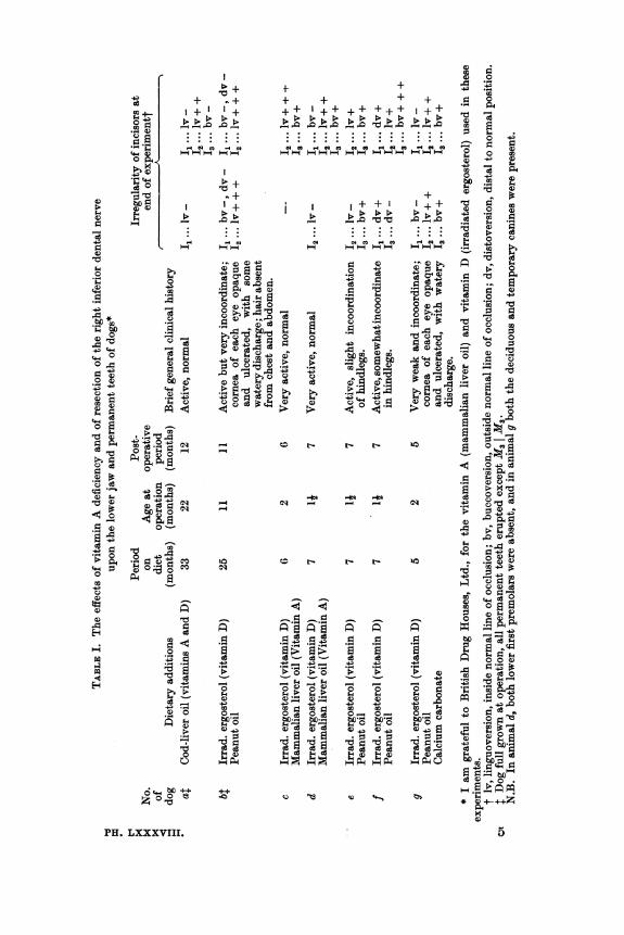
SERIES I (DoGs)With the exception of animals a and b (Table I), which were adults,
the excisions were performed on all of the dogs at an early age and theeffects of such treatment adjudged by observation of the condition of thepermanent dentition. Some of the animals received diets adequate, andothers deficient in vitamin A, as shown in Table I.
Age at beginning of the dietary period: 6-8 weeks.Basal diet: white bread, 120 g.; lean meat, 20 g.; separated milk
powder, 20 g.; bakers' yeast, 7-5 g.; sodium chloride, 1 g.; orange juice,6 c.c.
63
J. D. KING
The above results, together with the radiographs and other illus-trations shown in Pls. I and II, may be summarized as follows:
(a) Vitamin A deficiency. The effects of deficiency of vitamin A uponthe teeth and periodontal tissues of each dog were determined by thecondition of the whole of the upper jaw and, as shown in Table I, bythat of the left (unoperated) side of the mandible. In animals b, e, fand g (P1. I and II), receiving diets deficient in vitamin A, there wason the whole an increased deposition of tartar about the necks of thepermanent teeth, accompanied by varying degrees of gingivitis. Asseen in P1. II, figs. 2 and 4, dogs b and g, the alignment of the lowerincisors was irregular, the latter perhaps being caused by crowding of theteeth due to deficient growth of the mandible. Radiographs of the lowerjaws at death showed. the roots of the cheek teeth to be imperfectlydeveloped and the laminaT durae not well defined; the bone trabeculse inthe immediate vicinity of the tooth roots appeared unusually opaque toX-rays in some regions (P1. I, figs. 2 and 4). Histologically, degenerationof sensory nerves and hyperplasia and downward proliferation of thesubgingival epithelium were characteristic defects; in addition, theabnormal radiographic appearance of the tooth roots was seen to be dueto excessive deposition of cementum, which in some cases had invadedthe apical portion of the pulp chamber (P1. II, figs. 6 and 7). The additionof calcium carbonate to the ration of dog g (P1. I, fig. 4; P1. II, figs. 4and 7) produced no obvious improvement in the periodontal tissues or inthe alignment of the front teeth.
In contrast with the above, animals a, c and d showed much morenormal conditions. The amount of gingivitis and cervical tartar wasconsiderably reduced; the incisor teeth were in proper alignment;abnormalities of cementum and bone were absent; and the nervous andepithelial tissues presented a normal appearance (P1. I, figs. 1 and 3;P1. II, figs. 1, 3 and 5).
(b) Experimental nerve lesions. The effects due to resection of theright inferior dental nerve were assessed by comparing the right side ofthe lower jaw, peripheral to the site of operation, with the left (control)side. The most obvious result of the nerve lesion was the effectproduced upon the alignment of the front teeth. Pronouncedlinguoversion of the second incisor, and to a lesser extent buccoversionof the third incisor, was observed even when the diet contained liberalamounts of vitamins A and D and other essentials (P1. II, figs. 1 and 3,dogs a and c). In those animals receiving A-deficient rations, since lackof this vitamin was itself associated with similar conditions in the lower
64
.-4)-
C)* *90 &
0 .
-4 -44460&
44)
- 4.
+ ^ +
~. *, .> *-
4,.c N1-1 H C'* 1--
P> +4+
*tl064
Ca
40
4)i
CB-)
4)
C',
1>
Ca m
44)w.a O al
W
06
C1
_
_
4)o
._ .~
6464
PH. LXXXVIII.
4) 0
44'Go
+ + + + 00++ ++++++++ ++p +
p+
r$+ +
;-
E
..~~~ ~~0...*; P. ...P.... .. ... ....* ..._
@4@4@I@4 4O@@ c@@ @0 @2
4)4
+ 4 ^ 4
MI+~ I+t" @
* ... s e
*..*-* fl 0C)
0
--0C
-~~~~~64,W4o
o 0 0C 05A5 050644)-44 -
CB 4) 044-0
4) 4) 40*
4) Ca 4)Ca 4)~~~~~~~~~~4
4) o Q~~~~~~~~)46
oo-4Q
~~~ ~ ~ ~ ~~4) ,~~~~~~~~~~4-~~~~~ ~~ ~ ~ ~ *~~~~~~~ ~04)64
4)444)44 4)44~~~~46 4
4)
5.-
4)
.24)
64CH
0-4a
.4
4-4
*t bO
0
C4)
04)344
4) O
44.4
-44
@HO
w4"._
B*z 64
.~ 40@244C),4)
4)4)
66 J. D. KING
incisor region, the effects of the nerve operations cannot easily be judged(P1. II, figs. 2 and 4, dogs b and 9).
In the experiments described here no marked differences, macro-scopic, radiographic, or histological, in the condition of the periodontaltissues were observed between the operated and control sides of thelower jaw, even in the incisor region. When the diets were deficient, thechanges associated with lack of vitamin A were present; when there wasan adequate supply of this vitamin, the tissues appeared normal instructure.
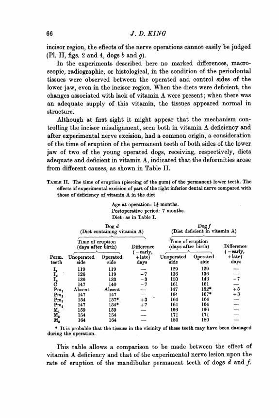
Although at first sight it might appear that the mechanism con-trolling the incisor misalignment, seen both in vitamin A deficiency andafter experimental nerve excision, had a common origin, a considerationof the time of eruption of the permanent teeth of both sides of the lowerjaw of two of the young operated dogs, receiving, respectively, dietsadequate and deficient in vitamin A, indicated that the deformities arosefrom different causes, as shown in Table II.
TABLE II. The time of eruption (piercing of the gum) of the permanent lower teeth. Theeffects of experimental excision of part of the right inferior dental nerve compared withthose of deficiency of vitamin A in the diet
Age at operation: 1j months.Postoperative period: 7 months.Diet: as in Table I.
Dog d Dog f(Diet containing vitamin A) (Diet deficient in vitamin A)
__ATime of eruption Time of eruption(days after birth) Difference (days after birth) Difference
C (- early, (-early,Perm. Unoperated Operated + late) Unoperated Operated + late)teeth side side days side side daysII 119 119 129 129I2 126 119 - 7 136 136I's 136 133 - 3 150 143 - 7C 147 140 - 7 161 161 -Pml Absent Absent 147 152* + 5Pm2 147 147 164 167* + 3Pm3 154 157* +3 164 164 -Pm4 147 154* +7 164 164ml 159 159 - 166 166M2 154 154 - 171 171
M3 164 164 180 180* It is probable that the tissues in the vicinity of these teeth may have been damaged
during the operation.
This table allows a comparison to be made between the effect ofvitamin A deficiency and that of the experimental nerve lesion upon therate of eruption of the mandibular permanent teeth of dogs d and f.
VITAMIN AND DENTAL NERVES
In the first instance, on the unoperated side it is evident that the teeth ofthe vitamin A-deficient dog f erupted, on an average, about 13 days laterthan the corresponding ones of dog d, which received a liberal supply ofvitamin A. The nerve operation, on the other hand, resulted in a slightacceleration of growth of the second or third incisor of the operated side,as compared with the corresponding teeth of the unoperated side of thesame animal; it will be noted that the incisor teeth so affected were thosewhich had previously been observed to be out of alignment (see Table I).The position of the canine of either dog, on account of the size ofthe toothand the length of its root, would hardly have been expected to be muchaffected. The proximity to the site of operation of the premolars peri-pheral to the nerve lesion renders it inadvisable to draw any conclusionsfrom the time of eruption of these teeth.
It would seem, therefore, that vitamin A deficiency and excision ofthe inferior dental nerve tend: to have reverse effects upon the rate oferuption of the lower incisor teeth, although their end result is similaras regards tooth alignment. On this basis, however, it is difficult toaccount for the incisor irregularity produced in dog a, all of whose teethwere fully erupted prior to the nerve operation.
SERIES 2 (RABBITS)
Experimental removal of part of the right inferior dental nerve wasalso performed on eleven rabbits. Since the teeth of these animals growfrom persistent pulps, by drilling small parallel holes in the cervicalenamel of the incisor of each side of the lower jaw and observing the rateat which these holes approached the biting edge, it was 'possible tocompare the rate of tooth growth on the operated and unoperated sides.This procedure was adopted in five of the operated rabbits and in sevenunoperated controls. In all these animals the distance from the lowerborder of the drilled hole to the biting edge of the tooth was recorded atfrequent intervals. In the other six operated animals, which wereallowed to survive for 1-4 months after the nerve had been excised, thegrowth of the teeth was not estimated, the object in this instance beingobservation of any possible pathological changes in the teeth and perio-dontal tissues over a varying postoperative period.
The diets of all the animals, experimental and control, included anadequate supply of fat-soluble vitamins, being composed of: "nibbed"oats, 40 g.; wheat bran, 10 g.; heated alfalfa, 10 g.; calcium carbonate,075 g.; lemon juice, 1 c.c.; vitamin D, 500 i.u.; and cabbage, 30 g. or
5-2
67
mammalian liver oil (vitamin A), 250 "blue" units. The average age ofthe animals at the time of operation was 3 months, when they werereceiving 70 g. of the above diet daily.
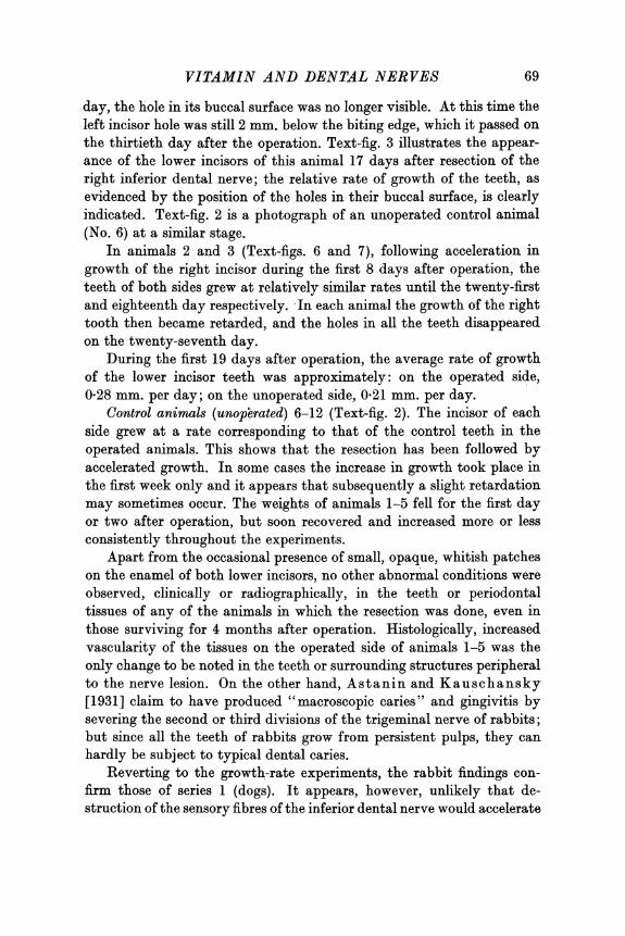
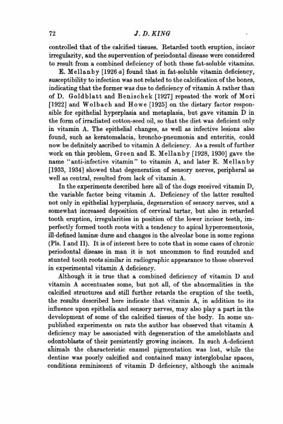
Results. The effects of the operation upon the rate of growth of theteeth of rabbits can best be described in conjunction with the accom-panying graphs (Text-figs. 5, 6 and 7), where AB represents the time
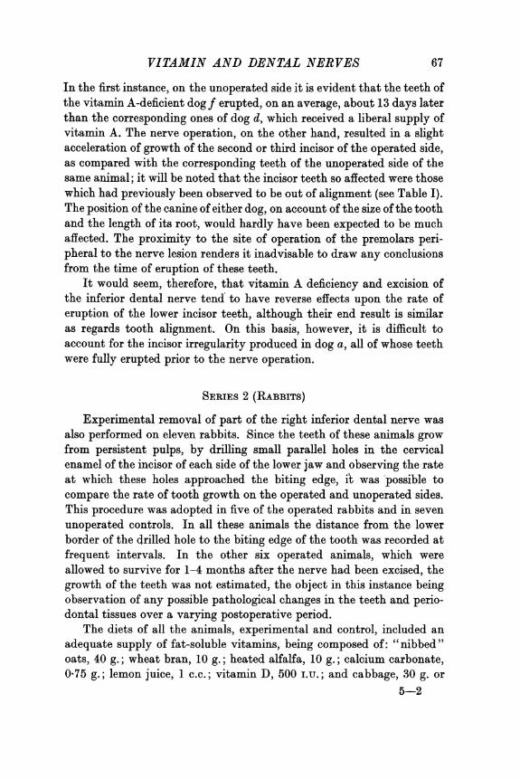
Fig. 2.
Fig. 1. Fig. 3.
~~~~~~Fig. 4.
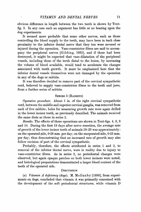
The effects of experimental resection of the inferior dental and cervical sympathetic nervesupon the rate of growth of the incisor teeth of rabbits. (See also p. 70, Text-figs. 5-10.)
Text-fig. 1. Radiograph of the lower jaw of a rabbit after excision of the right inferiordental nerve, showing the site of operation (0).
Text-fig. 2. Photograph of the incisors of control animal 6 (series 2), 17 days after holeshad been drilled in the cervical enamel of the lower teeth, showing their normal rateof growth. No nerve operation had been performed.
Text-fig. 3. Photograph of the incisors of animal 1 (series 2), 17 days after resection of theright inferior dental nerve. Note the more rapid rate ofgrowth of the lower tooth of theoperated side (0).
Text-fig. 4. Photograph of the incisors of animal 19 (series 3), 18 days after resection ofthe right cervical sympathetic trunk. Note the more rapid growth of the tooth of theoperated side (0).
after operation and BC the growth of the teeth during this period, theline passing horizontally through C being the level at which the drilledholes had grown out of the teeth. The following are typical of theseexperiments.
Animal 1 (Text-fig. 5). On the eighth day after operation it is seenthat the right incisor had grown distinctly faster than the left (control).This acceleration of growth increased steadily until, on the twenty-first
J. D. KINGB68
VITAMIN AND DENTAL NERVES
day, the hole in its buccal surface was no longer visible. At this time theleft incisor hole was still 2 mm. below the biting edge, which it passed onthe thirtieth day after the operation. Text-fig. 3 illustrates the appear-ance of the lower incisors of this animal 17 days after resection of theright inferior dental nerve; the relative rate of growth of the teeth, asevidenced by the position of the holes in their buccal surface, is clearlyindicated. Text-fig. 2 is a photograph of an unoperated control animal(No. 6) at a similar stage.
In animals 2 and 3 (Text-figs. 6 and 7), following acceleration ingrowth of the right incisor during the first 8 days after operation, theteeth of both sides grew at relatively similar rates until the twenty-firstand eighteenth day respectively. In each animal the growth of the righttooth then became retarded, and the holes in all the teeth disappearedon the twenty-seventh day.
During the first 19 days after operation, the average rate of growthof the lower incisor teeth was approximately: on the operated side,0-28 mm. per day; on the unoperated side, 021 mm. per day.
Control animals (unoperated) 6-12 (Text-fig. 2). The incisor of eachside grew at a rate corresponding to that of the control teeth in theoperated animals. This shows that the resection has been followed byaccelerated growth. In some cases the increase in growth took place inthe first week only and it appears that subsequently a slight retardationmay sometimes occur. The weights of animals 1-5 fell for the first dayor two after operation, but soon recovered and increased more or lessconsistently throughout the experiments.
Apart from the occasional presence of small, opaque, whitish patcheson the enamel of both lower incisors, no other abnormal conditions wereobserved, clinically or radiographically, in the teeth or periodontaltissues of any of the animals in which the resection was done, even inthose surviving for 4 months after operation. Histologically, increasedvascularity of the tissues on the operated side of animals 1-5 was theonly change to be noted in the teeth or surrounding structures peripheralto the nerve lesion. On the other hand, Astanin and Kauschansky[1931] claim to have produced "macroscopic caries" and gingivitis bysevering the second or third divisions of the trigeminal nerve of rabbits;but since all the teeth of rabbits grow from persistent pulps, they canhardly be subject to typical dental caries.
Reverting to the growth-rate experiments, the rabbit findings con-firm those of series 1 (dogs). It appears, however, unlikely that de-struction of the sensory fibres of the inferior dental nerve would accelerate
69
70 J. D. KING
tooth growth. It is of course possible that in rabbits the nerve lesion insome way affected the composition of the tooth, rendering it more or less
I OPIERAThD u6O
a CONTReL
WEMCHTIN ^RA
Fig. 5. Fig. 6. Fig. 7.
A DAY5 AFTER OR B A DAYS AFTER OP. B A DAYS AFTER OR B
Fig. 8. Fig. 9. Fig. 10.
The effect of experimental resection of the inferior dental and cervical sympathetic nervesupon the rate of growth of the incisor teeth of rabbits.
Text-figs. 5-7 illustrate graphically the rate of growth of the lower incisors of animals 1-3(series 2) after the inferior dental operation.
Text-figs. 8-10 illustrate the growth of the lower incisors of animals 19-21 (series 3) afterthe cervical sympathetic operation.
The distance BC=6-5 mm. in Text-figs. 5 and 6, and 6*0 mm. in Text-figs. 7-10; forfurther description of graphs see text, pp. 67 and 71.
brittle, but no such change was noted by the methods available. Cer-tainly the incisal edge on the operated side must, if the tooth grew faster,have been more susceptible to attrition than that of the control tooth,otherwise it would have projected beyond the latter. That there was no
VITAMIN AND DENTAL NERVES
obvious difference in length between the two teeth is shown by Text-fig. 3. In any case such an argument has little or no bearing upon thedog experiments.
It seemed more probable that some other nerves, such as thosecontrolling the blood supply to the teeth, may have been in huch closeproximity to the inferior dental nerve that they too were severed orinjured during the operation. Vaso-constrictor fibres are said to accom-pany the peripheral nerves [Gilding, 1932], and if these had beendestroyed, it might be expected that vaso-dilatation of the peripheralvessels, including those of the teeth distal to the lesion, by increasingthe volume of blood available, would tend to accelerate the changesassociated with tooth growth. It must be emphasized here that theinferior dental vessels themselves were not damaged by the operationin any of the dogs or rabbits.
It was therefore decided to remove part of the cervical sympatheticcord, believed to supply vaso-constrictor fibres to the teeth and jaws,from a further series of rabbits.
SERIES 3 (RABBITS)Operative procedure. About 1 in. cf the right cervical sympathetic
cord, between the middle and superior cervical ganglia, was removed fromeach of five rabbits; holes for measuring growth rate were again drilledin the lower incisor teeth, as previously described. The animals receivedthe same diets as those in series 2.
Results. The effects of these operations are shown in Text-figs. 4, 8, 9and 10. During the first 19 days after nerve resection, the average rateof growth of the lower incisor teeth of animals 19-23 was approximately:on the operated side, 0-28 mm. per day; on the unoperated side, 0-23 mm.per day; thus demonstrating that an increased rate of growth may alsofollow excision of part of the cervical sympathetic.
Probably, therefore, the effects attributed in series 1 and 2, toremoval of the inferior dental nerve, were in reality due to injury tovaso-constrictor fibres. As in series 2, no periodontal changes wereobserved, but again opaque patches on both lower incisors were noted,and histological preparations demonstrated a larger blood content of theteeth of the operated side.
DISCUSSION(a) Vitamin A deficiency (dogs). M. Mellanby [1930], from experi-
ments on dogs, concluded that vitamin A was primarily concerned withthe development of the soft periodontal structures, while vitamin D
71
J. D. KING
controlled that of the calcified tissues. Retarded tooth eruption, incisorirregularity, and the supervention of periodontal disease were consideredto result from a combined deficiency of both these fat-soluble vitamins.
E. Mellanby [1926 a] found that in fat-soluble vitamin deficiency,susceptibility to infection was not related to the calcification of the bones,indicating that the former was due to deficiency of vitamin A rather thanof D. Goldblatt and Benischek [1927] repeated the work of Mori[1922] and Wolbach and Howe [1925] on the dietary factor respon-sible for epithelial hyperplasia and metaplasia, but gave vitamin D inthe form of irradiated cotton-seed oil, so that the diet was deficient onlyin vitamin A. The epithelial changes, as well as infective lesions alsofound, such as keratomalacia, broncho-pneumonia and enteritis, couldnow be definitely ascribed to vitamin A deficiency. As a result of furtherwork on this problem, Green and E. Mellanby [1928, 1930] gave thename "anti-infective vitamin" to vitamin A, and later E. Mellanby[1933, 1934] showed that degeneration of sensory nerves, peripheral aswell as central, resulted from lack of vitamin A.
In the experiments described here all of the dogs received vitamin D,the variable factor being vitamin A. Deficiency of the latter resultednot only in epithelial hyperplasia, degeneration of sensory nerves, and asomewhat increased deposition of cervical tartar, but also in retardedtooth eruption, irregularities in position of the lower incisor teeth, im-perfectly formed tooth roots with a tendency to apical hypercementosis,ill-defined laminee durae and changes in the alveolar bone in some regions(Pls. I and II). It is of interest here to note that in some cases of chronicperiodontal disease in man it is not uncommon to find rounded andstunted tooth roots similar in radiographic appearance to those observedin experimental vitamin A deficiency.
Although it is true that a combined deficiency of vitamin D andvitamin A accentuates some, but not all, of the abnormalities in thecalcified structures and still further retards the eruption of the teeth,the results described here indicate that vitamin A, in addition to itsinfluence upon epithelia and sensory nerves, may also play a part in thedevelopment of some of the calcified tissues of the body. In some un-published experiments on rats the author has observed that vitamin Adeficiency may be associated with degeneration of the ameloblasts andodontoblasts of their persistently growing incisors. In such A-deficientamimals the characteristic enamel pigmentation was lost, while thedentine was poorly calcified and contained many interglobular spaces,conditions reminiscent of vitamin D deficiency, although the animals
72
VITAMIN AND DENTAL NERVES
were given ample amounts of vitamin D, calcium and phosphorus. Therat experiments confirm to some extent the findings of Wolbach andHowe [1933], who maintained that vitamin A played an extremelyimportant part in the calcification of the teeth of rats. But the experi-ments of these investigators were not decisive, since their A-deficientrations were also deficient in vitamin D, while in their adequate dietsvitamin A was given in the form of butter, a variable source of vitamin Awhich contains an appreciable amount of vitamin D.
It is usually considered that vitamin D controls calcification bymobilizing the mineral salts of the blood. It seems possible that vita-min A may affect calcification by its influence upon the morphology andconsequent activity of the lime-secreting cells, even when vitamin Dand mineral salts are available. If this is so, its absence may contributeto the development of periodontal disease and even dental caries bycausing defects of tooth structure as well as by increased susceptibilityto infection. It is hoped that the experimental production of suchdiseases, preliminary reports of which have already been published[King, 1935 a, b], may shed further light on this problem.
By means of a histological examination of human post-mortemmaterial Wilkinson [1935] showed that degenerated cells of thesubgingival epithelium may become calcified before the latter becomesseparated from the tooth; later, when separation does occur, he claimsthat the calcified cells are left adhering to the tooth as calculus, theformation of the latter being similar to calcification in cartilage asdescribed by Robison [1923]. In support of this theory Wilkinsoncites the experiments of Smith [1930] and Adamson [1929], as dem-onstrating that desquamated cells of the gingival epithelia liberate aphosphatase, which, by setting phosphates free in the presence of calciumions, allows calcium phosphate to be deposited in the dead cells.Wilkinson believes that calculus may thus be produced, resulting inthe inflammatory complications so often encountered in human perio-dontal disease. Certainly epithelial degeneration, calculus formation,and gingivitis are common sequelae to experimental vitamin A deficiency,so that since these conditions are also found in the human subject,whatever be the exact role of vitamin A in this syndrome, it may beinferred that lack of this vitamin, especially during the developmentalperiod, is an important predisposing factor in the aetiology of periodontaldisease.
(b) Experimental nerve lesions (dogs and rabbits). The relative uni-formity in results of the experiments in series 2 and 3 (rabbits) indicates
73
that the effects produced by resection of the inferior dental nerve in bothdogs and rabbits were caused by the unintentional severing of vaso-constrictor fibres in very close association with, and probably within theactual sheath of, the sensory nerve. It is probable, therefore, that inboth species the temporarily increased rate of growth of the teeth peri-pheral to the lesion was due to an increase in the volume of blood avail-able. Gilding [1932] showed that the distribution of the vaso-con-strictor nerves is strictly unilateral, stopping abruptly at the mid-line,in spite of the fact that the blood vessels of one side anastomose freelywith those of the other. The sensory distribution of the inferior dentalnerve, on the other hand, frequently extends for some little distanceacross the mid-line. The growth changes here reported were confined tothe operated side.
The temporary nature of such changes suggests that the walls of theaffected blood vessels soon recover the greater part, at least, of theirnormal tone, and this may perhaps be why no histological alterations inthe dental tissues were found by the methods described. It is true thatmicroscopic preparations of the rabbits' lower incisor teeth after deathshowed more blood in the tooth pulp of the operated side, but this maybe merely because the normally innervated vessels on the other side wereconstricted in the asphyxia induced by the chloroform used for killingthe animals. In this respect the animals in which the inferior dentalnerve was excised were like those in which the sympathetic was cut, sothat it is probable that the constrictor nerves had been severed in allthree series of animals and that the growth changes observed were dueto this.
Leist [1927] described an increased rate of growth of the teeth offive out of nine guinea-pigs after cauterizing the sympathetic fibresaround the carotid artery with 7 p.c. phenol; he also noted similar butless marked changes following such treatment of the internal maxillaryand inferior dental arteries of a few dogs. In the cervical sympatheticof the dog there are dilator as well as constrictor fibres [Dastre andMorat, 1884], but not in the rabbit or cat [Feldberg and Schiff,1926]. As the constrictors predominate over the dilators in the dog'ssplanchnic nerve [Dale, 1913], this may also be expected in its cervicalsympathetic.
Although resection of the inferior dental nerve, as previously de-scribed, has failed to cause lesions in the teeth or periodontal tissues, it ispossible that, by destruction of the whole sensory supply to one side ofone or both jaws and by preventing nerve regeneration for a sufficiently
74 J. D. KING
VITAMIN AND DENTAL NERVES
long period, pathological changes similar to those seen in vitamin Adeficiency may be produced. Experiments along these lines are now inprogress; in some, the sensory roots of the fifth nerve are resected, while inothers the Gasserian ganglion from which they originate is itself removed.
The results of the present nerve operations may have some bearingon the vexed problem of tooth eruption, for they show that, in additionto vitamins A and D, the blood supply and condition of the vascularnerves play a part in the development and growth of the teeth. G ottlieb[1927] claims that changes in the rate of growth of the teeth are ofoutstanding importance. He believes that eruption continues slowlyafter the teeth have come into occlusion, and that any conditions whichdisturb the synchronism between attrition of the tooth crowns and"postclinical" eruption accelerate the downgrowth of the subgingivalepithelium and the development of periodontal disease. If the mis-alignment of the right lower incisors following the operation in the adultdog which received vitamin A was due to changes in the blood supply,it seems possible that such changes, resulting in abnormal movementsof the teeth even after complete clinical eruption, may in turn give riseto changes in their supporting structures. The fact that in this dog theperiodontal tissues appeared free from defects and there was no down-growth of subgingival epithelium along the root or absorption of alveolarbone, does not seem to support Gottlieb's hypothesis, although moreexperiments on adult animals are necessary. Movements of the teethafter clinical eruption are, of course, quite commonly seen in the humanmouth, associated with trauma, pressure from other teeth, or extractionof adjacent or opposing teeth.
SUMMARY
In dogs receiving vitamin A-deficient diets:(1) eruption of teeth was delayed;(2) hypercementosis occurred and the laminae durao and bone of the
tooth sockets were malformed;(3) the alignment of the incisor teeth was irregular; in addition to the
periodontal and nervous defects described in previous papers.In animals (dogs and rabbits) in which the inferior dental nerve was
resected on one side:(1) the eruption in dogs, and in rabbits the growth of teeth was
accelerated;(2) the alignment of the incisor teeth was irregular; but no defects
in the dental or periodontal tissues were found.
75
76 J. D. KING
As similar changes were observed on resection of the cervical sym-pathetic, the effects of severing the inferior dental nerve are regarded asdue to damage to vasomotor fibres in it.
The expenses of this investigation have been defrayed by the Medical Research Council,to whom my thanks are due. I am also indebted to Mrs M. Mellanby and Prof. G. A.Clark for their valuable advice and criticism, and to Miss I. Jopling for clerical assistance.
REFERENCES
Adamson, K. T. (1929). Aust. J. Dent. 33, 245.Astanin, P. P. & Kauschansky, L. J. (1931). Dtsch. Mschr. Zahnheilk. 49, 12.Dale, H. H. (1913). J. Physiol. 40, 291.Dastre & Morat (1884). Systeme neurveux va8omoteur. Paris.Feldberg & Schiff (1926). Pfuigers Arch. 212, 365.Gilding, H. P. (1932). J. Physiol. 74, 34.Goldblatt, H. & Benischek, M. (1927). J. exp. Med. 46, 699.Gottlieb, B. (1927). J. Amer. dent. A8s. 14, 2178.Green, H. N. & Mellanby, E. (1928). Brit. med. J. 2, 691.Green, H. N. & Mellanby, E. (1930). J. Exp. Path. 11, 81.King, J. D. (1935 a). Dent. Rec. 55, 522.King, J. D. (1935 b), Brit. dent. J. 59, 233 and 305.Leist, M. (1927). Z. Stomat. No. 8.Mellanby, E. (1926 a). Brit. med. J. 1, 515.Mellanby, E. (1933). Edin. med. J. 40, No. 4.Mellanby, E. (1934). J. Path. Bact., Lond., 38, 391.Mellanby, M. (1929). Sp. Rep. Ser. Med. Res. Coun. No. 140.Mellanby, M. (1930). Ibid. No. 153.Mellanby, M. & King, J. D. (1934). Brit. dent. J. 56, 538.Mori, S. (1922). J. Amer. med. Ass. 79, 197.Robison, R. (1923). Biochem. J. 17, 286.Smith, G. H. (1930). Aust. J. exp. Biol. Med. Sci. 7, 45.Wilkinson, F. C. (1935). Dent. Rec. 55, No. 3, p. i.Wolbach, S. B. & Howe, P. R. (1925). J. exp. Med. 42, 753.Wolbach, S. B. & Howe, P. R. (1933). Amer. J. Path. 9, 275.
THE JOURNAL OF PHYSIOLOGY, VOL. 88, No. 1
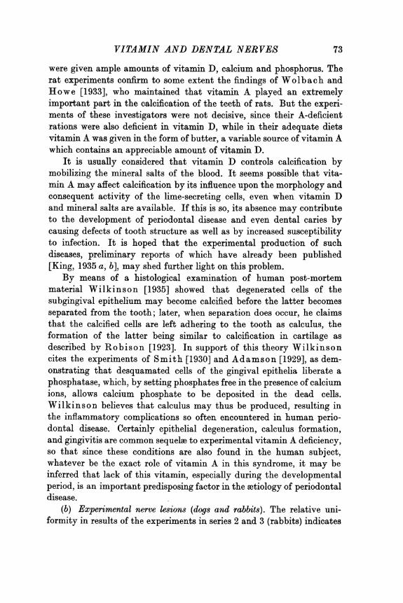
Fig. 1. Doga. Fig. 2. Dogb.
Fig. 3. Dog c. Fig. 4. Dogg.
To face p. 76
PLATE I
THE JOURNAL OF PHYSIOLOGY, VOL. 88, No. 1
at~~~~~~~~~~~~~~~~~7 0PF`7'
?jiJ
-0, ' . 2 _ __~~~~~~~~~~~~~~~~fd\|El
PLATE 1I
VITAMIN AND DENTAL NERVES 77
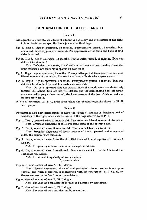
EXPLANATION OF PLATES I AND 11
PLATE I
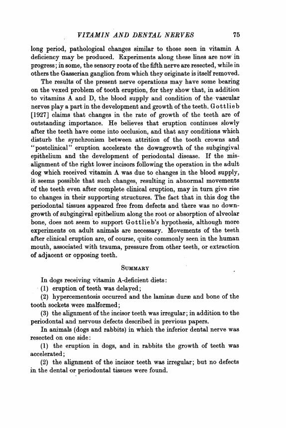
Radiographs to illustrate the effects of vitamin A deficiency and of resection of the rightinferior dental nerve upon the lower jaw and teeth of dogs.
Fig. 1. Dog a. Age at operation, 22 months. Postoperative period, 12 months. Dietcontained liberal supplies of vitamin A. The appearance of the teeth and bone of bothsides is normal.
Fig. 2. Dog b. Age at operation, 11 months. Postoperative period, 11 months. Diet wasdeficient in vitamin A.
Note. Defective tooth roots, ill-defined lamine dure and, surrounding these, thebone trabeculae are more radio-opaque on both sides.
Fig. 3. Dog c. Age at operation, 2 months. Postoperative period, 6 months. Diet includedliberal amounts of vitamin A. The teeth and bone of both sides appear normal.
Fig. 4. Dog g. Age at operation, 2 months. Postoperative period, 5 months. Diet wasdeficient in vitamin A but calcium carbonate was added.
Note. On both operated and unoperated sides the tooth roots are defectivelyformed, the laminae dure are not well-defined and the surrounding bone trabeculseare more radio-opaque than normal; the lower margin of the jaw of this animal wasinjured after death.
0, site of operation. A, B, C, areas from which the photomicrographs shown in P1. IIwere prepared.
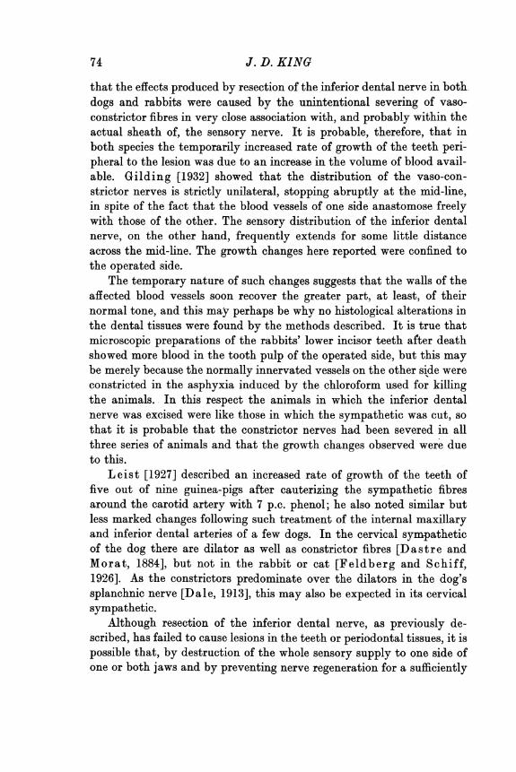
PLATE II
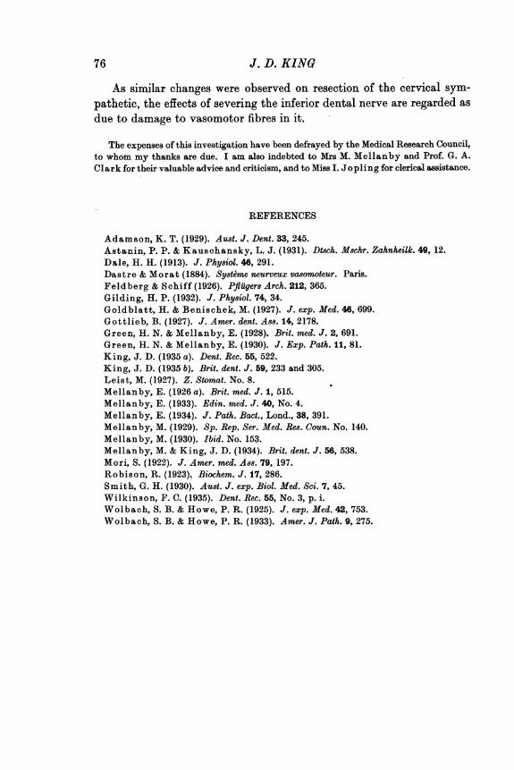
Photogrophs and photomicrographs to show the effects of vitamin A deficiency and ofresection of the right inferior dental nerve of the dogs referred to in P1. I.
Fig. 1. Dog a, operated when 22 months old. Diet contained liberal amount of vitamin A.Note. Irregular alignment of the lower front teeth of the operated side.
Fig. 2. Dog b, operated when 11 months old. Diet was deficient in vitamin A.Note. Irregular alignment of lower incisors of both operated and unoperated
sides; the canines were removed.
Fig. 3. Dog c, operated when 2 months old. Diet included liberal supplies of vitamins Aand D.
Note. Irregularity of lower incisors of the operated side.
Fig. 4. Dog g, operated when 2 months old. Diet was deficient in vitamin A but calciumcarbonate was added.
Note. Bilateral irregularity of lower incisors.0, operated side.
Fig. 5. Ground section of area A, P1. I, dog a.Note. Normal appearance of apical and peri-apical tissues; section is not quite
central, but, when considered in conjunction with the radiograph (P1. I, fig. 1), thetissues are seen to be free from obvious defects.
Fig. 6. Ground section of area B, P1. I, dog b.Note. Invasion and replacement of pulp and dentine by cementum.
Fig. 7. Ground section of area C, P1. I, dog g.Note. Invasion of pulp and dentine by cementum.