Embed Size (px)
Citation preview
BY :Dr.
Zeinab Hakim
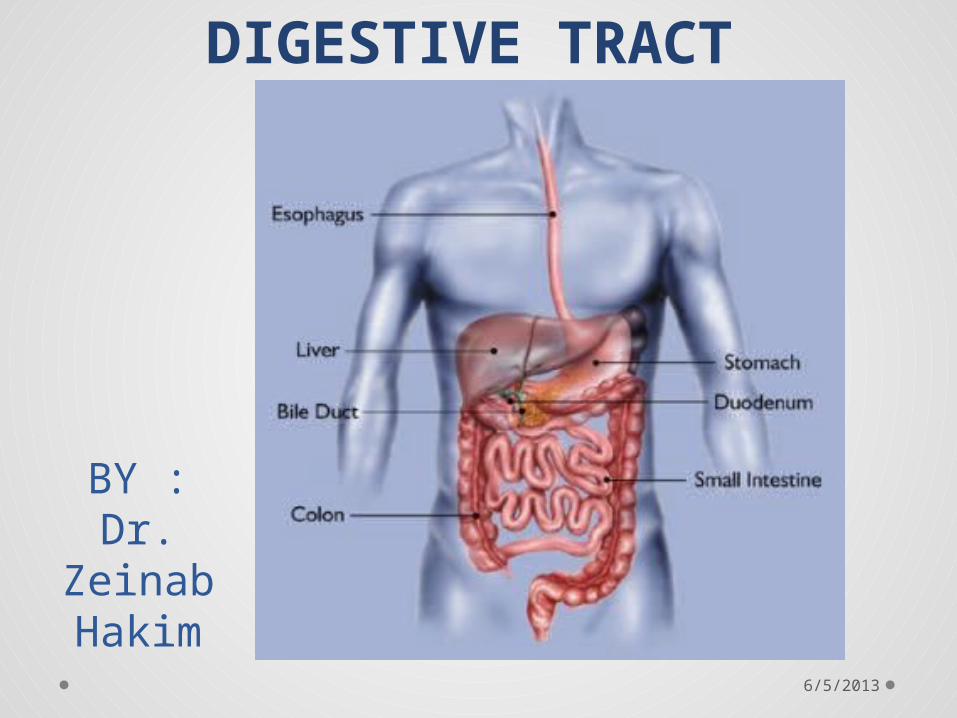
DIGESTIVE TRACT
6/5/2013

The roof of the mouth
anterior2/3rd by hard palate and posterior1/3rd by soft palate.
• The soft palate is made up of following five muscles.
1. Tensor veli palatini ( Trigeminal Nerve)2. Levator veli palatini3. Musculus uvulae4. Palatopharyngeus ( Glossopharyngeal 5. Palatoglossus
6/5/2013
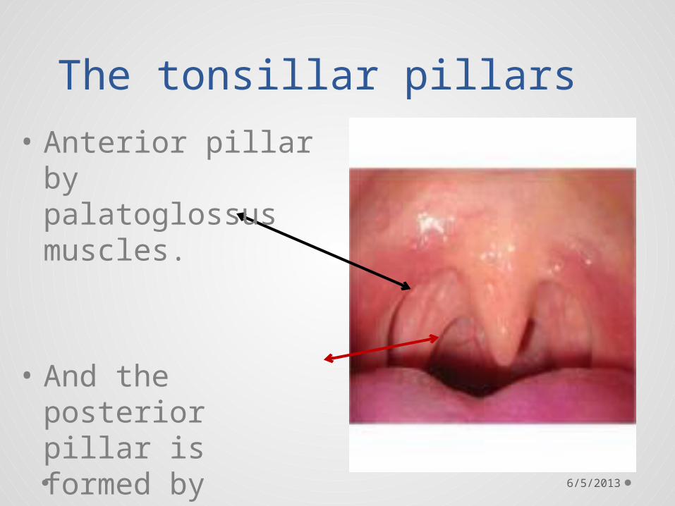
The tonsillar pillars
• Anterior pillar by palatoglossus muscles.
• And the posterior pillar is formed by palatopharyngeus muscles.
6/5/2013

6/5/2013

• Glossopharyngeal nerve
• General sensation carried by lingual nerve, a branch of trigeminal.
• The special sensation by chorda tympani nerve, a branch of facial nerve.
6/5/2013

The muscles of tongueThe extrinsic muscles
are:• Hyoglossus• Styloglossus• Genioglossus• Chondroglossus• Palatoglossus
The intrinsic muscles are:
• Longitudinalis linguae• Verticalis linguae• Transversus linguae
All muscles of tongue are supplied by hypoglossal nerve except palatoglosus which
is supplied by glossopharyngeal nerve6/5/2013
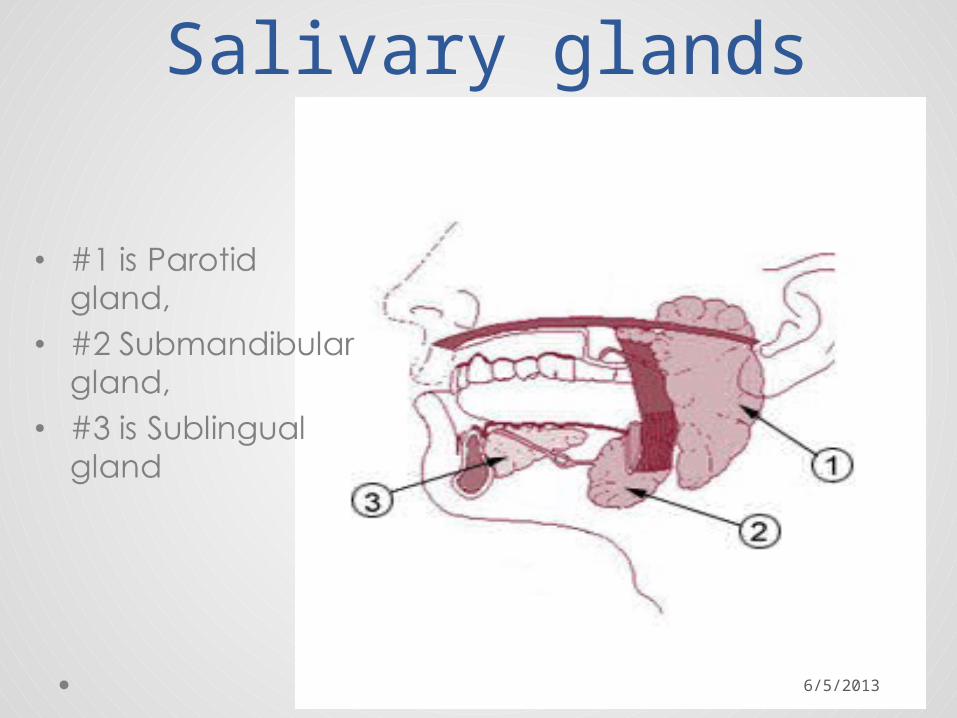
Salivary glands
6/5/2013
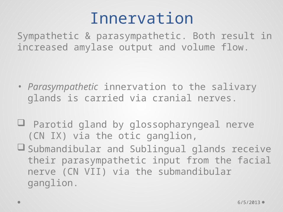
InnervationSympathetic & parasympathetic. Both result in increased amylase output and volume flow.
• Parasympathetic innervation to the salivary glands is carried via cranial nerves.
Parotid gland by glossopharyngeal nerve (CN IX) via the otic ganglion,
Submandibular and Sublingual glands receive their parasympathetic input from the facial nerve (CN VII) via the submandibular ganglion.
6/5/2013

Esophagus
• Muscular wall o upper third, or superior part: striated muscleo middle third, smooth muscle and striated muscleo inferior third: predominantly smooth muscles
• adventitia
Artery Esophageal arteries
Vein Esophageal veins
Nerve Celiac ganglia, vagus
Precursor Foregut
6/5/2013

Esophageal constrictions
• Normally, the esophagus has three anatomic constrictions at the following levels:
• At the esophageal inlet, where the pharynx joins the esophagus, behind the cricoid cartilage (14–16 cm from the incisor teeth).
• Where its anterior surface is crossed by the aortic arch and the left bronchus (25–27 cm from the incisor teeth).
• Where it pierces the diaphragm (36–38 cm from the incisor teeth).
6/5/2013

Barium swallow
6/5/2013

Stomach
6/5/2013

Artery
Right gastric artery, left gastric artery, right gastro-omental artery, left gastro-omental artery, short gastric arteries
Vein
Right gastric vein, left gastric vein, right gastro-omental vein, left gastro-omental vein, short gastric veins
Nerve Celiac ganglia, vagus
Lymph Celiac lymph nodes6/5/2013

Lesser omentum • The is a double fold of peritoneum arising from
the lesser curvature of stomach to the hilum of liver.
It carries within its folds, portal vein, hepatic artery, bile duct and gastric arteries.
6/5/2013
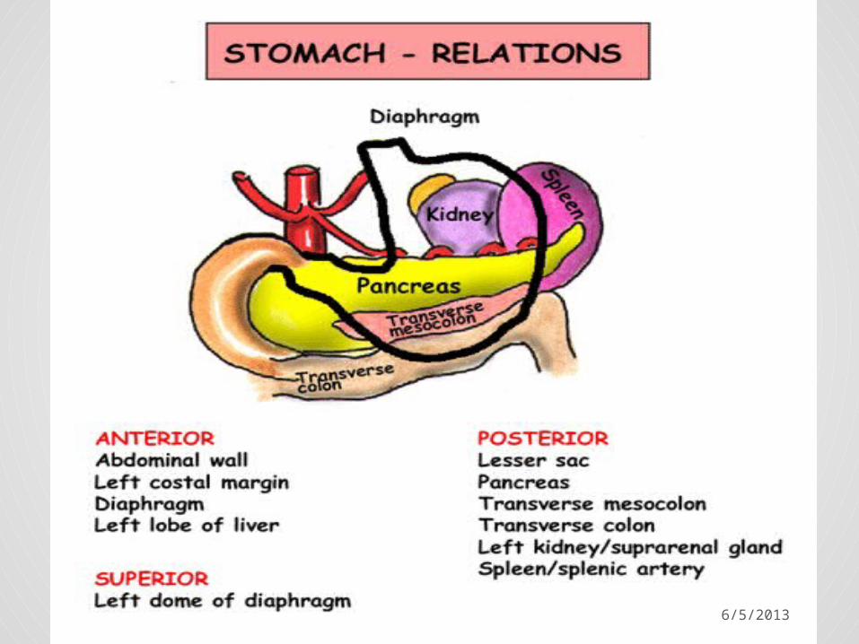
The greater omentum
• Is a double fold of peritoneum hanging from the greater curvature of stomach, extending up to left dome of diaphragm and enclosing spleen on one side and on the other side forms an apron like fold hanging from the greater curvature into the abdominal cavity. As it reflects back up to the transverse colon.
The fold of peritoneum after enclosing the transverse colon extends unto the posterior abdominal wall, as transverse mesocolon.
6/5/2013

6/5/2013

• The lesser sac, also known as the omental bursa, is the cavity in the abdomen that is formed by the lesser and greater omentum
• the greater sac, also known as the general cavity (of the abdomen) or peritoneum of the peritoneal cavity proper, is the cavity in the abdomen that is inside the peritoneum but outside of the lesser sac .
The connection between lesser sac and the greater sac via the epiploic foramen (also known as the Foramen of Winslow)
6/5/2013

6/5/2013

6/5/2013
6/5/2013

6/5/2013

Barium meal
6/5/2013

Small intestine
Artery Superior mesenteric artery
Vein Hepatic portal vein
Nerve Celiac ganglia, vagus
Lymph Intestinal lymph trunk
6/5/2013

Has three parts:• Duodenum: Here the digestive juices from the
pancreas hormones and the gall bladder (bile) mix. The digestive enzymes break down proteins and bile and emulsify fats into micelles. The duodenum contains Brunner's glands which produce bicarbonate. In combination with bicarbonate from pancreatic juice, this neutralizes HCl of the stomach.
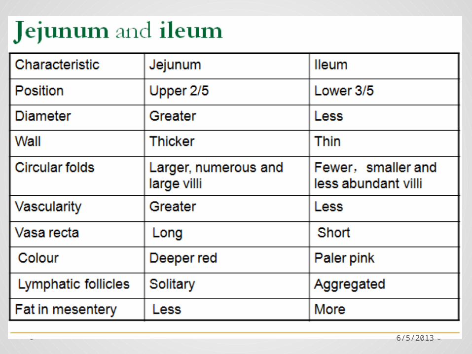
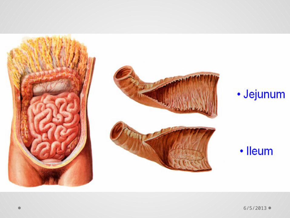
• Jejunum: This is the midsection of the intestine, connecting the duodenum to the ileum. It contains the plicae circulares, and villi to increase the surface area of that part of the GI Tract. Products of digestion (sugars, amino acids, fatty acids) are absorbed into the bloodstream.
• Ileum: Has villi and absorbs mainly vitamin B12 and bile acids, as well as any other remaining nutrients.
6/5/2013
6/5/2013
6/5/2013
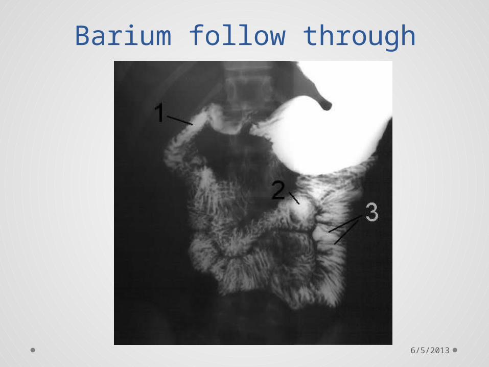
Barium follow through
6/5/2013

Large intestine
6/5/2013

6/5/2013

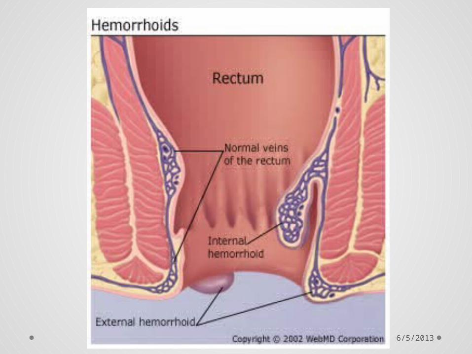
Rectum
Artery
Superior rectal artery (first two-thirds of rectum), middle rectal artery (last third of rectum)
Vein Superior rectal veins, middle rectal veins
Nerve Inferior anal nerves, Inferior mesenteric ganglia
Lymph
Inferior mesenteric lymph nodes, pararectal lymph nodes,internal iliac lymph nodes, Deep inguinal lymph nodes
Precursor Hindgut
6/5/2013
6/5/2013

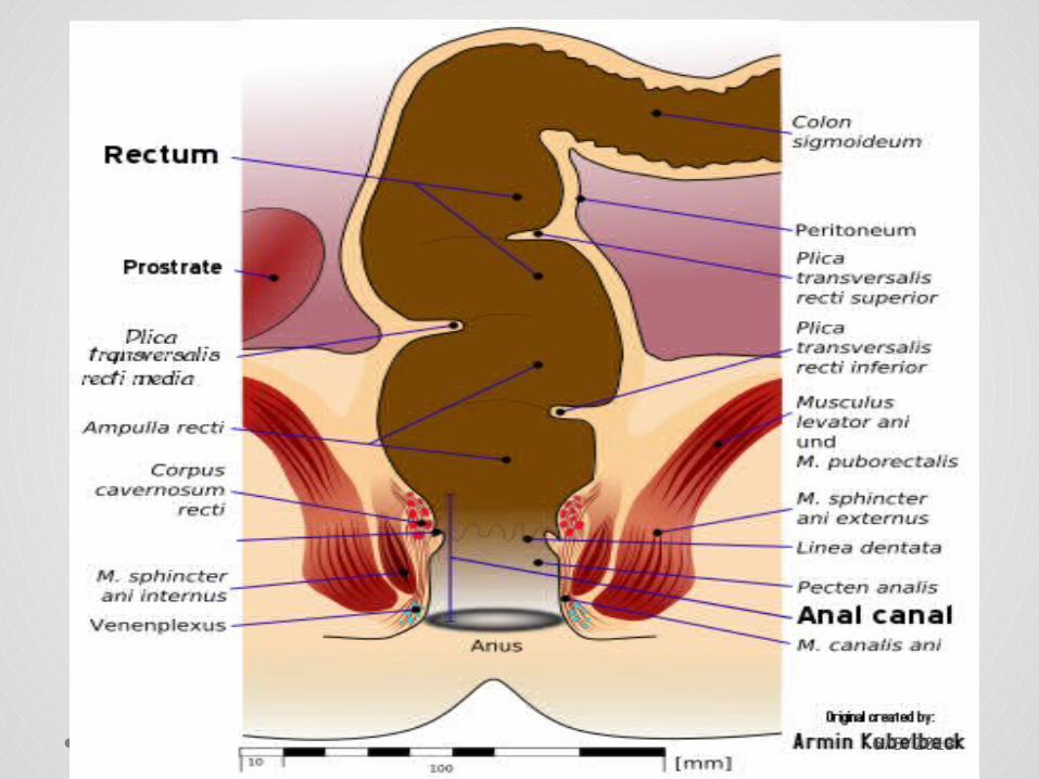
The posterior surface of entire rectum is retroperitoneal (extra peritoneal).• Its upper third is covered by peritoneum on the
front and sides.• The middle third is covered by peritoneum on the
front only.• The lower third is completely retroperitoneal (extra
peritoneal).
6/5/2013

Anal canal
Artery Inferior rectal artery
Vein Inferior rectal vein
Nerve Inferior rectal nerves
Lymph Superficial inguinal lymph nodes
Precursor Proctodeum
The anal canal is related to the perineal body in front and the anococcygeal body behind; both of these are fibromuscular structures.
6/5/2013

The anal canal is divided into two unequal sections, upper and lower.• The upper 2/3 has longitudinal folds or elevations of tunica
mucosa. Its mucosa is lined by simple columnar epithelium. Its lower ends are joined together by folds of mucus membrane called anal valves. The upper 2/3 of the anal canal is supplied by the superior rectal artery which is a branch of the inferior mesenteric artery.
• The lower 1/3 of the anal canal is lined by stratified squamous epithelium that blends with the skin. The lower third of the anal canal is supplied by the inferior rectal artery which is a branch of the internal pudendal artery.
A whitish line called Hilton's white line or pecten of Jon Stroud ( Pectinate or dentate ) indicates the junction between keratinized stratified squamous epithelium and non-keratinized stratified squamous epithelium.
6/5/2013

ANAL CANAL
6/5/2013
6/5/2013
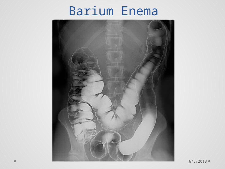
Barium Enema
6/5/2013

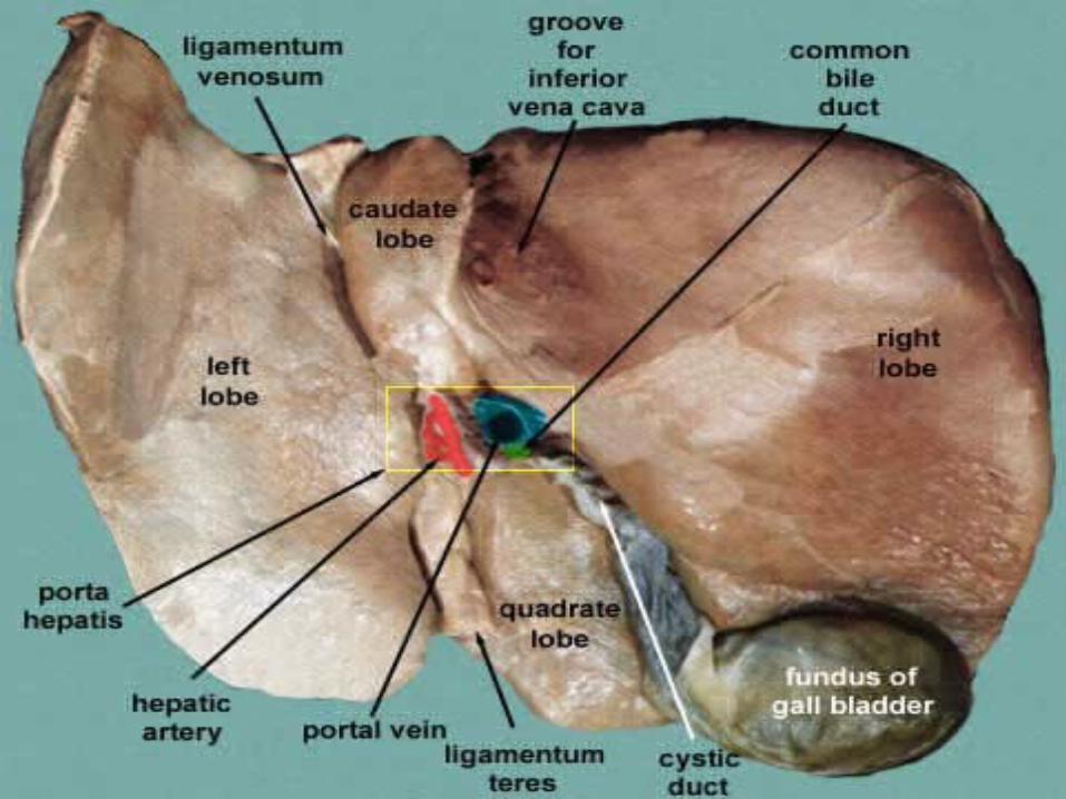
(1) right lobe, (2) left lobe, (3) caudate lobe, (4) quadrate lobe, (5) hepatic artery and portal vein, (6) hepatic lymph nodes, (7) gall bladder.
LIVER
6/5/2013
6/5/2013

6/5/2013

Ligament of liver• Falciform ligament• Coronary ligament• Right and left triangular ligaments• Gastro hepatic ligament• Duodenohepatic ligament• ligamentum venosum • ligamentum teres.
6/5/2013

Anterior abdominal walllayers from out to in
• Skin• Fascia
o Camper's fascia - fatty superficial layer.o Scarpa's fascia - deep fibrous layer.
• Muscle o Rectus abdominiso External oblique muscleo Internal oblique muscleo Transverse abdominal muscle
• Fascia transversalis• Peritoneum
6/5/2013

Inner surface of abdominal wall
Ligament/fold Remnant of Hernia
median umbilical ligament urachus -
medial umbilical ligament umbilical artery direct inguinal hernia
lateral umbilical fold inferior epigastric vessels
indirect inguinal hernia
The inner surface contains several ligaments separated by fossae:
6/5/2013

Peritoneum
• The peritoneum is the serous membrane that forms the lining covering of the abdominal cavity
Layers :• The outer layer, called the parietal peritoneum, is attached to the
abdominal wall and the pelvic walls.• The inner layer, the visceral peritoneum, is wrapped around the
internal organs that are located inside the intraperitoneal space. It is thinner than the parietal peritoneum.
• The potential space between these two layers is the peritoneal cavity; it is filled with a small amount (about 50 mL) of slippery serous fluid that allows the two layers to slide freely over each other.
• The term mesentery is often used to refer to a double layer of visceral peritoneum. There are often blood vessels, nerves, and other structures between these layers. The space between these two layers is technically outside of the peritoneal sac, and thus not in the peritoneal cavity.
6/5/2013

Peritoneal ligaments1. Falciform ligament2. Gastophrenic
ligament3. Gastro splenic
ligament4. Lien renal ligament5. Coronary ligament6. Triangular
ligaments7. Round ligament
6/5/2013

Mesenteries
Sources Structure From To Contains
Dorsal mesentery
Mesentery proper
Small intestine (jejunum and ileum)
Posterior abdominal wall
Superior mesenteric artery
Transverse mesocolon
Transverse colon
Posterior abdominal wall
Middle colic
Sigmoid mesocolon
Sigmoid colon Pelvic wall Sigmoid
arteries
Mesoappendix
Mesentery of ileum Appendix Appendicul
ar artery
6/5/2013
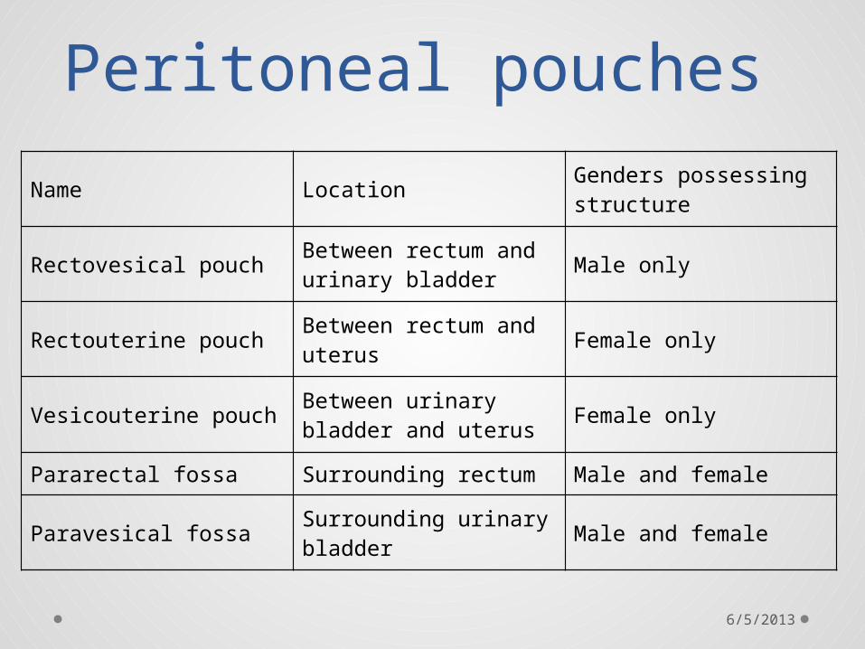
Peritoneal pouches
Name Location Genders possessing structure
Rectovesical pouch Between rectum and urinary bladder Male only
Rectouterine pouch Between rectum and uterus Female only
Vesicouterine pouch Between urinary bladder and uterus Female only
Pararectal fossa Surrounding rectum Male and female
Paravesical fossa Surrounding urinary bladder Male and female
6/5/2013

Intraperitoneal RetroperitonealInfraperitoneal / Subperitoneal
Stomach, First part of the duodenum jejunum, ileum, cecum, appendix,transverse colon, sigmoid colon, rectum (upper 1/3)
The rest of the duodenum, ascending colon, descending colon, rectum (middle 1/3)
Rectum (lower 1/3)
Liver,spleen, pancreas (only tail)
Pancreas (except tail)
Kidneys, adrenal glands, proximal ureters, renal vessels
Urinary bladder, distal ureters
In women: uterus, fallopian tubes, ovaries
Gonadal blood vessels
Inferior vena cava, aorta 6/5/2013

Inguinal ligament The inguinal ligament is a band running from the pubic tubercle to the anterior superior iliac spine.
It is formed by the external abdominal oblique aponeurosis and is continuous with the fascia lata of the thigh.
Structures that pass deep to the inguinal ligament include:• Psoas major, iliacus, pectineus• Femoral nerve, artery, and vein• Lateral cutaneous nerve of thigh• Lymphatics
6/5/2013

6/5/2013
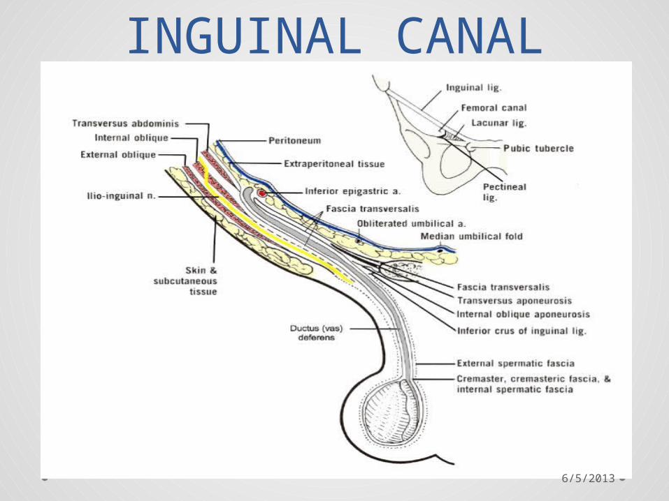
INGUINAL CANAL
6/5/2013

superior wall (roof):
• Medial crus of aponeurosis of external oblique
• Musculoaponeurotic arches of internal oblique and transverse abdominal
• Transversalis fascia
Anterior wall:•
aponeurosis of external oblique
•fleshy part of internal oblique (lateral third of canal only)
•superficial inguinal ring (medial third of canal only)
BOUNDARIES OF INGUINAL CANAL
posterior wall:•
transversalis fascia•
conjoint tendon (Inguinal falx,reflected part of inguinal ligament, medial third of canal only)
• deep inguinal ring (lateral third of canal only)
inferior wall (floor):•
inguinal ligament•
lacunar ligament (medial third of canal only)
• iliopubic tract (lateral third of canal only)
6/5/2013

Contents in males : the spermatic cord and its coverings + the ilioinguinal nerve*. in females : the round ligament of the uterus + the ilioinguinal nerve*.
The classic description of the contents of spermatic cord in the male are:• 3 arteries: artery to vas deferens (or ductus deferens),
testicular artery, cremasteric artery;• 3 fascial layers: external spermatic, cremasteric, and internal
spermatic fascia;• 3 other structures: pampiniform plexus, vas deferens (ductus
deferens), testicular lymphatics;• 3 nerves: genital branch of the genitofemoral nerve (L1/2),
autonomic and visceral afferent fibres, ilioinguinal nerve (N.B. outside spermatic cord but travels next to it)
* The ilioinguinal nerve passes through the superficial ring to descend into the scrotum, but does not formally run through the canal.
6/5/2013

Inguinal Hernia
Type Description
Relationship to inferior
epigastric vessels
Covered by internal
spermatic fascia
Usual onset
indirect inguinal hernia
protrudes through the inguinal ring and is
ultimately the result of the failure of
embryonic closure of the internal inguinal ring after the testicle
passes through it
Lateral Yes Congenital
direct inguinal hernia
enters through a weak point in the fascia of the abdominal wall
(Hesselbach triangle)
Medial No Adult
6/5/2013

6/5/2013