Embed Size (px)
Citation preview
ON A PAIR OF NEGRO FEMORA.' By THOMAS H. BRYCE,M.A., M.B., Lecturer on Anatomy, Queen Margaret College,University of Glasgow.
IN a recent number of this Journal I published some "1 Notes onthe Myology of a Negro." The pair of femora which form thesubject of the present paper belonged to the same individual,and I purpose to describe the characters of the bones, in theirrelation to certain muscular arrangements described in my formerpaper, and to discuss certain analogies between them and thecelebrated Trinil femur.
Right femur.-The upper end of the bone is much deformedby chronic rheumatic arthritis. The head is irregular, and itsarticular surface eburnated; the neck and trochanter major,as well as the subtrochanteric region of the shaft, are muchthickened by the irregular deposit of new bone. The rest ofthe shaft and the lower extremity show no signs of pathologicalchange.The linea aspera is somewhat prominent, giving a pilasteric
index of 109-2,-rather below the average for negro bones givenby Hepburn.2 The bicondylar width is 82 mm. The inter-condylar notch is narrow, and the curvature of the condyles isconsiderably sharper than in a European bone. The back ofeach condyle shows an accessory facet.
The popliteal surface measures 11 cm. in height. Theexternal supracondylar ridge is prominent; it attached a strongaccessory fasciculus of the biceps, which passed from the longhead to the femur. The internal supracondylar ridge is onlyslightly marked, while running down the space between thetwo lines is an oblique ridge, which gave attachment to thefemoral head of the biceps. This ridge commences close to theinternal supracondylar ridge above, but ends below in the
I Read at the meeting of the Anatomical Society of Great Britain andIreland, June 10, 1897.
2 Jour. Anat. and Phys., vol. xxxi. part 1, Oct. 1896.

ON A PAIR OF NEGRO FEMORA. 77
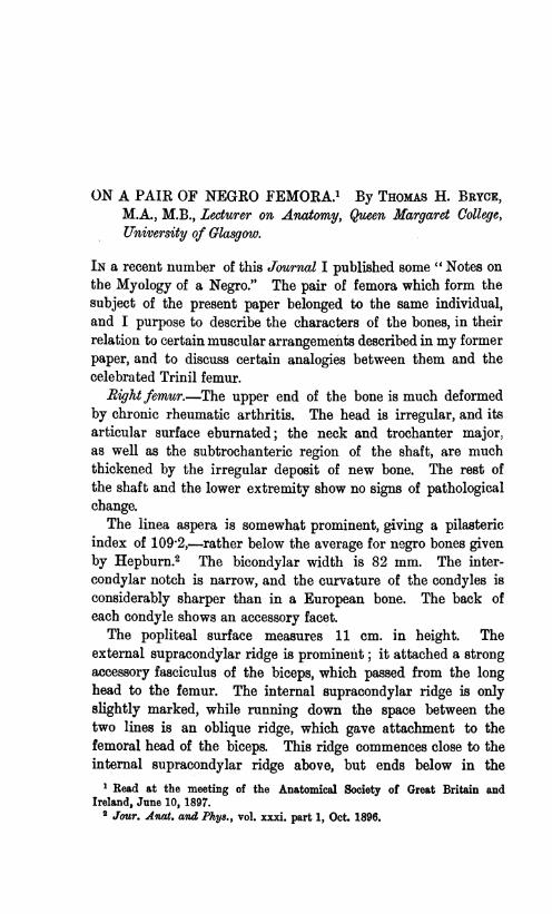
FIG. 2.FIG. 1.

MR THOMAS H. BRYCE.
middle line 4 cm. above the condyles. Below this level thereis a prominent mesial convexity running down to the inter-condylar notch, bounded on each side by a supracondylar hollow.The general effect is to give the popliteal surface a convex
contour, which is expressed by the following measurements,taken at a point 4 cm. above the highest point of the externalarticular surface, on the front of the bone (Manouvrier's 4 cm.line).' The antero-posterior diameter mp-that is, with theposterior limb of the callipers resting on the most prominentpoint of the popliteal surface-is 31 mm., while the diametermn-the posterior point being taken on the external supra-condylar ridge-is 30 mm. The transverse diameter is 36 mm.,giving the high popliteal index of 86&2. There is no suspicionof pathological change here, the convexity being due to thepresence of the ridge which gave attachment to the femoralhead of the biceps.
Left femur.-The total oblique length is 470 mm., just aboutthe average given by Hepburn; 2 the diameter of the head is 52mm. The subtrochanteric region of the shaft shows somedegree of flattening. The antero-posterior diameter is 23 mm.,the transverse 32-5 mm., giving a platymeric index of 7017, con-siderably below the average (77 7) given by Hepburn for nineNegro bones. The anterior part of the shaft is here somewhatconcave, due chiefly to the projection of the internal border,which rises into a prominent ridge opposite the lesser trochanter,but fades away again at the junction of the upper and middlethirds of the shaft. The spiral line runs into this ridge infront of the lesser trochanter, and is not continued upwards asthe anterior intertrochanteric line. The latter is only faintlymarked, the concavity of the anterior part of the shaft beingcontinued up on to the neck of the bone. The outer border orexternal infratrochanteric ridge (Turner)3 is rounded. The pro-jection of the linea aspera is not very marked,- the antero-posterior diameter is 29 mm., the tranverse 28-5 mm., giving apilasteric index of 101-7. Thus there is a low pilasteric associ-ated with a somewhat low platymeric index; whereas, according
1 Manouvrier, "Deuxihme 6tude sur le Pithecanthropus erectus," Bull. de laSoc. dAnthrop. de Paris, tome sixilme, 1896, fasc. v. (ivW serie).
2 Jour. Anat. and Phys., vol. xxxi. part 1, Oct. 1896.3 Challenger Reports, part xlvii. p. 97, and Proc. Scot. Soc. 4ntiq., May 1895.
78

ON A PAIR OF NEGRO FEMORA.
to Manouvrier, a low platymeric is generally associated with a highpilasteric index. In the middle section of the shaft the hollowfor the vastus externus is well marked, but there is no corre-sponding pilasteric excavation for the vastus internus, so thatthe inner face of the shaft is convex for the whole of itsextent. It has been suggested that subtrochanteric flatten-ing is due to increased development of the upper portion ofthe crureus and vastus internus (Manouvrier), or that thisportion of the bone is modified in shape by its being acted onby the opposing forces of these muscles and the gluteus maximus(Hepburn), the forces coming into action in the muscular effortof walking in a mountainous country. The degree of flatteningin this bone is not sufficiently marked to test completely thishypothesis, but it is interesting to note, in connection with it,the arrangement of the muscles supposed to be concerned. Thegluteus maximus was a powerful muscle, but attached just asusual; there is no third trochanter, and the lower part of itsimpression is a hollow, not a raised ridge, and placed on theposterior aspect of the bone. The vastus externus was verywell developed, and could not be separated from the crureusbelow, but above it came separately from the tubercle of thefemur, and the shaft in front of and below the great trochanter,as usual. The crureus and vastus internus were inseparableabove, but the latter was regarded as being confined to theinner face of the bone, behind the prominent inner border,while the upper part of the crureus, which was better developedthan is usual, arose from the hollowed-out anterior surface ofthe shaft and anterior intertrochanteric line.The bicondylar width is 82 cm., and the intercondylar notch
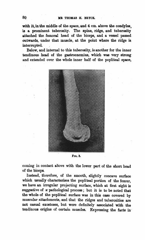
is narrower even than on the right bone, and the sharp cur-vature of the condyles, especially of the external, is striking(fig. 3).The popliteal surface extends 11 cm., 4 mm. above the con-
dyles; the external and internal supracondylar ridges are promi-nent, and supported by bony pillars or buttresses. Each risesabove into a spine; the outer of these spines attached theaccessory fasciculus of the biceps, the inner forms the upper endof a prominent oblique ridge, which passes downwards on to thepopliteal surface. This ridge is interrupted below, but in a line
79
MR THOMAS H. BRYCL
with it, in the middle of the space, and 4 cm. above the condylesis a prominent tuberosity. The spine, ridge,.and tuberosityattached the femoral head of the biceps, and a vessel passedoutwards, under that muscle, at the point where the ridge is.interrupted.
Below, and internal to this tuberosity, isanother for the innertendinous head of the gastrocnemius, which was very strongand extended over the whole inner half of the popliteal space,
FIG. 8.
coming in contact above with the lower part of the short headof the biceps.
Instead, therefore, of the smooth, slightly concave surfacewhich usually characterizes the popliteal portion of the femur,we have an irregular projecting surface, which at first sight issuggestive of a pathological process; but it is to be noted thatthe whole of the.popliteal surface was in this case covered bymuscular attachments, and that the ridges and tuberosities arenot casual exostoses, but were definitely associated with thetendinous origins of certain muscles. Expressing the facts in-
8Q

ON A PAIR OF NEGRO FEMORA.
figures, we find that at the 4 cm. line mp= 37 mm.; m.n.=35 mm.; transverse= 40 mm.; giving a popliteal index of 92-5.The pair of femora above described present a double analogy
to the Trinil fossil,-.1st, in respect of the signs of a pathologicalprocess associated with the formation of exostoses; 2nd, in re-spect of certain characters which are common to both, thoughthey may not be present in the same degree. Dubois claimedthat the fossil was unique in the possession of three characters-a concave anterior intertrochanteric line, a convex internalsurface, and a convex or rounded contour of the poplitealsurface.
ManouvrierI and Hepburn2 have, however, shown that allthese three characters may be present in certain rare specimens,in the large collections of human femora examined by them.With regard to the first two characters there is no dispute.They are present in many femora as well as in the pair whichare the subject of this paper, but there has been considerablediscussion as to the convexity of the popliteal space.Many regard the feature in the fossil as a pathological one,
but both the observers above named have described femorawhich approach closely the fossil in this respect, and are yetfree from the suspicion of being pathological. In explanationof the convexity of the popliteal surface, Manouvrier 1 has sug-gested that the bone retains its cylindrical character to a lowerlevel than usual, owing to the descent of the vastus externus.Hepburn,2 with more probability I think, suggests that thepopliteal surface in these cases has been occupied by muscle.He further suggests, from his observations on the limbs of theanthropoid apes, that the adductor magnus may descend furtherthan usual, and he desires information as to origin of thefemoral head of the biceps in the lower races of men.
In this instance I have been able to demonstrate that in theright bone, without any suspicion of its being pathological, theconvexity of the popliteal surface is due to the presence of aridge from which the femoral head of the biceps arose, while inthe left bone the impression for that muscle, and for the innerhead of the gastrocnemius are marked by bony prominences.The development of the spine and crest in the upper part
1 Loc. cit. 2 Loc. cit.VOL. XXXII. (N.S. VOL. XII.) F
81

82 ON A PAIR OF NEGRO FEMORA.
of the tendinous origin of the short head of the biceps isan instance of the very process suggested by Sir Wm. Turneras explaining the exostosis on the Trinil femur. The Negro wasthe subject of a disease associated with the formation of periostealnodes, and both tibiae showed irregular exostoses due to thiscause. It does not follow, however, that the exostoses on thepopliteal space are necessarily pathological, for they are strictlymuscular impressions. On the other hand, the thickening ofthe supracondylar ridges is difficult to account for unless it bepathological, and this throws the whole open to the samesuspicion.The interest of the specimens in relation to the Trinil femur
lies in this combination of the two factors, morphological andpathological; and the left bone suggests a simple and notimpossible hypothesis as to the cause of the convexity of thepopliteal surface of the fossil femur.