Embed Size (px)
Citation preview

CLINICAL MICROBIOLOGY REVIEWS, Apr. 2006, p. 403–434 Vol. 19, No. 20893-8512/06/$08.00�0 doi:10.1128/CMR.19.2.403–434.2006Copyright © 2006, American Society for Microbiology. All Rights Reserved.
Burn Wound InfectionsDeirdre Church,1,2,3* Sameer Elsayed,1,2 Owen Reid,3 Brent Winston,4 and Robert Lindsay3
Division of Microbiology, Calgary Laboratory Services,1 and Departments of Pathology & Laboratory Medicine,2 Surgery,3 andCritical Care,4 Calgary Health Region and University of Calgary, Calgary, Alberta, Canada
INTRODUCTION .......................................................................................................................................................404HUMAN SKIN—A MAJOR HOST DEFENSE......................................................................................................404BURN INJURY IN CIVILIANS................................................................................................................................405
Magnitude and Risk Factors of Civilian Burn Injury.......................................................................................405Pathogenesis and Etiology of Burns ....................................................................................................................405
Thermal injury ....................................................................................................................................................405Chemical injury...................................................................................................................................................406
Extent and Location of Burn Injury ....................................................................................................................406Inhalation Injury.....................................................................................................................................................407Early Excision and Burn Wound Closure ...........................................................................................................408
IMMUNOLOGICAL RESPONSE TO BURN INJURY ........................................................................................408Systemic Response to Burn Injury .......................................................................................................................409
Inflammatory response to burn injury ............................................................................................................409Anti-inflammatory response to burn injury ....................................................................................................409
Innate Immune System Response to Burn Injury..............................................................................................409Adaptive Immune System in Response to Burn Injury.....................................................................................410Altering the Immunologic Response to Burn Injury .........................................................................................410
EPIDEMIOLOGY OF BURN WOUND INFECTIONS ........................................................................................410Impact of Patient Demographics and Burn Severity.........................................................................................411Impact of Changes in Burn Wound Care............................................................................................................411
PATHOGENESIS OF BURN WOUND INFECTIONS .........................................................................................412Pathogenesis ............................................................................................................................................................412Biofilm Formation...................................................................................................................................................412Microbial Etiology ..................................................................................................................................................412Virulence Factors and Tissue Invasion................................................................................................................413
CLASSIFICATION OF BURN WOUND INFECTIONS.......................................................................................413Types of Burn Wound Infection............................................................................................................................414
Burn wound impetigo .........................................................................................................................................414Burn-related surgical wound infection ............................................................................................................414Burn wound cellulitis .........................................................................................................................................414Invasive infection in unexcised burn wounds .................................................................................................414
MICROBIOLOGICAL ANALYSIS OF BURN WOUND INFECTIONS ............................................................414Best Approach for Burn Wound Infection Surveillance....................................................................................414Burn Wound Sampling Techniques......................................................................................................................415
Superficial wound samples ................................................................................................................................415Tissue biopsy .......................................................................................................................................................416Sampling techniques for other microbial pathogens .....................................................................................416
Specimen Transport ...............................................................................................................................................416Analysis of Burn Wound Specimens ....................................................................................................................416
Gram stain...........................................................................................................................................................416Surface swab culture ..........................................................................................................................................416Quantitative tissue culture ................................................................................................................................417Histological analysis...........................................................................................................................................417
Distinguishing Burn Wound Colonization from Infection................................................................................418Antimicrobial Susceptibility Testing ....................................................................................................................418
Antimicrobial resistance and burn units.........................................................................................................418Burn unit antibiogram .......................................................................................................................................419
Other Types of Infection........................................................................................................................................419Fungal infections.................................................................................................................................................419Viral infections ....................................................................................................................................................419
* Corresponding author. Mailing address: Calgary Laboratory Ser-vices, 9-3535 Research Rd. N.W., Calgary, Alberta, Canada T2L 2K8.Phone: (403) 770-3281. Fax: (403) 770-3347. E-mail: [email protected].
403
on January 10, 2019 by guesthttp://cm
r.asm.org/
Dow
nloaded from

OTHER TYPES OF INFECTION IN BURN PATIENTS.....................................................................................419Sepsis and Toxic Shock Syndrome.......................................................................................................................420Pneumonia ...............................................................................................................................................................421Urinary Tract Infections ........................................................................................................................................421Catheter Infections and Suppurative Thrombophlebitis...................................................................................422Myonecrosis .............................................................................................................................................................422
PREVENTION OF BURN WOUND INFECTIONS ..............................................................................................422Topical Antimicrobial Therapy .............................................................................................................................423
Silver nitrate........................................................................................................................................................423Silver sulfadiazine...............................................................................................................................................423Mafenide acetate .................................................................................................................................................424Acticoat A.B. dressing/Silverlon........................................................................................................................424Mupirocin (Bactroban) ......................................................................................................................................424Nystatin ................................................................................................................................................................424Other topical antimicrobials .............................................................................................................................424
Prophylactic Systemic Antibiotics ........................................................................................................................425Selective Bowel Decontamination .........................................................................................................................425Prevention of Tetanus ............................................................................................................................................425Infection Control in the Burn Unit ......................................................................................................................426
FUTURE DIRECTIONS IN MICROBIAL BURN WOUND SURVEILLANCE................................................426REFERENCES ............................................................................................................................................................368
INTRODUCTION
Burns are one of the most common and devastating forms oftrauma. Patients with serious thermal injury require immediatespecialized care in order to minimize morbidity and mortality.Data from the National Center for Injury Prevention and Controlin the United States show that approximately 2 million fires arereported each year which result in 1.2 million people with burninjuries (7, 318, 319, 369). Moderate to severe burn injuries re-quiring hospitalization account for approximately 100,000 ofthese cases, and about 5,000 patients die each year from burn-related complications (7, 8, 215, 318, 319, 369). In Canada, theestimated numbers of burn victims and deaths in serious cases areproportionally smaller on a per capita basis (265, 349, 403).
The survival rates for burn patients have improved substantiallyin the past few decades due to advances in modern medical carein specialized burn centers. Improved outcomes for severelyburned patients have been attributed to medical advances in fluidresuscitation, nutritional support, pulmonary care, burn woundcare, and infection control practices. As a result, burn-relateddeaths, depending on the extent of injury, have been halvedwithin the past 40 years (7, 252, 320, 369, 373, 439). In patientswith severe burns over more than 40% of the total body surfacearea (TBSA), 75% of all deaths are currently related to sepsisfrom burn wound infection or other infection complicationsand/or inhalation injury (15, 20, 24, 32, 140).
This review focuses on modern aspects of the epidemiology,diagnosis, management, and prevention of burn wound infectionsand sepsis. Recent factors contributing to the development ofburn wound infection are also discussed, including the nature andextent of the burn injury itself and the secondary immunosup-pression resulting from thermal injury. The prevention of burnwound infection is reviewed in the context of newer therapeuticstrategies employed by specialized burn care facilities.
HUMAN SKIN—A MAJOR HOST DEFENSE
An intact human skin surface is vital to the preservation ofbody fluid homeostasis, thermoregulation, and the host’s pro-
tection against infection. The skin also has immunological,neurosensory, and metabolic functions such as vitamin D me-tabolism. Thermal injury creates a breach in the surface of theskin. A basic knowledge of skin anatomy and physiology isrequired to understand emergency burn assessment and ap-proaches to burn care (96, 114, 469).
Figure 1 provides a schematic representation of the skinlayers in relation to the depth of burn injury (96, 113, 369). Theskin is derived from ectoderm and mesoderm and has twoanatomic layers: the epidermis or outermost nonvascular layerconsists of several layers of epidermal cells that vary in thick-ness over various body surfaces, and the dermis or corium islargely made of collagen and contains the microcirculation, acomplex vascular plexus of arterioles, venules, and capillaries.The two skin layers are bound together by a complex mecha-nism that is essential for normal function. Epidermal append-ages are distributed throughout the dermis layer, including thesweat glands, sebaceous glands, and hair follicles. The dermallayer is capable of producing new epithelial cells to replacethose lost from the epidermis by burning or other injury to theskin because the shafts of these appendages are lined withepithelial cells. Nerve endings occur throughout both skin lay-
FIG. 1. Basic skin anatomy, showing the depth of injury for first-,second-, and third-degree burns. (Adapted from reference 369 withpermission of the publisher.)
404 CHURCH ET AL. CLIN. MICROBIOL. REV.
on January 10, 2019 by guesthttp://cm
r.asm.org/
Dow
nloaded from

ers, and the connective tissue of the dermis also provides a firmstructural base for the skin. Burn injury is a very painful formof trauma because of the multitude of pain receptors andnerves that traverse the skin layers. Beneath the skin lie thesubcutaneous tissues, muscle, and bone.
The skin is one of the largest organs in the human body, interms of both its overall size and weight. In an adult male theskin weighs between 6 and 10 kg (�13 and 22 lbs). The averageadult skin surface area is 1.5 to 2.0 square meters, in contrastto that of a newborn, whose skin surface area is only 0.2 to 0.3square meter. The two skin layers together are up to severalmillimeters thick, but both epidermal and dermal thicknessvaries depending on the body site. The epidermis is the thin-nest (0.05 mm) over the eyelid but thicker (up to 1 mm) overthe soles of the feet (114, 369). The dermis is thickest on theback. Males generally have thicker skin than females. Generalskin thickness peaks in midlife and gradually thins as part ofthe aging process (113, 114, 216, 217, 369). Infants, youngchildren, and elderly adults have a much thinner dermal layerto their skin, resulting in an increased propensity for deeperburn injury. Epidermal cells are constantly being shed andreplaced every month through a process that continuallypushes new cells to the surface. This natural process is de-signed to continually replenish and heal breaches in the out-ermost protective skin barrier, be it from the microtraumassustained as part of daily living or from overt injury. Theepidermis therefore heals itself after superficial injury.
Several important physiological functions of the skin arealtered by thermal injury. Survival of the severely burned pa-tient requires immediate access to a specialized burn care unit.Modern emergency burn resuscitation and ongoing treatmentare designed to alleviate the systemic changes that result fromacute disruption of a large part of the skin barrier. Meticulousattention is given to the replacement and prevention of fluidloss, the maintenance of body temperature homeostasis withina constant normal range, the easing of severe pain, and theprevention of infection.
BURN INJURY IN CIVILIANS
Magnitude and Risk Factors of Civilian Burn Injury
In North America, burn injury is one of the main causes ofinjury deaths, particularly in children under the age of 14 years(7, 68–70, 318, 320, 349). Although the age-adjusted death ratefrom burn injury in the United States has decreased substan-tially since 1985, the United States still has one of the highestper capita burn death rates of any industrialized country (318,320). Between 1993 and 1995, there were 18.7 burn-relateddeaths per million population in the United States, comparedwith 15 for Canada and 5.5 for Switzerland (318, 344, 349). Thehighest fatality rates occur among children 4 years of age oryounger and adults over the age of 55 years (252, 318, 320,457). Burn-related deaths in these two age groups account formore than two-thirds of all fire deaths. Males are twice as likelyto die of burn-related injury as females in all age groups.
Adult burn injury may also result from an industrial orwork-related accident or occur as a result of suicide attempts,assault, and unintentional injury due to alcohol and/or druguse (32, 211, 265, 332, 368). A significant proportion of adult
burn patients also suffer from a high degree of mental illness(344). Since legal action is taken in many of these cases, it isimportant to document the etiology and extent of the burninjury.
Burn injuries incur a significant cost to the health care sys-tem in North America and worldwide. In the United States andCanada there are currently 167 centers specializing in burncare, with over 2,000 beds (369). Although the overall hospi-talization rates from less-serious burn injuries have declined by50% since 1971, the proportion of patients admitted to burncenters has increased (7, 369). Recent estimates in the UnitedStates show that 45,000 patients are admitted to acute-carehospitals annually with burn injuries, and in approximately50% of these cases the extent of thermal injury is severeenough to warrant admission to a specialized burn center (7,68–70, 369). Burn care centers in North America currentlyadmit an average of more than 200 patients per year, whereasother hospital units admit an average of fewer than five burnpatients per year (7, 369).
Initial hospitalization costs and physicians’ fees for special-ized care of a patient with a major burn injury are currentlyestimated to be US$200,000 (292, 318, 369). Overall, costsescalate for major burn cases because of repeated admissionsfor reconstruction and rehabilitation therapy. In the UnitedStates, current annual estimates show that more than US$18billion is spent on specialized care of patients with major burninjuries (292, 318, 369).
Pathogenesis and Etiology of Burns
The breached skin barrier is the hallmark of thermal injury.The body tries to maintain homeostasis by initiating a processof contraction, retraction, and coagulation of blood vesselsimmediately after a burn injury. Three distinct zones have beendefined within the burn wound: (i) the zone of coagulation,which comprises the dead tissues that form the burn escharthat is located at the center of the wound nearest to the heatsource; (ii) the zone of stasis, which comprises tissues adjacentto the area of burn necrosis that is still viable but at risk forongoing ischemic damage due to decreased perfusion; and (iii)the zone of hyperemia, which comprises normal skin with min-imal cellular injury that has predominant vasodilation and in-creased blood flow as a response to injury (Fig. 2) (163, 195,369). Serious thermal injury causes total loss of the skin surfaceover large areas of the body. Because of the importance of theskin as a barrier to microbial host invasion, it is not surprisingthat the risk of subsequent burn wound infection and systemicinfection correlates with the size of the burn injury (377, 387).
Thermal injury. Direct contact with flame, a hot surface orhot liquid (scald), or a source of heat conduction, convection,or radiation causes a degree of cellular damage to the skin thatvaries with the temperature and duration of exposure (21, 179,222, 232, 299, 352, 355, 369). As the temperature rises, increas-ing molecular collisions occur, resulting in altered molecularconformation and the disruption of intermolecular bonds. Thisprocess leads to cell membrane dysfunction as ion channels aredisrupted, resulting in sodium and water intake. As the tem-perature rises further, protein denaturation occurs, oxygenradicals are liberated, and eventually cells die with the forma-tion of the burn eschar (299).
VOL. 19, 2006 BURN WOUND INFECTIONS 405
on January 10, 2019 by guesthttp://cm
r.asm.org/
Dow
nloaded from

Chemical injury. Chemical interaction may also damageprotein structures. A classification system that was described in1974 and remains in use groups chemicals according to theirmode of action (Table 1) (9, 56, 57, 74, 223, 305).
Extent and Location of Burn Injury
A burn patient is a trauma patient. The initial assessmentand resuscitation are therefore focused on the patient’s airway,breathing, and circulation and an examination for other majorinjuries besides the burn itself. Assessment of the burn injuryshould include a determination of the etiology of the burn aswell as the extent of the burn injury. Although the assessmentof the extent and depth of all types of burn injury is clinicallydifficult, chemical injuries are particularly challenging. The se-verity of injury is not only related to the areas and sites of skininjury, but also depends on the chemical agent and the dura-
tion of exposure. In these cases, morbidity may be high evenwith small areas of injury, such as alkali injury to the eye, withinhalation of vapors such as anhydrous ammonia due to thesystemic effects from absorption (9, 56).
Subjective clinical methods have historically been used todetermine the depth of burn injury. Body diagrams providean estimate of the percentage of total body surface area(%TBSA) of the burn exposure and injury and document apatient’s initial and clinical ongoing assessment in this regard(202, 369). Areas of partial and full-thickness burn injury aredescribed, noting areas of circumferential involvement andburn injury across joints. Figures 3 and 4 outline a schematicassessment of %TBSA for adult and pediatric burn patients,respectively, using the rule of nines. A Berkow’s percentagechart can also be used to obtain a more accurate estimate of%TBSA (40).
However, the clinical methods outlined above may not pro-vide sufficient accuracy of evaluation of burn depth to supportcrucial treatment decisions such as the extent of excision andgrafting required. Laser Doppler imaging (LDI) has recentlybeen shown to provide a more objective measurement onwhich to base the decision to operate (25, 199, 224). A recentprospective blinded trial compared the clinical outcome ofusing LDI versus clinical judgment to assess injury depth. Atotal of 23 burn patients and 41 wounds were analyzed by bothmethods. LDI and the surgeon agreed on determination of
FIG. 2. Zones of injury for superficial and deep second-degreeburns. (Adapted from reference 369 with permission of the publisher.)
FIG. 3. Body diagram for estimation of total burned surface area(%TBSA) in adults, using the rule of nines (numbers are for anterioronly and posterior only). (Adapted from reference 369 with permissionof the publisher.)
TABLE 1. Classification of chemicals that cause burn injurya
Class Example(s) Mode of action
Reducing agents Hydrochloric acid Bind free electrons intissue proteins
Oxidizing agents Sodium hypochlorite Oxidized on contactingproteins producingtoxic by-products
Corrosive agents Phenol Denatures tissue proteinsProtoplasmic
poisonsHydrofluoric acid Bind calcium or other
ions essential to cellfunction
Acetic acid
Vesicants Dimethyl sulforide Ischemia with anoxicnecrosisCantharides
Mustard gasDesiccants Sulfuric acid Dehydration
Muriatic acid Exothermic reaction
a Data are from references 9, 56, 57, 74, 223, and 305.
406 CHURCH ET AL. CLIN. MICROBIOL. REV.
on January 10, 2019 by guesthttp://cm
r.asm.org/
Dow
nloaded from

wound depth only 56% of the time (P � 0.031) (224). LDIagreed with wound biopsy confirmation when the scan indi-cated a need for excision. LDI also enabled excision to proceedearlier even when the surgeon’s clinical assessment agreed withthe LDI scan results. LDI may therefore be used as an effectiveaid to clinical judgment in modern burn centers for deciding toexcise burn wounds of indeterminate depth.
Inhalation Injury
Inhalation injury occurs in anywhere from 3 to 21% of burnpatients and is a major cause of mortality; 80% of fire-relateddeaths occur from hypoxia due to oxygen deprivation or frominhalation of the toxins found in smoke (15, 32, 43, 342, 372,425, 438). Inhalation injury of the lung in adults is usuallyproportional to the depth and extent of body surface areaburned (252, 279, 342, 440). Children have a lower rate ofinhalation injury because of the prominence of scald injury inthis group (200, 320). Hypoxia occurs when the carbon mon-oxide generated by combustion is inhaled and binds to hemo-globin. Even a relatively low concentration of carbon monox-ide in inhaled air can be significant because its affinity forhemoglobin is much greater than that of oxygen (e.g., 200-fold). Hydrogen cyanide and other agents generated in smokeare potent toxins and exacerbate the acidosis that occurs as aresult of burn injury (43, 275).
The pathogenesis of pulmonary injury from smoke inhala-tion has been well described (99–102, 201, 203, 433). Directheat injury is restricted to the upper airway above the glottisand is manifested by rapid swelling with the threat of obstruc-tion. Steam inhalation is the only type of heat that damages thelower respiratory tract. However, inhalation of smoke andproducts of combustion and destruction of the tracheobron-chial respiratory epithelium cause chemical injury. Inhalationinjury progresses during the first few days following a burn and
results in edema and sloughing of the respiratory tract mucosaand impairment of the normal mucociliary clearance mecha-nism. Damage of the mucociliary lining of the respiratory tractdecreases the clearance of invading microorganisms. Pulmo-nary edema results from direct microvascular injury and therelease of oxygen free radicals and inflammatory mediators.Cast formation due to aggregates of mucus and cellular debriscauses obstruction of moderate-size airways when the mucosasloughs. Disruption of endothelial and epithelial integrity re-sults in exudation of protein-rich plasma into terminal airways,which, in combination with atelectasis, leads to bacterialgrowth and the subsequent development of pneumonia. Smokeinhalation also destroys type II pneumocytes, which results inimpaired surfactant production (15, 251, 411).
Advances in respiratory resuscitation support in trauma in-tensive care units have improved the prognosis for burn pa-tients with inhalation injury (15, 104, 139, 275). Inhalationinjury should be suspected if the patient was burned in anenclosed space, has facial burns, and/or develops progressivehoarseness or stridor or a cough productive of carbonaceoussputum. The clinical effects of thermal inhalation injury typi-cally become manifest within a few hours after injury, whereaschemical injury of the lower respiratory tract progresses moreslowly (e.g., 1 to 2 days) (101, 322). Stridor that developsimmediately after heat injury associated with an increased re-spiratory rate, worsening hypoxemia, and trouble expectorat-ing secretions are signs of worsening edema of the upper air-way (e.g., glottis), and immediate airway intubation is requiredto maintain patency (15, 95, 275). Similar signs of impendingrespiratory failure also develop in burn patients with a smokeinhalation injury and require immediate respiratory resuscita-tion. Intubation and mechanical ventilation as well as intensivetracheobronchial care (e.g., regular airway suctioning and ther-apeutic bronchoscopy) are required to assist clearance of bron-chial mucus and debris (275, 312). High-frequency ventilation
FIG. 4. Body diagram for estimation of total burned surface area (%TBSA) in children, using the rule of nines (numbers include anterior andposterior). (Adapted from reference 369 with permission of the publisher.)
VOL. 19, 2006 BURN WOUND INFECTIONS 407
on January 10, 2019 by guesthttp://cm
r.asm.org/
Dow
nloaded from
may also be beneficial in the clearance of secretions and alsostabilizes collapsed and diseased lung segments (104, 275).
Patients with inhalation injury have greater fluid require-ments than those who have only sustained a cutaneous injury.More fluid must be given in the immediate period followingthermal injury in patients with inhalation injury (208, 369).Various agents have been administered, including inhaled hep-arin along with bronchodilators or free-radical scavengingagents such as dimethyl sulfoxide or N-acetylcysteine, in thetreatment of inhalation injury in order to decrease cast forma-tion and small-airway obstruction (15, 61, 229, 275). Nitricoxide is a potent vasodilator that has recently been adminis-tered as inhalation therapy to burn patients with acute respi-ratory distress syndrome due to lung injury in order to reduceventilation-perfusion mismatch by dilating blood vessels per-fusing lung alveoli (15, 122).
Early Excision and Burn Wound Closure
Prior to the widespread use of early surgical excision of burnwounds, conservative management was practiced. Colonizationof the burn wound was permitted to break down the burneschar so that it separated spontaneously. Daily cleansing andimmersion hydrotherapy were used to debride necrotic surfaceeschar (49, 73, 113, 216, 385). Skin grafting occurred only afterthe development of granulation tissue on the burn wound’ssurface.
Although early surgical excision and grafting have been re-peatedly attempted in the 20th century, the outcomes wereinitially poor (218, 219, 295, 296). However, an improved un-derstanding of the pathophysiology of burns allowed the ad-vancement of multiple intra- and postoperative medical andsurgical techniques that has resulted in gradual decreases inmorbidity and mortality (66, 87, 127, 181, 196, 200, 470). Med-ical support to maintain hemodynamic and respiratory func-tion within the trauma intensive care unit and operating the-ater, the provision of early adequate nutrition, and the use ofsurgical techniques that minimize blood and heat loss allowedthis approach to become the standard of care for large thermalinjuries in modern burn centers.
Early burn wound excision now occurs within the first fewdays after burn injury and has resulted in improved survival(30, 127, 170, 185, 196, 200, 253, 334, 390, 429). Full-thick-ness and deep partial-thickness wounds are excised as soonafter injury as possible once the patient has been hemody-namically stabilized. An appropriate burn care plan thatincludes a surgical timeline for wound closure must be de-veloped based on the age of the patients and their clinicalcondition and extent of burn injury. A more conservativesurgical approach may be required for patients with severeinhalation lung injury on ventilator support, the elderly, andthose with underlying medical conditions that increase therisk of operation (202, 217, 457).
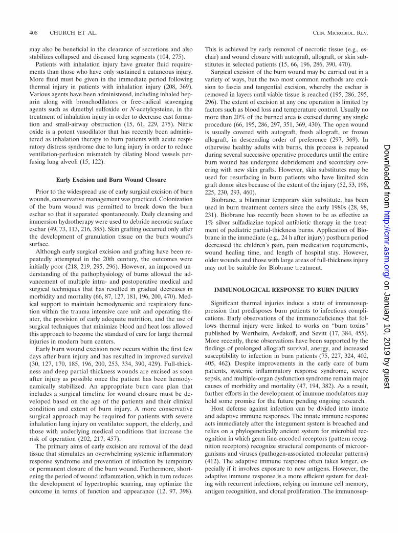
The primary aims of early excision are removal of the deadtissue that stimulates an overwhelming systemic inflammatoryresponse syndrome and prevention of infection by temporaryor permanent closure of the burn wound. Furthermore, short-ening the period of wound inflammation, which in turn reducesthe development of hypertrophic scarring, may optimize theoutcome in terms of function and appearance (12, 97, 398).
This is achieved by early removal of necrotic tissue (e.g., es-char) and wound closure with autograft, allograft, or skin sub-stitutes in selected patients (15, 66, 196, 286, 390, 470).
Surgical excision of the burn wound may be carried out in avariety of ways, but the two most common methods are exci-sion to fascia and tangential excision, whereby the eschar isremoved in layers until viable tissue is reached (195, 286, 295,296). The extent of excision at any one operation is limited byfactors such as blood loss and temperature control. Usually nomore than 20% of the burned area is excised during any singleprocedure (66, 195, 286, 297, 351, 369, 430). The open woundis usually covered with autograft, fresh allograft, or frozenallograft, in descending order of preference (297, 369). Inotherwise healthy adults with burns, this process is repeatedduring several successive operative procedures until the entireburn wound has undergone debridement and secondary cov-ering with new skin grafts. However, skin substitutes may beused for resurfacing in burn patients who have limited skingraft donor sites because of the extent of the injury (52, 53, 198,225, 230, 293, 460).
Biobrane, a bilaminar temporary skin substitute, has beenused in burn treatment centers since the early 1980s (28, 98,231). Biobrane has recently been shown to be as effective as1% silver sulfadiazine topical antibiotic therapy in the treat-ment of pediatric partial-thickness burns. Application of Bio-brane in the immediate (e.g., 24 h after injury) postburn perioddecreased the children’s pain, pain medication requirements,wound healing time, and length of hospital stay. However,older wounds and those with large areas of full-thickness injurymay not be suitable for Biobrane treatment.
IMMUNOLOGICAL RESPONSE TO BURN INJURY
Significant thermal injuries induce a state of immunosup-pression that predisposes burn patients to infectious compli-cations. Early observations of the immunodeficiency that fol-lows thermal injury were linked to works on “burn toxins”published by Wertheim, Avdakoff, and Sevitt (17, 384, 455).More recently, these observations have been supported by thefindings of prolonged allograft survival, anergy, and increasedsusceptibility to infection in burn patients (75, 227, 324, 402,405, 462). Despite improvements in the early care of burnpatients, systemic inflammatory response syndrome, severesepsis, and multiple-organ dysfunction syndrome remain majorcauses of morbidity and mortality (47, 194, 382). As a result,further efforts in the development of immune modulators mayhold some promise for the future pending ongoing research.
Host defense against infection can be divided into innateand adaptive immune responses. The innate immune responseacts immediately after the integument system is breached andrelies on a phylogenetically ancient system for microbial rec-ognition in which germ line-encoded receptors (pattern recog-nition receptors) recognize structural components of microor-ganisms and viruses (pathogen-associated molecular patterns)(412). The adaptive immune response often takes longer, es-pecially if it involves exposure to new antigens. However, theadaptive immune response is a more efficient system for deal-ing with recurrent infections, relying on immune cell memory,antigen recognition, and clonal proliferation. The immunosup-
408 CHURCH ET AL. CLIN. MICROBIOL. REV.
on January 10, 2019 by guesthttp://cm
r.asm.org/
Dow
nloaded from

pression associated with burn injuries has effects on both ofthese systems.
Many in vitro and in vivo studies have been conducted tocharacterize the immune responses and the relationships be-tween various cell types and inflammatory mediators. Severalreviews have been written on the topic, discussing the findingsof original works in more detail (82, 173, 194, 244, 412). Thisreview is a synthesis of summarized data and original researchthat have contributed to our current understanding of theimmune response following burn injury.
Systemic Response to Burn Injury
Local inflammation following injury is essential for woundhealing and host defense against infection. However, traumaor burns of sufficient magnitude can incite a systemic inflam-matory response, along a continuum from systemic inflamma-tory response syndrome through septic shock, which has theability to cause significant cellular and end-organ damage (46,47). Initially, the immunologic response to severe burn injury isproinflammatory but later becomes predominately anti-inflam-matory in an effort to maintain homeostasis and restore normalphysiology. Cytokines and cellular responses mediate both ofthese phases.
Inflammatory response to burn injury. Increased serum lev-els of proinflammatory cytokines characterize the systemic re-sponse to burns. Interleukin-1� (IL-1�) and tumor necrosisfactor alpha are produced by a wide variety of cells in responseto injury, of which leukocytes are key players. Both of thesecytokines contribute to the production of fever, acute-phaseproteins, and an overall state of catabolism. They also up-regulate the production of prostaglandin E2 (PGE2), IL-6, andplatelet-activating factor by endothelial cells and macrophages(80, 454). Levels of IL-6 are increased after injury through itsproduction by a number of different cells (1, 42). Like IL-1�and tumor necrosis factor alpha, IL-6 induces fever and theproduction of acute-phase reactants that contribute to T-cellactivation (471). Levels of IL-6 peak approximately 1 weekafter injury (178), and high levels have been associated withincreased rates of morbidity and mortality, for which it is likelya marker of disease severity rather than an etiologic factor.Gamma interferon (IFN-�) is another proinflammatory cyto-kine, produced by NK cells and Th-1 cells in response to injury.It has an important role in macrophage activation and thedifferentiation of CD4� T cells into Th-1 cells while inhibitingtheir differentiation into Th-2 cells (167). Cell types that areimportant in facilitating a proinflammatory response to injuryare proinflammatory macrophages and CD4� T helper cells.
Anti-inflammatory response to burn injury. The anti-inflam-matory response and the subsequent immunosuppression fol-lowing burn injury are characterized by a set of opposing celltypes and cytokines. The production and release of monocytes/macrophages are decreased following burn injury and sepsis(151). Under these circumstances, macrophages produce in-creased amounts of PGE2 and decreased amounts of IL-12,which have a cooperative effect on T-cell differentiation (82,166). T helper cells begin to preferentially differentiate intoTh-2 cells, which produce the anti-inflammatory cytokines IL-4and IL-10 (107, 167).
The exact sequence of events that result in immunosuppres-
sion after burn injury remains unknown; however, biochemicalchanges that may affect the immune system include those tothe endocrine system, the arachidonic acid cascade, and thecytokine network. Following severe burn injury, there is anincrease in the levels of vasopressin, aldosterone, growth hor-mone, cortisol, glucagon, and catecholamines (362, 454). Ele-vated levels of glucocorticoids inhibit the production of IFN-�and IL-2, but not IL-4 and IL-10 (132, 353, 454). Similarly,norepinephrine released early after injury inhibits Th-1 cellfunction, but not that of Th-2 cells (376). Increased productionof PGE2 by inhibitory macrophages has been observed aftersevere injury (454). PGE2 may have an important role in sec-ondary immunosuppression, as it has been shown to decreaselymphocyte proliferation, to decrease the levels of the proin-flammatory cytokines IL-1� and IL-2, to diminish the responseto IL-2, to inhibit the activity of NK cells, and to activatesuppressor T cells (4, 167). Many of the changes in cytokinelevels represent alterations of the adaptive immune systemfollowing burn injury, more specifically within the T-lympho-cyte population.
Innate Immune System Response to Burn Injury
Natural resistance to infection in traumatic wounds is pre-dominantly a function of the innate immune system. Followingthermal injury, the innate immune system responds immedi-ately by stimulating localized and systemic inflammatory reac-tions. The innate immune response participates in activatingthe adaptive immune response; however, in so doing it has anadverse affect on the burn victim’s ability to mount a vigorousimmune response to invading microorganisms and, therefore,predisposes the burn victim to infectious complications. Theinnate immune system itself is composed of natural barriers tomicrobial invasion as well as cellular (leukocyte) and humoral(complement) elements.
Before a pathogen can establish invasive infection within thehost it must break through the natural barriers of the skin ormucosa. For example, there is a loss of barrier function of thegastrointestinal epithelium in burn patients, which may be in-duced by up-regulation of the nitric oxide synthetase gene and theoverproduction of nitric oxide (311); postoperative changes, suchas decreased intestinal motility and mucus secretion; and in-creased exposure to endotoxin (4). The development of multiple-organ dysfunction syndrome in critically ill patients has also beenassociated with a derangement in intestinal permeability (109). Asa result, higher rates of bacterial translocation and endotoxinabsorption through the gastrointestinal mucosa may contribute tothe inflammatory response seen in burn patients.
The cellular elements of the innate immune system have im-portant roles in antimicrobial killing and in coordinating the im-mune response. Decreased macrophage and natural killer cellactivation results in reduced levels of IFN-� following burn injury(88, 187). The function of NK cells is diminished following sig-nificant injury (362). Neutrophil dysfunction after significant ther-mal injuries has also been reported (44, 137, 175, 242). Endothe-lial adherence of neutrophils is initially decreased after injury andthen increases (374); however, the site of endothelial adhesionmay not be at the point of injury, and this misguided neutrophiladhesion and activation contribute to neutrophil-mediated endo-thelial injury, which may play a significant role in the pathogenesis
VOL. 19, 2006 BURN WOUND INFECTIONS 409
on January 10, 2019 by guesthttp://cm
r.asm.org/
Dow
nloaded from

of systemic inflammatory response syndrome and multiple-organdysfunction syndrome.
Neutrophil chemotaxis and intracellular killing are impairedfollowing major burns (1, 173, 174). Diminished cytotoxic ac-tivity follows from a surge of degranulation early after injuryand a subsequent inability to replenish intralysosomal enzymesand defensins (173, 362). Macrophages also demonstrate di-minished phagocytic capacity following severe injury (5,381). Lower levels of major histocompatibility complex classII expression and antigen presentation disrupt their roles incoordination of the immune response (362, 413). They alsoproduce larger quantities of PGE2, resulting in the suppres-sion of B- and T-cell reactivity (281). Increased levels ofIL-4 and IL-10 inhibit macrophage antigen presentation,decrease the production of proinflammatory cytokines suchas IL-1�, and suppress bactericidal and fungicidal activity(110, 130, 138, 186, 329, 442, 447).
The complement cascade represents an important humoralcomponent of the innate immune system. Following significantburn injuries, the alternative pathway of the complement cas-cade is primarily depressed, while there is little effect on theclassical pathway (150). Complement levels fall in proportionto injury severity and then rise to supranormal levels (150).Activation of the complement cascade by thermal injury (39)increases levels of C3a and C5a, which may result in changes inblood pressure, vascular permeability, and leukocyte function(214, 473). Small amounts of C5a have been shown to stimulateleukocyte function; however, large amounts lead to suppres-sion of activity (453). Membrane attack complexes may targetnormal cells near the site of injury, contributing to reactive celllysis, which may induce end-organ damage (194). Lastly, in-creased levels of C3b may be directly immunosuppressive, asthey have been shown to decrease phagocytosis and contributeto lymphocyte dysfunction (4).
These alterations to the innate immune system have thecombined effect of increasing the burn patient’s exposure topathogens and decreasing the natural defenses that are respon-sible for counteracting them. Exposure to pathogens occurs viathe burn wound, invasive monitoring devices, and the gastro-intestinal tract, which loses some of its capacity to act as aneffective barrier to bacterial translocation. The effects of ananti-inflammatory cytokine milieu on NK cells, neutrophils,and macrophages impair the eradication of these pathogens bythe innate immune system. Furthermore, the activation ofcomplement following burn injury may be directly immunosup-pressive. As a result of these phenomena and subsequent al-terations to the adaptive immune system, burn patients aremore susceptible to wound infections, severe sepsis, and mul-tiple organ failure.
Adaptive Immune System in Response to Burn Injury
Following significant injury, several changes in the T-lym-phocyte population have been observed. Total numbers of Tlymphocytes fall in proportion to injury severity during the firstweek after injury (194, 362) and there is a decrease in T-cell-dependent immune functions (75, 227, 402, 405, 462). Dimin-ished T-cell proliferation in response to mitogens (210, 364,462) is associated with, and may be the result of, decreasedproduction of IL-2 and IFN-� by monocytes (133, 463). The
production of immunoglobulin G (IgG) in response to T-cell-dependent antigens is also impaired after serious injury;however, no impairment of antibody formation to T-cell-independent antigens has been observed (325). There is adecreased ratio of CD4� T helper cells to CD8-positive Tsuppressor cells (67, 327). After an initial proinflammatoryphase, injury results in a loss of Th-1 cells associated withdepressed levels of IL-1� and IFN-�. Concomitantly, Th-2lymphocytes are present in increased numbers along withhigher levels of the anti-inflammatory cytokines IL-4 andIL-10, which may inhibit Th-1 cell activation by suppressingantigen presentation (167).
A correlation between increased levels of IL-10 and septicevents has been reported (256, 395). It remains uncertainwhether the relative predominance of Th-2 cells over Th-1 cellsrepresents a phenotypic change or an increase in the rate ofapoptosis of Th-1 cells (244). Alterations in the balance be-tween T suppressor lymphocytes and T helper lymphocytesand the ratio of Th-1 to Th-2 cells appear to be importantetiologic factors in the suppression of the adaptive immuneresponse.
Altering the Immunologic Response to Burn Injury
Despite our increasingly detailed understanding of the im-munological suppression that follows thermal injuries, no at-tempts at directly modulating the immune response at a spe-cific site have been shown to be clinically effective. It isbecoming increasingly clear that any therapies directed at ad-dressing this immunodeficiency in burn patients will likely haveto target multiple points in the inflammatory response and theneuroendocrine axis.
Immune function in burn patients can only be restoredthrough intensive resuscitation and support. Early excision ofburn eschar and prompt wound coverage remove a significantinflammatory stimulus and restore the barrier function of theskin. Providing adequate analgesia and maintaining adequatetissue perfusion, ambient temperature, and blood volume helpoptimize the oxidative killing capacity of neutrophils (235).Early and adequate nutritional support is also important inrestoring protein synthesis and normal immune function. Re-search efforts have focused on the topic of immune-modifyingdiets, such as glutamine-enriched diets, and their clinical ben-efits (155). However, there is insufficient evidence to supportthe use of such diets in burn patients at this time.
EPIDEMIOLOGY OF BURN WOUND INFECTIONS
Burn wound infections are one of the most important andpotentially serious complications that occur in the acute periodfollowing injury (10, 11, 38, 108, 189, 243). The most importantpatient characteristics that influence morbidity and mortalityfrom burn wound infection and sepsis are outlined below. Inaddition, the impact of early excision on reducing burn woundinfections is discussed. Other factors that have played a signif-icant role in decreasing the overall fatality rates from burnwound infection and sepsis include the use of topical andprophylactic antibiotics and advances in infection control mea-sures in modern burn units (see Prevention of Burn WoundInfections, below).
410 CHURCH ET AL. CLIN. MICROBIOL. REV.
on January 10, 2019 by guesthttp://cm
r.asm.org/
Dow
nloaded from

Impact of Patient Demographics and Burn Severity
Very young children and the elderly have an increased riskof being burned and worse clinical outcomes than patients inother age groups (68–70, 72, 217, 320, 344). Individuals withdeliberate self-inflected burn injuries and the disabled havebeen shown to have more severe injuries and longer hospitalstays than those with accidental injuries (18, 211, 332). Obeseadults and those who have an underlying medical conditionsuch as diabetes have also been shown to have higher morbid-ity and mortality (169, 276, 290). AIDS patients appear to havemore complications due to infection, delayed wound healing,and increased mortality, although reported outcome data forhuman immunodeficiency virus-infected and AIDS patientsare limited (115, 289, 310, 400). It is expected that burn pa-tients with other types of severe immunosuppression wouldhave similar problems, particularly increased problems withwound infection and sepsis and a higher mortality, althoughthis group has not been studied.
Burns in the elderly constitute more severe injuries than inthe general population and result in a higher number of fatal-ities. A recent review of adult patients admitted to a burncenter over a 7-year period showed that 221 of 1,557 (11%)were �59 years of age and a higher proportion were women(279). Most elderly burn patients had one or more existingmedical conditions and impaired judgment and/or mobility.Approximately one-third of the elderly patients in this studyalso sustained smoke inhalation injury. Substance abuse was afactor in some elderly patients, because toxicology screeningshowed that 10% had used alcohol and almost one-third testedpositive for other drugs. Mortality was highest in elderly pa-tients who had more severe burns and/or smoke inhalationinjury that had existing underlying disease.
A recent study also assessed the factors affecting burn mor-tality in the elderly and analyzed changes that occurred overthe past three decades (252). The study included 201 patients75 years of age of older that had been admitted to a university-based burn center between 1972 and 2000. Almost half of thesepatients died (95, or 47.3%), and the severity of the burn injuryas measured by TBSA and the abbreviated burn severity indexwere both strongly correlated with mortality. Due to improvedburn care, however, the elderly are much less likely to die fromburns now than in the 1970s unless they have an inhalationinjury. Mortality increased significantly with inhalation injurydespite advances in intensive respiratory support.
Children have a much higher risk of being burned thanadults (344). In the United States in 2001 to 2002, an estimated92,500 children aged 14 years and under required emergencycare for burn-related injuries, and approximately 500 of thesechildren died (320). Approximately two-thirds of these chil-dren sustained thermal injuries, while children �4 years of ageare particularly prone to scald injury (320). Male children havea higher risk of burn injury and burn-related death than fe-males, and obese boys represented a disproportionate numberof the patients admitted to a pediatric burn center from 1991to 1997 (26). Children who show failure to thrive (e.g., heightand/or weight �5% of that expected by age) also have a higherrisk of burn injury, perhaps due to the combined effects ofmalnutrition and neglect or abuse (26, 344).
Impact of Changes in Burn Wound Care
Much of the steady decline in burn wound infections, sub-sequent tissue invasion and sepsis, and associated mortalitythat has been realized in the past 50 years has been attributedto the substantial advances that have occurred in burn woundcare, particularly early excision (15, 79, 171, 189, 196, 202, 209,291, 369). There was a substantial reduction in one burn centerin 1978 in the incidence of both burn wound infection andsepsis after the advent of early excision therapy (253). Duringthe study period, the incidence of burn wound sepsis fell from6% to 1% and the mortality rate for burn-related complica-tions decreased from 40% to 18%. However, there are onlytwo randomized, controlled trials of early excision versus con-servative exposure therapy, and neither of these studies dem-onstrated a significant reduction in burn wound infections inpatients with a major thermal injury (e.g., �15% TBSA) (127,170). Limited data have been published that provide a clearpicture of the epidemiology of different types of burn woundinfections according to the recently published classificationsystem (see Classification of Burn Wound Infections, below).
Most of our understanding of the epidemiology of burnwound infections has been gleaned from studies carried out inthe 1950s through 1990 during the preexcision era of burn care(273). It is not surprising that the overall morbidity and mor-tality of burn wound infections, tissue invasion, and secondarysepsis were extremely high during this time period because thegrowth of bacteria on the burn wound surface was controlledbut not eradicated. Case fatality rates were 40% or higherdepending on the extent of the burn injury (272, 285, 340, 341).Immediate colonization by the patient’s normal skin flora (i.e.,Staphylococcus aureus and Streptococcus pyogenes) occurredfollowing injury (23, 164, 249, 259, 333). Subsequent coloniza-tion by the patient’s own gut flora added to the complex mi-crobial ecology on the burn wound surface shortly thereafter(106, 248, 266, 267, 269, 371).
Nosocomial transmission of microorganisms to the burnwound also occurred by transfer from the hands of health carepersonnel and through immersion hydrotherapy treatment (73,273, 450, 468). Burn unit outbreaks of infection were attributedmainly to contaminated Hubbard hydrotherapy tanks or waterbut in other cases to contaminated surfaces such as the pa-tient’s mattress (126, 274, 280, 397, 436). Despite the recog-nized infection risk of immersion hydrotherapy treatment inburn units, this was standard practice in many specialized burncenters until the 1990s. In a survey of burn centers in NorthAmerica in 1990, 81.4% still used immersion hydrotherapyregardless of the size of the burn wound, and most centers alsocontinued this therapy throughout hospitalization on all pa-tients (385). Aside from microbial contamination of the tankwater, aerators and agitators in hydrotherapy tubs were dif-ficult to clean (280, 436) Hydrotherapy water continued tobe cross-contaminated between patients despite the removalof these devices from the tanks (436). Sodium hypochloriteand chloramine-T disinfectants added to the hydrotherapytank water decreased the microbial load on the burn woundsurface and health care workers’ hands (73, 414). However,the hydrotherapy water irritated the mucosal surfaces (e.g.,conjunctiva and nares) of the patient and health care per-sonnel, although this practice was effective in eliminating
VOL. 19, 2006 BURN WOUND INFECTIONS 411
on January 10, 2019 by guesthttp://cm
r.asm.org/
Dow
nloaded from

gram-negative microorganisms from burn wounds after sev-eral days of treatment (73).
Showering with a hand-held sprayer has gradually replacedhydrotherapy for cleansing and debridement of the burnwound. This practice decreases the transfer of bacteria onsurfaces to the patient’s burn wound. However, outbreaks re-lated to shower hydrotherapy have also recently been reported.Pseudomonas organisms were recovered from the hydrother-apy tank used to initially remove the patient’s adherent dress-ings in one outbreak (436), and another outbreak was caused bycontamination of the shower hand grip and showering stretcherby methicillin-resistant Staphylococcus aureus (MRSA) (126).Performing local wound care in the patient’s room has controlledburn unit outbreaks due to immersion hydrotherapy.
PATHOGENESIS OF BURN WOUND INFECTIONS
Pathogenesis
Thermal destruction of the skin barrier and concomitantdepression of local and systemic host cellular and humoralimmune responses are pivotal factors contributing to infectiouscomplications in patients with severe burns (4, 173, 182, 194,244). The burn wound surface (in deep partial-thickness and inall full-thickness burns) is a protein-rich environment consist-ing of avascular necrotic tissue (eschar) that provides a favor-able niche for microbial colonization and proliferation (29,129, 267, 268, 315). The avascularity of the eschar results inimpaired migration of host immune cells and restricts deliveryof systemically administered antimicrobial agents to the area,while toxic substances released by eschar tissue impair localhost immune responses (see Immunological Response to BurnInjury, above).
Although burn wound surfaces are sterile immediately fol-lowing thermal injury, these wounds eventually become colo-nized with microorganisms (129, 469). The nature and extentof the thermal injury along with the types and amounts ofmicroorganisms colonizing the burn wound appear to influencethe future risk of an invasive wound infection (29, 129, 268,315). Gram-positive bacteria that survive the thermal insult,such as staphylococci located deep within sweat glands andhair follicles, heavily colonize the wound surface within thefirst 48 h unless topical antimicrobial agents are used (6, 129,164). Eventually (after an average of 5 to 7 days), these woundsare subsequently colonized with other microbes, includinggram-positive bacteria, gram-negative bacteria, and yeasts de-rived from the host’s normal gastrointestinal and upper respi-ratory flora and/or from the hospital environment or that aretransferred via a health care worker’s hands (6, 129, 267, 268,356, 449, 450, 468).
Over the last several decades, gram-negative organisms haveemerged as the most common etiologic agents of invasive in-fection by virtue of their large repertoire of virulence factorsand antimicrobial resistance traits (84, 92, 162, 358, 360, 363,388, 401, 404, 436). If the patient’s host defenses and thera-peutic measures (including excision of necrotic tissue andwound closure) are inadequate or delayed, microbial invasionof viable tissue occurs, which is the hallmark of an invasiveburn wound infection (see “Histological analysis” under Anal-ysis of Burn Wound Specimens, below).
Biofilm Formation
Biofilms are complex communities of surface-attached ag-gregates of microorganisms embedded in a self-secreted extra-cellular polysaccharide matrix, or slime (419, 421). They arefound in a wide range of natural and artificial environmentsand provide their constituent microbial cells with a plethora ofprotected dynamic microenvironments (419, 421). Once ma-ture, biofilms act as efficient barriers against antimicrobialagents and the host immune system, resulting in persistentcolonization and/or infection at the site of biofilm formation(118, 326).
Although biofilms are best known for their role in foreigndevice-related infections, recent studies have confirmed the im-portance of biofilms in the pathogenesis of burn wound infec-tions (434). In animals with experimentally inflicted partial-thickness cutaneous burns, mature biofilms develop in 48 to72 h, while in vitro experiments with Pseudomonas aeruginosastrains recovered from human burn wounds demonstrate thatmature biofilms can form in about 10 h (184). Factors delayingthe formation of biofilms in vivo may be related to the need formicrobial nutrient replenishment, exposure to killing by theimmune system, and immediate wound cleansing (184).
Bacteria within a biofilm typically undergo a phenotypicchange whereby microbial virulence factor production isaltered and metabolic rate and motility are reduced (118,419, 421). Channels formed within the protective environ-ment of the biofilm facilitate the transport of nutrients andmicrobial waste products (118, 184, 419, 421). Intercellularsignaling molecules produced by bacteria within the biofilmare able to traverse these channels and influence the overallgrowth pattern and behavior of the biofilm in response tovarious host and environmental factors (258, 350, 419, 421).Persister cells within the biofilm are the cells that haveremained within the biofilm after treatment with antimicro-bial agents and antiseptics (434). These persister cells tem-porarily disable their inherent mechanisms of programmedcell death in the presence of harsh environmental conditionsand help in repopulating the biofilm, often leading to failurein biofilm eradication (419, 421).
Microbial Etiology
Bacteria rapidly colonize open skin wounds after burn in-jury. Microorganisms colonizing the burn wound originatefrom the patient’s endogenous skin and gastrointestinal andrespiratory flora (29, 129, 267, 268, 356). Microorganisms mayalso be transferred to a patient’s skin surface via contact withcontaminated external environmental surfaces, water, fomites,air, and the soiled hands of health care workers (450, 468).Immediately following injury, gram-positive bacteria from thepatient’s endogenous skin flora or the external environmentpredominantly colonize the burn wound (29, 164, 469). Endog-enous gram-negative bacteria from the patient’s gastrointesti-nal flora also rapidly colonize the burn wound surface in thefirst few days after injury (266, 267, 269, 357). Wound coloni-zation by yeasts and fungi usually occurs later due to the use ofbroad-spectrum antibiotic therapy (65, 105, 123). Microorgan-isms transmitted from the hospital environment tend to bemore resistant to antimicrobial agents than those originating
412 CHURCH ET AL. CLIN. MICROBIOL. REV.
on January 10, 2019 by guesthttp://cm
r.asm.org/
Dow
nloaded from

from the patient’s normal flora (84, 146, 148, 190, 358, 363).Table 2 lists the most common microorganisms colonizing andinfecting burn wounds.
Prior to the antibiotic era, Streptococcus pyogenes (group Abeta-hemolytic streptococci) was the predominant pathogenimplicated in burn wound infections and was a major cause ofdeath in severely burned patients (23, 246, 249). Staphylococ-cus aureus became the principal etiological agent of burnwound infections (250, 333) shortly after the introduction ofpenicillin G in the early 1950s, which resulted in the virtualelimination of Streptococcus pyogenes as a cause of infection inthermally injured patients. Although Staphylococcus aureusremains a common cause of early burn wound infection,Pseudomonas aeruginosa from the patient’s endogenous gas-trointestinal flora and/or an environmental source is themost common cause of burn wound infections in many cen-ters (6). The incidence of infections due to less commonly en-countered microbes, including other gram-positive and gram-neg-ative bacteria, fungi, and viruses, has also increased steadily insubsequent decades (3, 23, 37, 65, 92, 105, 143, 149, 360, 389)(Table 2). While less common, infections due to anaerobic bac-teria typically occur secondary to electrical burns or when openwound dressings are used in place of occlusive dressings (308).
The emergence worldwide of antimicrobial resistance among awide variety of human bacterial and fungal burn wound patho-gens, particularly nosocomial isolates, limits the available ther-apeutic options for effective treatment of burn wound infec-tions (6, 84, 120, 148, 190, 358). MRSA, methicillin-resistantcoagulase-negative staphylococci, vancomycin-resistant en-terococci, and multiply resistant gram-negative bacteria thatpossess several types of beta-lactamases, including extended-spectrum beta-lactamases, ampC beta-lactamases, and metallo-beta-lactamases, have been emerging as serious pathogens in
hospitalized patients (84, 92, 126, 152, 190, 241, 358). Fungalpathogens, particularly Candida spp., have increasingly be-come important opportunistic pathogens due to the use ofbroad-spectrum topical and systemic agents when infectionoccurs in the burned patient and have demonstrated increas-ing degrees of antifungal drug resistance (10, 19, 233, 302).
Virulence Factors and Tissue Invasion
The risk of invasive burn wound infection is influenced bythe extent and depth of the burn injury, various host factors,and the quantity and virulence of the microbial flora colonizingthe wound. Common burn wound pathogens such as Pseudo-monas aeruginosa and Staphylococcus aureus produce a num-ber of virulence factors that are important in the pathogenesisof invasive infection. Pseudomonas aeruginosa produces anumber of cell-associated (adhesins, alginate, pili, flagella,and lipopolysaccharide) and extracellular (elastase, exoen-zyme S, exotoxin A, hemolysins, iron-binding proteins, leu-kocidins, and proteases) virulence factors that mediate anumber of processes, including adhesion, nutrient acquisi-tion, immune system evasion, leukocyte killing, tissue de-struction, and bloodstream invasion (437, 441). Pseudomo-nas aeruginosa also carries many intrinsic and acquiredantimicrobial resistance traits that make infected burnwounds difficult to treat (212, 241).
Staphylococcus aureus also has a diverse array of virulencefactors that facilitate adherence to host tissues, immune systemevasion, and destruction of host cells and tissues, includingcoagulase, protein A, leukocidins, hemolysins, and superanti-gens (142). Resistance to methicillin in Staphylococcus aureus,and more recently emergence of resistance to glycopeptidesand oxazolidinones, also complicate the treatment of burnwound infections and sepsis caused by this highly virulent or-ganism (183, 288, 309, 391).
CLASSIFICATION OF BURN WOUND INFECTIONS
Burn wound infection is a serious problem because it causesa delay in epidermal maturation and leads to additional scartissue formation (118, 398). Invasion of microorganisms intothe tissue layers below the dermis may also result in bactere-mia, sepsis, and multiple-organ dysfunction syndrome (20, 272,346, 365). Clinical diagnosis of burn wound infection relies onregular monitoring of vital signs and inspection of the entireburn wound surface, preferably during each dressing change.Local signs of burn wound infection with invasion includeconversion of a partial-thickness injury to a full-thickness wound,rapidly extending cellulitis of healthy tissue surrounding theinjury, rapid eschar separation, and tissue necrosis.
Burn wound infections were previously classified based onchanges in the burn wound and/or eschar appearance, time ofoccurrence, and associated mortality into distinct conditions,including impetigo, cellulitis, and invasive infection. Due to theadvent of early excision therapy, new classifications for burnwound infections related to surgical wound infection at theexcision site(s) have been developed by a subcommittee of theCommittee on the Organization and Delivery of Burn Care ofthe American Burn Association (273, 331, 369). Each of thesedistinct clinical conditions that make up the spectrum of burn
TABLE 2. Microorganisms causing invasive burn wound infectiona
Group Species
Gram-positive organisms Staphylococcus aureusMethicillin-resistant S. aureusCoagulase-negative staphylococciEnterococcus spp.Vancomycin-resistant enterococci
Gram-negative organisms Pseudomonas aeruginosaEscherichia coliKlebsiella pneumoniaeSerratia marcescensEnterobacter spp.Proteus spp.Acinetobacter spp.Bacteroides spp.
Fungi Candida spp.Aspergillus spp.Fusarium spp.Alternaria spp.Rhizopus spp.Mucor spp.
Viruses Herpes simplex virusCytomegalovirusVaricella-zoster virus
a Data are from references 3, 23, 37, 65, 92, 105, 143, 149, 360, and 389.
VOL. 19, 2006 BURN WOUND INFECTIONS 413
on January 10, 2019 by guesthttp://cm
r.asm.org/
Dow
nloaded from

wound infections is described briefly below. Burn wound im-petigo may or may not be associated with systemic signs ofinfection, but fever (temperature of �38.4°C) or leukocytosis(white blood cell count of �10,000 cells/mm3) and/or throm-bocytopenia is present in all of the other types of burn woundinfections outlined. The development of burn wound cellulitisor invasive burn wound infection may also be heralded bybacteremia or septicemia.
In addition to burn wound surface and/or tissue cultures,patients with signs of systemic infections should have a com-plete septic workup that includes blood and urine cultures aswell as burn wound sample cultures. Effective treatment ofburn wound infections combines an increased frequency ofburn wound dressing changes with optimization of the patient’santimicrobial therapy regimen according to microbiology cul-ture and susceptibility results from burn wound cultures.
Types of Burn Wound Infection
Burn wound impetigo. Impetigo involves the loss of epithe-lium from a previously reepithelialized surface, such as graftedburns, partial-thickness burns allowed to close by secondaryintention, or healed donor sites. Burn wound impetigo is notrelated to inadequate excision of the burn, mechanical disrup-tion of the graft, or hematoma formation.
Burn-related surgical wound infection. Surgical wound in-fections in burn patients include both excised burn and donorsites that have not yet epithelialized. The wound has purulentexudate that is culture positive. Surgical wound infections inopen areas of the burn show loss of synthetic or biologicalcovering of the wound, changes in wound appearance (such ashyperemia), and erythema in the uninjured skin surroundingthe wound.
Burn wound cellulitis. Burn wound cellulitis results from anextension of infection into the healthy, uninjured skin and softtissues surrounding the burn wound or donor site. This condi-tion is recognized by extension of erythema in the uninjuredskin surrounding the burn beyond what is expected from theinjury itself. Burn wound cellulitis is not associated with othersigns of wound infection, but at least one of the followingmanifestations is present: localized pain or tenderness, swell-ing or heat at the affected site, progression of erythema andswelling, and signs of lymphangitis and/or lymphadenitis ex-tending from the affected skin area along routes of lymphaticdrainage to the area.
Invasive infection in unexcised burn wounds. Patients withareas of unexcised deep partial-thickness or full-thickness burnwound have an increased risk of developing an invasive infec-tion (10, 11, 27, 29, 253). This complication may be heralded bya rapid associated change in burn wound appearance or char-acter such as separation of the eschar or dark brown, black, orviolaceous discoloration of the eschar. Manifestations of inva-sive infection of unexcised burn wounds include inflammationof the surrounding uninjured skin, such as edema, erythema,warmth or tenderness, evidence of microbial invasion into ad-jacent viable tissue on histological examination, and positiveblood cultures with isolation of a pathogen in the absence ofanother identifiable source of infection and systemic signs ofsepsis, i.e., tachypnea, hypotension, oliguria, unexplained hy-perglycemia (e.g., increased serum glucose level that develops
at a previously tolerated level of dietary carbohydrate), and/ormental confusion. Effective treatment requires surgical exci-sion of the burn in addition to the medical measures outlinedpreviously.
MICROBIOLOGICAL ANALYSIS OF BURNWOUND INFECTIONS
Diagnosis of burn wound infection based on clinical signsand symptoms alone is difficult. Regular sampling of the burnwound either by surface swab or tissue biopsy for culture is alsodone to monitor for the presence of infection. Quantitativeculture of tissue biopsy samples and histological verification ofmicrobial invasion into viable unburned tissue have been the“gold standard” for confirming the presence of invasive burnwound infection, particularly in unexcised areas of eschar.More recently, however, the value of laborious and costlyquantitative burn wound tissue biopsy cultures has been ques-tioned (145, 282, 458). Many burn centers have correspond-ingly shifted to the more convenient practice of procuring burnwound surface swabs for qualitative or semiquantitative cul-ture for infection surveillance since the advent of early exci-sion. This section discusses the various diagnostic microbiolog-ical approaches to diagnosis of burn wound infection andcurrent recommendations for a best approach to burn woundinfection surveillance.
Best Approach for Burn Wound Infection Surveillance
Review of the studies that have compared burn wound in-fection surveillance by surface swabs and burn wound biopsyprovides conflicting results about the best approach. Conflict-ing results have been obtained by different studies for thefollowing reasons: burn patients do not have homogenous in-juries (e.g., the severity and extent of burn injury vary greatlyfrom patient to patient), various sampling techniques and lab-oratory methods have been used, and most comparative stud-ies were done before the advent of early excision therapy (41,48, 247, 254, 255, 282, 399, 407, 408, 423). Steer and colleagues(407, 408) have reported the largest recent studies that com-pared the results of surface swab versus biopsy cultures. Intheir initial study (408), a comparison was made of the quali-tative results and quantitative bacterial counts of 141 surfaceswabs and 141 wound biopsy samples taken from 74 burnpatients. Although there was significant correlation betweenthe bacterial counts obtained by biopsy and swab, the countsobtained by one method were poorly predictive of the countsobtained by the other. In addition, parallel cultures taken onmultiple occasions showed a significant correlation betweenbacterial counts obtained from two biopsies or two swabs takensimultaneously, but there was wide variation in bacterial den-sities from the same burn wound at the same time. Theseinvestigators concluded that the use of quantitative microbiol-ogy in burns is limited by the unreliability of a single surfaceswab or biopsy sample to represent the whole burn wound.
Steer and coworkers (407) subsequently performed a clini-cal-outcome study to determine the relationship between bac-terial counts obtained by burn wound biopsy culture and sur-face swabs. These investigators collected 69 paired biopsy-surface swab specimens from 47 patients (mean 16% TBSA
414 CHURCH ET AL. CLIN. MICROBIOL. REV.
on January 10, 2019 by guesthttp://cm
r.asm.org/
Dow
nloaded from

burned) on 64 separate occasions, either immediately prior toexcision and grafting or during routine dressing changes. Therewas a significant positive correlation between total bacterialcount by biopsy and total white cell count and a significantnegative correlation between total bacterial count by swab andpercent TBSA burned. No relationship was observed betweenclinical outcome and bacterial counts obtained with eithermethod. Hence, this study demonstrated that quantitative bac-teriology by burn wound biopsy or surface swab sample doesnot aid the prediction of sepsis or graft loss.
Loebl and colleagues (254, 255) originally demonstrated thatthe recovery of bacterial flora from the unexcised burn woundsurface showed poor correlation with that from tissue biopsysamples taken from deep sites beneath the eschar. Freshwaterand Su (145) also found that the results of quantitative burnwound cultures needed to be interpreted in conjunction withclinical observations of burn wound infection in order to be auseful guide to the management of burn patients with largeTBSA burns. Tahlan and colleagues (423), in a study compar-ing surface swabs and burn wound biopsy cultures in 17 pa-tients with second- and third-degree burns, found no differencein the types of microorganisms cultured from swabs versusthose cultured from biopsies. Levine and colleagues (247) ad-ditionally noted a linear numerical relationship between quan-titative surface swab and biopsy sample counts of viable bac-teria from burn wounds, whereby counts of 105 bacteria pergram of biopsy sample were equated with counts of 106 bac-teria obtained from surface swab samples.
McManus and colleagues found that quantitative culturesof tissue biopsy samples provided a better determination ofthe predominant bacterial types present in the burn wound(282). Herruzo-Cabrera and colleagues (205) showed that asemiquantitative surface swab method distinguished betweenwound contamination and infection, using 105 organisms/g as athreshold for the definition of infection by biopsy. Sjoberg andcolleagues (399) recently reported that quantitative tissue bi-opsies gave a better prediction of sepsis than surface swabs butconcluded that the amount of labor involved in collection andanalysis of multiple biopsy samples limited the clinical rele-vance of this approach. Bharadwaj and colleagues (41) alsoassessed the value of blood cultures in the diagnosis of burnwound sepsis compared to burn wound cultures by either swabor tissue biopsy. Fifty patients with burns ranging from 30 to50% TBSA were monitored for clinical signs of sepsis, and only62.5% had positive burn wound cultures according to surfaceswabs, compared to 87.5% who had a significant bacterialcount on biopsy sample culture. Blood cultures were found tobe of only prognostic value in this study (41). Blood cultureshave also been shown to be a late sign of invasive burn woundinfection even when they are positive (270).
The best approach for routine infection surveillance of burnwounds is to use the most appropriate sampling technique forthe type of burn wound area being cultured, since no singlemethod provides a clinically relevant, reliable result for unex-cised wounds (e.g., eschar) versus those that have been excised.Superficial swabs provide an adequate sampling of the micro-bial flora present on the wound surface and are the mostconvenient and least invasive approach currently available forsampling excised burn areas. Surface swabs are also the onlytype of sample that may be taken from areas where the skin is
too thin to do a biopsy, such as over the ears, eyes, and digits.However, quantitative cultures of burn wound tissue biopsysamples along with concomitant histological analysis are thepreferred infection surveillance approach for burn areas thathave not been or cannot be excised. Tissue biopsy samplesshould also be sent for quantitative culture from infected burnwound areas in patients with sepsis.
Simultaneous culture of quantitative tissue biopsy, blood,and urine samples provides the best approach for recovery andidentification of the causative organisms and their antimicro-bial susceptibilities in the septic burn patient. This method alsoprovides an accurate assessment of the depth and extent ofburn infection in areas of indeterminate injury. Tissue biopsyanalyses are also necessary in order to diagnose unusual typesof burn wound infection due to fungi and viruses (see OtherTypes of Infection in Burn Patients, below).
Burn Wound Sampling Techniques
A variety of different approaches have been described forassessing the nature and extent of microbial involvement inburn wounds, although the optimal sampling technique con-tinues to be debated. Infection surveillance of the burn woundrequires taking samples on a regular basis, by either biopsyingtissue or collecting surface swabs (55, 189, 202, 340, 341, 369,458, 459). Multiple samples from several areas of the burnwound should be collected in order to obtain the most accurateassessment of the types and amounts of microorganismspresent regardless of the sampling technique. Samples shouldbe collected frequently in the first few days to weeks followinginjury (e.g., daily or every 48 h during dressing changes) whenthe microbial flora is evolving. Sampling frequency may bedecreased to weekly once the burn wound has been excised,provided clinical signs of infection are not present.
Superficial wound samples. Clinical microbiology laborato-ries routinely provide semiquantitative or qualitative resultsfrom cultures of superficial wound samples. A number of tech-niques for the collection of burn wound surface cultures havebeen described over the last several decades, including thecollection of swabs or contact plates and capillarity gauze sam-pling (48, 160, 247, 399, 407, 408, 423, 459). Although each ofthese methods is described for historical completeness, mod-ern burn units universally rely on the collection of surfaceswabs. Specimens collected by superficial sampling of the burnwound surface must be done after the removal of dressings andtopical antibacterial agents and cleansing of the wound surfacewith 70% alcohol (41, 247, 407, 408).
Burn wound surface swabs are a convenient and effectivemethod for routinely collecting multiple superficial woundsamples (247, 408). Although there have been no studies thathave compared different commercially available swabs for theirability to recover pathogens from the burn wound surface, arecent general comparison of three swab transport systems(Starplex StarSwab II, Copan VI-Pak Amies agar gel andtransport swabs, and BBL Port-A-Cul) showed that the CopanVI-Pak system outperformed the other two by maintainingviability of both anaerobic and fastidious aerobic bacteria for24 h for the organisms evaluated (206). In order to obtainenough cellular material for culture, the end of a sterile swabis moved over a minimum 1-centimeter area of the open
VOL. 19, 2006 BURN WOUND INFECTIONS 415
on January 10, 2019 by guesthttp://cm
r.asm.org/
Dow
nloaded from

wound. Sufficient pressure should be applied to the tip of theswab to cause minimal bleeding in the underlying tissue. Eval-uations of the recovery of organisms using both dry and moist-ened swabs have shown that the moist-swab technique providesbetter reproducibility (48).
Capillarity gauze sample collections are done by applyinggauze squares moistened in nonbacteriostatic saline to theopen burn wound surface for several minutes, followed by useof the contaminated surface of the gauze to inoculate agarculture plates (160, 459). Although this method is relativelytime-consuming and expensive, it may be superior to swabcultures. Since the capillarity gauze surface permits a moreinclusive harvest of the resident bacteria, the quantitative cul-ture result is more reproducible (459).
Agar contact plates may also be applied directly to the openwound surface, but this method has not been adopted intoclinical practice because it is the least reproducible samplingtechnique, and culture medium sterility is not easily main-tained outside of the microbiology laboratory (85, 160, 161).
Tissue biopsy. Serial harvesting of multiple samples frombeneath the eschar for quantitative culture has historicallybeen the primary method used for accurate infection surveil-lance on the unexcised burn wound (254, 255, 282, 464, 465).The quantitative burn biopsy culture method was widelyadopted into practice following the studies by Loebl and col-leagues (254, 255). After the burn wound surface is cleansedwith isopropyl alcohol, two parallel incisions are made in theskin approximately 1 to 2 cm in length and 1.5 cm apart. Steriletissue forceps are then used to elevate and biopsy a samplewith a sterile scalpel from the subcutaneous tissue at sufficientdepth to obtain a small portion of the healthy underlying fat.Biopsy samples may also be collected by 3-mm punch biopsy.Tissue biopsy samples obtained by this method typically weighbetween 0.02 and 0.05 g. Biopsy specimens are then placed ona nonbacteriostatic moistened sterile gauze pad within a sterilecontainer in order to prevent tissue sample desiccation duringtransport.
Other investigators have also shown that quantitative burnwound biopsy cultures are more accurate than superficial sur-face cultures for diagnosing invasive infection in unexcisedburn wounds (41). However, tissue biopsy samples must betaken from deep sites beneath the eschar (145). Woolfrey andcolleagues (465) found poor reproducibility of quantitativebacterial counts between different eschar biopsy samples andshowed that even high bacterial tissue levels did not correlatewith the development of burn wound sepsis. Clinical microbi-ology laboratories may routinely perform burn wound surveil-lance cultures using quantitative methods on tissue biopsy sam-ples and correlate the results with the histological analysisperformed on a portion of the same biopsy sample.
Sampling techniques for other microbial pathogens. Lim-ited data are available regarding the optimal burn wound sam-pling technique to reliably detect other microbial pathogensthat may cause burn wound infection, including various anaer-obic bacteria, fungi, and viruses. Anaerobic swab systems andprereduced anaerobic media that provide an optimal environ-ment for the transport of inoculated surface swabs for cultureare commercially available (83). The Copan VI-Pak agar gelcollection system has recently been shown to maintain theviability of anaerobic bacteria for 24 h in transport (206).
However, tissue biopsy samples placed in nonbacteriostaticsaline-moistened gauze in a sterile container may be morereliable for recovery of all anaerobic species from burnwounds. For viruses and fungi, tissue biopsy for culture, im-munofluorescence testing (for viruses such as herpes simplexvirus), and histology appear to be the most reliable diagnosticmethods (see Histological analysis, below).
Specimen Transport
Although there are no published standards for transport ofburn wound specimens, both superficial swabs and tissue sam-ples should be received by the laboratory as soon after collec-tion as possible to ensure optimal recovery of all types ofmicroorganisms. The recovery of fastidious aerobes and anaer-obes may be impaired if transport to the laboratory is delayedeven though commercial swabs are directly inoculated intobuffered and prereduced media (e.g., most commonly Amies,Stuart’s, and PRAS). Tissue samples should be placed ontosterile nonbacteriostatic saline-moistened gauze in a leak-proof sterile container for immediate transport so that thelaboratory receives and inoculates that sample onto culturemedia within 1 to 2 h after collection (464). Liaison betweenclinicians and microbiologists is essential to establish and mon-itor expected transportation time thresholds for delivery ofburn wound biopsy specimens to the clinical microbiology lab-oratory.
Analysis of Burn Wound Specimens
The clinical microbiology laboratory, in order to recover andidentify all potential pathogens and to perform antibiotic sus-ceptibility testing, analyzes both superficial swab and tissuesamples. The primary analytical procedures used to cultureboth swab and tissue samples are outlined herein.
Gram stain. The utility of Gram staining for routine micro-biological analysis of burn wound surfaces was recently evalu-ated in 375 serially collected specimens from 50 burn patientsat our center (125). Overall, the degree of correlation betweensurface swab Gram stain and culture in that study was found tobe fair. While Gram staining may provide an index of thedegree of microbial colonization of the burn wound (125, 422),it is not suitable for diagnosing burn wound infection and doesnot provide information on the antimicrobial susceptibilityprofiles of microbes colonizing or infecting the burn wound.
Surface swab culture. Cultures of burn wound surfaceswabs are routinely performed to provide a qualitative orsemiquantitative result. However, methods have also beenpublished for reporting a quantitative result based on thearea of the surface of the burn wound sampled by the swab-bing procedure (247, 407).
Swabs are used to inoculate blood and MacConkey agarplates using a sterile loop and inoculating the surface using thefour-quadrant method. Plates are inspected for growth after24 h of aerobic incubation at 37°C. A qualitative microbiologyreport provides the identification of all potential pathogensregardless of amount and the results of antibiotic susceptibilitytesting by isolate. A semiquantitative microbiology report in-cludes an estimation of the relative predominance of all po-tential pathogens according to growth in each of the four
416 CHURCH ET AL. CLIN. MICROBIOL. REV.
on January 10, 2019 by guesthttp://cm
r.asm.org/
Dow
nloaded from

plated quadrants (e.g., 1�, 2�, 3�, and 4�) as well as iden-tification of each pathogen to the genus and/or species leveland their antibiotic susceptibility test results.
Quantitative counts may be reported from surface swab cul-tures provided a standard area was swabbed (e.g., 4 cm2 of theburn wound surface) (247, 407). A bacterial suspension is firstmade by vortexing the swab in 1 ml of Tween 80 for a minute.The bacterial suspension is then plated onto blood and Mac-Conkey agar in 0.1- and 0.01-ml quantities by spreading thesample evenly over the agar surface using a sterile spreadingrod. The plates are then incubated aerobically for 24 h at 37°C.Colony counts are done to obtain the counts per cm2 of thesurface of the burn for all potential pathogens isolated. Aquantitative microbiology culture report provides the exactamount per cm2 of all potential pathogens, including the iden-tification of each pathogen to the genus and/or species leveland their antibiotic susceptibility test results.
Quantitative tissue culture. Over three decades ago, Loebland colleagues (254, 255) developed and evaluated a methodfor quantitative bacterial cultures of burn wound samples thathave been widely adopted into practice. Their original methodwas based on the collection of burn wound biopsy specimensplaced in sterile nonbacteriostatic saline, followed by macera-tion and preparation of doubling 10-fold dilutions. A 0.1-mlaliquot of each dilution was plated onto the surface of nonse-lective agar medium, followed by overnight incubation at ap-propriate temperature and atmospheric conditions to supportmicrobial growth. Burn wound infection was present by quan-titative culture that demonstrated a bacterial potential patho-gen load in the tissue of �104 or �105 CFU/g of tissue. Sub-sequent clinical outcome studies performed by Loebl andcolleagues and others (254, 255, 282, 294, 343) demonstratedthat quantitative burn wound tissue biopsy culture results cor-relate well with histological evidence of bacterial invasion oftissues beneath the eschar and established that a tissue densityof �105 CFU/g tissue for potential pathogens established adiagnosis of burn wound infection and predicted the develop-ment of sepsis. Pruitt and Foley (343) also demonstrated thatquantitative cultures of 105 or more bacteria per gram of tissuecorrelated with a high (75%) mortality rate.
Clinical microbiology laboratories still use the originalmethod developed by Loebl (255) to perform quantitative tis-sue biopsy cultures. The burn wound tissue biopsy sample isfirst weighed and homogenized in 1 ml of 1% Tween 80 usinga disposable tissue grinder until only minimal tissue fibersremain. Bacterial suspensions in 0.1-ml and 0.01-ml quantitiesfrom the undiluted homogenized biopsy sample are thenspread evenly over the surface of blood and MacConkey agarplates using a sterile spreading rod. If high counts are sus-pected, then the original homogenized tissue is diluted 1:10 to1:10,000 and 0.1-ml and 0.01-ml quantities are also platedusing the same technique. The plates are incubated aerobicallyfor 24 h at 37°C. Colony counts are performed the next day inorder to obtain the bacterial count per gram of tissue. Allpotential pathogens present in significant amounts (e.g., �105
CFU/ml) are identified to the genus and/or species level andhave antibiotic susceptibility testing performed.
Buchanan and colleagues (64) have also reported a semi-quantitative modification of this method, which provided apredictive index of burn wound sepsis similar to that of quan-
titative biopsy culture. Real-time PCR has recently been usedexperimentally to quantitate Pseudomonas aeruginosa in woundbiopsy samples (336). Although molecular methods allow rapidquantitation of individual organisms, bacterial tissue biopsyculture is still necessary in order to identify and perform anti-biotic susceptibility testing on the different types of bacteriapresent in a given sample.
Histological analysis. Histological diagnosis of burn woundinfection is based on the observation of microorganisms invad-ing viable tissue beneath the eschar surface. Mitchell and col-leagues (294) performed one of the few studies that havecompared the results of histological analysis and those of quan-titative tissue biopsy culture. Tissue blocks were immediatelyimmersed in Bouin’s fixative solution for 2 h and then dehy-drated and embedded in paraffin using a modification of apreviously published rapid manual technique that allowed bi-opsy sections to be evaluated within 4 h. Microtome-cut tissuesections were stained with methylene blue, hematoxylin andeosin, and periodic acid-Schiff stains. Observation of 86 burnbiopsy specimens permitted the development of a grading sys-tem for microbial involvement based on the degree and depthof microbial penetration (Table 3). No growth of microorgan-isms on quantitative biopsy culture correlated with grades of 0and 1a on histological analysis, while grade Ib, II, and IIIbiopsy specimens gave positive quantitative cultures in therange of 103 to 106 organisms/gram or more, and all grade IVbiopsy specimens displayed counts of bacteria greater than 104
organisms/gram of tissue. Correlation with quantitative tissuecultures shows that grades 0 and 1a document colonization butnot infection of the burn wound, grades 1b and II documentincreasing colonization and early invasion of microorganismsinto the superficial dermis, and grades III and IV documentburn wound infection and the need for more aggressivetherapy.
Quantitative microbiology is not, however, a diagnostic sub-stitute for histological examination, since high tissue countsmay be found during colonization that do not correlate withmicroscopic tissue invasion (282, 294). Burn depth can also beaccurately assessed using histological measurement. A newtechnique that measures burn depth according to dermal mi-crovascular occlusion has recently been described (448). Fiveseparate sections are read in each burn biopsy sample, and theburn depth is expressed as a percentage of total dermal thick-ness. This histological assessment was found to correlate wellwith clinical estimation of burn depth as well as laser Dopplermeasurement. Thus, the primary advantages of histological
TABLE 3. Tissue biopsy histological grading forburn wound infectiona
Grade Histological description
0 .......................No microorganisms observed throughout the sectionI ........................Microorganisms limited to burn wound surface
Ia ..................Contamination by a few bacteriaIb..................Colonization by numerous organisms
II ......................Microorganisms penetrated superficial dermisIII .....................Bacterial colonization observed throughout dermisIV.....................Important microbial invasion occurred in burn wound
eschar of subjacent viable tissue and hypodermis
a Data are from reference 294.
VOL. 19, 2006 BURN WOUND INFECTIONS 417
on January 10, 2019 by guesthttp://cm
r.asm.org/
Dow
nloaded from

analysis are the provision of an accurate picture of the degreeand depth of colonization of microorganisms in the burn es-char and adjacent viable tissue that cannot be obtained bymicrobiological testing, and an accurate measurement of burndepth.
Distinguishing Burn Wound Colonization from Infection
Acute infection is clinically suspected when there is excessiveinflammatory response surrounding the wound, which mani-fests clinically as redness, pain, and edema surrounding thewound in association with purulence of the surface in thepresence of systemic signs of infection (i.e., fever and leuko-cytosis) (see Classification of Burn Wound Infections, above).Gram staining of a wound sample taken during acute infectionwill also show evidence of purulence, defined by the presenceof a moderate to high number of polymorphonuclear leuko-cytes (125). Because burn wound swabs may show purulencedue to the inflammatory response from the injury, the pres-ence of polymorphonuclear leukocytes must be correlatedwith the amounts and types of bacteria present (125, 159).Burn wound infection will demonstrate purulence and thepresence of a moderate to heavy amount of one or morepathogenic bacterial morphotypes that should be recoveredin culture in predominant amounts (see Microbial Etiology,above). However, histological analysis of tissue biopsy sam-ples may be required to definitively diagnose invasive burnwound infection (282, 294). Histological analysis shows in-vasion of bacteria into the dermis beneath the eschar andsurrounding healthy tissues (294).
Clinical symptoms and signs of infection will be absentwhen a burn wound is colonized. Colonization is presentwhen bacteria are cultured from the burn wound surface inthe absence of clinical or microscopic evidence of infection.Gram staining of a sample taken from a colonized woundnormally shows little or no purulence (e.g., no or few poly-morphonuclear leukocytes) (125). However, polymorphonu-clear leukocytes may be present in burn wound samplesbecause of the secondary inflammatory response due to theinjury (159). Gram stains typically show a mixture of normalskin flora and potential pathogens, with a lack of predomi-nance of any potential pathogen. Several species of bacteriaare found on normal human skin, including Staphylococcusspp., especially coagulase-negative staphylococci, Micrococ-cus spp., Corynebacterium spp., Propionibacterium acnes,Streptococcus viridans group, Neisseria spp., Brevibacteriumspp., and Peptococcus spp. (469). Bacteria will also growfrom colonized burn wounds, but typically in smalleramounts than are present during infection, and potentialpathogens are not predominant. The semiquantitative orquantitative wound bacteriologic cultures outlined abovetherefore assist in differentiation between colonized andinfected wounds (247). Histological analysis shows superfi-cial colonization of the burn wound surface by bacteria butno invasion into the deeper tissues (294).
Antimicrobial Susceptibility Testing
Although clinical microbiology laboratories perform con-ventional antibiotic susceptibility testing on bacterial patho-
gens isolated from burn swab and tissue samples, the informa-tion provided by these methods must be interpreted within thecontext of the burn wound milieu. Microorganisms entrenchedwithin a complex burn wound biofilm are in a physiologicallyand metabolically different state from their in vitro planktonicforms (419, 421). There are currently no published standardsfor the routine antibiotic susceptibility testing of bacteria thatform part of a biofilm, although recent studies suggest that amodified broth microdilution plate method may provide anaccurate in vitro result (77, 326). Only conventional brothmicrodilution methods provide antibiotic susceptibility MICinformation about planktonic forms (383), but most laborato-ries routinely use an automated system (i.e., VITEK fromBio-Merieux, Durham, NC; Phoenix from Becton Dickinson,Franklin Lakes, NJ; or Microscan from Dade-Behring, Deer-field, IL) to perform conventional antibiotic susceptibility test-ing in combination with disk diffusion, E-test, and agar dilutionfor confirmation of some automated system results. Most an-timicrobial agents have not been subjected to controlled clin-ical efficacy trials in the treatment of burn wound infections,and there are limited data about the ability of systemicallyadministered antibiotics to penetrate the devascularized burneschar (307, 339). Hence, burn patients infected with bacterialstrains that appear to be susceptible to a particular antimicro-bial agent in vitro may fail to respond clinically.
Most antimicrobial therapy prescribed for burn patients isadministered topically (see Topical Antimicrobial Therapy, be-low). Standard methods have not yet been published to allowclinical microbiology laboratories to routinely perform antibi-otic susceptibility testing of burn wound isolates against com-monly used topical antimicrobial agents. Although studies byNathan and colleagues (317) as well as others (193, 207, 367)have described an agar diffusion method for this purpose, itsuse remains experimental and it has not been widely adopteddue to lack of reproducibility and standardization. Pathogenicburn wound isolates are spread over the surface of an agarplate with 6-mm wells cut into the agar that contain variousconcentrations of the topical antibiotic being tested (317). Thesize of the zone of inhibition is predictive of the agent’s specificantibacterial activity. Although procedures have been studiedfor assessing the antimicrobial susceptibilities of bacteria grow-ing in a biofilm (77, 326), this method has not yet been widelyadopted into practice and may be too laborious for routine usein the clinical microbiology laboratory. Standardized methodsneed to be developed for routine antibiotic susceptibility test-ing of burn wound isolates against the topical antimicrobialagents currently used by burn units.
Antimicrobial resistance and burn units. Emerging antimi-crobial resistance trends in burn wound bacterial pathogensrepresent a serious therapeutic challenge for clinicians caringfor burn patients (6, 124, 129, 307). Antibiotic-resistant organ-isms such as MRSA, vancomycin-resistant enterococci, andmultiply-resistant gram-negative rods, including Pseudomonasaeruginosa, Acinetobacter spp., and various members of thefamily Enterobacteriaceae, have been associated with infectionsof the burn wound and other anatomic sites in patients withmajor thermal injury, occasionally in the form of nosocomialoutbreaks (51, 126, 136, 146, 212, 271, 274, 287, 337, 360, 361,363, 386, 397, 436, 437). Risk factors for acquisition of anantibiotic-resistant organism include receipt of antibiotics
418 CHURCH ET AL. CLIN. MICROBIOL. REV.
on January 10, 2019 by guesthttp://cm
r.asm.org/
Dow
nloaded from
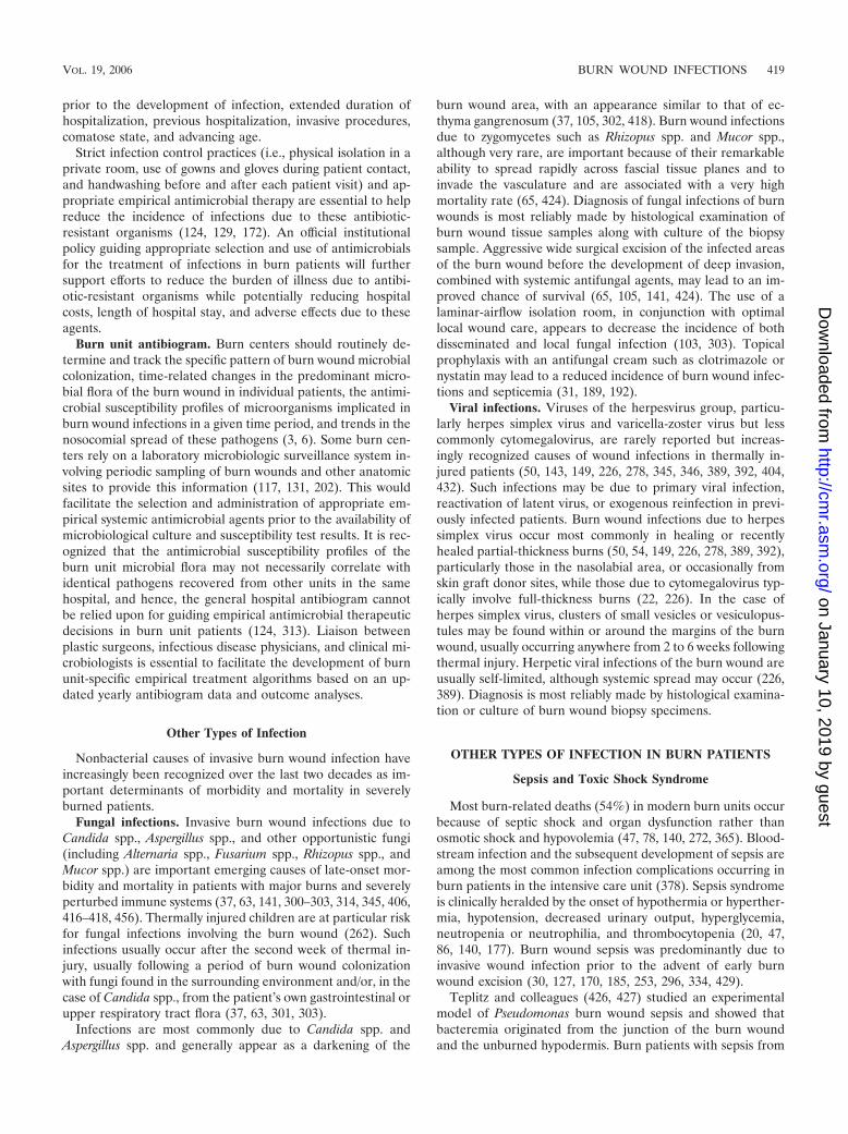
prior to the development of infection, extended duration ofhospitalization, previous hospitalization, invasive procedures,comatose state, and advancing age.
Strict infection control practices (i.e., physical isolation in aprivate room, use of gowns and gloves during patient contact,and handwashing before and after each patient visit) and ap-propriate empirical antimicrobial therapy are essential to helpreduce the incidence of infections due to these antibiotic-resistant organisms (124, 129, 172). An official institutionalpolicy guiding appropriate selection and use of antimicrobialsfor the treatment of infections in burn patients will furthersupport efforts to reduce the burden of illness due to antibi-otic-resistant organisms while potentially reducing hospitalcosts, length of hospital stay, and adverse effects due to theseagents.
Burn unit antibiogram. Burn centers should routinely de-termine and track the specific pattern of burn wound microbialcolonization, time-related changes in the predominant micro-bial flora of the burn wound in individual patients, the antimi-crobial susceptibility profiles of microorganisms implicated inburn wound infections in a given time period, and trends in thenosocomial spread of these pathogens (3, 6). Some burn cen-ters rely on a laboratory microbiologic surveillance system in-volving periodic sampling of burn wounds and other anatomicsites to provide this information (117, 131, 202). This wouldfacilitate the selection and administration of appropriate em-pirical systemic antimicrobial agents prior to the availability ofmicrobiological culture and susceptibility test results. It is rec-ognized that the antimicrobial susceptibility profiles of theburn unit microbial flora may not necessarily correlate withidentical pathogens recovered from other units in the samehospital, and hence, the general hospital antibiogram cannotbe relied upon for guiding empirical antimicrobial therapeuticdecisions in burn unit patients (124, 313). Liaison betweenplastic surgeons, infectious disease physicians, and clinical mi-crobiologists is essential to facilitate the development of burnunit-specific empirical treatment algorithms based on an up-dated yearly antibiogram data and outcome analyses.
Other Types of Infection
Nonbacterial causes of invasive burn wound infection haveincreasingly been recognized over the last two decades as im-portant determinants of morbidity and mortality in severelyburned patients.
Fungal infections. Invasive burn wound infections due toCandida spp., Aspergillus spp., and other opportunistic fungi(including Alternaria spp., Fusarium spp., Rhizopus spp., andMucor spp.) are important emerging causes of late-onset mor-bidity and mortality in patients with major burns and severelyperturbed immune systems (37, 63, 141, 300–303, 314, 345, 406,416–418, 456). Thermally injured children are at particular riskfor fungal infections involving the burn wound (262). Suchinfections usually occur after the second week of thermal in-jury, usually following a period of burn wound colonizationwith fungi found in the surrounding environment and/or, in thecase of Candida spp., from the patient’s own gastrointestinal orupper respiratory tract flora (37, 63, 301, 303).
Infections are most commonly due to Candida spp. andAspergillus spp. and generally appear as a darkening of the
burn wound area, with an appearance similar to that of ec-thyma gangrenosum (37, 105, 302, 418). Burn wound infectionsdue to zygomycetes such as Rhizopus spp. and Mucor spp.,although very rare, are important because of their remarkableability to spread rapidly across fascial tissue planes and toinvade the vasculature and are associated with a very highmortality rate (65, 424). Diagnosis of fungal infections of burnwounds is most reliably made by histological examination ofburn wound tissue samples along with culture of the biopsysample. Aggressive wide surgical excision of the infected areasof the burn wound before the development of deep invasion,combined with systemic antifungal agents, may lead to an im-proved chance of survival (65, 105, 141, 424). The use of alaminar-airflow isolation room, in conjunction with optimallocal wound care, appears to decrease the incidence of bothdisseminated and local fungal infection (103, 303). Topicalprophylaxis with an antifungal cream such as clotrimazole ornystatin may lead to a reduced incidence of burn wound infec-tions and septicemia (31, 189, 192).
Viral infections. Viruses of the herpesvirus group, particu-larly herpes simplex virus and varicella-zoster virus but lesscommonly cytomegalovirus, are rarely reported but increas-ingly recognized causes of wound infections in thermally in-jured patients (50, 143, 149, 226, 278, 345, 346, 389, 392, 404,432). Such infections may be due to primary viral infection,reactivation of latent virus, or exogenous reinfection in previ-ously infected patients. Burn wound infections due to herpessimplex virus occur most commonly in healing or recentlyhealed partial-thickness burns (50, 54, 149, 226, 278, 389, 392),particularly those in the nasolabial area, or occasionally fromskin graft donor sites, while those due to cytomegalovirus typ-ically involve full-thickness burns (22, 226). In the case ofherpes simplex virus, clusters of small vesicles or vesiculopus-tules may be found within or around the margins of the burnwound, usually occurring anywhere from 2 to 6 weeks followingthermal injury. Herpetic viral infections of the burn wound areusually self-limited, although systemic spread may occur (226,389). Diagnosis is most reliably made by histological examina-tion or culture of burn wound biopsy specimens.
OTHER TYPES OF INFECTION IN BURN PATIENTS
Sepsis and Toxic Shock Syndrome
Most burn-related deaths (54%) in modern burn units occurbecause of septic shock and organ dysfunction rather thanosmotic shock and hypovolemia (47, 78, 140, 272, 365). Blood-stream infection and the subsequent development of sepsis areamong the most common infection complications occurring inburn patients in the intensive care unit (378). Sepsis syndromeis clinically heralded by the onset of hypothermia or hyperther-mia, hypotension, decreased urinary output, hyperglycemia,neutropenia or neutrophilia, and thrombocytopenia (20, 47,86, 140, 177). Burn wound sepsis was predominantly due toinvasive wound infection prior to the advent of early burnwound excision (30, 127, 170, 185, 253, 296, 334, 429).
Teplitz and colleagues (426, 427) studied an experimentalmodel of Pseudomonas burn wound sepsis and showed thatbacteremia originated from the junction of the burn woundand the unburned hypodermis. Burn patients with sepsis from
VOL. 19, 2006 BURN WOUND INFECTIONS 419
on January 10, 2019 by guesthttp://cm
r.asm.org/
Dow
nloaded from

invasive burn wound infection have transient or intermittentbacteremia or fungemia from seeding of microorganisms intothe bloodstream, but positive blood cultures are a late sign ofinfection (41, 109, 123, 152, 270, 444). Bacteremia also occursfrom endogenous intestinal flora because of the decreasedblood flow and gut perfusion that occur following thermalinjury (248, 266, 371). Peak endotoxin concentrations havebeen documented to occur within 12 h after injury (283). Fol-lowing burn injury in an animal model, potent gut vasocon-strictors, including thromboxane and systemic vasopressin, arereleased, and their levels increase in parallel in the gut (257,266, 380). However, preventing gut mucosal atrophy and im-proving gut mucosal blood flow through early enteral feedingof burn patients have reduced subsequent translocation andendogenous bacteremia (185).
Mason and colleagues (272) performed the largest study ofbacteremia and mortality in 5,882 burn patients consecutivelyadmitted to a major burn center over a 25-year period from1959 to 1983. They developed a predictor of mortality indexbased on the 75% of patients who did not have bacteremia.Comparison between the observed and predicted levels ofmortality in subsets of patients with bacteremia showed thatthose with gram-negative bacteremia had a significantly in-creased mortality, whereas those with gram-positive bactere-mia had no increase in their attributed mortality. Overall,sepsis resulted in a 21% increase in total mortality in patientswith bacteremia due to either gram-negative organisms oryeasts. Most of these deaths (75%) also occurred in patientswhose predicted mortality from burn injury in the absence ofbacteremia was low.
Although the incidence of invasive wound infection as theprimary source for burn wound sepsis has decreased substan-tially since the advent of early excision therapy, septic shockand multiple-organ dysfunction syndrome remain importantcauses of death after burn trauma despite immediate admis-sion to an intensive care unit (46, 47, 452). In the last twodecades, sepsis in burn patients was more often secondary tocatheter-related infection or pneumonia rather than a result ofthe burn wound itself (94, 165, 347, 359, 393). Fitzwater andcolleagues (140) recently studied the risk factors and temporalrelationship between the development of multiple-organ dys-function syndrome and sepsis; 175 adult patients with a �20%TBSA burn injury were studied, and 27% developed severemultiple-organ dysfunction syndrome, while 17% developedcomplicated sepsis, and almost a quarter of the patients died.Full-thickness burn size, age, and inhalation injury were asso-ciated with the development of multiple-organ dysfunctionsyndrome, sepsis, and death. Infection preceded multiple-or-gan dysfunction syndrome in 83% of the patients.
Burn patients may also manifest signs of toxic shock syn-drome if they become colonized or infected with a strain ofStaphylococcus aureus that produces TSST-1 toxin (59, 119,121, 461). However, differentiating this severe form of toxicosisfrom systemic inflammatory response syndrome or even themetabolic response to the burn injury itself may be difficult,particularly in the immediate postinjury period. Toxic shocksyndrome may be more prevalent in children with burn injury,and the associated mortality can be high, especially if there isa delay in recognition and appropriate management (59). Fewtoxic shock syndrome cases have been reported from adult
burn centers (461). The Centers for Disease Control andPrevention criteria should be applied to define cases of toxicshock syndrome (76). Confirmation of the production ofTSST-1 toxin can be obtained by sending a patient’s Staph-ylococcus aureus isolate(s) to a reference laboratory for test-ing to detect toxin production. Antistaphylococcal antibodytiters are not useful in the diagnosis of toxic shock syn-drome, since many patients who do not have acute diseasewill have positive titers (121).
Burn patients with sepsis should be examined immediatelyto determine the site and source of infection, including inspec-tion of the entire burn wound surface. Diagnostic tests shouldbe done to identify the site and source of infection, includingblood, urine, and sputum cultures. Empirical broad-spectrumantibiotic therapy directed at the most recent bacteria isolatedfrom burn wound cultures and other sources should be insti-tuted promptly (86, 108, 307). Patients in the intensive careunit with bloodstream infections may develop severe sepsis ifinitial empirical antimicrobial treatment provides inadequateactivity against the organism causing infection (474). Addi-tional drugs that block a part of the septic cascade may also bebe administered to burn patients with severe sepsis. Activatedprotein C appears to hold the most promise for improvingoutcomes in intensive care unit patients with severe sepsis, butsepsis drug intervention trials have shown divergent results forother agents, including cytokine inhibitors, antiendotoxin, andother naturally occurring anticoagulants (338).
Pneumonia
Pulmonary complications are common in burn patients withinhalation lung injury. Burn patients with severe inhalationinjury requiring prolonged intubation are also at risk for de-veloping ventilator-associated pneumonia (VAP) (35, 86, 306,378, 431, 445, 446). VAP is defined as pneumonia that devel-ops more than 48 h after intubation (e.g., late onset) in amechanically ventilated patient who had no signs of this com-plication at the time the endotracheal tube was inserted. Theestimated prevalence of nosocomial pneumonia in the inten-sive care unit setting ranges from 10% to 65%, and mortalityrates are �25% in most reported studies (60, 89, 228, 466).
The nosocomial bacteria that usually cause VAP tend to bemore antibiotic resistant (306). Ramzy and colleagues (356)demonstrated that although the organisms colonizing the burnwound were similar to those recovered from quantitative cul-ture of bronchoalveolar lavage fluid samples in pediatric burnpatients with VAP, quantitative wound culture was not predic-tive of cross-infection in the lung. Bronchoalveolar lavage withcollection of samples from the deep lung should therefore beperformed in order to rapidly establish the microbial etiologyof the pneumonia (34, 356, 445, 446). Quantitative culture ofbronchoalveolar lavage fluid samples has shown that the pres-ence of a bacterial pathogen(s) at �104 CFU/ml of bronchoal-veolar lavage fluid is diagnostic of VAP in intensive care unitpatients (34, 91, 396).
Invasive pulmonary diagnostic procedures such as bron-choalveolar lavage allow more appropriate use of antibiotictherapy but have not been shown to change the overall mor-tality from VAP (396). The results of preliminary bronchoal-veolar lavage fluid sample tests such as the Gram stain are
420 CHURCH ET AL. CLIN. MICROBIOL. REV.
on January 10, 2019 by guesthttp://cm
r.asm.org/
Dow
nloaded from

unreliable in directing prescription of initial empirical antibi-otic therapy (93, 111). Laupland and colleagues (239) recentlyevaluated the utility of using a rapid bacterial ATP assay toscreen bronchoalveolar lavage fluid samples from 477 intensivecare unit patients with suspected VAP. This rapid screen dem-onstrated excellent performance compared to quantitative cul-tures in accurately detecting bronchoalveolar lavage fluid sam-ples with significant bacterial counts. This diagnostic strategymay allow physicians to selectively prescribe antibiotics tothose with a positive screening assay. Antibiotic therapy forVAP in the burn patient should be tailored to the reportedantibiotic susceptibility profile of the primary bacterial patho-gen(s) causing the pneumonia (323).
Burn patients may also have pulmonary complications evenwhen the lungs have not sustained direct thermal damage (101,201). Atelectasis and hypostatic pneumonia are common dueto the hyperventilation and decreased lung expansion that oc-cur in patients with large burns, �30% TBSA, due in part tochest wall restriction from circumferential eschar formation(101). Burn patients also have a high risk of repeated aspira-tion episodes (99, 101), and respiratory therapy with regularsuctioning of upper airway secretions and expectoration ofsputum in addition to escharotomy are critical to maintainingpulmonary function (261, 275).
Endotracheal or sputum samples should be sent for cultureto determine the microbial etiology of bronchopneumonia. Aclinical history of burn injury should be provided to the clinicalmicrobiology laboratory on the requisition. Endotracheal andsputum samples are initially microscopically screened for con-tamination by saliva (e.g., presence of �25 epithelial cells perhigh-power field) to determine if the specimen has been ade-quately collected (33). All potential pathogens isolated froman adequately collected sputum sample from a burn patientshould be identified and tested for antibiotic susceptibility(307). The culture results from sputum samples should becorrelated to those from the burn wound since there is oftencross-colonization between the burn wound and the tracheo-bronchial tree (356).
Although hematogenous pneumonia is a much less commoncomplication in burn patients today than in prior decades, it isnevertheless a serious complication that is largely preventable.Blood cultures should be drawn prior to the start of antibiotictherapy in burn patients with fever and suspected pneumoniain order to document bacteremia (86). The occurrence of tran-sient bacteremia associated with manipulation of the burnwound has been well documented (304, 379). The bacteremiaoccurrence rate ranges from 1.6 to 60% of severe burn cases,with the risk being proportional to the extent of the burn andthe duration and intensity of manipulation (304). However, theincidence of bacteremia after burn wound manipulation hasbeen substantially reduced by early excision therapy (30, 170,185, 253).
The source of infection in bacteremic burn patients must berapidly identified and eradicated to prevent secondary seedingof the lungs and other deep tissues through sustained high-grade circulation of microorganisms in the blood (86). Appro-priate empirical systemic antibiotic therapy should be directedagainst the organisms recovered from recent cultures of theburn wound surface and other sources such as sputum, urine,and blood samples. Antibiotic regimens should be altered as
necessary based on the results of the antibiotic susceptibilityprofile(s) of the isolate(s) recovered from the patient’s bloodcultures (307). Sustained high-grade bacteremia without anobvious focus of infection should be attributed to endocarditicor endovascular infection and treated as such.
Urinary Tract Infections
Burn patients may develop a urinary tract infection in asso-ciation with prolonged bladder catheterization. Patients willdevelop significant bacteriuria after 72 h of urinary catheterinsertion, so these devices should be removed after the initialperiod of fluid resuscitation and output monitoring (156, 172,450, 468). Recent use of silver-impregnated Foley catheters inburn patients showed promise in reducing the incidence ofurinary tract infections compared to that in patients with stan-dard Foley catheters for a similar period of time (321).
In order to minimize contamination, urine samples for cul-ture should not be collected from the drainage bag, but shouldbe aspirated through the rubber catheter using a large-boreneedle. Immediate removal of the urine catheter and institu-tion of an appropriate antibiotic(s) based upon the latest urineculture report should be used to treat urinary tract infections.Candiduria may represent contamination of the urine from theperiurethral area or vaginal area in women. However, repeatedisolation of Candida spp. from urine samples in the burn pa-tient should not be ignored because it may signal that thepatient has candidemia as a sign of disseminated candidiasis(213, 316). Antifungal therapy should be instituted immedi-ately in burn patients with an active or disseminated infectiondue to Candida spp. While Candida albicans and most non-C.albicans Candida spp. currently remain susceptible to flucon-azole, one of the newer antifungal agents, either casopofunginor voriconazole, may be required to treat serious infectionsdue to Candida glabrata and Candida krusei because they aretypically resistant to fluconazole (19, 234, 240, 277). Most Can-dida spp. currently remain susceptible to amphotericin B.
Catheter Infections and Suppurative Thrombophlebitis
Burn patients are particularly susceptible to the complica-tions associated with insertion of intravenous and intra-arterialcatheters and lines, particularly infection. Catheter-associatedinfections have been reported to affect from 8 to 57% of burnpatients (144, 245). Infected peripheral and central cathetersare a significant source of sepsis in the burn patient (86, 165,263, 347, 359, 393, 450). Suppurative thrombophlebitis mayalso be diagnosed in 5 to 10% of hospitalized burn patientswith severe burn injury, �20% TBSA, and the mortalityreaches 60% even in those who receive prompt treatment (58,165, 328, 348, 359, 410, 443).
Suppurative thrombophlebitis should be suspected in burnpatients who have high-grade bacteremia without an obviousfocus of infection, including endocarditis. Suppurative throm-bophlebitis serves as a source of bacteremia, sepsis syndrome,and the seeding of other deep organ tissues (i.e., endocarditisand brain abscesses) (359). Since only a third of cases havelocal signs of infection over the affected vein, a diagnosis ofsuppurative thrombophlebitis is difficult to make (165, 328,348, 410). An infected vein may only be identified through
VOL. 19, 2006 BURN WOUND INFECTIONS 421
on January 10, 2019 by guesthttp://cm
r.asm.org/
Dow
nloaded from

sequential venotomy and examination of veins for intraluminalpus by vein wall biopsy for histological examination for bacte-rial colonization of the intimal surface. Careful nursing recordsmust therefore be kept about sequential peripheral and centralintravenous catheter locations.
Burn patients have a high frequency of peripheral intravenousand central line-related infections for a number of reasons,including a prolonged need for intravenous infusion, frequenthyperalimentation fluids, frequent episodes of bacteremia sec-ondary to wound manipulation, a high density of surface mi-croorganisms, and the impairment of host defenses due to theburn injury (144, 245). A recent study of burn patients dem-onstrated a high correlation between the microorganisms cul-tured from the catheter tip and hubs within 48 h of insertion,and the incidence of catheter-associated infection was in-versely proportional to the distance of the line insertion sitefrom the burn wound (354). Catheter infections in burn pa-tients likely arise from the adherence and migration of burnwound flora microorganisms along the catheter surface to thetip, with the creation of a thick biofilm (326, 378, 419, 421).Although peripheral and central catheters should be insertedthrough an area of normal skin in order to minimize the de-velopment of infection, this may not be possible in patientswith severe burns. It may also be necessary to cut down into ausable vein or artery through the burn wound itself in burnpatients with extensive injuries to a large area of the cutaneoussurface.
Rates of catheter-associated infection can nevertheless bedecreased with the use of rigorous aseptic technique during theinsertion procedure, the use of Teflon catheters, change of theperipheral intravenous equipment, and rotation of the periph-eral catheter site every 72 h (263, 264). In addition, cathetersimpregnated with either antibiotics or antiseptic (chlorohexi-dine and silver sulfadiazine) may decrease the incidence ofcentral catheter infection in burn patients, although compara-tive studies are needed (370). New catheters impregnated withnanocrystalline silver show decreased microbial colonization,but there have been no studies of the use of these devices inburn patients (375).
Treatment of suppurative thrombophlebitis requires promptinstitution of empirical systemic antibiotic therapy directedagainst the burn wound flora and operative excision of theinfected vein or vein segment. However, these measures areadjunctive to prompt removal of the infected line and reloca-tion of a clean device at another site in a healthy noninfectedvein (328, 348, 410).
Myonecrosis
Burn patients may sustain deep tissue injuries beneath thecutaneous layers that provide a focus for infection to develop.Muscle necrosis and pyomyositis occur in thermally injuredextremities with circumferential full-thickness injury due tovascular compromise. These serious infections may be difficultto diagnose in limbs that are already edematous and painfuldue to the thermal injury. In particular, direct electrical contactresults in deep muscular necrosis and delayed infection (180,232, 352). Effective treatment of these serious infections re-quires prompt surgical excision of the affected deep tissues
involved along with institution of broad-spectrum antibioticsagainst aerobic and anaerobic microorganisms (300, 308).
PREVENTION OF BURN WOUND INFECTIONS
Several studies have demonstrated the role of topical anti-microbials in decreasing morbidity and mortality in patientswith major burn injuries (partial- or full-thickness skin involve-ment), particularly before early excision (189, 298, 307). Arecent study conducted by the U.S. Army Burn Center com-pared the levels of mortality of adult patients according to ageand burn size before (1950 to 1963) and after (1964 to 1968)the introduction of mafenide acetate topical antibiotic therapy(62). Use of mafenide acetate was associated with a greaterthan 10% reduction in mortality for those with burns of 40 to79% TBSA, but its use had only a minimal effect on mortalityin patients with smaller or much larger burn injuries.
The efficacy of various topical antimicrobials in common usein modern burn centers is dynamic due to the ability of micro-organisms to develop resistance rapidly (6). The sustained po-tency of individual agents depends on the extent of use and theresident nosocomial flora within any specialized burn center.In order to accurately detect and track emerging trends intopical antimicrobial resistance in modern burn units, it isessential that standard reproducible methods be published forclinical implementation (see Antibiotic Susceptibility Testing,above).
Topical Antimicrobial Therapy
Widespread application of an effective topical antimicrobialagent substantially reduces the microbial load on the openburn wound surface and reduces the risk of infection (189, 298,307). By controlling infection, effective topical antimicrobialtherapy decreases the conversion of partial-thickness to full-thickness wounds, but its use is adjunctive to early excisiontherapy. Selection of topical antimicrobial therapy should bebased on the agent’s ability to inhibit the microorganisms re-covered from burn wound surveillance cultures and monitoringof the nosocomial infections acquired in the burn unit. Pre-scription is also based on the individual preparation of thetopical agent (e.g., ointment or cream versus solution or dress-ing) and its pharmacokinetic properties. Burn units may rotatethe use of various topical antimicrobial preparations on a reg-ular basis to decrease the potential for development of antibi-otic resistance (6, 124, 313). Topical antibiotic agents shouldfirst be applied directly to the patient’s dressings before appli-cation to the burn wound to prevent contamination of theagent’s container by burn wound flora.
Table 4 outlines the most widely used topical antimicrobialagents and new silver nanocrystalline dressings that are basedon the bactericidal properties of the silver ion (134, 189, 298).The inhibitory action of silver is due to its strong interactionwith thiol groups present in the respiratory enzymes in thebacterial cell (237, 238). Silver has also been shown to interactwith structural proteins and preferentially bind with DNA nu-cleic acid bases to inhibit replication (236, 237). For this rea-son, silver has recently been shown to be highly toxic to kera-tinocytes and fibroblasts and may delay burn wound healing ifapplied indiscriminately to debrided healing tissue areas (53,
422 CHURCH ET AL. CLIN. MICROBIOL. REV.
on January 10, 2019 by guesthttp://cm
r.asm.org/
Dow
nloaded from

90, 236). Moist exposure therapy using a moisture-retentiveointment (MEBO-Julphar; Gulf Pharmaceutical Industries,United Arab Emirates) has recently been shown to act as aneffective antibacterial agent while promoting rapid autolysisdebridement and optimal moist wound healing in partial-thick-ness injury (13, 16). Moisture-retentive ointment also resultedin earlier recovery of keratinocytes with improved wound heal-ing and decreased scar formation (14). The topical antimicro-bial agents reviewed are primarily used in burn center patientswith full-thickness or deep partial-thickness burn wounds.
Silver nitrate. Silver nitrate is rarely used nowadays inmodern burn units because the deposition of silver discolorsthe wound bed and other topical agents are available thatare easier to use and have less potential toxicity. Silvernitrate is most effective before the burn wound becomescolonized. The burn wound needs to be cleansed of emol-lients and other debris before a multilayered dressing isapplied to the burn wound and subsequently saturated withsilver nitrate solution. Effective use of this preparationtherefore requires continuous application with secondaryocclusive dressings, making examination of the wound dif-ficult. The silver ion in AgNO3 also quickly binds to elemen-tal chlorine ions, so that repeated or large-surface applicationof this solution may lead to electrolyte imbalance (e.g., hypo-natremia and hypochloremia) (189, 298). Silver nitrate anti-bacterial activity is limited to the burn wound surface (204,409). This agent demonstrates bacteriostatic activity againstgram-negative aerobic bacteria such as Pseudomonas aerugi-nosa and Escherichia coli, but it is not active against othergenera, including Klebsiella, Providencia, and Enterobacter(189, 260). Silver nitrate also has limited antifungal activity, sothat nystatin should be used concomitantly (192, 467).
Silver sulfadiazine. Silver sulfadiazine is the most commonlyused topical antibiotic agent for both ambulatory and hospi-talized burn patients. This agent is a combination of sodiumsulfadiazine and silver nitrate. The silver ion binds to the mi-croorganism’s nucleic acid, releasing the sulfadiazine, whichthen interferes with the metabolism of the microbe (237). It iseasy to use and painless when applied and can be used with or
without a dressing. Limited systemic toxicity with repeateddaily or twice-daily application has occurred aside from thedevelopment of leukopenia (81, 147). Silver sulfadiazine hasexcellent broad-spectrum antibacterial coverage againstPseudomonas aeruginosa and other gram-negative entericbacteria, although some resistance has recently been re-ported (189, 335). This agent also has some activity againstCandida albicans, but enhanced antifungal activity can beachieved by using nystatin in combination with silver sulfa-diazine (192, 467).
Although silver sulfadiazine dissociates more slowly thansilver nitrate, there is still poor penetration into the wound(204, 409). Silver sulfadiazine is only absorbed within the sur-face epidermal layer, which limits its effectiveness in somepatients with severe injuries. In Europe, a combination ofcerium nitrate and silver sulfadiazine (Flammacerium; SolvayDuphar, The Netherlands) has been used to combat this prob-lem (153, 154). Flammacerium has been shown to reduce theinflammatory response to burn injury, decrease bacterial col-onization, and provide a firm eschar for easier wound manage-ment (154).
Mafenide acetate. Topical mafenide acetate cream allowsopen burn wound therapy and regular examination of the burnwound surface because it is used without dressings. The burnwound surface is cleansed of debris prior to application of thecream, and the treated burn wound surface is left exposed afterthe cream is applied for maximal antimicrobial effect. Mafenideacetate is applied a minimum of twice daily and has been shownto penetrate the burn eschar (409). The 5% solution must beapplied to saturate gauze dressings, and these need to be changedevery 8 hours for maximal effect. Mafenide acetate solutionappears to be as effective as the cream preparation when usedin this way (135, 189).
Mafenide acetate (Sulfamylon) cream has a broad spectrumof activity against gram-negative bacteria, particularly Pseudo-monas aeruginosa, but has little activity against gram-positiveaerobic bacteria such as Staphylococcus aureus (189). Thisagent also inhibits anaerobes such as Clostridium spp. Becauseprotracted use of mafenide acetate favors the overgrowth of
TABLE 4. Profile of commonly used topical antimicrobial agents a
Topical agent Preparation Escharpenetration Antibacterial activity b Major toxicity
Silver nitrate (AgNO3) 0.5% solution None Bacteriostatic against aerobic gram-negativebacilli, P. aeruginosa, limited antifungal
Electrolyteimbalance
Silver sulfadiazine (Silvodene,Flamazine, Thermazine,Burnazine)
1% water-soluble cream(oil-in-water emulsion)
None Bactericidal against aerobic gram-negativebacilli, P. aeruginosa, some C. albicans
Leukopenia
Mafenide acetate(Sulfamylon)
10% water-soluble cream(oil-in-water emulsion),5% solution
Limited Broad spectrum against aerobic gram-negativebacilli, P. aeruginosa, anaerobes
Metabolic acidosis
Nanocrystalline silverdressings (Acticoat A.B.dressing, Silverlon)
Dressing consisting oftwo sheets of high-density polyethylenemesh coated withnanocrystalline silver
Moderate Potent activity against aerobic gram-negativebacilli, P. aeruginosa, aerobic gram-positivebacilli, MRSA, VRE, multidrug-resistantEnterobacteriaceae
Limited toxicity
a Data are from references 134, 189, and 298.b VRE, vancomycin-resistant enterococci.
VOL. 19, 2006 BURN WOUND INFECTIONS 423
on January 10, 2019 by guesthttp://cm
r.asm.org/
Dow
nloaded from

Candida albicans and other fungi, this agent should be used incombination with nystatin to prevent this complication due toits limited antifungal activity (192, 467).
Despite its antibacterial potency, mafenide acetate is not aswidely used as other agents because of its toxicity profile. Thiscompound is converted to p-sulfamylvanzoic acid by mono-amide oxidase, a carbonic anhydrase inhibitor, causing metabolicacidosis in the burn patient (189, 298). In burn patients withinhalation injury and a concomitant respiratory acidosis, theuse of mafenide acetate over a large burn surface area or therepeated application of this compound can be fatal. Mafenideacetate also decreases the breaking strength of healed woundsand delays healing (53).
Acticoat A.B. dressing/Silverlon. This product is a special-ized dressing that consists of two sheets of high-density poly-ethylene mesh coated with nanocrystalline silver (e.g., ionicsilver with a rayon-polyester core) (112, 435, 472). The morecontrolled and prolonged release of nanocrystalline silver tothe burn wound area allows less-frequent dressing changes,reducing the risk of tissue damage, nosocomial infection, pa-tient discomfort, and the overall cost of topical therapy (112,188). Nanocrystalline dressings may also provide better pene-tration of unexcised burn wounds because of their prolongedmechanism of action. Acticoat A.B. dressing with SILCRYST�
(Smith & Nephew Wound Management, Largo, FL), Silverlon(Argentum Medical, L.L.C., Lakemont, GA), and Silvasorb(Medline Industries Inc., Mundelein, IL) provides the mostcomprehensive broad-spectrum bactericidal coverage againstimportant burn wound pathogens of any topical antimicrobialpreparation currently marketed (112, 188). These dressingshave potent antibacterial activity against most aerobic gram-negatives, including Pseudomonas aeruginosa and antibiotic-resistant members of the family Enterobacteriaceae as well asaerobic gram-positive bacteria, including MRSA and vancomy-cin-resistant enterococci (112, 188, 472). If the burn woundsurface has minimal exudates, these specialized dressings canremain in place for several days and retain antibacterial activity(188). This approach is replacing the use of other silver-basedtopical antibiotics in many burn centers.
Mupirocin (Bactroban). Mupirocin (pseudomonic acid A) isa fermentation product of Pseudomonas fluorescens (189, 330).This antibiotic has potent inhibitory activity against gram-pos-itive skin flora such as coagulase-negative staphylococci andStaphylococcus aureus, including MRSA (287, 366, 367, 420).Although primarily marketed for nasal decontamination,mupirocin has increasingly been used as a burn wound topicalagent in burn units in North America, where MRSA has be-come a problem (126, 287). Mupirocin is currently not licensedin Europe for use as a topical burn wound agent. Varioustopical antibiotic preparations, including 1% silver sulfadia-zine, 2% mupirocin, and 2% fusidic acid, were recently com-pared for their antibacterial effect in an MRSA-infected full-skin-thickness rat burn model (2). All of these agents werefound to be equally effective against MRSA in reducing localburn wound bacterial counts and preventing systemic infection.Burn centers where MRSA is a problem may therefore rotatethe use of topical mupirocin in combination with these otheragents in order to decrease the development of resistance.
Nystatin. Nystatin (Mycostatin or Nilstat) is produced byStreptomyces noursei and has potent antifungal effect by bind-
ing to the sterols in the fungal cell membrane (189, 330). Alower concentration (3 �g/ml) of this agent inhibits Candidaalbicans, but a higher concentration (6.25 �g/ml) is needed toinhibit other Candida spp. and fungi (19, 302). A recent studyof nystatin powder at a concentration of 6 million units/gshowed that this approach was effective in treating four burnpatients with severe angioinvasive fungal infections due to ei-ther Aspergillus or Fusarium spp. (31). Both superficial anddeep-tissue burn wound infections were eradicated using nys-tatin powder without any other interventions or adverse effectson wound healing (31, 302). However, since nystatin has noactivity against bacteria, it should be used in combination witha topical agent that has activity against the broad spectrum ofpathogenic bacteria that cause burn wound colonization andinfection (189).
Other topical antimicrobials. Several other topical antimicro-bials have also been used for topical burn therapy, includinggentamicin sulfate (0.1% water-soluble cream), betadine (10%povidone-iodine ointment), bacitracin-polymyxin ointment, andnitrofurantoin (189, 330). However, these compounds are nolonger used extensively because significant resistance has devel-oped and/or they have been shown to be toxic or ineffective atcontrolling localized burn wound infections. Topical bacitracin-polymyxin is primarily used as a nonadherent, nontoxic petro-leum-based ointment for skin graft dressings and for dressingpartial-thickness burn wounds, particularly in children (330).
Prophylactic Systemic Antibiotics
Studies of the clinical benefit of prophylactic courses ofsystemic antibiotics in burn patients in decreasing the occur-rence of burn wound infections have not demonstrated im-proved outcome compared to the use of topical therapy alongwith surgical excision. In a recent study of pediatric burn pa-tients, the efficacy of antibiotic prophylaxis was studied in 77children; 47 children received prophylactic antibiotics, whilethe rest received no prophylaxis (128). Children in both groupswith wound colonization and infection had a larger burn injury,but the administration of antibiotics did not prevent the devel-opment of burn wound infection. The group of children thatreceived prophylactic antibiotics had a higher burn wound in-fection rate (21.3% versus 16.7%, P � 0.05) (128). Most of thechildren who developed sepsis were also on prophylactic anti-biotics. Use of prophylactic antibiotics also promoted the de-velopment of other secondary infections (i.e., upper and lowerrespiratory tract and urinary tract infections and otitis media).Overall length of hospital stay was also prolonged in the chil-dren receiving prophylactic antibiotics.
Administration of systemic antibiotic therapy may also causeantibiotic-associated diarrhea due to the overgrowth of toxi-genic strains of Clostridium difficile (176, 404, 415). Exposure toprophylactic antibiotic therapy may also increase the resistanceof endogenous and pathogenic bacteria to a wide variety ofantibiotics, making the subsequent treatment of clinically overtinfections in the burn patient more difficult (6, 307).
Systemic antibiotic administration in burn patients shouldtherefore only be used selectively and for a short period oftime. Because of the secondary bacteremia associated withprolonged burn wound manipulation and/or excision, prophy-lactic systemic antibiotic therapy may be given immediately
424 CHURCH ET AL. CLIN. MICROBIOL. REV.
on January 10, 2019 by guesthttp://cm
r.asm.org/
Dow
nloaded from

before, during, and for one or two doses after the procedure,particularly in burn patients with extensive injury (e.g., 40%TBSA) (304). Culture-specific laboratory information obtainedfrom bacterial culture and susceptibility results for the burnwound and other sources (i.e., blood, urine, and respiratorycultures) should be used to guide the selection of effectiveantimicrobial agents for use as preoperative prophylaxis as wellas treatment of overt clinical infections (307).
Selective Bowel Decontamination
The relationship between the translocation of bowel micro-flora due to increased intestinal permeability and the subse-quent colonization of the burn wound by enteric gram-negativemicroorganisms has been well described (168, 257). Severalstudies compared the ability of oral prophylactic antibioticregimens to selectively decontaminate the normal bowel florain burn patients, thereby reducing burn wound colonizationand infection (106, 220, 221, 267, 269). Jarrett and colleagues(220, 221) showed that burn wound colonization was delayedand the rate of infection was reduced in patients who receivedan oral bowel flora-suppressive antibiotic regimen (neomycin,erythromycin, and nystatin). However, this broad-spectrum de-contamination regimen not only suppressed aerobic gram-neg-ative bacteria but also destroyed the anaerobic flora that isimportant in host “colonization resistance.” Manson and col-leagues (267, 269) later showed that selective bowel decontam-ination using oral polymyxin either alone or in combinationwith oral trimethoprim-sulfamethoxazole and/or amphotericinB prevented burn wound colonization. Selective oral boweldecontamination therapy, however, was never widely adoptedas routine therapy and became unnecessary with the advent ofearly-excision burn wound therapy.
Recent studies have shown that early enteral feeding incombination with excision of the burn wound in severely burn-injured children improved their clinical recovery and outcome.Hart and colleagues (185) found that early enteral feedingdiminished the incidence of wound colonization and infectionby bowel flora and sepsis but did not accelerate the hypermeta-bolic state induced by the burn injury. Early enteral feeding islikely effective because it increases circulation to the bowel,thereby decreasing ischemia postinjury and the translocationof bowel flora.
Prevention of Tetanus
Since thermal injury creates an open dirty wound, burn pa-tients are prone to develop tetanus (45, 116, 394). Burn centersroutinely administer human tetanus immunoglobulin (250 to500 IU) to provide immediate passive immunization regardlessof the patient’s active immunization status. Active immuniza-tion with tetanus toxoid is also given (0.5 ml intramuscularly)to burn patients who have not received a complete primaryimmunizing series or who have not received a tetanus toxoidbooster within the past 10 years (45, 116). For children �7years of age, a trivalent diphtheria-pertussis-tetanus vaccine isadministered, while adults are given tetanus-diphtheria vac-cine. Patients who have never been immunized against tetanusor who have been partially immunized should also receive
subsequent doses of tetanus vaccine to ensure antibody levelsare protective (45, 116).
Infection Control in the Burn Unit
Modern burn centers have a contained perimeter that isdesigned to minimize the unnecessary traffic of health careworkers and visitors alike through the unit (36, 191, 202, 449).Cross-contamination is further diminished within the unit byhousing burn patients in individual nursing units composed ofindividual isolation rooms, each with its own laminar airflow(103, 449). Nosocomial outbreaks due to antibiotic-resistantorganisms have been described in modern burn units becausecritically ill burn patients and equipment had to be movedbetween the burn unit and the trauma intensive care unit (36).Modern burn unit designs should allow all intensive and burncare procedures, including ventilation and operative proce-dures, to be done within the burn center itself, or, as a mini-mum, the facility design should minimize the need to transferpatients out of the burn unit for different aspects of their care.A recent study showed that the rate of cross-colonization withresistant organisms in 66 critically ill children with severe burnsand inhalation injury on ventilator support during a 5-yearperiod was extremely low (3.2 cases per 1,000 patient-days) insuch a facility (451).
Modern infection control practice has been effective in re-ducing or eliminating endemic pathogenic and/or antibiotic-resistant organisms, preventing the establishment of newly in-troduced pathogenic and/or antibiotic-resistant organisms asthe predominant nosocomial flora of the burn unit, and pre-venting reseeding of such strains back into the burn unit frompatients housed in the adjacent convalescent ward (284, 449).The infection control program for burn centers requires strictcompliance with a number of environmental control measuresthat include strictly enforced hand washing and the universaluse of personal protective equipment (i.e., gowns, gloves, andmasks) (284, 449). Health care personnel must be gowned(including use of disposable or reusable gowns and disposableplastic aprons to prevent soiling of health care workers’ cloth-ing during wound care procedures) and gloved at each entry tothe burn patient’s isolation room. Monitoring and diagnosticequipment is housed in each burn patient’s room to preventcross-contamination between patients. All equipment in theisolation room must be regularly cleaned with appropriatedisinfectants. Procedures that may predispose burn patients tocross-contamination, such as exposure hydrotherapy, are keptto a minimum (126, 436, 449). Most burn wound care in unitspracticing early excision therapy is now performed at the pa-tient’s bedside.
Cohort nursing care is another important component ofenvironmental control that is utilized in the burn unit (71, 428).Nurses and other health care personnel are assigned to care fora specific patient or group of patients as a team, and themovements of assigned personnel between patients are strictlylimited. Convalescent burn patients are also separated fromthose with an acute injury because they represent a reservoirfor more antibiotic-resistant organisms that may have beenacquired during a prolonged hospital stay. Admission surveil-lance cultures are also done to screen burn patients for colo-nization by antibiotic-resistant organisms (e.g., MRSA and
VOL. 19, 2006 BURN WOUND INFECTIONS 425
on January 10, 2019 by guesthttp://cm
r.asm.org/
Dow
nloaded from

vancomycin-resistant enterococci). Patients who are colonizedon admission or who acquire an antibiotic-resistant organismduring their burn unit stay are physically isolated from otherburn unit patients.
Infection control practitioners also play an integral part inany burn center’s prevention program. Burn wound infectionsshould be rigorously monitored according to the standard def-initions previously provided (see Classification of Burn WoundInfections, above) in order to generate accurate epidemiolog-ical data about infection rates. Routine surveillance shouldalso be carried out for other types of nosocomial infectionscommonly diagnosed in burn patients, including catheter-re-lated infections, pneumonia, and urinary tract infections. In allcases, published standard definitions should be used in identi-fying these types of infection complications (157, 158). Labo-ratory surveillance cultures (e.g., culture of nasal, rectal, orgroin swabs for MRSA and culture of rectal swabs for vanco-mycin-resistant enterococci) as well as routine microbial sur-veillance cultures of the burn wound and other sources (i.e.,blood, respiratory, and urine samples) should be monitored torapidly identify epidemic pathogens and/or antibiotic-resistantstrains so that control measures can be immediately imple-mented (6, 124). Antibiotic utilization should be rotated orchanged based on monitoring of antibiotic resistance trends(e.g., antibiograms) within individual burn centers (3). Finally,adverse outcomes, including morbidity and mortality due toburn wound infection, sepsis, or another nosocomial infectioncomplication, should be monitored in burn patients accordingto the extent of burn injury in order to assess the effectivenessof existing infection control practices within the institute’smodern burn therapy program.
FUTURE DIRECTIONS IN MICROBIAL BURNWOUND SURVEILLANCE
Infection control programs need to document and reportburn wound infections according to the recently establisheddefinitions of the classification system. Future studies of burnwound infections should use this standardized burn woundclassification system so that clinical outcomes can be comparedfor burn patients with a specific condition (e.g., burn woundcellulitis) (273, 331). More research is required to determinethe best methods for sampling excised and unexcised burnwound areas over the course of a severe deep partial-thicknessand/or full-thickness injury. Reproducible standardized meth-ods should be developed so that clinical microbiology labora-tories can routinely test burn wound bacterial isolates for sus-ceptibility to the topical antimicrobial agents on formulary at aparticular burn center. A rotation program for topical antimi-crobial use may also retard the development of resistance.Laboratory surveillance should include the reporting of burnunit-specific antibiograms for topical antimicrobial agents oncestandardized methods are available for performing susceptibil-ity testing.
REFERENCES
1. Abraham, E., and A. A. Freitas. 1989. Hemorrhage produces abnormalitiesin lymphocyte function and lymphokine generation. J. Immunol. 142:899–906.
2. Acikel, C., O. Oncul, E. Ulkur, I. Bayram, B. Celikoz, and S. Cavuslu. 2003.Comparison of silver sulfadiazine 1%, mupirocin 2%, and fusidic acid 2%
for topical antibacterial effect in methicillin-resistant staphylococci-in-fected, full-skin thickness rat burn wounds. J. Burn Care Rehabil. 24:37–41.
3. Agnihotri, N., V. Gupta, and R. M. Joshi. 2004. Aerobic bacterial isolatesfrom burn wound infections and their antibiograms—a five-year study.Burns 30:241–243.
4. Alexander, J. W. 1990. Mechanism of immunologic suppression in burninjury. J. Trauma 30:S70–S75.
5. Altman, L. C., C. T. Furukawa, and S. J. Klebanoff. 1977. Depressedmononuclear leukocyte chemotaxis in thermally injured patients. J. Immu-nol. 119:199–205.
6. Altoparlak, U., S. Erol, M. N. Akcay, F. Celebi, and A. Kadanali. 2004. Thetime-related changes of antimicrobial resistance patterns and predominantbacterial profiles of burn wounds and body flora of burned patients. Burns30:660–664.
7. American Burn Association. 2000. Burn incidence and treatment in the US:2000 fact sheet. http://www.ameriburn.org. [Online.]
8. American Burn Association. 2000. Burn incidence and treatment in theU.S. National health interview survey (1991–1993 data). American BurnAssociation, Philadelphia, Pa.
9. Amshel, C. E., M. H. Fealk, B. J. Phillips, and D. M. Caruso. 2000.Anhydrous ammonia burns: case report and review of the literature. Burns26:493–497.
10. Appelgren, P., V. Bjornhagen, K. Bragderyd, C. E. Jonsson, and U. Ransjo.2002. A prospective study of infections in burn patients. Burns 28:39–46.
11. Arons, M. S. 1965. Burn wound infection—a review. Conn. Med. 29:718–722.
12. Atiyeh, B. S., C. A. Amm, and K. A. El Musa. 2003. Improved scar qualityfollowing primary and secondary healing of cutaneous wounds. AestheticPlast. Surg. 27:411–417.
13. Atiyeh, B. S., R. Dham, M. Kadry, A. F. Abdallah, M. Al-Oteify, O. Fathi,and A. Samir. 2002. Benefit-cost analysis of moist exposed burn ointment.Burns 28:659–663.
14. Atiyeh, B. S., K. A. El-Musa, and R. Dham. 2003. Scar quality and physi-ologic barrier function restoration after moist and moist-exposed dressingsof partial-thickness wounds. Dermatol. Surg. 29:14–20.
15. Atiyeh, B. S., S. W. Gunn, and S. N. Hayek. 2005. State of the art in burntreatment. World J. Surg. 29:131–148.
16. Atiyeh, B. S., J. Ioannovich, G. Magliacani, M. Masellis, M. Costagliola, R.Dham, and M. Al-Farhan. 2002. Efficacy of moist exposed burn ointment inthe management of cutaneous wounds and ulcers: a multicenter pilot study.Ann. Plast. Surg. 48:226–227.
17. Avdakoff, V. 1876. Modifications pathologiques des tissus apres brulures.Vestrik 16:4.
18. Backstein, R., W. Peters, and P. Neligan. 1993. Burns in the disabled. Burns19:192–197.
19. Baddley, J. W., and S. A. Moser. 2004. Emerging fungal resistance. Clin.Lab. Med. 24:721–735, vii.
20. Baker, C. C., C. L. Miller, and D. D. Trunkey. 1979. Predicting fatal sepsisin burn patients. J. Trauma 19:641–648.
21. Baker, S. P., B. O’Neill, N. J. Ginsberg, and G. Li. 1992. Fire burns andlightening, p. 161–173. In Injury fact book. Oxford University Press, NewYork, N.Y.
22. Bale, J. F., Jr., G. P. Kealey, C. L. Ebelhack, C. E. Platz, and J. A. Goeken.1992. Cytomegalovirus infection in a cyclosporine-treated burn patient:case report. J. Trauma 32:263–267.
23. Bang, R. L., R. K. Gang, S. C. Sanyal, E. M. Mokaddas, and A. R. Lari.1999. Beta-haemolytic Streptococcus infection in burns. Burns 25:242–246.
24. Bang, R. L., P. N. Sharma, S. C. Sanyal, and I. Al Najjadah. 2002. Septi-caemia after burn injury: a comparative study. Burns 28:746–751.
25. Banwell, P. E., M. P. Tyler, A. M. Watts, A. H. Roberts, and D. A.McGrouther. 1999. Burn depth estimation: use of laser Doppler flowmetry.Plast. Reconstr. Surg. 103:334–335.
26. Barillo, D. J., T. S. Burge, D. T. Harrington, E. C. Coffey, K. Z. Shirani, andC. W. Goodwin. 1998. Body habitus as a predictor of burn risk in children:do fat boys still get burned? Burns 24:725–727.
27. Barillo, D. J., A. T. McManus, L. C. Cancio, A. Sofer, and C. W. Goodwin.2003. Burn center management of necrotizing fasciitis. J. Burn Care Reha-bil. 24:127–132.
28. Barret, J. P., P. Dziewulski, P. I. Ramzy, S. E. Wolf, M. H. Desai, and D. N.Herndon. 2000. Biobrane versus 1% silver sulfadiazine in second-degreepediatric burns. Plast. Reconstr. Surg. 105:62–65.
29. Barret, J. P., and D. N. Herndon. 2003. Effects of burn wound excision onbacterial colonization and invasion. Plast. Reconstr. Surg. 111:744–750.
30. Barret, J. P., and D. N. Herndon. 2003. Modulation of inflammatory andcatabolic responses in severely burned children by early burn wound exci-sion in the first 24 hours. Arch. Surg. 138:127–132.
31. Barret, J. P., P. I. Ramzy, J. P. Heggers, C. Villareal, D. N. Herndon, andM. H. Desai. 1999. Topical nystatin powder in severe burns: a new treat-ment for angioinvasive fungal infections refractory to other topical andsystemic agents. Burns 25:505–508.
32. Barrow, R. E., M. Spies, L. N. Barrow, and D. N. Herndon. 2004. Influence
426 CHURCH ET AL. CLIN. MICROBIOL. REV.
on January 10, 2019 by guesthttp://cm
r.asm.org/
Dow
nloaded from

of demographics and inhalation injury on burn mortality in children. Burns30:72–77.
33. Bartlett, J. G. 1987. Diagnosis of bacterial infections of the lung. Clin. ChestMed. 8:119–134.
34. Baselski, V. S., and R. G. Wunderink. 1994. Bronchoscopic diagnosis ofpneumonia. Clin. Microbiol. Rev. 7:533–558.
35. Baughman, R. P. 2005. Diagnosis of ventilator-associated pneumonia. Mi-crobes Infect. 7:262–267.
36. Bayat, A., H. Shaaban, A. Dodgson, and K. W. Dunn. 2003. Implications forBurns Unit design following outbreak of multi-resistant Acinetobacter in-fection in ICU and Burns Unit. Burns 29:303–306.
37. Becker, W. K., W. G. Cioffi, Jr., A. T. McManus, S. H. Kim, W. F. McManus,A. D. Mason, and B. A. Pruitt, Jr. 1991. Fungal burn wound infection. A10-year experience. Arch. Surg. 126:44–48.
38. Beetstra, J. E. 1986. Recognition and treatment of burn wound infection.Can. Oper. Room Nurs. J. 4:28–32.
39. Bengtson, A., and M. Heideman. 1987. Anaphylatoxin formation in plasmaand burn bullae fluid in the thermally injured patient. Burns Incl. Therm.Inj. 13:185–189.
40. Berkow, S. G. 1924. A method for estimating the extensiveness of lesions(burns and scalds) based on surface area proportions. Arch. Surg. 8:138–148.
41. Bharadwaj, R., B. N. Joshi, and S. A. Phadke. 1983. Assessment of burnwound sepsis by swab, full thickness biopsy culture and blood culture—acomparative study. Burns Incl. Therm. Inj. 10:124–126.
42. Biffl, W. L., E. E. Moore, F. A. Moore, and V. M. Peterson. 1996. Interleu-kin-6 in the injured patient. Marker of injury or mediator of inflammation?Ann. Surg. 224:647–664.
43. Birky, M. M., and F. B. Clarke. 1981. Inhalation of toxic products fromfires. Bull. N. Y. Acad. Med. 57:997–1013.
44. Bjornson, A. B., H. S. Bjornson, and W. A. Altemeier. 1981. Serum-medi-ated inhibition of polymorphonuclear leukocyte function following burninjury. Ann. Surg. 194:568–575.
45. Bleck, T. P. 2005. Clostridium tetani (tetanus), p. 2817–2822. In G. L.Mandell, J. E. Bennett, and R. Dolin (ed.), Principles and practice ofinfectious diseases. Elsevier Churchill Livingstone, Philadelphia, Pa.
46. Bone, R. C. 1996. Immunologic dissonance: a continuing evolution in ourunderstanding of the systemic inflammatory response syndrome (SIRS) andthe multiple organ dysfunction syndrome (MODS). Ann. Intern. Med.125:680–687.
47. Bone, R. C., W. J. Sibbald, and C. L. Sprung. 1992. The ACCP-SCCMconsensus conference on sepsis and organ failure. Chest 101:1481–1483.
48. Bornside, G. H., and B. B. Bornside. 1979. Comparison between moist swaband tissue biopsy methods for quantitation of bacteria in experimentalincisional wounds. J. Trauma 19:103–105.
49. Boswick, J. A., Jr. 1967. Long-term management of the burn patient. Mod.Treat. 4:1282–1290.
50. Bourdarias, B., G. Perro, M. Cutillas, J. C. Castede, M. E. Lafon, and R.Sanchez. 1996. Herpes simplex virus infection in burned patients: epidemi-ology of 11 cases. Burns 22:287–290.
51. Boyce, J. M., R. L. White, W. A. Causey, and W. R. Lockwood. 1983. Burnunits as a source of methicillin-resistant Staphylococcus aureus infections.JAMA 249:2803–2807.
52. Boyce, S. T., R. J. Kagan, K. P. Yakuboff, N. A. Meyer, M. T. Rieman, D. G.Greenhalgh, and G. D. Warden. 2002. Cultured skin substitutes reducedonor skin harvesting for closure of excised, full-thickness burns. Ann. Surg.235:269–279.
53. Boyce, S. T., A. P. Supp, V. B. Swope, and G. D. Warden. 1999. Topicalsulfamylon reduces engraftment of cultured skin substitutes on athymicmice. J. Burn Care Rehabil. 20:33–36.
54. Brandt, S. J., C. G. Tribble, A. D. Lakeman, and F. G. Hayden. 1985.Herpes simplex burn wound infections: epidemiology of a case cluster andresponses to acyclovir therapy. Surgery 98:338–343.
55. Bretano, L., and A. C. Gravens. 1967. A method for quantitation of bacteriain burn wounds. Appl. Microbiol. 15:671.
56. Brodovsky, S. C., C. A. McCarty, G. Snibson, M. Loughnan, L. Sullivan, M.Daniell, and H. R. Taylor. 2000. Management of alkali burns: an 11-yearretrospective review. Ophthalmology 107:1829–1835.
57. Brodzka, W., H. L. Thornhill, and S. Howard. 1985. Burns: causes and riskfactors. Arch. Phys. Med. Rehabil. 66:746–752.
58. Brook, I., and E. H. Frazier. 1996. Aerobic and anaerobic microbiology ofsuperficial suppurative thrombophlebitis. Arch. Surg. 131:95–97.
59. Brown, A. P., K. Khan, and S. Sinclair. 2003. Bacterial toxicosis/toxic shocksyndrome as a contributor to morbidity in children with burn injuries. Burns29:733–738.
60. Brown, D. L., E. S. Hungness, R. S. Campbell, and F. A. Luchette. 2001.Ventilator-associated pneumonia in the surgical intensive care unit.J. Trauma 51:1207–1216.
61. Brown, M., M. Desai, L. D. Traber, D. N. Herndon, and D. L. Traber. 1988.Dimethylsulfoxide with heparin in the treatment of smoke inhalation injury.J. Burn Care Rehabil. 9:22–25.
62. Brown, T. P., L. C. Cancio, A. T. McManus, and A. D. Mason, Jr. 2004.
Survival benefit conferred by topical antimicrobial preparations in burnpatients: a historical perspective. J. Trauma 56:863–866.
63. Bruck, H. M., G. Nash, J. M. Stein, and R. B. Lindberg. 1972. Studies onthe occurrence and significance of yeasts and fungi in the burn wound. Ann.Surg. 176:108–110.
64. Buchanan, K., D. M. Heimbach, B. H. Minshew, and M. B. Coyle. 1986.Comparison of quantitative and semiquantitative culture techniques forburn biopsy. J. Clin. Microbiol. 23:258–261.
65. Burdge, J. J., F. Rea, and L. Ayers. 1988. Noncandidal, fungal infections ofthe burn wound. J. Burn Care Rehabil. 9:599–601.
66. Burke, J. F., C. C. Bondoc, and W. C. Quinby. 1974. Primary burn excisionand immediate grafting: a method shortening illness. J. Trauma 14:389–395.
67. Burleson, D. G., A. D. Mason, Jr., and B. A. Pruitt, Jr. 1988. Lymphoidsubpopulation changes after thermal injury and thermal injury with infec-tion in an experimental model. Ann. Surg. 207:208–212.
68. Burn Foundation. 1999. Burn incidence and treatment in the United States:1999 fact sheet. http://www.burnfoundation.org. [Online.]
69. Burn Foundation. 1999. Adult burn fact sheet. http://www.burnfoundation.org. [Online.]
70. Burn Foundation. 1999. Pediatric burn fact sheet. http://www.burnfoundation.org. [Online.]
71. Busby, H. C. 1979. Nursing management of the acute burn patient andnursing management of optimal burn recovery. J. Contin. Educ. Nurs.10:16–30.
72. Cadier, M. A., and P. G. Shakespeare. 1995. Burns in octogenarians. Burns21:200–204.
73. Cardany, C. R., G. T. Rodeheaver, J. H. Horowitz, J. G. Kenney, and R. F.Edlich. 1985. Influence of hydrotherapy and antiseptic agents on burnwound bacterial contamination. J. Burn Care Rehabil. 6:230–232.
74. Cartotto, R. C., W. J. Peters, P. C. Neligan, L. G. Douglas, and J. Beeston.1996. Chemical burns. Can. J. Surg. 39:205–211.
75. Casson, P., A. C. Solowey, J. M. Converse, and F. T. Rapaport. 1966.Delayed hypersensitivity status of burned patients. Surg. Forum 17:268–270.
76. Centers for Disease Control. 1980. Follow-up on toxic shock syndrome.Morb. Mortal. Wkly. Rep. 29:442.
77. Ceri, H., M. Olson, D. Morck, D. Storey, R. Read, A. Buret, and B. Olson.2001. The MBEC assay system: multiple equivalent biofilms for antibioticand biocide susceptibility testing. Methods Enzymol. 337:377–385.
78. Chai, J., Z. Sheng, L. Diao, H. Yang, J. Gao, and M. Xu. 2000. Effect ofextensive excision of burn wound with invasive infection on hypermetabo-lism in burn patients with sepsis. Zhonghua Wai Ke Za Zhi 38:405–408.
79. Chai, J., Z. Sheng, H. Yang, L. Diao, and L. Li. 2000. Successful treatmentof invasive burn wound infection with sepsis in patients with major burns.Chin. Med. J. (Engl. Ed.) 113:1142–1146.
80. Chaudry, I. H., and A. Ayala. 1993. Mechanism of increased susceptibilityto infection following hemorrhage. Am. J. Surg. 165:59S–67S.
81. Choban, P. S., and W. J. Marshall. 1987. Leukopenia secondary to silversulfadiazine: frequency, characteristics and clinical consequences. Am.Surg. 53:515–517.
82. Cioffi, W. G. 2001. What’s new in burns and metabolism. J. Am. Coll. Surg.192:241–254.
83. Citron, D. M. 1984. Specimen collection and transport, anaerobic culturetechniques, and identification of anaerobes. Rev. Infect. Dis. 6(Suppl. 1):S51–S58.
84. Clark, N. M., J. Patterson, and J. P. Lynch III. 2003. Antimicrobial resis-tance among gram-negative organisms in the intensive care unit. Curr.Opin. Crit. Care 9:413–423.
85. Clarkson, J. G., C. G. Ward, and H. C. Polk. 1967. Quantitative bacterio-logic study of the burn wound surface. Surg. Forum 18:506–507.
86. Cohen, J., C. Brun-Buisson, A. Torres, and J. Jorgensen. 2004. Diagnosis ofinfection in sepsis: an evidence-based review. Crit. Care Med. 32:S466–S494.
87. Cole, J. K., L. H. Engrav, D. M. Heimbach, N. S. Gibran, B. A. Costa, D. Y.Nakamura, M. L. Moore, C. B. Blayney, and C. L. Hoover. 2002. Earlyexcision and grafting of face and neck burns in patients over 20 years. Plast.Reconstr. Surg. 109:1266–1273.
88. Collart, M. A., D. Belin, J. D. Vassalli, S. de Kossodo, and P. Vassalli. 1986.Gamma interferon enhances macrophage transcription of the tumor necro-sis factor/cachectin, interleukin 1, and urokinase genes, which are con-trolled by short-lived repressors. J. Exp. Med. 164:2113–2118.
89. Cook, D. J., S. D. Walter, R. J. Cook, L. E. Griffith, G. H. Guyatt, D. Leasa,R. Z. Jaeschke, and C. Brun-Buisson. 1998. Incidence of and risk factors forventilator-associated pneumonia in critically ill patients. Ann. Intern. Med.129:433–440.
90. Cooper, M. L., S. T. Boyce, J. F. Hansbrough, T. J. Foreman, and D. H.Frank. 1990. Cytotoxicity to cultured human keratinocytes of topical anti-microbial agents. J. Surg. Res. 48:190–195.
91. Croce, M. A., T. C. Fabian, E. W. Mueller, G. O. Maish III, J. C. Cox, T. K.Bee, B. A. Boucher, and G. C. Wood. 2004. The appropriate diagnosticthreshold for ventilator-associated pneumonia using quantitative cultures.J. Trauma 56:931–934.
VOL. 19, 2006 BURN WOUND INFECTIONS 427
on January 10, 2019 by guesthttp://cm
r.asm.org/
Dow
nloaded from

92. Dalamaga, M., K. Karmaniolas, C. Chavelas, S. Liatis, H. Matekovits, andI. Migdalis. 2003. Stenotrophomonas maltophilia: a serious and rare com-plication in patients suffering from burns. Burns 29:711–713.
93. Davis, K. A., M. J. Eckert, R. L. Reed, T. J. Esposito, J. M. Santaniello, S.Poulakidas, and F. A. Luchette. 2005. Ventilator-associated pneumonia ininjured patients: do you trust your Gram’s stain? J. Trauma 58:462–466.
94. Davis, K. A., J. M. Santaniello, L. K. He, K. Muthu, S. Sen, S. B. Jones,R. L. Gamelli, and R. Shankar. 2004. Burn injury and pulmonary sepsis:development of a clinically relevant model. J. Trauma 56:272–278.
95. DeBoer, S., C. Felty, and M. Seaver. 2004. Burn care in EMS. Emerg. Med.Serv. 33:69–76.
96. DeBoer, S., and A. O’Connor. 2004. Prehospital and emergency departmentburn care. Crit. Care Nurs. Clin. N. Am. 16:61–73.
97. Deitch, E. A., T. M. Wheelahan, M. P. Rose, J. Clothier, and J. Cotter. 1983.Hypertrophic burn scars: analysis of variables. J. Trauma 23:895–898.
98. Demling, R. H. 1995. Use of Biobrane in management of scalds. J. BurnCare Rehabil. 16:329–330.
99. Demling, R. H., L. DeSanti, and D. Orgill. 2005. Tracheobronchitis frominhational injury. http://www.burnsurgery.org. [Online.]
100. Demling, R. H., L. DeSanti, and D. Orgill. 2005. Pulmonary problems in theinflammation-infection phase. http://www.burnsurgery.org. [Online.]
101. Demling, R. H., L. DeSanti, and D. P. Orgill. 2005. Pulmonary problems inthe burn patient. http://www.burnsurgery.org. [Online.]
102. Demling, R. H., C. LaLonde, Y. P. Liu, and D. G. Zhu. 1989. The lunginflammatory response to thermal injury: relationship between physiologicand histologic changes. Surgery 106:52–59.
103. Demling, R. H., A. Perea, J. Maly, J. A. Moylan, F. Jarrett, and E. Balish.1978. The use of a laminar airflow isolation system for the treatment ofmajor burns. Am. J. Surg. 136:375–378.
104. Derdak, S. 2005. High-frequency oscillatory ventilation for adult acuterespiratory distress syndrome: a decade of progress. Crit. Care Med. 33:S113–S114.
105. Desai, M. H., and D. N. Herndon. 1988. Eradication of Candida burnwound septicemia in massively burned patients. J. Trauma 28:140–145.
106. Deutsch, D. H., S. F. Miller, and R. K. Finley, Jr. 1990. The use of intestinalantibiotics to delay or prevent infections in patients with burns. J. BurnCare Rehabil. 11:436–442.
107. DiPiro, J. T., T. R. Howdieshell, J. K. Goddard, D. B. Callaway, R. G.Hamilton, and A. R. Mansberger, Jr. 1995. Association of interleukin-4plasma levels with traumatic injury and clinical course. Arch. Surg. 130:1159–1162.
108. Dodd, D., and H. R. Stutman. 1991. Current issues in burn wound infec-tions. Adv. Pediatr. Infect. Dis. 6:137–162.
109. Doig, C. J., L. R. Sutherland, J. D. Sandham, G. H. Fick, M. Verhoef, andJ. B. Meddings. 1998. Increased intestinal permeability is associated withthe development of multiple organ dysfunction syndrome in critically illICU patients. Am. J. Respir. Crit. Care Med. 158:444–451.
110. Donnelly, R. P., M. J. Fenton, J. D. Kaufman, and T. L. Gerrard. 1991. IL-1expression in human monocytes is transcriptionally and posttranscription-ally regulated by IL-4. J. Immunol. 146:3431–3436.
111. Duflo, F., B. Allaouchiche, R. Debon, F. Bordet, and D. Chassard. 2001. Anevaluation of the Gram stain in protected bronchoalveolar lavage fluid forthe early diagnosis of ventilator-associated pneumonia. Anesth. Analg. 92:442–447.
112. Dunn, K., and V. Edwards-Jones. 2004. The role of Acticoat with nano-crystalline silver in the management of burns. Burns 30(Suppl. 1):S1–S9.
113. Dyer, C. 1988. Burn wound management: an update. Plast. Surg. Nurs.8:6–12.
114. Dyer, C., and D. Roberts. 1990. Thermal trauma. Nurs. Clin. N. Am.25:85–117.
115. Edge, J. M., A. E. Van der Merwe, C. H. Pieper, and P. Bouic. 2001. Clinicaloutcome of HIV positive patients with moderate to severe burns. Burns27:111–114.
116. Edlich, R. F., L. G. Hill, C. A. Mahler, M. J. Cox, D. G. Becker, J. H.Horowitz, L. S. Nichter, M. L. Martin, and W. C. Lineweaver. 2003. Man-agement and prevention of tetanus. J. Long-Term Eff. Med. Implants 13:139–154.
117. Edlich, R. F., G. T. Rodeheaver, M. Spengler, J. Herbert, and M. T.Edgerton. 1977. Practical bacteriologic monitoring of the burn victim. Clin.Plast. Surg. 4:561–569.
118. Edwards, R., and K. G. Harding. 2004. Bacteria and wound healing. Curr.Opin. Infect. Dis. 17:91–96.
119. Edwards-Jones, V., M. M. Dawson, and C. Childs. 2000. A survey into toxicshock syndrome (TSS) in UK burns units. Burns 26:323–333.
120. Edwards-Jones, V., and J. E. Greenwood. 2003. What’s new in burn micro-biology? James Laing Memorial Prize essay 2000. Burns 29:15–24.
121. Edwards-Jones, V., and S. G. Shawcross. 1997. Toxic shock syndrome in theburned patient. Br. J. Biomed. Sci. 54:110–117.
122. Eichacker, P. Q. 1997. Inhaled nitric oxide in adult respiratory distresssyndrome: do we know the risks versus benefits? Crit. Care Med. 25:563–565.
123. Ekenna, O., R. J. Sherertz, and H. Bingham. 1993. Natural history of
bloodstream infections in a burn patient population: the importance ofcandidemia. Am. J. Infect. Control 21:189–195.
124. Elliott, T. S., and P. A. Lambert. 1999. Antibacterial resistance in theintensive care unit: mechanisms and management. Br. Med. Bull. 55:259–276.
125. Elsayed, S., D. B. Gregson, T. Lloyd, M. Crichton, and D. L. Church. 2003.Utility of Gram stain for the microbiological analysis of burn wound sur-faces. Arch. Pathol. Lab. Med. 127:1485–1488.
126. Embil, J. M., J. A. McLeod, A. M. Al-Barrak, G. M. Thompson, F. Y. Aoki,E. J. Witwicki, M. F. Stranc, A. M. Kabani, D. R. Nicoll, and L. E. Nicolle.2001. An outbreak of methicillin resistant Staphylococcus aureus on a burnunit: potential role of contaminated hydrotherapy equipment. Burns 27:681–688.
127. Engrav, L. H., D. M. Heimbach, J. L. Reus, T. J. Harnar, and J. A. Marvin.1983. Early excision and grafting vs. nonoperative treatment of burns ofindeterminant depth: a randomized prospective study. J. Trauma 23:1001–1004.
128. Ergun, O., A. Celik, G. Ergun, and G. Ozok. 2004. Prophylactic antibioticuse in pediatric burn units. Eur. J. Pediatr. Surg. 14:422–426.
129. Erol, S., U. Altoparlak, M. N. Akcay, F. Celebi, and M. Parlak. 2004.Changes of microbial flora and wound colonization in burned patients.Burns 30:357–361.
130. Essner, R., K. Rhoades, W. H. McBride, D. L. Morton, and J. S. Economou.1989. IL-4 down-regulates IL-1 and TNF gene expression in human mono-cytes. J. Immunol. 142:3857–3861.
131. Fabri, P. J. 1986. Monitoring of the burn patient. Clin. Plast. Surg. 13:21–27.132. Faist, E., T. S. Kupper, C. C. Baker, I. H. Chaudry, J. Dwyer, and A. E.
Baue. 1986. Depression of cellular immunity after major injury. Its associ-ation with posttraumatic complications and its reversal with immunomodu-lation. Arch. Surg. 121:1000–1005.
133. Faist, E., A. Mewes, T. Strasser, A. Walz, S. Alkan, C. Baker, W. Ertel, andG. Heberer. 1988. Alteration of monocyte function following major injury.Arch. Surg. 123:287–292.
134. Falcone, A. E., and J. A. Spadaro. 1986. Inhibitory effects of electricallyactivated silver material on cutaneous wound bacteria. Plast. Reconstr.Surg. 77:455–459.
135. Falcone, P. A., H. N. Harrison, G. O. Sowemimo, and G. P. Reading. 1980.Mafenide acetate concentrations and bacteriostasis in experimental burnwounds treated with a three-layered laminated mafenide-saline dressing.Ann. Plast. Surg. 5:266–269.
136. Falk, P. S., J. Winnike, C. Woodmansee, M. Desai, and C. G. Mayhall. 2000.Outbreak of vancomycin-resistant enterococci in a burn unit. Infect. Con-trol Hosp. Epidemiol. 21:575–582.
137. Fikrig, S. M., S. C. Karl, and K. Suntharalingam. 1977. Neutrophil che-motaxis in patients with burns. Ann. Surg. 186:746–748.
138. Fiorentino, D. F., A. Zlotnik, T. R. Mosmann, M. Howard, and A. O’Garra.1991. IL-10 inhibits cytokine production by activated macrophages. J. Im-munol. 147:3815–3822.
139. Fitzpatrick, J. C. and W. G. Cioffi, Jr. 1997. Ventilatory support followingburns and smoke-inhalation injury. Respir. Care Clin. N. Am. 3:21–49.
140. Fitzwater, J., G. F. Purdue, J. L. Hunt, and G. E. O’Keefe. 2003. The riskfactors and time course of sepsis and organ dysfunction after burn trauma.J. Trauma 54:959–966.
141. Foley, F. D., and J. M. Shuck. 1968. Burn-wound infection with phycomy-cetes requiring amputation of hand. JAMA 203:596.
142. Foster, T. J. 2004. The Staphylococcus aureus “superbug.” J. Clin. Investig.114:1693–1696.
143. Frame, J. D., L. Kangesu, and W. M. Malik. 1992. Changing flora in burnand trauma units: experience in the United Kingdom. J. Burn Care Rehabil.13:281–286.
144. Franceschi, D., R. L. Gerding, G. Phillips, and R. B. Fratianne. 1989. Riskfactors associated with intravascular catheter infections in burned patients:a prospective, randomized study. J. Trauma 29:811–816.
145. Freshwater, M. F., and C. T. Su. 1980. Potential pitfalls of quantitative burnwound biopsy cultures. Ann. Plast. Surg. 4:216–218.
146. Fuchs, P. C., J. Kopp, H. Hafner, U. Kleiner, and N. Pallua. 2002. MRSA-retrospective analysis of an outbreak in the burn centre Aachen. Burns28:575–578.
147. Fuller, F. W., and P. E. Engler. 1988. Leukopenia in non-septic burnpatients receiving topical 1% silver sulfadiazine cream therapy: a survey.J. Burn Care Rehabil. 9:606–609.
148. Gales, A. C., R. N. Jones, J. Turnidge, R. Rennie, and R. Ramphal. 2001.Characterization of Pseudomonas aeruginosa isolates: occurrence rates,antimicrobial susceptibility patterns, and molecular typing in the globalSENTRY Antimicrobial Surveillance Program, 1997–1999. Clin. Infect.Dis. 32(Suppl. 2):S146–S155.
149. Gallagher, W. F. 1970. Burn-wound infection with viruses. N. Engl. J. Med.282:1272.
150. Gallinaro, R., W. G. Cheadle, K. Applegate, and H. C. Polk, Jr. 1992. Therole of the complement system in trauma and infection. Surg. Gynecol.Obstet. 174:435–440.
151. Gamelli, R. L., L. K. He, and H. Liu. 1994. Marrow granulocyte-macro-
428 CHURCH ET AL. CLIN. MICROBIOL. REV.
on January 10, 2019 by guesthttp://cm
r.asm.org/
Dow
nloaded from

phage progenitor cell response to burn injury as modified by endotoxin andindomethacin. J. Trauma 37:339–346.
152. Gang, R. K., S. C. Sanyal, R. L. Bang, E. Mokaddas, and A. R. Lari. 2000.Staphylococcal septicaemia in burns. Burns 26:359–366.
153. Garner, J. P., and P. S. Heppell. 2005. The use of Flammacerium in Britishburns units. Burns 31:379–382.
154. Garner, J. P., and P. S. Heppell. 2005. Cerium nitrate in the managementof burns. Burns 31:539–547.
155. Garrel, D., J. Patenaude, B. Nedelec, L. Samson, J. Dorais, J. Champoux,M. D’Elia, and J. Bernier. 2003. Decreased mortality and infectious mor-bidity in adult burn patients given enteral glutamine supplements: a pro-spective, controlled, randomized clinical trial. Crit. Care Med. 31:2444–2449.
156. Gastmeier, P., O. Weigt, D. Sohr, and H. Ruden. 2002. Comparison ofhospital-acquired infection rates in paediatric burn patients. J. Hosp. Infect.52:161–165.
157. Gaynes, R. P., D. H. Culver, T. G. Emori, T. C. Horan, S. N. Banerjee, J. R.Edwards, W. R. Jarvis, J. S. Tolson, T. S. Henderson, and J. M. Hughes.1991. The National Nosocomial Infections Surveillance System: plans forthe 1990s and beyond. Am. J. Med. 91:116S–120S.
158. Gaynes, R. P., and S. Solomon. 1996. Improving hospital-acquired infectionrates: the CDC experience. Joint Comm. J. Qual. Improv. 22:457–467.
159. Gelfand, J. A. 1984. Infections in burn patients: a paradigm for cutaneousinfection in the patient at risk. Am. J. Med. 76:158–165.
160. Georgiade, N. G., M. C. Lucas, W. M. O’Fallon, and S. Osterhout. 1970. Acomparison of methods for the quantitation of bacteria in burn wounds. I.Experimental evaluation. Am. J. Clin. Pathol. 53:35–39.
161. Georgiade, N. G., M. C. Lucas, and S. Osterhout. 1970. A comparison ofmethods for the quantitation of bacteria in burn wounds. II. Clinical eval-uation. Am. J. Clin. Pathol. 53:40–42.
162. Geyik, M. F., M. Aldemir, S. Hosoglu, and H. I. Tacyildiz. 2003. Epidemi-ology of burn unit infections in children. Am. J. Infect. Control 31:342–346.
163. Gibran, N. S., and D. M. Heimbach. 2000. Current status of burn woundpathophysiology. Clin. Plast. Surg. 27:11–22.
164. Gibson, C. D., Jr., and W. C. Thompson, Jr. 1955. The response of burnwound staphylococci to alternating programs of antibiotic therapy. Anti-biot. Annu. 3:32–34.
165. Gillespie, P., H. Siddiqui, and J. Clarke. 2000. Cannula related suppurativethrombophlebitis in the burned patient. Burns 26:200–204.
166. Goebel, A., E. Kavanagh, A. Lyons, I. B. Saporoschetz, C. Soberg, J. A.Lederer, J. A. Mannick, and M. L. Rodrick. 2000. Injury induces deficientinterleukin-12 production, but interleukin-12 therapy after injury restoresresistance to infection. Ann. Surg. 231:253–261.
167. Gosain, A., and R. L. Gamelli. 2005. A primer in cytokines. J. Burn CareRehabil. 26:7–12.
168. Gosain, A., and R. L. Gamelli. 2005. Role of the gastrointestinal tract inburn sepsis. J. Burn Care Rehabil. 26:85–91.
169. Gottschlich, M. M., T. Mayes, J. C. Khoury, and G. D. Warden. 1993.Significance of obesity on nutritional, immunologic, hormonal, and clinicaloutcome parameters in burns. J. Am. Diet. Assoc. 93:1261–1268.
170. Gray, D. T., R. W. Pine, T. J. Harnar, J. A. Marvin, L. H. Engrav, and D. M.Heimbach. 1982. Early surgical excision versus conventional therapy inpatients with 20 to 40 percent burns. A comparative study. Am. J. Surg.144:76–80.
171. Greenfield, E., and B. Jordan. 1996. Advances in burn wound care. Crit.Care Nurs. Clin. N. Am. 8:203–215.
172. Greenfield, E., and A. T. McManus. 1997. Infectious complications: pre-vention and strategies for their control. Nurs. Clin. N. Am. 32:297–309.
173. Griswold, J. A. 1993. White blood cell response to burn injury. Semin.Nephrol. 13:409–415.
174. Grogan, J. B. 1976. Altered neutrophil phagocytic function in burn patients.J. Trauma 16:734–738.
175. Grogan, J. B., and R. C. Miller. 1973. Impaired function of polymorpho-nuclear leukocytes in patients with burns and other trauma. Surg. Gynecol.Obstet. 137:784–788.
176. Grube, B. J., D. M. Heimbach, and J. A. Marvin. 1987. Clostridium difficilediarrhea in critically ill burned patients. Arch. Surg. 122:655–661.
177. Gullo, A. 1999. Sepsis and organ dysfunction/failure. An overview. MinervaAnestesiol. 65:529–540.
178. Guo, Y., C. Dickerson, F. J. Chrest, W. H. Adler, A. M. Munster, and R. A.Winchurch. 1990. Increased levels of circulating interleukin 6 in burn pa-tients. Clin. Immunol. Immunopathol. 54:361–371.
179. Haberal, M., Z. Oner, H. Gulay, U. Bayraktar, and N. Bilgin. 1989. Severeelectrical injury. Burns Incl. Therm. Inj. 15:60–63.
180. Haberal, M. A. 1995. An eleven-year survey of electrical burn injuries.J. Burn Care Rehabil. 16:43–48.
181. Hansbrough, J. F. 1994. The promises of excisional therapy of burnwounds: have they been achieved? J. Intensive Care Med. 9:1–3.
182. Hansbrough, J. F., T. O. Field, Jr., M. A. Gadd, and C. Soderberg. 1987.Immune response modulation after burn injury: T cells and antibodies.J. Burn Care Rehabil. 8:509–512.
183. Haraga, I., S. Nomura, S. Fukamachi, H. Ohjimi, H. Hanaki, K. Hiramatsu,
and A. Nagayama. 2002. Emergence of vancomycin resistance during ther-apy against methicillin-resistant Staphylococcus aureus in a burn patient—importance of low-level resistance to vancomycin. Int. J. Infect. Dis. 6:302–308.
184. Harrison-Balestra, C., A. L. Cazzaniga, S. C. Davis, and P. M. Mertz. 2003.A wound-isolated Pseudomonas aeruginosa grows a biofilm in vitro within10 hours and is visualized by light microscopy. Dermatol. Surg. 29:631–635.
185. Hart, D. W., S. E. Wolf, D. L. Chinkes, R. B. Beauford, R. P. Mlcak, J. P.Heggers, R. R. Wolfe, and D. N. Herndon. 2003. Effects of early excision andaggressive enteral feeding on hypermetabolism, catabolism, and sepsis aftersevere burn. J. Trauma 54:755–761.
186. Hart, P. H., G. F. Vitti, D. R. Burgess, G. A. Whitty, D. S. Piccoli, and J. A.Hamilton. 1989. Potential antiinflammatory effects of interleukin 4: sup-pression of human monocyte tumor necrosis factor alpha, interleukin 1, andprostaglandin E2. Proc. Natl. Acad. Sci. USA 86:3803–3807.
187. Hayes, M. P., S. L. Freeman, and R. P. Donnelly. 1995. IFN-gamma primingof monocytes enhances LPS-induced TNF production by augmenting bothtranscription and MRNA stability. Cytokine 7:427–435.
188. Heggers, J., R. E. Goodheart, J. Washington, L. McCoy, E. Carino, T. Dang,P. Edgar, C. Maness, and D. Chinkes. 2005. Therapeutic efficacy of three silverdressings in an infected animal model. J. Burn Care Rehabil. 26:53–56.
189. Heggers, J. P., H. Hawkins, P. Edgar, C. Villarreal, and D. N. Herndon.2002. Treatment of infections in burns, p. 120–169. In D. N. Herndon (ed.),Total burn care. Saunders, London, England.
190. Heggers, J. P., L. McCoy, B. Reisner, M. Smith, P. Edgar, and R. J.Ramirez. 1998. Alternate antimicrobial therapy for vancomycin-resistantenterococci burn wound infections. J. Burn Care Rehabil. 19:399–403.
191. Heggers, J. P., and M. C. Robson. 1986. Infection control in burn patients.Clin. Plast. Surg. 13:39–47.
192. Heggers, J. P., M. C. Robson, D. N. Herndon, and M. H. Desai. 1989. Theefficacy of nystatin combined with topical microbial agents in the treatmentof burn wound sepsis. J. Burn Care Rehabil. 10:508–511.
193. Heggers, J. P., V. Velanovich, M. C. Robson, S. M. Zoellner, R. Schileru, J.Boertman, and X. T. Niu. 1987. Control of burn wound sepsis: a comparisonof in vitro topical antimicrobial assays. J. Trauma 27:176–179.
194. Heideman, M., and A. Bengtsson. 1992. The immunologic response tothermal injury. World J. Surg. 16:53–56.
195. Heimbach, D., R. Mann, and L. Engrav. 2002. Evaluation of the burnwound management decisions, p. 101–108. In D. Herndon (ed.), Total burncare. Saunders, London, England.
196. Heimbach, D. M. 1987. Early burn excision and grafting. Surg. Clin. N. Am.67:93–107.
197. Heimbach, D. M., T. D. Kirksey, J. M. Shuck, and B. A. Pruitt, Jr. 1969.Gastrointestinal complications of burns. GP. 40:110–115.
198. Heitland, A., A. Piatkowski, E. M. Noah, and N. Pallua. 2004. Update onthe use of collagen/glycosaminoglycate skin substitute-six years of experi-ences with artificial skin in 15 German burn centers. Burns 30:471–475.
199. Hemington-Gorse, S. J. 2005. A comparison of laser Doppler imaging withother measurement techniques to assess burn depth. J. Wound. Care 14:151–153.
200. Herndon, D. N., D. Gore, M. Cole, M. H. Desai, H. Linares, S. Abston, T.Rutan, T. Van Osten, and R. E. Barrow. 1987. Determinants of mortality inpediatric patients with greater than 70% full-thickness total body surfacearea thermal injury treated by early total excision and grafting. J. Trauma27:208–212.
201. Herndon, D. N., F. Langner, P. Thompson, H. A. Linares, M. Stein, andD. L. Traber. 1987. Pulmonary injury in burned patients. Surg. Clin. N. Am.67:31–46.
202. Herndon, D. N., and M. Spies. 2001. Modern burn care. Semin. Pediatr.Surg. 10:28–31.
203. Herndon, D. N., D. L. Traber, G. D. Niehaus, H. A. Linares, and L. D.Traber. 1984. The pathophysiology of smoke inhalation injury in a sheepmodel. J. Trauma 24:1044–1051.
204. Herruzo-Cabrera, R., V. Garcia-Torres, J. Rey-Calero, and M. J. Vizcaino-Alcaide. 1992. Evaluation of the penetration strength, bactericidal efficacyand spectrum of action of several antimicrobial creams against isolatedmicroorganisms in a burn centre. Burns 18:39–44.
205. Herruzo-Cabrera, R., M. J. Vizcaino-Alcaide, C. Pinedo-Castillo, and J.Rey-Calero. 1992. Diagnosis of local infection of a burn by semiquantitativeculture of the eschar surface. J. Burn Care Rehabil. 13:639–641.
206. Hindiyeh, M., V. Acevedo, and K. C. Carroll. 2001. Comparison of threetransport systems (Starplex StarSwab II, the new Copan Vi-Pak Amies AgarGel collection and transport swabs, and BBL Port-A-Cul) for maintenanceof anaerobic and fastidious aerobic organisms. J. Clin. Microbiol. 39:377–380.
207. Holder, I. A., M. Schwab, and L. Jackson. 1979. Eighteen months of routinetopical antimicrobial susceptibility testing of isolates from burn patients:results and conclusions. J. Antimicrob. Chemother. 5:455–463.
208. Holm, C., J. Tegeler, M. Mayr, U. Pfeiffer, G. Henckel von Donnersmarck,and W. Muhlbauer. 2002. Effect of crystalloid resuscitation and inhalationinjury on extravascular lung water: clinical implications. Chest 121:1956–1962.
VOL. 19, 2006 BURN WOUND INFECTIONS 429
on January 10, 2019 by guesthttp://cm
r.asm.org/
Dow
nloaded from

209. Honari, S. 2004. Topical therapies and antimicrobials in the management ofburn wounds. Crit. Care Nurs. Clin. N. Am. 16:1–11.
210. Horgan, A. F., M. V. Mendez, D. S. O’Riordain, R. G. Holzheimer, J. A.Mannick, and M. L. Rodrick. 1994. Altered gene transcription after burninjury results in depressed T-lymphocyte activation. Ann. Surg. 220:342–351.
211. Horner, B. M., H. Ahmadi, R. Mulholland, S. R. Myers, and J. Catalan.2005. Case-controlled study of patients with self-inflicted burns. Burns31:471–475.
212. Hsueh, P. R., L. J. Teng, P. C. Yang, Y. C. Chen, S. W. Ho, and K. T. Luh.1998. Persistence of a multidrug-resistant Pseudomonas aeruginosa clone inan intensive care burn unit. J. Clin. Microbiol. 36:1347–1351.
213. Huang, C. T., and H. S. Leu. 1995. Candiduria as an early marker ofdisseminated infection in critically ill surgical patients. J. Trauma 39:616.
214. Hugli, T. E. 1984. Complement and cellular triggering reactions. Introduc-tory remarks. Fed. Proc. 43:2540–2542.
215. Hunt, J. L. 2000. The 2000 presidential address. Back to the future: theABA and burn prevention. J. Burn Care Rehabil. 21:474–483.
216. Hunt, J. L., B. G. McGranahan, and B. A. Pruitt, Jr. 1973. Burn-woundmanagement. Heart Lung 2:690–695.
217. Hunt, J. L., and G. F. Purdue. 1992. The elderly burn patient. Am. J. Surg.164:472–476.
218. Janzekovic, J. 1970. A new concept in the early excision and immediategrafting of burns. J. Trauma 10:1103–1108.
219. Janzekovic, Z. 1975. The burn wound from the surgical point of view.J. Trauma 15:42–62.
220. Jarrett, F., E. Balish, J. A. Moylan, and S. Ellerbe. 1978. Clinical experiencewith prophylactic antibiotic bowel suppression in burn patients. Surgery83:523–527.
221. Jarrett, F., C. K. Chan, E. Balish, and J. Moylan. 1976. Antibiotic bowelpreparation and burn wound colonization. Surg. Forum 27:67–68.
222. Jay, K. M., R. H. Bartlett, R. Danet, and P. A. Allyn. 1977. Burn epidemi-ology: a basis for burn prevention. J. Trauma 17:943–947.
223. Jelenko, C., III. 1974. Chemicals that “burn.” J. Trauma 14:65–72.224. Jeng, J. C., A. Bridgeman, L. Shivnan, P. M. Thornton, H. Alam, T. J.
Clarke, K. A. Jablonski, and M. H. Jordan. 2003. Laser Doppler imagingdetermines need for excision and grafting in advance of clinical judgment:a prospective blinded trial. Burns 29:665–670.
225. Jones, I., L. Currie, and R. Martin. 2002. A guide to biological skin sub-stitutes. Br. J. Plast. Surg. 55:185–193.
226. Kagan, R. J., S. Naraqi, T. Matsuda, and O. M. Jonasson. 1985. Herpessimplex virus and cytomegalovirus infections in burned patients. J. Trauma25:40–45.
227. Kay, G. D. 1957. Prolonged survival of a skin homograft in a patient withvery extensive burns. Ann. N. Y. Acad. Sci. 64:767–774.
228. Keith, D. D., K. M. Garrett, G. Hickox, B. Echols, and E. Comeau. 2004.Ventilator-associated pneumonia: improved clinical outcomes. J. Nurs.Care Qual. 19:328–333.
229. Kimura, R., L. D. Traber, D. N. Herndon, G. D. Neuhaus, and D. L. Traber.1988. Treatment of smoke-induced pulmonary injury with nebulized di-methylsulfoxide. Circ. Shock 25:333–341.
230. Klein, M. B., L. H. Engrav, J. H. Holmes, J. B. Friedrich, B. A. Costa, S.Honari, and N. S. Gibran. 2005. Management of facial burns with a colla-gen/glycosaminoglycan skin substitute-prospective experience with 12 con-secutive patients with large, deep facial burns. Burns 31:257–261.
231. Klein, R. L., B. F. Rothmann, and R. Marshall. 1984. Biobrane—a usefuladjunct in the therapy of outpatient burns. J. Pediatr. Surg. 19:846–847.
232. Koumbourlis, A. C. 2002. Electrical injuries. Crit. Care Med. 30:S424–S430.233. Kuhn, D. M., and M. A. Ghannoum. 2004. Candida biofilms: antifungal
resistance and emerging therapeutic options. Curr. Opin. Investig. Drugs5:186–197.
234. Kullberg, B. J., J. D. Sobel, M. Ruhnke, P. G. Pappas, C. Viscoli, J. H. Rex,J. D. Cleary, E. Rubinstein, L. W. Church, J. M. Brown, H. T. Schlamm,I. T. Oborska, F. Hilton, and M. R. Hodges. 2005. Voriconazole versus aregimen of amphotericin B followed by fluconazole for candidaemia innon-neutropenic patients: a randomised non-inferiority trial. Lancet 366:1435–1442.
235. Kuroda, T., T. Harada, H. Tsutsumi, and M. Kobayashi. 1997. Hypernatremicsuppression of neutrophils. Burns 23:338–340.
236. Lansdown, A. B. 2002. Silver. 2. Toxicity in mammals and how its productsaid wound repair. J. Wound Care 11:173–177.
237. Lansdown, A. B. 2002. Silver. 1. Its antibacterial properties and mechanismof action. J. Wound Care 11:125–130.
238. Lansdown, A. B., A. Williams, S. Chandler, and S. Benfield. 2005. Silverabsorption and antibacterial efficacy of silver dressings. J. Wound Care14:155–160.
239. Laupland, K. B., D. L. Church, and D. B. Gregson. 2005. Validation of arapid diagnostic strategy for determination of significant bacterial counts inbronchoalveolar lavage samples. Arch. Pathol. Lab. Med. 129:78–81.
240. Laupland, K. B., D. B. Gregson, D. L. Church, T. Ross, and S. Elsayed.2005. Invasive Candida species infections: a 5 year population-based assess-ment. J. Antimicrob. Chemother. 56:532–537.
241. Laupland, K. B., M. D. Parkins, D. L. Church, D. B. Gregson, T. J. Louie,J. M. Conly, S. Elsayed, and J. D. Pitout. 2005. Population-based epidemi-ological study of infections caused by carbapenem-resistant Pseudomonasaeruginosa in the Calgary Health Region: importance of metallo-beta-lactamase (MBL)-producing strains. J. Infect. Dis. 192:1606–1612.
242. Lavaud, P., J. Mathieu, P. Bienvenu, M. Braquet, P. Gerasimo, J. F.Kergonou, and R. Ducousso. 1988. Modulation of leucocyte activation inthe early phase of the rabbit burn injury. Burns Incl. Therm. Inj. 14:15–20.
243. Lawrence, J. C., and J. Soothill. 1989. Burn wound infection. Burns 15:342.244. Lederer, J. A., M. L. Rodrick, and J. A. Mannick. 1999. The effects of injury
on the adaptive immune response. Shock 11:153–159.245. Lesseva, M. 1998. Central venous catheter-related bacteraemia in burn
patients. Scand. J. Infect. Dis. 30:585–589.246. Lesseva, M., B. P. Girgitzova, and C. Bojadjiev. 1994. Beta-haemolytic
streptococcal infections in burned patients. Burns 20:422–425.247. Levine, N. S., R. B. Lindberg, A. D. Mason, Jr., and B. A. Pruitt, Jr. 1976.
The quantitative swab culture and smear: a quick, simple method for de-termining the number of viable aerobic bacteria on open wounds.J. Trauma 16:89–94.
248. LeVoyer, T., W. G. Cioffi, Jr., L. Pratt, R. Shippee, W. F. McManus, A. D.Mason, Jr., and B. A. Pruitt, Jr. 1992. Alterations in intestinal permeabilityafter thermal injury. Arch. Surg. 127:26–29.
249. Liedberg, N. C., L. R. Kuhn, B. A. Barnes, E. Reiss, and W. H. Mspacher.1954. Infection in burns. II. The pathogenicity of streptococci. Surg. Gynecol.Obstet. 98:693–699.
250. Lilly, H. A., E. J. Lowbury, M. D. Wilkins, and J. S. Cason. 1979. Staphy-lococcal sepsis in a burns unit. J. Hyg. (London) 83:429–435.
251. Linares, H. A., D. N. Herndon, and D. L. Traber. 1989. Sequence ofmorphologic events in experimental smoke inhalation. J. Burn Care Reha-bil. 10:27–37.
252. Lionelli, G. T., E. J. Pickus, O. K. Beckum, R. L. Decoursey, and R. A.Korentager. 2005. A three decade analysis of factors affecting burn mor-tality in the elderly. Burns 31:958–963.
253. Lloyd, J. R., and D. W. Hight. 1978. Early laminar excision: improvedcontrol of burn wound sepsis by partial dermatome debridement. J. Pediatr.Surg. 13:698–706.
254. Loebl, E. C., J. A. Marvin, E. L. Heck, P. W. Curreri, and C. R. Baxter.1974. The use of quantitative biopsy cultures in bacteriologic monitoring ofburn patients. J. Surg. Res. 16:1–5.
255. Loebl, E. C., J. A. Marvin, E. L. Heck, P. W. Curreri, and C. R. Baxter.1974. The method of quantitative burn-wound biopsy cultures and its rou-tine use in the care of the burned patient. Am. J. Clin. Pathol. 61:20–24.
256. Lyons, A., J. L. Kelly, M. L. Rodrick, J. A. Mannick, and J. A. Lederer.1997. Major injury induces increased production of interleukin-10 by cellsof the immune system with a negative impact on resistance to infection.Ann. Surg. 226:450–458.
257. Macintire, D. K., and T. L. Bellhorn. 2002. Bacterial translocation: clinicalimplications and prevention. Vet. Clin. N. Am. Small Anim. Pract. 32:1165–1178.
258. Mack, D., P. Becker, I. Chatterjee, S. Dobinsky, J. K. Knobloch, G. Peters,H. Rohde, and M. Herrmann. 2004. Mechanisms of biofilm formation inStaphylococcus epidermidis and Staphylococcus aureus: functional mole-cules, regulatory circuits, and adaptive responses. Int. J. Med. Microbiol.294:203–212.
259. MacMillan, B. G. 1980. Infections following burn injury. Surg. Clin. N. Am.60:185–196.
260. MacMillan, B. G., E. O. Hill, and W. A. Altemeier. 1967. Use of topicalsilver nitrate, mafenide, and gentamicin in the burn patient. A comparativestudy. Arch. Surg. 95:472–481.
261. Madden, M. R., J. L. Finkelstein, and C. W. Goodwin. 1986. Respiratorycare of the burn patient. Clin. Plast. Surg. 13:29–38.
262. Majeski, J. A., and B. G. MacMillan. 1977. Fatal systemic mycotic infec-tions in the burned child. J. Trauma 17:320–322.
263. Maki, D. G. 1976. Preventing infection in intravenous therapy. Hosp. Pract.11:95–104.
264. Maki, D. G., J. T. Botticelli, M. L. LeRoy, and T. S. Thielke. 1987. Pro-spective study of replacing administration sets for intravenous therapy at48- vs 72-hour intervals. 72 hours is safe and cost-effective. JAMA 258:1777–1781.
265. Mandelcorn, E., M. Gomez, and R. C. Cartotto. 2003. Work-related burninjuries in Ontario, Canada: has anything changed in the last 10 years?Burns 29:469–472.
266. Manson, W. L., J. M. Coenen, H. J. Klasen, and E. H. Horwitz. 1992.Intestinal bacterial translocation in experimentally burned mice withwounds colonized by Pseudomonas aeruginosa. J. Trauma 33:654–658.
267. Manson, W. L., H. J. Klasen, E. W. Sauer, and A. Olieman. 1992. Selectiveintestinal decontamination for prevention of wound colonization in severelyburned patients: a retrospective analysis. Burns 18:98–102.
268. Manson, W. L., P. C. Pernot, V. Fidler, E. W. Sauer, and H. J. Klasen. 1992.Colonization of burns and the duration of hospital stay of severely burnedpatients. J. Hosp. Infect. 22:55–63.
269. Manson, W. L., A. W. Westerveld, H. J. Klasen, and E. W. Sauer. 1987.
430 CHURCH ET AL. CLIN. MICROBIOL. REV.
on January 10, 2019 by guesthttp://cm
r.asm.org/
Dow
nloaded from

Selective intestinal decontamination of the digestive tract for infectionprophylaxis in severely burned patients. Scand. J. Plast. Reconstr. Surg.Hand Surg. 21:269–272.
270. Marvin, J. A., E. L. Heck, E. C. Loebl, P. W. Curreri, and C. R. Baxter.1975. Usefulness of blood cultures in confirming septic complications inburn patients: evaluation of a new culture method. J. Trauma 15:657–662.
271. Maslow, J. N., T. Glaze, P. Adams, and M. Lataillade. 2005. Concurrentoutbreak of multidrug-resistant and susceptible subclones of Acinetobacterbaumannii affecting different wards of a single hospital. Infect. ControlHosp. Epidemiol. 26:69–75.
272. Mason, A. D., Jr., A. T. McManus, and B. A. Pruitt, Jr. 1986. Associationof burn mortality and bacteremia. A 25-year review. Arch. Surg. 121:1027–1031.
273. Mayhall, C. G. 2003. The epidemiology of burn wound infections: then andnow. Clin. Infect. Dis. 37:543–550.
274. Mayhall, C. G., V. A. Lamb, W. E. Gayle, Jr., and B. W. Haynes, Jr. 1979.Enterobacter cloacae septicemia in a burn center: epidemiology and controlof an outbreak. J. Infect. Dis. 139:166–171.
275. McCall, J. E., and T. J. Cahill. 2005. Respiratory care of the burn patient.J. Burn Care Rehabil. 26:200–206.
276. McCampbell, B., N. Wasif, A. Rabbitts, L. Staiano-Coico, R. W. Yurt, andS. Schwartz. 2002. Diabetes and burns: retrospective cohort study. J. BurnCare Rehabil. 23:157–166.
277. McCormack, P. L., and C. M. Perry. 2005. Caspofungin: a review of its usein the treatment of fungal infections. Drugs 65:2049–2068.
278. McGill, S. N., and R. C. Cartotto. 2000. Herpes simplex virus infection in apaediatric burn patient: case report and review. Burns 26:194–199.
279. McGill, V., A. Kowal-Vern, and R. L. Gamelli. 2000. Outcome for olderburn patients. Arch. Surg. 135:320–325.
280. McGuckin, M. B., R. J. Thorpe, and E. Abrutyn. 1981. Hydrotherapy: anoutbreak of Pseudomonas aeruginosa wound infections related to Hubbardtank treatments. Arch. Phys. Med. Rehabil. 62:283–285.
281. McLoughlin, G. A., A. V. Wu, I. Saporoschetz, R. Nimberg, and J. A.Mannick. 1979. Correlation between anergy and a circulating immunosup-pressive factor following major surgical trauma. Ann. Surg. 190:297–304.
282. McManus, A. T., S. H. Kim, W. F. McManus, A. D. Mason, Jr., and B. A.Pruitt, Jr. 1987. Comparison of quantitative microbiology and histopathol-ogy in divided burn-wound biopsy specimens. Arch. Surg. 122:74–76.
283. McManus, A. T., R. B. Lindberg, B. A. Pruitt, Jr., and A. D. Mason, Jr.1979. Association of endotoxemia and bacteremia in burn patients: a pro-spective study. Prog. Clin. Biol. Res. 29:275–278.
284. McManus, A. T., A. D. Mason, Jr., W. F. McManus, and B. A. Pruitt, Jr.1994. A decade of reduced gram-negative infections and mortality associ-ated with improved isolation of burned patients. Arch. Surg. 129:1306–1309.
285. McManus, W. F., C. W. Goodwin, A. D. Mason, Jr., and B. A. Pruitt, Jr.1981. Burn wound infection. J. Trauma 21:753–756.
286. McManus, W. F., A. D. Mason, Jr., and B. A. Pruitt, Jr. 1989. Excision ofthe burn wound in patients with large burns. Arch. Surg. 124:718–720.
287. Meier, P. A., C. D. Carter, S. E. Wallace, R. J. Hollis, M. A. Pfaller, andL. A. Herwaldt. 1996. A prolonged outbreak of methicillin-resistant Staph-ylococcus aureus in the burn unit of a tertiary medical center. Infect.Control Hosp. Epidemiol. 17:798–802.
288. Meka, V. G., S. K. Pillai, G. Sakoulas, C. Wennersten, L. Venkataraman,P. C. DeGirolami, G. M. Eliopoulos, R. C. Moellering, Jr., and H. S. Gold.2004. Linezolid resistance in sequential Staphylococcus aureus isolates as-sociated with a T2500A mutation in the 23S rRNA gene and loss of a singlecopy of rRNA. J. Infect. Dis. 190:311–317.
289. Mele, J. A., III, S. A. Linder, R. Calabria, and C. J. Ikeda. 1998. HIVseropositivity in a burn center’s population. J. Burn Care Rehabil. 19:138–141.
290. Memmel, H., A. Kowal-Vern, and B. A. Latenser. 2004. Infections in diabeticburn patients. Diabetes Care 27:229–233.
291. Merz, J., C. Schrand, D. Mertens, C. Foote, K. Porter, and L. Regnold.2003. Wound care of the pediatric burn patient. AACN Clin. Issues 14:429–441.
292. Miller, T. R., N. M. Pindus, J. B. Douglas, and B. Rossman. 1995. Databookon nonfatal injury: incidence, costs and consequences. The Urban InstitutePress, Washington, D.C.
293. Mis, B., E. Rolland, and V. Ronfard. 2004. Combined use of a collagen-based dermal substitute and a fibrin-based cultured epithelium: a steptoward a total skin replacement for acute wounds. Burns 30:713–719.
294. Mitchell, V., J. P. Galizia, and L. Fournier. 1989. Precise diagnosis ofinfection in burn wound biopsy specimens. Combination of histologic tech-nique, acridine orange staining, and culture. J. Burn Care Rehabil. 10:195–202.
295. Monafo, W. W. 1974. Tangential excision. Clin. Plast. Surg. 1:591–601.296. Monafo, W. W., C. E. Aulenbacher, and C. Pappalardo. 1972. Early tan-
gential excision of the eschars of major burns. Arch. Surg. 104:503–508.297. Monafo, W. W., and P. Q. Bessey. 2002. Wound care, p. 109–119. In D.
Herndon (ed.), Total burn care. Saunders, London, England.
298. Monafo, W. W., and M. A. West. 1990. Current treatment recommendationsfor topical burn therapy. Drugs 40:364–373.
299. Moritz, A. R., and F. C. Heuriquez. 1947. Studies of thermal injury. II. Therelative importance of time and surface temperature in the causation ofcutaneous burns. Am. J. Pathol. 23:695–720.
300. Mousa, H. A. 1997. Aerobic, anaerobic and fungal burn wound infections.J. Hosp. Infect. 37:317–323.
301. Mousa, H. A. 1999. Fungal infection of burn wounds in patients with openand occlusive treatment methods. East. Mediterr. Health J. 5:333–336.
302. Mousa, H. A., and S. M. al-Bader. 2001. Yeast infection of burns. Mycoses44:147–149.
303. Mousa, H. A., S. M. al-Bader, and D. A. Hassan. 1999. Correlation betweenfungi isolated from burn wounds and burn care units. Burns 25:145–147.
304. Mozingo, D. W., A. T. McManus, S. H. Kim, and B. A. Pruitt, Jr. 1997.Incidence of bacteremia after burn wound manipulation in the early post-burn period. J. Trauma 42:1006–1010.
305. Mozingo, D. W., A. A. Smith, W. F. McManus, B. A. Pruitt, Jr., and A. D.Mason, Jr. 1988. Chemical burns. J. Trauma 28:642–647.
306. Mueller, E. W., S. D. Hanes, M. A. Croce, G. C. Wood, B. A. Boucher, andT. C. Fabian. 2005. Effect from multiple episodes of inadequate empiricantibiotic therapy for ventilator-associated pneumonia on morbidity andmortality among critically ill trauma patients. J. Trauma 58:94–101.
307. Murphy, K. D., J. O. Lee, and D. N. Herndon. 2003. Current pharmaco-therapy for the treatment of severe burns. Expert Opin. Pharmacother.4:369–384.
308. Murray, P. M., and S. M. Finegold. 1984. Anaerobes in burn-wound infec-tions. Rev. Infect. Dis. 6(Suppl. 1):S184–S186.
309. Mutnick, A. H., V. Enne, and R. N. Jones. 2003. Linezolid resistance since2001: SENTRY Antimicrobial Surveillance Program. Ann. Pharmacother.37:769–774.
310. Mzezewa, S., K. Jonsson, E. Sibanda, M. Aberg, and L. Salemark. 2003.HIV infection reduces skin graft survival in burn injuries: a prospectivestudy. Br. J. Plast. Surg. 56:740–745.
311. Nadler, E. P., J. S. Upperman, E. C. Dickinson, and H. R. Ford. 1999. Nitricoxide and intestinal barrier failure. Semin. Pediatr. Surg. 8:148–154.
312. Nakae, H., H. Tanaka, and H. Inaba. 2001. Failure to clear casts andsecretions following inhalation injury can be dangerous: report of a case.Burns 27:189–191.
313. Namias, N., L. Samiian, D. Nino, E. Shirazi, K. O’Neill, D. H. Kett, E.Ginzburg, M. G. McKenney, D. Sleeman, and S. M. Cohn. 2000. Incidenceand susceptibility of pathogenic bacteria vary between intensive care unitswithin a single hospital: implications for empiric antibiotic strategies.J. Trauma 49:638–645.
314. Nash, G., F. D. Foley, M. N. Goodwin, Jr., H. M. Bruck, K. A. Greenwald,and B. A. Pruitt, Jr. 1971. Fungal burn wound infection. JAMA 215:1664–1666.
315. Nasser, S., A. Mabrouk, and A. Maher. 2003. Colonization of burn woundsin Ain Shams University Burn Unit. Burns 29:229–233.
316. Nassoura, Z., R. R. Ivatury, R. J. Simon, N. Jabbour, and W. M. Stahl.1993. Candiduria as an early marker of disseminated infection in criticallyill surgical patients: the role of fluconazole therapy. J. Trauma 35:290–294.
317. Nathan, P., E. J. Law, D. F. Murphy, and B. G. McMillan. 1979. A labo-ratory method for selection of topical antimicrobial agents to treat infectedburn wounds. Burns 4:177–187.
318. National Center for Injury Prevention and Control. 2002. United Statesfire/burn deaths and rates per 100,000. Office of Statistics and Program-ming, Centers for Disease Control and Prevention, Atlanta, Ga.
319. National Fire Data Center. 1993. Fire in the United States, 1983 to 1990.National Fire Data Center, Emmitsburg, Md.
320. National Safe Kids Campaign. 2002. Burn injury fact sheet. http://www.safekids.ca. [Online.]
321. Newton, T., J. M. Still, and E. Law. 2002. A comparison of the effect of earlyinsertion of standard latex and silver-impregnated latex foley catheters onurinary tract infections in burn patients. Infect. Control Hosp. Epidemiol.23:217–218.
322. Nguyen, T. T., D. A. Gilpin, N. A. Meyer, and D. N. Herndon. 1996. Currenttreatment of severely burned patients. Ann. Surg. 223:14–25.
323. Niederman, M. S. 2004. Therapy of ventilator-associated pneumonia: whatmore can we do to use less antibiotics? Crit. Care Med. 32:2344–2345.
324. Ninnemann, J. L., J. C. Fisher, and H. A. Frank. 1978. Prolonged survivalof human skin allografts following thermal injury. Transplantation 25:69–72.
325. Nohr, C. W., N. V. Christou, H. Rode, J. Gordon, and J. L. Meakins. 1984.In vivo and in vitro humoral immunity in surgical patients. Ann. Surg.200:373–380.
326. Olson, M. E., H. Ceri, D. W. Morck, A. G. Buret, and R. R. Read. 2002.Biofilm bacteria: formation and comparative susceptibility to antibiotics.Can. J. Vet. Res. 66:86–92.
327. O’Mahony, J. B., J. J. Wood, M. L. Rodrick, and J. A. Mannick. 1985.Changes in T lymphocyte subsets following injury. Assessment by flowcytometry and relationship to sepsis. Ann. Surg. 202:580–586.
328. O’Neill, J. A., Jr., B. A. Pruitt, Jr., F. D. Foley, and J. A. Moncrief. 1968.
VOL. 19, 2006 BURN WOUND INFECTIONS 431
on January 10, 2019 by guesthttp://cm
r.asm.org/
Dow
nloaded from

Suppurative thrombophlebitis—a lethal complication of intravenous ther-apy. J. Trauma 8:256–267.
329. Oswald, I. P., T. A. Wynn, A. Sher, and S. L. James. 1992. Interleukin 10inhibits macrophage microbicidal activity by blocking the endogenous pro-duction of tumor necrosis factor alpha required as a costimulatory factorfor interferon gamma-induced activation. Proc. Natl. Acad. Sci. USA 89:8676–8680.
330. Palmieri, T. L., and D. G. Greenhalgh. 2002. Topical treatment of pediatricpatients with burns: a practical guide. Am. J. Clin. Dermatol. 3:529–534.
331. Peck, M. D., J. Weber, A. McManus, R. Sheridan, and D. Heimbach. 1998.Surveillance of burn wound infections: a proposal for definitions. J. BurnCare Rehabil. 19:386–389.
332. Pham, T. N., J. R. King, T. L. Palmieri, and D. G. Greenhalgh. 2003.Predisposing factors for self-inflicted burns. J. Burn Care Rehabil. 24:223–227.
333. Phillips, L. G., J. P. Heggers, M. C. Robson, J. A. Boertman, T. Meltzer,and D. J. Smith, Jr. 1989. The effect of endogenous skin bacteria on burnwound infection. Ann. Plast. Surg. 23:35–38.
334. Pietsch, J. B., D. T. Netscher, H. S. Nagaraj, and D. B. Groff. 1985. Earlyexcision of major burns in children: effect on morbidity and mortality.J. Pediatr. Surg. 20:754–757.
335. Pirnay, J. P., D. De Vos, C. Cochez, F. Bilocq, J. Pirson, M. Struelens, L.Duinslaeger, P. Cornelis, M. Zizi, and A. Vanderkelen. 2003. Molecularepidemiology of Pseudomonas aeruginosa colonization in a burn unit: per-sistence of a multidrug-resistant clone and a silver sulfadiazine-resistantclone. J. Clin. Microbiol. 41:1192–1202.
336. Pirnay, J. P., V. D. De, L. Duinslaeger, P. Reper, C. Vandenvelde, P.Cornelis, and A. Vanderkelen. 2000. Quantitation of Pseudomonas aerugi-nosa in wound biopsy samples: from bacterial culture to rapid ‘real-time’polymerase chain reaction. Crit. Care 4:255–261.
337. Podnos, Y. D., M. E. Cinat, S. E. Wilson, J. Cooke, W. Gornick, and L. D.Thrupp. 2001. Eradication of multi-drug resistant Acinetobacter from anintensive care unit. Surg. Infect. (Larchmont) 2:297–301.
338. Polderman, K. H., and A. R. Girbes. 2004. Drug intervention trials in sepsis:divergent results. Lancet 363:1721–1723.
339. Polk, R. E., C. G. Mayhall, J. Smith, G. Hall, B. J. Kline, E. Swensson, andB. W. Haynes. 1983. Gentamicin and tobramycin penetration into burneschar. Pharmacokinetics and microbiological effects. Arch. Surg. 118:295–302.
340. Pruitt, B. A., Jr. 1990. Infection and the burn patient. Br. J. Surg. 77:1081–1082.
341. Pruitt, B. A., Jr. 1984. The diagnosis and treatment of infection in the burnpatient. Burns Incl. Therm. Inj. 11:79–91.
342. Pruitt, B. A., Jr., W. G. Cioffi, T. Shimazu, H. Ikeuchi, and A. D. Mason, Jr.1990. Evaluation and management of patients with inhalation injury.J. Trauma 30:S63–S68.
343. Pruitt, B. A., Jr., and F. D. Foley. 1973. The use of biopsies in burn patientcare. Surgery 73:887–897.
344. Pruitt, B. A., C. W. Goodwin, and D. Mason Jr. 2002. Epidemiological,demographic, and outcome characteristics of burn injury, p. 16–30. In D.Herndon (ed.), Total burn care. Saunders, London, England.
345. Pruitt, B. A., Jr., and A. T. McManus. 1992. The changing epidemiology ofinfection in burn patients. World J. Surg. 16:57–67.
346. Pruitt, B. A., Jr., A. T. McManus, S. H. Kim, and C. W. Goodwin. 1998.Burn wound infections: current status. World J. Surg. 22:135–145.
347. Pruitt, B. A., Jr., W. F. McManus, S. H. Kim, and R. C. Treat. 1980.Diagnosis and treatment of cannula-related intravenous sepsis in burnpatients. Ann. Surg. 191:546–554.
348. Pruitt, B. A., Jr., J. M. Stein, F. D. Foley, J. A. Moncrief, and J. A. O’Neill,Jr. 1970. Intravenous therapy in burn patients. Suppurative thrombophle-bitis and other life-threatening complications. Arch. Surg. 100:399–404.
349. Public Health Agency of Canada. 1999. Mechanism of injury, mortality byprovince/territory: deaths per 100,000 crude rates. Health Surveillance andEpidemiology Division, CHHD. http://dsol-smed.phac-aspc.gc.ca. [Online.]
350. Purevdorj, B., J. W. Costerton, and P. Stoodley. 2002. Influence of hydro-dynamics and cell signaling on the structure and behavior of Pseudomonasaeruginosa biofilms. Appl. Environ. Microbiol. 68:4457–4464.
351. Quinby, W. C., Jr., J. F. Burke, and C. C. Bondoc. 1981. Primary excisionand immediate wound closure. Intensive Care Med. 7:71–76.
352. Rai, J., M. G. Jeschke, R. E. Barrow, and D. N. Herndon. 1999. Electricalinjuries: a 30-year review. J. Trauma 46:933–936.
353. Ramirez, F., D. J. Fowell, M. Puklavec, S. Simmonds, and D. Mason. 1996.Glucocorticoids promote a TH2 cytokine response by CD4� T cells invitro. J. Immunol. 156:2406–2412.
354. Ramos, G. E., A. N. Bolgiani, O. Patino, G. E. Prezzavento, P. Guastavino,R. Durlach, L. B. Fernandez Canigia, and F. Benaim. 2002. Catheterinfection risk related to the distance between insertion site and burnedarea. J. Burn Care Rehabil. 23:266–271.
355. Ramzy, P. I., J. P. Barret, and D. N. Herndon. 1999. Thermal injury. Crit.Care Clin. 15:333–352, ix.
356. Ramzy, P. I., D. N. Herndon, S. E. Wolf, O. Irtun, J. P. Barret, R. J.Ramirez, and J. P. Heggers. 1998. Comparison of wound culture and
bronchial lavage in the severely burned child: implications for antimicrobialtherapy. Arch. Surg. 133:1275–1280.
357. Ramzy, P. I., S. E. Wolf, O. Irtun, D. W. Hart, J. C. Thompson, and D. N.Herndon. 2000. Gut epithelial apoptosis after severe burn: effects of guthypoperfusion. J. Am. Coll. Surg. 190:281–287.
358. Rennie, R. P., R. N. Jones, and A. H. Mutnick. 2003. Occurrence andantimicrobial susceptibility patterns of pathogens isolated from skin andsoft tissue infections: report from the SENTRY Antimicrobial SurveillanceProgram (United States and Canada, 2000). Diagn. Microbiol. Infect. Dis.45:287–293.
359. Reper, P., P. Van Der Rest, A. Creemers, D. Vandenen, and A. Vanderkelen.2001. Medical treatment of a central vein suppurative thrombosis withcerebral metastatic abscesses in a burned child. Burns 27:662–663.
360. Revathi, G., J. Puri, and B. K. Jain. 1998. Bacteriology of burns. Burns24:347–349.
361. Revathi, G., K. P. Shannon, P. D. Stapleton, B. K. Jain, and G. L. French.1998. An outbreak of extended-spectrum, beta-lactamase-producing Sal-monella senftenberg in a burns ward. J. Hosp. Infect. 40:295–302.
362. Rich, R. R. 2001. Clinical immunology principles and practices, p. 44.3.Mosby, Baltimore, Md.
363. Richard, P., R. Le Floch, C. Chamoux, M. Pannier, E. Espaze, and H.Richet. 1994. Pseudomonas aeruginosa outbreak in a burn unit: role ofantimicrobials in the emergence of multiply resistant strains. J. Infect. Dis.170:377–383.
364. Riddle, P. R., and M. C. Berenbaum. 1967. Postoperative depression of thelymphocyte response to phytohaemagglutinin. Lancet i:746–748.
365. Robson, M. C. 1988. Burn sepsis. Crit. Care Clin. 4:281–298.366. Rode, H., D. Hanslo, P. M. De Wet, A. J. Millar, and S. Cywes. 1989.
Efficacy of mupirocin in methicillin-resistant Staphylococcus aureus burnwound infection. Antimicrob. Agents Chemother. 33:1358–1361.
367. Rodgers, G. L., J. E. Mortensen, M. C. Fisher, and S. S. Long. 1997. In vitrosusceptibility testing of topical antimicrobial agents used in pediatric burnpatients: comparison of two methods. J. Burn Care Rehabil. 18:406–410.
368. Rossignol, A. M., J. A. Locke, C. M. Boyle, and J. F. Burke. 1986. Epide-miology of work-related burn injuries in Massachusetts requiring hospital-ization. J. Trauma 26:1097–1101.
369. Roth, J. J., and W. B. Hughes. 2004. The essential burn unit handbook.Quality Medical Publishing, St. Louis, Mo.
370. Rupp, M. E., S. J. Lisco, P. A. Lipsett, T. M. Perl, K. Keating, J. M. Civetta,L. A. Mermel, D. Lee, E. P. Dellinger, M. Donahoe, D. Giles, M. A. Pfaller,D. G. Maki, and R. Sherertz. 2005. Effect of a second-generation venouscatheter impregnated with chlorhexidine and silver sulfadiazine on centralcatheter-related infections: a randomized, controlled trial. Ann. Intern.Med. 143:570–580.
371. Ryan, C. M., S. H. Bailey, E. A. Carter, D. A. Schoenfeld, and R. G.Tompkins. 1994. Additive effects of thermal injury and infection on gutpermeability. Arch. Surg. 129:325–328.
372. Ryan, C. M., D. A. Schoenfeld, W. P. Thorpe, R. L. Sheridan, E. H. Cassem,and R. G. Tompkins. 1998. Objective estimates of the probability of deathfrom burn injuries. N. Engl. J. Med. 338:362–366.
373. Saffle, J. R., B. Davis, and P. Williams. 1995. Recent outcomes in thetreatment of burn injury in the United States: a report from the AmericanBurn Association Patient Registry. J. Burn Care Rehabil. 16:219–232.
374. Salo, M. 1992. Effects of anaesthesia and surgery on the immune response.Acta Anaesthesiol. Scand. 36:201–220.
375. Samuel, U., and J. P. Guggenbichler. 2004. Prevention of catheter-relatedinfections: the potential of a new nano-silver impregnated catheter. Int. J.Antimicrob. Agents 23(Suppl. 1):S75–S78.
376. Sanders, V. M., R. A. Baker, D. S. Ramer-Quinn, D. J. Kasprowicz, B. A.Fuchs, and N. E. Street. 1997. Differential expression of the beta2-adren-ergic receptor by Th1 and Th2 clones: implications for cytokine productionand B cell help. J. Immunol. 158:4200–4210.
377. Santaniello, J. M., F. A. Luchette, T. J. Esposito, H. Gunawan, R. L. Reed,K. A. Davis, and R. L. Gamelli. 2004. Ten year experience of burn, trauma,and combined burn/trauma injuries comparing outcomes. J. Trauma 57:696–700.
378. Santucci, S. G., S. Gobara, C. R. Santos, C. Fontana, and A. S. Levin. 2003.Infections in a burn intensive care unit: experience of seven years. J. Hosp.Infect. 53:6–13.
379. Sasaki, T. M., G. W. Welch, D. N. Herndon, J. Z. Kaplan, R. B. Lindberg,and B. A. Pruitt, Jr. 1979. Burn wound manipulation-induced bacteremia.J. Trauma 19:46–48.
380. Saydjari, R., G. I. Beerthuizen, C. M. Townsend, Jr., D. N. Herndon, andJ. C. Thompson. 1991. Bacterial translocation and its relationship to vis-ceral blood flow, gut mucosal ornithine decarboxylase activity, and DNA inpigs. J. Trauma 31:639–643.
381. Schildt, B. E. 1970. Function of the RES after thermal and mechanicaltrauma in mice. Acta Chir. Scand. 136:359–364.
382. Schwacha, M. G., L. T. Holland, I. H. Chaudry, and J. L. Messina. 2005.Genetic variability in the immune-inflammatory response after major burninjury. Shock 23:123–128.
383. Sepandj, F., H. Ceri, A. Gibb, R. Read, and M. Olson. 2004. Minimum
432 CHURCH ET AL. CLIN. MICROBIOL. REV.
on January 10, 2019 by guesthttp://cm
r.asm.org/
Dow
nloaded from

inhibitory concentration (MIC) versus minimum biofilm eliminating con-centration (MBEC) in evaluation of antibiotic sensitivity of gram-negativebacilli causing peritonitis. Perit. Dial. Int. 24:65–67.
384. Sevitt, S. 1957. Burns: pathology and therapeutic applications, p. 171–176.Buttersworth, London, England.
385. Shankowsky, H. A., L. S. Callioux, and E. E. Tredget. 1994. North Amer-ican survey of hydrotherapy in modern burn care. J. Burn Care Rehabil.15:143–146.
386. Sherertz, R. J., and M. L. Sullivan. 1985. An outbreak of infections withAcinetobacter calcoaceticus in burn patients: contamination of patients’mattresses. J. Infect. Dis. 151:252–258.
387. Sheridan, R. L. 2000. Evaluating and managing burn wounds. Dermatol.Nurs. 12:17–18.
388. Sheridan, R. L., C. M. Ryan, M. S. Pasternack, J. M. Weber, and R. G.Tompkins. 1993. Flavobacterial sepsis in massively burned pediatric pa-tients. Clin. Infect. Dis. 17:185–187.
389. Sheridan, R. L., J. T. Schulz, J. M. Weber, C. M. Ryan, M. S. Pasternack,and R. G. Tompkins. 2000. Cutaneous herpetic infections complicatingburns. Burns 26:621–624.
390. Sheridan, R. L., R. G. Tompkins, and J. F. Burke. 1994. Management ofburn wounds with prompt excision and immediate closure. J. Intensive CareMed. 9:6–17.
391. Sheridan, R. L., J. Weber, J. Benjamin, M. S. Pasternack, and R. G.Tompkins. 1994. Control of methicillin-resistant Staphylococcus aureus in apediatric burn unit. Am. J. Infect. Control 22:340–345.
392. Sheridan, R. L., J. M. Weber, M. M. Pasternak, J. M. Mulligan, and R. G.Tompkins. 1999. A 15-year experience with varicella infections in a pedi-atric burn unit. Burns 25:353–356.
393. Sheridan, R. L., J. M. Weber, H. F. Peterson, and R. G. Tompkins. 1995.Central venous catheter sepsis with weekly catheter change in paediatricburn patients: an analysis of 221 catheters. Burns 21:127–129.
394. Sherman, R. T. 1970. The prevention and treatment of tetanus in the burnpatient. Surg. Clin. N. Am. 50:1277–1281.
395. Sherry, R. M., J. I. Cue, J. K. Goddard, J. B. Parramore, and J. T. DiPiro.1996. Interleukin-10 is associated with the development of sepsis in traumapatients. J. Trauma 40:613–616.
396. Shorr, A. F., J. H. Sherner, W. L. Jackson, and M. H. Kollef. 2005. Invasiveapproaches to the diagnosis of ventilator-associated pneumonia: a meta-analysis. Crit. Care Med. 33:46–53.
397. Simor, A. E., M. Lee, M. Vearncombe, L. Jones-Paul, C. Barry, M. Gomez,J. S. Fish, R. C. Cartotto, R. Palmer, and M. Louie. 2002. An outbreak dueto multiresistant Acinetobacter baumannii in a burn unit: risk factors foracquisition and management. Infect. Control Hosp. Epidemiol. 23:261–267.
398. Singer, A. J., and S. A. McClain. 2002. Persistent wound infection delaysepidermal maturation and increases scarring in thermal burns. WoundRepair Regen. 10:372–377.
399. Sjoberg, T., S. Mzezewa, K. Jonsson, V. Robertson, and L. Salemark. 2003.Comparison of surface swab cultures and quantitative tissue biopsy culturesto predict sepsis in burn patients: a prospective study. J. Burn Care Rehabil.24:365–370.
400. Sjoberg, T., S. Mzezewa, K. Jonsson, and L. Salemark. 2004. Immuneresponse in burn patients in relation to HIV infection and sepsis. Burns30:670–674.
401. Skoll, P. J., D. A. Hudson, and J. A. Simpson. 1998. Aeromonas hydrophilain burn patients. Burns 24:350–353.
402. Slade, M. S., R. L. Simmons, E. Yunis, and L. J. Greenberg. 1975. Immu-nodepression after major surgery in normal patients. Surgery 78:363–372.
403. Smartrisk. 1998. The economic burden of unintentional injury in Canada.http://www.smartrisk.ca. [Online.]
404. Smith, D. J., Jr., and P. D. Thomson. 1992. Changing flora in burn andtrauma units: historical perspective—experience in the United States.J. Burn Care Rehabil. 13:276–280.
405. Solowey, A. C., and F. T. Rapaport. 1966. The immunologic response torepeated individual-specific skin allografts. Transplantation 4:178–181.
406. Spebar, M. J., and B. A. Pruitt, Jr. 1981. Candidiasis in the burned patient.J. Trauma 21:237–239.
407. Steer, J. A., R. P. Papini, A. P. Wilson, D. A. McGrouther, and N. Park-house. 1996. Quantitative microbiology in the management of burn pa-tients. II. Relationship between bacterial counts obtained by burn woundbiopsy culture and surface alginate swab culture, with clinical outcomefollowing burn surgery and change of dressings. Burns 22:177–181.
408. Steer, J. A., R. P. Papini, A. P. Wilson, D. A. McGrouther, and N. Park-house. 1996. Quantitative microbiology in the management of burn pa-tients. I. Correlation between quantitative and qualitative burn woundbiopsy culture and surface alginate swab culture. Burns 22:173–176.
409. Stefanides, M. M., Sr., C. E. Copeland, S. D. Kominos, and R. B. Yee. 1976.In vitro penetration of topical antiseptics through eschar of burn patients.Ann. Surg. 183:358–364.
410. Stein, J. M., and B. A. Pruitt, Jr. 1970. Suppurative thrombophlebitis. Alethal iatrogenic disease. N. Engl. J. Med. 282:1452–1455.
411. Stein, M. D., D. N. Herndon, J. M. Stevens, L. D. Traber, and D. L. Traber.
1986. Production of chemotactic factors and lung cell changes followingsmoke inhalation in a sheep model. J. Burn Care Rehabil. 7:117–121.
412. Steinstraesser, L., Y. Oezdogan, S. C. Wang, and H. U. Steinau. 2004. Hostdefense peptides in burns. Burns 30:619–627.
413. Stephan, R. N., A. Ayala, J. M. Harkema, R. E. Dean, J. R. Border, and I. H.Chaudry. 1989. Mechanism of immunosuppression following hemorrhage:defective antigen presentation by macrophages. J. Surg. Res. 46:553–556.
414. Steve, L., P. Goodhart, and J. Alexander. 1979. Hydrotherapy burn treat-ment: use of chloramine-T against resistant microorganisms. Arch. Phys.Med. Rehabil. 60:301–303.
415. Still, J., E. Law, B. Friedman, T. Newton, and J. Wilson. 2002. Clostridiumdifficile diarrhea on a burn unit. Burns 28:398–399.
416. Still, J. M., Jr., E. J. Law, G. I. Pereira, and E. Singletary. 1993. Invasiveburn wound infection due to Curvularia species. Burns 19:77–79.
417. Still, J. M., K. Orlet, and E. J. Law. 1994. Trichosporon beigelii septicaemiain a burn patient. Burns 20:467–468.
418. Stone, H. H., J. Z. Cuzzell, L. D. Kolb, M. S. Moskowitz, and J. E.McGowan, Jr. 1979. Aspergillus infection of the burn wound. J. Trauma19:765–767.
419. Stoodley, P., K. Sauer, D. G. Davies, and J. W. Costerton. 2002. Biofilms ascomplex differentiated communities. Annu. Rev. Microbiol. 56:187–209.
420. Strock, L. L., M. M. Lee, R. L. Rutan, M. H. Desai, M. C. Robson, D. N.Herndon, and J. P. Heggers. 1990. Topical Bactroban (mupirocin): efficacyin treating burn wounds infected with methicillin-resistant staphylococci.J. Burn Care Rehabil. 11:454–459.
421. Sutherland, I. W. 2001. The biofilm matrix—an immobilized but dynamicmicrobial environment. Trends Microbiol. 9:222–227.
422. Taddonio, T. E., P. D. Thomson, M. J. Tait, J. K. Prasad, and I. Feller.1988. Rapid quantification of bacterial and fungal growth in burn wounds:biopsy homogenate Gram stain versus microbial culture results. Burns Incl.Therm. Inj. 14:180–184.
423. Tahlan, R. N., R. K. Keswani, S. Saini, and O. P. Miglani. 1984. Correlationof quantitative burn wound biopsy culture and surface swab culture to burnwound sepsis. Burns Incl. Therm. Inj. 10:217–224.
424. Tang, D., and W. Wang. 1998. Successful cure of an extensive burn injurycomplicated with mucor wound sepsis. Burns 24:72–73.
425. Tasaki, O., C. W. Goodwin, D. Saitoh, D. W. Mozingo, S. Ishihara, W. W.Brinkley, W. G. Cioffi, Jr., and B. A. Pruitt, Jr. 1997. Effects of burns oninhalation injury. J. Trauma 43:603–607.
426. Teplitz, C., D. Davis, A. D. Mason, Jr., and J. A. Moncrief. 1964. Pseudo-monas burn wound sepsis. I. Pathogenesis of experimental Pseudomonasburn wound sepsis. J. Surg. Res. 54:200–216.
427. Teplitz, C., D. Davis, H. L. Walker, G. L. Raulston, A. D. Mason, Jr., andJ. A. Moncrief. 1964. Pseudomonas burn wound sepsis. II. Hematogenousinfection at the junction of the burn wound and the unburned hypodermis.J. Surg. Res. 54:217–222.
428. Thompson, J. T., J. W. Meredith, and J. A. Molnar. 2002. The effect of burnnursing units on burn wound infections. J. Burn Care Rehabil. 23:281–286.
429. Thompson, P., D. N. Herndon, S. Abston, and T. Rutan. 1987. Effect ofearly excision on patients with major thermal injury. J. Trauma 27:205–207.
430. Tompkins, R. G., J. P. Remensnyder, J. F. Burke, D. M. Tompkins, J. F.Hilton, D. A. Schoenfeld, G. E. Behringer, C. C. Bondoc, S. E. Briggs, andW. C. Quinby, Jr. 1988. Significant reductions in mortality for children withburn injuries through the use of prompt eschar excision. Ann. Surg. 208:577–585.
431. Torres, A., and S. Ewig. 2004. Diagnosing ventilator-associated pneumonia.N. Engl. J. Med. 350:433–435.
432. Torstenson, O. L., W. J. Meyer, and P. G. Quie. 1970. Burn-wound infectionwith viruses. N. Engl. J. Med. 282:1272–1273.
433. Traber, D. L., D. N. Herndon, and K. Soejima. 2005. The pathopysiology ofinhalation injury, p. 221–231. In D. N. Herndon (ed.), Total burn care.Saunders, London, England.
434. Trafny, E. A. 1998. Susceptibility of adherent organisms from Pseudomonasaeruginosa and Staphylococcus aureus strains isolated from burn wounds toantimicrobial agents. Int J. Antimicrob. Agents 10:223–228.
435. Tredget, E. E., H. A. Shankowsky, A. Groeneveld, and R. Burrell. 1998. Amatched-pair, randomized study evaluating the efficacy and safety of Acti-coat silver-coated dressing for the treatment of burn wounds. J. Burn CareRehabil. 19:531–537.
436. Tredget, E. E., H. A. Shankowsky, A. M. Joffe, T. I. Inkson, K. Volpel, W.Paranchych, P. C. Kibsey, J. D. Alton, and J. F. Burke. 1992. Epidemiologyof infections with Pseudomonas aeruginosa in burn patients: the role ofhydrotherapy. Clin. Infect. Dis. 15:941–949.
437. Tredget, E. E., H. A. Shankowsky, R. Rennie, R. E. Burrell, and S. Logsetty.2004. Pseudomonas infections in the thermally injured patient. Burns 30:3–26.
438. Tredget, E. E., H. A. Shankowsky, T. V. Taerum, G. L. Moysa, and J. D.Alton. 1990. The role of inhalation injury in burn trauma. A Canadianexperience. Ann. Surg. 212:720–727.
439. U.S. Department of Health and Human Services. 1993. Injury mortality:national summary of injury mortality data, 1984–1990. U.S. Department ofHealth and Human Services, Washington, D.C.
VOL. 19, 2006 BURN WOUND INFECTIONS 433
on January 10, 2019 by guesthttp://cm
r.asm.org/
Dow
nloaded from

440. Valova, M., R. Konigova, L. Broz, R. Zajicek, and P. Toupalik. 2002. Earlyand late fatal complications of inhalation injury. Acta Chir. Plast. 44:51–54.
441. Van Delden, C., and B. H. Iglewski. 1998. Cell-to-cell signaling and Pseudo-monas aeruginosa infections. Emerg. Infect. Dis. 4:551–560.
442. Vannier, E., L. C. Miller, and C. A. Dinarello. 1992. Coordinated antiin-flammatory effects of interleukin 4: interleukin 4 suppresses interleukin 1production but up-regulates gene expression and synthesis of interleukin 1receptor antagonist. Proc. Natl. Acad. Sci. USA 89:4076–4080.
443. Villani, C., D. H. Johnson, and B. A. Cunha. 1995. Bilateral suppurativethrombophlebitis due to Staphylococcus aureus. Heart Lung 24:342–344.
444. Vindenes, H., and R. Bjerknes. 1993. The frequency of bacteremia andfungemia following wound cleaning and excision in patients with largeburns. J. Trauma 35:742–749.
445. Wahl, W. L., K. S. Ahrns, M. M. Brandt, S. A. Rowe, M. R. Hemmila, andS. Arbabi. 2005. Bronchoalveolar lavage in diagnosis of ventilator-associ-ated pneumonia in patients with burns. J. Burn Care Rehabil. 26:57–61.
446. Wahl, W. L., G. A. Franklin, M. M. Brandt, L. Sturm, K. S. Ahrns, M. R.Hemmila, and S. Arbabi. 2003. Does bronchoalveolar lavage enhance ourability to treat ventilator-associated pneumonia in a trauma-burn intensivecare unit? J. Trauma 54:633–638.
447. Wang, P., P. Wu, M. I. Siegel, R. W. Egan, and M. M. Billah. 1994. IL-10inhibits transcription of cytokine genes in human peripheral blood mono-nuclear cells. J. Immunol. 153:811–816.
448. Watts, A. M., M. P. Tyler, M. E. Perry, A. H. Roberts, and D. A.McGrouther. 2001. Burn depth and its histological measurement. Burns27:154–160.
449. Weber, J., and A. McManus. 2004. Infection control in burn patients. Burns30:A16–A24.
450. Weber, J. M., R. L. Sheridan, M. S. Pasternack, and R. G. Tompkins. 1997.Nosocomial infections in pediatric patients with burns. Am. J. Infect. Con-trol 25:195–201.
451. Weber, J. M., R. L. Sheridan, J. T. Schulz, R. G. Tompkins, and C. M.Ryan. 2002. Effectiveness of bacteria-controlled nursing units in preventingcross-colonization with resistant bacteria in severely burned children. In-fect. Control Hosp. Epidemiol. 23:549–551.
452. Weber, J. M., and D. M. Tompkins. 1993. Improving survival: infectioncontrol and burns. AACN Clin. Issues Crit. Care Nurs. 4:414–423.
453. Webster, R. O., S. R. Hong, R. B. Johnston, Jr., and P. M. Henson. 1980.Biologial effects of the human complement fragments C5a and C5ades Argon neutrophil function. Immunopharmacology 2:201–219.
454. Weissman, C. 1990. The metabolic response to stress: an overview andupdate. Anesthesiology 73:308–327.
455. Wertheim, G. 1868. Uber die Vesandesungen bei vesbrennungen, p. 309.Weiner Medical Press, Vienna, Austria.
456. Wheeler, M. S., M. R. McGinnis, W. A. Schell, and D. H. Walker. 1981.Fusarium infection in burned patients. Am. J. Clin. Pathol. 75:304–311.
457. Wibbenmeyer, L. A., M. J. Amelon, L. J. Morgan, B. K. Robinson, P. X.Chang, R. Lewis, and G. P. Kealey. 2001. Predicting survival in an elderlyburn patient population. Burns 27:583–590.
458. Williams, H. B., W. C. Breidenbach, W. B. Callaghan, G. K. Richards, andJ. J. Prentis. 1984. Are burn wound biopsies obsolete? A comparative study
of bacterial quantitation in burn patients using the absorbent disc andbiopsy techniques. Ann. Plast. Surg. 13:388–395.
459. Winkler, M., G. Erbs, W. Konig, and F. E. Muller. 1987. Comparison of 4methods of bacterial count determination in burn wounds. Zentbl. Bakte-riol. Mikrobiol. Hyg. A 265:82–98. (In German.)
460. Wisser, D., and J. Steffes. 2003. Skin replacement with a collagen baseddermal substitute, autologous keratinocytes and fibroblasts in burn trauma.Burns 29:375–380.
461. Withey, S. J., N. Carver, J. D. Frame, and C. C. Walker. 1999. Toxic shocksyndrome in adult burns. Burns 25:659–662.
462. Wolfe, J. H., A. V. Wu, N. E. O’Connor, I. Saporoschetz, and J. A. Mannick.1982. Anergy, immunosuppressive serum, and impaired lymphocyte blas-togenesis in burn patients. Arch. Surg. 117:1266–1271.
463. Wood, J. J., M. L. Rodrick, J. B. O’Mahony, S. B. Palder, I. Saporoschetz,P. D’Eon, and J. A. Mannick. 1984. Inadequate interleukin 2 production. Afundamental immunological deficiency in patients with major burns. Ann.Surg. 200:311–320.
464. Woolfrey, B. F. 1986. Burn wound biopsy techniques. J. Clin. Microbiol.24:503. (Letter.)
465. Woolfrey, B. F., J. M. Fox, and C. O. Quall. 1981. An evaluation of burnwound quantitative microbiology. I. Quantitative eschar cultures. Am. J.Clin. Pathol. 75:532–537.
466. Woske, H. J., T. Roding, I. Schulz, and H. Lode. 2001. Ventilator-associatedpneumonia in a surgical intensive care unit: epidemiology, etiology andcomparison of three bronchoscopic methods for microbiological specimensampling. Crit. Care 5:167–173.
467. Wright, J. B., K. Lam, D. Hansen, and R. E. Burrell. 1999. Efficacy oftopical silver against fungal burn wound pathogens. Am. J. Infect Control27:344–350.
468. Wurtz, R., M. Karajovic, E. Dacumos, B. Jovanovic, and M. Hanumadass.1995. Nosocomial infections in a burn intensive care unit. Burns 21:181–184.
469. Wysocki, A. B. 2002. Evaluating and managing open skin wounds: coloni-zation versus infection. AACN Clin. Issues 13:382–397.
470. Xiao-Wu, W., D. N. Herndon, M. Spies, A. P. Sanford, and S. E. Wolf. 2002.Effects of delayed wound excision and grafting in severely burned children.Arch. Surg. 137:1049–1054.
471. Xing, Z., J. Gauldie, G. Cox, H. Baumann, M. Jordana, X. F. Lei, and M. K.Achong. 1998. IL-6 is an antiinflammatory cytokine required for controllinglocal or systemic acute inflammatory responses. J. Clin. Investig. 101:311–320.
472. Yin, H. Q., R. Langford, and R. E. Burrell. 1999. Comparative evaluation ofthe antimicrobial activity of ACTICOAT antimicrobial barrier dressing.J. Burn Care Rehabil. 20:195–200.
473. Yurt, R. W., and B. A. Pruitt, Jr. 1986. Base-line and postthermal injuryplasma histamine in rats. J. Appl. Physiol. 60:1782–1788.
474. Zaragoza, R., A. Artero, J. J. Camarena, S. Sancho, R. Gonzalez, and J. M.Nogueira. 2003. The influence of inadequate empirical antimicrobial treat-ment on patients with bloodstream infections in an intensive care unit. Clin.Microbiol. Infect. 9:412–418.
434 CHURCH ET AL. CLIN. MICROBIOL. REV.
on January 10, 2019 by guesthttp://cm
r.asm.org/
Dow
nloaded from