Embed Size (px)
Citation preview
Burn Injuries
Dr. Thuc The BachDr. Melanie Walker
Huntington Memorial HospitalPasadena, California
Basic Demographics
Hard to estimate statistics for Vietnam2 million burns require medical attention each year in the USA
Basic demographics
14,000 deathsMost deaths are due to smoke inhalationHouse fires responsible for 50% of deaths
Anatomy and Physiology of Skin
Skin is the largest organ of the body0.25 m2 in children1.8 m2 in adults
It is also the most exposed organ
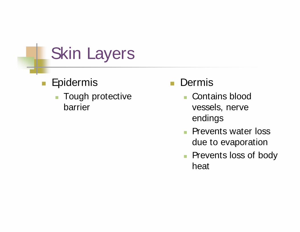
Skin Layers
EpidermisTough protective barrier
DermisContains blood vessels, nerve endingsPrevents water loss due to evaporationPrevents loss of body heat
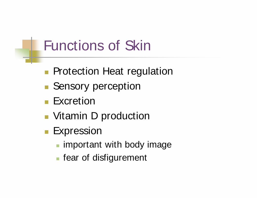
Functions of Skin
Protection Heat regulation Sensory perception Excretion Vitamin D productionExpression
important with body imagefear of disfigurement
Depth of Burns
May be difficult to determineIn some cases, may not be known until after healing has occurred

Traditional Classification
1st degree2nd degree3rd degree
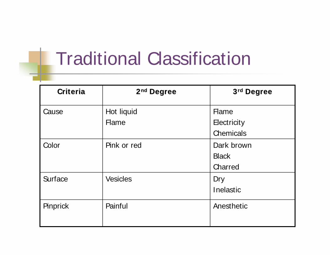
Traditional Classification
AnestheticPainfulPinprick
DryInelastic
VesiclesSurface
Dark brownBlackCharred
Pink or redColor
FlameElectricityChemicals
Hot liquidFlame
Cause
33rdrd DegreeDegree22ndnd DegreeDegreeCriteriaCriteria
Alternative Classification
Partial ThicknessSuperficial Deep
Full thickness
Partial Thickness
Characterized by varying depth from epidermis (outer layer of skin) to the dermis (middle layer of skin)
Superficial - includes only the epidermis Deep - involve entire epidermis and part of the dermis Generally heals spontaneously
Full Thickness
Includes destruction of epidermis and the entire dermis as well as possible damage to the SQ, muscle and bone
Requires skin graft
Determining Severity of Injury
Size (surface area)DepthAgePrior status of health of victimLocation of burnSeverity of associated injury
Pathology of Burns
Burns will cause rapid loss of intravascular fluid and proteinVolume loss is greatest in first 6-8 hours
Metabolic Response to Burns
As with any other major injury:Body secretions of catecholamine, cortisone, ADH, aldosterone and glucagonProfound hypermetabolic state that requires excess nutrients and oxygenEvaporative water loss from burn wounds may reach 300 cc/m2/h (normal = 15)Heat loss may reach 580 Kcal/hour
Fighting the Metabolic Response
Aggressive nutritional supportRapid wound closureControl pain and stressPrevent sepsis
Hypovolemic State: First 48°
Rapid fluid shiftsCapillary permeability with burns increases with vasodilation Fluid loss deep in woundsMetabolic acidosis
Protein lossHemoconcentration- Hct increases Low blood volume, oliguria Hyponatremia
K -- damaged cells release K
Diuretic Phase: 48-72° After Injury
Capillary membrane integrity returns Edema fluid shifts back into vessels - blood volume increases Hemodilution - low Hct, decreased potassium as it moves back into the cell or is excreted in urine with the diuresis
Fluid overload can occur due to increased intravascular volume Metabolic acidosis -HCO3 loss in urine, increase in fat metabolismIncrease in renal blood flow - result in diuresis (unless renal damage)
General Indications for Fluid Resuscitation
Burns > 20% of BSA with adults Burns > 10% of BSA with children Age > 65 or < 2
Acute Resuscitation
AirwayBreathingCirculationAnalgesia
Acute ResuscitationAssessment: Objective
how burn occurred, when Durationtype of agent
Subjective: previous medical problems size and depth of burnage body part involved mechanism of injury
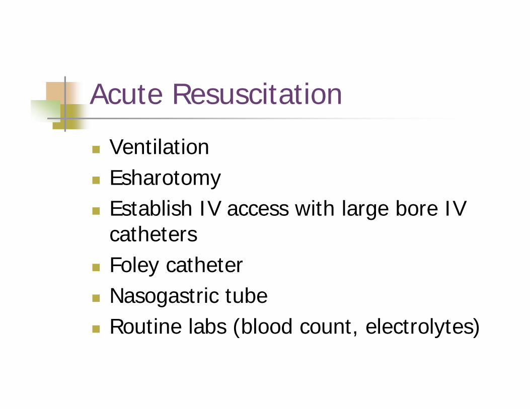
Acute Resuscitation
VentilationEsharotomyEstablish IV access with large bore IV cathetersFoley catheterNasogastric tubeRoutine labs (blood count, electrolytes)
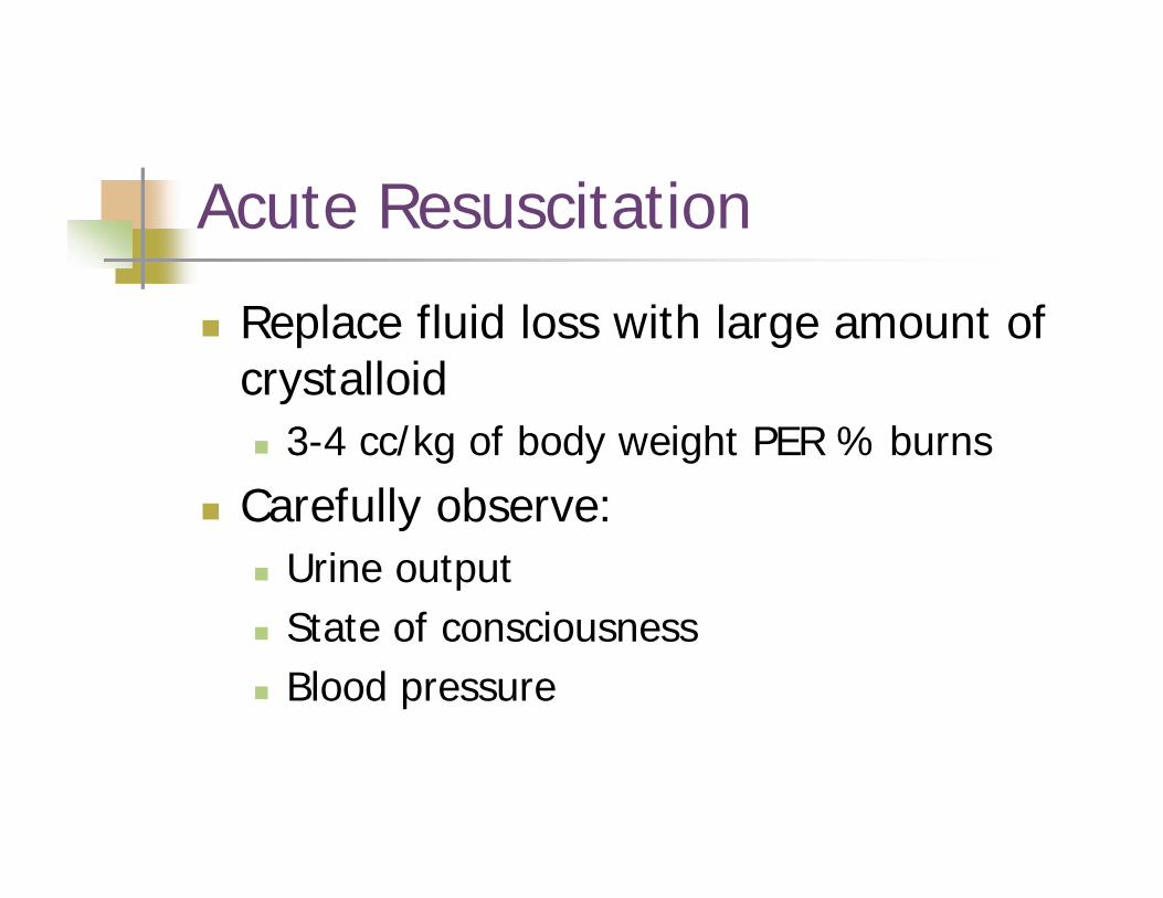
Acute Resuscitation
Replace fluid loss with large amount of crystalloid
3-4 cc/kg of body weight PER % burns
Carefully observe:Urine outputState of consciousnessBlood pressure
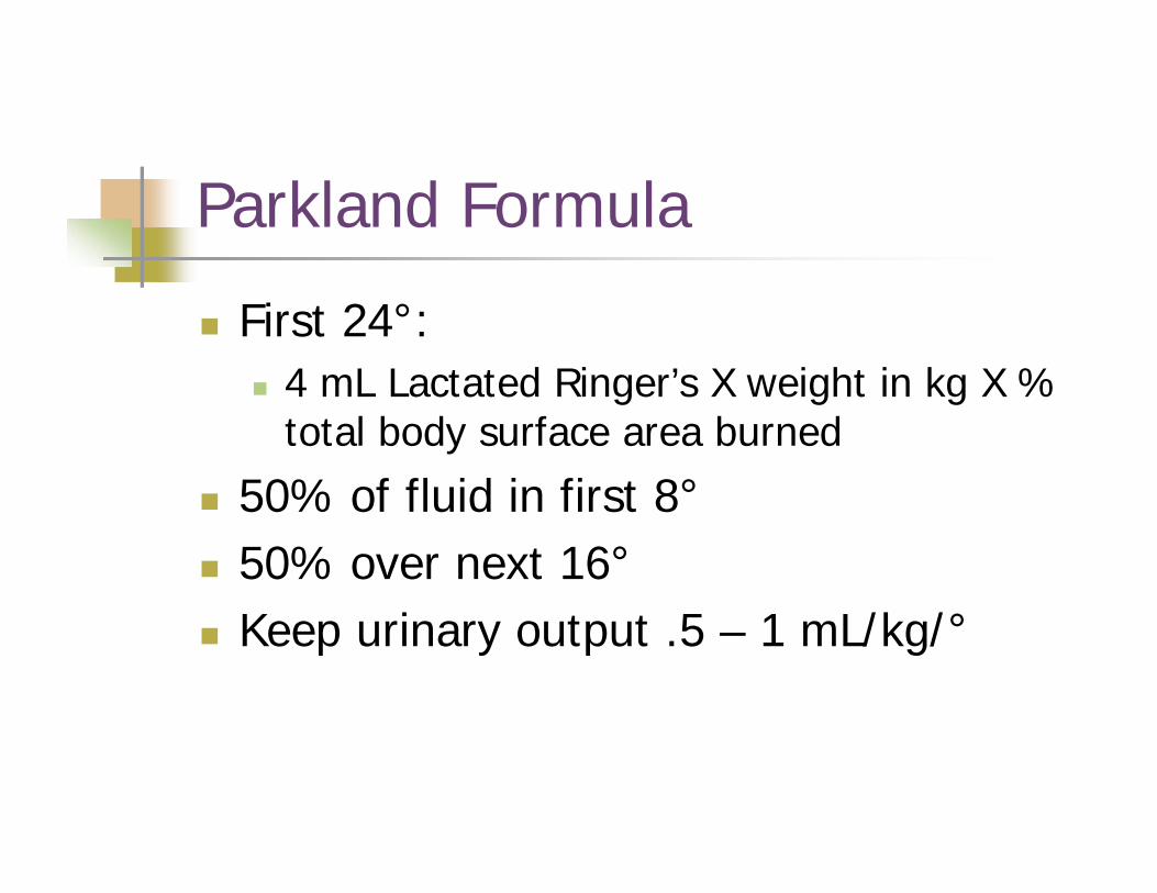
Parkland Formula
First 24°: 4 mL Lactated Ringer’s X weight in kg X % total body surface area burned
50% of fluid in first 8°50% over next 16°Keep urinary output .5 – 1 mL/kg/°
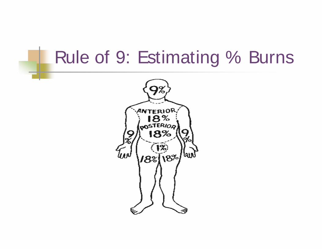
Rule of 9: Estimating % Burns
Signs of Adequate Fluid Resuscitation
Clear sensoriumPulse < 120 beats per minute Urine output for adults 30 - 50 cc/hour Systolic blood pressure > 100 mm Hg Blood pH within normal range 7.35 -7.45
Acute Resuscitation: Crystalloids
Isotonicmost common are lactated Ringers or NaCl (0.9%)these do not generate a difference in osmotic pressure between the intravascular and interstitial spacessubsequently LARGE amounts of fluid are required
Acute Resuscitation: Crystalloids
Hypertonic salt solutions create an osmotic pull of fluid from the interstitial space back to the depleted intravascular space helps decrease the amount of fluid needed during resuscitationdecreases the development of edema, pulmonary edema, and CHF
Acute Resuscitation: Colloids
Replacement begins during the second 24° following the burn to replace intravascular volume Once capillary permeability significantly decreases
Post-Resuscitation Period: The Second 24 Hours
IV fluid should consist of glucose in water and plasma to maintain adequate circulating volumeCalorie and protein needs may be twice normal
Oral feeding if possibleParenteral (IV) feeding may be necessary
Post-Resuscitation Period
Antibiotic use is controversialVitamin CVitamin A
Wound Care PrincipalsGoals
close woundprevent infection reduce scarring and contractures provide for comfort
Wound cleaningDebridement
mechanical surgical
Topical antibacterial therapy
Topical Antibacterial Agents
Silvadene creamTransient leukopenia
SulfamyalonMetabolic acidosisPain
Silver nitrateWater toxicity
Dressing the Burn: Open Technique
Partial thickness exudate dries in 48 to 72 hours forming a hard crustepithelialization occurs beneath the crust and may take 14 to 21 days to healcrust then falls off spontaneously
Full thickness dead skin is dehydrated and converted to black leathery escar in 48 to 72 hoursloose escar is gradually removed
Dressing the Burn: Closed Technique
Wound is washed and sterile dressings changed each shift or dailyDressing consists of gauze wraps and ointments if available
Dressing the Burn: Semi-Open Technique
Consists of covering the wound with topical antimicrobial agents and gauzeAdvantages:
speeds debridementdevelops granulation tissues faster makes skin grafting possible sooner
Biological Dressings
Homograftssame species (cadaver skin) temporary coverage
Heterograftsanother species (pig skin)temporary coverage
Autograftspatients own skin permanent coverage
Wound Care: Grafting
Indications for grafting full thickness burnspriority areaswound bed pink, firm, free of exudatebacterial count < 100,000/gram of tissue
Care of grafts - assess
Rehabilitation
Care of healing skin Wash dailyKeep clean and dry
Pressure garmentsPrevent scaring and contractures
Promote mobility
Inhalation Injuries
Major cause of death from burn injuryDirect inhalation of dry heat cause
Burn injury of upper airwayRarely occurs below the level of the vocal cords
TreatmentEndotracheal intubationTracheostomy
Inhalation Injury: Carbon Monoxide
Should be considered when accident occurs in a closed spaceSymptoms
HeadacheConfusion, HallucinationMild dyspneaComa
Treatment100% oxygen