Embed Size (px)
Citation preview
www.iajpr.com
Pag
e33
24
Indo American Journal of Pharmaceutical Research, 2013 ISSN NO: 2231-6876
Journal home page:
http://www.iajpr.com/index.php/en/
INDO AMERICAN
JOURNAL OF
PHARMACEUTICAL
RESEARCH
Buccal Patches: A Review
Farheen Fiza*, Bharadwaj Sudhir, Jat R.C, Arjariya Priyanka, Sharma Garima, Ratnakar Deepti,
Arjariya Priyanka, Khan Imran, Tiwari Rahul, Rathore Singh Arvind
Shri ram college of pharmacy Banmore, Morena (M.P.) India
Corresponding author
Fiza Farheen M.pharm (Pharmaceutics)
Shriram college of pharmacy,Banmore
AB Express highway, Near Gwalior
(M.P.) INDIA, Mail ID: [email protected], Mob: +91-9907713357
Copy right © 2013 This is an Open Access article distributed under the terms of the Indo American journal of Pharmaceutical Research, which
permits unrestricted use, distribution, and reproduction in any medium, provided the original work is properly cited.
ARTICLE INFO ABSTRACT
Article history Received 11/04/2013
Available online
28/04/2013
Keywords Buccal mucosa, hepatic
first-pass metabolism,
patch covers.
Drugs that are administered via the buccal mucosa directly enter the systemic
circulation, thereby avoiding hepatic first-pass metabolism. Therefore, this
administration route is useful for improving the bioavailability of drugs that are
subject to an extensive first-pass effect when delivered orally. For the oral mucosal
route of drug administration, various types of dosage forms can be prepared. A
sublingual tablet can afford rapid drug absorption and a prompt pharmacological
effect; however, the duration of delivery is short owing to the inevitable loss of a
large proportion of the administered dose due to swallowing. To avoid such losses,
a patch can be formulated that is located on the buccal mucosa of the oral cavity.
However, this approach is limited by the thicker dimensions of the buccal
membrane compared to the others that line the oral cavity, and constraints impelled
by the delivery system itself (the amount of drug reaching the systemic circulation
is limited by the area of the mucosa that the patch covers, which, for patient
comfort reasons, is relatively small). Direct access to the systemic circulation
through the internal jugular vein bypasses drugs from the hepatic first pass
metabolism leading to high bioavailability.
Please cite this article in press as Fiza Farheen et.al. Buccal Patches: A Review. Indo American Journal of Pharm
Research.2013:3(4).
www.iajpr.com
Pag
e33
25
Vol 3, Issue 4, 2013. Fiza Farheen et. al. ISSN NO: 2231-6876
Introduction
Buccal route of drug delivery is a good alternative, amongst the various routes of drug delivery.[1] Buccal drug
delivery is most advantageous because it abundant blood supply in buccal mucosa, bypassing the hepatic
firstpass effect and accessibility.[2] However, peroral administration of drugs has disadvantages such
as hepatic first pass metabolism and enzymatic degradation within the GI tract, that prohibit oral administration
of certain classes of drugs especially peptides and proteins. Consequently, other absorptive mucosae are
considered as potential sites for drug administration.[3] Oral cavity has been investigated for number of
applications including the treatment of periodontal disease bacterial and fungal infection, aphthous and dental
stomatitis. Over the last two decades mucoadhesion has become of interest for its systemic delivery by retaining
a formulation intimate contact with buccal cavity.[4] The term bio adhesion has been used to define the
attachment of a synthetic natural macromolecule to a biological tissue for an extended period of time. When a
substrate is a mucosal system adheres and interacts primarily with the mucus layer, this phenomenon being
referred to as mucoadhesion.[5] The adhesive properties of such drug delivery platforms can reduce the
enzymatic degradation due to the increased intimacy between the delivery vehicle and the absorbing
membrane.[6] The use of mucoadhesive polymers in buccal drug delivery has a greater
application.Various mucoadhesive devices, including tablets, films, patches, disks, strips, ointments and gels,
have recently been developed. However, buccal patch offer greater flexibility and comfort than the other
devices. In addition, a patch can circumvent the problem of the relatively short residence time of oral gels on
mucosa, since the gels are easily washed away by saliva. Buccal route of drug delivery provides the direct
access to the systemic circulation through the jugular vein bypassing the first pass hepatic metabolism leading
to high bioavailability.[7]
Advantages
Bypass of the gastrointestinal tract and hepatic portal system, increasing the bioavailability of orally
administered drugs that otherwise undergo hepatic first metabolism.
Improved patient compliance due to the elimination of associated pain with injections; administration of
drugs in unconscious or incapacitated patients.
Sustained drug delivery.
A relatively rapid onset of action can be achieved relative to the oral route, and the formulation can be
removed if therapy is required to be discontinued.
Increased ease of drug administration.[8]
Disadvantages
Limited absorption area- the total surface area of the membranes of the oral cavity available for drug
absorption is 170 cm2 of which ~50 cm2 represents non-keratinized tissues, including buccal
membrane.[9]
The barriers such as saliva,mucus,membrane coating granules, basement membrane etc retard the rate
and extent of drug absorption through the buccal mucosa.[10]
Continuous secretion of the saliva(0.5-2 l/day)leads to subsequent dilution of the drug.[11]
The hazard of choking by involuntarily swallowing the delivery system is a concern.
Swallowing of saliva can also potentially lead to the loss of dissolved or suspended drug and ultimately
the involuntary removal of the dosage form.[12]
www.iajpr.com
Pag
e33
26
Vol 3, Issue 4, 2013. Fiza Farheen et. al. ISSN NO: 2231-6876
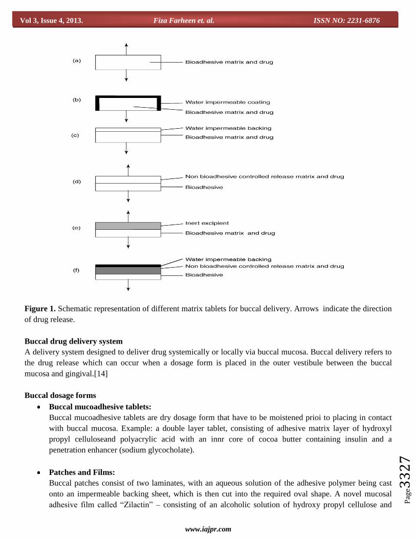
Structure & Design of Buccal Dosage Form
Structure and design: Drug delivery designed for the buccal mucosa contains a polymeric adhesive
component. When in contact with the saliva, the adhesive attaches to the mucosa causing immediate and rapid
drug delivery. Transmucosal drug delivery systems can be unidirectional or bi-directional. Unidirectional
patches release the drug only into the mucosa, while bi-directional patches release the drug in both the mucosa
and the mouth. The buccal patch is designed in either a matrix configuration with drug, adhesive, and additives
mixed together, or a reservoir system that contains a cavity for the drug and additives separate from the
additives. An impermeable backing is applied to control the direction of drug delivery, to reduce patch
deformation and disintegration while in the mouth; and to prevent drug loss. Additionally, the patch can be
constructed to undergo minimal degradation in the mouth, or can be designed to dissolve almost immediately.
Buccal dosage form for buccal delivery: In the past decades, to till now, different drug delivery systems
intended for buccal administration have been developed. The most common buccal dosage forms are tablets and
patches. Such type of form must be of a small size and a suitable geometry so as to not interfere with
physiological function of the mouth, even after their hydration in the oral cavity. One of the requirements is that
they do not adhere too tightly because it is undesirable to exert too much force to remove the formulation/
dosage form after use, otherwise the mucosa could be injured. An alternative is the use of formulations that
dissolve or disintegrate completely during the application period. Moreover, in the case of Transmucosal
administration, Drug release should be unidirectional (towards the mucosa), and the release into the saliva
should be avoided.
Matrix type: The buccal patch designed in a matrix configuration contains drug, adhesive, and additives mixed
together.
Reservoir types: The buccal patch designed in a reservoir system contains a cavity for the drug and additives
separate from the adhesive. An impermeable backing is applied to control the direction of drug delivery; to
reduce patch deformation and disintegration while in the mouth; and to prevent drug loss. Additionally, the
patch can be constructed to undergo minimal degradation in the mouth, or can be designed to dissolve almost
immediately.
Patches: Patches are laminated and generally consist of an impermeable backing layer and a drug-containing
layer that has mucoadhesive properties and from which the drug is released in a controlled manner. Moreover,
buccal patches for systemic delivery of tyrotropin-releasing hormone, octreotide, oxytocin, buserelin, calcitonin
and leuenkephalinhave been studied.
Novel drug delivery system: Novel drug delivery systems, such as lipophilic gel, buccal spray and
phospholipids vesicles have been recently proposed to deliver peptides via the buccal route. A novel liquid
aerosol formulation (Oralin, Generex Biotechnology) has been already developed. This system allows precise
insulin dose delivery via a metered dose inhaler in the form of fine aerosolized droplets directed into the mouth.
This oral aerosol formulation is rapidly absorbed through the buccal mucosal epithelium, and it provides the
plasma insulin levels necessary to control postprandial glucose rise in diabetic patients. This novel, pain-free,
oral insulin formulation has a number of advantages including rapid absorption, a simple (user-friendly)
administration technique, precise dosing control (comparable to injection within one unit) and bolus delivery of
drug.[13]
www.iajpr.com
Pag
e33
27
Vol 3, Issue 4, 2013. Fiza Farheen et. al. ISSN NO: 2231-6876
Figure 1. Schematic representation of different matrix tablets for buccal delivery. Arrows indicate the direction
of drug release.
Buccal drug delivery system
A delivery system designed to deliver drug systemically or locally via buccal mucosa. Buccal delivery refers to
the drug release which can occur when a dosage form is placed in the outer vestibule between the buccal
mucosa and gingival.[14]
Buccal dosage forms
Buccal mucoadhesive tablets:
Buccal mucoadhesive tablets are dry dosage form that have to be moistened prioi to placing in contact
with buccal mucosa. Example: a double layer tablet, consisting of adhesive matrix layer of hydroxyl
propyl celluloseand polyacrylic acid with an innr core of cocoa butter containing insulin and a
penetration enhancer (sodium glycocholate).
Patches and Films:
Buccal patches consist of two laminates, with an aqueous solution of the adhesive polymer being cast
onto an impermeable backing sheet, which is then cut into the required oval shape. A novel mucosal
adhesive film called “Zilactin” – consisting of an alcoholic solution of hydroxy propyl cellulose and
www.iajpr.com
Pag
e33
28
Vol 3, Issue 4, 2013. Fiza Farheen et. al. ISSN NO: 2231-6876
three organic acids. The film which is applied to the oral mucosal can be retained in place for at least 12
hours even when it is challenged with fluids.[15]
Semisolid Preparations (Ointment and Gels):
Bioadhesive gels or ointment have less patient acceptability than solid bioadhesive dosage form, and
most of the dosage forms are used only for localized drug therapy within the oral cavity.one of the
original oral mucoadhesive delivery systems- “orabase”- consists of finely ground pectin, gelatin and
sodium carboxy methylcellulose dispersed in a poly (ethleene) and a ground pectin, gelatin and sodium
carboxy methylcellulose dispersed in poly (ethylene) and a mineral oil gel base, which can be
maintained at its site of application for 15-150 minutes.[16]
Powders:
Hydroxpropyl cellulose and beclomethasone in powder form when sprayed onto the oral mucosa of rats,
a significant increase in the residence time relative to an oral solution is seen, and 2.5% of
beclomethasone is retained on buccal mucosa for over 4 hours.[17]
Mechanism of buccal absorption
Buccal drug absorption occurs by passive diffusion of the nonionized species, a process governed
primarily by a concentration gradient, through the intercellular spaces of the epithelium. The passive transport
of non-ionic species across the lipid membrane of the buccal cavity is the primary transport mechanism. The
buccal mucosa has been said to be a lipoidal barrier to the passage of drugs, as is the case with many other
mucosal membrane and the more lipophilic the drug molecule, the more readily it is absorbed. The dynamics of
buccal absorption of drugs could be adequately described by first order rate process. Several potential barriers
to buccal drug absorption have been identified. Dearden and Tomlison (1971) pointed out that salivary secretion
alters the buccal absorption kinetics from drug solution by changing the concentration of drug in the mouth. The
linear relationship between salivary secretion and time is given as follows:
where,
=
M – Mass of drug in mouth at time t
K – Proportionality constant
C – Concentration of drug in mouth at time
Vi- The volume of solution put into mouth cavity and Vt- Salivary secretion rate
Factors affecting buccal absorption
The oral cavity is a complex environment for drug delivery as there are many interdependent and
independent factors which reduce the absorbable concentration at the site of absorption.
1.Membrane Factors
This involves degree of keratinization, surface area available for absorption, mucus layer of salivary pellicle,
intercellular lipids of epithelium, basement membrane and lamina propria. In addition, the absorptive membrane
thickness, blood supply/ lymph drainage, cell renewal and enzyme content will all contribute to reducing the
rate and amount of drug entering the systemic circulation.
2.Environmental Factors
A.) Saliva: The thin film of saliva coats throughout the lining of buccal mucosa and is called salivary pellicle or
film. The thickness of salivary film is 0.07 to 0.10 mm. The thickness, composition and movement of this film
www.iajpr.com
Pag
e33
29
Vol 3, Issue 4, 2013. Fiza Farheen et. al. ISSN NO: 2231-6876
affect the rate of buccal absorption.
B.) Salivary glands: The minor salivary glands are located in epithelial or deep epithelial region of buccal
mucosa. They constantly secrete mucus on surface of buccal mucosa. Although, mucus helps to retain
mucoadhesive dosage forms, it is potential barrier to drug penetration.
C.) Movement of buccal tissues: Buccal region of oral cavity shows less active movements. The
mucoadhesive polymers are to be incorporated to keep dosage form at buccal region for long periods to
withstand tissue movements during talking and if possible during eating food or swallowing.[18]
Composition of buccal patches
A. Active Pharmaceutical ingredient (API):
The buccal film technology has the potential for delivery of variety of APIs. However since the size of
the dosage form has limitation, high dose molecules are difficult to be incorporated in buccal film.
Generally 5%w/w to 30%w/w of active pharmaceutical ingredients can be incorporated in the buccal
patches.[19]
B. Polymers (adhesive layer):
Polymer hydration and swelling properties probably play the main role. The polymer hydration and
consequently the mucus dehydration could cause an increase in mucous cohesive properties that
promote mucoadhesion. Swelling should favor polymer chain flexibility and interpenetration between
polymer and mucin chains. So, depending on the type of formulation,polymers with different
characteristics have to be considered.
Examples: Hydroxy ethylcellulose, hydroxypropyl cellulose, polyvinyl pyrrolidone,
polyvinyl alcohol, carbopol and other mucoadhesive polymers.[20]
C. Diluents:
Lactose DC is selected as diluent for its high aqueous solubility, its flavouring characteristics, and its
physico-mechanical properties, which make it suitable for direct compression. other example :
microcrystalline starch and starch.
D. Sweetening agents:
Sucralose, aspartame, mannitol, etc.
E. Flavouring agents:
Menthol, vanillin,clove oil, Peppermint oil, cinnamon oil, spearmint oil, oil of nutmeg are
examples of flavor oils while vanilla, cocoa, coffee, chocolate and etc.[21]
F. Backing layer:
Ethyl cellulose, etc.
G. Penetration enhancer:
Cyano acrylate, EDTA, Ctric acid etc.
H. Plasticizers:
PEG-100, 400, propylene glycol, etc.[22]
Method of preparation
Two methods used to prepare adhesive patches include,
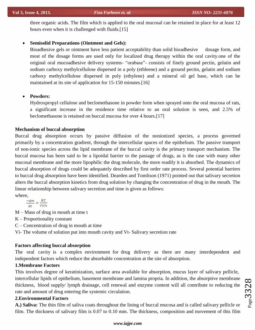
Solvent casting:
In this, all patch excipients including the drug codispersed in an organic solvent and coated Onto a sheet of
release liner. After solvent evaporation, a thin layer of the protective backing material is laminated onto the
sheet of coated release liner to form a laminate that is die-cut to form patches of the desired size and geometry.
www.iajpr.com
Pag
e33
30
Vol 3, Issue 4, 2013. Fiza Farheen et. al. ISSN NO: 2231-6876
The solvent casting method is simple, but suffers from some disadvantages, including long processing time,
high cost, and environmental concerns due to the solvents used. Thes drawbacks can be overcome by the hot-
melt extrusion method.[23]
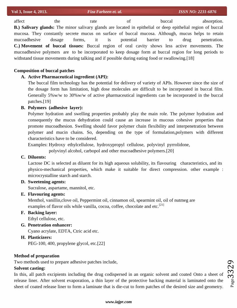
Direct milling:
In this, patches are manufactured without the use of solvents (solvent-free). Drug and excipients are
mechanically mixed by direct milling or by kneading, usually without the presence of any liquids.After the
mixing process, the resultant material is rolled on a release liner until the desired thickness is achieved. An
impermeable backing membrane may also be applied to control the direction of drug release, prevent drug loss,
and minimize deformation and disintegration of the device during application period.[24]
While there are only minor or even no differences in patch performance between patches fabricated with the
two processes, the solvent-free process is preferred because there is no possibility of residual solvents and no
associated solvent-related health issues.[25]
www.iajpr.com
Pag
e33
31
Vol 3, Issue 4, 2013. Fiza Farheen et. al. ISSN NO: 2231-6876
List of drug delivered via buccal route
In an effort to determine the feasibility of buccal route as a novel route of drug delivery, several drugs have
been studied. The variation in class of compounds illustrates that the pharmaceutical industries have an
alternative and novel routes of administration for existing drugs.[26]
Active Ingredients:
Acitretin
Acyclovir
Arecoline
Buprenorpine
Carbamazepine
Chitosan
Chlorpheniramine maleate
Metronidazole
Morphine sulphate
Nicotine
Nifedipine
Omeprazole
Oxytocin
Piroxicam
Ergotamine tartrate (etc).
Evaluation
Surface pH:
The surface pH of the buccal patch was determined in order to investigate the possibility of any side effects in
vivo. As an acidic or alkaline pH may cause irritation to the buccal mucosa, it was determined to keep the
surface pH as close to neutral as possible. A combined glass electrode was used for this purpose.The patches
were allowed to swell by keeping it in contact with 1 ml of distilled water (pH 6.5 ± 0.05) for 2 hours at room
temperature, and pH was note down by bringing the electrode in contact with the surface of the patch and
allowing it to equilibrate for 1 minute.[27]
Swelling studies:
Weight and area increase due to swelling were measured. Weight increase due to swelling: A drug-loaded patch
of 1x1 cm2 was weighed on a preweighed cover slip. It was kept in a petridish and 50 ml of phosphate buffer,
pH 6.6 was added. After every five minutes, the cover slip was removed and weighed upto 30 minutes. The
difference in the weights gives the weight increase due to absorption of water and swelling of patch. Area
increase due to swelling: A drug loaded patch size of 1x1 cm2 was cut and placed in a petridish. A graph paper
was placed beneath the petridish, to measure the increase in the area. 50ml of phosphate buffer, pH 6.6, was
poured into the petridish. An increase in the length and breadth of the patch was noted at 5 min intervals for 60
min and area was calculated.[28]
The percent swelling, %S, was calculated using the following equation:
Xt - Xo
%S = x 100
Xo
www.iajpr.com
Pag
e33
32
Vol 3, Issue 4, 2013. Fiza Farheen et. al. ISSN NO: 2231-6876
Where Xt is the weight or area of the swollen patch after time t
Xo is the original patch weight or area at zero time
Thickness measurements:
The thickness of each film is measured at five different locations (centre and four corners) using an electronic
digital micrometer.
Thermal analysis study:
Thermal analysis study is performed using differential scanning calorimeter (DSC).
Morphological characters:
Morphological characters are studied by using scanning electron microscope (SEM).[29]
Palatability test:
Palatability study is conducted on the basis of taste, after bitterness and physical appearance. All the batches are
rated A,B and C grades as per the criteria. When the formulation scores at least one A grade, formulation is
considered as average. When the formulation score two a geade then it would be considered as good and the one
with all three A grade it would be the very good formulation.[30]
Grades: A = very good, B = good, C = poor
Folding endurance:
The test is performed by repeated folding of the film at the same place until film failure. A maximum of 300
times is sometimes reported as a limit to the test, and the value is reported as the number of times the film can
be folded prior to rupture.[31]
In vitro drug release:
The United States Pharmacopeia (USP) XXIII rotating paddle method used to study the drug Release from the
bilayered and multilayered patches. The dissolution medium consisted of phosphate buffer pH 6.8. the release
was performed at 37 0C ± 0.50
0C,with a rotation speed of 50 rpm. The backing layer of buccal patches attached
to the glass disk with instant adhesive (cyanoacrylate adhesive). The disk was allocated to the bottom of the
dissolution vessel. Samples (5ml) were withdrawn at predetermined time intervals and replaced with fresh
medium. The samples filtered through whatman filter paper and analyzed after appropriate dilution by UV
spectrophotometry at suitable nm.[32]
In vitro drug permeation:
The in vitro buccal drug permeation study of Drugs through the buccal mucosa (sheep and rabbit) performed
using Keshary-Chien/Franz type glass diffusion cell at 37°C± 0.2°C. Fresh buccal mucosa mounted between the
donor and receptor compartments. The buccal tablet was placed with the core facing the mucosa and the
compartments clamped together. The donor compartment filled with 1 ml of phosphate buffer pH 6.8. The
receptor compartment was filled with phosphate buffer pH 7.4, and the hydrodynamics in the receptor
compartment maintained by stirring with a magnetic bead at 50 rpm. A one ml sample can be withdrawn at
predetermined time intervals and analyzed for drug content at suitable nm using a UVspectrophotometer.[33]
Stability study in Human saliva:
Stability study of fast dissolving films is carried out for all the batches according to ICH guidelines. After
predetermined time intervals, the films are evaluated for the drug content,disintegration time and physical
appearance. The stability study of optimized mucoadhesive patch formulation was performed at 400C, 37 ±50C
& 75±5% RH for three months. The value of all parameter after three months remain same as their values and
www.iajpr.com
Pag
e33
33
Vol 3, Issue 4, 2013. Fiza Farheen et. al. ISSN NO: 2231-6876
minor changes occur in value of volume entrapment efficiency, % elongation & % drug release after 8 hour
which was considerable.[34-35]
Ex vivo mucoadhesive strength:
A modified balance method used for determining the ex vivo mucoadhesive strength. Fresh buccal mucosa
(sheep and rabbit) obtained, used within 2 hours of slaughter. The mucosal membrane separated by removing
underlying fat and loose tissues. The membrane washed with distilled water and then with phosphate buffer pH
6.8 at 370C. The buccal mucosa cut into pieces and washed with phosphate buffer pH 6.8. A piece of buccal
mucosa was tied to the glass vial, which was filled with phosphate buffer. The two sides of the balance made
equal before the study, by keeping a 5 g weight on the right-hand pan. A weight of 5 g was removed from the
right-hand pan, which lowered the pan along with the tablet over the mucosa. The balance was kept in this
position for 5 minutes contact time. The water (equivalent to weight) was added slowly with an infusion set
(100 drops/min) to the righthand pan until the tablet detached from the mucosal surface. This detachment force
gave the mucoadhesive strength of the buccal tablet in grams. The glass vial was tightly fitted into a glass
beaker (filled with phosphate buffer pH 6.8, at 37°C ±1°C) so that it just touched the mucosal surface. The
buccal tablet was stuck to the lower side of a rubber stopper with cyanoacrylate adhesive.[36]
Conclusion:
Buccal region provides a convenient route of administration for both local and systemic drug actions.Controlled
buccal drug delivery systems, where the drug delivery is directed towards buccal mucosa by protecting the local
environment is also gaining interest. Currently solid dosage forms, liquids and gels applied to oral cavity are
commercially successful. The future direction of buccal adhesive drug delivery lies in vaccine formulations and
delivery of small proteins/peptides. Microparticulate bioadhesive systems are particularly interesting as they
offer protection to therapeutic entities as well as the enhanced absorption that result from increased contact time
provided by the bioadhesive component. Exciting challenges remain to influence the bioavailability of drugs
across the buccal mucosa. Many issues are yet to be resolved before the safe and effective delivery through
buccal mucosa. In mucoadhesive placebo buccal patches we can use any potent drugs which fulfill the criteria
for buccal patch as drug delivery system.
References
1. Shoba Rani R Hiremath; Industrial Pharmacy, Orient Longman private limited, 2008; First edition,
73-77.
2. Sang-Chul Shin, Jin-Pil Bum, Jun-Shik Choi. Enhanced bioavailability by buccal administration of
triamcinolone acetonide from the bioadhesive gels in rabbits, Int. J. Pharmaceutics., 2009; 209:37-
43.
3. Giradkar KP, Design development and in vitro evaluation of bioadhesive dosage form for buccal
route, International journal of pharma research & development, 2010, 2.
4. Giradkar MA, Channawar AD, Kajale E, Sridhar RS, Kamble BV, Chandewar. Design,
development and in vitro evaluation of bioadhesive dosage form for buccal route. Int. J. Pharm.
Res. Dev., 2010; 2(6):1-20.
5. Calum R, Park, Dale L, Munday. Development and evaluation of a biphasic buccal adhesive tablet
for nicotine replacement therapy. Int. J. Pharm., 2002; 237:215-26.
www.iajpr.com
Pag
e33
34
Vol 3, Issue 4, 2013. Fiza Farheen et. al. ISSN NO: 2231-6876
6. Subhash V, Madhuri Channawar, Anil V, Unmesh, Kailash R. Chitosan based sustained release
mucoadhesive buccal patches Containing verapamil HCl, Int. J. Pharm. Pharm. Sci., 2009; 1(1):
216-29.
7. Shidhaye SS, Mucoadhesive bilayered patches for administration of sumatriptan, AAPS pharm sci
tech, 2009, 9(3).
8. Edsman K, Pharmaceutical applications of mucoadhesion for the non-oral routes, Journal of
pharmacy & pharmacology, 2005, 57, 3-19.
9. Surender Verma, MahimaKaul, ArunaRawat, SapnaSaini. An overview on buccal drug
delivery system. Int J Pharm Sci Res 2011;2(6):1303-21.
10. Miller NS, Johnston TP. The use of mucoadhesive polymers in buccal drug delivery,
Advanced Drug Delivery Reviews 2005;57:1666–91.
11. Patel KV, Patel ND, Dodiya HD, Shelat PK. Buccal bioadhesive drug delivery system:An
Overview.Int J Pharm Bio Arch 2011;2(2): 600-9.
12. Yajaman S., Bandyopadhyay AK. Buccal bioadhesive drug delivery- A promising option for
orally less efficient drugs. J Controlled Release 2006;114:15–40.
13. Shimamoto T. Androl J 1987; 8 (1): S14-S16.
14. Amir H, Systemic drug delivery via the buccal mucosal route, Pharmaceutical technology, 2001, 1-
27.
15. Sudhakar Y, Knotsu K, Bandopadhyay AK. Bio adhesive drug delivery – A promising option for
orally less efficient drugs. J Control Rel 2006;114:15-40.
16. Rajashree Mashru, Vijay Sutariya, Mayur Sankalia, Jolly Sankalia. Transbuccal delivery of
lamotrigine across procine buccal mucosa in vitro determination of routes of buccal transport. J
Pharm Pharmaceut Sci 2005;8:54-62.
17. Ceschel GC, Maffei P, Moretti MD, Demontis LS, Peana A. International Journal of
Pharmaceutics 2000;195:45.
18. Haas J, Lehr CM. Developments in the area of bioadhesive drug delivery systems. Expert Opin
Biol Ther 2002;2:287-298.
19. DeVries M.E, Ph.D. Thesis, University of Leiden, Leiden, The Netherlands, 1991.
20. Dixit R.P., Puthli S. P. , Oral strip technology: Overview and future potential, Journal of
Controlled Release,2009, 139, 94–107.
21. Rossi Silvia, Sandri Giuseppina, Caramella Carla M., Buccal drug delivery: A challenge already
won?, Drug Discovery Today: Technologies, 2005, 2(1).
22. Smart, J.D., The role of water movement and polymer hydration in mucoadhesion. In
Bioadhesive drug delivery systems (Mathiowitz, E., Chickering, III D.E.,Lehr, C.M., eds), Marcel
Dekker, 1999, PP11–23.
23. Nazilasalamat M, Montakarn C, Thomas J. The use of mucoadhesive polymers in buccal
drug delivery. AdvDrug Del Rev 2005;57:1666-91.
24. Gandhi RB, Robinson JR. Oral cavity as a site for bioadhesive drug delivery. Adv Drug Del Rev
1994;13:43-74.
25. Amir HS. Buccal mucosa as a route for systemic drug delivery: A review. J Pharm
Pharmceut Sci 1998;1(1):15-30.
26. Salamat-Miller N, Chittchang M, Johnston TP, The use of mucoadhesive polymers in buccal drug
delivery, Advance DrugDelivery Review, Nov 2005; 57(11): 1666-1691
27. Peppas NA, Buri PA. Surface, interfacial and molecular aspects of polymer bioadhesion
www.iajpr.com
Pag
e33
35
Vol 3, Issue 4, 2013. Fiza Farheen et. al. ISSN NO: 2231-6876
on soft tissues. J ContrRel1985;2:257–75.
28. Bottenberg P, Cleymaet R, Muynek CD, Remon JP, Coomans D, Slop D. Development
and testing of bioadhesive, floride-containg slow-release tablets for oral use. J PharmPharmacol
1991;43:457-64.
29. Steward A, The Effect of Enhancers on the Buccal Absorption of Hybrid (BDBB) Alpha-
Interferon, Int.J. Pharm, 104, 1994, 145–149.
30. Patel R, Shardul N, Patel J, Baria A. overview on buccal mucoadhesive films. Arch
Pharm Sci& Res2009;1(2):212-7.
31. Anders R., Merkle H., Evaluation of laminated muco-adhesive patches for buccal drug delivery,
International Journal of Pharmaceutics, 1989, 49, 231– 240.
32. Apoorva M, Neha C, Geeta A. Formulation and characterization of fast dissolving buccal
films: A review. Der Pharmacia Lettre 2011;3(1):152-65.
33. Leung SS, Robinson JR. Polymer structure features contributing to mucoadhesion: II. J
ContrRel 1990;12:187–94.
34. Amit Khairnar, Parridhi J, Dheeraj B, Dinesh J. Developmement of mucoadhesive buccal
patch containing aceclofenac: in vitro evaluations. Int J PharmTech Res2009;1(4):978-81.
35. Subhash VD, Madhuri A, channavar, Anil VC, Umesh MJ, Kailash RB. Chitosan based
release mucoadhesive patches containing verapamil Hcl. Int J Pharm PharmceutSci 2009 Nov-
Dec;1(1):216-29.
36. Manishkumar, Garimagarg, Pushpendrakumar, Kulkarni GT, Arunkumar. Design and in
vitro evaluation of mucoadhesivebuccal films containing famotidine. Int J Pharm
Pharmceut Sci;2(3):86-90.
DGFGDF451001254
Submit your next manuscript to IAJPR and take advantage of: • Access Online first • Double blind peer review policy • No space constraints • Rapid publication • International recognition Submit your manuscript at: [email protected]