Embed Size (px)
Citation preview

1
B.Sc. (Hon’s) (Fifth Semester) Examination, 2014
Zoology
Paper: LZC-503
(Reproductive and Development Biology)
Model answer
Section A
1.
(i) (c)
(ii) (b)
(iii) (b)
(iv) (c)
(v) (b)
(vi) (b)
(vii) (d)
(viii) (c)
(ix) (c)
(x) (a)
Section B
Answer No. 2.
Fertilization is the phenomenon of fusion of gametes to initiate the development of a new
individual organism. In animals, the process involves the fusion of an ovum with a sperm, which
eventually leads to the development of an embryo.
In sea urchin, fertilization is external. Generally the events which occur during or after the
fertilization phenomenon comes under the post fertilization events. In sea urchin it mainly
includes:
1. Block to polyspermy
2. Egg activation
3. Intiation of cleavage
1. Block to polyspermy
Although many sperm attach to the coats surrounding the egg, it is important that only
one sperm fuses with the egg plasma membrane and delivers its nucleus into the egg.
Two mechanisms are used by animals to ensure that only one sperm fertilizes a given
egg: the fast block to polyspermy and the slow block to polyspermy.

2
Figure showing the different types of block to polyspermy
a. Fast block to polyspermy:
In sea urchin, a fast block to polyspermy occurs within a tenth of a second of fusion.
The fast block to polyspermy involves the opening of Na+ channels in the egg plasma
membrane. Na+ flows into the egg cell, depolarizing the membrane. This
depolarization prevents additional sperm from fusing to the egg plasma membrane.
The egg plasma membrane is restored to its normal -70mV potential within minutes
of fusion as the Na+ channels close, other + ions flow out of the cell, and Na+ is
pumped out.
b. Slow block to polyspermy:
The slow block to polyspermy begins within 10 seconds of fusion of the sperm and
egg plasma membranes. A compound called inositol triphosphate (IP3) causes the
release of Ca++ from intracellular stores in the egg endoplasmic reticulum. Ca++ is
first released at the site of sperm entry, and during the next minute, a wave of free
Ca++ passes through the egg. This Ca++ results in the fusion of cortical vesicles
with the egg plasma membrane, releasing their contents into the space surrounding
the egg, called the perivitelline space. This raises the vitelline membrane, and
inactivates bindin receptors on the vitelline membrane. Thus, any additional sperm
are released from the vitelline membrane and no more bind.
2. Egg Activation:
Ca++ release at fertilization results in an increase in metabolic activity within the egg,
apparently due to an increase in the intracellular pH of the egg. Diacyl gycerol (DAG)
causes protein phosphorylation cascades to be initiated, with one result being the

3
phosphorylation and activation of a plasma membrane Na+:H+ ion exchanger. Na+ is
pumped into the cell, H+ is pumped out of the cell, and the pH inside the cell increases.
3. Initiation of cleavage:
After fertilization, the zygote begins cleavage. In typical sea urchins, cleavage occurs in a
stereotyped way, producing progressively smaller cells (blastomeres). In the early
cleavages, the orientation of the mitotic spindle changes in a specific manner, resulting in
divisions that are either vertical or horizontal. Sea urchins undergo radial cleavage, as do
"typical" deuterstomes, such as chordates, ascidians, and other echinoderms. Like
embryonic cleavages in other organisms, sea urchin cleavages result in more cells, but
without an increase in the total cellular volume of the embryo.
Answer No. 3.
Cleavage is the division of cells in the early embryo. The zygotes of many species undergo rapid
cell cycles with no significant growth, producing a cluster of cells the same size as the original
zygote. The different cells derived from cleavage are called blastomeres and form a compact
mass called the morula. Cleavage ends with the formation of the blastula.
Characterstics of cleavage and mitosis
Cleavage Mitosis
Cleavage refers to the process of division of
cytoplasm (Cytokinesis) in the animal cells.
Cleavage in the animal cells takes place after the
telophase of mitosis.
Mitosis is a type of cell division in which a cell
nucleus divides into two identical nuclei.
Depends on a transient structure based on actin
and myosin filaments, the contractile ring
Depends on a transient microtubule-based
structure, the mitotic spindle
In embryology, it also refers to the first process of
development that follows fertilization. In this
process, a large single cell zygote is divided into
smaller cells known as blastomeres.
In embryology, mitosis is the process by which
cells divide. The dividing cell is what causes an
embryo to become a fetus.
Mitosis consists of DNA replication, cytokinesis, and cell growth, resulting in two cells
of the same size as the parent cell. Cleavage, is similar to mitosis, but lacks the growth phase.
The fertilized egg splits into 2, 4, 8, then 16 cells in very rapid succession. As a result, the

4
fertilized egg (quite large compared to most cells) splits into 16 cells of a much smaller size
(with a combined size equal to the parent cell).
Types of cleavage
There are two main types of cleavage
I. Holoblastic (complete) cleavage
Cleavage found in eggs that contain no (mammals) or only moderate amounts (frog)
of yolk, cytokinesis divides the cells completely.
It may be equal or unequal type.
II. Meroblastic (incomplete) cleavage
Cleavage found in eggs that contain a large amount of yolk, cytokinesis does not
divide the egg completely.
I. Holoblastic (complete) cleavage II. Meroblastic (incomplete) cleavage
A. Isolecithal (sparse, evenly distributed yolk)
• 1. Radial cleavage
(echinoderms, hemichordates, amphioxus)
• 2. Spiral cleavage (annelids,
most mollusks, flatworms)
• 3. Bilateral cleavage (tunicates)
• 4. Rotational cleavage (placental
mammals, nematodes, marsupials)
B. Mesolecithal (moderate vegetal yolk
disposition)
• Displaced radial cleavage (amphibians, some
fish [the lampreys, gars and bowfins)
A. Telolecithal (dense yolk throughout most of cell)
• 1. Bilateral cleavage (cephalopod molluscs)
• 2. Discoidal cleavage
(some fish [the hagfishes, chondrichthyans and
most teleosts], sauropsids [reptiles and
birds], monotremes)
B. Centrolecithal (yolk in center of egg)
• Superficial cleavage (most insects)

5

6
Answer No. 4.
Gastrulation is a phase early in the embryonic development of most animals, during which the
single-layered blastula is reorganized into a trilaminar ("three-layered") structure known as the
gastrula. These three germ layers are known as the ectoderm, mesoderm, and endoderm.
Gastrulation is characterized by cell movement and reorganization within the embryo
(morphogenetic movements) to the interior of the embryo, forming three primary germ layers:
ectoderm, mesoderm, and endoderm.
Mammalian gastrulation:
In mammals gastrulation takes place after the formation of the blastula. Gastrulation is followed
by organogenesis, when individual organs develop within the newly formed germ layers. Each
layer gives rise to specific tissues and organs in the developing embryo. The ectoderm gives rise
to epidermis, and to the neural crest and other tissues that will later form the nervous system. The
mesoderm is found between the ectoderm and the endoderm and gives rise to somites, which
form muscle; the cartilage of the ribs and vertebrae; the dermis, the notochord, blood and blood
vessels, bone, and connective tissue. The endoderm gives rise to the epithelium of the digestive
system and respiratory system, and organs associated with the digestive system, such as the liver
and pancreas.
Major events occur during mammalian gastrulation are:
1. Formation of primitive streak
2. Formation of organizer
3. Occurrence of morphogenetic movements
4. Initiation of neurulation
5. Development of extra embryonic membranes
6. Formation of ecto, endo and mesoderm.

7
7. Decision of fate map
Mammalian gastrulation begins with formation of a primitive streak in the epiblast and
continues much like birds. Mammalian eggs have little or no yolk, but mammalian gastrulation is
nonetheless similar to bird gastrulation due to evolutionary remnants from ancestral reptiles that
laid very yolky eggs. In humans, gastrulation occurs after implantation, around days 14-16 after
fertilization in human embryogenesis.
Answer No. 5.
Metamorphosis
Metamorphosis is the process in which an animal undergoes noticeable changes in its physical
appearance as it moves from one stage of its life to another. Metamorphosis is a biological
process by which an animal physically develops after birth or hatching, involving a conspicuous
and relatively abrupt change in the animal's body structure through cell growth and
differentiation.
Animals that commonly undergo metamorphosis include insects, amphibians and some fishes.
Metamorphosis is associated with adaptive changes in the way an organism interacts with its
environment. For example, adult amphibians often eat very different foods than their larvae.
Thus, adults and larvae do not compete for food. A second example of the adaptive significance
of metamorphosis is in barnacles in which adults are sessile but the larvae are free-swimming.
Thus, the dispersal of larvae gives adults the opportunity to colonize new habitats where the local
environment might be more favorable.
Types of metamorphosis
1. Progressive metamorphosis
Metamorphoses in which larvae possess primitive characters and as they attain adulthood
they develop advanced characters.
Example: In amphibians and majority of insects.
2. Retrogressive metamorphosis
Metamorphoses in which larvae possess advanced characters such as locomotory organs
and sense organs but as they metamorphose into adult, these advanced features
degenerate and adult develops primitive features. This type of metamorphosis is called
retrogressive
Example: Herdmania

8
Metamorphosis in frog:
Amphibians are vertebrates that include frogs, toads, newts and salamanders. This means that
amphibian larvae (eggs and tadpoles) live in water, but gradually change to become better suited
to live on land. The female amphibian can lay as many as 4,000 eggs. When the eggs hatch, the
tadpoles consume the soft egg jelly, called the egg sac.
Metamorphosis in frog:
Morphological changes associated with metamorphosis
In amphibians, metamorphosis is generally associated with the changes that prepare an aquatic
organism for a primarily terrestrial existence. In anurans (frogs and toads), the metamorphic
changes are more dramatic, and almost every organ is subject to modification. Regressive
changes include the loss of the tadpole's horny teeth and internal gills, as well as the destruction
of the tail. At the same time, constructive processes such as limb development and dermoid
gland morphogenesis are also evident. The means of locomotion changes as the paddle tail
recedes while the hindlimbs and forelimbs develop. The tadpole's cartilaginous skull is replaced
by the predominantly bony skull of the frog. The horny teeth used for tearing pond plants
disappear as the mouth and jaw take a new shape, and the tongue muscle develops. Meanwhile,
the large intestine characteristic of herbivores shortens to suit the more carnivorous diet of the
adult frog. The gills regress, and the gill arches degenerate. The lungs enlarge, and muscles and
cartilage develop for pumping air in and out of the lungs. The sensory apparatus changes, too, as
the lateral line system of the tadpole degenerates, and the eye and ear undergo further
differentiation. The middle ear develops, as does the tympanic membrane characteristic of frog
and toad outer ears. In the eye, both nictitating membranes and eyelids emerge.
Biochemical changes associated with metamorphosis
In addition to the obvious morphological changes, important biochemical transformations occur
during metamorphosis. In tadpoles, the major retinal photopigment is porphyropsin. During

9
metamorphosis, the pigment changes to rhodopsin, the characteristic photopigment of terrestrial
and marine vertebrates. Tadpole hemoglobin is changed into an adult hemoglobin that binds
oxygen more slowly and releases it more rapidly than does tadpole hemoglobin. The liver
enzymes change also, reflecting the change in habitat. Tadpoles, like most freshwater fishes, are
ammonotelic; that is, they excrete ammonia. Adult frogs are ureotelic, excreting urea, like most
terrestrial vertebrates, which requires less water than excreting ammonia. During metamorphosis,
the liver begins to synthesize the urea cycle enzymes necessary to create urea from carbon
dioxide and ammonia.
System Larva Adult
Locomotory Aquatic; tail fins Terrestrial; tailless tetrapod
Respiratory Gills, skin, lungs; larval
hemoglobins
Skin, lungs; adult hemoglobins
Circulatory Aortic arches; aorta; anterior,
posterior, and common jugular
veins
Carotid arch; systemic arch; cardinal veins
Nutritional Herbivorous: long spiral gut;
intestinal symbionts; small
mouth, horny jaws, labial teeth
Carnivorous: Short gut; proteases; large mouth
with long tongue
Nervous Lack of nictitating membrane;
porphyropsin, lateral line system,
Mauthner's neurons
Development of ocular muscles, nictitating
membrane, rhodopsin; loss of lateral line
system, degeneration of Mauthner's neurons;
tympanic membrane
Excretory Largely ammonia, some urea
(ammonotelic)
Largely urea; high activity of enzymes of
ornithine-urea cycle (ureotelic)
Integumental Thin, bilayered epidermis with
thin dermis;no mucous glands or
granular glands
Stratified squamous epidermis with adult
keratins; well-developed dermis contains
mucous glands and granular glands secreting
antimicrobial peptides

10
Hormonal control of amphibian metamorphosis
The control of metamorphosis by thyroid hormones was demonstrated by Guder-natsch
(1912), who discovered that tadpoles metamorphosed prematurely when fed powdered sheep
thyroid gland. In a complementary study, Allen (1916) found that when he removed or destroyed
the thyroid rudiment from early tadpoles (thus performing a thyroidectomy), the larvae never
metamorphosed, instead becoming giant tadpoles.
The metamorphic changes of frog development are all brought about by the secretion of the
hormones thyroxine (T4) and triiodothyronine (T3) from the thyroid during metamorphosis. It is
thought that T3 is the more important hormone, as it will cause metamorphic changes in
thyroidectomized tadpoles in much lower concentrations than will T4.
Answer No.6.
Sexual reproduction in most species is regulated by regular endocrine changes, or cycles,
in the female. These cycles begin postnatally, function for variable times and can then decrease
or cease entirely. There are a number of different species-specific female hormonal cycles which
can regulate reproduction.
In mammals mainly two reproductive cycles are found
a. Estrous cycle
b. Menstrual cycle
A. Estrous cycle
The estrous cycle (oestrous cycle) comprises the recurring physiologic changes that are
induced by reproductive hormones in most mammalian therian females. Estrous cycles start
after sexual maturity in females and are interrupted by anestrous phases or pregnancies.
Typically, estrous cycles continue until death.
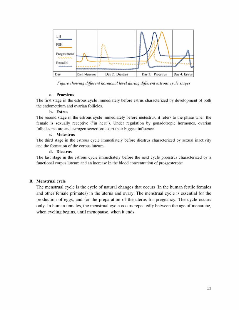
It comprises of 4 stages:
11
Figure showing different hormonal level during different estrous cycle stages
a. Proestrus
The first stage in the estrous cycle immediately before estrus characterized by development of both
the endometrium and ovarian follicles.
b. Estrus
The second stage in the estrous cycle immediately before metestrus, it refers to the phase when the
female is sexually receptive ("in heat"). Under regulation by gonadotropic hormones, ovarian
follicles mature and estrogen secretions exert their biggest influence.
c. Metestrus
The third stage in the estrous cycle immediately before diestrus characterized by sexual inactivity
and the formation of the corpus luteum.
d. Diestrus
The last stage in the estrous cycle immediately before the next cycle proestrus characterized by a
functional corpus luteum and an increase in the blood concentration of prosgesterone
B. Menstrual cycle
The menstrual cycle is the cycle of natural changes that occurs (in the human fertile females
and other female primates) in the uterus and ovary. The menstrual cycle is essential for the
production of eggs, and for the preparation of the uterus for pregnancy. The cycle occurs
only. In human females, the menstrual cycle occurs repeatedly between the age of menarche,
when cycling begins, until menopause, when it ends.

12
Figure showing different hormonal level during different menstrual cycle stages
It comprises of three ovarian and three uterine cycle phases:
Ovarian cycle phases:
1. Follicular phase
The follicular phase is the first part of the ovarian cycle. During this phase, the
ovarian follicles mature and get ready to release an egg. Through the influence of a
rise in follicle stimulating hormone (FSH) during the first days of the cycle, a few
ovarian follicles are stimulated.
2. Ovulatory phase
Ovulation is the second phase of the ovarian cycle in which a mature egg is released
from the ovarian follicles into the oviduct. During the follicular phase, estradiol
suppresses production of luteinizing hormone (LH) from the anterior pituitary gland.
3. Luteal phase
The luteal phase is the final phase of the ovarian cycle and it corresponds to the
secretory phase of the uterine cycle. During the luteal phase, the pituitary hormones
FSH and LH cause the remaining parts of the dominant follicle to transform into the
corpus luteum, which produces progesterone.
Uterine cycle phase:
1. Menstruation phase

13
Menstruation is the first phase of the uterine cycle.
2. Proliferative phase
The proliferative phase is the second phase of the uterine cycle when estrogen causes
the lining of the uterus to grow, or proliferate, during this time. As they mature, the
ovarian follicles secrete increasing amounts of estrogen.
3. Secretory phase
The secretory phase is the final phase of the uterine cycle and it corresponds to the
luteal phase of the ovarian cycle. During the secretory phase, the corpus luteum
produces progesterone, which plays a vital role in making the endometrium receptive
to implantation.
Answer No.7.
Extraembryonic membranes are membranous structures that appear in parallel with the
embryo and play important roles in the embryonic development. They form from the embryo but
do not become part of the individual organism after its birth.
The presence of each extraembryonic membrane varies according to the vertebrate class.
In fishes and amphibians only the yolk sac is present. In reptiles, birds and mammals besides the
yolk sac there are also the amnion, the chorion and the allantois.
The embryos of reptiles, birds, and mammals produce
4 extraembryonic membranes, the
• amnion
• yolk sac
• chorion
• allantois
In birds and most reptiles, the embryo with its
extraembryonic membranes develops within a shelled
egg.
• The amnion protects the embryo in a sac filled with amniotic fluid.
• The yolk sac contains yolk — the sole source of food until hatching. Yolk is a mixture of
proteins and lipoproteins.
• The chorion lines the inner surface of the shell (which is permeable to gases) and
participates in the exchange of O2 and CO2 between the embryo and the outside air.
• The allantois stores metabolic wastes (chiefly uric acid) of the embryo and, as it grows
larger, also participates in gas exchange.
Amnion
• Surrounds the embryo in fluid‐filled sac ( Amniotic fluid)
• Innermost membrane

14
• Protects embryo ( Shock absorption, Temperature fluctuations, Prevents desiccation)
Yolk sac
• Surrounds the yolk
• Uptake and modification modification of yolk lipids to lipoproteins
• Provides the nourishment required for embryonic growth
Chorion
• Surrounds the embryo
• Outermost membrane
• Chorion + Allantois = “exchange organ”
Oviparous ‐Gas exchange
Viviparous ‐Gas and nutrient exchange
Allantois
• Outpocketing of hindgut
• Waste removal
Removes metabolic wastes produced by the embryo “primitive bladder
With these four membranes, the developing embryo is able to carry on essential metabolism
while sealed within the egg. Surrounded by amniotic fluid, the embryo is kept as moist as a fish
embryo in a pond.
Although (most) mammals do not make a shelled egg, they do also enclose their embryo in an
amnion. For this reason, the reptiles, birds, and mammals are collectively referred to as the
amniota.
Answer No. 8.
In all sexually reproducing coelomates, there are four main stages of embryogenesis,
namely; fertilization, cleavage, gastrulation, and organogenesis. Fertilization is the fusion of
haploid male and female gametes to form a diploid zygote. Zygote is the new cell, which is also
known as fertilized ovum. In the process of cleavage, the zygote rapidly divides into many cells,
without increasing the overall size of it and ends up with a structure called blastula.

15
Blastula
Blastula represents the first important stage after the fertilization and plays an important role in
the development of organisms. It is a hollow, spherical, one celled thick structure formed by the
process called blastulation. Both holoblastic and meroblastic cleavages give rise to blastula. The
cavity inside the blastula is called blastocoel, and its outer single cell layer is called blastoderm.
Gastrula
The continuous development of blastula finally results the gastrula. The conversion process of
the blastula into gastrula is called ‘gastrulation’. Gastrulation is followed by the organogenesis.
Gastrula is composed of three primary germ layers, which eventually give rise to organs in the
late embryo. The primary germ layers are ectoderm, mesoderm, and endoderm. Ectoderm is the
outermost layer of gastrula, which differentiates into skin, brain, spinal cord, and nerves of
embryo. Mesoderm is the middle layer, which forms muscles, connective tissues, reproductive
organs, cartilage, bones, and dermis of skin and dentine of teeth. Endoderm is the innermost
layer of the embryo and basically differentiates into primitive gut.
Difference between Blastula and Gastrula
(i) During the embryogenesis process, formation of blastula is followed by gastrula.
(ii) Formation of blastula is called blastulation, whereas the formation of gastrula is
called gastrulation.
(iii) Rapid mitotic divisions of zygote results blastula while slow mitotic divisions of
blastula results gastrula.
(iv) During the formation of blastula, cells do not move, but during the formation of
gastrula, cell masses move by morphogenetic movements.
(v) Three primary germ layers are present in gastrula unlike in the blastula.

16
(vi) Blastula is often called a pre-embryo, whereas gastrula is referred to as a mature
embryo.
(vii) Gastrula has more cells than blastula.
(viii) Gastrula has differentiated cells, while blastula has undifferentiated cells.
Types of Blastula:
In chordate the blastula are of following type –
a. Coeloblastula
A blastula having cavity inside is called coeloblastula. It is formed through
complete holoblastic cleavage. The blastoderm is formed of a single layer of cells.
Its blastocoel is filled with mucopolysaccaharide.
e.g. Echinoderms and Amphibians
b. Discoblastula
It is formed as a result of discoidal cleavage. The blastocoel is small & it is called
as subgerminal space. It is situated below the blastoderm.

17
eg. Bony fishes, reptile, birds & prototherian mammals.
c. Blastocyst
It is formed in mammals as a result of holoblastic cleavage.
Outer cells are called as trophoblasts or cells of Rauber. They form a trophoderm. This layer get
attached to the uterine wall. Inner cells form the embryo are called as inner cell mass.
eg. Mammals (Human)