Embed Size (px)
Citation preview
Basidiomycota (club fungi)
B.Sc Botany (Hons)
Semester II
Mycology and Phytopathology
The Basidiomycota (colloquially basidiomycetes) are a large group of fungi with over 30 000 species.
They include many familiar mushrooms and toadstools, bracket fungi, puffballs, earth balls, earth stars, stinkhorns, false truffles, jelly fungi and some less familiar forms.
Also classified here are the rust and smut fungi, which are pathogens of higher plants and may cause serious crop diseases.
Most basidiomycetes are terrestrial with wind-dispersed spores, but some grow in freshwater or marine habitats.
Many are saprotrophic and are involved in litter and wood decay, but there are also pathogens of trees.
the honey fungus, Armillaria, which attacks numerous tree species, and Heterobasidion annosum, which can seriously damage conifer plantations.
1. The somatic phase consists of a well developed, septate, filamentous mycelium which passes chiefly through two stages:
a) primary mycelium-
it is formed by the germination of a basidiospore and contains a single haploid nucleus in each cell.
It bears neither sex organs nor any basidia and basidiospores.
It is short lived
b) Secondary or dikaryotic mycelium
It constitutes the main food absorbing phase and consists of cells each containing two haploid nuclie.
It is long lived and plays prominent role in the life cycle.
In Homobasidiomycetidae it may continue to grow for years producing fructifications every year by the interweaving of hyphae.
The fructifications bear basidia and basidiospores
In Heterobasidiomycetidae it forms teleutospores or brand spores which germinate to produce basidia bearing basidiospores.
2.Except in rusts and smuts the septal pore in the Basidiomycetes is complex, It is dolipore septa with parenthesome type.
3.The motile cells are absent in the life cycle
4. The clamp connections on the dikaryotic hyphae are of universal occurrence
5.Asexual reproduction by spores plays an insignificant role in the life cycle.
The homobasidiomycetes do not form any asexual spores.
The heterobasidiomycetes form them in the dikaryotic mycelium and produces uredospores and aecidiospores in the rust.
6.The sex organs are lacking in the Basidiomycetes. The sexual process is represented by plasmogamy and karyogamy. Karyogamy is immediately followed by meiosis.
7. Basidium is the characteristic reproductive organ of Basidiomycetes in which both karyogamy and meiosis takes place.
8.Typically the basidium bears four basidiospores exogenously. The number, however varies from one to many depending on species.
9. The basidiospore germinates to produce the primary mycelium.
Resembles with Ascomycota
1. Both include parasitic as well as saprophytic species.
2. Terrestrial mycelium consists of septa.
3. The septa have each a central pore.
4. Motile cells completely absent.
5. The sexual process comprises plasmogamy and karyogamy. The latter is immediately followed by meiosis.
6. The delayed fusion of nuclei of opposite strains after plasmogamy has resulted in the origin and establishment of a binucleate or dikaryophase in the life cycle.
7. The characteristic reproductive organ, basidium of Basidiomycetes and ascus of Ascomycetes resemble each other in development and cytology till the initiation of spores. Both arises from binucleate cells, the basidium from the dikaryotic hyphae and ascus from the ascogenous hyphae. Karyogamy and meiosis both occur in the basidium as in the ascus.
8.The basidiospores and ascospores are ususally haploid and uninucleate.
9. The basidiocarp in Basidiomycetes and ascocarp in Ascomycetes (not homologous structure)
10.A clamp connection of the Basidiomycetes is considered homologous in sturcture and analogous in function to the hook of the Ascomycetes.
The Basidiomycetes differ from the Ascomycetes in the following respects:
1. The septal pore in most of the Basidiomycetes is not a simple pore as in Ascomycetes but is a complex structure known as dolipore. The actual pore is barrel shaped. It is surrounded by a swollen rim which is a part of the annular septum. The opening of the pore is guarded by a curved pore cap called parenthesome.
2. The primary mycelium is short lived whereas in Ascomycetes it is long lived and dominanat.
3. The primary mycelium in the Basidiomycetes bears neither the sex organs nor basidia or basidiospores.
4. The conidia (uredospores and aeciospores) are borne on the secondary mycelium whereas in Ascomycetes they are borne on haplomycelium
5. Except the Uredinales all traces of sexual apparatus have been lost throughout the classes.
6. Presence of clamp connection is a characteristic feature of secondary mycelium
7. The dikaryotic (secondary) mycelium is long lived, independent structure whereas ascogenous hypae are short lived, homologous and occur inside the fruit body.
8.The fruit bodies in Basidiomycetes consists entirely of dikaryotic hyphae whereas in Ascomycetes the basal hyphae, peridia and paraphyses are haploid.
9.The basidium produced 4 basidiospores whereas ascus produced 8 ascospores.
10. The basidiospores are produced exogenously whereas ascospores are produced endogenously.
1. Homobasidiomycetes. Fungi with holobasidia,
e.g. Agaricus and Polypores.
2.Heterobasidiomycetes. Fungi with heterobasidia, i.e. jelly fungi and their allies.
3. Urediniomycetes. Rust fungi.
4. Ustilaginomycetes. Smut fungi.
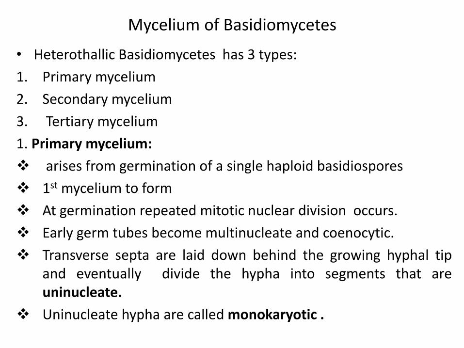
Mycelium of Basidiomycetes
• Heterothallic Basidiomycetes has 3 types:
1. Primary mycelium
2. Secondary mycelium
3. Tertiary mycelium
1. Primary mycelium:
arises from germination of a single haploid basidiospores
1st mycelium to form
At germination repeated mitotic nuclear division occurs.
Early germ tubes become multinucleate and coenocytic.
Transverse septa are laid down behind the growing hyphal tip and eventually divide the hypha into segments that are uninucleate.
Uninucleate hypha are called monokaryotic .
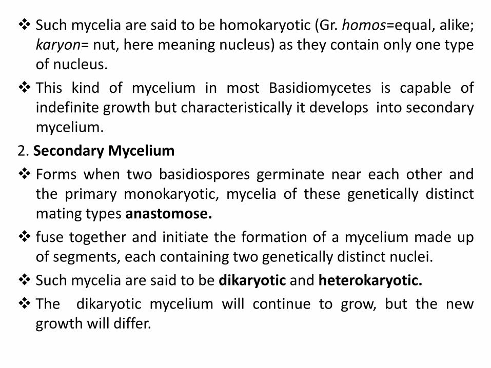
Such mycelia are said to be homokaryotic (Gr. homos=equal, alike; karyon= nut, here meaning nucleus) as they contain only one type of nucleus.
This kind of mycelium in most Basidiomycetes is capable of indefinite growth but characteristically it develops into secondary mycelium.
2. Secondary Mycelium
Forms when two basidiospores germinate near each other and the primary monokaryotic, mycelia of these genetically distinct mating types anastomose.
fuse together and initiate the formation of a mycelium made up of segments, each containing two genetically distinct nuclei.
Such mycelia are said to be dikaryotic and heterokaryotic.
The dikaryotic mycelium will continue to grow, but the new growth will differ.
3. Tertiary mycelium
arises when secondary mycelium transforms into organized, specialized tissues that comprise the basidiocarps of the most complex species.
The hyphae constituting the basidiocarp may be differentiated several types.
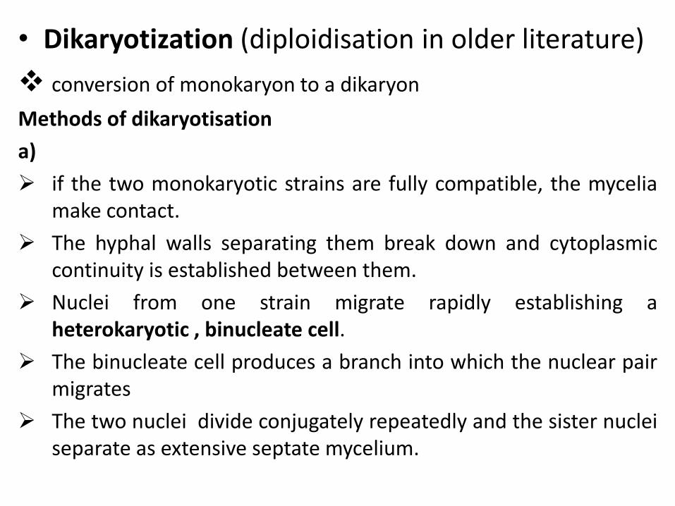
• Dikaryotization (diploidisation in older literature)
conversion of monokaryon to a dikaryon
Methods of dikaryotisation
a)
if the two monokaryotic strains are fully compatible, the mycelia make contact.
The hyphal walls separating them break down and cytoplasmic continuity is established between them.
Nuclei from one strain migrate rapidly establishing a heterokaryotic , binucleate cell.
The binucleate cell produces a branch into which the nuclear pair migrates
The two nuclei divide conjugately repeatedly and the sister nuclei separate as extensive septate mycelium.
Migration is accompanied by nuclear division, in order to permit the doubling of the number of nuclei in both mycelia
b).
In another very common method of dikaryotisation, there a division of the nuclei in the above formed binucleate cell followed by migration of the daughter nuclei into primary mycelium of the opposite mating type,
eg. The ‘a’ nucleus moves into ‘b’ mycelium, while a ‘b’ nucleus moves into the ‘a’ mycelium.
The foreign nucleus in each mycelium then divides rapidly and its progeny migrating from one cell to another completing dikaryotising both parent mycelia
c)
Dikaryotisation can occur by the deposition of an oidium from one strain close to the mycelium of another.
A hypha from the mycelium shows chemotropic curvature towards the oidium and fuses with it
If the strains are compatible, nuclear migration follows, which associated with the breakdown of dolipore/parenthesome complex leading to the transfer of a compatible nucleus from one compartment to another.
Eg. Coprinus cinereus
d)
Dikaryotisation can also occur if a monokaryon encounters a dikaryon carrying nuclei of a compatible mating type.
Migration is unilateral instead of reciprocal
Eg. Autoecious rusts
Clamps connection
In most Basidiomycetes, the most conspicuous feature of the secondary mycelium is the presence of clamp connections.
Visible as a lateral bulge in the hyphal wall adjacent to a transverse septum.
Clamp connections are special devices for maintaining the dikaryotic condition as growth continues.
Ensured each segment of a dikaryotic hypha contains two genetically distinct nuclei.
In the terminal segment of a dikaryotic hypha, a clamp connection develops between the position of a pair of nuclei ‘a’ and ‘b’ following the sequence
The apical cell produces a short backward pointing side branch or a hook (clamp) and a daughter nucleus moves into it, the other nucleus remaining in the hypha but close to the branch.
Simultaneous nuclear division follows
One division becomes oriented obliquely so that one daughter nucleus a forms in the clamp connection and the other a’ in the dividing cell
Division of the second nucleus is oriented along the longitudinal axis of the dividing cell
One of the daughter nucleus b forms near one end of the cell and other b’ approaches the nucleus a’ of the first division near the other end of the cell
The hook bends and fuses with the subterminal cell forming a bridge through which one of the daughter nuclei a passes to the other end of its cell which already contains one nucleus b
A septum forms to close the hook at the point of its origin
Another septum is form vertically under the bridge to divide the parent cell into daughter cells with a and b in one daughter cell and a’ and b’ in another.
Dolipore septa (L. dolium= large jar, cask).
Except in rusts and smuts the septal pore in the Basidiomycetes is complex, It is dolipore septa with parenthesome type.
The transverse septa which divide both monokaryotic and dikaryotic hyphae into segments are incomplete
They contain a central pore which lies between adjacent segments.
The septal pore is surrounded by a barrel-shaped flange of thickened wall material.
Such septa, which are characteristic of Basidiomycete mycelia, are known as dolipore septa.
Septal development begins by centripetal ingrowth of a membrane on which wall material (glucan and chitin) is deposited at both faces from associated vesicles.
The thickening surrounding the pore results from more rapid deposition of wall material.
The septal pore on each side of the septum is covered by a dome shaped membranous structure known as the septal pore cap or parenthesome (parenthesis= round bracket).
They are shaped like parentheses and therefore named so.
It is a specialized portion of endoplasmic reticulum and is the integral and functional part of the septal apparatus.
Various types of septal pore caps have been demonstrated in Basidiomycetes.
In Homobasidiomycetes, the parenthesome are perforated.
In many Heterobasidiomycetes, there is only a single perforation or it appears as continuous non-porus structure.
The variation in the parenthesome or dolipore complex are useful in distinguishing individual species.
An important role of the dolipore/parenthesome complex is to secure the integrity of hyphal cells.
And to maintain intracellular communication and transport of some organelles.
It acts as a screen or sieve possibly permitting the passage of most organelles (small tubular and filamentous structures, small vesicles, tubular endoplasmic reticulum and mitochondria) from one cell to the next while not letting pass nuclei possibly due to their larger size.
The migration of nuclei following plasmogamy between between two sexually compatible monokaryotic mycelia is associated with enzymatic dissolotion of the dolipore.
Another important function of dolipores is the repair of hyphal damage, whereby damaged segments of the septal pores are rapidly plugged by electron dense materials.
Asexual reproduction in Basidiomycota
1. By conidia
Conidia production is not so common in this group.
They are produced in the rusts, smuts, and some other Basidiomycetes
In Smuts they are budded off from the basidiospores and the mycelium.
The uredospores of rusts are also of conidial nature and function.
The conidia in the Basidiomycetes are produced by the dikaryotic mycelium.
They serve to propagate the dikaryophase in the life cycle.
2. By Oidia
These are small, hyaline, thin walled unicellular sections or fragments of the mycelium.
They may be uninucleate or binucleate according as they are produced by breaking up of the primary or secondary mycelium.
They usually do not round up or secrete thick walls to become spore like.
They germinate by means of germ tubes and grow into new mycelia.
In some species the oidia are segmented from special, short lateral hyphal branches called the oidiophores.
The oidia serve a double function.
They may either germinate to form primary mycelia or bring about diplodization.
In the later case the germinating oidium acts as a spermatium and fuses with the hyphal cell of an opposite strain.
3. Budding and fragmentation.
Unimportant vegetative means of asexual reproduction in Basidiomycetes.

Basidiocarps (Gr. Basidium= small base; karp=fruit) In higher Basidiomycota (class Homobasidiomycetes) the
secondary mycelium develops fruiting bodies called basidiocarps.
The basidiocarps are usually massive aerial sporophores which bear basidia.
They are of various sizes, types, texture and forms.
They may be thin, crust-like, gelatinous, papery, thick and fleshy, leathery, corky, woody and spongy.
It range from microscopic to macroscopic bodies 3 feet or more in diameter. (eg. Rigidiporus ulmarius 147cm and 5lb)
They may be umbrella-shaped, fan shaped and the like.
The portion of the secondary mycelium which forms the frutifications(basidiocarps) is sometimes, called the tertiary or generative mycelium. It is dikaryotic.
Basidiocarps may be open or remain completely closed.
In species with basidiocarps that remain closed , the spores are liberated only by the disintegration of the basidiocarps or with its fracture by external forces including ingestion of insects and rodents.
The basidium in most species arew formed in definite layers called hymenia (comparable to asci).
Hymenia also contains elements like basidioles and cystidia.
Basidioles probable function is to provide support for fertile basidia.
Cystidia are sterile elements and act as air drops and aid in the evaporation of moisture and other volatile compounds.
Basidium
A basidium may be defined as a structure bearing on its surface a definite number of basidiospores that are formed as a result of karyogamy and meiosis.
Usually there are four spores, but in some cases there are one (Itersonilia perplexans), two (Agaricus bisporus), or more than four (Phallus impudicus) basidiospores per basidium.
The simple club shaped basidium originates as a terminal cell of a binucleate hypha.
It is delimited from the rest of the hypha by a septum over which the clamp connection is generally found
At first narrow and elongated, the basidium soon enlarges and becomes broader.
The two nuclei within the young basidium undergo karyogamy and diploid nucleus soon undergoes meiosis, producing four haploid nuclei.
In the mean time four small, curved and tapering outgrowths, termed sterigmata, push out at the top of the basidium.
The tips of sterigmata enlarge, eventually forming the basidiospore initials.
The contents of the basidium are then pushed into the basidiospore initial.
Meiosis occurs in the upper part of the basidium.
In narrow basidia the plane of the second meiotic nuclear division lies parallel to the long axis of the basidium. This type of nuclear division is termed as chiastic (Gr. Chiastos=crossed,arranged diagonally).
And the basidia formed by chiastic nuclear division is called chiastobasidia.
Transverse nuclear division then its called stichic (Gr. Stichos=a line or row of things) and the basidium is called stichobasidium.
This characters are of Taxonomic relevance. Chiastobasidia (mushrooms and toadstool; stichobasidium (bracket fungi)
basidium is divided into three parts on the basis of nuclear behaviour
1. Probasidium: karyogamy takes place
2. Metabasidium: meisois occurs
3. Sterigmata: parts between metabasidium and basidiospore.

Types of basidia
• Holobasidia= no septa, hymenium freely exposed. Eg.Homobasidiomycetes
• Phragmobasidia or heterobasidia (Gr.Phragmo=hedge or barricade; heteros= other, different), hymenium freely exposed eg. Heterobasidiomycetes
• Basidiomycota classification
• Homobasidiomycetes
• Heterobasidiomycetes
• Urediniomycetes
• Ustilaginomycetes
• Gasteromycetes- hymenium not freely exposed