Embed Size (px)
Citation preview
1
Bronchial Asthma
and the
Atopic Syndrome
A Biochemical Study Proposing
a Deficit of Lipoprotein LipaseBy Alexander Dunedin
Abstract: A thesis on bronchial asthma and the atopic
conditions existing as manifestations of a breakdown in
prostaglandin metabolism resultant from a chemical
depletion of lipoprotein lipase (a regulating enzyme), or
a deficit of the elemental components required to
synthesise the enzyme.
This work is licensed under the Creative Commons Attribution-NoDerivs 3.0 Unported License. To
view a copy of this license, visit http://creativecommons.org/licenses/by-nd/3.0/ or send a letter to
Creative Commons, 444 Castro Street, Suite 900, Mountain View, California, 94041, USA.
2
Bronchial Asthma and the Atopic Syndrome
Index
Paper 1: Foreword 6
1.1: Glucuronic acid 71.2: Glucosamine 81.3: Glucuronidation 91.4: A simple explanation of allergic conditions 121.5: Glucuronic acid 151.6: Glucosamine 171.7: Summary 19
Paper 2: Asthma and Atopy 23
2.1: Asthma 242.2: Status Asthmaticus 262.3: Atopy 272.4: Allergy 282.5: Allergens 292.6: Hay Fever 302.7: Eczema 312.8: Dermatitis 322.9: Gastroenteritis 332.10: Diarrhoea 342.11: Crohn’s disease 352.12: Anaphylaxis 362.13: Definition 37
Paper 3: Asthma and Inflammation 40
3.1: The definition of asthma 413.2: Immunological response to the antigenic substance 423.3: Immunoglobulins 443.4: Mast cells 463.5: Anaphylaxis 483.6: Summation 49
3
Paper 4: Asthma and Inflammation 52
4.1: The clinical definition of asthma 534.2: Dyspnoea 554.3: Oedema 564.4: Eosinophilia 574.5: Eosinophils 584.6: Smooth muscle 594.7: Autonomic nervous system 604.8: Connective tissue 614.9: Mast cells 624.10: Synopsis 63
Paper 5: Cellular Mediators of Inflammation 66
5.1: The mediators of the inflammatory process 685.2: Interactions between possible mediators 695.3: Mast cells 715.4: Neutrophilic Polymorphonuclear Leukocytes 735.5: Eosinophilic Polymorphonuclear Leukocytes 765.6: Mononuclear cells 775.7: Lymphocytes 785.8: A broad overview 81
Paper 6: Humoral Mediators of Inflammation 86
6.1: Histamine 886.2: Serotonin 926.3: Catecholamines 946.4: Adrenaline 956.5: Noradrenaline 966.6: Peptides and Proteins 976.7: Kinins 986.8: Activated complement components 1016.9: Components of the blood clotting system 1036.10: Prostaglandins 1046.11: Heparin 1086.12: Mucopolysaccharides 1096.13: Summary 110
Paper 7: Hypothesis of Biological Adaptation 118
7.1: Selfish Brain Theory 120
4
Paper 8: The Hormonal Pathology of Asthma 120
8.1: Pathological examination 1228.2: Smooth muscle 1238.3: Prostaglandins 1258.4: Edema 1278.5: Dose dependent 1308.6: Eicosanoids 1318.7: Eicosanoids 1328.8: Prostaglandins 1368.9: Death 1388.10: Stimuli 1398.11: Iteration 140
Paper 9: The Metabolism of Prostaglandins 146
9.1: Prostaglandins 1489.2: Glucuronic acid 1519.3: Compounds 1529.4: Glucuronidation 1539.5: Lipoprotein lipase 1549.6: Glucuronic acid 1569.7: Summary 157
Paper 10: Heparin and its Actions 161
10.1: Anti-inflammatory agent 16310.2: Lungs 16410.3: Heparin sulphate 16510.4: Lipoproteins 16710.5: Highly sulphated heparin glycosaminoglycans 16910.6: Component 17010.7: Lipase 17110.8: Arylsulfatase 17310.9: Hypothesis 175
Paper 11: Glucuronidation 179
11.1: Acid polysaccharides 18111.2: Organic acids in the urine 18311.3: Foreign organic compounds 18411.4: Direct chemical interactions with heparin 18511.5: Summary 186
5
Paper 12: Hypothesis on Atopy’s Treatment 188
12.1: Anti-inflammatory agent 19112.2: Effects of heparin on lipoprotein lipase 19312.3: Heparin 19412.4: Glucuronic acid residues 19512.5: Eosinophilic leukocytes 19612.6: Oxazepam, morphine and 3-hydroxyantipyrine 19712.7: Hyaluronic acid 19912.8: Summary 200
Paper 13: Recapitulation of Hypothesis 203
Paper 14: Addendum - Extended Analysis 213
Bronchial Asthma 215Allergic Challenge Asthma 224Mast Cells 227Allergic Response 231Inflammation 235Inositol 238Histamine 239Eosinophil 240Arylsulfatase 244Prostaglandins 246M2 Acetylcholine Muscarinic Receptors 252Heparin 254Glycosaminoglycans 263Eczema 267Crohn’s disease 269Arthritis 274Hay Fever 278Anaphylactic Shock 279
Bibliography of references 280
Index 306
6
Bronchial Asthma and the Atopic Syndrome
Paper 1: Foreword
This document is a hypothesis of the cause of bronchial asthma
and the atopic syndrome. Key terms are shown in bold and
underlined. These are points of reference to published texts.
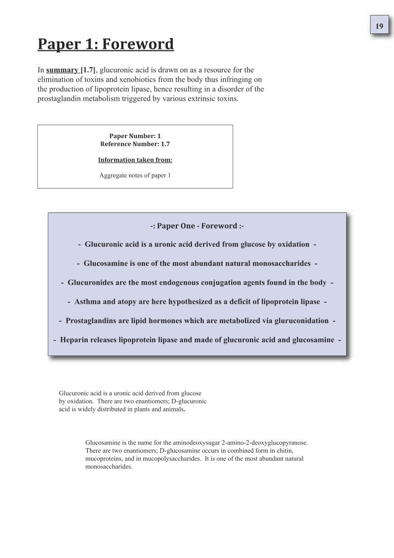
In summary [1.7], glucuronic acid is drawn on as a resource for the
elimination of toxins and xenobiotics from the body thus infringing on
the production of lipoprotein lipase, hence resulting in a disorder of the
prostaglandin metabolism triggered by various extrinsic toxins.
A simple explanation of allergic conditions [1.4] is inflammation. Allergic
conditions are manifestations of the same type of inflammation occurring in
different mucosal tissues of the body. The hypothesis here being presented is
that the inflammation arises as a direct result of a deficit of glucuronic acid
[1.5] and glucosamine [1.6] which one-at-the-same-time are required to control
prostaglandin hormones via the enzyme lipoprotein lipase.
The principal papers provide an index leading through an increasingly detailed
definition and breakdown of atopy and its theorized cause. Two substances -
glucuronic acid [1.1] and glucosamine [1.2] - are required for the construction
of glycosaminoglycans, heparin and active lipoprotein lipase. Glucuronic acid is
also required for the elimination of certain prostaglandins, toxins and xenobiotics
from the body in a process known as glucuronidation [1.3].
When a key term has been made in the narrative you will be able to find in
the appendix the details of the author; the title of the publication; the date of
and edition of publication from which it has been taken; the publisher; a
surmising of the content, the page of reference; all followed by the
unexpurgated reference. This has been done so the facts may be easily
compared against the original texts.
7
The principal papers provide an index leading through an increasingly detailed definition and
breakdown of atopy and its theorized cause. Two substances - glucuronic acid [1.1] and
glucosamine - are required for the construction of glycosaminoglycans, heparin and active
lipoprotein lipase. Glucuronic acid is also required for the elimination of certain
prostaglandins, toxins and xenobiotics from the body in a process known as glucuronidation.
Paper 1: Foreword
Paper Number: 1
Reference Number: 1.1
Information taken from:
Oxford Dictionary Of Biochemistry And Molecular Biology
Revised Edition 2000
Managing Editor Dr A D Smith University College London
General Editors: Professor S P Datta University College London
Dr G H Smith University College London
Professor P N Campbell (Chairman) University College London
Dr R Bentley University of Pittsburgh
Dr H A McKenzie Australian Defence Force Ac.
© The General Editors, 1997
Oxford University Press Inc., New York
ISBN 0 19 850673 2
Page 267; Glucuronic acid
Glucuronic Acid
abbr. (sometimes): GA; symbol: GlcA (or (formerly) GlcU or GlcUA); the uronic acid
formally derived from glucose by oxidation of the hydroxymethylene group at C-6 to a
carboxyl group [1]. There are two enantiomers; D-glucuronic acid is widely distributed in
plants and animals, where it usually occurs as glucuronides [2].
Notes:
Glucuronic acid is a uronic acid derived from glucose by oxidation [1].
There are two enantiomers; D-glucuronic acid is widely distributed in
plants and animals [2].
8
The principal papers provide an index leading through an increasingly detailed definition
and breakdown of atopy and its theorized cause. Two substances - glucuronic acid and
glucosamine [1.2] - are required for the construction of glycosaminoglycans, heparin and
active lipoprotein lipase. Glucuronic acid is also required for the elimination of certain
prostaglandins toxins and xenobiotics from the body in a process known as glucuronidation.
Paper 1: Foreword
Paper Number: 1
Reference Number: 1.2
Information taken from:
Oxford Dictionary Of Biochemistry And Molecular Biology
Revised Edition 2000
Managing Editor Dr A D Smith University College London
General Editors: Professor S P Datta University College London
Dr G H Smith University College London
Professor P N Campbell (Chairman) University College London
Dr R Bentley University of Pittsburgh
Dr H A McKenzie Australian Defence Force Ac.
© The General Editors, 1997
Oxford University Press Inc., New York
ISBN 0 19 850673 2
Page 265; Glucosamine
Glucosamine
Symbol: GlcpN; the trivial name for the aminodeoxysugar 2-amino-2-deoxyglucopyranose [1];
there are two enantiomers. D-Glucosamine (symbol: D-GlcpN), formerly known as chitosamine,
occurs in combined form in chitin, in mucoproteins, and in mucopolysaccharides [2], and is one
of the most abundant natural monosaccharides [3].
Notes:
Glucosamine is the name for the aminodeoxysugar 2-amino-2-deoxyglucopyranose
[1]. There are two enantiomers; D-glucosamine occurs in combined form in chitin,
mucoproteins, and in mucopolysaccharides [2]. It is one of the most abundant natural
monosaccharides [3].
9
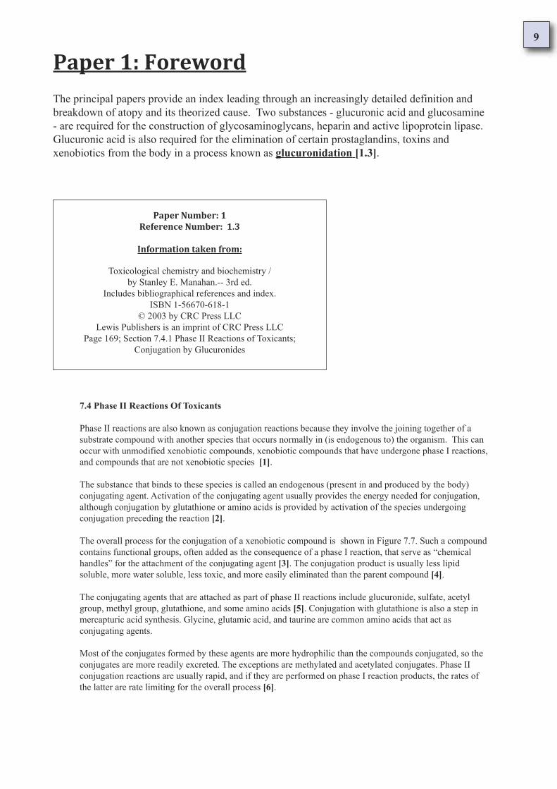
The principal papers provide an index leading through an increasingly detailed definition and
breakdown of atopy and its theorized cause. Two substances - glucuronic acid and glucosamine
- are required for the construction of glycosaminoglycans, heparin and active lipoprotein lipase.
Glucuronic acid is also required for the elimination of certain prostaglandins, toxins and
xenobiotics from the body in a process known as glucuronidation [1.3].
Paper 1: Foreword
Paper Number: 1
Reference Number: 1.3
Information taken from:
Toxicological chemistry and biochemistry /
by Stanley E. Manahan.-- 3rd ed.
Includes bibliographical references and index.
ISBN 1-56670-618-1
© 2003 by CRC Press LLC
Lewis Publishers is an imprint of CRC Press LLC
Page 169; Section 7.4.1 Phase II Reactions of Toxicants;
Conjugation by Glucuronides
7.4 Phase II Reactions Of Toxicants
Phase II reactions are also known as conjugation reactions because they involve the joining together of a
substrate compound with another species that occurs normally in (is endogenous to) the organism. This can
occur with unmodified xenobiotic compounds, xenobiotic compounds that have undergone phase I reactions,
and compounds that are not xenobiotic species [1].
The substance that binds to these species is called an endogenous (present in and produced by the body)
conjugating agent. Activation of the conjugating agent usually provides the energy needed for conjugation,
although conjugation by glutathione or amino acids is provided by activation of the species undergoing
conjugation preceding the reaction [2].
The overall process for the conjugation of a xenobiotic compound is shown in Figure 7.7. Such a compound
contains functional groups, often added as the consequence of a phase I reaction, that serve as “chemical
handles” for the attachment of the conjugating agent [3]. The conjugation product is usually less lipid
soluble, more water soluble, less toxic, and more easily eliminated than the parent compound [4].
The conjugating agents that are attached as part of phase II reactions include glucuronide, sulfate, acetyl
group, methyl group, glutathione, and some amino acids [5]. Conjugation with glutathione is also a step in
mercapturic acid synthesis. Glycine, glutamic acid, and taurine are common amino acids that act as
conjugating agents.
Most of the conjugates formed by these agents are more hydrophilic than the compounds conjugated, so the
conjugates are more readily excreted. The exceptions are methylated and acetylated conjugates. Phase II
conjugation reactions are usually rapid, and if they are performed on phase I reaction products, the rates of
the latter are rate limiting for the overall process [6].
10
7.4.1 Conjugation by Glucuronides
Glucuronides are the most common endogenous conjugating agents in the body. They react with xenobiotics
through the action of uridine diphosphate glucuronic acid (UDPGA). This transfer is mediated by glucuronyl
transferase enzymes [7]. These enzymes occur in the endoplasmic reticulum, where hydroxylated phase I
metabolites of lipophilic xenobiotic compounds are produced [8]. As a result, the lifetime of the phase I
metabolites is often quite brief because the conjugating agent is present where they are produced. A generalized
conjugation reaction of UDPGA with a xenobiotic compound can be represented as the following:
DIAGRAM OMITED
In this reaction HX–R represents the xenobiotic species in which HX is a functional group (such as –OH) and R
is an organic moiety, such as the phenyl group (benzene ring less a hydrogen atom). The kind of enzyme that
mediates this type of reaction is UDP glucuronyltransferase. Glucuronide conjugation products may be classified
according to the element to which the glucuronide is bound [9]. The atoms to which the glucuronide most readily
attaches are electron rich, usually O, N, or S (nucleophilic heteroatoms in the parlance of organic chemistry)
[10].
Example glucuronides involving O, N, and S atoms are shown in Figure 7.8. When the functional group through
which conjugation occurs is a hydroxyl group, –OH (HX in Reaction 7.4.1), an ether glucuronide is formed [11].
A carboxylic acid group for HX gives an ester glucuronide [12]. Glucuronides may be attached directly to N as
the linking atom, as is the case with aniline glucuronide [13] in Figure 7.8, or through an intermediate O atom
[14].
An example of the latter is N-hydroxyacetylaminoglucuronide, for which the structure is shown in Figure 7.9.
This species is of interest because it is a stronger carcinogen than its parent xenobiotic compound, N-
hydroxyacetylaminofluorene, contrary to the decrease in toxicity that usually results from glucuronide
conjugation [15].
The carboxylic acid (–CO2H group) in glucuronides is normally ionized at the pH of physiological media, which
is a major reason for the water solubility of the conjugates [16]. When the compound conjugated (called the
aglycone) is of relatively low molecular mass, the conjugate tends to be eliminated through urine. For heavier
aglycones, elimination occurs through bile [17].
Enterohepatic circulation provides a mechanism by which the metabolic effects of some glucuronide conjugates
Notes:
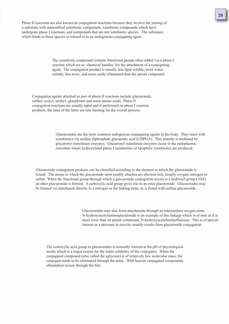
Phase II reactions are also known as conjugation reactions because they involve the joining of a
substrate with unmodified xenobiotic compounds, xenobiotic compounds which have undergone
phase I reactions, and compounds that are not xenobiotic species [1]. The substance which
binds to these species is refered to as an endogenous conjugating agent [2].
The xenobiotic compound contains functional groups often added via a phase I reaction which
act as ‘chemical handles’ for the attachment of a conjugating agent [3]. The conjugation
product is usually less lipid soluble, more water soluble, less toxic, and more easily eliminated
than the parent compound [4].
Conjugation agents attached as part of phase II reactions include glucuronide, sulfate, acetyl,
methyl, glutathione and some amino acids [5]. Phase II conjugation reactions are usually rapid
and if performed on phase I reaction products, the rates of the latter are rate limiting for the
overall process [6].
11
Glucuronides are the most common endogenous conjugating agents in the body. They react with
xenobiotics via uridine diphosphate glucuronic acid (UDPGA). This transfer is mediated by
glucuronyl transferase enzymes [7]. Glucuronyl transferase enzymes occur in the endoplasmic
reticulum where hydroxylated phase I metabolites of lipophilic xenobiotics are produced [8].
Glucuronide conjugation products can be classified according to the element to which the
glucuronide is bound [9]. The atoms to which the glucuronide most readily attaches are electron
rich, usually oxygen, nitrogen or sulfur [10]. When the functional group through which a
glucuronide conjugation occurs is a hydroxyl group (-OH), an ether glucuronide is formed [11].
A carboxylic acid group gives rise to an ester glucuronide [12]. Glucuronides may be formed via
attachment directly to a nitrogen as the linking atom, as is found with aniline glucuronide [13].
Glucuronides may also form attachments through an intermediate oxygen atom [14].
N-hydroxyacetylaminoglucuronide is an example of this linkage which is of note as it
is more toxic than its parent compound, N-hydroxyacetylaminofluorene. This is of
special interest as a decrease in toxicity usually results from glucuronide conjugation [15].
The carboxylic acid group in glucuronides is normally ionized at the pH of physiological media
which is a major reason for the water solubility of the conjugates [16]. When the conjugated
compound (also called the aglycone) is of relatively low molecular mass, the conjugate tends to
be eliminated through the urine. With heavier conjugated compounds, elimination occurs
through the bile [17].
Some glucuronide conjugates enter a recycling process in which the glucuronide conjugate
released into the intestine with bile becomes deconjugated and reabsorbed. This has been known
to amplify the metabolic effects of some glucuronide conjugates [18].
12
A simple explanation of allergic conditions [1.4] is inflammation. Allergic conditions
are manifestations of the same type of inflammation occurring in different mucosal
tissues of the body. The hypothesis here being presented is that the inflammation arises
as a direct result of a deficit of glucuronic acid and glucosamine which one-at-the-same-
time are required to control prostaglandin hormones via the enzyme lipoprotein lipase.
Paper 1: Foreword
Paper Number: 1
Reference Number: 1.4
Information taken from:
Delineation of hypothesis being posited
as the cause of allergic conditions
A Simplified Explanation of Allergic Conditions
Asthma and allergic conditions are clasified in two ways according to how they are
triggered. One type is triggered Intrinsically (from inside the body); the other is triggered
Extrinsically (from outside the body). Both types of allergic condition trigger the same
underlying reaction which results in inflammation of mast cell containing tissues.
Extrinsic Triggers: Toxins and Xenobiotics
Nutrients are taken in from the environment, lifestyle and diet where upon they are broken
down by the digestive system before being assembled into the bone, tissue, and chemical
messengers which the body needs. A healthy body maintains and regulates all of its tissues
whilst protecting them from any toxic elements.
Toxins and xenobiotics enter the body through eating, drinking and
breathing, and because these toxins do not naturally have a place in
the metabolism of the body, they must be chemically inactivated
(detoxified) and then eliminated from the body.
To protect itself, the body utilizes certain substances (glucuronic acid) which react with
the toxins and xenobiotics allowing them to be excreted. If these substances are not
chemically inactivated and removed then they accumulate and interfere with the bodys
natural chemical metabolism acting as irritants and poisons.
13
When there is a lack of available glucuronic acid the body uses the immune
system to cause inflammation which in turn releases enough glucuronic acid
from storage cells (mast cells) so to conjugate with and excrete the toxins.
Certain toxins trigger allergic reactions because glucuronic acid is also required for
the construction of the active enzyme lipoprotein lipase. This enzyme regulates the
lipid hormones known as prostaglandins which control unconscious bodily functions
such as the lungs expanding. In this scenario, toxins and xenobiotics compete with
prostaglandins for chemical inactivation.
Intrinsic Triggers: Hormones and Enzymes
Prostaglandins are lipid hormones which stimulate
unconscious functions in the body like the lungs expanding,
the skin sweating, the bowel moving and the heart beating.
In the normal metabolism of the prostaglandins, these hormones require
to be quickly chemically deactivated once they have served their
purpose. If they are not, the functions which they stimulate do not work
as they should and become overstimulated and unco-ordinated.
Examples of this breakdown in regulation are when the lungs do not
deflate properly after expansion (bronchial asthma) or when the skin
does not stop itching after being scratched (eczema/psoriasis).
When the body lacks the regulating enzyme (lipoprotein lipase) the body is forced to compensate;
it does this by quickly requisiting resources in the body through causing inflammation with the
immune system. As a result cells are damaged but the required component building blocks for the
enzyme are made available and the functional deficit is balanced.
14
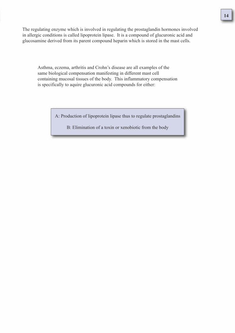
The regulating enzyme which is involved in regulating the prostaglandin hormones involved
in allergic conditions is called lipoprotein lipase. It is a compound of glucuronic acid and
glucosamine derived from its parent compound heparin which is stored in the mast cells.
Asthma, eczema, arthritis and Crohn’s disease are all examples of the
same biological compensation manifesting in different mast cell
containing mucosal tissues of the body. This inflammatory compensation
is specifically to aquire glucuronic acid compounds for either:
A: Production of lipoprotein lipase thus to regulate prostaglandins
B: Elimination of a toxin or xenobiotic from the body
15
A simple explanation of allergic conditions is inflammation. Allergic conditions are
manifestations of the same type of inflammation occurring in different mucosal tissues of
the body. The hypothesis here being presented is that the inflammation arises as a direct
result of a deficit of glucuronic acid [1.5] and glucosamine which one-at-the-same-time
are required to control prostaglandin hormones via the enzyme lipoprotein lipase.
Paper 1: Foreword
Paper Number: 1
Reference Number: 1.5
Information taken from:Glucuronidation of oxidized fatty acids and prostaglandins B1
and E2 by human hepatic and recombinant UDP glucuronosyltransferases
Joanna M. Little,* Mika Kurkela, † Julia Sonka,* Sirkku Jäntti, † Raimo Ketola, †
Stacie Bratton,* Moshe Finel, † and Anna Radominska-Pandya 1,
*Department of Biochemistry and Molecular Biology,* University of Arkansas for
Medical Sciences, Little Rock, AR; and Viikki Drug Discovery Technology Center,
† Faculty of Pharmacy, University of Helsinki, Helsinki, Finland
1To whom correspondence should be addressed.
e-mail: [email protected]
Manuscript received 15 March 2004 and in revised form 16 June 2004.
Published, JLR Papers in Press, July 1, 2004.
DOI 10.1194/jlr.M400103-JLR200
Copyright © 2004 by the American Society for
Biochemistry and Molecular Biology, Inc.
1694 Journal of Lipid Research Volume 45, 2004
Page 1696 Glucuronidation of lipid substrates
Glucuronidation of lipid substrates by human recombinant UGTs
Human hepatic and intestinal microsomes from a single donor, recombinant UGT isoforms from the 1A family
expressed in Sf9 cells as His tag proteins, and UGT2B7 expressed in HK293 cells were screened for their ability to
glucuronidate AA, 15-HETE, 20-HETE, PGE2, and PGB1; the results are summarized in Table 1 [1].
Glucuronidation assays using UGTs from the 1A family identified UGT1A1, 1A3, 1A4, 1A9, and 1A10 as being
active in the glucuronidation of both AA and 15-HETE [2]. AA, which is glucuronidated on the carboxyl function,
was the best substrate for all recombinant UGTs [3]. Interestingly, 20-HETE was not accepted as a substrate by
UGT1A9 or 1A10 under the experimental conditions used.
Both PGB1and PGE2 were glucuronidated by all recombinant UGTs under investigation, with the exception of
UGT1A4 [4]. PGB1, which contains only one hydroxyl group, as opposed to PGE2, which contains two hydroxyl
groups, one on the cyclic ring and one on the side chain, was a much better substrate for all recombinant UGTs [5].
UGT1A3 was able to glucuronidate both HETE derivatives; however, 20-HETE was the better substrate by a factor
of almost 2 [6].
Abbreviations: AA, arachidonic acid; GlcUA, glucuronic acid; HETE, hydroxyeicosatetraenoic acid; HI, human
intestine; HL, human liver; 13-HODE, 13-hydroxyoctadecadienoic acid; LA, linoleic acid; LCMS, liquid
chromatography-mass spectrometry; OFA, oxidized fatty acid; 13-OXO, 13-oxooctadecadienoic acid; PG,
prostaglandin; UDPGlcUA, UDP-glucuronic acid; UGT, UDP-glucuronosyltransferase; UGTs, UDP-
glucuronosyltransferases
16
Notes:
Human cells were screened for their ability to glucuronidate Arachidonic Acid (AA), 15-
Hydroxyeicosatetraenoic acid (15-HETE), 20-Hydroxyeicosatetraenoic acid (20-HETE),
Prostaglandin 2 (PG2) and Prostaglandin B1 (PGB1) [1].
Glucuronidation assays using UDP-glucuronosyltransferases (UGTs) from the 1A family identified
UDP-glucuronosyltransferase 1A1, 1A3, 1A4, 1A9 and 1A10 as being active in the glucuronication
of both Arachidonic Acid (AA) and 15-Hydroxyeicosatetraenoic acid [2].
Arachidonic acid, which is glucuronidated on the carboxyl function, was the best substrate for all
UDP-glucuronosyltransferases [3]. Both Prostaglandin B1 (PGB1) and Prostaglandin E2 (PGE2)
were glucuronidated by all UDP-glucuronosyltransferases under investigation except UDP-
glucuronosyltransferase 1A4 [4].
Prostaglandin B1 (PGB1) was a better substrate for all UDP-glucuronosyltransferases [5]. UDP-
glucuronosyltransferase 1A3 was able to glucuronidate both 15- and 20-Hydroxyeicosatetraenoic
acid, however 20-Hydroxyeicosatetraenoic acid was a better substrate by a factor of nearly 2 [6].
17
A simple explanation of allergic conditions is inflammation. Allergic conditions are
manifestations of the same type of inflammation occurring in different mucosal tissues of the
body. The hypothesis here being presented is that the inflammation arises as a direct result of a
deficit of glucuronic acid and glucosamine [1.6] which one-at-the-same-time are required to
control prostaglandin hormones via the enzyme lipoprotein lipase.
Paper 1: Foreword
Paper Number: 1
Reference Number: 1.6
Information taken from:
Journal of Lipid Research Volume 37,1996 p.693
Lipoprotein lipase and lipolysis: central roles in
lipoprotein metabolism and atherogenesis
Ira J. Goldberg
Department of Medicine, Columbia University
College of Physicians and Surgeons, 630 West 168th
Street, New York, NY 10032
Page 696; LPL hydrolysis of chylomicrons
LPL Hydrolysis of Chylomicrons
The observations that heparin released LPL into the bloodstream and that LPL binding to endothelial cells was
markedly decreased by HSPGdegrading enzymes [1] suggested that LPL is associated with HSPG [2]. HSPG
are members of the family of proteoglycans, negatively charged polysaccharides that are components of cell
membranes and the extracellular matrix, and are important in cell adhesion and growth.
The two major parts of the proteoglycan molecule are the glycosaminoglycans (GAG-carbohydrate chains) and
the core proteins [4]. The major classes of sulfated GAG, chondroitin sulfate (CS), dermatan sulfate (DS),
heparan sulfate (HS), and keratin sulfate, differ in their component sugars [5]. HS is a polymer composed of
repeating disaccharide units of a hexuronic acid (either glucuronic acid or iduronic acid) and glucosamine [6].
The glucosamine residues are either N-acetylated or N-sulfated and both hexuronate and glucosamine residues
may be O-sulfated in varying positions [7]. This leads to a highly variable structure that depends on tissue of
origin, molecular environment, and cell growth state [8]. Heparin differs from HS in extent of N-acetylation, N-
and O-sulfation, and content of iduronate [9].
Abbreviations: LPL, lipoprotein lipase; TG, triglyceride; HSPG, heparan sulfate proteoglycans; CS, chondriotin
sulfate; DS, dermatin sulfate; GAG, glycosaminoglycans; GPI, glycosylphosphotidylinositol; PIPLC,
phosphoinositol specific phospholipase C; LRP. LDL receptor-related protein; HTGL, hepatic triglyceride
lipase; CAD, coronary artery disease; LDL, low density lipoprotein; VLDL, very low density lipoprotein; HDL,
high density lipoprotein.
18
Notes:
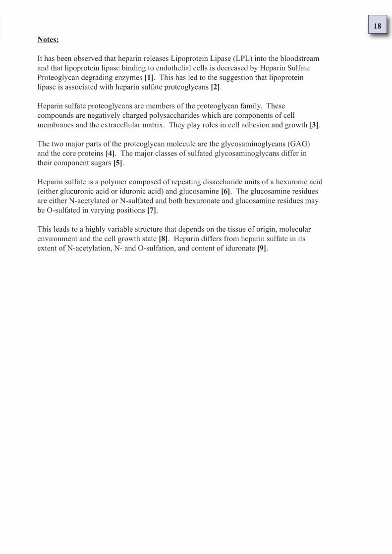
It has been observed that heparin releases Lipoprotein Lipase (LPL) into the bloodstream
and that lipoprotein lipase binding to endothelial cells is decreased by Heparin Sulfate
Proteoglycan degrading enzymes [1]. This has led to the suggestion that lipoprotein
lipase is associated with heparin sulfate proteoglycans [2].
Heparin sulfate proteoglycans are members of the proteoglycan family. These
compounds are negatively charged polysaccharides which are components of cell
membranes and the extracellular matrix. They play roles in cell adhesion and growth [3].
The two major parts of the proteoglycan molecule are the glycosaminoglycans (GAG)
and the core proteins [4]. The major classes of sulfated glycosaminoglycans differ in
their component sugars [5].
Heparin sulfate is a polymer composed of repeating disaccharide units of a hexuronic acid
(either glucuronic acid or iduronic acid) and glucosamine [6]. The glucosamine residues
are either N-acetylated or N-sulfated and both hexuronate and glucosamine residues may
be O-sulfated in varying positions [7].
This leads to a highly variable structure that depends on the tissue of origin, molecular
environment and the cell growth state [8]. Heparin differs from heparin sulfate in its
extent of N-acetylation, N- and O-sulfation, and content of iduronate [9].
19
Paper 1: Foreword
Paper Number: 1
Reference Number: 1.7
Information taken from:
Aggregate notes of paper 1
Glucuronic acid is a uronic acid derived from glucose
by oxidation. There are two enantiomers; D-glucuronic
acid is widely distributed in plants and animals.
Glucosamine is the name for the aminodeoxysugar 2-amino-2-deoxyglucopyranose.
There are two enantiomers; D-glucosamine occurs in combined form in chitin,
mucoproteins, and in mucopolysaccharides. It is one of the most abundant natural
monosaccharides.
In summary [1.7], glucuronic acid is drawn on as a resource for the
elimination of toxins and xenobiotics from the body thus infringing on
the production of lipoprotein lipase, hence resulting in a disorder of the
prostaglandin metabolism triggered by various extrinsic toxins.
-: Paper One - Foreword :-
- Glucuronic acid is a uronic acid derived from glucose by oxidation -
- Glucosamine is one of the most abundant natural monosaccharides -
- Glucuronides are the most endogenous conjugation agents found in the body -
- Asthma and atopy are here hypothesized as a deficit of lipoprotein lipase -
- Prostaglandins are lipid hormones which are metabolized via gluruconidation -
- Heparin releases lipoprotein lipase and made of glucuronic acid and glucosamine -
20
The xenobiotic compound contains functional groups often added via a phase I
reaction which act as ‘chemical handles’ for the attachment of a conjugating
agent. The conjugation product is usually less lipid soluble, more water
soluble, less toxic, and more easily eliminated than the parent compound.
Conjugation agents attached as part of phase II reactions include glucuronide,
sulfate, acetyl, methyl, glutathione and some amino acids. Phase II
conjugation reactions are usually rapid and if performed on phase I reaction
products, the rates of the latter are rate limiting for the overall process.
Glucuronides are the most common endogenous conjugating agents in the body. They react with
xenobiotics via uridine diphosphate glucuronic acid (UDPGA). This transfer is mediated by
glucuronyl transferase enzymes. Glucuronyl transferase enzymes occur in the endoplasmic
reticulum where hydroxylated phase I metabolites of lipophilic xenobiotics are produced.
Glucuronide conjugation products can be classified according to the element to which the glucuronide is
bound. The atoms to which the glucuronide most readily attaches are electron rich, usually oxygen, nitrogen or
sulfur. When the functional group through which a glucuronide conjugation occurs is a hydroxyl group (-OH),
an ether glucuronide is formed. A carboxylic acid group gives rise to an ester glucuronide. Glucuronides may
be formed via attachment directly to a nitrogen as the linking atom, as is found with aniline glucuronide.
Glucuronides may also form attachments through an intermediate oxygen atom.
N-hydroxyacetylaminoglucuronide is an example of this linkage which is of note as it is
more toxic than its parent compound, N-hydroxyacetylaminofluorene. This is of special
interest as a decrease in toxicity usually results from glucuronide conjugation.
Phase II reactions are also known as conjugation reactions because they involve the joining of
a substrate with unmodified xenobiotic compounds, xenobiotic compounds which have
undergone phase I reactions, and compounds that are not xenobiotic species. The substance
which binds to these species is refered to as an endogenous conjugating agent.
The carboxylic acid group in glucuronides is normally ionized at the pH of physiological
media which is a major reason for the water solubility of the conjugates. When the
conjugated compound (also called the aglycone) is of relatively low molecular mass, the
conjugate tends to be eliminated through the urine. With heavier conjugated compounds,
elimination occurs through the bile.
21
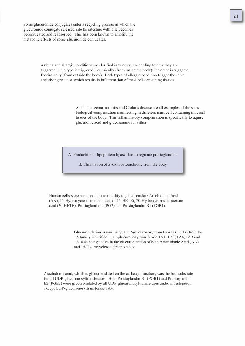
Some glucuronide conjugates enter a recycling process in which the
glucuronide conjugate released into he intestine with bile becomes
deconjugated and reabsorbed. This has been known to amplify the
metabolic effects of some glucuronide conjugates.
Asthma and allergic conditions are clasified in two ways according to how they are
triggered. One type is triggered Intrinsically (from inside the body); the other is triggered
Extrinsically (from outside the body). Both types of allergic condition trigger the same
underlying reaction which results in inflammation of mast cell containing tissues.
Asthma, eczema, arthritis and Crohn’s disease are all examples of the same
biological compensation manifesting in different mast cell containing mucosal
tissues of the body. This inflammatory compensation is specifically to aquire
glucuronic acid and glucosamine for either:
A: Production of lipoprotein lipase thus to regulate prostaglandins
B: Elimination of a toxin or xenobiotic from the body
Human cells were screened for their ability to glucuronidate Arachidonic Acid
(AA), 15-Hydroxyeicosatetraenoic acid (15-HETE), 20-Hydroxyeicosatetraenoic
acid (20-HETE), Prostaglandin 2 (PG2) and Prostaglandin B1 (PGB1).
Glucuronidation assays using UDP-glucuronosyltransferases (UGTs) from the
1A family identified UDP-glucuronosyltransferase 1A1, 1A3, 1A4, 1A9 and
1A10 as being active in the glucuronication of both Arachidonic Acid (AA)
and 15-Hydroxyeicosatetraenoic acid.
Arachidonic acid, which is glucuronidated on the carboxyl function, was the best substrate
for all UDP-glucuronosyltransferases. Both Prostaglandin B1 (PGB1) and Prostaglandin
E2 (PGE2) were glucuronidated by all UDP-glucuronosyltransferases under investigation
except UDP-glucuronosyltransferase 1A4.
22
It has been observed that heparin releases Lipoprotein Lipase (LPL) into the bloodstream
and that lipoprotein lipase binding to endothelial cells is decreased by Heparin Sulfate
Proteoglycan degrading enzymes. This has led to the suggestion that lipoprotein lipase is
associated with heparin sulfate proteoglycans.
Prostaglandin B1 (PGB1) was a better substrate for all UDP-glucuronosyltransferases. UDP-
glucuronosyltransferase 1A3 was able to glucuronidate both 15- and 20-Hydroxyeicosatetraenoic
acid, however 20-Hydroxyeicosatetraenoic acid was a better substrate by a factor of nearly 2.
This leads to a highly variable structure that depends on the tissue of origin, molecular
environment and the cell growth state. Heparin differs from heparin sulfate in its extent
of N-acetylation, N- and O-sulfation, and content of iduronate.
Heparin sulfate is a polymer composed of repeating disaccharide units of a hexuronic
acid (either glucuronic acid or iduronic acid) and glucosamine. The glucosamine
residues are either N-acetylated or N-sulfated and both hexuronate and glucosamine
residues may be O-sulfated in varying positions.
The two major parts of the proteoglycan molecule are the
glycosaminoglycans (GAG) and the core proteins. The major classes
of sulfated glycosaminoglycans differ in their component sugars.
Heparin sulfate proteoglycans are members of the proteoglycan family. These
compounds are negatively charged polysaccharides which are components of cell
membranes and the extracellular matrix. They play roles in cell adhesion and growth.
23
Bronchial Asthma and the Atopic Syndrome
Paper 2: Asthma and Atopy
Asthma [2.1] is considered an idiopathic disease which increasingly affects more
and more people around the world. The asthmatic experiences varying degrees of
difficulty in breathing during episodes refered to as asthma attacks. When an attack
is severe it is technically refered to as Status Asthmaticus [2.2].
Asthma is a syndrome which is part of a more general term -
Atopy [2.3]; this term must be defined before asthma gains its
context. Atopic conditions are conditions in which the subject
experiences a hypersensitive reaction known as an Allergy [2.4].
Allergies are symptoms of the response to stimulae known as Allergens [2.5];
these are substances that cause a hypersensitive reaction. There are many
different allergens which trigger an atopic response. Environmental and
psychological factors can also trigger the hypersensitive reaction
Other conditions which are manifestations of Atopy are Hay Fever [2.6],
Eczema [2.7], Dermatitis [2.8], Gastroenteritis [2.9], Diarrhoea [2.10],
and Crohn’s Disease [2.11]. All these atopic conditions have in common
the biochemical events known as Anaphylaxis [2.12].
A summary definition [2.13] is given before
the cells and tissues which are affected in
asthma and atopy are defined.
24
Paper 2: Asthma and Atopy
Asthma [2.1] is considered an idiopathic disease which increasingly affects more
and more people around the world. The asthmatic experiences varying degrees of
difficulty in breathing during episodes refered to as asthma attacks. When an attack
is severe it is technically refered to as Status Asthmaticus.
Paper Number: 2
Reference Number: 2.1
Information taken from:Oxford Reference Concise Medical Dictionary
Fourth Edition 1994
Oxford University Press
Concise definition of asthma
Page 53; asthma
Asthma n.
The condition of subjects with widespread narrowing of the bronchial airways, which changes in
severity over short periods of time (either spontaneously or under treatment) and leads to coughing,
wheezing and difficulty in breathing [1]. Bronchial asthma may be precipitated by exposure to one
or more of a wide range of stimuli, including allergens, drugs (such as aspirin and other Non
Steroidal Anti-Inflammatory Drugs and beta blockers), exertion, emotion, infections, and air
pollution [2].
The onset of asthma is usually early in life and in Atopic subjects (see Atopy) may be accompanied
by other manifestations of hypersensitivity, such as hay fever and dermatitis; however, the onset
may be delayed into adulthood or even middle or old age [3].
Treatment is with bronchodilators, with or without corticosteroids, usually administered via aerosol
or dry powder inhalers, or if the condition is more sever, via nebuliser. Oral corticosteroids are
reserved for patients who fail to respond adequately to these measures. Severe asthmatic attacks
may need large doses of oral corticosteroids (see status asthmaticus) [4].
Avoidance of known allergens, especially the house dust mite, allergens arising from domestic pets,
and food additives, will help reduce the frequency of attacks, as will the discouragement of smoking
[5]. Cardiac asthma occurs in left ventricular heart failure and must be distinguished from bronchial
asthma, as it is quite different. Adjective - asthmatic [6].
25
Notes:
In bronchial asthma there is a breakdown in the regular functioning of the lung
manifest as narrowing of the bronchial airways [1]. An attack is triggered when
contact is made with specific substances (such as aspirin and other non-steroidal
anti-inflammatory drugs, and beta blockers), exertion, emotion, infections and air
pollution [2].
Asthma can arise at any time of life. Asthma may be accompanied by various
manifestations of hypersensitivity such as hay fever and dermatitis [3]. It is
generally treated with bronchodilators, sometimes with corticosteroids [4].
Avoidance of allergens reduces the frequency of attacks. Allergens include house
dust mite, allergens deriving from domestic pets and food additives [5]. It should
be noted that bronchial asthma is different from cardiac asthma [6].
26
Paper 2: Asthma and Atopy
Asthma is considered an idiopathic disease which increasingly affects more and
more people around the world. The asthmatic experiences varying degrees of
difficulty in breathing during episodes refered to as asthma attacks. When an attack
is severe it is technically refered to as Status Asthmaticus [2.2].
Paper Number: 2
Reference Number: 2.2
Information taken from:Oxford Reference Concise Medical Dictionary
Fourth Edition 1994
Oxford University Press
Concise definition of status asthmaticus
Page 626; status asthmaticus
Status Asthmaticus
A severe attack of asthma, which often follows a period of poorly controlled asthma
[1]. Patients are distressed and very breathless and may die from respiratory failure if
not vigorously treated with inhaled oxygen, nebulized or intravenous bronchodilators,
and corticosteroid therapy [2]; Sedatives are absolutely contraindicated. These
patients need hospital care in an intensive care unit [3].
Notes:
A severe asthma attack generally progresses after a time of poor control [1]. and in
an acute case, complete functional breakdown of the lungs can lead to death if not
acutely treated with oxygen, nebulized or intravenous bronchodilators and
corticosteroid therapy [2]. Sedatives should on no account be used [3].
27
Paper 2: Asthma and Atopy
Asthma is a syndrome which is part of a more general term -
Atopy [2.3]; this term must be defined before asthma gains its
context. Atopic conditions are conditions in which the subject
experiences a hypersensitive reaction known as an Allergy.
Paper Number: 2
Reference Number: 2.3
Information taken from:Oxford Reference Concise Medical Dictionary
Fourth Edition 1994
Oxford University Press
Concise definition of atopy
Page 55; atopy
Atopy n.
A form of allergy in which there is a hereditary or constitutional tendency
to develop hypersensitivity reactions (e.g. hay fever, allergic asthma,
atopic eczema) in response to allergens (atopens). Adjective - Atopic [1].
Notes:
Asthma, eczema and hayfever are all different forms of the allergic reaction
which occur in hypersensitive people. Collectively these conditions are
termed as atopy, or atopic conditions [1].
28
Paper 2: Asthma and Atopy
Asthma is a syndrome which is part of a more general term -
Atopy; this term must be defined before asthma gains its context.
Atopic conditions are conditions in which the subject experiences
a hypersensitive reaction known as an Allergy [2.4].
Paper Number: 2
Reference Number: 2.4
Information taken from:Oxford Reference Concise Medical Dictionary
Fourth Edition 1994
Oxford University Press
Concise definition of allergy
Page 19; allergy
Allergy n.
A disorder in which the body becomes hypersensitive to particular antigens (called allergens),
which provoke characteristic symptoms whenever they are subsequently inhaled, ingasted,
injected or otherwise contacted [1].
Normally antibodies in the bloodstream destroy specific antigens without further trouble. In an
allergic person, however, the reaction of allergen with tissue bound antibody (reagin) also leads,
as a side effect, to cell damage, release of histamine and serotonin (5-Hydroxytryptamine),
inflammation, and all the symptoms of the particular allergy [2].
Different allergies afflict different tissues and may have either local or general effects, varying
from asthma and hay fever to severe dermatitis and gastroenteritis or extremely serious shock
(see anaphylaxis), Adjective- allergic [3].
Notes:
An allergy is a hypersensitive reaction the body takes to a particular substance [1]. Normally
a healthy body does not react to these substances, however an atopic individuals body does
react by causing a form of inflammation [2]. Different allergies cause inflammation in
different tissues [3].
29
Allergies are symptoms of the response to stimulae known as Allergens [2.5];
these are substances that cause a hypersensitive reaction. There are many
different allergens which trigger an atopic response. Environmental and
psychological factors can also trigger the hypersensitive reaction
Paper 2: Asthma and Atopy
Paper Number: 2
Reference Number: 2.5
Information taken from:Oxford Reference Concise Medical Dictionary
Fourth Edition 1994
Oxford University Press
Concise definition of allergen
Page 19; allergen
Allergen n.
Any antigen that causes allergy in a hypersensitive person. Allergens are
diverse and affect different tissues and organs [1]. Pollens, fur, feathers,
mould, and dust may cause hay fever; house mites have been implicated in
some forms of asthma;
drugs, dyes, cosmetics, and a host of other chemicals can cause rashes and
dermatitis; some food allergies may cause diarrhoea or constipation or
simulate acute bacterial food poisoning [2].
When a patients allergen has been identified (see patch test), it may be
possible to alleviate or prevent allergic attacks. Adjective - allergenic [3].
Notes:
An allergen describes any substance that triggers an allergic reaction in a hypersensitive
person. Allergens may affect different tissues and organs [1]. Pollens, fur, feathers, mould,
dust, house mites, drugs, dyes, cosmetics, food allergies and other chemicals are amongst
known allergens [2]. Allergies to substances can be identified by use of a patch test [3].
30
Paper 2: Asthma and Atopy
Other conditions which are manifestations of Atopy are Hay Fever [2.6],
Eczema, Dermatitis, Gastroenteritis, Diarrhoea, and Crohn’s Disease. All
these atopic conditions have in common the biochemical events known as
Anaphylaxis.
Paper Number: 2
Reference Number: 2.6
Information taken from:Oxford Reference Concise Medical Dictionary
Fourth Edition 1994
Oxford University Press
Concise definition of hay fever
Page 295; hay fever
Hay Fever
A form of allergy due to the pollen of grasses, trees, and other plants,
characterised by inflammation of the membrane lining the nose and
sometimes of the conjunctiva (vernal conjunctivitis) [1].
The symptoms of sneezing, running or blocked nose, and watering eyes are
due to histamine release and often respond to treament with antihistamines
[2]. If the allergen is identified, it may be possible to undertake
desensitisation. Medical name: allergic rhinitis.
Notes:
Hay fever is the allergy which manifests as inflammation of themembrane of the nose and sometimes of the conjunctiva [1].
Hay fever often responds to treatment with antihistamines [2].
31
Paper 2: Asthma and Atopy
Other conditions which are manifestations of Atopy are Hay Fever,
Eczema [2.7], Dermatitis, Gastroenteritis, Diarrhoea, and Crohn’s
Disease. All these atopic conditions have in common the biochemical
events known as Anaphylaxis.
Paper Number: 2
Reference Number: 2.7
Information taken from:Oxford Reference Concise Medical Dictionary
Fourth Edition 1994
Oxford University Press
Concise definition of eczema
Page 206; eczema
Eczema n.
A common itchy skin disease characterized by reddening (erythema)
and vesicle formation, which may lead to weeping and crusting [1].
It is endogenous or constitutional, i.e. ouside agents do not play a primary role
(compare dermatitis), but in some contexts, the terms ‘dermatitis’ and ‘eczema’ are
used interchangeably [2]. There are five main types:
(1) Atopic eczema, which affects up to 20% of the population and is associated with
asthma and hay fever [3]; (2) Seborrhoeic eczema (or dermatitis), involves the scalp,
eyelids, nose, and lips and is associated with the presence of pityrosporum yeasts; (3)
Discoid (or nummular) eczema, which is characterized by coin-shaped lesions and
occurs only in adults; (4) Pompholyx, affecting the hands and feet; (5) Gravitational
(or stasis) eczema, associated with poor venous circulation and incorrectly known as
varicose eczema.
Treatment of eczema is with topic or systemic corticosteroids but emolients are very
important, especially in treating mild cases. Adjective - eczematous [4].
Notes:
Eczema is an itchy skin disease characterized by reddening and vesicle formation which
may lead to weeping and crusting [1]. It is an endogenous condition [2]. Of all
eczema, an estimated 20% is atopic and is associated with asthma and hay fever [3].
Treatment is topic or with corticosteroids [4].
32
Paper 2: Asthma and Atopy
Other conditions which are manifestations of Atopy are Hay Fever,
Eczema, Dermatitis [2.8], Gastroenteritis, Diarrhoea, and Crohn’s
Disease. All these atopic conditions have in common the biochemical
events known as Anaphylaxis.
Paper Number: 2
Reference Number: 2.8
Information taken from:Oxford Reference Concise Medical Dictionary
Fourth Edition 1994
Oxford University Press
Concise definition of dermatitis
Page 177; dermatitis
Dermatitis n.
An inflammatory condition of the skin caused by outside agents (compare eczema, an endogenous
disease in which such agents do not play a primary role) [1]. Primary irritant dermatitis may occur
in anyone who has sufficient contact with such irritants as acids, alkalis, solvents, and (especially)
detergents. It is the commonest cause of occupational dermatitis in hairdressers, nurses, cooks, etc.
(see also napkin rash).
In allergic contact dermatitis, skin changes resembling those of eczema develop as a delayed
reaction with a particular allergen, which may be present at low concentrations. The commonest
example in women is nickel dermatitis from jewellery, jeans studs, etc.; in men chromium
dermatitis is relatively common. Treatment of dermatitis depends upon the cause [2].
Dermatitis herpetiformis is an uncommon very itchy rash with symmetrical blistering, especially on
the knees, elbows, buttocks, and shoulders. It is associated with gluten sensitivity and responds
well to treatment with dapsone.
Notes:
Dermatitis is extrinsically triggered inflammation of the
skin. Strongly associated with eczema [1], the skin
changes in dermatitis resemble those seen in eczema [2].
33
Paper 2: Asthma and Atopy
Other conditions which are manifestations of Atopy are Hay Fever,
Eczema, Dermatitis, Gastroenteritis [2.9], Diarrhoea, and Crohn’s
Disease. All these atopic conditions have in common the biochemical
events known as Anaphylaxis.
Paper Number: 2
Reference Number: 2.9
Information taken from:Oxford Reference Concise Medical Dictionary
Fourth Edition 1994
Oxford University Press
Concise definition of gastroenteritis
Page 266; gastroenteritis
Gastroenteritis n.
Inflammation of the stomach and intestine. It is usually due to acute infection by
viruses or bacteria or to food-poisoning toxins and causes vomiting and diarrhoea.
The illness usually last 3 - 5 days. Fluid loss is sometimes severe especially in
infants, and intravenous fluid replacement may be necessary [1].
Notes:
Gastroenteritis is an example of the allergic reaction occurring in the stomach
and intestine. The inflammation is usually triggered by infection or to toxins.
The result is diarrhoea and vomiting [1].
34
Paper 2: Asthma and Atopy
Other conditions which are manifestations of Atopy are Hay Fever,
Eczema, Dermatitis, Gastroenteritis, Diarrhoea [2.10], and Crohn’s
Disease. All these atopic conditions have in common the biochemical
events known as Anaphylaxis.
Paper Number: 2
Reference Number: 2.10
Information taken from:Oxford Reference Concise Medical Dictionary
Fourth Edition 1994
Oxford University Press
Concise definition of diarrhoea
Page 182; diarrhoea
Diarrhoea n.
Frequent bowel evacuation or the passage of abnormally soft or liquid faeces. It may be
caused by intestinal infections, other forms of intestinal inflammation (such as colitis or
Crohn’s disease), malabsorption, anxiety, and the irritable bowel syndrome. Severe or
prolonged diarrhoea may lead to excess losses of fluid, salts, and nutrients in the faeces [1].
Notes:
Diarrhoea as a result of intestinal inflammation leads to loss of fluid,
salts and malabsorption of nutrients. Diarrhoea is a factor in Crohn’s
disease and inflammation of the colon (colitis) [1].
35
Paper 2: Asthma and Atopy
Other conditions which are manifestations of Atopy are Hay Fever, Eczema,
Dermatitis, Gastroenteritis, Diarrhoea, and Crohn’s Disease [2.11]. All these
atopic conditions have in common the biochemical events known as Anaphylaxis.
Paper Number: 2
Reference Number: 2.11
Information taken from:Oxford Reference Concise Medical Dictionary
Fourth Edition 1994
Oxford University Press
Concise definition of Crohn’s disease
Page 157; Crohn’s disease
Crohn’s Disease
A condition in which segments of the alimentary tract become inflammed, thickened, and ulcerated.
it usually affects the terminal part of the ileum; its acute form (acute ileitis) may mimic appendicitis.
Chronic disease often causes partial obstruction of the intestine, leading to pain, diarrhoea, and
malabsorption [1].
Fistulae around the anus, between adjacent loops of intestine, or from intestine to skin, bladder, etc.,
are characteristic complications. The cause is unknown. Treatment includes rest, corticosteroids,
immunosuppressive drugs, antibiotics, dietary modification, or (in some cases) surgical removal of
the affected part of the intestine. Alternative names: Regional enteritis, regional ileitis [2].
Notes:
Crohn’s disease is the term used to describe inflammation of various parts of
the digestive tract [1]. Treatments include corticosteroids, immunosuppressive
drugs, antibiotics and dietary modification [2].
36
Paper 2: Asthma and Atopy
Other conditions which are manifestations of Atopy are Hay Fever, Eczema,
Dermatitis, Gastroenteritis, Diarrhoea, and Crohn’s Disease. All these atopic
conditions have in common the biochemical events known as Anaphylaxis [2.12].
Paper Number: 2
Reference Number: 2.12
Information taken from:Oxford Reference Concise Medical Dictionary
Fourth Edition 1994
Oxford University Press
Concise definition of anaphylaxis
Page 30; anaphylaxis
Anaphylaxis n.
An abnormal reaction to a particular antigen, in which histamine is released from tissues and
causes either local or widespread symptoms. An allergic attack (see allergy) is an example of
localized anaphylaxis [1].
Rarer, but much more serious is anaphylactic shock; an extreme and generalized allergic reaction
in which widespread release of histamine causes swelling (oedema), constriction of the
bronchioles, heart failure, circulatory collapse, and sometimes death. Adjective - anaphylactic
Notes:
Anaphylaxis is an abnormal reaction to a particular antigen. An allergic attack (allergy)
is an example of localized anaphylaxis [1]. Anaphylactic shock is the term used for a
generalized allergic reaction which strongly affects various tissues and systems of the
body. During anaphylactic shock there is constriction of the bronchioles, heart failure,
circulatory collapse, and sometimes death [2].
37
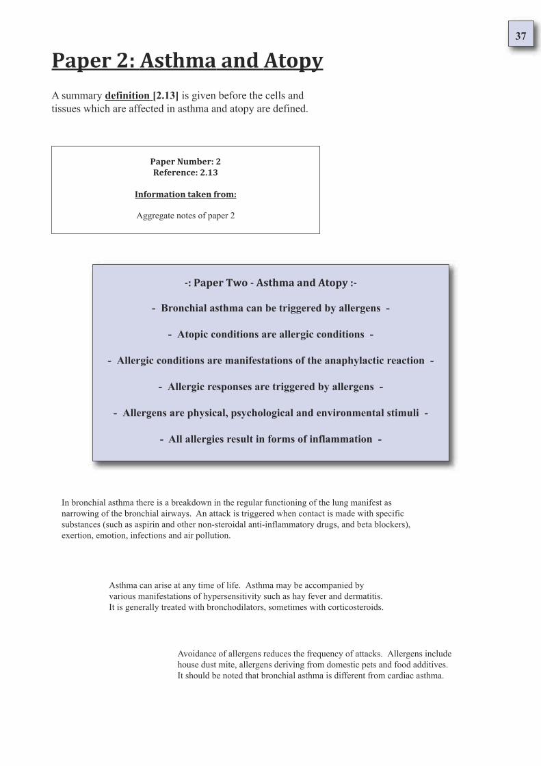
Paper 2: Asthma and Atopy
A summary definition [2.13] is given before the cells and
tissues which are affected in asthma and atopy are defined.
Paper Number: 2
Reference: 2.13
Information taken from:
Aggregate notes of paper 2
-: Paper Two - Asthma and Atopy :-
- Bronchial asthma can be triggered by allergens -
- Atopic conditions are allergic conditions -
- Allergic conditions are manifestations of the anaphylactic reaction -
- Allergic responses are triggered by allergens -
- Allergens are physical, psychological and environmental stimuli -
- All allergies result in forms of inflammation -
In bronchial asthma there is a breakdown in the regular functioning of the lung manifest as
narrowing of the bronchial airways. An attack is triggered when contact is made with specific
substances (such as aspirin and other non-steroidal anti-inflammatory drugs, and beta blockers),
exertion, emotion, infections and air pollution.
Asthma can arise at any time of life. Asthma may be accompanied by
various manifestations of hypersensitivity such as hay fever and dermatitis.
It is generally treated with bronchodilators, sometimes with corticosteroids.
Avoidance of allergens reduces the frequency of attacks. Allergens include
house dust mite, allergens deriving from domestic pets and food additives.
It should be noted that bronchial asthma is different from cardiac asthma.
38
A severe asthma attack generally progresses after a time of poor control. and
in an acute case, complete functional breakdown of the lungs can lead to death
if not acutely treated with oxygen, nebulized or intravenous bronchodilators
and corticosteroid therapy. Sedatives should on no account be used.
Asthma, eczema and hayfever are all different forms of the allergic
reaction which occur in hypersensitive people. Collectively these
conditions are termed as atopy, or atopic conditions.
An allergy is a hypersensitive reaction the body takes to a particular substance.
Normally a healthy body does not react to these substances, however an atopic
individuals body does react by causing a form of inflammation. Different
allergies cause inflammation in different tissues.
An allergen describes any substance that triggers an allergic reaction in a hypersensitive
person. Allergens may affect different tissues and organs. Pollens, fur, feathers, mould,
dust, house mites, drugs, dyes, cosmetics, food allergies and other chemicals are amongst
known allergens. Allergies to substances can be identified by use of a patch test.
Hay fever is the allergy which manifests as inflammation of the
membrane of the nose and sometimes of the conjunctiva. Hay
fever often responds to treatment with antihistamines.
Eczema is an itchy skin disease characterized by reddening and vesicle
formation which may lead to weeping and crusting. It is an endogenous
condition. Of all eczema, an estimated 20% is atopic and is associated
with asthma and hay fever. Treatment is topic or with corticosteroids.
Dermatitis is extrinsically triggered inflammation of
the skin. Strongly associated with eczema, the skin
changes in dermatitis resemble those seen in eczema.
39
Gastroenteritis is an example of the allergic reaction occurring in
the stomach and intestine. The inflammation is usually triggered
by infection or to toxins. The result is diarrhoea and vomiting.
Diarrhoea as a result of intestinal inflammation leads to loss of
fluid, salts and malabsorption of nutrients. Diarrhoea is a factor
in Crohn’s disease and inflammation of the colon (colitis).
Crohn’s disease is the term used to describe inflammation of various
parts of the digestive tract. Treatments include corticosteroids,
immunosuppressive drugs, antibiotics and dietary modification.
Anaphylaxis is an abnormal reaction to a particular antigen. An allergic attack (allergy) is an
example of localized anaphylaxis. Anaphylactic shock is the term used for a generalized allergic
reaction which strongly affects various tissues and systems of the body. During anaphylactic shock
there is constriction of the bronchioles, heart failure, circulatory collapse, and sometimes death.
40
Bronchial Asthma and the Atopic Syndrome
Paper 3: Asthma and Inflammation
The definition of Asthma [3.1] starts with the
immune response. Thus the scenario begins :-
The subject comes into contact with an allergen;
There then occcrs an immunological response to the antigenic substance [3.2]
which involves the function of the immunoglobulins [3.3]. Primarily it is
immunoglobulin E which is involved in atopy, having the effect of sponsoring
anaphylaxis in the cells and tissues.
The body synthesises immunoglobulin E and releases it into the
circulation where it binds to cell surfaces, especially to mast cells [3.4]
and circulating basophils (also called circulating mast cells).
When an allergen come into proximity with the cell cultures it combines
with the cell-bound immunoglobulin E. The result of this is anaphylaxis
(either localized or general) which consequently causes inflammation.
The compounds and substances which are released and activated during
inflammation have the clinical definition of anaphylaxis [3.5].
The summation [3.6] of events is that the
immune system causes the mast cells to release
their contents into the surrounding tissues.
41
Paper 3: Asthma and Inflammation
The definition of Asthma [3.1] starts with the
immune response. Thus the scenario begins :-
The subject comes into contact with an allergen;
Paper Number: 3
Reference Number: 3.1
Information taken from:The Nutrition and Health Dictionary
Copyright 1995
Chapman & Hall
Definition of asthma
Page 33; asthma
Asthma
A respiratory problem whose chief feature is laboured or difficult breathing. Often accompanied
by characteristic wheezing or whistling sounds. The whistling or wheezing sounds of asthma are
loudest when breathing air out.
The symptoms of asthma are caused by changes in the respiratory system, the system of
passageways that carries air from the mouth into the lungs. In asthma, the small air passageways
go into spasms, narrowing the width of the tubes and thus making the passage of air in and out of
the lungs more difficult [1].
In addition to muscle spasm, there is an outpouring of mucus into the small air passages, further
obstructing the flow of air [2]. Finally, the mucosa become inflammed and swollen, thus
narrowing the air passageways even further, much as an accumulation of rust on the inner surface
of a pipe would paritally obstruct the flow of water through a pipe [3].
All these changes, the spasms, the swelling, and the out pouring of mucus, occur together,
affecting to a greater or lesser degree almost all of the air passages. The resistance to the flow of
air, particularly during expiration, or breathing out, is significantly increased, and the individual
must work harder to move air in and out of the lungs, which results in a whistling sound. Asthma
is considered to be the result of an allergy [4].
Notes:
In asthma the muscles of the air passageways go into spasm [1], there occurs an
exodus of mucus into the lungs [2], the mucosa become inflammed and swollen [3],
and the resistance of air flow is significantly increased particularly whilst breathing
out. Asthma is considered to be the result of an allergy [4].
42
Paper 3: Asthma and Inflammation
There then occcrs an immunological response to the antigenic substance [3.2]
which involves the function of the immunoglobulins. Primarily it is
immunoglobulin E which is involved in atopy, having the effect of sponsoring
anaphylaxis in the cells and tissues.
Paper Number: 3
Reference Number: 3.2
Information taken from:Basic Pharmacology
Basic Principles in Therapeutics
Second edition 1978
Kenneth L. Melmon, M.D.
Howard F. Morrelli, M. D.
The Classic Drug Reaction:
The immunlogical response to an antigenic substance
Page 957 - 958
The Immunological Response to an Antigenic Substance
The physician who never uses penicillin on any patient because he has “seen” an anaphylactic reaction that
appears to create unnecessary suffering is like the physician who uses it casually because he has “never had”
any problems with it.
The antibodies produced by penicillin appear specific for different aspects of the chemistry of the penicillin;
i.e. at least three types of immunoglobulin E antibody can be made. One is to the benzylpenicilloye moiety
(BPO), a frequent antigen and so-called “major” determinant; one is to penicilloate, and one is to the other
chemicals related to penicillin that are often responsible for antigenicity, i.e., the so called “minor
determinants” (Tsuji et al., 1974; Yamana et al., 1975) [1].
Although each of these antibodies can be distinctly different molecules, the antibody-combining site
recognizes the whole molecule and can, in part be carrier specific. Thus it is not surprising to find cross
reactivity to a variety of the chemicals that have the penicillin structure in common (Adkinson et al., 1971) [2].
Immediate hypersensitivity is clearly caused by the immunoglobulin E (IgE) molecule; there may be some
types of immediate reactions that might be mediated by Immunoglobulin G. Some combination of
Immunoglobulin M plus other antibodies may account for late hypersensitivity reactions and Immunoglobulin
G may contribute to the accelerated response. The skin tests we will discuss usually detect tissue-fixed (skin)
Immunoglobulin E and thus have proven useful in screening for those patients who are not likely to have
immediate types of reactions to either the major or minor antigenic determinants of the penicillin molecule.
Degradation of the penicillin molecule to a reactive proimmunogen can proceed without enzymatic
intervention. This event with penicillin is in marked contrast to most other drugs that must undergo enzymatic
degradation (usually in the liver) before they become antigenic or before they conjugate with endogenous
protein to form complete antigens [3].
43
This difference in active versus spontaneous degradation may be the reason that hypersensitivity reactions
to penicillin do not express themselves in the liver. That is, INH, for instance, is degraded in the liver
when the antigen or reactive metabolic intermediate may express its effects as clinically important
hepatitis.
The skin-sensitizing (IgE) antibodies in penicillin allergy are most commonly developed to the penicilloyl
metabolite (“major” determinant). IgE is thought to be responsible for anaphylactic reactions, and may
also be produced in response to the unaltered drug or to another metabolite (e.g. penaldate, penicilloate,
or penicillamine (Levine, 1966; Turk and Baker, 1968) [4].
IgM and IgG antibodies may be produced after exposure of patients to penicillin and are usually specific
for the penicilloyl metabolite (“major” determinant). The “minor” detertminants (unaltered drug or other
drug metabolites) may be responsible for disastrous allergic reactions, and the nomenclature (major and
minor determinants) is misleading. Better terms might be “frequent” and “infrequent” determinants of
antigenicity. There are many possible determinants of antigenicity after administration of penicilin.
Although IgE antibodies may cause immediate anaphylactic reactions, the significance of IgM and IgG
antibodies is less clear. IgG antibodies may act as blocking antibodies to prevent or modify the course of
reactions in patients who are skin-test-sensitive to antigens but who develop skin rashes rather than
anaphylaxis on exposure to the allergen. In some patients, the concentration of IgG falls during the acute
allergic reaction and rises during the recovery period.
Hemaglutinating antibodies (IgG) are usually specific for the penicilloyl determinant, but these antibodies
may be found in 60 to 100% of populations, if sensitive methods for their detection are used. Although
petients with a history of allergy have higher concentrations of IgG than normals, it is not known if these
antibodies are responsible for most reactions [5].
In addition, lymphocytes have been “sensitized” to components of the penicillin’s (David, 1973,
Gimenez-Camarasa, 1975; Reidenberg and caccese, 1975). These lymphocytes, on contact with the
antigen, release a substance that causes blood monocytes to migrate across endothelial linings of vessels,
become converted to macrophages, and destroy tissues by release of lysosomal enzymes [6].
This hypothesis may explain those allergic reactions to pencillin that stimulate serum sicknesses. The
reader is referred to Chapter 13 where a detailed discussion of the mechanisms by which humoral or cell
mediated immune-generated events is presented.
Notes:
During an allergic immune response the body produces antibodies known as
immunoglobulins [1]. These antibodies react with molecules that have a
similar chemical structure [2]. Some chemicals need to be changed before
being recognized by the body as foreign matter (allergens) [3].
Immunoglobulin E is responsible for the anaphylactic reaction and can also be
produced in response the unaltered drug or to another metabolite [4].
It has been observed that Immunoglobulin G concentrations fall during the
allergic reaction and rise after. Immunoglobulin G levels are higher in allergic
patients [5]. Lymphocytes are immune cells that on exposure to allergens
encourage the production of macrophages which digest tissues [6].
44
Paper 3: Asthma and Inflammation
There then occcrs an immunological response to the antigenic substance which
involves the function of the immunoglobulins [3.3]. Primarily it is
immunoglobulin E which is involved in atopy, having the effect of sponsoring
anaphylaxis in the cells and tissues.
Paper Number: 3
Reference Number: 3.3
Information taken from:Joan F. Zilva
P.R. Pannall
Clinical Chemistry in Diagnosis and Treatment
Third edition 1979
Lloyd - Luke (Medical Books) Ltd
Function of the Immunoglobulins
Page 317 - 319 Immunoglobulin E
Function Of The Immunoglobulins
The immune response mechanism of the body consists of a cellular and humoral component.
Although we are concerned here only with the humoral component - the immunoglobulins - the
student should remember that both are necessary: He should consult a textbook of immunology
for details of the cellular mechanism. The specific functions of each class of immunoglobulin
will be discussed briefly, and followed by an outline of changes in disease [1].
Immunoglobulin G (MW: 160 000);
IgG accounts for about 75 per cent of circulating immunoglobulins and contains most of the
normal plasma antibodies. It is a relatively small molecule, and is present in very low
concentration throughout the extracellular compartment: Its most important function is
stimulated by soluble antigens such as bacterial toxins.
IgG deficiency is characterised by recurrent pyogenic infections of tissue spaces by toxin-
producing organisms such as staphylococci and streptococci: pulmonary and subcutaneous
infections are common. IgG can bind complement and cross the placental barrier.
In the first few months of life endogenous IgG levels are very low. Maternal IgG which has
been transported across the placenta provides antibody cover during this period. Adult
concentrations of this class of protein are not reached before the age of 3 to 5 years [2].
Immunoglobulin E (MW: 200 000);
IgE is synthesised by plasma cells beneath the mucosae of the gastrointestinal and respiratory
tracts and by those in the lymphoid tissue of the nasopharynx [3]. It is present in nasal and
bronchial secretions.
45
Circulating IgE is rapidly bound to cell surfaces, particularly to those of mast cells and circulating
basophils, and plasma levels are therefore very low [4]. Combination of antigen with this cell-bound
antibody results in the cells releasing mediators and accounts for immediate hypersensitivity reactions
such as occur in hay fever [5].
Desensitisation therapy of allergic disorders aims at stimulating production of circulating IgG against
the offending antigen, to prevent it reaching cell-bound IgE. - Raised concentrations are found in several
diseases with an allergic component such as some cases of eczema, asthma and parasitic infestations [6].
Notes:
The total immune response consists of both cellular and humoral components [1].
Immunoglobulin G is stimulated by soluble antigens such as bacterial toxins,
deficiency of this antibody is characterised by pus forming (pyogenic) infections [2].
Immunoglobulin E is the antibody made by the plasma cells of the alimentary tract,
the respiratory tract and by the lymphoid tissue of the nasopharynx [3]. Present in
nasal and bronchial secretions, circulating immunoglobulin E rapidly binds to cell
surfaces - especially of mast cells and circulating basophils [4].
Antigen combines with cell-bound Immunoglobulin E - this causes the cells to
release their contents and an immediate hypersensitivity reaction (anaphylaxis)
ensues [5]. Raised levels of IgE are found in allergic conditions such as asthma,
eczema and parasitic infestations [6].
46
Paper 3: Asthma and Inflammation
The body synthesises immunoglobulin E and reseasles it into the
circulation where it binds to cell surfaces, especially to mast cells [3.4]
and circulating basophils (also called circulating mast cells).
Paper Number: 3
Reference Number: 3.4
Information taken from:Kenneth L. Melmon, M.D.
Howard F. Morrelli, M.D.
Clinical Pharmacology
Basic Principles in Therapeutics
Second edition 1978
Bailliere, Tindall london
Cellular mediators of inflammation
Page 671 Mast cells
Mast Cells
Mast cells are mainly found in loose connective tissue, frequently near vascular channels. Their
most distinguishing features are their membrane-bound, spheroid, basophilic secretory granules,
which may be so numerous that other cytoplasmic structures are obscured [1].
These granules contain large amounts of heparin and other mucopolysaccharides, histamine, and
several proteolytic and esterolytic enzyme systems. Serotonin is present in the mast cells of some
species, man being a notable exception [2].
Much is known regarding the structure and composition of mast cells, but their physiologic function
remains unidentified. However it seems certain that the mast cell and the basophil (circulating mast
cells) contribute to the development of an inflammatory process (Lewis et al., 1975). Changes in
mast cell density and mast cell granules are thought to be characteristic of many inflammatory
lesions. Mast cell preparations (either in vitro or in vivo during anaphylaxis) have been used as
experimental models of inflammation [3].
The mast cell is readily degranulated (see previous section on histamine), and the granule-bound
chemical substances are released into extracellular fluid [4]. Histamine and probably heparin can
have important influences on an inflammatory process, but much less is known about the granule
esterase's and protease's that are also released from the granules of the mast cell and basophil
(Benditt, 1968; Lewis et al., 1975).
These enzymes may alter the course of inflammation by interactions with other cells or substances
to release, activate, or destroy kinins, complement, and blood coagulation factors, to alter
membranes of other cells such as granulocytes so that additional intracellular constituents are
released, or to alter the structure and characteristics of adjacent connective tissue [5].
47
Notes:
Mast cells are mainly found in loose connective tissue often near vascular channels. They are
distinguishable by copious amounts of secretory granules on the membrane [1].
The granules contain large amounts of heparin and mucopolysaccharides, histamine and
several proteolytic and esterolytic enzyme systems which catalyse the splitting of esters into
constituent alcohols and acids, and proteins into smaller peptides and amino acids [2].
Changes in mast cell density and mast cell granules are characteristic of many inflammatory
lesions. The mast cells are intimately involved in the inflammatory process and are used as
models of inflammation in anaphylaxis [3]. During the inflammatory process the mast cell
degranulates (excretes its contents) into the extracellular fluid [4].
Histamine and heparin are both important factors in the inflammatory process. The digestive
enzyme systems that mast cells release interact with kinins, complement, blood coagulation
factors, polymorphonuclear leukocyte membranes and the structure of connective tissue [5].
48
Paper 3: Asthma and Inflammation
When an allergen come into proximity with the cell cultures it combines
with the cell-bound immunoglobulin E. The result of this is anaphylaxis
(either localized or general) which consequently causes inflammation.
The compounds and substances which are released and activated during
inflammation have the clinical definition of anaphylaxis [3.5].
Paper Number: 3
Reference Number: 3.5
Information taken from:Kenneth L. Melmon, M.D.
Howard F. Morrelli, M.D.
Clinical Pharmacology
Basic Principles in Therapeutics
Second edition 1978
Bailliere, Tindall london
Clinical definition of anaphylaxis
Page 689 - 690 Inflammation
Anaphylaxis
Release of mediators of inflammation with systemic effects. In man, the signs and symptoms of
anaphylaxis are bronchial muscle contraction, hyperperistalsis, widespread increased capillary
permeability and vasodilitation with peripheral vascular collapse due to the release of multiple
chemicals during antigen - antibody interaction. Mediators that most likely participate in human
disease include histamine, slow reacting substance A, and bradykinin (Kaplan et al., 1971) [1].
Although anaphylaxis shares many common pathogenetic factors with urticaria, life-threatening
respiratory involvement and/or severe hypotension and subsequent cardiac abnormalities may occur
at any time during the course of anaphylaxis. Major determinants of the severity of the reaction
appear to be the nature, rate, and magnitude of the antigen - antibody reaction [2].
Notes:
Anaphylaxis is the release of mediators of inflammation with systemic effects. Signs and
symptoms are bronchial muscle contraction, hyperperistalsis, widespread increased capillary
permeability and vasodilation with peripheral vascular collapse due to antibody-to-antibody
interaction [1].
General anaphylaxis shares many common factors with urticaria (nettle rash) however
anaphylaxis is considered life threatening due to its potent effects on respiration, blood
pressure and the heart [2].
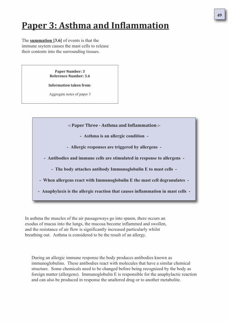
49
The summation [3.6] of events is that the
immune ssytem causes the mast cells to release
their contents into the surrounding tissues.
Paper 3: Asthma and Inflammation
Paper Number: 3
Reference Number: 3.6
Information taken from:
Aggregate notes of paper 3
-: Paper Three - Asthma and Inflammation :-
- Asthma is an allergic condition -
- Allergic responses are triggered by allergens -
- Antibodies and immune cells are stimulated in response to allergens -
- The body attaches antibody Immunoglobulin E to mast cells -
- When allergens react with Immunoglobulin E the mast cell degranulates -
- Anaphylaxis is the allergic reaction that causes inflammation in mast cells -
In asthma the muscles of the air passageways go into spasm, there occurs an
exodus of mucus into the lungs, the mucosa become inflammed and swollen,
and the resistance of air flow is significantly increased particularly whilst
breathing out. Asthma is considered to be the result of an allergy.
During an allergic immune response the body produces antibodies known as
immunoglobulins. These antibodies react with molecules that have a similar chemical
structure. Some chemicals need to be changed before being recognized by the body as
foreign matter (allergens). Immunoglobulin E is responsible for the anaphylactic reaction
and can also be produced in response the unaltered drug or to another metabolite.
50
The total immune response consists of both cellular and humoral components.
Immunoglobulin G is stimulated by soluble antigens such as bacterial toxins,
deficiency of this antibody is characterised by pus forming (pyogenic) infections.
Mast cells are mainly found in loose connective tissue
often near vascular channels. They are distinguishable by
copious amounts of secretory granules on the membrane.
It has been observed that Immunoglobulin G concentrations fall during the allergic reaction and
rise after. Immunoglobulin G levels are higher in allergic patients. Lymphocytes are immune cells
that on exposure to allergens encourage the production of macrophages which digest tissues.
Changes in mast cell density and mast cell granules are characteristic of many
inflammatory lesions. The mast cells are intimately involved in the inflammatory process
and are used as models of inflammation in anaphylaxis. During the inflammatory process
the mast cell degranulates (excretes its contents) into the extracellular fluid.
The granules contain large amounts of heparin and mucopolysaccharides,
histamine and several proteolytic and esterolytic enzyme systems which
catalyse the splitting of esters into constituent alcohols and acids, and
proteins into smaller peptides and amino acids.
Antigen combines with cell-bound Immunoglobulin E - this causes the
cells to release their contents and an immediate hypersensitivity
reaction (anaphylaxis) ensues. Raised levels of IgE are found in
allergic conditions such as asthma, eczema and parasitic infestations.
Immunoglobulin E is the antibody made by the plasma cells of the alimentary
tract, the respiratory tract and by the lymphoid tissue of the nasopharynx. Present
in nasal and bronchial secretions, circulating immunoglobulin E rapidly binds to
cell surfaces - especially of mast cells and circulating basophils.
51
Anaphylaxis is the release of mediators of inflammation with systemic effects.
Signs and symptoms are bronchial muscle contraction, hyperperistalsis,
widespread increased capillary permeability and vasodilation with peripheral
vascular collapse due to antibody-to-antibody interaction.
General anaphylaxis shares many common factors with urticaria
(nettle rash) however anaphylaxis is considered life threatening due
to its potent effects on respiration, blood pressure and the heart.
Histamine and heparin are both important factors in the inflammatory
process. The digestive enzyme systems that mast cells release interact with
kinins, complement, blood coagulation factors, polymorphonuclear
leukocyte membranes and the structure of connective tissue.
52
Bronchial Asthma and the Atopic Syndrome
Paper 4: Asthma and Inflammation
The clinical definition of asthma [4.1] describes the characteristic coughing and
wheezing along with dyspnoea [4.2] (a word which describes the difficulty in breathing)
the sufferer experiences. Along with these symptoms there is oedema [4.3] - the
accumulation of fluid in the affected tissue.
On a cellular scale there is eosinophilia [4.4], which describes an increase in the
number of eosinophils [4.5] - a particular kind of cell of the immune system.
There is also involvement from other cells of the immune system which work in
combination with each other to create a series of microscopic events in the body.
During an asthmatic episode, progressively the smooth muscle [4.6] exhibits a loss of
control (dyspnoea). The smooth muscle of the body carries out unconscious actions under
the regulation of the Autonomic Nervous System. The Autonomic Nervous System [4.7] is
in command of many things which are not consciously managed; these include the regular
beating of the heart, sweating and breathing.
Over time the immune system affects the smooth muscle, respiratory cells and
connective tissue [4.8] in adverse physiological ways. Hypertrophication of
the smooth muscle and a loss of vitality in the respiratory cells and connective
tissue occurs. All of these tissues contain mast cells [4.9].
In synopsis [4.10]: During an asthmatic episode a breakdown in the regular control of the
lungs (smooth muscle; autonomic nervous system) results in difficulty of breathing; the mast
cells are targeted by the immune system (immunoglobulin E) resulting in the release of their
intracellular contents into the body. The result of the immune systems action is ultimately
localised inflammation of the lung (anaphylaxis) and its associated tissues.
53
Paper 4: Asthma and Inflammation
Paper Number: 4
Reference Number: 4.1
Information taken from:Kenneth L. Melmon, M.D.
Howard F. Morrelli, M.D.
Clinical Pharmacology
Basic Principles in therapeutics
Second edition 1978
Bailliere, Tindall london
Clinical definition of asthma
Page 483 - 484 Asthma
The clinical definition of asthma [4.1] describes the characteristic coughing and
wheezing along with dyspnoea (a word which describes the difficulty in breathing) the
sufferer experiences. Along with these symptoms there is oedema - the accumulation
of fluid in the affected tissue.
Asthma
Bronchial asthma is characterised by episodic obstruction of airways that is clinically manifested by cough,
wheezing, and dyspnoea and is frequently associated with eosinophilia in the blood and sputum [1]. Accurate
identification of asthma, therefore, depends on the history, the presence of wheezing during an attack, and the
findings in the blood and sputum. The diagnosis is supported by the demonstration of significant reversibility
of the obstruction to airflow when aerosolised bronchodilators are used. This is measured as an improvement
of 15% or more in the FEV1 [2].
The pathologic findings in the lungs of patients who died during an acute asthmatic episode are well
documented. macroscopically, the lungs are over distended, and numerous tenacious mucous plugs are found
in the bronchi. Histologic examination shows a dense exudate in the bronchial lumen with a mucous and
serous component and many eosinophils and effete columnar respiratory cells (Dunnill, 1971). The bronchial
mucous membrane and submucosa are thickened and infiltrated with eosinophils, and the bronchial smooth
muscle is hypertrophied (Dunnill et al., 1969) [3].
Asthma may be related to specific allergies ( "extrinsic" ), if no allergy can be demonstrated, it is considered
"intrinsic". Extrinsic asthma most commonly occurs in young persons with a family history of atopy and in
persons who develop wheezing after exposure to house dust, pollen, or other common allergens. These
patients' allergic diathesis can be verified by demonstrating a positive "type 1" reaction on prick testing of the
skin with the suspected allergen (Gell and Coombs, 1968) [4].
Most atopic subjects are sensitive to several allergens. Sensitisation occurs after initial contact with antigen
causes the formation of specific IgE antibodies that attack to the surface of mast cells (Ishizaka and Ishizaka,
1970) [5]. Subsequent exposure causes release of various chemical mediators by the mast cells. These include
histamine, slow-reacting substance of anaphylaxis, eosinophilic chemotactic factor, bradykinin, 5-
hydroxytryptamine, prostaglandin F2 alpha, and several others whose significance is not certain [6].
54
These mediators act directly on the bronchial wall and initiate reflexes that cause bronchoconstriction,
mucosal edema, and hypersecretion [7]. As type 1 reactions occur within minutes of exposure to the
allergen, the patients often give a history of almost immediate wheezing on contact with allergen.
Onset of non-atopic or intrinsic asthma commonly occurs later in life (over 35 years of age) [8]. There
is no personal of family history of allergy or seasonal variation in the episodes of bronchospasm. The
wheezing frequently is severe and perennial and may show marked resistance to bronchodilator
therapy. It is in this group that most deaths from asthma occur (Cochrane and Clark, 1975) [9].
Table 10- 5 summarises the contrasting features of extrinsic and intrinsic asthma. Although there is
much overlap in the general management of intrinsic and extrinsic asthma, many patients (particularly
children) need to have their treatment tailored for their particular disease to give maximum therapeutic
benefit with minimum side effects [10].
Notes:
Bronchial asthma is characterised by obstructed airways manifesting coughing,
wheezing and dyspnoea. The condition is associated with eosinophils in the blood,
saliva and mucus [1]. Diagnosis is decided on a case history of wheezing and the ability
to reverse airflow obstruction with bronchodilators [2].
A patient who has died from asthma has over inflated lungs, abnormally sticky mucus
and many eosinophils in the mucus. The bronchial mucus membrane and submucosa
are inflammed and the bronchial smooth muscle is abnormally enlarged [3]. If the
asthma is related to specific allergies it is considered extrinsic, if no allergy can be
demonstrated it is considered intrinsic.
Extrinsic asthma is the most common kind found in youths with a family history of
allergy and those commonly exposed to allergens. Patients can be confirmed by a
positive "type 1" reaction on a skin prick test with possible allergens [4]. Most atopic
subjects are sensitive to more than two allergens. After initial contact with antigens the
formation of Immunoglobulin E antibodies occurs which then attach themselves to the
surface of the mast cells [5].
Subsequent exposure to the allergen causes the release of histamine, slow reacting
substance of anaphylaxis, eosinophilic chemotactic factor, bradykinin, serotonin, and
prostaglandin F2 alpha [6]. These mediators cause bronchoconstriction, mucosal
accumulation of fluid, and hypersecretion. Immediate hypersensitivity reactions happen
within minutes of contact with the allergen. Patients often give a history of this [7].
Intrinsic asthma commonly occurs in patients over 35 years of age [8], generally, there
is no personal or family history of allergy nor any seasonal variation in the condition.
The wheezing is often severe, lasts a long time and may show marked resistance to
bronchodilators. It is in the intrinsic group that most deaths occur [9]. There is much
overlap in the treatment of intrinsic and extrinsic asthma [10].
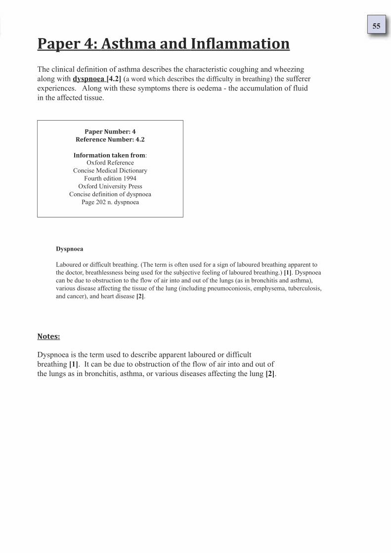
55
Paper 4: Asthma and Inflammation
Paper Number: 4
Reference Number: 4.2
Information taken from:Oxford Reference
Concise Medical Dictionary
Fourth edition 1994
Oxford University Press
Concise definition of dyspnoea
Page 202 n. dyspnoea
The clinical definition of asthma describes the characteristic coughing and wheezing
along with dyspnoea [4.2] (a word which describes the difficulty in breathing) the sufferer
experiences. Along with these symptoms there is oedema - the accumulation of fluid
in the affected tissue.
Dyspnoea
Laboured or difficult breathing. (The term is often used for a sign of laboured breathing apparent to
the doctor, breathlessness being used for the subjective feeling of laboured breathing.) [1]. Dyspnoea
can be due to obstruction to the flow of air into and out of the lungs (as in bronchitis and asthma),
various disease affecting the tissue of the lung (including pneumoconiosis, emphysema, tuberculosis,
and cancer), and heart disease [2].
Notes:
Dyspnoea is the term used to describe apparent laboured or difficult
breathing [1]. It can be due to obstruction of the flow of air into and out of
the lungs as in bronchitis, asthma, or various diseases affecting the lung [2].
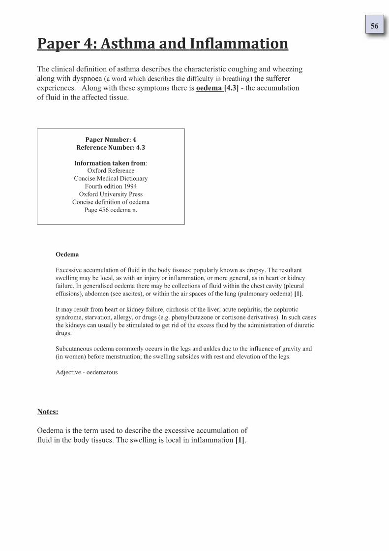
56
Paper 4: Asthma and Inflammation
Paper Number: 4
Reference Number: 4.3
Information taken from:Oxford Reference
Concise Medical Dictionary
Fourth edition 1994
Oxford University Press
Concise definition of oedema
Page 456 oedema n.
The clinical definition of asthma describes the characteristic coughing and wheezing
along with dyspnoea (a word which describes the difficulty in breathing) the sufferer
experiences. Along with these symptoms there is oedema [4.3] - the accumulation
of fluid in the affected tissue.
Oedema
Excessive accumulation of fluid in the body tissues: popularly known as dropsy. The resultant
swelling may be local, as with an injury or inflammation, or more general, as in heart or kidney
failure. In generalised oedema there may be collections of fluid within the chest cavity (pleural
effusions), abdomen (see ascites), or within the air spaces of the lung (pulmonary oedema) [1].
It may result from heart or kidney failure, cirrhosis of the liver, acute nephritis, the nephrotic
syndrome, starvation, allergy, or drugs (e.g. phenylbutazone or cortisone derivatives). In such cases
the kidneys can usually be stimulated to get rid of the excess fluid by the administration of diuretic
drugs.
Subcutaneous oedema commonly occurs in the legs and ankles due to the influence of gravity and
(in women) before menstruation; the swelling subsides with rest and elevation of the legs.
Adjective - oedematous
Notes:
Oedema is the term used to describe the excessive accumulation of
fluid in the body tissues. The swelling is local in inflammation [1].
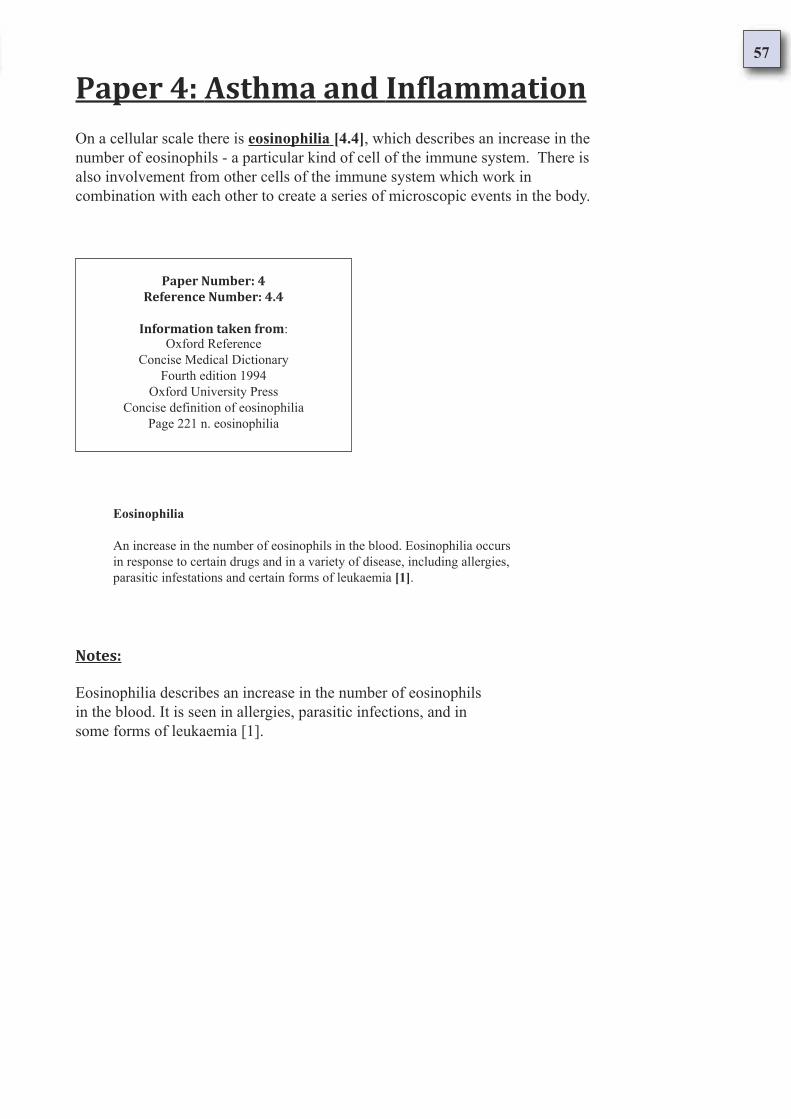
57
Paper 4: Asthma and Inflammation
Paper Number: 4
Reference Number: 4.4
Information taken from:Oxford Reference
Concise Medical Dictionary
Fourth edition 1994
Oxford University Press
Concise definition of eosinophilia
Page 221 n. eosinophilia
On a cellular scale there is eosinophilia [4.4], which describes an increase in the
number of eosinophils - a particular kind of cell of the immune system. There is
also involvement from other cells of the immune system which work in
combination with each other to create a series of microscopic events in the body.
Eosinophilia
An increase in the number of eosinophils in the blood. Eosinophilia occurs
in response to certain drugs and in a variety of disease, including allergies,
parasitic infestations and certain forms of leukaemia [1].
Notes:
Eosinophilia describes an increase in the number of eosinophils
in the blood. It is seen in allergies, parasitic infections, and in
some forms of leukaemia [1].
58
Paper 4: Asthma and Inflammation
Paper Number: 4
Reference Number: 4.5
Information taken from:Oxford Reference
Concise Medical Dictionary
Fourth edition 1994
Oxford University Press
Concise definition of eosinophil
Page 221 n. eosinophil
On a cellular scale there is eosinophilia, which describes an increase in the
number of eosinophils [4.5] - a particular kind of cell of the immune system.
There is also involvement from other cells of the immune system which work in
combination with each other to create a series of microscopic events in the body.
Eosinophil
A variety of white blood cell distinguished by the presence in its cytoplasm
of coarse granules that stain orange-red with Romanowsky stains [1].
The function of the eosinophil is poorly understood, but it is capable of
ingesting foreign particles, is present in large numbers in lining or covering
surfaces within the body, and is involved in allergic responses. There are
normally 40 - 400 x 10 to the six eosinophils per litre of blood [2].
Notes:
Eosinophils are a kind of white blood cell [1] that take part in allergic
responses and ingest foreign particles. Large numbers are found in the
lining and covering of the surfaces within the body [2].
59
Paper 4: Asthma and Inflammation
Paper Number: 4
Reference Number: 4.6
Information taken from:Oxford Reference
Concise Medical Dictionary
Fourth edition 1994
Oxford University Press
Concise definition of smooth muscle
Page 609 smooth muscle (involuntary muscle)
During an asthmatic episode, progressively the smooth muscle [4.6] exhibits a loss of
control (dyspnoea). The smooth muscle of the body carries out unconscious actions under
the regulation of the Autonomic Nervous System. The Autonomic Nervous System is in
command of many things which are not consciously managed; these include the regular
beating of the heart, sweating and breathing.
Smooth Muscle (Involuntary Muscle)
Muscle that produces slow long-term contractions of which the individual is unaware [1].
Smooth muscle occurs in hollow organs, such as the stomach, intestine, blood vessels, and
bladder [2]. It consists of spindle-shaped cells within a network of connective tissue and is
under the control of the autonomic nervous system. Compare straited muscle [3].
Notes:
Smooth muscle is known as involuntary muscle and produces slow long
term contractions which are not consciously controlled [1]. It is part of
hollow organs such as the stomach, intestine, bladder, and blood vessels [2].
Made up of spindle shaped cells in a network of connective tissue, smooth
muscle is under the control of the autonomic nervous system [3].
60
Paper 4: Asthma and Inflammation
Paper Number: 4
Reference Number: 4.7
Information taken from:Oxford Reference
Concise Medical Dictionary
Fourth edition 1994
Oxford University Press
Concise definition of the autonomic nervous system
Page 59 autonomic nervous system
During an asthmatic episode, progressively the smooth muscle exhibits a loss of control
(dyspnoea). The smooth muscle of the body carries out unconscious actions under the
regulation of the Autonomic Nervous System. The Autonomic Nervous System [4.7] is in
command of many things which are not consciously managed; these include the regular
beating of the heart, sweating and breathing.
Autonomic Nervous System
The part of the nervous system responsible for the control of bodily functions that are not
consciously directed, including regular beating of the heart, intestinal movements, sweating ,
salivation, etc [1].
The autonomic system is subdivided into sympathetic and parasympathetic nervous systems.
Sympathetic nerves lead from the middle section of the spinal cord and parasympathetic nerves
from the brain and lower spinal cord [2].
The heart, smooth muscles, and most glands receive fibres of both kinds and the interplay of
sympathetic and parasympathetic reflex activity (the actions are often antagonistic) governs
their working. Sympathetic nerve endings liberate noradrenaline as a neurotransmitter;
parasympathetic nerve endings release acetylcholine [3].
Notes:
The autonomic nervous system controls unconscious movements and body
functions that include the regular beating of the heart, intestinal movements,
sweating and salivation [1]. There are two parts to the autonomic nervous
system: The sympathetic nervous system and the parasympathetic system [2].
The heart, smooth muscle and most glands are connected to both parts of the
autonomic nervous system. The sympathetic portion works with the
parasympathetic portion in a reflex mechanism [3].
61
Paper 4: Asthma and Inflammation
Paper Number: 4
Reference Number: 4.8
Information taken from:Oxford Reference
Concise Medical Dictionary
Fourth edition 1994
Oxford University Press
Concise definition of connective tissue
Page 146 connective tissue
Over time the immune system affects the smooth muscle, respiratory cells and
connective tissue [4.8] in adverse physiological ways. Hypertrophication of
the smooth muscle and a loss of vitality in the respiratory cells and connective
tissue occurs. All of these tissues contain mast cells.
Connective Tissue
The tissue that supports, binds, or separates more specialised tissues and organs, or functions
as packing tissue of the body [1]. It consists of an amorphous ground substance of
mucopolysaccharides in which may be embedded white (collagenous), yellow (elastic), and
reticular fibres, fat cells, fibroblasts, mast cells, and macrophages [2].
Variations in chemical composition of the ground substance and in the proportions and
quantities of cells and fibres give rise to tissues of widely differing characteristics, including
bone, cartilage, tendons, and ligaments as well as adipose, areolar, and elastic tissues [3].
Notes:
Connective tissue is the tissue that supports, binds, or provides a
partition between specialised tissues and organs in the body. As
well as this it functions as the packing tissue of the body [1].
Made up of a shapeless ground substance of mucopolysaccharides it
may contain collagen, elastin, reticular fibres, fat cells, fibroblasts,
macrophages and mast cells [2].
The chemical composition of the ground substance varies widely
giving rise to a variety of tissues including bone, cartilage, tendons,
ligaments as well as adipose, elastic and areolar tissues [3].

62
Paper 4: Asthma and Inflammation
Paper Number: 4
Reference Number: 4.9
Information taken from:Oxford Reference
Concise Medical Dictionary
Fourth edition 1994
Oxford University Press
Concise definition of mast cell
Page 392 mast cell
Over time the immune system affects the smooth muscle, respiratory cells and
connective tissue in adverse physiological ways. Hypertrophication of the
smooth muscle and a loss of vitality in the respiratory cells and connective
tissue occurs. All of these tissues contain mast cells [4.9].
Mast Cell
A large cell in connective tissue with many coarse cytoplasmic granules.
These granules contain chemicals heparin, histamine and serotonin, which
are released during inflammation and allergic responses [1].
Notes:
The mast cell is the large cell that is found in connective tissue. The mast
cell has many cytoplasmic granules which contain heparin, histamine
and serotonin. During inflammation and allergic responses the granules
are excreted [1].
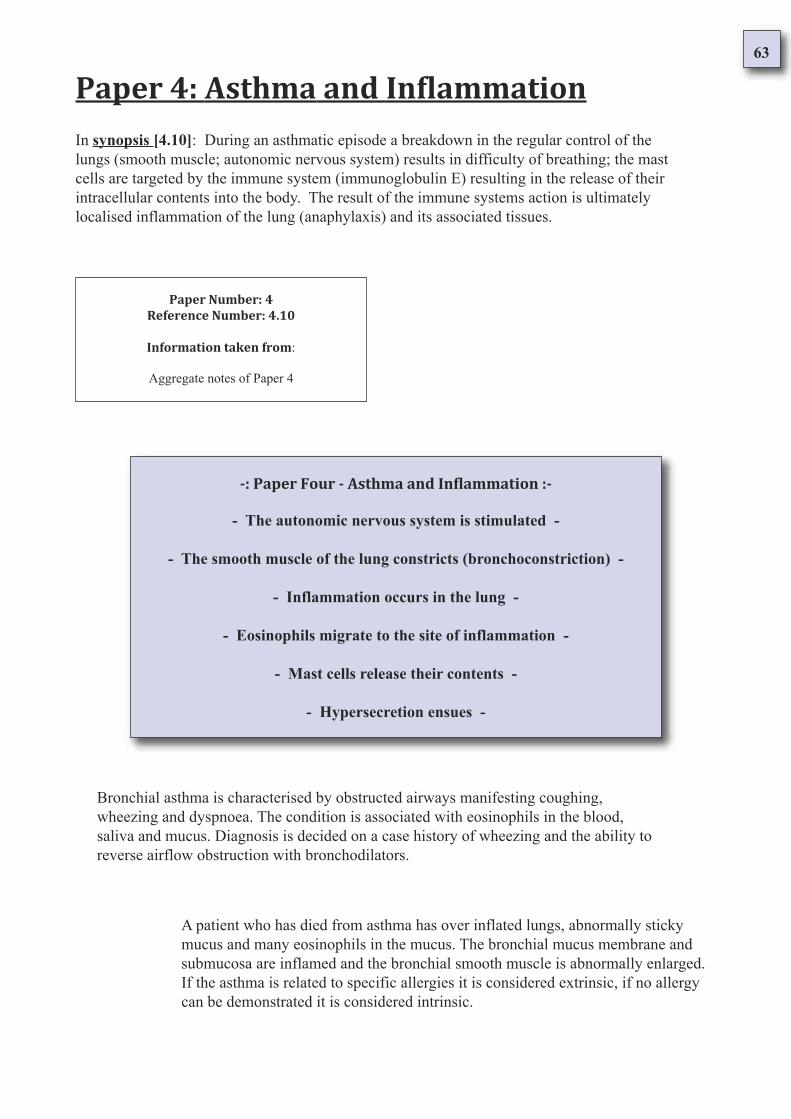
63
Paper 4: Asthma and Inflammation
Paper Number: 4
Reference Number: 4.10
Information taken from:
Aggregate notes of Paper 4
In synopsis [4.10]: During an asthmatic episode a breakdown in the regular control of the
lungs (smooth muscle; autonomic nervous system) results in difficulty of breathing; the mast
cells are targeted by the immune system (immunoglobulin E) resulting in the release of their
intracellular contents into the body. The result of the immune systems action is ultimately
localised inflammation of the lung (anaphylaxis) and its associated tissues.
-: Paper Four - Asthma and Inflammation :-
- The autonomic nervous system is stimulated -
- The smooth muscle of the lung constricts (bronchoconstriction) -
- Inflammation occurs in the lung -
- Eosinophils migrate to the site of inflammation -
- Mast cells release their contents -
- Hypersecretion ensues -
Bronchial asthma is characterised by obstructed airways manifesting coughing,
wheezing and dyspnoea. The condition is associated with eosinophils in the blood,
saliva and mucus. Diagnosis is decided on a case history of wheezing and the ability to
reverse airflow obstruction with bronchodilators.
A patient who has died from asthma has over inflated lungs, abnormally sticky
mucus and many eosinophils in the mucus. The bronchial mucus membrane and
submucosa are inflamed and the bronchial smooth muscle is abnormally enlarged.
If the asthma is related to specific allergies it is considered extrinsic, if no allergy
can be demonstrated it is considered intrinsic.
64
Dyspnoea is the term used to describe apparent laboured or
difficult breathing. It can be due to obstruction of the flow
of air into and out of the lungs as in bronchitis, asthma, or
various diseases affecting the lung.
Oedema is the term used to describe the
excessive accumulation of fluid in the body
tissues. The swelling is local in inflammation.
Eosinophilia describes an increase in the number of
eosinophils in the blood. It is seen in allergies, parasitic
infections, and in some forms of leukaemia.
Extrinsic asthma is the most common kind found in youths with a family history of allergy
and those commonly exposed to allergens. Patients can be confirmed by a positive "type 1"
reaction on a skin prick test with possible allergens. Most atopic subjects are sensitive to
more than two allergens. After initial contact with antigens the formation of Immunoglobulin
E antibodies occurs which then attach themselves to the surface of the mast cells.
Subsequent exposure to the allergen causes the release of histamine, slow reacting
substance of anaphylaxis, eosinophilic chemotactic factor, bradykinin, serotonin, and
prostaglandin F2 alpha. These mediators cause bronchoconstriction, mucosal
accumulation of fluid, and hypersecretion. Immediate hypersensitivity reactions happen
within minutes of contact with the allergen. Patients often give a history of this.
Intrinsic asthma commonly occurs in patients over 35 years of age, generally,
there is no personal or family history of allergy nor any seasonal variation in the
condition. The wheezing is often severe, lasts a long time and may show marked
resistance to bronchodilators. It is in the intrinsic group that most deaths occur.
There is much overlap in the treatment of intrinsic and extrinsic asthma.
65
Eosinophils are a kind of white blood cell that take part in
allergic responses and ingest foreign particles. Large numbers are
found in the lining and covering of the surfaces within the body.
Smooth muscle is known as involuntary muscle and produces slow long term
contractions which are not consciously controlled. It is part of hollow organs
such as the stomach, intestine, bladder, and blood vessels. Made up of spindle
shaped cells in a network of connective tissue, smooth muscle is under the
control of the autonomic nervous system.
The autonomic nervous system controls unconscious movements and body
functions that include the regular beating of the heart, intestinal movements,
sweating and salivation. There are two parts to the autonomic nervous system:
The sympathetic nervous system and the parasympathetic system.
Connective tissue is the tissue that supports, binds, or provides a
partition between specialised tissues and organs in the body. As
well as this it functions as the packing tissue of the body.
The mast cell is the large cell that is found in connective tissue.
The mast cell has many cytoplasmic granules which contain
heparin, histamine and serotonin. During inflammation and
allergic responses the granules are excreted.
The heart, smooth muscle and most glands are connected to both
parts of the autonomic nervous system. The sympathetic portion
works with the parasympathetic portion in a reflex mechanism.
Made up of a shapeless ground substance of mucopolysaccharides it may
contain collagen, elastin, reticular fibres, fat cells, fibroblasts, macrophages
and mast cells. The chemical composition of the ground substance varies
widely giving rise to a variety of tissues including bone, cartilage, tendons,
ligaments as well as adipose, elastic and areolar tissues.
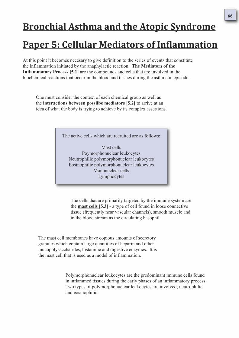
66
Bronchial Asthma and the Atopic Syndrome
Paper 5: Cellular Mediators of Inflammation
At this point it becomes necesary to give definition to the series of events that constitute
the inflammation initiated by the anaphylactic reaction. The Mediators of the
Inflammatory Process [5.1] are the compounds and cells that are involved in the
biochemical reactions that occur in the blood and tissues during the asthmatic episode.
One must consider the context of each chemical group as well as
the interactions between possilbe mediators [5.2] to arrive at an
idea of what the body is trying to achieve by its complex assertions.
The active cells which are recruited are as follows:
Mast cells
Poymorphonuclear leukocytes
Neutrophilic polymorphonuclear leukocytes
Eosinophilic polymorphonuclear leukocytes
Mononuclear cells
Lymphocytes
The cells that are primarily targeted by the immune system are
the mast cells [5.3] - a type of cell found in loose connective
tissue (frequently near vascular channels), smooth muscle and
in the blood stream as the circulating basophil.
The mast cell membranes have copious amounts of secretory
granules which contain large quantities of heparin and other
mucopolysaccharides, histamine and digestive enzymes. It is
the mast cell that is used as a model of inflammation.
Polymorphonuclear leukocytes are the predominant immune cells found
in inflammed tissues during the early phases of an inflammatory process.
Two types of polymorphonuclear leukocytes are involved; neutrophilic
and eosinophilic.
67
Neutrophilic Polymorphonuclear Leukocytes [5.4] are cells of the immune system which
protect the body by ingesting and ultimately destroying potentially harmful organisms and
foreign material. Emigrating to the sites of inflammation, they act in conjunction with the
complement systems to disrupt mast cells, increase vessel permeability and release enzymes
that degrade proteins, mucopolysaccharides and other substances.
Lymphocytes [5.7] are immune cells which mediate adaptive
immunity producing and secreting immunoglobulins and
regulating and overall inflammatory response.
Mononuclear cells [5.6] are cells that digest cellular
debris, injured cells, some types of bacteria, fungi,
protozoa and other factors that stimulate inflammation.
Eosinophilic Polymorphonuclear Leukocytes [5.5] are cells of the immune system
which are a prominent feature of many allergic and immediate hypersensitive reactions.
They are selective to mast cells and contain many of the digestive enzymes present in
neutrophils but one particular enzyme - arylsulfatase - is present in higher
concentrations and is a strong inactivator of anaphylaxis. These cells degrade heparin.
A broad overview [5.8] of the cellular events must be
considered to develop an understanding of the
chemical events which the immune system sponsors.
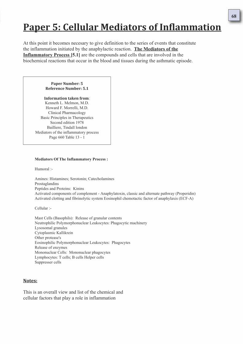
68
Paper 5: Cellular Mediators of Inflammation
Paper Number: 5
Reference Number: 5.1
Information taken from:Kenneth L. Melmon, M.D.
Howard F. Morrelli, M.D.
Clinical Pharmacology
Basic Principles in Therapeutics
Second edition 1978
Bailliere, Tindall london
Mediators of the inflammatory process
Page 660 Table 13 - 1
Mediators Of The Inflammatory Process :
Humoral :-
Amines: Histamines; Serotonin; Catecholamines
Prostaglandins
Peptides and Proteins: Kinins
Activated components of complement - Anaphylatoxin, classic and alternate pathway (Properidin)
Activated clotting and fibrinolytic system Eosinophil chemotactic factor of anaphylaxis (ECF-A)
Cellular :-
Mast Cells (Basophils): Release of granular contents
Neutrophilic Polymorphonuclear Leukocytes: Phagocytic machinery
Lysosomal granules
Cytoplasmic Kallikrein
Other protease's
Eosinophilic Polymorphonuclear Leukocytes: Phagocytes
Release of enzymes
Mononuclear Cells: Mononuclear phagocytes
Lymphocytes: T cells; B cells Helper cells
Suppresser cells
Notes:
This is an overall view and list of the chemical and
cellular factors that play a role in inflammation
At this point it becomes necesary to give definition to the series of events that constitute
the inflammation initiated by the anaphylactic reaction. The Mediators of the
Inflammatory Process [5.1] are the compounds and cells that are involved in the
biochemical reactions that occur in the blood and tissues during the asthmatic episode.
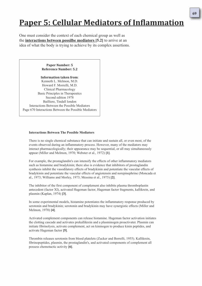
69
Paper 5: Cellular Mediators of Inflammation
Paper Number: 5
Reference Number: 5.2
Information taken from:Kenneth L. Melmon, M.D.
Howard F. Morrelli, M.D.
Clinical Pharmacology
Basic Principles in Therapeutics
Second edition 1978
Bailliere, Tindall london
Interactions Between the Possible Mediators
Page 670 Interactions Between the Possible Mediators
Interactions Between The Possible Mediators
There is no single chemical substance that can initiate and sustain all, or even most, of the
events observed during an inflammatory process. However, many of the mediators may
interact pharmacologically; their appearance may be sequential, or all may simultaneously
appear (Miller and Melmon, 1970; Webster et al., 1972) [1].
For example, the prostaglandin's can intensify the effects of other inflammatory mediators
such as histamine and bradykinin; there also is evidence that inhibitors of prostaglandin
synthesis inhibit the vasodilatory effects of bradykinin and potentiate the vascular effects of
bradykinin and potentiate the vascular effects of angiotensin and norepinephrine (Moncada et
al., 1973; Williams and Morley, 1973; Messina et al., 1975) [2].
The inhibitor of the first component of complement also inhibits plasma thromboplastin
antecedent (factor XI), activated Hageman factor, Hageman factor fragments, kallikrein, and
plasmin (Kaplan, 1974) [3].
In some experimental models, histamine potentiates the inflammatory response produced by
serotonin and bradykinin; serotonin and bradykinin may have synergistic effects (Miller and
Melmon, 1970) [4].
Activated complement components can release histamine. Hageman factor activation initiates
the clotting cascade and activates prekallikrein and a plasminogen proactivater. Plasmin can
initiate fibrinolysis, activate complement, act on kininogen to produce kinin peptides, and
activate Hageman factor [5].
Thrombin releases serotonin from blood platelets (Zucker and Borrelli, 1955). Kallikrein,
fibrinopeptides, plasmin, the prostaglandin's, and activated components of complement all
possess chemotactic activity [6].
One must consider the context of each chemical group as well as
the interactions between possilbe mediators [5.2] to arrive at an
idea of what the body is trying to achieve by its complex assertions.
70
Notes:
Many chemicals are released during inflammation, these are 'humoral' mediators that are
released by the cells involved in anaphylaxis. The anaphylactic reaction is a complicated
series of events, which involves chemical mediators interacting and influencing each other as
well as tissues to achieve a particular physiological aim. Examples of these chemical inter-
relationships follow [1]:
Prostaglandin's are lipid hormones that when present may increase the vasodilatory effects of
histamine and bradykinin . Inhibiting the prostaglandins has the effect of inhibiting vascular
dilation and the body's sensitivity to bradykinin, angiotensin and norepinephrine [2].
Inhibitition of the first component of complement also inhibits components of blood
coagulation (thromboplatin antecedent - factor XI; activated Hageman factor; Hageman factor
fragments), kallikrein (which produces bradykinin), and plasmin (which digests the protein of
blood clots - fibrin) [3].
Histamine potentiates the inflammatory response produced by serotonin and bradykinin in
some models (both are known to act on smooth muscle and cause vascular dilation) [4].
The activation of complement can result in histamine release. Hageman factor, (which
initiates blood clotting), also activates prekallikrein and a plasminogen proactivater. Plasmin
can initiate breakdown of fibrin, activate complement, causes kinins to be produced, and
activates Hageman factor in blood coagulation [5].
Thrombin (the precursor to fibrin) stimulates the release of serotonin from blood platelets.
Kallikrein, fibrinopeptides, plasmin, prostaglandin's, and complement all have chemotactic
activity [6].
It is plain to see that the chemical mediators of anaphylaxis interact with each other and that
cells influence the chemical substances and vice versa [7]. A closer look at the cells that are
the target of all the attentions; the cells which act on them, and the substances they utilise
follows.
Certain cells have receptors for activated components of complement and immunoglobulins; therefore,
interrelationships exist not only between the various chemical substances that may be important in the
initiation and progression of inflammation, but also between these substances and the cellular
components involved in an inflammatory process [7].
Undoubtedly there are additional contributors to the inflammatory process, but their roles and
identification are only now emerging. There is a considerable body of knowledge related to the
mechanisms of inflammation. However, we have not yet seen extensive applications of this
information to therapeutics. Because use of basic pathogenetic information in other areas has been
fruitful, we can logically expect major advances in anti inflammatory therapeutics in the future.
71
Paper 5: Cellular Mediators of Inflammation
Paper Number: 5
Reference Number: 5.3
Information taken from:Kenneth L. Melmon, M.D.
Howard F. Morrelli, M.D.
Clinical Pharmacology
Basic Principles in Therapeutics
Second edition 1978
Bailliere, Tindall london
Cellular mediators of inflammation
Page 671 Mast cells
Mast Cells
Mast cells are mainly found in loose connective tissue, frequently near vascular channels. Their most
distinguishing features are their membrane-bound, spheroid, basophilic secretory granules, which may be
so numerous that other cytoplasmic structures are obscured. These granules contain large amounts of
heparin and other mucopolysaccharides, histamine, and several proteolytic and esterolytic enzyme
systems. Serotonin is present in the mast cells of some species, man being a notable exception [1].
Much is known regarding the structure and composition of mast cells, but their physiologic function
remains unidentified. However it seems certain that the mast cell and the basophil (circulating mast cells)
contribute to the development of an inflammatory process (Lewis et al., 1975). Changes in mast cell
density and mast cell granules are thought to be characteristic of many inflammatory lesions. Mast cell
preparations (either in vitro or in vivo during anaphylaxis) have been used as experimental models of
inflammation [2].
The mast cell is readily degranulated (see previous section on histamine), and the granule-bound chemical
substances are released into extracellular fluid [3]. Histamine and probably heparin can have important
influences on an inflammatory process, but much less is known about the granule esterase's and protease's
that are also released from the granules of the mast cell and basophil (Benditt, 1968; Lewis et al., 1975).
These enzymes may alter the course of inflammation by interactions with other cells or substances to
release, activate, or destroy kinins, complement, and blood coagulation factors, to alter membranes of
other cells such as granulocytes so that additional intracellular constituents are released, or to alter the
structure and characteristics of adjacent connective tissue [4].
Notes:
Mast cells are cells mainly found in loose connective tissue (see smooth
muscle) and are often near vascular channels. They are distinguished by
copious amounts of secretory granules on the membrane.
The cells that are primarily targeted by the immune system are
the mast cells [5.3] - a type of cell found in loose connective
tissue (frequently near vascular channels), smooth muscle and
in the blood stream as the circulating basophil.
72
The secretory granules contain large amounts of heparin and mucopolysaccharides (both
of which are compounds of glucuronic acid and glucosamine), histamine and several
enzyme systems which catalyse the splitting of esters into constituent alcohol's and
acids, and proteins into smaller peptides and amino acids [1].
The mast cells are the target cells intimately involved in the inflammatory process and
are used as models of inflammation caused by anaphylaxis [2]. During inflammation the
mast cell is stimulated to excrete its granules into the extracellular fluid [3].
Histamine and heparin are both important influences in the inflammatory process. The
enzyme systems of mast cells may act on kinins, complement, blood coagulation factors,
leukocyte membranes and on the structure and characteristics of connective tissue [4].
73
Paper 5: Cellular Mediators of Inflammation
Paper Number: 5
Reference Number: 5.4
Information taken from:Kenneth L. Melmon, M.D.
Howard F. Morrelli, M.D.
Clinical Pharmacology
Basic Principles in Therapeutics
Second edition 1978
Bailliere, Tindall london
Cellular mediators of inflammation
Page 671 Neutrophilic Polymorphonuclear Leukocytes
Neutrophilic Polymorphonuclear Leukocytes
Neutrophilic leukocytes protect the body by ingesting and ultimately destroying potentially harmful
organisms and foreign material. Phagocytosis occurs after the leukocytes have marginated along the
endothelium at the site of the inflammatory process [1].
They then emigrate through the vessel wall, attracted by many substances released or activated by the
inflammatory stimulus, and ingest and digest or kill the "noxious" agent. Unfortunately, little is known of
the mechanisms responsible for each of these steps, but they are probable directly influenced by the
presence of serum factors such as complement [2].
The leukocytes can also influence other aspects of the inflammatory response in addition to the
destruction of a harmful stimulus. Leukocytes are essential in the Schwartzman phenomenon, Arthus'
reaction, the acute inflammatory process of gout, and probably in other conditions. Although the extent of
the participation of leukocytes is not fully defined, release of their chemical contents appears to be one
mechanism of action [3].
The lysosomes of leukocytes contain a protease that can cleave C5, the fifth component of complement,
to yield a product that, in turn, selectively releases other lysosomal enzymes from intact leukocytes.
Therefore, activation of either the classic or alternate complement pathway results in the production of
factors capable of stimulating lysosomal release (Goldstein et al., 1973; Goldstein and Weissmann,
1974a) [4].
The lysosomal substances influence inflammatory processes by disrupting mast cells, increasing the
permeability of vessels, furthering chemotaxis, or injuring normal tissue. Lysosomal constituents can
degrade collagen, elastin, cartilage, hyaluronate, chondroitin sulphate, nucleic acids, complement
components, fibrin, plasminogen, coagulation factors, and kininogen [5].
Neutrophilic Polymorphonuclear Leukocytes [5.4] are cells of the immune system which
protect the body by ingesting and ultimately destroying potentially harmful organisms and
foreign material. Emigrating to the sites of inflammation, they act in conjunction with the
complement systems to disrupt mast cells, increase vessel permeability and release enzymes
that degrade proteins, mucopolysaccharides and other substances.
74
Notes:
Neutrophilic Polymorphonuclear Leukocytes (neutrophils) are a kind of immune cell that act to
digest bacteria and foreign materials at the endothelium of the inflammed tissue [1]. These cells
migrate through vessel walls attracted by the noxious substances released during inflammation
[2]. Neutrophils are involved in inflammatory reactions, are essential in the Schwartzman
phenomenon, Arthus’ reaction and the inflammation of gout. They act by releasing their chemical
contents [3].
The digestive enzymes neutrophils release affect complement, which in turn prompts other
leukocytes to release their lysosomal enzymes [4]. The enzymes they release disrupt mast cells,
make blood vessels more permeable for cell migration and digest substances such as collagen,
elastin, cartilage, hyaluronate, chondroitin sulphate, nucleic acids, complement, fibrin,
plasminogen, coagulation factors and kininogen [5].
Neutrophils also release fungicidal and bactericidal proteins as well as non-enzymic substances
that act to increase capillary permeability and body temperature [6]. The enzymes are released in
two ways. One is termed 'Regurgitation During Feeding', where lysosomal enzymes are
discharged into the surrounding tissue whist neutrophils digest substances internally. There is no
leakage of cytoplasmic constituents and no evidence of cellular injury.
The other mechanism of discharge is termed 'Reverse Endocytosis'. Intact leukocytes selectively
release lysosomal constituents (exocytosis) when they encounter immune complexes on solid
surfaces [7]. Enzyme release is influenced by chemicals that alter intracellular cyclic nucleotide
levels and substances that influence the formation of microtubules ("intracellular veins") [8].
A series of nonenzymatic factors have also been isolated from lysosomes that induce capillary
permeability and provoke fever (Goldstein and Weissmann, 1974a). Some proteins in the lysosome
are fungicidal and bactericidal (Weissmann, 1967) [6].
Lysosomal enzymes are released by two mechanisms. One mechanism is called "regurgitation
during feeding"; the lysosomal contents are released into the surrounding medium by cells
engaging in endocytosis (Weissmann et al., 1971).
There is no leakage of cytoplasmic constituents and no evidence of cellular injury. Another
mechanism of discharge of lysosomal enzymes from intact leukocytes involves selective release
(exocytosis) of lysosomal constituents when leukocytes encounter immune complexes on solid
surfaces; The process has been termed "reverse endocytosis" (Weissmann et al., 1972) [7].
The release of lysosomal contents can be modified by pharmacologic agents that alter intracellular
concentrations of cyclic nucleotides (cAMP, cGMP) and agents that promote or reduce assemble of
microtubules (Goldstein and Weissmann, 1974b; Ignarro, 1975) [8].
Plasmin-generating activity of a kallikrein activator is present on the cell surface and a kininase is
found in the granules of neutrophils and eosinophils (Melmon and Cline, 1968). The kinin-
generating capability of these cells is likely due to plasmin production and probably can account
for the delayed appearance of kinin-like substances in experimental inflammatory processes
(Kellermeyer and Graham, 1968; Miller et al., 1975) [9].
Although the optimal pH of kinin generation from the cytoplasm is 7 to 8, cathepsins are active in
the potentially acid environment of inflammatory foci, and granulocyte kinin production continues
at pH 5.5 (Greenbaum et al., 1966) [10].

75
The surface of the neutrophil cell can stimulate the formation of plasmin and kinins,
as well as this, an enzyme which digests kinins is found in the granules of both
neutrophilic and eosinophilic leukocytes [9].
The pH value of the tissue environment affects the production of kinins but
cathepsins (enzymes which digest proteins) and kinins produced by granulocytes
(white blood cells which possess granules in their cytoplasm - neutrophils,
eosinophils, basophils) continue to be synthesised where there is pH variation [10].
76
Paper 5: Cellular Mediators of Inflammation
Paper Number: 5
Reference Number: 5.5
Information taken from:Kenneth L. Melmon, M.D.
Howard F. Morrelli, M.D.
Clinical Pharmacology
Basic Principles in Therapeutics
Second edition 1978
Bailliere, Tindall london
Cellular mediators of inflammation
Page 672 Eosinophilic Polymorphonuclear Leukocytes
Eosinophilic Polymorphonuclear Leukocytes
Eosinophilic leukocytes are a prominent feature of many allergic and immediate hypersensitive reactions.
In addition to activated complement factors and kallikrein, an eosinophil chemotactic factor of anaphylaxis
(ECF-A) released from sensitised mast cells selectively attracts eosinophils (Kay et al., 1971) [1].
Eosinophils phagocytize and have membrane bound granules that contain many of the hydrolytic enzymes
present in neutrophils. One enzyme present in higher concentrations than in polymorphonuclear cells is
arylsulfatase, which is a potent inactivater of slow reacting substance of anaphylaxis (SRS-A) (Wasserman et
al., 1975) [2]. Other factors in eosinophils inhibit histamine release, neutralise heparin, and activate
plasminogen; thus, these cells may play a role in limiting allergic reactions (Goetzl et al., 1975) [3].
Notes:
Eosinophilic Polymorphonuclear Leukocytes (eosinophils) are immune cells that play a major
part in allergic and immediate hypersensitivity reactions. They are attracted by complement,
kallikrein and a substance released by sensitised mast cells [1].
These leukocytes engulf and digest bacteria and foreign materials in a similar way to
neutrophils. Eosinophils have a higher level of the enzyme - arylsulfatase - which strongly
inactivates anaphylactic events by acting on aromatic and sulphur containing compounds [2].
Eosinophils inhibit histamine release, neutralise heparin (sulphur compound) and activate
plasminogen. These cells play an important role in limiting allergic reactions [3].
Eosinophilic Polymorphonuclear Leukocytes [5.5] are cells of the immune system
which are a prominent feature of many allergic and immediate hypersensitive reactions.
They are selective to mast cells and contain many of the digestive enzymes present in
neutrophils but one particular enzyme - arylsulfatase - is present in higher
concentrations and is a strong inactivator of anaphylaxis. These cells degrade heparin.
77
Paper 5: Cellular Mediators of Inflammation
Paper Number: 5
Reference Number: 5.6
Information taken from:Kenneth L. Melmon, M.D.
Howard F. Morrelli, M.D.
Clinical Pharmacology
Basic Principles in Therapeutics
Second edition 1978
Bailliere, Tindall london
Cellular mediators of inflammation
Page 672 Mononuclear cells
Mononuclear Cells
Mononuclear phagocytes. Polymorphonuclear leukocytes are the predominant cells in inflammed
tissues during the early phases of an inflammatory process. Later, monocytes and macrophages are
more numerous [1].
The factors that attract and stimulate the mononuclear cells seem to differ from those that influence
the polymorphonuclear leukocytes. The mononuclear phagocytes (monocytes and macrophages)
ingest cellular debris, injured cells, some types of bacteria, fungi, protozoa, and other factors that
stimulate inflammation [2].
The mononuclear phagocytes contain many potent hydrolytic enzyme systems that are capable of
degrading most of the known macromolecular constituents of tissues and bacteria (Cohn, 1965).
Mononuclear phagocytes probably contribute to various immune phenomena by processing antigens
into immunogenic complexes (Cohn, 1965; Sell and Asofsky, 1968; Weiser et al., 1969) [3].
The monocytic phagocytes are important factors in the bodies resistance against certain types of
infection as well as during tissue repair and healing. Although the role of the monocyte in
inflammation has been extensively described, we have little information on ways to influence its
function by pharmacologic means [4].
Notes:
Mononuclear cells are involved more in the later stages of inflammation [1]. There are two
types of mononuclear cell; monocytes and macrophages. Both ingest undesirable substances
(phagocytosis) [2]. Mononuclear phagocytes have enzymes capable of breaking down most
tissues and bacteria [3] and are involved in immune resistance to infection, tissue repair and
healing [4].
Mononuclear cells [5.6] are cells that digest cellular
debris, injured cells, some types of bacteria, fungi,
protozoa and other factors that stimulate inflammation.
78
Paper 5: Cellular Mediators of Inflammation
Paper Number: 5
Reference Number: 5.7
Information taken from:Kenneth L. Melmon, M.D.
Howard F. Morrelli, M.D.
Clinical Pharmacology
Basic Principles in Therapeutics
Second edition 1978
Bailliere, Tindall london
Cellular mediators of inflammation
Page 672 - 674 Lymphocytes
Lymphocytes
The lymphocytes mediate both types of responses necessary for adaptive immunity: humoral and cell
mediated immunity. Humoral immunity involves the production and secretion of antibody
(immunoglobulins can combine with and frequently neutralise specific antigens) [1].
The reaction of the antibody and antigen is called and immediate reaction or immediate
hypersensitivity. The clinical manifestations of this reaction (anaphylaxis, serum sickness, Arthus
reaction) will depend upon the type of antibody produced and the tissues in which the reaction occurs
[2].
Antibodies are produced by plasma cells derived from specialised lymphocytes called B cells. Other
lymphocytes, called T cells, are involved in cellular immunity, which instead is mediated by cells
specifically altered as a result of antigenic stimulation. The cell mediated type of immunity is
responsible for the rejection of tissue grafts and for delayed hypersensitivity reactions, such as the
tuberculin reaction [3].
Both types of lymphocytes (T and B) are derived from hematopoietic stem cells that migrate to the
primary lyphoid organs, the thymus and "bursa" or is phylogenetic counterpart. The stem cells
differentiate along lymphoid cell lines in these tissues [4].
Thymus derived cells, T cells, populate the thymus dependent areas of lymphoid tissues and recirculate
from blood to lymph. Non thymus derived cells (B cells) colonise the thymus independent areas of the
secondary lymphoid organs such as the lymph follicle [5].
After interaction with antigen, the T cells differentiate into cells involved in cellular immunity and B
cells to antibody producing cells. For optimal function of either the T or B lymphocytes, both types of
cells are necessary. For example, if a deficiency of T helper cells exist, the B cells cannot effectively
produce antibodies after antigenic stimulation. Neither type of cell has a pure function nor can one type
function independently of the other cell type [6].
Lymphocytes [5.7] are immune cells which mediate adaptive
immunity producing and secreting immunoglobulins and
regulating and overall inflammatory response.
79
The mature, antigen sensitive T lymphocyte is capable of specific antigen recognition and can initiate
immune responses (Basten et al., 1971). The thymus derived lymphocytes appear to have multiple
functions in immunity: (1) Helper cells and suppresser cells can modulate B cell production of antibody;
(2) They modulate the extent of effecter T cell expression of delayed hypersensitivity, such as mixed
lymphocyte reactions, and graft versus host reactions [7].
T cells (Paul and Bencerraf, 1977) may play an important role on B cell responses, as evidenced by
procedures that deplete T cells (such as thymectomy, treatment of cells with anti-lymphocyte serum) or
separation of a sub-population of T cells by virtue of their adherence to insolublised conjugate of
histamines with albumin (Mozes et al., 1974; Shearer et al., 1974).
These manoeuvre result in increased antibody production against T independent antigens (Kerbel and
Eidinger, 1971; Shearer et al., 1974). In tolerant animals, T cells exert an active suppresser effect on IgG
antibody production (Gershon and Kondo, 1971; Basten et al., 1974; Segal et al., 1974).
The mechanism of suppression of the immune response by T cells is unknown, but in addition to their
effects on antibody production , it appears that T suppresser cells are also involved in regulation of T cell
mediated delayed hypersensitivity (Zembala and Asherson, 1973). In antibody production, however, it is
not known whether the suppressive effect is exerted directly on B cells, on macrophages that process
antigens, or on other T cells that would ordinarily facilitate the B cell response.
Knowledge of the mechanism of T cell suppression may be required to understand surveillance against
antigens. Such knowledge also can help with proper design of experiments to effectively modulate T and B
cell function pharmacologically (e.g. with the drugs that increase or decrease their cyclic AMP
concentrations) and with the effective design of unconventional new drugs that might selectively alter the
functions of the cells that serve as targets for the T suppresser population.
Transfer factor is a low molecular weight RNA complex from leukocyte extracts that converts "naive"
lymphocytes to antigen responsive cells. This results in a cascade effect so that a large population of
antigen responsive cells is recruited to deal more effectively with the noxious agent (Lawrence, Harvey
Lectures, in press) [8].
Transfer factors have specificity for subgroups of antigen sensitive T cells as opposed to specificity for
various antigens. Transfer factors are now being purified to the extent that they can be chemically
characterised and perhaps synthesised [9].
Their clinical efficacy has not been established, but preliminary studies suggest their anti-tumour and anti-
infective actions (e.g. against coccidioidomycosis and lepromatous leprosy) and their use in therapy of
immunodeficiency states (Bullock et al., 1972; Spitler et al., 1972; Ammann et al., 1974; Catanzaro et al.,
1974; Levin et al., 1975). Further testing of these agents is required before recommendation of their use is
possible.
The T cells are believed to be regulators of inflammatory responsiveness, because they either promote or
suppress functions of other lymphocytes in both cellular and humoral immunity; They activate
macrophages and enhance microbicidal activity; They produce factors (such as lymphokines) that can
enhance migration and function of other mononuclear and polymorphonuclear leukocytes (Bloom and
Bennett, 1970; McDevitt et al., 1974; Gershon, 1974; Mitchell, in press) [10].
Almost any process that causes tissue injury can trigger the series of events included in the inflammatory
and possibly the immune process. Such stimuli may not produce inflammation or an immune response by
identical mechanisms. The release or activation of histamine, serotonin, bradykinin, complement
components, and coagulation factors may contribute to inflammation [11];
Many other factors (e.g. slow reacting substances, prostaglandin's changes in hydrogen ion concentration
and oxygen tension), undoubtedly play some part in an inflammatory process. The emigration of
granulocytes and mononuclear cells and their interaction have early direct and indirect effects by causing
release of cellular contents. In a later phase of inflammation, these cells may be primarily involved in the
immune process [12].
80
Notes:
Lymphocytes are immune cells involved in adaptive immunity and the production of
immunoglobulins (antibodies) [1]. When an antibody combines with an antigen an
immediate hypersensitivity reaction results. How the reaction manifests itself depends on
the antibody and tissue involved [2].
There are two kinds of lymphocyte:- The B cell lymphocyte which produces the antibodies
involved in 'humoral immunity' - and the - T cell lymphocyte which is involved in 'cellular
immunity' by adapting to the antigens they encounter. The T cells are responsible for tissue
rejection and delayed hypersensitivity [3].
The lymphocytes are produced by the process of 'haemopoiesis' in the bone marrow and
then migrate to lymphoid tissues and bursa where they mature [4]. T cells are named thus
because they are found in the thymus dependent tissues whereas the B cells are found in
tissues that are independent of the thymus organ [5]. After contact with antigens T cells
adapt to the antigen and B cells produce antibodies. Both T and B lymphocytes act in a co-
ordinated manner to provide effective immunity [6].
Matured antigen sensitive T cells act to specific substances and initiate appropriate immune
responses. T cells influence the antibody production of B cells and the extent of the
hypersensitive reaction [7]. They are made antigen sensitive by way of ribonucleic acid
complex termed 'transfer factor' [8]. Transfer factors are specific to groups of T cells that
have antigen sensitive qualities [9].
T cells act to either promote or suppress the lymphocyte population involved in cellular and
humoral immunity, as well as activate macrophage activity [10]. Many processes that
produce tissue injury involve inflammation and the immune system. Many humoral
substances are involved [11]. Granulocytes and mononuclear cells have early actions in
releasing cellular contents, and in the later phase of the inflammatory response, are a
primary aspect [12].
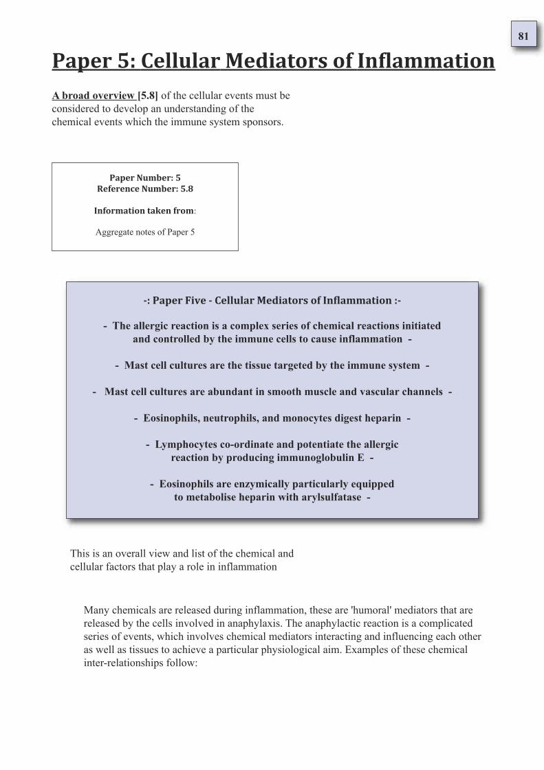
81
Paper 5: Cellular Mediators of Inflammation
Paper Number: 5
Reference Number: 5.8
Information taken from:
Aggregate notes of Paper 5
A broad overview [5.8] of the cellular events must be
considered to develop an understanding of the
chemical events which the immune system sponsors.
-: Paper Five - Cellular Mediators of Inflammation :-
- The allergic reaction is a complex series of chemical reactions initiated
and controlled by the immune cells to cause inflammation -
- Mast cell cultures are the tissue targeted by the immune system -
- Mast cell cultures are abundant in smooth muscle and vascular channels -
- Eosinophils, neutrophils, and monocytes digest heparin -
- Lymphocytes co-ordinate and potentiate the allergic
reaction by producing immunoglobulin E -
- Eosinophils are enzymically particularly equipped
to metabolise heparin with arylsulfatase -
Many chemicals are released during inflammation, these are 'humoral' mediators that are
released by the cells involved in anaphylaxis. The anaphylactic reaction is a complicated
series of events, which involves chemical mediators interacting and influencing each other
as well as tissues to achieve a particular physiological aim. Examples of these chemical
inter-relationships follow:
This is an overall view and list of the chemical and
cellular factors that play a role in inflammation
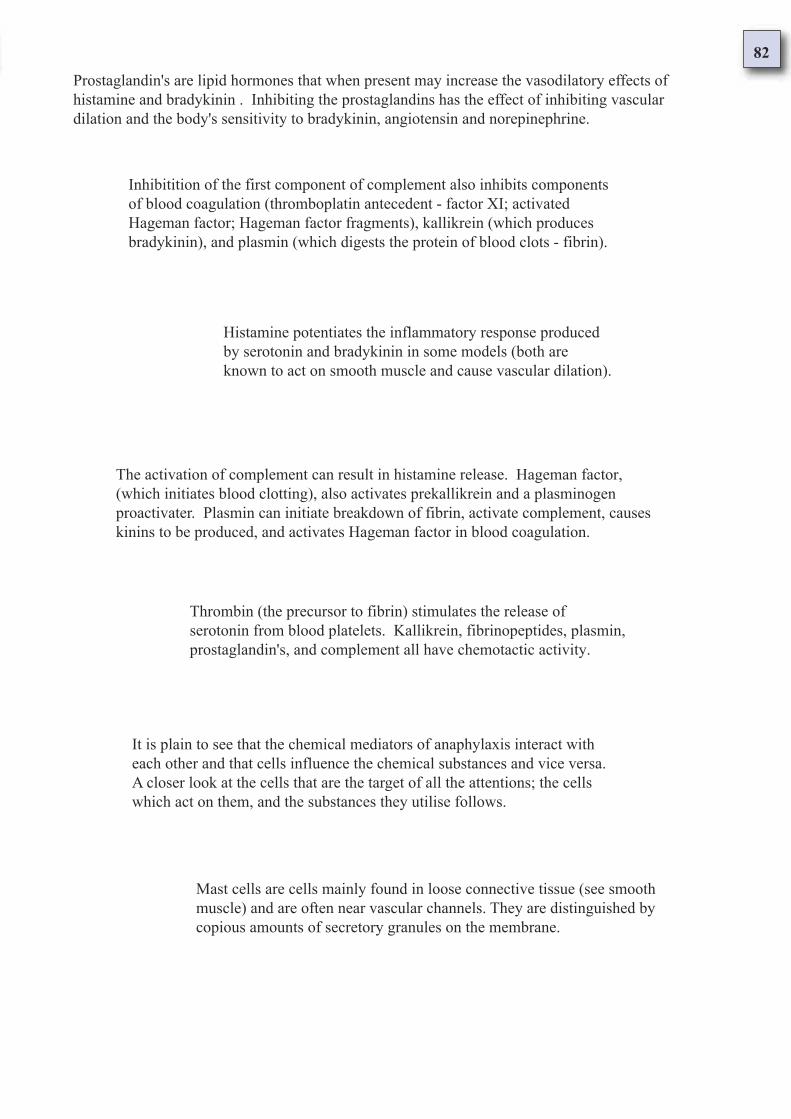
82
Prostaglandin's are lipid hormones that when present may increase the vasodilatory effects of
histamine and bradykinin . Inhibiting the prostaglandins has the effect of inhibiting vascular
dilation and the body's sensitivity to bradykinin, angiotensin and norepinephrine.
Inhibitition of the first component of complement also inhibits components
of blood coagulation (thromboplatin antecedent - factor XI; activated
Hageman factor; Hageman factor fragments), kallikrein (which produces
bradykinin), and plasmin (which digests the protein of blood clots - fibrin).
Histamine potentiates the inflammatory response produced
by serotonin and bradykinin in some models (both are
known to act on smooth muscle and cause vascular dilation).
The activation of complement can result in histamine release. Hageman factor,
(which initiates blood clotting), also activates prekallikrein and a plasminogen
proactivater. Plasmin can initiate breakdown of fibrin, activate complement, causes
kinins to be produced, and activates Hageman factor in blood coagulation.
Thrombin (the precursor to fibrin) stimulates the release of
serotonin from blood platelets. Kallikrein, fibrinopeptides, plasmin,
prostaglandin's, and complement all have chemotactic activity.
It is plain to see that the chemical mediators of anaphylaxis interact with
each other and that cells influence the chemical substances and vice versa.
A closer look at the cells that are the target of all the attentions; the cells
which act on them, and the substances they utilise follows.
Mast cells are cells mainly found in loose connective tissue (see smooth
muscle) and are often near vascular channels. They are distinguished by
copious amounts of secretory granules on the membrane.
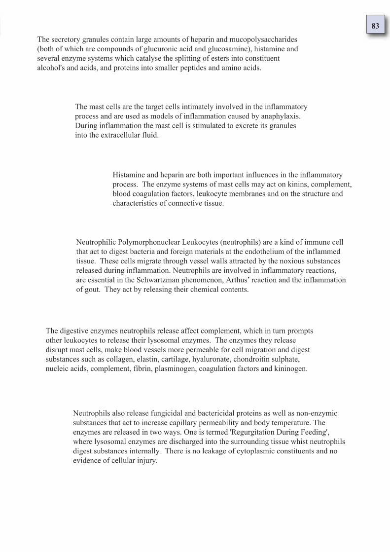
83
The secretory granules contain large amounts of heparin and mucopolysaccharides
(both of which are compounds of glucuronic acid and glucosamine), histamine and
several enzyme systems which catalyse the splitting of esters into constituent
alcohol's and acids, and proteins into smaller peptides and amino acids.
The mast cells are the target cells intimately involved in the inflammatory
process and are used as models of inflammation caused by anaphylaxis.
During inflammation the mast cell is stimulated to excrete its granules
into the extracellular fluid.
Histamine and heparin are both important influences in the inflammatory
process. The enzyme systems of mast cells may act on kinins, complement,
blood coagulation factors, leukocyte membranes and on the structure and
characteristics of connective tissue.
Neutrophilic Polymorphonuclear Leukocytes (neutrophils) are a kind of immune cell
that act to digest bacteria and foreign materials at the endothelium of the inflammed
tissue. These cells migrate through vessel walls attracted by the noxious substances
released during inflammation. Neutrophils are involved in inflammatory reactions,
are essential in the Schwartzman phenomenon, Arthus’ reaction and the inflammation
of gout. They act by releasing their chemical contents.
The digestive enzymes neutrophils release affect complement, which in turn prompts
other leukocytes to release their lysosomal enzymes. The enzymes they release
disrupt mast cells, make blood vessels more permeable for cell migration and digest
substances such as collagen, elastin, cartilage, hyaluronate, chondroitin sulphate,
nucleic acids, complement, fibrin, plasminogen, coagulation factors and kininogen.
Neutrophils also release fungicidal and bactericidal proteins as well as non-enzymic
substances that act to increase capillary permeability and body temperature. The
enzymes are released in two ways. One is termed 'Regurgitation During Feeding',
where lysosomal enzymes are discharged into the surrounding tissue whist neutrophils
digest substances internally. There is no leakage of cytoplasmic constituents and no
evidence of cellular injury.
84
Eosinophilic Polymorphonuclear Leukocytes (eosinophils) are immune cells
that play a major part in allergic and immediate hypersensitivity reactions.
They are attracted by complement, kallikrein and a substance released by
sensitised mast cells.
Mononuclear cells are involved more in the later stages of inflammation. There
are two types of mononuclear cell; monocytes and macrophages. Both ingest
undesirable substances (phagocytosis). Mononuclear phagocytes have enzymes
capable of breaking down most tissues and bacteria and are involved in immune
resistance to infection, tissue repair and healing.
The other mechanism of discharge is termed 'Reverse Endocytosis'. Intact leukocytes
selectively release lysosomal constituents (exocytosis) when they encounter immune
complexes on solid surfaces. Enzyme release is influenced by chemicals that alter
intracellular cyclic nucleotide levels and substances that influence the formation of
microtubules ("intracellular veins").
The surface of the neutrophil cell can stimulate the formation of plasmin
and kinins, as well as this, an enzyme which digests kinins is found in the
granules of both neutrophilic and eosinophilic leukocytes.
The pH value of the tissue environment affects the production of kinins but
cathepsins (enzymes which digest proteins) and kinins produced by granulocytes
(white blood cells which possess granules in their cytoplasm - neutrophils,
eosinophils, basophils) continue to be synthesised where there is pH variation.
These leukocytes engulf and digest bacteria and foreign materials in a
similar way to neutrophils. Eosinophils have a higher level of the enzyme
- arylsulfatase - which strongly inactivates anaphylactic events by acting
on aromatic and sulphur containing compounds.
Eosinophils inhibit histamine release, neutralise heparin
(sulphur compound) and activate plasminogen. These cells
play an important role in limiting allergic reactions.
85
Lymphocytes are immune cells involved in adaptive immunity and the production
of immunoglobulins (antibodies). When an antibody combines with an antigen an
immediate hypersensitivity reaction results. How the reaction manifests itself
depends on the antibody and tissue involved.
There are two kinds of lymphocyte:- The B cell lymphocyte which
produces the antibodies involved in 'humoral immunity' - and the - T cell
lymphocyte which is involved in 'cellular immunity' by adapting to the
antigens they encounter. The T cells are responsible for tissue rejection
and delayed hypersensitivity.
The lymphocytes are produced by the process of 'haemopoiesis' in the bone marrow
and then migrate to lymphoid tissues and bursa where they mature. T cells are named
thus because they are found in the thymus dependent tissues whereas the B cells are
found in tissues that are independent of the thymus organ. After contact with antigens
T cells adapt to the antigen and B cells produce antibodies. Both T and B lymphocytes
act in a co-ordinated manner to provide effective immunity.
Matured antigen sensitive T cells act to specific substances and initiate
appropriate immune responses. T cells influence the antibody production of B
cells and the extent of the hypersensitive reaction. They are made antigen
sensitive by way of ribonucleic acid complex termed 'transfer factor'. Transfer
factors are specific to groups of T cells that have antigen sensitive qualities.
T cells act to either promote or suppress the lymphocyte population involved in cellular and
humoral immunity, as well as activate macrophage activity. Many processes that produce
tissue injury involve inflammation and the immune system. Many humoral substances are
involved. Granulocytes and mononuclear cells have early actions in releasing cellular
contents, and in the later phase of the inflammatory response, are a primary aspect.
86
Bronchial Asthma and the Atopic Syndrome
Paper 6: Humoral Mediators of Inflammation
Many substances are released during inflammation
and each compound serves a specific role in the
body. The following are mediators of anaphylaxis:
Histamine [6.1] as a substance is concerned with vascular dilation and
it increases the permeability of blood vessels and tissues. It facilitates
oedema and the migration of immune cells and compounds across
tissue barriers. Histamine produces pruritus but not pain.
Serotonin[6.2] as a substance causes vasodilation and
increases blood flow. It is involved in the functioning of
smooth muscle and non-immediate communication of pain.
The catecholamines [6.3]: Adrenaline [6.4] and noradrenaline [6.5] are
suggested to contribute to the development of hemorrhagic lesions as well as to
act as local anti-inflammatory hormones. These substances affect the sympathetic
portion of the autonomic nervous system and relax the smooth muscle.
Peptides and Proteins [6.6]: This category covers the intrinsic coagulation
system, fibrinolytic system, components of complement and components of the
kinin generation system. These groups are inter-utilized by the body sharing key
components. The groups work in chain reaction existing under suppression by
inhibitors. They play roles in oedema.
Kinins [6.7] are polypeptides which function to produce arterial and venular dilationby direct action on smooth muscle. They are involved in pain production and aresuggested to induce leukocyte adherence and migration during inflammation.
Activated complement components [6.8] is a group of compounds
which act to cause late-acting, extensive cellular damage by fixing to
cells and causing their mediators to be released and activated.
87
In summary [6.13] the immune system during inflammation is causing the
deliberate degranulation and damaging of specific cells - The mast cells.
The body employs cells of the immune system which have the ability to
attack and digest not only allergens and bacteria but the bodies own tissues.
Mucopolysaccharides [6.12] are a class of compound that consist in general
of a sugar (example: glucosamine), a uronic acid (example: glucuronic acid)
and a protein which is covalently linked. They provide the tissues with
resistance to compression and act as lubricants. They are part of a broader
group known as the glycosaminoglycans.
Preventing the coagulation of fibrinogen, heparin also
functions as the structural precursor of lipoprotein lipase; an
enzyme involved in the metabolism of lipids in the body.
Heparin [6.11] is a naturally occurring anticoagulant in the blood and
other tissues. It is comprised of glucosamine, glucuronic acid and
varying proportions of sulphate and acetyl groups. The heparin content
of tissues correlates directly with the number of mast cells present.
Prostaglandins [6.10] are lipid compounds which produce an array of
metabolic effects. Widely distributed throughout the body, each prostaglandin
has its own spectrum of activities which is tissue specific. Prostaglandins have
chemical properties which are not dose-dependent and are physiologically
potent substances important to inflammation.
Components of the blood clotting system [6.9] interact with
other plasma proteins such as the components of complement,
fibrin, and the kinin system. Their function in the body is to clot
- a suppressing factor being heparin.
Both leukocytes and lung macrophages are receptive to components of
complement. Complement causes the degranulation of mast cells which
occurs during smooth muscle contraction. It enhances phagocytosis and
changes in vascular permeability.
88
Paper 6: Humoral Mediators of Inflammation
Paper Number: 6
Reference Number: 6.1
Information taken from:Kenneth L. Melmon, M.D.
Howard F. Morrelli, M.D.
Clinical Pharmacology
Basic Principles in Therapeutics
Second edition 1978
Bailliere, Tindall london
Functional action of histamine
Page 660 Histamine
Histamine
The notable actions of histamine are its effects on the vascular system, smooth muscle, and exocrine
glands. Histamine dilates arterioles and venules in most species. Histamine induced vasodilation in
independent of innervation and is only partly suppressed by antihistamines, but many be completely
overcome by sympathomimetic amines (Goodman and Gilman, 1975) [1].
Increases in capillary permeability have been attributed to histamine, but there is no evidence that it
affects the smallest blood vessels (those containing a single layer of endothelium with a basement
membrane). Histamine increases the permeability of small veins (up to 50 micrometers in diameter) by
causing separation of the endothelial cells (Majno and Palade, 1961; Majno et al., 1961; Marchesi,
1962) [2].
When histamine is injected into human skin, a characteristic triple response occurs (Lewis, 1927). This
series of events involve the local venules, arterioles, capillaries (vessels up to 50 micrometers in
diameter that may lack thick muscle coats), and sensory nerves.
The triad consists of : - (1) A localise red spot, or "flush", representing the immediate and direct
vasodilatory effect of histamine; - (2) A bright red flush or "flare" of irregular outline extending for 1cm
or more beyond the original red spot (believed to be produced by the reflex vasodilation of the adjacent
small vessels) - (3) A localised collection of edema fluid (a wheal) secondary to the extravasation of
plasma fluid through the abnormally permeable walls of the small vessels [3].
Other important effects of histamine include pruritis, but not pain, when the amine is applied to a blister
base or infected intradermally; unusually high doses of the amine may produce diapedesis of a small
number of leukocytes (Spector and Willoughby, 1964a) [4].
In many species, repeated doses of histamine result in tachyphylaxis (Naranjo, 1966). Histamine
markedly enhances the endothelial deposition of immune complexes; antihistamines will retard their
deposition (Cochrane, 1963) [5].
Histamine [6.1] as a substance is concerned with vascular dilation and
it increases the permeability of blood vessels and tissues. It facilitates
oedema and the migration of immune cells and compounds across
tissue barriers. Histamine produces pruritus but not pain.
89
The properties of histamine make it a candidate for influencing the acute phase of inflammation.
Antihistamines, if useful at all, would have their major effects in this early phase. Whether late phases of
inflammation are cause-and-effect related to early phases in unknown, but his consideration may become
fundamental for the proper use of anti-inflammatory agents.
Histamine is formed by decarboxylation of histidine and is widely distributed in the body. Free histamine is
found only in trace amounts in most tissues but may be quite active in terms of the role it can play during an
inflammatory process (Ivy and Bachrach, 1966; Erjavec et al., 1967) [6]. Mast cells provide the major
storage site for histamine, but the amine is also present in granulocytes. Histamine is stored in the mast cell
(Uvnas, 1964, 1967) [7].
The extrusion of granules is associated with events that alter the mast cell membrane, such as decreases in
pH and changes in the ionic milieu and temperature. Factors involved in the release from the granulocyte
include similar changes, plus phagocytosis and damage of its cell membrane produced by antigen-antibody
reaction [8]. Release of histamines of substance contained in the granular fraction of the cell (lysozyme,
myeloperoxidase, alkaline phosphatase, cathepsin, etc.) is capable of contributing to inflammation and is
inhibited by drugs that activate the adenylyl cyclase system or interfere with degranulation [9].
These observations, and the fact that inhibitors of proteolytic enzymes decrease granule extrusion, indicate
an enzymatic basis for release of granules and their contents, presumably including histamine (Lichenstein
and Margolis, 1968; Miller and Melmon, 1970; Bourne and Melmon, 1971) [10].
Anoxia and lack of glucose slow the extrusion of granules; therefore, the process requires energy. Histamine
release may involve activation of an enzyme, located on the mast cell membrane, that alters the permeability
characteristics of the membrane and results in entry of the mast cell granules into the extracellular fluid.
Almost any agent that causes tissue injury also liberates histamine (Spector and Willoughby, 1963a, 1964a,
1964b; Beraldo et al., 1966) [11].
Proof of the relevance of release of a mediator in the inflammatory process requires serial measurements and
correlation of changes in concentration of the substance with morphologic abnormalities (Reichgott and
Melmon, 1971). The role of histamine during an inflammatory process may be defined in part by the effects
of pharmacologic antagonists on the peripheral effects of the amine. By heating the skin of a guinea pig, an
early and delayed phase of increased small vessel permeability was demonstrated (Sevitt, 1958).
Two phases of inflammation may be observed in many other types of injury. The initial phase can be
suppressed by pre-treatment of the experimental animals with antihistamines (Spector and Willoughby,
1965) [12]. In some types of inflammation, such as severe tissue destruction produced by high temperatures
or prolonged heating, the early response is less noticeable, and the overall response may appear to be an
accelerated late phase that is not altered by antihistamines [13].
X-ray damage to rat intestine causes abnormal vascular permeability within 24 hours. If antihistamines are
given at the time or irradiation, the increased vascular permeability is delayed for an additional 24 hours;
however, by 72 hours, the severity of the reaction is equal to that of controls (Spector and Willoughby,
1964a).
The role histamine plays in the development of an inflammatory process is early, transient, incomplete, and
not essential for the development of the most characteristic changes that produce lasting tissue alteration
[14]. Therefore, antihistaminic's or inhibitors of histidine decarboxylase have limited but specific usefulness
as anti-inflammatory agents. Conversely, if an agent that alters mast cell or granulocyte function also affects
the development or maintenance of an inflammatory process, the drug may affect substances in addition to
or other than histamine [15].
Finding that T lymphocytes with histamine receptors may act as suppresser cells inhibiting immune
responsiveness suggests that histamines role in inflammatory disorders is more complex than was previously
thought (Shearer et al., 1972; Mozes et al., 1974; Roszkowski et al., 1977) [16]. Histamine, along with
prostaglandin's and adrenergic catecholamines, may play a major role in modulating the inflammatory
response in both delayed and immediate hypersensitivity (Figure 13-1; Table 13-2) [17].
90
The demonstration of two subclasses of histamine receptors based on studies of agonists (2-methyl-
histamine or 4-methyl-histamine) and antagonists (mempyramine, i.e., "classic" antihistamine or
burimamide) as either H1 or H2 with selective tissue distribution and second messenger mediators has
simultaneously clarified and complicated the pharmacology of histamine (Black et al., 1972; Chand and
Eyre, 1975; Beaven, 1976) [18].
Histamine receptors on leukocytes that are involved in blocking further histamine release or in inhibiting
lymphocyte-mediated cytotoxicity appear to be of the H2 type (Plaut et al., 1971; Lichtenstein and Gillespie,
1973; Plaut et al., 1975; Roszkowski et al., 1977) [19].
Notes:
Histamine is a substance that acts on the vascular system, smooth muscle and secretory glands.
It dilates both minor arteries and small veins acting independently of the motor nervous
system. It can be partially suppressed by antihistamine drugs but is fully suppressed by amines
such as noradrenaline [1].
Histamine increases the permeability of veins of up to 50 micrometers in diameter by
separating the endothelial cells [2]. When it is injected a characteristic triple response reaction
occurs. First a localised red spot appears with immediate vasodilation, then follows a bright
red flush extending out from the primary site for about a centimetre or more. Thirdly oedemic
fluid gathers in a localised area, termed a 'wheal', as the leakage of plasma through
hyperpermeable vessel walls occurs [3].
Histamine produces itching but no pain and in high doses allows the migration of leukocytes
through vessel walls [4]. Repeated doses cause a reduction in its chemical potential.
Histamine acts to enhance the placement of immunoglobulins on endothelial tissues and
antihistamines hinder this process [5].
Histamine is made by the decarboxylation of the essential amino acid histidine which is found
widely distributed throughout the body. Free histamine is present only in trace amounts [6] but
rather is stored in mast cells (also granulocytes) [7].
Mast cell degranulation is associated with changes in pH levels, ionic environment and
temperature. Granulocytes degranulate to these factors, phagocytosis and antibody-antigen
reactions [8].
The release of histamine contributes to the inflammatory process [9]. Inhibiting protein
digesting enzymes results in lessened degranulation [10] and inadequate glucose or oxygen
slows down cell degranulation. Almost any substance that causes tissue injury results in
histamine release as well [11].
Two stages of inflammation can be observed in many types of injury, antihistamines act to
suppress the first [12]. In inflammation caused by high or prolonged temperatures causative of
severe tissue destruction, the early stage is less prominent than the late stage [13].
91
Histamine’s role in inflammation is preparatory and is not essential for the
development of lasting tissue changes [14]. Antihistamine drugs and inhibitors of
histidine decarboxylase have limited usefulness as anti-inflammatory treatments [15].
T cell lymphocytes possess histamine receptors [16]. Histamine, prostaglandin's and
adrenergic catecholamines are three major factors that act in delayed and immediate
hypersensitivity reactions [17]. Of the two types of histamine receptor (h1 or h2) [18],
the leukocytes have the h2 type. Leukocytes limit histamine release by suppressing
lymphocyte immune actions (lymphocyte mediated cell damage) [19].
92
Paper 6: Humoral Mediators of Inflammation
Paper Number: 6
Reference Number: 6.2
Information taken from:Kenneth L. Melmon, M.D.
Howard F. Morrelli, M.D.
Clinical Pharmacology
Basic Principles in Therapeutics
Second edition 1978
Bailliere, Tindall london
Functional action of serotonin
Page 661 Serotonin (5-hydroxytryptamine)
Serotonin (5-Hydroxytryptamine)
The cardiovascular effects of exogenously administered serotonin are complex and variable because the
direct and reflex actions of the amine may occur sequentially or sometimes almost simultaneously. In
addition, the response varies depending on the route, speed and frequency of administration.
When serotonin is administered, the resultant vasodilation and increased blood flow resemble the
changes observed during inflammation. Infusion of serotonin into the brachial artery of normal subjects
causes the fingers to redden and then become a dusky blue colour. The colour changes are thought to be
related to dilation and later constriction of the minute vessels of the skin [1].
In rodents, the combination of arteriolar dilation, venular constriction, and separation of endothelial
cells occurring after subcutaneous administration of serotonin produces leakage of plasma from
venules. In man serotonin has no prominent effects on vascular permeability. When applied to the base
of a blister, serotonin causes severe pain, which may be delayed in onset but persists for long periods
(Spector and Willoughby, 1965) [2]. The amine has only a modest and most likely unimportant effect on
the emigration of leukocytes from blood vessels (Spector and Willoughby, 1964b) [3].
The results of studies on inflammation in different species show variability between species and often
little relation to the changes seen in man. Implied is the need for studies of inflammation in man, the
need for scepticism about facts extrapolated from animals to man but not tested in man, and the need to
recognise the inadequacies of in vitro preparations used to screen new anti-inflammatory drugs.
Most serotonin in man (90-95%) is synthesised and localised in the enterochromaffin cells of the
gastrointestinal mucosa and the serotoninergic cells of the brain; some is present in blood platelets and
spleen [4].
Platelets acquire the free amine by active transport from the blood. Once inside the platelet, serotonin is
protected from metabolism until the platelet disintegrates. The mechanism for the release of stored
serotonin is not well understood but appears to be related to platelet aggregation (possibly dependent on
ATP and ADP), platelet breakdown (related to death, physical damage, or antigen-antibody alteration of
its membrane), or direct damage to enterochromaffin cells [5].
Serotonin[6.2] as a substance causes vasodilation and
increases blood flow. It is involved in the functioning of
smooth muscle and non-immediate communication of pain.
93
Notes:
Serotonin has the action of causing vascular dilation and increasing blood flow [1]. When
serotonin is applied to the base of a blister severe pain occurs which is delayed in onset but
lasts for long periods [2]. Serotonin mildly effects the migration of leukocytes [3].
Over 90% of serotonin is made and localised in the gastrointestinal mucosa and the brain, but
some is present in the spleen and blood platelets [4]. Platelets take free serotonin from the
blood by way of active transport where it is stored until the platelet expires.
The release of stored serotonin involves platelet aggregation, breakdown of platelets or
damage to cells in the intestine that contain catecholamines [5]. Serotonin is found in
inflammatory secretions up to an hour after initiation, and when serotonin is present so is
histamine. Inhibiting serotonin has no effect on the vascular system during inflammation [6].
Serotonin is present in inflammatory exudates for as long as one hour after injury. When ever serotonin
is present in inflammatory exudates, histamine is also present. Inhibitors of serotonin fail to influence
the vascular changes of an inflammatory process in man (Spector and Willoughby, 1963) [6].
The limited permeability enhancing properties of serotonin are species dependent. During inflammatory
processes in man, this property seem negligible. However, because of the limited information available
regarding possible interaction of serotonin with other vasoactive substances, the precise role of the
amine in inflammation must be kept open for review. The active contribution of a mediator may not be
dramatic, but it may be critical for the direct effects of other mediators.
There is ample evidence that the mediators are physically, chemically, and pharmacologically
interrelated (Miller and Melmon, 1970; Kaplan et al., 1971). Under such circumstances it would not be
surprising to find that anti-serotonin activities may be manifested inconsistently as anti-histaminic or
anti-adrenergic effects.
94
Paper 6: Humoral Mediators of Inflammation
Paper Number: 6
Reference Number: 6.3
Information taken from:Kenneth L. Melmon, M.D.
Howard F. Morrelli, M.D.
Clinical Pharmacology
Basic Principles in Therapeutics
Second edition 1978
Bailliere, Tindall london
Functional action of catecholamines
Page 663 Catecholamines
Catecholamines
The catecholamines are not generally considered as mediators of inflammation. In certain
situations, however, they, like serotonin, may alter the manifestations of inflammation.
Epinephrine may contribute to the development of hemorrhagic lesions observed in some types
of inflammatory processes, such as the Schwartzman phenomenon (McKay et al., 1969).
The catecholamines may act locally during inflammatory processes as endogenous anti
inflammatory hormones (see chapter 6) (Miller and Melmon, 1970) [1]. Experimental studies
suggest that tissue injury results in augmented synthesis and destruction of catecholamines
(Spector and Willoughby, 1965) [2].
The presence of receptors for catecholamines on leukocytes that are coupled to adenylyl cyclase
suggests that catecholamines are capable of modulating the inflammatory response in vivo
(Bourne et al., 1974). More information is required, however, before the direct and indirect roles
of the catecholamines during inflammatory processes can be defined [3].
Notes:
The catecholamines act locally as anti-inflammatory hormones [1]. Studies show that
tissue injury results in increased catecholamine metabolism [2]. Leukocytes are known
to possess receptors for catecholamines which are coupled to adenylyl cyclase [3].
The catecholamines [6.3]: Adrenaline and noradrenaline are suggested to
contribute to the development of hemorrhagic lesions as well as to act as local
anti-inflammatory hormones. These substances affect the sympathetic portion of
the autonomic nervous system and relax the smooth muscle.
95
Paper 6: Humoral Mediators of Inflammation
Paper Number: 6
Reference Number: 6.4
Information taken from:Oxford Reference
Concise Medical Dictionary
Fourth edition 1994
Oxford University Press
Concise definition of adrenaline
Page 12 n. adrenaline
Adrenaline (Epinephrine)
An important hormone secreted by the medulla of the adrenal gland. It has the function of preparing the
body for 'fright, flight, or fight' and has widespread effects on circulation, the muscles, and sugar
metabolism [1]. The action of the heart is increased, the rate and depth of breathing are increased, and
the metabolic rate is raised; the force of muscular contraction improves and the onset of muscular
fatigue is delayed [2].
At the same time the blood supply to the bladder and intestines is reduced, their muscle walls relax and
the sphincters contract [3]. Sympathetic nerves were originally thought to act by releasing adrenaline at
their endings, and were therefore called adrenergic nerves. In fact the main substance released is the
related substance noradrenaline, which also forms a portion of the adrenal secretion [4].
Adrenaline given by injection is valuable for the relief of bronchial asthma, because it relaxes
constricted airways. It is also used during surgery or by injection through an endoscope to reduce blood
loss by constricting vessels in the skin or mucous membranes. It is included in some local anaesthetic
solutions, particularly those used in dentistry, to prolong anaesthesia [5].
Notes:
Adrenaline is a catecholamine hormone secreted by the adrenal medulla which acts on the
circulation, muscles and sugar metabolism [1]. It increases the heart action, rate and depth
of breathing, and metabolic rate whilst reinforcing muscular contraction and delaying
muscle fatigue [2]. It reduces the blood supply to the bladder and intestines, relaxes their
smooth muscle (involuntary) and makes the sphincter muscle (voluntary) contract [3].
It is the sympathetic nerves of the autonomic nervous system that release adrenaline in
conjunction with noradrenaline [4]. Adrenaline is used to relieve bronchial asthma by
relaxing the constricted smooth muscle airways. It is also used during surgical procedures
to limit blood loss by constricting blood vessels in the skin and mucous membranes [5].
The catecholamines: Adrenaline [6.4] and noradrenaline are suggested to
contribute to the development of hemorrhagic lesions as well as to act as local
anti-inflammatory hormones. These substances affect the sympathetic portion of
the autonomic nervous system and relax the smooth muscle.
96
Paper 6: Humoral Mediators of Inflammation
Paper Number: 6
Reference Number: 6.5
Information taken from:Oxford Reference
Concise Medical Dictionary
Fourth edition 1994
Oxford University Press
Concise definition of adrenaline
Page 449 n. noradrenaline
Noradrenaline (Norepinephrine)
A hormone, closely related to adrenaline and with similar actions, secreted by the medulla of
the adrenal gland and also released as a neurotransmitter by sympathetic nerve endings [1].
Among its many actions are constriction of small blood vessels leading to an increase in
blood pressure, increased blood flow through the coronary arteries and a slowing of the
heart rate, increase in the rate and depth of breathing, and relaxation of the smooth muscle in
intestinal walls [2].
Notes:
Noradrenaline is a catecholamine hormone closely related to adrenaline. Secreted by the
adrenal medulla it has similar actions to adrenaline and acts as a neurotransmitter [1]. Among
its actions are causing the constriction of small blood vessels, increasing blood pressure and
circulation through the coronary arteries. It slows heart rate, increases respiration and relaxes
smooth muscle [2].
The catecholamines: Adrenaline and noradrenaline [6.5] are suggested to
contribute to the development of hemorrhagic lesions as well as to act as local
anti-inflammatory hormones. These substances affect the sympathetic portion of
the autonomic nervous system and relax the smooth muscle.
97
Paper 6: Humoral Mediators of Inflammation
Paper Number: 6
Reference Number: 6.6
Information taken from:Kenneth L. Melmon, M.D.
Howard F. Morrelli, M.D.
Clinical Pharmacology
Basic principles in Therapeutics
Second edition 1978
Bailliere, Tindall london
Functioning action of peptides and proteins
Page 663 Peptides and Proteins
Peptides And Proteins
Understanding the roles of peptides and proteins during the inflammatory process is a fascinating
but difficult task. They are intimately involved in the pathogenesis of inflammation. The intrinsic
coagulation system, fibrinolytic system, components of complement, and components of the kinin
generating system are complex serum proteins [1].
Each system is usually suppressed by inhibitors, and activation requires a number of intermediary
steps. The systems and processes of activation are interrelated and share key components of the
other systems. For example, kallikrein and c'1 esterase probably have a common inhibitor [2].
Activation of Hageman factor can result in three effects: (1) Activation of prekallikrein to kallikrein
and production of the kinin peptides; (2) Interaction with a plasminogen proactivator eventually
leading to plasmin production; and (3) Inhibition of the coagulation cascade as the first step in the
intrinsic clotting system. Each of these processes can make important contributions to the
production of an inflammatory condition.
Notes:
The blood coagulation system, fibrinolytic system, parts of the complement system
and parts of the kinin generation system are all complexes of protein [1]. Each system
has inhibitors and the systems and activators of them share key components [2].
Peptides and Proteins [6.6]: This category covers the intrinsic coagulation
system, fibrinolytic system, components of complement and components of the
kinin generation system. These groups are inter-utilized by the body sharing key
components. The groups work in chain reaction existing under suppression by
inhibitors. They play roles in oedema.
98
Paper 6: Humoral Mediators of Inflammation
Paper Number: 6
Reference Number: 6.7
Information taken from:Kenneth L. Melmon, M.D.
Howard F. Morrelli, M.D.
Clinical Pharmacology
Basic principles in Therapeutics
Second edition 1978
Bailliere, Tindall london
Functioning action of peptides and proteins
Page 663 Kinins
Kinins
The term "kinin" refers to several polypeptides similar to bradykinin in structure and pharmacologic
effect. The three that occur naturally in man include bradykinin, lysyl-brady-kinin (kallidin), and
methionyllysyl-bradykinin [1].
Bradykinin may be considered a prototype for the kinins. It is a linear nonapeptide with a molecular
weight of 1060 that has been isolated from plasma and synthesised (Webster and Pierce, 1963). In man,
bradykinin is one of the most potent endogenous vasodilators known. The peptide produces arterial and
venular dilation by direct action on smooth muscle (Kellermeyer and Graham, 1968) [2].
Bradykinin increases venular permeability, causing formation of wheal at intradermal concentrations as
low as 10-9 M. On a molar basis, the peptide is said to be 15 times more active than histamine in
producing this effect [3].
Bradykinin is a very powerful pain producing agent when applied to a blister base or injected intra-
arteriolly or intradermally in humans (Kellermeyer and Graham, 1968). The peptide may also cause
leukocyte adherence and migration during inflammation, but the evidence for these effects is
inconclusive and may be dependent on the model chosen for experimental study [4].
Potentially, 4 to 11 mg of bradykinin can be derived from 1 litre of human plasma. Under normal
conditions almost all of the peptide exists in an inactive precursor form and is liberated from a plasma
alpha-2-globulin (kininogen) by the peptidase or esterolytic action of enzymes called kallikrein's [5].
Presumably, kininogen is produced by the liver, but the factors controlling its production have not been
defined. The highest concentrations of kallikrein's are in glandular tissue (parotids, pancreas, sweat
glands, etc.), plasma, and urine in man [6].
Many reactions involved in kinin generations are well characterised (Erdos, 1966b; Schachter, 1969;
Pisano and Austen, 1976). Plasma kallikrein ordinarily exists in an inactive form called prekallikrein
and requires activation by other enzymes. Activation of kallikrein is usually associated with initiation of
coagulation, temperature changes, immune mechanism, and local tissue factors [7].
Kinins [6.7] are polypeptides which function to produce arterial and venular dilationby direct action on smooth muscle. They are involved in pain production and aresuggested to induce leukocyte adherence and migration during inflammation.
99
Notes:
Three kinins occur in man, each is similar in structure and action to bradykinin [1]. Bradykinin is
considered to be the prototype for the kinins and is one of the most powerful natural dilators of
the vascular system. It produces arterial and venular dilation by directly acting on the smooth
muscle [2].
Bradykinin increases the permeability of venules, producing wheal and flare like that of
histamine, however 15 times less is needed for the same reaction [3]. Bradykinin is a strong pain
producing agent and may cause the adherence and migration of leukocytes during inflammation
[4]. Existing as the inactive precursor kininogen it becomes activated by the peptidase or
esterolytic enzymes known as kallikreins [5].
The highest concentrations of kallikreins in the glandular tissues, plasma and urine [6].
Kallikreins exist as the inactive precursor prekallikrein and is activated by blood coagulation,
temperature changes, immunoactivity and local tissue factors [7].
Hageman factor (factor XII) is usually activated first and is responsible for the conversion of
prekallikrein to kallikrein. In order for Hageman factor to be completely activated, prekallikrein and
high molecular weight kininogen are required [8]. Deficiency of prekallikrein (Fletcher factor
deficiency) is characterised by low or diminished activity of Hageman factor, abnormalities of
coagulation, fibrinolysis, chemotactic activity, and kinin production (Weiss et al., 1974) [9].
Deficiency of high molecular weight kininogen (Williams trait) will also result in abnormalities of
Hageman factor-dependant pathways (Colman et al., 1975, 1976) [10]. As with histamine release,
almost any process causing tissue injury can trigger the series of events resulting in the production of
bradykinin. All such stimuli may not effect kinin generation by similar sequences, but activation of
surface active clotting factors, plasmin, or thrombin or disturbances of granulocyte membranes, can
contribute to kinin generation (Kaplan et al., 1971; Miller 1975) [11].
Once formed, bradykinin has a very short half life in the circulation (measured in seconds). Blood
plasma, erythrocytes, granulocytes and most tissues contain enzymes called kininase's, which are
capable of rapidly inactivating bradykinin (Erdos, 1966a) [12]. A bradykinin like substance that
produces pain has been isolated from human blister fluid and from inflammatory exudates and synovial
fluid during acute arthritides of varying etiologies (Nies and Melmon, 1968) [13].
Bradykinin may play a role in the development of thermic edema (Rocha et Silva, 1964) and may be
important in the pathogenesis of endotoxic shock (Nies et al., 1968a; 1968b; Nies and Melmon, 1971;
Nies et al., 1971) [14].
Pulmonary and renal angiotensin converting enzymes probably are also important kininase's. Although
the significance of this association is not clear at the represent time, it does point out the potential for
interactions between the two systems in inflammation where the vascular state and subsequent
reactions determine the intensity and course of the inflammatory process [15].
Furthermore, urinary kallikrein seems to have an important homeostatic role related to salt metabolism
and blood pressure (Geller et al., 1972; Margolius et al., 1972a, 1972b, 1974; Webster et al., 1976) [16].
Plasma kallikrein might, independent of kinin formation, contribute to altered vascular permeability in
inflammatory conditions.
Finally, although it is speculative, it is conceivable that patients with inflammation may develop
abnormalities in homeostasis for blood pressure and those with abnormalities of blood pressure could
have anomalous responses to an inflammatory stimulus [17].
100
Hageman factor (involved in blood coagulation) is usually activated first and brings about,
prekallikrein's conversion to kallikrein. Both kininogen and prekallikreins are required to
fulfil the activation of Hageman factor [8].
Deficiency of prekallikrein results in the low activity of Hageman factor, abnormal
coagulation, fibrinolysis, chemotactic activity and kinin production [9]. Deficiency of
kininogen also results in abnormalities of Hageman factor reliant systems [10].
Almost any tissue injury results in the production of bradykinin. Surface active clotting
factors, plasmin, thrombin, and granulocyte activity influences kinin production [11].
Bradykinin has a short half life in circulation. Blood plasma, erythrocytes, granulocytes and
most tissue contain enzymes that deactivate bradykinin's [12].
Blister fluid, inflammatory exudates and synovial fluid all exhibit a bradykinin like
substance capable of producing pain [13]. Bradykinin has an involvement in heat induced
oedema and anaphylaxis [14].
Lung and kidney enzymes involved in angiotensin metabolism (a hormone which affects
blood pressure) are likely to be involved in interactions with kinin production during
inflammation [15].
Urinary kallikrein has an important role in maintaining a balance between salt metabolism
and blood pressure [16]. Plasma kallikrein that is independent of kinin formation may
contribute to alterations in vascular permeability during inflammatory conditions [17].
101
Paper 6: Humoral Mediators of Inflammation
Paper Number: 6
Reference Number: 6.8
Information taken from:Kenneth L. Melmon, M.D.
Howard F. Morrelli, M.D.
Clinical Pharmacology
Basic principles in Therapeutics
Second edition 1978
Bailliere, Tindall london
Functioning action of peptides and proteins
Page 664 Activated Complement Components
Activated Complement Components
The human complement system consists of at least 11 distinct serum proteins and three inhibitors (those of
C1, C3 and C6). The 11 complement proteins in the classic pathway have been divided into three functional
units that react in fixed sequences and are directed against cell membranes [1]: The Recognition Unit (C1q,
C1r, C1s);- The Activation unit (C2, C3, C4);- The Membrane Attack Unit (C5, C6, C7, C8, C9) (Alper and
Rosen, 1975; Muller-Eberhard, 1975; Volvyrn, 1976) [2].
An alternate (or properdin-dependent) pathway for complement activation also exists. The alternative
pathway involves at least five proteins with activation of C3 that bypasses C1, C4 and C2 [3]. The alternate
pathway is initiated by complex polysaccharides, aggregates of IgA, and some bacterial endotoxins; the
activation may be independent of immunoglobulin (Ruddy et al., 1972; Vogt, 1974; Fearon and Austen,
1975). The mechanism of activation of the alternate pathway is not well understood [4].
C3, once activated by either the classic or alternate pathway, interacts with other serum components to
activate the late acting complement factors that cause extensive cellular damage. Some cells such as
leukocytes, lung macrophages, and those in the glomerulus appear to possess receptors for components of
complement (Arnaiz-Vellena and Hay, 1975; Gelfand et al., 1975; Rabellino and Mecalfe, 1975; Reynolds et
al., 1975) [5].
When cells such as platelets or mast cells that contain biologically active mediators are the target of a
complement reaction, their mediators may be released and activated. Activation of complement is not
restricted to cell membranes of individual circulating cells [6].
Blood vessel walls are also sites (Arthus reaction and glomerulonephritis) in which deposition of immune
complexes and fixation of complement have been demonstrated. Activation of the complement system need
not inevitably progress to its full expression of cell membrane lesions and cytotoxicity, but intermediate
reaction products and complexes are formed that have pharmacologic properties important to the
inflammatory process [7].
Activated complement components [6.8] is a group of compounds which act to cause late-
acting, extensive cellular damage by fixing to cells and causing their mediators to be released
and activated. Both leukocytes and lung macrophages are receptive to components of
complement. Complement causes the degranulation of mast cells which occurs during
smooth muscle contraction. It enhances phagocytosis and changes in vascular permeability.
102
Notes:
The complement system is made up of upwards of 11 serum proteins and 3 inhibitors. These act in
sequence directly against cell membranes [1]. The proteins are divided into a recognition unit, an
activation unit and a membrane attack unit [2]. An alternative activation unit also exists [3], which
is initiated by complex polysaccharides, IgA aggregates and various bacterial toxins [4].
Leukocytes, lung macrophages, and kidney macrophages all possess receptors for complement [5].
Complement can release and activate cell contents from platelets and mast cells [6]. Blood vessel
walls are also targets of immune complexes and complement [7].
Anaphylatoxin is a substance generated from complement which acts to release mediators from
mast cells, cause smooth muscle contraction and influence vascular permeability. Anaphylatoxin is
involved in the complement reaction of anaphylaxis and allergic reactions [8]. Complement of the
recognition unit promotes the activation of the other complement units that act to degranulate mast
cells, release histamine and contract smooth muscle.
Activated complement is involved in the inflammatory process due to its ability to attract immune
cells (chemotaxis) and enhance phagocytosis [9]. The complement component involved in forming
anaphylatoxin also causes immune adherence, conglutination, chemotaxis and enhances
phagocytosis by granulocytes [10].
Anaphylatoxin, a compound generated from either the C3 or C5 components of complement, is a
substance of low molecular weight that can release histamine from mast cells, cause smooth muscle
contraction, and change in vascular permeability. It has been implicated in the development of
anaphylaxis and other allergic reactions, but its precise biologic significance has not been fully
determined [8].
The activated C1 component of complement promotes activation of other complement components and
causes degranulation of mast cells, resulting in histamine release and in smooth muscle contraction.
Activated components of complement can also participate in the inflammatory process by virtue of their
chemotactic properties and their ability to enhance phagocytosis [9]. In addition to forming
anaphylatoxin, C3 causes immune adherence, conglutination, chemotaxis, and enhanced phagocytosis
by granulocytes. C5, C6 and C7 can form complexes that are chemotactic and can release histamine
independently of anaphylatoxin formation (Muller-Eberhard, 1968) [10].
Principle: The complex interrelationships among proteins, peptides and amine demonstrate that there is
no simple approach to the inflammatory process or its treatment. Deficiency of C1 esterase inhibitor can
be demonstrated in a unique clinical syndrome: hereditary angioneurotic edema (Donaldson and Rosen,
1966; Schur and Austin, 1968; Donaldson, 1972). Patients with this genetically determined serum
protein abnormality may have life threatening symptoms and signs similar to those of severe allergy and
are unresponsive to the therapy used for the more common varieties of angioneurotic edema
(Thorvaldsson et al., 1969).
Although the biochemical hallmark of this disease is deficiency of C1 esterase, the relationship of the
protein deficiency to the expressions of the angioneurotic edema remains unclear (Frank et al., 1976).
Thus not only may the active components of a system be involved in an inflammatory response, but
malfunction of the modulators of the system must also be considered as key disturbances in an
inflammatory reaction.
103
Paper 6: Humoral Mediators of Inflammation
Paper Number: 6
Reference Number: 6.9
Information taken from:Kenneth L. Melmon, M.D.
Howard F. Morrelli, M.D.
Clinical Pharmacology
Basic principles in Therapeutics
Second edition 1978
Bailliere, Tindall london
Functioning action of peptides and proteins
Page 667 Components of the Blood Clotting System
Components Of The Blood Clotting System
The coagulation system is extremely complex. Activation of the intrinsic clotting system is initiated by
activation of Hageman factor (factor XII); this agent then interacts with the complement and kinin-
kallikrein systems [1]. In addition to promoting the generation of vasoactive substances, activated
Hageman factor by itself is capable of producing increased vascular permeability [2].
Other components of the clotting system may have important functions in the development of an
inflammatory process, e.g. fibrin, which can be leukotactic and is an essential component for the
development of the classic Schwartzman reaction (Vassallli and McCluskey, 1964; McKay et al., 1969).
Plasmin interacts with other plasma proteins such as components of complement, fibrin and the kinin
system. Such interactions might be significant in terms of an inflammatory process, but proof of the
relevance of these interactions to the inflammatory process has not yet been obtained (Eisen, 1969;
Hamberg, 1969) [3].
In disseminated intravascular coagulation (e.g. associated with endotoxemia), the inter-relationships of
the inflammatory process with abnormalities of coagulation may be critical to the outcome of the disease.
The best "anti inflammatory agent" in such a clinical setting is the drug that can stop the coagulation
process - heparin [4].
Notes:
Blood coagulation is complicated. Hageman factor initiates the intrinsic clotting system and goes
on to interact with complement and kinin systems [1]. Activated Hageman factor promotes
vasoactive substances and vascular permeability [2]. Plasmin, the enzyme which digests fibrin
also interacts with complement and the kinin system [3]. Heparin is used in clinical settings as an
anti-inflammatory agent.[4].
Components of the blood clotting system [6.9] interact with
other plasma proteins such as the components of complement,
fibrin, and the kinin system. Their function in the body is to clot
- a suppressing factor being heparin.
104
Paper 6: Humoral Mediators of Inflammation
Paper Number: 6
Reference Number: 6.10
Information taken from:Kenneth L. Melmon, M.D.
Howard F. Morrelli, M.D.
Clinical Pharmacology
Basic principles in Therapeutics
Second edition 1978
Bailliere, Tindall london
Functioning action of prostaglandins
Page 667 Prostaglandins
Prostaglandin's
The term "prostaglandin's" was first introduced by Von Euler in 1935 to describe a smooth muscle
stimulating lipid that he believed to be derived from the prostate. The term is now the generic name for
a family of naturally occurring lipids characterised by a unique oxygenated fatty acid structure
incorporating a five-membered ring (Bergstrom et al., 1963, 1965, 1968; Caton, 1973) [1].
More than a dozen compounds are recognised members of the principal classes of prostaglandin's, their
precursors, and metabolites. Prostaglandin's are named by letters according to specific functional groups
attached to the five membered ring of "prostanoic acid" and the first numerical subscript denoting the
number of double bonds contained in the side chains. The alpha and beta subscripts for PFG designate
whether the hydroxyl group at C9 is on the same side of the molecule as the carboxylic acid group,
(alpha), or is on the opposite side, (beta). The naturally occurring F prostaglandin's all have the alpha
configurations (Pike, 1972; Caton, 1973) [2].
The prostaglandin's are considered pathogenically important in a number of settings, but they have not
been definitely proven essential to any of them. As a group, they can produce an array of metabolic
effects. Individually, each prostaglandin has its own spectrum of activities that is both tissue and species
specific. Even in the same organ system prostaglandin's can oppose the action of others [3].
For example, different prostaglandin's F1 alpha and F2 alpha contract all segments of human fallopian
tubes; PFG3 relaxes the same segments; PGE1 and PGE2 contract the uterine end but relax the ovarian
end (Sandberg et al., 1965). PGE1 stimulates the uterus of the human, cat, guinea pig and rat, but
relaxes the rabbit uterus (Berti and Naimzada, 1965; Horton et al., 1965).
Prostaglandins [6.10] are lipid compounds which produce an array of
metabolic effects. Widely distributed throughout the body, each prostaglandin
has its own spectrum of activities which is tissue specific. Prostaglandins have
chemical properties which are not dose-dependent and are physiologically
potent substances important to inflammation.
105
The diverse activity of the prostaglandin's in a single organ system is confusing enough, but, in
addition, they can produce both direct and indirect effects in virtually every mammalian tissue studied
[4]. The prostaglandin's have been suggested as mediators of inflammation. They are released during
inflammatory reactions (Greaves et al., 1971). Small amounts of PGE1 and PGE2 produce a wheal and
flare at the site of injection partially by release of vasoactive amines (Crunkhorn and Willis, 1971;
Sondergaard and Greaves, 1971) [5].
There are intricate relationships between intracellular calcium ions, adeylyl cyclase activity, biologic
effects of cyclic AMP, and intracellular prostaglandin concentrations (Silver and Smith, 1975) [6].
Perturbation of tissue membranes may change the biosynthesis of prostaglandin's and their precursors
that also have potent biologic effects. All can contribute to the intensity and duration of responses to an
inciting inflammatory stimulus [7].
Since low concentrations of cyclic AMP can stimulate the activity of at least one of the enzymes
involved in the metabolism of the prostaglandin's, a negative feedback control mechanism may be
operative (Pike, 1972) [8]. In some tissues certain prostaglandin's inhibit cyclic AMP production in
response to hormonal stimulation (Hittelman and Butcher, 1973; Kahn and Lands, 1973) [9].
There is no direct evidence for an essential role for prostaglandin's in the cellular responses we are
considering, but their potency, presence, and the effects of drugs that reverse their actions all suggest
that they are important mediators in the inflammatory response.
Detailed information concerning the biosynthesis of prostaglandin's is available elsewhere (Goodman
and Gilman, 1975). Prostaglandin's are formed from membrane bound stores of polyunsaturated
essential fatty acids by the series of complex reactions. The requirements for the enzymatic synthesis
are 20-carbon fatty acids that have multiple unsaturated sites. Molecular oxygen, co-factors acting as
reducing agents, and the multi-enzyme complex prostaglandin synthetase are also required. The
enzymes that synthesis prostaglandin's are in all mammalian tissues except red blood cells (Ramwell
and Shaw, 1970) [10].
At each step the reaction can be inhibited or stimulated. The mechanisms controlling biosynthesis of the
prostaglandin's are not known. Recent detailed studies of the biosynthesis of the prostaglandin's have
resulted in the isolation and identification of new intermediates in this pathway as well as some untested
antagonists of specific intermediates [11].
These highly reactive, but short lived, intermediates, called endoperoxides and thromboxanes, have
potent biological effects intracellularly. In some systems, the effects previously ascribed to
prostaglandin's result from the endoperoxides. These substances are primarily metabolised to
nonprostanoate structures with only a small amount converted to prostaglandin (Hamberg et al., 1974a,
1974b). The mechanism by which the prostaglandin's and prostaglandin intermediates reach the
extracellular environment in unknown [12].
The edema produced by the prostaglandin's is not dose dependent and is substantially less than can be
produced with equimolar quantities of histamine or bradykinin (Williams and Morley, 1973) [13].
Leukocytes produce prostaglandin's; their production is enhanced during phagocytosis. PGE1 in minute
amounts is chemotactic for leukocytes; by virtue of their ability to synthesise prostaglandin's,
leukocytes can significantly contribute to the production of prostaglandin's found in some inflammatory
lesions (Higgs et al., 1975) [14].
In addition, increased prostaglandin's are produced during antigenic stimulation of the spleen (Webb
and Osheroff, 1976) and by dispersed synovial cells derived from patients with rheumatoid arthritis
(Dayer et al., 1976) [15].
Different prostaglandin's synthesised locally can contribute to both the continuation and termination of
an inflammatory process depending on the specific prostaglandin synthesised. For example, PGE1
decreases inflammation by inhibiting aggregation of leukocytes and lysosomal release from them
(Emmons et al., 1967; Robison et al., 1971; Weissmann et al., 1975) [16].
106
Notes:
Prostaglandins are a family of smooth muscle stimulating lipids made from oxygenated fatty acids
[1]. More than a dozen compounds are considered prostaglandins [2] and collectively they produce
various metabolic effects. Individually each prostaglandin has wide activity which is specific to
tissues, even in the same system of the body the actions of prostaglandin's can oppose each other
[3]. They can produce direct and indirect effects in virtually all mammalian tissues [4].
However PGE1 can also increase the intensity of an inflammatory process by virtue of its chemotactic
properties (Higgs et al., 1975). PGE2 stimulates platelet aggregation and may contribute to the
development of an inflammatory process.
These mixed effects of prostaglandin's, including their ability to increase the concentration of cellular
cyclic AMP in a variety of formed elements of the blood, may help to explain the anti inflammatory
properties of some inflammatory exudates (Atkinson et al., 1971) [17]. Prostaglandin's are released
during fever and can produce fever and can produce fever themselves when administered into the
cerebral ventricles in experimental animals or when given systematically to human subjects (Feld
Fledberg and Gupta, 1973). Pyrogen induced fever can be diminished by administration of inhibitors of
prostaglandin synthesis such as aspirin or indomethacin [18].
The prostaglandin's can influence the course of inflammation through their interaction with other
mediator substances, e.g. additive effects with histamine or bradykinin and alteration in vascular and
metabolic responses to norepinephrine, bradykinin, and angiotensin (Steinberg et al., 1964; Moncada et
al., 1973; Williams and Morley, 1973; Messina et al., 1975).
In concentrations likely to be found at sites of inflammation, the prostaglandin's do not cause pain
themselves, but can sensitise the pain receptors to mechanical and chemical stimulation. The analgesic
action of drugs such as aspirin may be partly due to the reversal of prostaglandin induced sensitisation
of nerves during an inflammatory process (Ferreira et al., 1973; Moncada et al., 1973) [19].
As mentioned previously, the prostaglandin's can stimulate cyclic AMP production, but, in addition, the
prostaglandin's also modify the second messenger system for other hormones (the adenylyl cyclase -
cyclic AMP - effecter system) (Pike, 1972) [20].
The prostaglandin's are rapidly metabolised in the liver, lungs and other tissues (Samuelsson et al.,
1971). Their biologic half life in the circulation is only a few minutes (Raz, 1972). More than 80% of
prostaglandin's E1, E2 and F2 alpha is metabolised during a single passage through the lungs or liver,
but even small amounts of these potent substances may be pharmacologically or therapeutically
important (Vane, 1969) [21].
Generally, human tissues do not store prostaglandin's; rather it appears that their pathophysiologic
effects occur as a result of immediate synthesis and release from cells and tissues whose membranes are
stimulated (Ramwell and Shaw, 1970; Silver et al., 1972, 1973). Prostaglandin's are local hormones;
they apparently influence the metabolic events in the same cells or the neighbours of cells that
synthesise and metabolise them [22].
The presence of the asymmetric centres of the prostaglandin molecules enables chemists to synthesise
many isomers and congeners. In some cases, the analogues have entirely different pharmacologic
effects and pharmacokinetic behaviour.
These findings are being used to seek analogues for therapeutic uses either in mimicking selected
effects of prostaglandin's, in competitively blocking some of their effects, or in designing agonists or
antagonists that are tissue specific. Generally, metabolism of the prostaglandin's results in diminished
biologic activity; 15 keto-PGF2 alpha has potent bronchoactivity and is a notable exception [23].
107
They are released during inflammatory reactions and are known to produce wheal and flare
reactions by the release of vasoactive amines [5]. There are interrelations between
prostaglandin's, calcium, adenylyl cyclase and cyclic AMP [6]. Disturbance of the tissue
membrane influences prostaglandin synthesis and as substances they contribute to the
intensity and duration of the inflammatory reaction [7].
Low cyclic AMP levels can stimulate the activity of at least one of the enzymes involved in
prostaglandin metabolism [8]. In some tissues prostaglandins inhibit cyclic AMP
production in response to hormonal stimulation [9].
Formed from polyunsaturated essential fatty acids that are bound to cell membranes, they
require 20-carbon fatty acids which have multiple unsaturated sites, molecular oxygen,
cofactor reducing agents and the multi-enzyme complex prostaglandin synthetase. The
enzymes required to make prostaglandins are present in all mammalian tissues except in red
blood cells [10], and synthesis can be inhibited or stimulated at each metabolic stage [11].
Intermediates of the prostaglandin family have potent, highly reactive, short-lived actions
and are mainly metabolised to non-prostaglandin related substances; only a small proportion
are converted to formal prostaglandins [12]. The oedema caused by prostaglandin's is not
dose-dependent. Molecule for molecule substantially less prostaglandin's are required to
produce oedema in comparison to histamine or bradykinin [13].
Prostaglandin production in leukocytes is enhanced during phagocytosis having the effect of
attracting leukocytes to the place of formation. Leukocytes can contribute to prostaglandin
levels during inflammation [14]. Levels increase when tissues are stimulated by allergens.
Prostaglandin's are released by the cells of joints in rheumatoid arthritis [15].
Local prostaglandin levels influence the continuation or cessation of inflammation [16].
Understanding the action of the prostaglandins may explain the anti-inflammatory
properties of inflammatory exudates [17].
Prostaglandins affect the body's temperature and inhibitors of prostaglandins (such as
aspirin) reduce body temperature [18]. Prostaglandin's also sensitise pain receptors to
stimulus, and inhibitors (such as aspirin) possess an analgesic effect [19].
Prostaglandin's stimulate cyclic AMP levels and change the 'second messenger' system
which acts to carry a chemical message from a hormone into the cell [20].
Rapidly metabolised in the liver, lungs and other tissues, a large proportion of these lipid
hormones are metabolised during a single passage through the lungs or liver [21]. Generally
they are formed after a cell or a tissue membrane is stimulated as opposed to the body
having stores of them.
Prostaglandins are local hormones which have influence in the same or neighbouring cells
that produced them [22]. In general the metabolism of the prostaglandin's leads to a decline
in activity but the prostaglandin 15 keto PGF2 alpha (which has a potent action on the
lungs) is an exception [23].
108
Paper 6: Humoral Mediators of Inflammation
Paper Number: 6
Reference Number: 6.11
Information taken from:Percy J. Russell
Anita Williams
The Nutrition and Health Dictionary
Copyright 1995
Chapman & Hall
Definition of heparin
Page 213 Heparin
Heparin
The naturally occurring anticoagulant in blood and other tissues containing glucosamine,
glucuronic acid, and varying proportions of sulphate and acetyl groups. Its structure is not entirely
clear [1]. It is produced by the mast cells of the connective tissue and is stored as granules within
the cells. The heparin content of tissues correlates with the number of mast cells present [2].
Heparin is secreted into the intercellular substance and functions there to prevent the fibrinogen
that escapes from capillaries from forming fibrin clots [3]. It also functions in the formation or
activation of lipoprotein lipase, which clears chylomicrons from the blood plasma [4].
Notes:
Heparin is a natural anti-coagulant in the body consisting of glucosamine, glucuronic acid,
sulphate and acetyl groups [1]. It is produced and stored by the mast cells in connective
tissue, the heparin content of tissues serves as a relative index to the number of mast cells
present [2]. Heparin is secreted into the intercellular substance to prevent fibrinogen
clotting [3]. A function of heparin is to make lipoprotein lipase which is an enzyme which
metabolises lipids in the body [4].
Heparin [6.11] is a naturally occurring anticoagulant in the blood and other tissues. It is
comprised of glucosamine, glucuronic acid and varying proportions of sulphate and acetyl
groups. The heparin content of tissues correlates directly with the number of mast cells
present. Preventing the coagulation of fibrinogen, heparin also functions as the structural
precursor of lipoprotein lipase; an enzyme involved in the metabolism of lipids in the body.
109
Paper 6: Humoral Mediators of Inflammation
Paper Number: 6
Reference Number: 6.12
Information taken from:E. A. Newsholme
A. R. Leech
Biochemistry For The Medical Sciences
Reprinted January 1992
John Wiley & Sons
Definition and function of Glycosaminoglycans
Page 596 Glycosaminoglycans
Glycosaminoglycans
A large number of the polysaccharides that occur in mammalian connective tissue are
glycosaminoglycans. They form a fairly homogenous group of carbohydrates and are
broadly defined by possession of the following characteristics [1]:
(1) They contain a repeating disaccharide unit, consisting of a sugar (such as glucosamine,
usually N-acetylated) and a uronic acid (except in keratan sulphate).
(2) The uronic acid is usually esterified with sulphate (except in hyaluronate).
(3) The polysaccharide is usually covalently linked to a protein to form a proteoglycan (also
known as a mucopolysaccharide) [2].
Most glycosaminoglycan chains contain fewer than 100 monosaccharide units but there may
be as many as 50 chains attached to a single core protein in the proteoglycan. Hyaluronate
chains are longer and may contain 5000 monosaccharide residues .
Notes:
Glycosaminoglycans refers to a large number of polysaccharides that are found in connective
tissues. They are all fairly uniform having similar characteristics which are [1]: They contain
a repeating sugar unit (such as glucosamine) and a uronic acid (such as glucuronic acid)
which is often esterified with sulphate. If the polysaccharide is covalently linked to a protein
it makes a mucopolysaccharide [2].
Mucopolysaccharides [6.12] are a class of compound that consist in general
of a sugar (example: glucosamine), a uronic acid (example: glucuronic acid)
and a protein which is covalently linked. They provide the tissues with
resistance to compression and act as lubricants. They are part of a broader
group known as the glycosaminoglycans.
110
Paper 6: Humoral Mediators of Inflammation
Paper Number: 6
Reference Number: 6.13
Information taken from:
Aggregate notes for Paper 6
-: Paper Six - Humoral Mediators of Inflammation :-
- Histamine, serotonin and kinins are released which dilate
the vascular system by acting on the smooth muscle -
- Adrenaline and noradrenaline are released
which both act to relax smooth muscle -
- Components of complement act to degranulate
mast cells and contract smooth muscle -
- The blood clotting system shares components with the
complement system and the kinin generating system -
- The prostaglandins are lipid hormones that act on smooth muscle and
contribute to the intensity and duration of the inflammatory reaction -
- Heparin and mucopolysaccharides are released from
mast cells during the ensuing inflammatory reaction -
- Control of smooth muscle is the major
theme in anaphylactic reactions -
In summary [6.13] the immune system during inflammation is causing the
deliberate degranulation and damaging of specific cells - The mast cells.
The body employs cells of the immune system which have the ability to
attack and digest not only allergens and bacteria but the bodies own tissues.
Histamine is a substance that acts on the vascular system, smooth muscle and
secretory glands. It dilates both minor arteries and small veins acting
independently of the motor nervous system. It can be partially suppressed by
antihistamine drugs but is fully suppressed by amines such as noradrenaline.
111
Histamine increases the permeability of veins of up to 50 micrometers in diameter by separating the
endothelial cells. When it is injected a characteristic triple response reaction occurs. First a localised
red spot appears with immediate vasodilation, then follows a bright red flush extending out from the
primary site for about a centimetre or more. Thirdly oedemic fluid gathers in a localised area,
termed a 'wheal', as the leakage of plasma through hyperpermeable vessel walls occurs.
Histamine produces itching but no pain and in high doses allows the migration of
leukocytes through vessel walls. Repeated doses cause a reduction in its
chemical potential. Histamine acts to enhance the placement of immunoglobulins
on endothelial tissues and antihistamines hinder this process.
Histamine is made by the decarboxylation of the essential amino acid
histidine which is found widely distributed throughout the body. Free
histamine is present only in trace amounts but rather is stored in mast
cells (also granulocytes).
Mast cell degranulation is associated with changes in pH levels,
ionic environment and temperature. Granulocytes degranulate
to these factors, phagocytosis and antibody-antigen reactions.
The release of histamine contributes to the inflammatory process. Inhibiting
protein digesting enzymes results in lessened degranulation and inadequate
glucose or oxygen slows down cell degranulation. Almost any substance that
causes tissue injury results in histamine release as well.
Two stages of inflammation can be observed in many types of injury,
antihistamines act to suppress the first. In inflammation caused by high
or prolonged temperatures causative of severe tissue destruction, the
early stage is less prominent than the late stage.
Histamine’s role in inflammation is preparatory and is not essential for the
development of lasting tissue changes. Antihistamine drugs and inhibitors of
histidine decarboxylase have limited usefulness as anti-inflammatory treatments.
112
Serotonin has the action of causing vascular dilation and increasing
blood flow. When serotonin is applied to the base of a blister severe
pain occurs which is delayed in onset but lasts for long periods.
Serotonin mildly effects the migration of leukocytes.
The catecholamines act locally as anti-inflammatory hormones. Studies show that
tissue injury results in increased catecholamine metabolism. Leukocytes are known
to possess receptors for catecholamines which are coupled to adenylyl cyclase.
Adrenaline is a catecholamine hormone secreted by the adrenal medulla which acts on the
circulation, muscles and sugar metabolism. It increases the heart action, rate and depth of
breathing, and metabolic rate whilst reinforcing muscular contraction and delaying muscle
fatigue. It reduces the blood supply to the bladder and intestines, relaxes their smooth
muscle (involuntary) and makes the sphincter muscle (voluntary) contract.
T cell lymphocytes possess histamine receptors. Histamine, prostaglandin's and
adrenergic catecholamines are three major factors that act in delayed and immediate
hypersensitivity reactions. Of the two types of histamine receptor (h1 or h2), the
leukocytes have the h2 type. Leukocytes limit histamine release by suppressing
lymphocyte immune actions (lymphocyte mediated cell damage).
Over 90% of serotonin is made and localised in the gastrointestinal
mucosa and the brain, but some is present in the spleen and blood
platelets. Platelets take free serotonin from the blood by way of
active transport where it is stored until the platelet expires.
The release of stored serotonin involves platelet aggregation, breakdown of platelets or
damage to cells in the intestine that contain catecholamines. Serotonin is found in
inflammatory secretions up to an hour after initiation, and when serotonin is present so is
histamine. Inhibiting serotonin has no effect on the vascular system during inflammation.
It is the sympathetic nerves of the autonomic nervous system that release adrenaline in
conjunction with noradrenaline. Adrenaline is used to relieve bronchial asthma by relaxing
the constricted smooth muscle airways. It is also used during surgical procedures to limit
blood loss by constricting blood vessels in the skin and mucous membranes.
113
Noradrenaline is a catecholamine hormone closely related to adrenaline. Secreted by
the adrenal medulla it has similar actions to adrenaline and acts as a neurotransmitter.
Among its actions are causing the constriction of small blood vessels, increasing blood
pressure and circulation through the coronary arteries. It slows heart rate, increases
respiration and relaxes smooth muscle.
The blood coagulation system, fibrinolytic system, parts of the complement system
and parts of the kinin generation system are all complexes of protein. Each system
has inhibitors and the systems and activators of them share key components.
Three kinins occur in man, each is similar in structure and action to
bradykinin. Bradykinin is considered to be the prototype for the kinins and is
one of the most powerful natural dilators of the vascular system. It produces
arterial and venular dilation by directly acting on the smooth muscle.
Bradykinin increases the permeability of venules, producing wheal and flare like that of
histamine, however 15 times less is needed for the same reaction. Bradykinin is a strong
pain producing agent and may cause the adherence and migration of leukocytes during
inflammation. Existing as the inactive precursor kininogen it becomes activated by the
peptidase or esterolytic enzymes known as kallikrein's.
The highest concentrations of kallikreins in the glandular tissues,
plasma and urine. Kallikreins exist as the inactive precursor
prekallikrein and is activated by blood coagulation, temperature
changes, immunoactivity and local tissue factors.
Hageman factor (involved in blood coagulation) is usually activated first and
brings about, prekallikrein's conversion to kallikrein. Both kininogen and
prekallikreins are required to fulfil the activation of Hageman factor.
Deficiency of prekallikrein results in the low activity of Hageman factor,
abnormal coagulation, fibrinolysis, chemotactic activity and kinin
production. Deficiency of kininogen also results in abnormalities of
Hageman factor reliant systems.
114
Almost any tissue injury results in the production of bradykinin. Surface active clotting
factors, plasmin, thrombin, and granulocyte activity influences kinin production.
Bradykinin has a short half life in circulation. Blood plasma, erythrocytes, granulocytes
and most tissue contain enzymes that deactivate bradykinin's.
Blister fluid, inflammatory exudates and synovial fluid all exhibit
a bradykinin like substance capable of producing pain. Bradykinin
has an involvement in heat induced oedema and anaphylaxis.
Lung and kidney enzymes involved in angiotensin metabolism (a hormone
which affects blood pressure) are likely to be involved in interactions with
kinin production during inflammation.
Urinary kallikrein has an important role in maintaining a balance between
salt metabolism and blood pressure. Plasma kallikrein that is independent
of kinin formation may contribute to alterations in vascular permeability
during inflammatory conditions.
The complement system is made up of upwards of 11 serum proteins and 3 inhibitors.
These act in sequence directly against cell membranes. The proteins are divided into a
recognition unit, an activation unit and a membrane attack unit. An alternative activation
unit also exists, which is initiated by complex polysaccharides, IgA aggregates and
various bacterial toxins.
Leukocytes, lung macrophages, and kidney macrophages all possess
receptors for complement. Complement can release and activate cell
contents from platelets and mast cells. Blood vessel walls are also
targets of immune complexes and complement.
Anaphylatoxin is a substance generated from complement which acts to release mediators
from mast cells, cause smooth muscle contraction and influence vascular permeability.
Anaphylatoxin is involved in the complement reaction of anaphylaxis and allergic reactions.
Complement of the recognition unit promotes the activation of the other complement units
that act to degranulate mast cells, release histamine and contract smooth muscle.
115
Blood coagulation is complicated. Hageman factor initiates the intrinsic clotting
system and goes on to interact with complement and kinin systems. Activated
Hageman factor promotes vasoactive substances and vascular permeability. Plasmin,
the enzyme which digests fibrin also interacts with complement and the kinin system.
Heparin is a known anti-inflammatory agent..
Prostaglandins are a family of smooth muscle stimulating lipids made from oxygenated fatty
acids. More than a dozen compounds are considered prostaglandins and collectively they produce
various metabolic effects. Individually each prostaglandin has wide activity which is specific to
tissues, even in the same system of the body the actions of prostaglandin's can oppose each other.
They can produce direct and indirect effects in virtually all mammalian tissues.
Activated complement is involved in the inflammatory process due to its ability to
attract immune cells (chemotaxis) and enhance phagocytosis. The complement
component involved in forming anaphylatoxin also causes immune adherence,
conglutination, chemotaxis and enhances phagocytosis by granulocytes.
They are released during inflammatory reactions and are known to produce wheal and
flare reactions by the release of vasoactive amines. There are interrelations between
prostaglandin's, calcium, adenylyl cyclase and cyclic AMP. Disturbance of the tissue
membrane influences prostaglandin synthesis and as substances they contribute to the
intensity and duration of the inflammatory reaction.
Low cyclic AMP levels can stimulate the activity of at least one
of the enzymes involved in prostaglandin metabolism. In some
tissues prostaglandins inhibit cyclic AMP production in
response to hormonal stimulation.
Formed from polyunsaturated essential fatty acids that are bound to cell membranes, they
require 20-carbon fatty acids which have multiple unsaturated sites, molecular oxygen,
cofactor reducing agents and the multi-enzyme complex prostaglandin synthetase. The
enzymes required to make prostaglandins are present in all mammalian tissues except in red
blood cells, and synthesis can be inhibited or stimulated at each metabolic stage.
Intermediates of the prostaglandin family have potent, highly reactive, short-lived actions
and are mainly metabolised to non-prostaglandin related substances; only a small
proportion are converted to formal prostaglandins. The oedema caused by prostaglandin's
is not dose-dependent. Molecule for molecule substantially less prostaglandin's are
required to produce oedema in comparison to histamine or bradykinin.
116
Heparin is a natural anti-coagulant in the body consisting of glucosamine,
glucuronic acid, sulphate and acetyl groups. It is produced and stored by
the mast cells in connective tissue, the heparin content of tissues serves as
a relative index to the number of mast cells present.
Prostaglandin production in leukocytes is enhanced during phagocytosis having the
effect of attracting leukocytes to the place of formation. Leukocytes can contribute to
prostaglandin levels during inflammation. Levels increase when tissues are stimulated
by allergens. Prostaglandin's are released by the cells of joints in rheumatoid arthritis.
Local prostaglandin levels influence the continuation or cessation of
inflammation. Understanding the action of the prostaglandins may
explain the anti-inflammatory properties of inflammatory exudates.
Prostaglandins affect the body's temperature and inhibitors of
prostaglandins (such as aspirin) reduce body temperature.
Prostaglandin's also sensitise pain receptors to stimulus, and
inhibitors (such as aspirin) possess an analgesic effect.
Prostaglandin's stimulate cyclic AMP levels and change
the 'second messenger' system which acts to carry a
chemical message from a hormone into the cell.
Rapidly metabolised in the liver, lungs and other tissues, a large proportion of
these lipid hormones are metabolised during a single passage through the
lungs or liver. Generally they are formed after a cell or a tissue membrane is
stimulated as opposed to the body having stores of them.
Prostaglandins are local hormones which have influence in the same or
neighbouring cells that produced them. In general the metabolism of the
prostaglandin's leads to a decline in activity but the prostaglandin 15 keto
PGF2 alpha (which has a potent action on the lungs) is an exception.
117
Glycosaminoglycans refers to a large number of polysaccharides that are found in
connective tissues. They are all fairly uniform having similar characteristics which
are: They contain a repeating sugar unit (such as glucosamine) and a uronic acid
(such as glucuronic acid) which is often esterified with sulphate. If the
polysaccharide is covalently linked to a protein it makes a mucopolysaccharide.
Heparin is secreted into the intercellular substance to prevent
fibrinogen clotting. A function of heparin is to make lipoprotein
lipase which is an enzyme which metabolises lipids in the body.
118
Bronchial Asthma and the Atopic Syndrome
Paper 7: Hypothesis of Biological Adaptation
In a state of deficiency the body draws progressively upon more important stores
and resources of substances it requires to balance an immediate metabolic deficit;
The body draws upon biologically less important substances to manufacture from
them biologically more important and immediately required substances.
The principle is that the body will look
after a more important tissue before it
looks after a less important tissue.
Importance in these terms refers to a relative scale of absolute
requirement and immediacy of need. The economics of
alternative/opportunity costs and the logistics biology must be
taken as factors in the interpretation of biological function.
The body draws upon the least important and most plentiful resources
first. The body prioritizes the functions, organs, tissues and chemical
stores according to how essential they are in the functioning of the body.
In short there is a hierarchy amongst the tissues of the body.
We are literally an assembly of what we eat,
drink and breath. Ultimately the body relies
upon the diet for all its building blocks and fuel.
A medical condition manifests itself when the first metabolic actionis impaired - that is to say - a metabolically active substance isnegatively affected and the function which it supports is impededforcing a biological adaptation of normal metabolism to occur.
There is a hierarchical system in the biology of the organism. In the
‘Selfish Brain Theory [7.1]’ a model newly describes a ‘principle of
balance’ in biological systems. The various organs and tissues must
compete for allocation of a limited number of resources.
119
The more active the substance that is requisited for reassignment of its
constituents - the more important the function that is affected in the
running of the body - the more dramatic is the symptom of deficit.
The greater the stage of deficiency
the more important the function or
tissue which is affected.
Deficit may be brought about by lack of the substance in the diet,
by increased utilization of the substance so that demand outstrips
supply, or by the destruction of the substance.
As deficiency progresses the body is forced to requisite resources so to
supply more vital functions with the components which are required to
support the biological action. An increasing amount of symptoms occur
with an increasing deficit of resources. When a biological function
breaks down it is related as a symptom.
Paper Number: 7
Reference Number: 7.1
Information taken from:Neurosci Biobehav Rev. 2004 Apr;28(2):143-80.
The selfish brain: competition for energy resources.
Peters A, Schweiger U, Pellerin L, Hubold C, Oltmanns KM,
Conrad M, Schultes B, Born J, Fehm HL.
Department of Internal Medicine, University of Luebeck,
Ratzeburger Allee 160, D-23538 Germany. achim.peters@uni-
luebeck.de
The brain occupies a special hierarchical position in the organism [1]. It is separated from
the general circulation by the blood-brain barrier has high energy consumption and a low
energy storage capacity, uses only specific substrates, and it can record information from
the peripheral organs and control them. Here we present a new paradigm for the regulation
of energy supply within the organism. The brain gives priority to regulating its own
adenosine triphosphate (ATP) concentration [2]. In that postulate, the peripheral energy
supply is only of secondary importance [3]. The brain has two possibilities to ensure its
energy supply: allocation or intake of nutrients. The term ‘allocation’ refers to the
allocation of energy resources between the brain and the periphery [4].
Neocortex and the limbic-hypothalamus-pituitary–adrenal (LHPA) System control the
allocation and intake. In order to keep the energy concentrations constant, the following
mechanisms are available to the brain: (1) high and low-affinity ATP-sensitive potassium
channels measure the ATP concentration in neurons of the neocortex and generate a
‘glutamate command’ signal.
This signal affects the brain ATP concentration by locally (via astrocytes) stimulating
glucose uptake across the blood-brain barrier and by systemically (via the LHPA system)
inhibiting glucose uptake into the muscular and adipose tissue.(2) High-affinity
mineralocorticoid and low-affinity glucocorticoid receptors determine the state of balance,
i.e. the set point, of the LHPA system.
This set point can permanently and pathologically be displaced by extreme stress situations
(chronic metabolic and psychological stress, traumatization,etc.), by starvation, exercise,
infectious diseases, hormones, drugs, substances of abuse, or chemicals disrupting the
endocrine system [5]. Disorders in the ‘energy on demand’ processor the LHPA-system can
influence the allocation of energy and in so doing alter the body mass of the organism.
120
Paper 7: Hypothesis of Biological Adaptation
There is a hierarchical system in the biology of the organism. In the
‘Selfish Brain Theory [7.1]’ a model newly describes a ‘principle of
balance’ in biological systems. The various organs and tissues must
compete for allocation of a limited number of resources.
121
Notes:
There is a hierarchical system in the biology of the organism. The
brain occupies a special position in this scheme [1]. To illustrate this,
brain gives priority to regulating its own ATP concentration [2].
It has been postulated that the peripheral energy supply is only of
secondary importance [3]. The brain has two possibilities to ensure
energy supply: allocation or intake. ‘Allocation’ refers to the
allocation of energy resources between the brain and the periphery [4].
System control can be permanently displaced by extreme stress,
starvation, exercise, infectious diseases, hormones, drugs, substances
of abuse, or chemical disruption of the endocrine system [5].
In the ‘Selfish Brain Theory’ a model newly describes a ‘principle of
balance’ in biological systems [6]. The various organs and tissues
must compete for allocation of a limited number of resources [7].
In summary, the presented model includes a newly discovered‘ principle of balance’
of how pairs of high and low-affinity receptors can originate set points in biological
systems. In this ‘Selfish Brain Theory’, the neocortex and limbic system play a
central role in the pathogenesis of diseases such as anorexia nervosa and obesity [6].
How does the human organism control its energy supply? The answer to this question
is the key to treating many diseases: obesity and the so-called metabolic syndrome
with diabetes mellitus, hyperlipoproteinemia, hypertension and cardiovascular
diseases belonging to these disorders.
Gynecological diseases including polycystic ovaries or Psychiatric disorders such as
depression or eating disorders are also associated with disrupted regulation of energy
supplies. Two different processes can be distinguished that regulate energy
metabolism: energy supply (appetite, intake of foods) and allocation (assignment).
The various organs of the body must compete for the allocation of a limited number
of energy resources [7].
122
Bronchial Asthma and the Atopic Syndrome
Paper 8: The Hormonal Pathology of Asthma
After death has been caused by asthma, pathological examination [8.1] of the asthmatic
lungs shows them to be over-distended and numerous tenacious mucous plugs are found in
the bronchi. There is a dense exudate in the bronchial lumen with many eosinophils and
effect columnar respiratory cells. The bronchial mucous membrane and submucosa are
thickened and infiltrated with eosinophils and the bronchial smooth muscle is hypertrophied.
The lungs are hollow organs which are under smooth muscle
control. Smooth muscle [8.2] is under the control of the
autonomic nervous system which utilizes prostaglandin hormones.
Prostaglandins [8.3] are a group of locally acting lipid hormones
which regulate multiple functions in the body. Edema [8.4] produced
by prostaglandins is not dose-dependent [8.5] and they hold
immediate and powerful physiological effects in the tissues and body.
Prostaglandins [8.8] require to be deactivated very quickly after they have been made
active. If these hormones are not deactivated quickly and metabolised, the function the
prostaglandin performs continues to be stimulated. In example; if the lungs expand and
subsequently only recieve the stimulus to expand, they will remain expanded until the
instigating factor is metabolised.
The cysteinyl-leukotrienes and prostaglandins are collectively known
as eicosanoids [8.7]. They represent a family of lipid mediators
which have an important role in the pathophysiology of asthma.
Eicosanoids [8.6] collectively refers to the compounds prostaglandins,
thromboxane, leukotrienes and epoxyeicosatrienoic eicosanoids.
Eicosanoids are so named due to the twenty carbon atoms derived from
the fatty acid arachidonic acid (eicosa: Greek; the number twenty).
123
The fact that in asthma the lungs are fully expanded at the time of
death [8.9] is an indication that the chemical stimulus which caused the
expansion was not effectively metabolised. This could be viewed as a
breakdown in the metabolic regulation of smooth muscle stimulation.
As a chemical family, prostaglandins have diverse roles in the body activated by, andreacting to, a great deal of stimuli ranging across emotion, temperature, immune response,exertion, blood pressure and many more factors. All these stimuli [8.10] have beenindicated as triggers of the anaphylactic reaction found at the heart of all atopic conditions.
The tenacious exudate in the lungs is an indication of a lack of the agent that
maintains the mucus from becoming too sticky. It is feasible that heparin
may play some role in the structure or in maintaining the glycosaminoglycan
structures in mucus so that it may hold water molecules.
Glucuronic acid is responsible for the conjugation and elimination
from the body of various toxins. As well as this role, it also serves
as a structual component of heparin and lipoprotein lipase.
Anaphylactic reactions all exhibit inflammation of mast cell cultures
producing degranulation of their cellular contents. Heparin is stored
in mast cell granules. Mast cell population is directly proportional
to heparin content of tissue.
The asthmatic condition appears to be manifesting a deficit of:
The enzyme that degrades specific prostaglandins (lipoprotein lipase)
The agent that keeps mucous from becoming too sticky (heparin)
The substance required to eliminate toxins and prostaglandins from the body (glucuronic acid)
To iterate [8.11], the hypothetical scheme presented in this thesis focuses on the interactions
between glucuronic acid and its compounds and the metabolism of prostaglandins.
Glycosaminoglycans, heparin and lipoprotein lipase are all compounds of glucuronic acid.
Heparin is the structural precursor to lipoprotein lipase which is involved in regulating
prostaglandin hormone metabolism and hence smooth muscle function.
124
Paper 8: The Hormonal Pathology of Asthma
Paper Number: 8
Reference Number: 8.1
Information taken from:Kenneth L. Melmon, M.D
Howard F. Morrelli, M.D
Clinical Pharmacology
Basic Principles in therapeutics
Second edition 1978
Bailliere, Tindall london
Clinical definition of asthma
Page 483 - 484 Asthma
The pathologic findings in the lungs of patients who died during an acute asthmatic episode are well
documented. macroscopically, the lungs are over distended [1], and numerous tenacious mucous plugs
are found in the bronchi [2]. Histologic examination shows a dense exudate in the bronchial lumen [3]
with a mucous and serous component and many eosinophils and effete columnar respiratory cells
(Dunnill, 1971). The bronchial mucous membrane and submucosa are thickened and infiltrated with
eosinophils, and the bronchial smooth muscle is hypertrophied (Dunnill et al., 1969) [4].
Notes:
Post mortem examination of the lungs of a patient who died during an
acute asthma episode show that the lungs are over-distended [1] and
numerous tenacious mucous plugs are found in the bronchi [2].
A dense exudate is found in the bronchial lumen which has a mucous
and serous component [3]. Eosinophils are found to have infiltrated
the bronchial lumen as well as the bronchial mucous membrane and
submucosa. The bronchial smooth muscle is hypertrophied [4].
After death has been caused by asthma, pathological examination [8.1] of the asthmatic
lungs shows them to be over-distended and numerous tenacious mucous plugs are found in
the bronchi. There is a dense exudate in the bronchial lumen with many eosinophils and
effect columnar respiratory cells. The bronchial mucous membrane and submucosa are
thickened and infiltrated with eosinophils and the bronchial smooth muscle is hypertrophied.
125
Paper 8: The Hormonal Pathology of Asthma
Paper Number: 8
Reference Number: 8.2
Information taken from:J Clin Invest. 2006 May 1; 116(5): 1400–1409.
Copyright © 2006, American Society for Clinical Investigation
Airway smooth muscle prostaglandin-EP1 receptors directly modulate
β2–adrenergic receptors within a unique heterodimeric complex
Dennis W. McGraw,[1] Kathryn A. Mihlbachler,[2] Mary Rose Schwarb,[2] Fahema F.
Rahman,[1] Kersten M. Small,[1] Khalid F. Almoosa,[1] and Stephen B. Liggett[2]
1: Pulmonary Division, Department of Medicine,
University of Cincinnati College of Medicine,
Cincinnati, Ohio, USA.
2: Cardiopulmonary Genomics Program,
University of Maryland School of Medicine,
Baltimore, Maryland, USA.
Address correspondence to: Stephen B. Liggett, University of Maryland School of Medicine,
20 Penn Street, HSF-2, Room S-112, Baltimore, Maryland 21201, USA.
Phone: (410) 706-6256; Fax: (410) 706-6262; E-mail: sligg001/at/umaryland.edu .
PGE2 is a cyclooxygenase-derived product of arachidonic acid metabolism that plays a major role in
regulating such diverse physiological processes as smooth muscle tone, inflammation, and pain
(reviewed in ref. 17) [1]. The biological effects of PGE2 are often multifaceted and can appear
contradictory to traditional signaling paradigms. As introduced earlier, some of these properties of
PGE2 are due to the fact that it is an endogenous agonist for 4 receptor subtypes coupled to several
signal transduction cascades.
The EP1 receptor is linked to IP3 and [Ca2+]i turnover through Gq-mediated signal transduction (18),
whereas the EP2 and EP4 receptors stimulate adenylyl cyclase and cAMP production through Gs (19–
21). Splice variants of the EP3 receptor give rise to multiple isoforms that apparently can couple to Gi,
Gs, or Gq (22, 23).
Since one or more of these receptors may be expressed by a cell or tissue, the integrated response to
PGE2 often represents the net effect of additive or opposing signal transduction events. Moreover,
temporal and spatial changes in patterns of EP receptor expression in response to various stimuli (e.g.,
injury and inflammation) provide an additional level of dynamic regulation and complexity to PGE2-
regulated processes in the airways.
For example, recent studies of the PGE2-mediated fibroproliferative response due to intratracheal
bleomycin have shown an approximately 50% decrease in fibroblast EP2 receptor mRNA without
significant change in the other EP subtypes (24), whereas FITC-mediated fibrosis was associated with
decreases in both EP2 and EP4 transcript expression (24).
The lungs are hollow organs which are under smooth muscle
control. Smooth muscle [8.2] is under the control of the
autonomic nervous system which utilizes prostaglandin hormones.
126
Notes:
Prostaglandin E2 is a cyclooxygenase-derived product of arachidonic acid which plays a
major role in smooth muscle tone, inflammation and pain [1]. Prostaglandin E2 is
produced endogenously by airway smooth muscle cells, bronchial and alveolar epithelial
cells, fibroblasts, and inflammatory cells in the lung [2].
Increased levels have been reported in human asthma [3]. Cox-2, an initial enzyme for
prostanoid receptor synthesis, and prostaglandin E2 precursors are also found increased in
asthmatic airways [3].
In nonasthmatic humans, prostaglandin E2 exerts a net bronchodilation, however, in
asthmatics the response is highly variable, sometimes resulting in signficant
bronchoconstriction [4]. Prostaglandin E2 may act as an intermediary through which
inflammatory cytokines can modulate other receptor systems which govern airway tone [5].
Specifically, prostaglandin E2 appears to mediate the inhibitory effects of the inflammatory
cytokines TGF beta and interleukin-1 on Beta-2 adrenergic receptor signal transduction in
cultured airway smooth muscle [6]. These inflammatory cytokines stimulate COX-2 and
production of prostaglandin E2 by airway smooth muscle[7].
Because PGE2 is produced endogenously within the lung and activates pathways that may both enhance and
inhibit ASM contraction, it may have particular relevance in asthma. PGE2 is produced by ASM cells,
bronchial and alveolar epithelial cells, fibroblasts, and inflammatory cells in the lung [2], and increased levels
have been reported in some studies with human asthma (25, 26) [3].
Furthermore, an initial enzyme for prostanoid receptor synthesis (COX-2), as well as PGE2 precursors, are
increased in asthmatic airways (27, 28) [3]. Following its discovery, PGE2 was found to relax ASM and thus
has generally been considered to be a bronchoprotective prostanoid. The bronchodilatory effect of PGE2
appears to be due to activation of the EP2 receptor subtype, since PGE2-mediated bronchodilation is present in
EP1-, EP3-, and EP4-null mice but absent in EP2-null mice (29).
Interestingly, in nonasthmatic humans, PGE2 exerts a net bronchodilation; in asthmatics, though, the response
is highly variable, sometimes resulting in significant bronchoconstriction (30) [4]. The complex nature of
PGE2 action in the asthmatic milieu is further illustrated in that it may serve as an intermediary through which
inflammatory cytokines can modulate other receptor systems that govern airway tone [5]. In particular, PGE2
appears to mediate the inhibitory effects of the inflammatory cytokines TGFβ and interleukin-1 on β2AR
signal transduction in cultured ASM (31–34) [6].
These inflammatory cytokines stimulate COX-2 and production of PGE2 by ASM, and it has been proposed
that the resulting increase in cAMP production following EP2 receptor activation leads to desensitization of
the β2AR (3) [7]. However, the mechanism(s) by which this inhibition occurs has not been completely
elucidated, nor has the role of other EP receptor subtypes in mediating this effect been explored.
127
Paper 8: The Hormonal Pathology of Asthma
Paper Number: 8
Reference Number: 8.3
Information taken from:Allergy. 1984 Aug;39(6):413-20
Leukotrienes and prostaglandins in asthma.
Bisgaard H.
Research Fellow, The Danish Medical Research Council,
Copenhagen, Denmark
Correspondence Address: Hans Bisgaard, Research Fellow,
Vester Søgade 20.6 tv, DK-1601 Copenhagen, Denmark
Copyright Munksgaard 1984
Leukotrienes and prostaglandins possess properties which are central in the asthmatic reaction. They
are bronchoconstrictors, they inhibit the mucociliary clearance, increase blood flow and permeability
and thereby induce edema formation, and they attract and activate leukocytes [1]. They are formed
partly by allergic reactions and partly by a large number of other more non-specific reactions [2].
Finally, the concentration of prostanoids has been found increased in the asthmatic reaction in vivo.
The leukotrienes have not been traced in vivo in asthmatic attacks so far, but have been found in vivo
in man in a specific type I allergic conjunctival reaction. Much evidence suggests that these mediators
are relevant in asthmatic diseases, even though prostaglandin inhibitors have no effect in asthma.
There still remains the need to investigate the influence on asthmatic diseases by as yet unavailable
leukotriene blocking agents. Even though leukotrienes are judged today to be important mediators in
asthma, it does not seem reasonable to expect that a single mediator is responsible for asthmatic
diseases.
Rather, it seems quite likely that asthma is caused by a complex interplay of a large number of
mediators, circulating hormones, nervous mechanisms, receptor abnormalities, intracellular metabolic
defects, etc.
Despite this complexity, investigations in recent years have increased the knowledge of the
biochemistry and human physiological effects of leukotrienes and prostaglandins which has created an
improved understanding of the asthmatic reaction's pathophysiology, contributed a pharmacological
rationale for previously used therapy, and stimulated new perspectives for specific pharmacological
research.
Prostaglandins [8.3] are a group of locally acting lipid hormones which regulate multiple
functions in the body. Edema produced by prostaglandins is not dose-dependent and they hold
immediate and powerful physiological effects in the tissues and body.

128
Notes:
Leukotrienes and prostaglandins are central to the asthmatic reaction. They act
as bronchoconstrictors, inhibit mucociliary clearance, increase blood flow and
permeability, induce edema as well as attract and activate leukocytes [1].
Prostaglandins and leukotrienes are formed partly by allergic reactions and part
by many non-specific reactions [2]. The concentration of prostanoids has been
found increased in the asthmatic reaction in vivo, however prostaglandin
inhibitors have not been found to have any effect in asthma [3].
129
Paper 8: The Hormonal Pathology of Asthma
Paper Number: 8
Reference Number: 8.4
Information taken from:Science Vol 294 30 November 2001
Prostaglandins and Leukotrienes: Advances in Eicosanoid Biology
Colin D. Funk
www.sciencemag.org
Center for Experimental Therapeutics and Department
of Pharmacology, University of Pennsylvania,
Philadelphia, PA 19104, USA. E-mail: [email protected]
Inflammation
Scientists have been grappling for years over the specific mechanisms of how prostaglandins mediate their
effects on the cardinal signs of acute inflammation [1]: pain, vasodilation (swelling and redness), and fever.
COX-1 is expressed in nearly all tissues, whereas COX-2 is absent in most (some exceptions are the
glomerulus and certain brain regions) until induced by various inflammatory insults in monocytes or mast
cells or by shear stress in endothelium [2].
In most instances, COX-1 expression is marginally affected by inflammatory stimuli. However, exceptions
to the “constitutive” mode of COX-1 prostanoid synthesis are known (e.g., both COX-1 and COX-2 are
expressed in the inflamed synovia of joints) [3].
Most of the traditional NSAIDs do not distinguish between the two COX isoforms [4]. Coxibs, however,
were developed specifically with the promise that they would selectively block synthesis of
“proinflammatory” prostaglandins derived from the induced COX-2 enzyme while leaving intact the COX-
1-derived “homeostatic” prostaglandins involved in renal water and electrolyte balance, gastric
cytoprotection, and platelet aggregation (40) [5].
Two years of clinical use in pain management indicate that coxibs are as effective as traditional
nonselective NSAIDs and also reveal a 50 % reduction in adverse gastrointestinal events (40). Although
indications of potentially deleterious actions of COX-2 inhibitors (e.g., causing acute tubulointerstitial
nephritis or decreased cardioprotection) have been reported (49, 50), the case to support an increased
incidence of adverse events compared with traditional NSAIDs has not been developed.
Vasodilation and increased permeability of postcapillary venules, early events in the inflammatory
response, reflect the effects of COX-2–derived prostaglandins and leukotrienes at sites of inflammation [7].
Prostaglandins synergize with other mediators (e.g., bradykinin, histamine) to elicit enhanced vascular
permeability and edema.
Prostaglandins are a group of locally acting lipid hormones which
regulate multiple functions in the body. Edema [8.4] produced by
prostaglandins is not dose-dependent and they hold immediate and
powerful physiological effects in the tissues and body.
130
Notes:
For years scientists have been trying to understand the specific mechanisms of how
prostaglandins mediate acute inflammation [1]. Cyclooxygenase 1 (COX-1) expression is
expressed in nearly every tissue, whilst cyclooxygenase 2 (COX-2) is absent in most until
it is induced by various inflammatory insults in monocytes or mast cells, or by stress in
endothelium [2].
Generally, cyclooxygenase 1 expression is marginally affected by inflammatory stimuli,
however exceptions are known such as in the inflamed synovia of joints where both
cyclooxygenase 1 and 2 are found expressed [3]. Most traditional Non-Steroidal Anti-
Inflammatory Drugs (NSAIDs) do not distinguish between the two COX isoforms [4].
Selective inhibitors of cyclooxygenase-2 (coxibs) were developed to specifically block
synthesis of ‘proinflammatory’ prostaglandins derived from the cyclooxygenase 2 enzyme
whilst not affecting the cyclooxygenase 1 derived ‘homeostatic’ prostaglandins involved in
renal water and electrolyte balance, gastric cytoprotection and platelet aggregation [5].
Selective inhibitors of cyclooxygenase 2 were found to be as effective as traditional
NSAIDs as well as being able to produce a 50% reduction in adverse gastrointestinal
events [6].
These molecules can be viewed within the context of a complex milieu of parenchymal and inflammatory
cells, an array of cytokine and other noneicosanoid mediators, and extracellular matrix interactions combined
with the overall physiological status of the host. To complicate matters, prostaglandins may act as both
proinflammatory and anti-inflammatory mediators depending on the context, which is due in part to the array
of EP-type prostaglandin receptors with opposing signal transduction pathways [8].
How tissues and cells sort out the mixed signals has been reviewed recently (48). The temporal sequence of
events in acute inflammation may be governed by eicosanoid profile switching such that eicosanoids made
during the initial phase are gradually replaced by other lipid mediators in the resolution phase (51).
In vitro evidence indicates that monocytes and/or macrophages can undergo a shift in eicosanoid products (52,
53), a process perhaps mediated by altered gene expression of the synthases downstream of COX-1 and COX-
2 or by specific compartmentalization of the enzymes to various stimuli.
Combined data from several murine inflammation models support a complex regulatory network in
eicosanoid signaling (48, 51, 54). The 5-LO pathway leading to leukotriene formation has long been
recognized as a proinflammatory cascade. LTB4 promotes neutrophil chemotaxis and adhesion to vascular
endothelium through specific integrins.
The cysteinyl leukotrienes cause plasma leakage from postcapillary venules and enhance mucus secretion [9].
LTD4 and another 5-LO– derived eicosanoid, 5-oxo- ETE, are eosinophil chemoattractants [10]. The use of 5-
lipoxygenase, FLAP-, LTA4H-, and LTC4S-deficient mice has enabled a detailed examination of the
leukotrienes in murine models, firmly establishing their roles in allergic inflammation .

131
Vasodilation and increased permeabililty of postcapillary venules are early events in the
inflammatory response and reflect the effect of cyclooxygenase 2 derived prostaglandins and
leukotrienes at sites of inflammation [7]. Prostaglandins can act as both proinflammatory and
anti-inflammatory mediators depending on the context [8].
The 5-lipoxygenase pathway (5-LO) leading to leukotriene formation has been noted as a
proinflammatory cascade. The cysteinyl leukotrienes are know to cause plasma leakage from
postcapillary venules and enhance mucus secretion. Leukotriene D4 (LTD4) and 5-oxo-6,8,
11,14-eicosatetraenoic acid (5-oxo-ETE) are eosinophil chemoattractants [10].
132
Paper 8: The Hormonal Pathology of Asthma
Paper Number: 8
Reference Number: 8.5
Information taken from:Kenneth L. Melmon, M.D.
Howard F. Morrelli, M.D.
Clinical Pharmacology
Basic principles in Therapeutics
Second edition 1978
Bailliere, Tindall london
Functioning action of prostaglandin's
Page 667 Prostaglandin's
At each step the reaction can be inhibited or stimulated. The mechanisms controlling biosynthesis of the
prostaglandin's are not known. Recent detailed studies of the biosynthesis of the prostaglandin's have resulted in
the isolation and identification of new intermediates in this pathway as well as some untested antagonists of
specific intermediates.
These highly reactive, but short lived, intermediates, called endoperoxides and thromboxanes, have potent
biological effects intracellularly. In some systems, the effects previously ascribed to prostaglandin's result from
the endoperoxides. These substances are primarily metabolised to nonprostanoate structures with only a small
amount converted to prostaglandin (Hamberg et al., 1974a, 1974b). The mechanism by which the prostaglandin's
and prostaglandin intermediates reach the extracellular environment in unknown.
The edema produced by the prostaglandin's is not dose dependent [1] and is substantially less than can be
produced with equimolar quantities of histamine or bradykinin (Williams and Morley, 1973). Leukocytes
produce prostaglandin's; their production is enhanced during phagocytosis. PGE1 in minute amounts is
chemotactic for leukocytes; by virtue of their ability to synthesise prostaglandin's, leukocytes can significantly
contribute to the production of prostaglandin's found in some inflammatory lesions (Higgs et al., 1975). In
addition, increased prostaglandin's are produced during antigenic stimulation of the spleen (Webb and Osheroff,
1976) and by dispersed synovial cells derived from patients with rheumatoid arthritis (Dayer et al., 1976) [3].
Notes:
Edema produced by prostaglandins is not dose dependent [1]. Leukocytes produce prostaglandins
and can significantly contribute to the production of prostaglandins in some inflammatory lesions
[2]. Increased prostaglandins are produced during antigenic stimulation of the spleen as well as by
dispersed synovial cells derived from patients with rheumatoid arthritis [3].
Prostaglandins are a group of locally acting lipid hormones which regulate multiple
functions in the body. Edema produced by prostaglandins is not dose-dependent [8.5] and
they hold immediate and powerful physiological effects in the tissues and body.
133
Paper 8: The Hormonal Pathology of Asthma
Paper Number: 8
Reference Number: 8.6
Information taken from:Color Atlas of Physiology
5th edition, completely revised and expanded
Professor Agamemnon Despopoulos, M.D.
Formerly: Ciba Geigy, Basel
Professor Stefan Silbernagl, M.D.
Institute of Physiology, University of Wuerzburg
Wuerzburg, Germany
186 color plates by Ruediger Gay and Astried Rothenburger
Thieme: Stuttgart · New York
Despopoulos, Color Atlas of Physiology © 2003 Thieme
Page 269 Eicosanoids
Eicosanoids. Prostaglandins (PG), thromboxane (TX), leukotrienes and
Epoxyeicosatrienoic eicosanoids (Greek eicosa- = twenty [C atoms]) derived in humans
from the fatty acid arachidonic acid (AA). (Prostaglandins derived from AA have the index
number 2) [1].
AA occurs as an ester in the phospholipid layer of the cell membranes and is obtained from
dietary sources (meat), synthesized from linoleic acid, an essential fatty acid, and released
by phospholipase A2 (p. 252) [2].
Notes:
Eicosanoids include the compounds prostaglandins, thromboxane, leukotrienes and
epoxyeicosatrienoic eicosanoids. Eicosa is twenty in Greek. Eicosanoids are so named
due to the twenty carbon atoms derived from the fatty acid arachidonic acid [1].
Arachidonic acid is found as an ester in the phospholipid layer of cell membranes and is
obtained from dietary sources, synthesized from the essential fatty acid linoleic acid,
and released by the action of phospholipase A2 [2].
Eicosanoids [8.6] collectively refers to the compounds prostaglandins,
thromboxane, leukotrienes and epoxyeicosatrienoic eicosanoids.
Eicosanoids are so named due to the twenty carbon atoms derived from
the fatty acid arachidonic acid (eicosa: Greek; the number twenty).
134
Paper 8: The Hormonal Pathology of Asthma
Paper Number: 8
Reference Number: 8.7
Information taken from:Am J Respir Crit Care Med Vol 160. pp 1905–1909, 1999
Internet address: www.atsjournals.org
Induced Sputum Eicosanoid Concentrations In Asthma
Ian D. Pavord, Richard Ward, Gerrit Woltmann,
Andrew J. Wardlaw, James R. Sheller, and Ryszard Dworski
Department Of Respiratory Medicine And Thoracic Surgery,
Glenfield Hospital, Leicester, United Kingdom, And Vanderbilt University
School Of Medicine, Nashville, Tennessee
Correspondence and requests for reprints should be addressed to Dr. I. D. Pavord,
Department of Respiratory Medicine and Thoracic Surgery, Glenfield Hospital,
Leicester LE3 9QP, UK. E-mail: [email protected]
We have shown that induced sputum cysteinyl-leukotriene concentration was significantly greater in
subjects with asthma than in normal control subjects, but there were no significant differences in
concentrations of other eicosanoids. Sputum cysteinyl-leukotriene concentrations were greater in
subjects with more persistent and severe asthma, suggesting that they might be more functionally
important in these groups. Most of these subjects were treated with inhaled corticosteroids, supporting
the theory that corticosteroids do not directly reduce cysteinyl-leukotriene production (14) [1].
We found considerable within-subject variability of most sputum eicosanoid concentrations, which
could reflect variable airway production of these mediators or problems with the methodology. The
fact that sputum PGE2 could be measured repeatably is more in keeping with the former. We have
addressed a number of potential problems with using induced sputum to assess airway eicosanoid
production by performing appropriate control experiments.
Ex vivo production, or breakdown of, seems unlikely since immediate incubation of sputum with
leukotriene and prostaglandin biosynthesis and breakdown inhibitors did not affect sputum eicosanoid
concentrations. Similarly, important interference in the cysteinyl-leukotriene assay by DTT is unlikely
since concentrations were not significantly different in sputum treated with and that treated without
DTT.
We cannot exclude the possibility that the cysteinyl-leukotriene concentration in sputum from subjects
with asthma might be increased by the effect of hypertonic saline on mast cells and other mediator-
producing cells in the airway. However, sputum concentrations of the mast cell product PGD2 were
not elevated and subjects were pretreated with albuterol before sputum induction, which would be
expected to reduce mediator release.
The cysteinyl-leukotrienes and prostaglandins are collectively known
as eicosanoids [8.7]. They represent a family of lipid mediators
which have an important role in the pathophysiology of asthma.
135
Furthermore, concentrations of cysteinyl-leukotrienes were greatest in spontaneously produced sputum
from subjects with acute severe asthma [2]. It is possible that our inclusion of spontaneous sputum has
biased our comparisons and correlations, but we consider this unlikely since inflammatory cell counts
and the concentration of most inflammatory mediators have been shown to be similar in induced and
spontaneous sputum samples (15).
A further concern was variable dilution of sputum eicosanoids by contaminating saliva. It is difficult to
design control spiking experiments because of the different physical properties of sputum and
contaminating fluid, so we attempted to minimize this by selecting sputum from surrounding fluid.
Our median squamous cell contamination was less than 5%, suggesting that any effect of salivary
contamination was small.
Earlier investigators measured slow-reacting substance-type activity in sputum from asthmatics using
a bioassay or by high pressure liquid chromatography with mixed results (16, 17). More recently
cysteinyl-leukotrienes have been shown to be present in increased concentrations in bronchial wash
and bronchoalveolar lavage (BAL) samples from subjects with stable asthma (3), after allergen
challenge in atopic asthma (14, 18) and after aspirin challenge in aspirin-sensitive asthma (12) [3].
Our findings using newer techniques to obtain and process sputum support these earlier findings. The
relatively noninvasive nature of sputum induction and the fact that cysteinyl- leukotriene
concentrations are present in considerably greater concentration in induced sputum than in BAL
suggest that this technique has a number of advantages over bronchoscopic methods [4].
LTE4, the end product of LTC4 and D4 metabolism, can be measured in the urine, and there is
increasing interest in the use of measures of urinary excretion of LTE4 to assess airway cysteinyl-
leukotriene production (5, 6, 19) [5]. Intervention studies show that urinary LTE4 concentrations
increase after allergen challenge in atopic asthma (5) and after aspirin challenge in subjects with
aspirin-induced asthma (6) [6].
Urinary LTE4 concentrations may also be increased in subjects with acute severe asthma (5, 20). Only
4 to 9% of an intravenous or inhaled dose of LTC4 appears in the urine (19), suggesting that this
technique might be relatively insensitive to minor differences or changes in airway cysteinyl-
leukotriene concentrations. In support of this, urinary LTE4 excretion is similar in subjects with stable
asthma and in normal control subjects (5).
Furthermore, exercise-induced asthma, which is effectively inhibited by treatment with leukotiene
receptor antagonists, suggesting it is at least partly due to airway cysteinyl-leukotriene production (1,
21), is not always associated with significant changes in urinary LTE4 excretion (21, 22). We have
shown that induced sputum cysteinyl-leukotriene concentration are significantly greater in subjects
with stable asthma, suggesting that measurement in sputum is more sensitive than measurement in
urine.
Although the magnitude of the differences in sputum concentrations of PGD2, PGF2a, and TXB2 was
similar to the differences in cysteinyl-leukotrienes, there was more within-category variability and the
differences were not statistically significant. This is in keeping with the variable effect of nonsteroidal
anti-inflammatory drugs on airway function and responsiveness in stable asthma (2).
Our findings contrast with those of a number of bronchoscopy studies showing increased
concentrations or proportions of broncoconstrictor prostaglandins in bronchial wash and BAL in stable
asthma and after allergen challenge (4, 14, 23) [8].
Possible explanations for the difference include greater precision of bronchoscopic measurements,
difference in subjects studied, or spurious elevation of airway prostaglandin production caused by
trauma of the inflamed airway wall in asthma during bronchoscopy. Assessment of airway
prostaglandin production using both techniques in the same subjects might help resolve this issue.

136
Notes:
Cysteinyl-leukotriene concentration was found increased in asthmatics. Sputum
cysteinyl-leukotriene concentrations were greater in subjects with persistent and more
severe asthma [1]. Concentrations of cysteinyl-leukotrieines were highest in
spontaneously produced sputum from subjects with acute severe asthma [2].
Cysteinyl-leukotrienes are found increased in bronchial wash and bronchoalveolar
lavage samples from asthmatics [3]. Cysteinyl-leukotriene concentrations are
significantly greater in induced sputum than in bronchoalveolar lavage samples [4].
leukotriene E4 (LTE4) is the end product of leukotriene C4 (LTC4) and leukotriene D4
(LTD4). Leukotriene E4 can be measured in the urine [5]. Urinary leukotriene E4
concentrations are found increased after allergen challenge in atopic asthma and after
aspirin challenge in subjects with aspirin induced asthma [6].
Our technique allows us to obtain simultaneous information on cellular markers of airway
inflammation. We did not find a positive correlation between the sputum eosinophil count and the
sputum cysteinyl-leukotriene or bronchoconstrictor prostaglandin concentration in our heterogeneous
population of subjects with asthma, suggesting that additional cell types might be an important source
or that the cell producing these eicosanoids is not proportionately represented in sputum.
We did find a negative correlation between the sputum eosinophil count and the supernatant PGE2
concentration [9]. One interpretation of this relationship is that PGE2 exerts an anti-inflammatory
effect in the airway. The fact that PGE2 and its analogues have a number of bronchoprotective and
anti-inflammatory effects in vitro (24), and inhaled PGE2 inhibits the late bronchoconstrictor (25) and
inflammatory (26) response to allergen in atopic asthma, would support such a role.
Another possible explanation for the relationship between the sputum eosinophil count and PGE2
concentration is that active eosinophilic airway inflammation damages cells producing PGE2 such as
airway epithelial cells. However, sputum concentrations of PGF2a (which is produced by similar cell
types to PGE2) (27) correlated positively with the sputum eosinophil count, arguing against this
interpretation [10].
Finally we have considered whether the reduced PGE2 concentration in the sputum supernatant of
samples containing a high proportion of eosinophils may be due to reduced numbers of macrophages
since the macrophage is a potential source of PGE2 (27). The lack of correlation between the sputum
differential macrophage count and sputum supernatant PGE2 concentration suggests that this is not the
case.
In conclusion, we have shown that eicosanoids are present in high concentrations in induced sputum
and that concentrations of cysteinyl-leukotrienes are present in significantly (Pavord, Ward, Woltmann,
et al.: Induced Sputum Eicosanoid Concentrations in Asthma 1909) higher concentrations in subjects
with asthma than in normal control subjects [11].
We suggest that this technique is a potentially useful method to assess airway production of
eicosanoids and to relate this to the underlying cellular inflammation. The noninvasive nature of
sputum induction suggests that this technique might be particularly useful for serial assessment after
intervention with drugs and/or bronchial challenge. Our estimates of within-subject repeatability of
sputum eicosanoid concentrations will help in the planning of these studies.

137
Exercise induced asthma is effectively inhibited by treatment with leukotriene
receptor antagonists [7]. Studies showed increased concentrations or proportions of
bronchoconstrictor prostaglandins in bronchial wash and bronchoalveolar lavage in
stable asthma and after allergen challenge [8].
A negative correlation was found between sputum eosinophil count and supernatant
prostaglandin E2 (PGE2) concentration [9]. Sputum concentrations of prostaglandin
F2 alpha (PGF2a) correlated positively with the sputum eosinophil count [10].
Eicosanoids are present in high concentrations in induced sputum, and concentrations
of cysteinyl-leukotrienes are present in significantly higher concentrations in those
with asthma than in normal control subjects [11].
138
Paper 8: The Hormonal Pathology of Asthma
Paper Number: 8
Reference Number: 8.8
Information taken from:Biochemistry: the Molecular Basis of Life
by McKee, Trudy; McKee, James R.
Publisher: Oxford University Press, USA
ISBN-13: 9780195305753
ISBN: 0195305752
Page 338 The Eicosanoids
The Eicosanoids
The eicosanoids are a diverse group of extremely powerful hormone-like molecules produced in most
mammalian tissues. They mediate a wide variety of physiological processes. Examples include smooth muscle
contraction, inflammation, pain perception, and blood flow regulation [1]. Eicosanoids are also implicated in
several diseases such as myocardial infarct and rheumatoid arthritis [2].
Because they are generally active within the cell in which they are produced, the eicosanoids are called
autocrine regulators instead of hormones [3]. Most eicosanoids are derived from arachidonic acid (20:4A5-8-u-
14), which is also called 5,8,11,14-eicosatetraenoic acid. (Arachidonic acid is synthesized from linoleic acid by
adding a three-carbon unit followed by decarboxylation and desaturation.) [4]
Production of eicosanoids begins after arachidonic acid is released from membrane phospholipid molecules by
the enzyme phospholipase A2 [5]. The eicosanoids, which include the prostaglandins, thromboxanes, and
leukotrienes (Figure 11 A), are extremely difficult to study because they are active for short periods (often
measured in seconds or minutes) [6]. In addition, they are produced only in small amounts [7].
Notes:
The eicosanoids are a diverse group of extremely powerful hormone-like molecules produced in
most tissues. They mediate many physiological processes including smooth muscle contraction,
inflammation, pain perception, and blood flow regulation [1]. Eicosanoids have been implicated
in diseases including myocardial infarct and rheumatoid arthritis [2].
As they generally act in the cell in which they are synthesized, they are autocrine regulators
rather than hormones [3]. Arachidonic acid is synthesized from linoleic acid via addition of a
three carbon unit, decarboxylation and desaturation. Arachidonic acid is also known as
5,8,11,14-eicosatetraenoic acid [4].
Prostaglandins [8.8] require to be deactivated very quickly after they have been made
active. If these hormones are not deactivated quickly and metabolised, the function the
prostaglandin performs continues to be stimulated. In example; if the lungs expand and
subsequently only recieve the stimulus to expand, they will remain expanded until the
instigating factor is metabolised.

139
Eicosanoids are produced after arachidonic acid is released from membrane phospholipid
molecules by the action of phospholipase A2 [5]. The eicosanoids (prostaglandins,
thromboxanes, and leukotrienes) have been hard to study due to the fact that they are active for
very short periods (seconds or minutes) [6]. They are also produced in small quantities [7].

140
Paper 8: The Hormonal Pathology of Asthma
Paper Number: 8
Reference Number: 8.9
Information taken from:Kenneth L. Melmon, M.D.
Howard F. Morrelli, M.D.
Clinical Pharmacology
Basic Principles in therapeutics
Second edition 1978
Bailliere, Tindall london
Clinical definition of asthma
Page 483 Asthma
The pathologic findings in the lungs of patients who died during an acute asthmatic episode are well
documented. Macroscopically, the lungs are over distended [1], and numerous tenacious mucous
plugs are found in the bronchi [2]. Histologic examination shows a dense exudate in the bronchial
lumen with a mucous and serous component and many eosinophils and effete columnar respiratory
cells (Dunnill, 1971). The bronchial mucous membrane and submucosa are thickened and infiltrated
with eosinophils [3], and the bronchial smooth muscle is hypertrophied (Dunnill et al., 1969) [4].
Notes:
The post mortem examination of asthmatic shows that the lungs
are abnormally expanded or increased in size [1]. Numerous
sticky mucous plugs are found in the bronchi, the exudate is dense
in the bronchial lumen with many eosinophils and effete columnar
respiratory cells [2].
The bronchial mucous membrane and submucosa are swollen and
infiltrated with eosinophils [3]. The bronchial smooth muscle is
found hypertrophied [4].
The fact that in asthma the lungs are fully expanded at the time of
death [8.9] is an indication that the chemical stimulus which caused the
expansion was not effectively metabolised. This could be viewed as a
breakdown in the metabolic regulation of smooth muscle stimulation.
141
Paper 8: The Hormonal Pathology of Asthma
Paper Number: 8
Reference Number: 8.10
Information taken from:Oxford Reference
Concise Medical Dictionary
Fourth Edition 1994
Oxford University Press
Concise definition of asthma
Page 53 asthma n.
ASTHMA N.
The condition of subjects with widespread narrowing of the bronchial airways, which changes in
severity over short periods of time (either spontaneously or under treatment) and leads to coughing,
wheezing and difficulty in breathing. Bronchial asthma may be precipitated by exposure to one or
more of a wide range of stimuli, including allergens, drugs (such as aspirin and other Non Steroidal
Anti Inflammatory Drugs and beta blockers), exertion, emotion, infections, and air pollution [1].
Notes:
Amongst the stimuli cited to invoke an
asthmatic episode are allergens, drugs, exertion,
emotion, infections and air pollution [1].
As a chemical family, prostaglandins have diverse roles in the body activatedby, and reacting to, a great deal of stimuli ranging across emotion, temperature,immune response, exertion, blood pressure and many more factors. All thesestimuli [8.10] have been indicated as triggers of the anaphylactic reactionfound at the heart of all atopic conditions.
142
Paper Number: 8
Reference Number: 8.11
Information taken from:
Aggregate notes for Paper 8
-: Paper Eight - The Hormonal Pathology of Asthma :-
- Post mortem exam shows lungs over distended with
numerous tenacious mucous plugs -
- A dense exudate is found in the bronchial lumen -
- Eosinophils are found infiltrated in the bronchial lumen -
- Increased prostaglandins and leukotrienes
are central to the asthmatic reaction -
- Cysteinyl leukotrienes are found in increased levels in asthmatics -
- Edema produced by prostaglandins is not dose dependent -
Paper 8: The Hormonal Pathology of Asthma
To iterate [8.11], the hypothetical scheme presented in this thesis focuses on the interactions
between glucuronic acid and its compounds and the metabolism of prostaglandins.
Glycosaminoglycans, heparin and lipoprotein lipase are all compounds of glucuronic acid.
Heparin is the structural precursor to lipoprotein lipase which is involved in regulating
prostaglandin hormone metabolism and hence smooth muscle function.
Post mortem examination of the lungs of a patient who died during an
acute asthma episode show that the lungs are over-distended and
numerous tenacious mucous plugs are found in the bronchi.
A dense exudate is found in the bronchial lumen which has a mucous
and serous component. Eosinophils are found to have infiltrated the
bronchial lumen as well as the bronchial mucous membrane and
submucosa. The bronchial smooth muscle is hypertrophied.
143
Prostaglandin E2 is a cyclooxygenase-derived product of arachidonic acid which plays a major role in
smooth muscle tone, inflammation and pain. Prostaglandin E2 is produced endogenously by airway
smooth muscle cells, bronchial and alveolar epithelial cells, fibroblasts, and inflammatory cells in the
lung. Increased levels have been reported in human asthma. Cox-2, an initial enzyme for prostanoid
receptor synthesis, and prostaglandin E2 precursors are also found increased in asthmatic airways.
In nonasthmatic humans, prostaglandin E2 exerts a net bronchodilation, however, in
asthmatics the response is highly variable, sometimes resulting in signficant
bronchoconstriction. Prostaglandin E2 may act as an intermediary through which
inflammatory cytokines can modulate other receptor systems which govern airway tone.
Specifically, prostaglandin E2 appears to mediate the inhibitory effects of the
inflammatory cytokines TGF beta and interleukin-1 on Beta-2 adrenergic receptor signal
transduction in cultured airway smooth muscle. These inflammatory cytokines stimulate
COX-2 and production of prostaglandin E2 by airway smooth muscle.
Leukotrienes and prostaglandins are central to the asthmatic reaction. They act
as bronchoconstrictors, inhibit mucociliary clearance, increase blood flow and
permeability, induce edema as well as attract and activate leukocytes.
Prostaglandins and leukotrienes are formed partly by allergic reactions and part by
many non-specific reactions. The concentration of prostanoids has been found
increased in the asthmatic reaction in vivo, however prostaglandin inhibitors have
not been found to have any effect in asthma.
Edema produced by prostaglandins is not dose dependent. Leukocytes produce prostaglandins
and can significantly contribute to the production of prostaglandins in some inflammatory
lesions. Increased prostaglandins are produced during antigenic stimulation of the spleen as
well as by dispersed synovial cells derived from patients with rheumatoid arthritis.
For years scientists have been trying to understand the specific mechanisms of
how prostaglandins mediate acute inflammation. Cyclooxygenase 1 (COX-1)
expression is expressed in nearly every tissue, whilst cyclooxygenase 2 (COX-2)
is absent in most until it is induced by various inflammatory insults in monocytes
or mast cells, or by stress in endothelium.
Generally, cyclooxygenase 1 expression is marginally affected by inflammatory stimuli,
however exceptions are known such as in the inflamed synovia of joints where both
cyclooxygenase 1 and 2 are found expressed. Most traditional Non-Steroidal Anti-
Inflammatory Drugs (NSAIDs) do not distinguish between the two COX isoforms.
144
Selective inhibitors of cyclooxygenase-2 (coxibs) were developed to specifically
block synthesis of ‘proinflammatory’ prostaglandins derived from the
cyclooxygenase 2 enzyme whilst not affecting the cyclooxygenase 1 derived
‘homeostatic’ prostaglandins involved in renal water and electrolyte balance,
gastric cytoprotection and platelet aggregation.
Eicosanoids include the compounds prostaglandins, thromboxane, leukotrienes and
epoxyeicosatrienoic eicosanoids. Eicosa is twenty in Greek. Eicosanoids are so named
due to the twenty carbon atoms derived from the fatty acid arachidonic acid.
Cysteinyl-leukotriene concentration was found increased in asthmatics. Sputum
cysteinyl-leukotriene concentrations were greater in subjects with persistent and
more severe asthma. Concentrations of cysteinyl-leukotrieines were highest in
spontaneously produced sputum from subjects with acute severe asthma.
Arachidonic acid is found as an ester in the phospholipid layer of cell membranes
and is obtained from dietary sources, synthesized from the essential fatty acid
linoleic acid, and released by the action of phospholipase A2.
The 5-lipoxygenase pathway (5-LO) leading to leukotrine formation has been noted as a
proinflammatory cascade. The cysteinyl leukotrienes are known to cause plasma leakage
from postcapillary venules and enhance mucus secretion. Leukotriene D4 (LTD4) and
5-oxo-6,8, 11,14-eicosatetraenoic acid (5-oxo-ETE) are eosinophil chemoattractants.
Selective inhibitors of cyclooxygenase 2 were found to be as effective as traditional
NSAIDs as well as being able to produce a 50% reduction in adverse gastrointestinal
events. Vasodilation and increased permeabililty of postcapillary venules are early events
in the inflammatory response and reflect the effect of cyclooxygenase 2 derived
prostaglandins and leukotrienes at sites of inflammation. Prostaglandins can act as both
proinflammatory and anti-inflammatory mediators depending on the context.
Cysteinyl-leukotrienes are found increased in bronchial wash and bronchoalveolar lavage samples
from asthmatics. Cysteinyl-leukotriene concentrations are significantly greater in induced sputum
than in bronchoalveolar lavage samples. Leukotriene E4 (LTE4) is the end product of leukotriene
C4 (LTC4) and leukotriene D4 (LTD4). Leukotriene E4 can be measured in the urine. Urinary
leukotriene E4 concentrations are found increased after allergen challenge in atopic asthma and
after aspirin challenge in subjects with aspirin induced asthma.
145
The eicosanoids are a diverse group of extremely powerful hormone-like molecules produced in
most tissues. They mediate many physiological processes including smooth muscle contraction,
inflammation, pain perception, and blood flow regulation. Eicosanoids have been implicated in
diseases including myocardial infarct and rheumatoid arthritis.
As they generally act in the cell in which they are synthesized, they are autocrine
regulators rather than hormones. Arachidonic acid is synthesized from linoleic acid via
addition of a three carbon unit, decarboxylation and desaturation. Arachidonic acid is
also known as 5,8,11,14-eicosatetraenoic acid.
A negative correlation was found between sputum eosinophil count and supernatant
prostaglandin E2 (PGE2) concentration. Sputum concentrations of prostaglandin F2 alpha
(PGF2a) correlated positively with the sputum eosinophil count. Eicosanoids are present in
high concentrations in induced sputum, and concentrations of cysteinyl-leukotrienes are present
in significantly higher concentrations in those with asthma than in normal control subjects.
Exercise induced asthma is effectively inhibited by treatment with leukotriene
receptor antagonists. Studies showed increased concentrations or proportions
of bronchoconstrictor prostaglandins in bronchial wash and bronchoalveolar
lavage in stable asthma and after allergen challenge.
Eicosanoids are produced after arachidonic acid is released from membrane phospholipid
molecules by the action of phospholipase A2. The eicosanoids (prostaglandins, thromboxanes,
and leukotrienes) have been hard to study due to the fact that they are active for very short
periods (seconds or minutes). They are also produced in small quantities.
The post mortem examination of asthmatic shows that the lungs are
abnormally expanded or increased in size. Numerous sticky mucous plugs are
found in the bronchi, the exudate is dense in the bronchial lumen with many
eosinophils and effete columnar respiratory cells.
The bronchial mucous membrane and submucosa are swollen and infiltrated
with eosinophils. The bronchial smooth muscle is found hypertrophied.
Amongst the stimuli cited to invoke an asthmatic episode are allergens, drugs,
exertion, emotion, infections and air pollution.
146
Bronchial Asthma and the Atopic Syndrome
Paper 9: The Metabolism of Prostaglandins
There are more than a dozen recognized members of the prostaglandin family.
Prostaglandin’s are named by letters according to the chemical groups attached to
the five membered ring of “prostanoic acid” - the first numerical subscript gives
the number of double bonds contained within the side chains.
The alpha and beta subscripts for prostaglandin’s tell if the hydroxyl group
is on the same side of the molecule as the carboxylic acid group (alpha), or
is on the opposite side (beta). The naturally occurring F prostaglandins all
have alpha configuration.
Prostaglandins are formed from membrane-bound stores of polyunsaturated essential fatty
acids by a series of complex reactions. The requirements for synthesis are 20-carbon fatty
acids that have multiple unsaturated sites; molecular oxygen; co-factor reducing agents,
and the multi-enzyme complex prostaglandin synthetase. The enzyme that synthesizes
prostglandin’s is found in all mammalian tissues except red blood cells.
Prostaglandins are rapidly metabolized in the liver, lungs and other tissues. Their
biologic half-life in the circulation is only a few minutes. More than 80% of
prostaglandins E1, E2 and F2 alpha are metabolized during a single passage through
the lungs or liver, but even in small amounts these substances have potent effect.
Generally, human tissues do not store prostaglandins; their effects
occur as a result of immediate synthesis and release from the cells
whose membranes are stimulated. Prostaglandins are local hormones;
they influence events in the same or neighbouring cells that synthesize
and metabolize them.
Individually, each prostglandin has its own spectrum of activites that
is tissue specific. Even in the same organ prostglandin’s can oppose
the action of other prostaglandins. They can produce both direct and
indirect effects in virtually every mammalian tissue studied.
147
Generally, metabolism of the prostaglandins results in
diminished biological activity, but 15 keto-PGF2 alpha, which
possesses a potent action on the lungs, is a notable exception.
In summary [9.7] prostaglandins are an intrinsic example of chemical sensitivity.
Glucuronic acid is required to detoxify many intrinsic and extrinsical chemicals and
toxins we come into contact with, thus competitively interfere with the formation of
lipoprotein lipase and glucuronidation via depletion of available resources.
Conjugation of prostaglandins with glucuronic acid is a significant part of the regular
functioning of the lungs and tissues. A breakdown in this mechanism results in an
asthmatic episode constituting smooth muscle disfunction resulting in dyspnoea and
immune system mediated inflammation technically refered to as anaphylaxis.
Glucuronic acid [9.6] is the chemical moiety of the enzyme lipoprotein lipase
which conjugates with the hydroxyl group and the carboxylic acid group found in
prostaglandins. Once conjugated the lipid autocrine regulator (prostaglandin) is
inactivated and can then be metabolized and excreted as a waste product.
Glucuronidation[9.4] is the process whereby glucuronic acid is
used to conjugate with chemical and bacterial substances so they
may be metabolized and excreted from the body.
Various endogenously produced prostaglandins [9.1] require glucuronic acid to be
metabolized prior to being excreted from the body. Glucuronic acid [9.2] is also
required to eliminate chemical and bacterial toxins. Endogenous compounds have
to compete with exogenous compounds [9.3] for glucuronidation.
Compounds of glucuronic acid found in the body include glycosaminoglycans,
heparin and lipoprotein lipase. Heparin is stored in mast cell granules and
lipoprotein lipase [9.5] is derived from heparin.
It it proposed that forms of the anaphylaxic reaction manifest a biological adapation to
requisite more glucuronic acid from mast cells via heparin release and subsequent
lipoprotein lipase synthesis so that prostaglandin hormones may be metabolized as normal
and excreted from the body, thus enabling smooth muscle to function as normal.
Paper Number: 9
Reference Number: 9.1
Information taken from:Glucuronidation of oxidized fatty acids and prostaglandins B1and E2
by human hepatic and recombinant UDP glucuronosyltransferases
Journal of Lipid Research
Volume 45, 2004; Pages 1694 -1703
Copyright © 2004 by the American Society for Biochemistry
and Molecular Biology, Inc.
Joanna M. Little,[1] Mika Kurkela, [2] Julia Sonka,[1] Sirkku Jäntti, [2]
Raimo Ketola, [2] Stacie Bratton,[1] Moshe Finel,[2] and Anna
Radominska-Pandya,
[1]: Department of Biochemistry and Molecular Biology,
University of Arkansas for Medical Sciences, Little Rock, AR;
and Viikki Drug Discovery Technology Center,
[2]: Faculty of Pharmacy, University of Helsinki,
Helsinki, Finland
DOI 10.1194/jlr.M400103-JLR200
Although it has previously been reported that 20-HETEglucuronide is present in urine from normal human
subjects at concentrations much higher than PGE2 (23), this is the first report that 20-HETE glucuronide is
formed in vitro by human UGT isoforms [1]. 20-HETE is formed as a result of the cytochrome P450-
mediated metabolism of AA [2]. 20-HETE is a potent vasoconstrictor (41–43) and may also play an
important role in renal function and the pathogenesis of hypertension (44)[3]. hydroxylated derivatives of
AA such as 15- and 20-HETE are usually produced in the body after the consumption of foods rich in fats or
under certain pathological conditions (25) [4].
The glucuronidation of HETEs can be considered a biotransformation process that results in the excretion of
these lipids from the body after conversion to more soluble compounds [5]. However, because HETEs are
also important signaling molecules, their biotransformation via glucuronidation could be, as in the case of
PGs, considered a termination of biological activity [6].
Our studies have identified human recombinant UGT2B7 as being capable of forming a PGE2 glucuronide. It
has been demonstrated previously that metabolism of PGE2 in isolated hepatocytes leads to a series of
products including glucuronide conjugates (27) [7]. However, the in vitro formation of this compound is a
new discovery. PGE2 is a major product of AA metabolism via cyclooxygenase-2 and is also the major
prostanoid product of AA metabolism in colorectal tissue (45) [8].
Because PGE2 is actively glucuronidated by human intestinal mucosa, one can speculate about the
significance of this reaction. Because UGT2B7 is the only isoform that has been identified to date that is able
to glucuronidate PGE2, and because it is a major isoform expressed in human colon, the increased expression
of UGT2B7 in the colon may protect against the accumulation of this PG [9].
148
Paper 9: The Metabolism of Prostaglandins
Various endogenously produced prostaglandins [9.1] require glucuronic acid to be
metabolized prior to being excreted from the body. Glucuronic acid is also required
to eliminate chemical and bacterial toxins. Endogenous compounds have to
compete with exogenous compounds for glucuronidation.
149
Notes:
20-hydroxyeicosatetraenoic acid (20-HETE) glucuronide is present in normal human urine and
is formed in vitro by human UDP-glucuronosyltransferase (UGT) isoforms [1]. 20-HETE is
produced by the action of cytochrome P450 metabolism of arachidonic acid [2]. 20-HETE is a
potent vasoconstrictor and has been postulated to play a role in renal function and the
pathogenesis of hypertension [3].
Hydroxylated derivatives of arachidonic acid such as 15- and 20-hydroxyeicosatetraenoic acid
are produced in the body after consumption of fat-rich foods or under certain pathological
conditions [4]. Glucuronidation of hydroxyeicosatetraenoic acids is a biotransformation
process which results in the excretion of these lipids from the body after conversion into more
soluble compounds [5].
Hydroxyeicosatetraenoic acids (HETEs) are important signaling molecules and their
biotransformation via glucuronidation (as in the case of prostaglandins) is considered a termination
of their biological activity [6]. Human recombinant UDP-glucuronosyltransferase-2B7
(UGT2B7) can form a prostaglandin E2 glucuronide and metabolism of PGE2 leads to a series
of products including glucuronide conjugates in isolated hepatocytes [7].
Prostaglandin E2 is a major product of arachidonic acid metabolism via cyclooxygenase-2 and
is the major prostanoid product of arachidonic acid metabolism in colorectal tissue [8].
Prostaglandin E2 is actively glucuronidated by human intestinal mucosa.
On the other hand, lower expression may promote the accumulation of this PG and, thereby, the
development of cancer. As presented in Fig. 4, the glucuronidation of PGE2 in small intestinal
segment S-2 and colon is relatively high in some donors but demonstrates very significant individual
variation. High expression of UGT2B7 in small intestine and colon, and the ability of this isoform to
glucuronidate PGE2, could be a very important colon cancer protective pathway.
Additionally, we examined the glucuronidation of another PG, PGB1, for which the physiological role
is not yet clearly defined. The fact that PGB1 is glucuronidated with relatively high affinity by
UGT1A1, and with slightly lower affinity by UGT2B7, is also a new discovery [10]. To our
knowledge, this is the first demonstration of glucuronidation of PGB1 by human UGTs and
demonstrates a new biotransformation pathway for this product of AA metabolism.
This may represent a detoxification step or, if PGB1 is a ligand for nuclear receptors, as are other
OFAs, may provide additional evidence that UGTs, like other drug-metabolizing enzymes, are
involved in controlling steady-state concentrations of signaling molecules and/or ligands for nuclear
receptors, as has been discussed by Nebert (46, 47) [11]. In summary, our present studies have
identified novel substrates for in vitro glucuronidation: AA, PGB1, PGE2 and 20-HETE [12]. We have
demonstrated previously that LA and its derivatives, LA-9,10- and LA-12,13-diols, as well as 13-
HODE and 13-OXO, are excellent substrates for UGT2B7 (7, 8, 28) [13].
This work establishes the role of human recombinant UGT2B7 and several isoforms from the UGT1A
family in the glucuronidation of physiologically and pharmacologically important lipid compounds,
such as PGs, HETEs, and AA [14]. Because glucuronidation is the most effective detoxification
process, it can be postulated that UGT2B7 is involved in the glucuronidation of these lipids when they
accumulate above normal physiological concentrations [15]. On the other hand, the UGTs from the 1A
family, especially 1A1 and 1A9, may be responsible for the glucuronidation of oxidized lipid
substrates at physiological concentrations [16].
150
UDP-glucuronosyltransferase-2B7 (UGT2B7) is the isoform which has been identified to
glucuronidate prostaglandin E2 and is the major isoform expressed in human colon. The
increased expression of this enzyme in the colon has been suggested to protect against the
accumulation of this prostaglandin [9].
Prostaglandin B1 is glucuronidated with relatively high affinity for UDP-glucuronosyltransferase-
1A1 (UGT1A1), and a lower affinity for UDP-glucuronosyltransferase-2B7 (UGT2B7) [10]. This has
been interpreted to possibly represent a detoxification step and contribute to evidence that UDP-
glucuronosyltransferases (like other drug-metabolizing enzymes) are involved in controlling steady state
concentrations of signaling molecules and/or ligands for nuclear receptors [11].
Substrates for glucuronidation which have been identified include arachidonic acid, prostaglandin
B1, prostaglandin E2, 20-hydroxyeicosatetraenoic acid [12]. Linoleic acid and it s derivatives,
linoleic acid-9,10- and linoleic acid-12,13-diols, as well as 13-oxooctadecadienoic acid (13-HODE)
and 13-hydroxyoctadecadienoic acid (13-OXO) have also been identified as substrates for
glucuronidation via UDP-glucuronosyltransferase-2B7 (UGT2B7) [13].
Human recombinant UDP-glucuronosyltransferase-2B7 (UGT2B7) and several isoforms from
the UDP-glucuronosyltransferase-1A family are established in their role of glucuronidating
physiologically and pharmacologically important lipid compounds such as prostaglandins,
hydroxyeicosatetraenoic acids, and arachidonic acid [14].
As glucuronidation is the most effective detoxification process, it has been postulated that UDP-
glucuronosyltransferase-2B7 (UGT2B7) is involved in the glucuronidation of these lipids when
they accumulate above normal levels [15]. It is also recognized that other UDP-
glucuronosyltransferases from the 1A family (esp. 1A1 and 1A9) may also be responsible
for the glucuronidation of oxidized lipid substrates [16].
Paper Number: 9
Reference Number: 9.2
Information taken from:Linda Lazarides
The Nutritional Health Bible
First edition copyright 1997
Harper Collins Publishers
Page 93 Glucuronic Acid
Glucuronic Acid
A substance derived from glucose, which can
combine with chemical and bacterial toxins and
convert them to a form ready for excretion [1].
Notes:
Glucuronic acid is a substance derived from
glucose that is required for the excretion of
chemical and bacterial toxins [1].
151
Paper 9: The Metabolism of Prostaglandins
Various endogenously produced prostaglandins require glucuronic acid to be
metabolized prior to being excreted from the body. Glucuronic acid [9.2] is also
required to eliminate chemical and bacterial toxins. Endogenous compounds have
to compete with exogenous compounds for glucuronidation.
Paper Number: 9
Reference Number: 9.3
Information taken from:Biochemical Pharmacology
Volume 49, Issue 1, 6 January 1995, Pages 65-68
Copyright © 1995 Published by Elsevier Science Inc.
Gábor Bánhegyi[1], István Mucha[2], Tamás Garzó[1],
Ferenc Antoni[1] and József Mandl[1],
[1]: 1st Institute of Biochemistry, Semmelweis University of
Medicine, Budapest, Hungary
[2]: Institute of Isotopes of the Hungarian Academy of Sciences,
Budapest, Hungary
Endotoxin inhibits glucuronidation in the liver :
An effect mediated by intercellular communication
Endotoxin [lipopolysaccharide (LPS) 50 μg/mL] added to the perfusion medium increased glucose
production and inhibited the glucuronidation of p-nitrophenol in perfused mouse liver both in
recirculating and non-recirculating systems, while sulfation of p-nitrophenol was unchanged [1].
The effects of endotoxin could be prevented by the addition of cyclooxygenase inhibitors, while PGD2
and PGE2 also caused a decrease in p-nitrophenol glucuronidation in perfused liver. In isolated
hepatocytes endotoxin failed to affect p-nitrophenol conjugation, while PGD2 and PGE2 decreased the
rate of it [2]. Our results suggest that endotoxin inhibits glucuronidation through an intercellular
communication presumably mediated by eicosanoids [3].
Notes:
Endotoxin was shown to inhibit the glucuronidation of p-nitrophenol in perfused
mouse liver [1]. Prostaglandin D2 and E2 also caused a decrease in p-nitrophenol
glucuronidation in perfused liver [2]. It has been suggested that endotoxin inhibits
glucuronidation through intracellular communication mediated by eicosanoids [3].
152
Various endogenously produced prostaglandins require glucuronic acid to be
metabolized prior to being excreted from the body. Glucuronic acid is also required
to eliminate chemical and bacterial toxins. Endogenous compounds have to
compete with exogenous compounds [9.3] for glucuronidation.
Paper 9: The Metabolism of Prostaglandins
Paper Number: 9
Reference Number: 9.4
Information taken from:Linda Lazarides
The Nutritional Health Bible
First edition copyright 1997
Harper Collins Publishers
Page 60 Glucuronidation
Glucuronidation
Glucuronic acid is a metabolite of glucose. It can conjugate with chemical and bacterial toxins such
as alcohols, phenols, enols, carboxylic acid, amines, hydroxyamines, carbamides, sulphonamides
and thiols, as well as some normal metabolites in a process known as glucuronidation [1]. For most
individuals glucuronidation is a supplementary detoxification pathway. It is a secondary, slower
process than sulphation or glycination, but is important if the latter pathways are diminished or
saturated [2].
Obese people seem to have an enhanced capacity to detoxify molecules that can use the
glucuronidation pathway. However, damage to the capacity for oxidative phosphorylation, which
takes place in the mitochondria, is likely to diminish the capacity for glucuronide conjugation. If
the livers detoxification pathways are excessively stimulated and overly utilised, they eventually
become depleted or begin to respond poorly - being suppressed by toxic chemicals [3].
Notes:
Glucuronic acid is a metabolite that conjugates with chemical and bacterial toxins in a
process known as glucuronidation. It is known to conjugate alcohols, phenols, enols,
carboxylic acid, amines, hydroxyamines, carbamides, sulphonamides and thiols [1].
As a detoxification pathway it is secondary to sulphation or glycination and is a slower
process. When these other pathways become saturated it becomes primarily important [2].
If detoxification pathways are excessively stimulated and over utilized they become
depleted and respond poorly. Toxic chemicals suppress these pathways [3].
153
Glucuronidation [9.4] is the process whereby glucuronic acid is
used to conjugate with chemical and bacterial substances so they
may be metabolized and excreted from the body.
Paper 9: The Metabolism of Prostaglandins
Paper Number: 9
Reference Number: 9.5
Information taken from:E. A. Newsholme
A. R. Leech
Biochemistry For The Medical Sciences
Reprinted January 1992
John Wiley & Sons
Definition and function of Glycosaminoglycans
Page 596 Glycosaminoglycans
A large number of the polysaccharides that occur in mammalian connective tissue are
glycosaminoglycans. They form a fairly homogenous group of carbohydrates and are broadly
defined by possession of the following characteristics [1]:
They contain a repeating disaccharide unit, consisting of a sugar (such as glucosamine, usually N-
acetylated) and a uronic acid (except in keratan sulphate). The uronic acid is usually esterified with
sulphate (except in hyaluronate) [2]. The polysaccharide is usually covalently linked to a protein to
form a proteoglycan (also known as a mucopolysaccharide) [3].
Most glycosaminoglycan chains contain fewer than 100 monosaccharide units but there may be as
many as 50 chains attached to a single core protein in the proteoglycan. Hyaluronate chains are
longer and may contain 5000 monosaccharide residues.
Functions of Glycosaminoglycans
Glycosaminoglycans possess an exceptionally large negative charge density and therefore assume a
random, extended, conformation. This accounts for the highly viscous solutions and gels (in which
water molecules are 'trapped' in a loose framework of polysaccharide molecules) which these
substances form and also for the lubricant action of , for example, hyaluronate, in synovial fluid [4].
In combination with protein, the sulphates of chondroitin, dermatan and keratan (each with a
distinctive monosaccharide composition) form the ground substance of connective tissue. They
provide the resistance of such tissues to compression, while the protein fibres of collagen and elastin
embedded within them provide, respectively, the resistance to stretch and the elasticity characteristic
of particular connective tissues.
Heparin, although structurally similar to other glycosaminoglycans, does not occur in connective
tissue but is stored in mast cells beneath the epithelium of blood vessels. Here it acts as an
anticoagulant and as a structural component of lipoprotein lipase [5].
154
Paper 9: The Metabolism of Prostaglandins
Compounds of glucuronic acid found in the body include glycosaminoglycans,
heparin and lipoprotein lipase. Heparin is stored in mast cell granules and
lipoprotein lipase [9.5] is derived from heparin.
155
Like all other cell constituents, glycosaminoglycans undergo continual turnover in the tissues. Their
degradation is brought about by lysosomal enzymes and a number of diseases are caused by the failure of
this system to work smoothly.
In rheumatoid arthritis, excessive release of lysosomal enzymes degrades the articular cartilage covering
the surfaces of bone in apposition in the joints. A number of rare inborn errors of metabolism are known in
which particular lysosomal enzymes are absent so that partially degraded glycosaminoglycans accumulate
and cause problems (often by damaging the lysosomes so that their contents leak into the cytosol).
Notes:
A large number of polysaccharides which occur in mammalian connective tissueare glycosaminoglycans [1]. They form a fairly homogenous group ofcarbohydrates which generally contain a repeating disaccharide unit consisting ofa sugar (such as n-acetylglucosamine) and a uronic acid (often esterified withsulphate) [2]. When the polysaccharide is covalently linked to a protein they forma proteoglycan (mucopolysaccharide) [3].
Glycosaminoglycans have an exceptionally large negative charge density andassume a random, extended conformation of gels in which water molecules aretrapped. It is this chemical property which contributes to the lubricant action [4].Heparin is a glycosaminoglycan which is stored in mast cells. Heparin acts as ananticoagulant and as a structural component of lipoprotein lipase [5].
Glycosaminoglycans undergo continuual turnover in the tissues via lysosomalenzyme action. Various diseases are caused by failures of this system [6]. Inrheumatoid arthritis there is excessive release of lysosomal enzymes whichdegrade the articular cartilage covering the surfaces of bone in the joints [7].
Paper Number: 9
Reference Number: 9.6
Information taken from:John Daintith
Oxford Dictionary of Chemistry
Third edition 1996
Oxford University Press
Page 225 Glucuronic Acid
Glucuronic Acid
A compound - OC6 H9 O6 - derived from the oxidation of glucose. It is an important
constituent of gums and mucilages. Glucuronic acid can combine with hydroxyl (-OH),
carboxyl (-COOH), or amino (-NH2) groups to form a glucuronide [1].
The addition of a glucuronide group to a molecule (glucuronidation) generally increases the
solubility of a compound; hence glucuronidation plays an important role in the excretion of
foreign substances [2].
Notes:
Glucuronic acid is an important part of gums and mucilages. As a substance it
combines with hydroxyl groups (-OH), carboxyl groups (-COOH), and amino
groups (-NH2) to form a glucuronide [1]. Glucuronidation increases the solubility
of chemicals which utilize the pathway for excretion [2].
156
Glucuronic acid [9.6] is the chemical moiety of the enzyme lipoprotein lipase
which conjugates with the hydroxyl group and the carboxylic acid group found in
prostaglandins. Once conjugated the lipid autocrine regulator (prostaglandin) is
inactivated and can then be metabolized and excreted as a waste product.
Paper 9: The Metabolism of Prostaglandins
Paper Number: 9
Reference Number: 9.7
Information taken from:
Aggregate notes from paper 9
157
Paper 9: The Metabolism of Prostaglandins
In summary [9.7] prostaglandins are an intrinsic example of chemical sensitivity.
Glucuronic acid is required to detoxify many intrinsic and extrinsical chemicals and
toxins we come into contact with, thus competitively interfere with the formation of
lipoprotein lipase and glucuronidation via depletion of available resources.
-: Paper Nine - The Metabolism of Prostaglandins :-
- Glucuronidation of fatty acids is a biotransformation
process which increases their solubility -
- Prostglandin glucuronidation is considered a
termination of their biological activity -
- Glucuronidation is the most effective detoxification process -
- Detoxification pathways may become depleted and respond poorly -
- Glucuronosyltrnasferase enzymes are suggested
to protect against prostaglandin accumulation -
- Heparin, a glycosaminoglycan, contains glucuronic acid
and is a structural precursor to lipoprotein lipase -
20-hydroxyeicosatetraenoic acid (20-HETE) glucuronide is present in normal human
urine and is formed in vitro by human UDP-glucuronosyltransferase (UGT) isoforms.
20-HETE is produced by the action of cytochrome P450 metabolism of arachidonic
acid. 20-HETE is a potent vasoconstrictor and has been postulated to play a role in
renal function and the pathogenesis of hypertension.
158
Substrates for glucuronidation which have been identified include arachidonic acid,
prostaglandin B1, prostaglandin E2, 20-hydroxyeicosatetraenoic acid. Linoleic acid and it s
derivatives, linoleic acid-9,10- and linoleic acid-12,13-diols, as well as 13-oxooctadecadienoic
acid (13-HODE) and 13-hydroxyoctadecadienoic acid (13-OXO) have also been identified as
substrates for glucuronidation via UDP-glucuronosyltransferase-2B7 (UGT2B7).
Prostaglandin B1 is glucuronidated with relatively high affinity for UDP-
glucuronosyltransferase-1A1 (UGT1A1), and a lower affinity for
UDP-glucuronosyltransferase-2B7 (UGT2B7). This has been interpreted to possibly
represent a detoxification step and contribute to evidence that UDP-
glucuronosyltransferases (like other drug-metabolizing enzymes) are involved in controlling
steady state concentrations of signaling molecules and/or ligands for nuclear receptors.
UDP-glucuronosyltransferase-2B7 (UGT2B7) is the isoform which has been
identified to glucuronidate prostaglandin E2 and is the major isoform expressed
in human colon. The increased expression of this enzyme in the colon has been
suggested to protect against the accumulation of this prostaglandin.
Prostaglandin E2 is a major product of arachidonic acid metabolism via
cyclooxygenase-2 and is the major prostanoid product of arachidonic acid
metabolism in colorectal tissue. Prostaglandin E2 is actively
glucuronidated by human intestinal mucosa.
Hydroxyeicosatetraenoic acids (HETEs) are important signaling molecules and their
biotransformation via glucuronidation (as in the case of prostaglandins) is considered a termination
of their biological activity. Human recombinant UDP-glucuronosyltransferase-2B7 (UGT2B7)
can form a prostaglandin E2 glucuronide and metabolism of PGE2 leads to a series of
products including glucuronide conjugates in isolated hepatocytes.
Hydroxylated derivatives of arachidonic acid such as 15- and 20-hydroxyeicosatetraenoic
acid are produced in the body after consumption of fat-rich foods or under certain
pathological conditions. Glucuronidation of hydroxyeicosatetraenoic acids is a
biotransformation process which results in the excretion of these lipids from the body
after conversion into more soluble compounds.

159
Glucuronic acid is a substance derived from
glucose that is required for the excretion of
chemical and bacterial toxins.
Endotoxin was shown to inhibit the glucuronidation of p-nitrophenol in perfused
mouse liver. Prostaglandin D2 and E2 also caused a decrease in p-nitrophenol
glucuronidation in perfused liver. It has been suggested that endotoxin inhibits
glucuronidation through intracellular communication mediated by eicosanoids.
Glucuronic acid is a metabolite that conjugates with chemical and
bacterial toxins in a process known as glucuronidation. It is known
to conjugate alcohols, phenols, enols, carboxylic acid, amines,
hydroxyamines, carbamides, sulphonamides and thiols.
As a detoxification pathway it is secondary to sulphation or glycination and is a slower
process. When these other pathways become saturated it becomes primarily important. If
detoxification pathways are excessively stimulated and over utilized they become depleted
and respond poorly. Toxic chemicals suppress these pathways.
As glucuronidation is the most effective detoxification process, it has been postulated that
UDP-glucuronosyltransferase-2B7 (UGT2B7) is involved in the glucuronidation of these
lipids when they accumulate above normal levels. It is also recognized that other UDP-
glucuronosyltransferases from the 1A family (esp. 1A1 and 1A9) may also be responsible
for the glucuronidation of oxidized lipid substrates.
Human recombinant UDP-glucuronosyltransferase-2B7 (UGT2B7) and several
isoforms from the UDP-glucuronosyltransferase-1A family are established in their role
of glucuronidating physiologically and pharmacologically important lipid compounds
such as prostaglandins, hydroxyeicosatetraenoic acids, and arachidonic acid.
160
Glucuronic acid is an important part of gums and mucilages. As a substance it
combines with hydroxyl groups (-OH), carboxyl groups (-COOH), and amino
groups (-NH2) to form a glucuronide. Glucuronidation increases the solubility of
chemicals which utilize the pathway for excretion.
Glycosaminoglycans undergo continuual turnover in the tissues via lysosomalenzyme action. Various diseases are caused by failures of this system. Inrheumatoid arthritis there is excessive release of lysosomal enzymes whichdegrade the articular cartilage covering the surfaces of bone in the joints.
A large number of polysaccharides which occur in mammalian connectivetissue are glycosaminoglycans. They form a fairly homogenous group ofcarbohydrates which generally contain a repeating disaccharide unitconsisting of a sugar (such as n-acetylglucosamine) and a uronic acid (oftenesterified with sulphate). When the polysaccharide is covalently linked to aprotein they form a proteoglycan (mucopolysaccharide).
Glycosaminoglycans have an exceptionally large negative charge density andassume a random, extended conformation of gels in which water molecules aretrapped. It is this chemical property which contributes to the lubricant action.Heparin is a glycosaminoglycan which is stored in mast cells. Heparin acts as ananticoagulant and as a structural component of lipoprotein lipase.
161
Bronchial Asthma and the Atopic Syndrome
Heparin is the best known anti-inflammatory agent [10.1].
Heparin is a member of the glycosaminoglycan family which is
commonly known for its natural anticoagulant properties which
is found especially in the lungs [10.2] and blood vessels.
Heparin sulfate [10.3] is a polymer of repreating dissaccharide unites of a
hexuronic acid (glucuronic acid or iduronic acid) and glucosamine. The
glucosamine residues are either N-acetylated or N-sulfated, and both hexuronate
and glucosamine residues can be O-sulfated in varying positions.
Heparin sulfate is a polymer of repeating dissacharide units of a hexuronic acid
(glucuronic acid or iduronic acid) and glucosamine. The glucosamine residues
are either N-acetylated or N-sulfated, and both hexuronate and glucosamine
residues can be O-sulfated in varying positions.
Paper 10: Heparin and it’s Actions
Heparin sulfate, chondroitin sulfate, dermatan sulfate and keratin sulfate are all part of
the sulfated glycosaminoglycan family. Proteoglycan molecules consist of
glycosaminoglycans and core proteins. Heparin sulfate proteoglycans are members of
the proteoglycans that are components of the cell membranes and extracellular matrix.
Glycosaminoglycans determine the affinity of low density lipoprotein
(LPL) for heparin sulfate proteoglycan. Low density lipoprotein (LPL)
is associated with Hparin sulfate proteoglycans. Lipoproteins [10.4]
are compounds of lipid and protein found in the blood and lymph.
Specificity of protein binding to heparin sulfate proteoglycan often depends on the
structural features of the glycosaminoglycan chains. Low density lipoprotein binds
more tightly to highly sulfated heparin glycosaminoglycan. Low density lipoprotein
binds more tightly to highly sulfated heparin glycosaminoglycan [10.5].
162
Low density lipoprotein binding to endothelial cells is decreased by heparin
sulfate degrading enzymes. Heparin causes release of lipoprotein lipase into the
bloodstream. Heparin differs from heparin sulfate in the extent of N-acetylation,
N- and O-sulfation and content of iduronate.
Heparin is stored in mast cells where it acts as an
anticoagulant and a structural component [10.6]
of lipoprotein lipase [10.7].
During an asthmatic episode, inflammation and degranulation of the
mast cells is produced by the action of the immune system. The body
increases the numbers of eosinophils especially, which contain
exceptionally high levels of the enzyme arylsulfatase [10.8].
Arylsulfatase acts to split the sulfate groups from heparin sulfate and similar
compounds. This acts to increase heparin levels and switch the specificity of
lipoprotein away from acting on low density lipoprotein to some other
mechanism of action which is involved in the termination of the anaphylactic
inflammation and ultimately the symptoms of the atopic condition.
The hypothesis [10.9] here presented suggests the mechanism of action of this
termination of the anaphylactic series of events as teh making available resources of
glucuronic acid for phase II conjugation and biological inactivation of both eicosanoids
(prostaglandins, prostacyclins, leukotrienes, etc) and xenobiotic allergens which have
competitively inhibited the normal metabolism and excretion of autocrine regulators.
163
Paper 10: Heparin and it’s Actions
Paper Number: 10
Reference Number: 10.1
Information taken from:Kenneth L. Melmon, M.D.
Howard F. Morrelli, M.D.
Clinical Pharmacology
Basic principles in Therapeutics
Second edition 1978
Bailliere, Tindall london
Functioning action of peptides and proteins
Page 667 Components of the Blood Clotting System
COMPONENTS OF THE BLOOD CLOTTING SYSTEM
The coagulation system is extremely complex. Activation of the intrinsic clotting system is initiated by
activation of Hageman factor (factor XII); this agent then interacts with the complement and kinin-
kallikrein systems. In addition to promoting the generation of vasoactive substances, activated Hageman
factor by itself is capable of producing increased vascular permeability.
Other components of the clotting system may have important functions in the development of an
inflammatory process, e.g. fibrin, which can be leukotactic and is an essential component for the
development of the classic Schwartzman reaction (Vassallli and McCluskey, 1964; McKay et al., 1969).
Plasmin interacts with other plasma proteins such as components of complement, fibrin and the kinin
system. Such interactions might be significant in terms of an inflammatory process, but proof of the
relevance of these interactions to the inflammatory process has not yet been obtained (Eisen, 1969;
Hamberg, 1969).
In disseminated intravascular coagulation (e.g. associated with endotoxemia), the interrelationships of the
inflammatory process with abnormalities of coagulation may be critical to the outcome of the disease. The
best "anti inflammatory agent" in such a clinical setting is the drug that can stop the coagulation process -
heparin [1].
Notes:
Interrelationships between the inflammatory process with abnormalities of
coagulation have been noted. The best known anti-inflammatory agent in such
clinical settings is heparin which can stop the coagulation process [1].
Heparin is the best known anti-inflammatory agent [10.1].
Heparin is a member of the glycosaminoglycan family which is
commonly known for its natural anticoagulant properties which
is found especially in the lungs and blood vessels.
164
Paper 10: Heparin and it’s Actions
Paper Number: 10
Reference Number: 10.2
Information taken from:Oxford Reference
Concise Science Dictionary
New edition 1991
Oxford University Press
Definition of heparin
Page 320 Heparin
Heparin
A mucopolysaccharide with anticoagulant properties, occurring in
vertebrate tissues, especially the lungs and blood vessels [1].
Notes:
Heparin is the mucopolysaccharide which has anticoagulant
properties. It is found especially in the lungs and blood vessels [1].
Heparin is a known anti-inflammatory agent. Heparin is a
member of the glycosaminoglycan family which is commonly
known for its natural anticoagulant properties which is found
especially in the lungs [10.2] and blood vessels.
165
Paper 10: Heparin and it’s Actions
Paper Number: 10
Reference Number: 10.3
Information taken from:Lipoprotein lipase and lipolysis: central roles in
lipoprotein metabolism and atherogenesis
Journal of Lipid Research Volume 37,1996 Page 696
Ira J. Goldberg
Department of Medicine, Columbia University
College of Physicians and Surgeons, 630 West 168th
Street, New York, NY 10032
The observations that heparin released LPL into the bloodstream [1] (4) and that LPL binding to
endothelial cells was markedly decreased by HSPGdegrading enzymes [2] (51) suggested that LPL is
associated with HSPG. HSPG are members of the family of proteoglycans, negatively charged
polysaccharides that are components of cell membranes and the extracellular matrix, and are important
in cell adhesion and growth [3].
The two major parts of the proteoglycan molecule are the glycosaminoglycans (GAG-carbohydrate
chains) and the core proteins [4]. The major classes of sulfated GAG, chondroitin sulfate (CS), dermatan
sulfate (DS), heparan sulfate (HS), and keratin sulfate, differ in their component sugars.
HS is a polymer composed of repeating disaccharide units of a hexuronic acid (either glucuronic acid or
iduronic acid) and glucosamine [5]. The glucosamine residues are either N-acetylated or N-sulfated and
both hexuronate and glucosamine residues may be O-sulfated in varying positions (52) [6].
This leads to a highly variable structure that depends on tissue of origin, molecular environment, and
cell growth state. Heparin differs from HS in extent of N-acetylation, N- and O-sulfation, and content of
iduronate [7].
Notes:
Heparin causes release of lipoprotein lipase in the bloodstream [1]. Lipoprotein lipase
binding to endothelial cells was markedly decreased by Heparin Sulfate Proteoglycan
(HSPG) degrading enzymes [2]. Heparin sulfate proteoglycans are members of the
proteoglycan family which are negatively charged polysaccharides known to be
components of cell membranes and the extracellular matrix[3].
Heparin sulfate [10.3] is a polymer of repreating dissaccharide unites of a
hexuronic acid (glucuronic acid or iduronic acid) and glucosamine. The
glucosamine residues are either N-acetylated or N-sulfated, and both hexuronate
and glucosamine residues can be O-sulfated in varying positions.
166
Proteoglycan molecules are composed of glycosaminoglycan chains and core proteins [4].
Heparin sulfate is a polymer composed of repeating disaccharide unites of hexuronic acid
(either glucuronic acid or iduronic acid) and glucosamine [5].
The glucosamine residues are either N-acetylated or N-sulfated and both hexuronate and
glucosamine residues may be O-sulfated in varying positions [6]. Heparin differs from
Heparin Sulfatein the extent of N-acetylation, N- and O-sulfation and content of iduronate [7].
167
Paper 10: Heparin and it’s Actions
Paper Number: 10
Reference Number: 10.4
Information taken from:Percy J. Russell
Anita Williams
The Nutrition and Health Dictionary
Copyright 1995
Chapman & Hall
Definition of lipoproteins
Page 249 Lipoproteins
Lipoproteins
Conjugated proteins that incorporate lipids as a part of their structure. Lipoproteins are associated with a
wide variety of complexes that are significant in both structure and function of cells [1]. Lipoproteins
appear to be of two types. Those that exist as relatively discrete and identifiable macromolecules are
called "soluble types". Those that are aggregates of the complex membrane structure called "membrane
types" of structural lipoproteins [2].
Most soluble lipoproteins circulate in the blood plasma. Some are involved in blood clotting [3]. The
ratio of lipid to protein in the serum lipoprotein varies widely, and these variations affect their density.
Lipoproteins differ in their densities and flotation rates (Sf values), depending upon the kind and quantity
of lipid associated with the protein [4].
Lipoproteins fall into four general classifications which depend on their density [5]. The HDL (high
density lipoprotein) and LDL (low density lipoprotein) levels in the serum are more sensitive indexes of
coronary heart disease than total cholesterol [6].
The cholesterol in LDL has a direct relationship to coronary heart disease whereas the cholesterol in HDL
has an inverse relationship. In others, it is the distribution of the total cholesterol between the LDL and
HDL rather than the total blood cholesterol that better defines the risk of coronary heart disease[7].
Notes:
Lipoproteins are compounds that contain both protein and lipids [1]. They are
divided into two types; soluble types and membrane types [2]. Soluble lipoproteins
are found in the blood plasma and some are involved in blood clotting [3].
Glycosaminoglycans determine the affinity of low density lipoprotein
(LPL) for heparin sulfate proteoglycan. Low density lipoprotein (LPL)
is associated with Hparin sulfate proteoglycans. Lipoproteins [10.4]
are compounds of lipid and protein found in the blood and lymph.

168
Lipoproteins have different densities and floatation rates according to the
proportions of lipid and proteins incorporated in them [4], lipoproteins are
classed according to their densities [5].
High and low density lipoproteins present in blood serum are good indexes of
coronary heart disease [6]. The cholesterol in low density lipoproteins has a
direct relationship to coronary heart disease whereas the cholesterol in high
density lipoproteins has an inverse relationship [7].
169
Paper 10: Heparin and it’s Actions
Specificity of protein binding to HSPG often depends on the structural features of the GAG chains [1].
Specific oligosaccharides with high affinity for antithrombin and several growth factors have been
sequenced. LPL is no exception to this pattern; GAG also determine the affinity of LPL for HSPG [2].
LPL binds more tightly to highly sulfated heparin GAG [3] (59) and treatment of cells with chlorate,
which reduces sulfate of GAG, decreases LPL binding to adipocytes [4] (60).
Notes:
The specificity of protein binding to Heparin Sulfate Proteoglycan (HSPG) often depends on
the structural features of the glycosaminoglycan chains [1]. The glycosaminoglycan chain
determines the affinity of lipoprotein lipase (LPL) for heparin sulfate proteoglycan [2].
Lipoprotein lipase binds more tightly to highly sulfated heparin glycosaminoglycan [3].
Treatment of cells with chlorate reduces the sulfate of glycosaminoglycans and decreases
lipoprotein lipase binding to adipocytes [4].
Paper Number: 10
Reference Number: 10.5
Information taken from:Lipoprotein lipase and lipolysis: central roles in
lipoprotein metabolism and atherogenesis
Journal of Lipid Research Volume 37,1996 Page 697
Ira J. Goldberg
Department of Medicine, Columbia University
College of Physicians and Surgeons, 630 West 168th
Street, New York, NY 10032
Specificity of protein binding to heparin sulfate proteoglycan often depends on the
structural features of the glycosaminoglycan chains. Low density lipoprotein binds
more tightly to highly sulfated heparin glycosaminoglycan. Low density lipoprotein
binds more tightly to highly sulfated heparin glycosaminoglycan [10.5].
170
Paper 10: Heparin and it’s Actions
Paper Number: 10
Reference Number: 10.6
Information taken from:E. A. Newsholme
A. R. Leech
Biochemistry For The Medical Sciences
Reprinted January 1992
John Wiley & Sons
Definition and function of
Glycosaminoglycans
Page 596 Glycosaminoglycans
Heparin, although structurally similar to other glycosaminoglycans, does not occur in
connective tissue but is stored in mast cells beneath the epithelium of blood vessels.
Here it acts as an anticoagulant and as a structural component of lipoprotein lipase [1].
Notes:
Heparin is structurally similar to other glycosaminoglycans
and stored in mast cells where it acts as an anticoagulant
and as a structural component of lipoprotein lipase [1].
Heparin is stored in mast cells where it acts as an
anticoagulant and a structural component [10.6]
of lipoprotein lipase.

171
Paper 10: Heparin and it’s Actions
Paper Number: 10
Reference Number: 10.7
Information taken from:Lipoprotein lipase and lipolysis: central roles in
lipoprotein metabolism and atherogenesis
Ira J. Goldberg
Department of Medicine, Columbia University
College of Physicians and Surgeons, 630
West 168th Street, New York, NY 10032
Journal of Lipid Research Volume 37,1996 Page 696
The observations that heparin released LPL into the bloodstream (4) [1] and that LPL binding to endothelial
cells was markedly decreased by HSPGdegrading enzymes (51) suggested that LPL is associated with HSPG
[2]. HSPG are members of the family of proteoglycans, negatively charged polysaccharides that are
components of cell membranes and the extracellular matrix, and are important in cell adhesion and growth [3].
The two major parts of the proteoglycan molecule are the glycosaminoglycans (GAG-carbohydrate chains)
and the core proteins [4]. The major classes of sulfated GAG, chondroitin sulfate (CS), dermatan sulfate (DS),
heparan sulfate (HS), and keratin sulfate, differ in their component sugars [5].
HS is a polymer composed of repeating disaccharide units of a hexuronic acid (either glucuronic acid or
iduronic acid) and glucosamine [6]. The glucosamine residues are either N-acetylated or N-sulfated and both
hexuronate and glucosamine residues may be O-sulfated in varying positions (52). This leads to a highly
variable structure that depends on tissue of origin, molecular environment, and cell growth state [7].
Heparin differs from HS in extent of N-acetylation, N- and O-sulfation, and content of iduronate [8]. Vascular
endothelial cells synthesize a variety of HSPG whose core proteins are integral membrane proteins or are
membrane-associated through a glycosylphosphotidylinositol (GPI) linkage. In other cell types, some
proteoglycans associate with cell surfaces by binding to receptors for either the GAG or core protein [9].
Heparin is stored in mast cells where it acts as
an anticoagulant and a structural component of
lipoprotein lipase [10.7].
Notes:
Heparin releases Lipoprotein Lipase (LPL) into the bloodstream [1] and lipoprotein lipase
binding to endothelial cells was markedly decreased by Heparin Sulphate Proteoglycan
(HSPG) degrading enzymes suggests lipoprotein lipase is associate with heparin sulphate
proteoglycans [2].
172
Heparin sulphate proteoglycans are members of the family of proteoglycans composed of
negatively charged polysaccharides which are constituents of cell membranes and the
extracellular matrix as well as being involved in cell adhesion and growth [3].
The two main parts of the proteoglycan molecule are the glycosaminoglycans and the core
proteins [4]. The classes of sulphated glycosaminoglycan differ in their component sugars;
chondroitin sulphate, dermatan sulphate, heparin sulphate, and keratin sulphate are all
members of this class [5].
Heparin sulphate is a polymer of repeating disaccharide units of a hexuronic acid (glucuronic
or iduronic acid) and glucosamine [6]. The glucosamine residues are either N-acetylated or
N-sulphated and both hexuronate and glucosamine residues may be O-sulphated in varying
positions [7].
Heparin differs from heparin sulphate in extent of N-acetylation, N- and O-sulphation and
content of iduronate [8]. Vascular endothelial cells synthesize a variety of heparin sulphate
proteoglycans whose core proteins are membrane proteins or are associated to the membrane
through a glycosylphosphotidylinositol linkage. In other cell types, some proteoglycans
associate with cell surfaces by binding to receptors for either the glycosaminoglycan or the
core protein [9].
173
Paper 10: Heparin and it’s Actions
Paper Number: 10
Reference Number: 10.8
Information taken from:The Heparin/Heparan Sulfate 2-O-Sulfatase from Flavobacterium heparinum
A Structural And Biochemical Study Of The Enzyme
Active Site And Saccharide Substrate Specificity
Received for publication, November 8, 2002, and in revised form, December 16, 2002
Published, JBC Papers in Press, January 7, 2003, DOI 10.1074/jbc.M211425200
Rahul Raman‡§, James R. Myette‡¶, Zachary Shriver‡, Kevin Pojasek‡,
Ganesh Venkataraman**, and Ram Sasisekharan‡ ‡‡
From the ‡Division of Biological Engineering and **
Division of Health Sciences and Technology,
Massachusetts Institute of Technology, Cambridge, Massachusetts 02139
Heparin and heparin sulfate glycosaminoglycans (HSGAGs)1 are structurally complex linear
polysaccharides (1, 2) composed of repeating disaccharides of uronic acid (L-iduronic or
Dglucuronic)linked to -D-glucosamine [1]. This structural complexity derives principally from the
variable chemical modifications made to the polysaccharide chain. Such modifications include
acetylation or sulfation at the N-position of the glucosamine, epimerization of glucuronic acid to
iduronic acid, and additional O-sulfation at the 2-O-position of the uronic acid in addition to the 3-
O, 6-O-position of the adjoining glucosamine [2].
It is a highly variable sulfation pattern, in particular, which ascribes to each GAG chain a unique
structural signature. In turn, this signature dictates specific GAG-protein interactions underlying
critical biological processes related to cell and tissue function [3]. Given this critical structure-
function relationship of GAG sulfation, enzymes that can hydrolyze these sulfates in a structurally
specific manner are important tools for the determination of GAG fine structure to better ascertain
these structure-function relationships.
In the previous paper (21), we described the cloning, recombinant expression, and biochemical
characterization of one such sulfatase, the 2-O-sulfatase from Flavobacterium heparinum. As
members of a large enzyme family, the sulfatases hydrolyze a wide array of sulfate esters (for a
review, see Refs. 3 and 4) [4].
Their respective substrates include sulfated complex carbohydrates such as the glycosaminoglycans
(GAGs), steroids, sphingolipids, xenobiotic compounds, and amino acids such as tyrosine [5].
Additionally, many of these enzymes are able to hydrolyze in vitro smaller synthetic substrates (e.g.
4-nitrophenyl sulfate and catechol sulfate) [6].
It is for this reason that these enzymes are often generically described as “arylsulfatases” (even
when their preferred in vivo substrate is ill defined). Despite their disparate substrate specificities,
the members of this enzyme family share both considerable structural homology and a common
catalytic mechanism with one another (5) [7].
During an asthmatic episode, inflammation and degranulation of the
mast cells is produced by the action of the immune system. The body
increases the numbers of eosinophils especially, which contain
exceptionally high levels of the enzyme arylsulfatase [10.8].
174
Notes:
Heparin and heparin sulfate glycosaminoglycans (HSGAGs) are structurally complex linear
polysaccharides composed of repeating disaccharides of uronic acid (either L-iduronic acid
or D-glucuronic acid) linked to a D-glucosamine [1].
Structural complexity derives from the various chemical modifications made to the
polysaccharide chain such as acetylation or sulfation of the N-position of the glucosamine,
epimerization of glucuronic acid to iduronic acid, and additional O-sulfation at the 2-O-
position of the uronic acid as well as the 3-O, 6-O position of the adjacent glucosamine [2].
The sulfation pattern is highly variable which gives each glycosaminoglycan chain a unique
structure. The structure dictates the interactions which occur between the
glycosaminoglycans and proteins [3].
The sulfatases are members of a large enzyme family and hydrolyze a wide variety of
sulfate esters [4]. Their substrates include sulfated complex carbohydrates such as the
glycosaminoglycans, steroids, sphingolipids, xenobiotic compounds, and amino acids such
as tyrosine [5]. Many can hydrolyze small synthetic substrates in vitro such as 4-
nitrophenyl sulfate and catechol sulfate [6].
For this reason these enzymes are generally described as arylsulfatases. Despite having
disparate substrate specificities the arylsulfatases share considerable structural homology
and a common catalytic mechanism [7].
175
Paper 10: Heparin and it’s Actions
Paper Number: 10
Reference Number: 10.9
Information taken from:
Aggregate notes of paper 10
The hypothesis [10.9] here presented suggests the mechanism of action of this
termination of the anaphylactic series of events as teh making available resources of
glucuronic acid for phase II conjugation and biological inactivation of both eicosanoids
(prostaglandins, prostacyclins, leukotrienes, etc) and xenobiotic allergens which have
competitively inhibited the normal metabolism and excretion of autocrine regulators.
-: Paper Ten - Heparin and it’s Actions :-
- Heparin is both an anti inflammatory agent and an anticoagulant -
- Heparin causes the release of lipoprotein lipase -
- Heparin is a structural component of lipoprotein lipase -
- The sulfation pattern gives each glycosaminoglycan chain a unique structure -
- Heparin sulfate and related glycosmainoglycans are composed of glucuronic acid -
- Sulfatases hydrolyze a wide variety of sulfate esters -
Interrelationships between the inflammatory process with abnormalities of
coagulation have been noted. The best known anti-inflammatory agent in such
clinical settings is heparin which can stop the coagulation process.
Heparin is the mucopolysaccharide which has
anticoagulant properties. It is found especially
in the lungs and blood vessels.
176
Heparin causes release of lipoprotein lipase in the bloodstream. Lipoprotein lipase
binding to endothelial cells was markedly decreased by Heparin Sulfate Proteoglycan
(HSPG) degrading enzymes. Heparin sulfate proteoglycans are members of the
proteoglycan family which are negatively charged polysaccharides known to be
components of cell membranes and the extracellular matrix.
Lipoproteins are compounds that contain both protein and lipids. They are
divided into two types; soluble types and membrane types. Soluble lipoproteins
are found in the blood plasma and some are involved in blood clotting.
The specificity of protein binding to Heparin Sulfate Proteoglycan (HSPG)
often depends on the structural features of the glycosaminoglycan chains.
The glycosaminoglycan chain determines the affinity of lipoprotein lipase
(LPL) for heparin sulfate proteoglycan.
High and low density lipoproteins present in blood serum are good indexes of
coronary heart disease. The cholesterol in low density lipoproteins has a direct
relationship to coronary heart disease whereas the cholesterol in high density
lipoproteins has an inverse relationship.
Lipoproteins have different densities and floatation rates
according to the proportions of lipid and proteins incorporated
in them, lipoproteins are classed according to their densities.
The glucosamine residues are either N-acetylated or N-sulfated and
both hexuronate and glucosamine residues may be O-sulfated in
varying positions. Heparin differs from Heparin Sulfatein the extent of
N-acetylation, N- and O-sulfation and content of iduronate.
Proteoglycan molecules are composed of glycosaminoglycan
chains and core proteins. Heparin sulfate is a polymer
composed of repeating disaccharide unites of hexuronic acid
(either glucuronic acid or iduronic acid) and glucosamine.

177
Heparin is structurally similar to other glycosaminoglycans
and stored in mast cells where it acts as an anticoagulant
and as a structural component of lipoprotein lipase.
Heparin releases Lipoprotein Lipase (LPL) into the bloodstream and
lipoprotein lipase binding to endothelial cells was markedly decreased
by Heparin Sulphate Proteoglycan (HSPG) degrading enzymes suggests
lipoprotein lipase is associate with heparin sulphate proteoglycans.
Heparin differs from heparin sulphate in extent of N-acetylation, N- and O-sulphation and content
of iduronate. Vascular endothelial cells synthesize a variety of heparin sulphate proteoglycans
whose core proteins are membrane proteins or are associated to the membrane through a
glycosylphosphotidylinositol linkage. In other cell types, some proteoglycans associate with cell
surfaces by binding to receptors for either the glycosaminoglycan or the core protein.
Heparin sulphate is a polymer of repeating disaccharide units of a hexuronic
acid (glucuronic or iduronic acid) and glucosamine. The glucosamine residues
are either N-acetylated or N-sulphated and both hexuronate and glucosamine
residues may be O-sulphated in varying positions.
The two main parts of the proteoglycan molecule are the glycosaminoglycans
and the core proteins. The classes of sulphated glycosaminoglycan differ in
their component sugars; chondroitin sulphate, dermatan sulphate, heparin
sulphate, and keratin sulphate are all members of this class.
Heparin sulphate proteoglycans are members of the family of
proteoglycans composed of negatively charged polysaccharides which
are constituents of cell membranes and the extracellular matrix as well
as being involved in cell adhesion and growth.
Lipoprotein lipase binds more tightly to highly sulfated heparin
glycosaminoglycan. Treatment of cells with chlorate reduces the
sulfate of glycosaminoglycans and decreases lipoprotein lipase
binding to adipocytes.
178
Heparin and heparin sulfate glycosaminoglycans (HSGAGs) are
structurally complex linear polysaccharides composed of repeating
disaccharides of uronic acid (either L-iduronic acid or D-glucuronic
acid) linked to a D-glucosamine.
For this reason these enzymes are generally described as arylsulfatases.
Despite having disparate substrate specificities the arylsulfatases share
considerable structural homology and a common catalytic mechanism.
The sulfatases are members of a large enzyme family and hydrolyze a wide variety
of sulfate esters. Their substrates include sulfated complex carbohydrates such as
the glycosaminoglycans, steroids, sphingolipids, xenobiotic compounds, and amino
acids such as tyrosine. Many can hydrolyze small synthetic substrates in vitro such
as 4-nitrophenyl sulfate and catechol sulfate.
The sulfation pattern is highly variable which gives each
glycosaminoglycan chain a unique structure. The structure dictates the
interactions which occur between the glycosaminoglycans and proteins.
Structural complexity derives from the various chemical modifications made to
the polysaccharide chain such as acetylation or sulfation of the N-position of
the glucosamine, epimerization of glucuronic acid to iduronic acid, and
additional O-sulfation at the 2-O-position of the uronic acid as well as the 3-O,
6-O position of the adjacent glucosamine.
179
Bronchial Asthma and the Atopic Syndrome
In hypothesis, the antagonistic qualities of allergenic chemicals can be attributed to the
compounds possessing the same chemical groups that the prostaglandin hormones possess.
A deficit of lipoprotein lipase arises through a competitive requirement of glucuronic acid
which is needed both to conjugate with and excrete specific chemical toxins from the
body,and bioregulate the eicosanoid metabolism through glucuronidation.
As the prostaglandin hormones require the same method of detoxification as
extrinsic allergens, when a deficit of glucuronic acid arises prostaglandin
hormones remain active longer than they should because they have to compete
with toxins for glucuronidation and biological inactivation.
The body which exists in significant deficit
will possess sparse stores of glucuronic acid
which are drawn upon in two instances:
Paper 11: Glucuronidation
[A] Stimulus causes prostaglandin hormone levels to increase
which in turn increases the requirement for lipoprotein lipase,
heparin, glycosaminoglycans and glucuronic acid.
[B] Certain Xenobiotics, toxins and waste substances raise the
requirement for glucuronic acid because the substances require
to conjugated with glucuronic aicd and excreted from the body
via the glucuronidation pathway.
A deficiency of glucuronic acid results in the overstimulation of smooth muscle
systems controlled by prostaglandins. This overstimulation exist until the body
requisites enough glucuronic acid from mast cells (by the action of the immune system
causing inflammation via anaphylaxis) to detoxify the molar sum total of the active
eicosanoids and xenobiotic toxins which require glucuronidation.
180
Glucuronic acid compounds in the body are classed as Acid Polysaccharides [11.1].
As well as being an important constituent of glycosaminoglycans and
mucopolysaccharides, glucuronic aicd is used to conjugate foreign matter so it may
be excreted as Organic Acids in the Urine [11.2].
Glucuronidation is an important pathway for the
metabolism of Foreign Organic Compounds [11.3];
these conjugates are termed glucuronides.
The specific chemical groups that require glucuronic acid for excretion possess allergenic qualities.
These include alcohols, phenols, enols, carbamides, sulphonamides, thiols, some antibiotics, female
sex hormones, hydroxyl containing compounds (prostaglandins), carboxylic acid containing
compounds (prostaglandins), amine containing compounds and benzoic acid. These substances and
chemical groups are found naturally in the body, in the diet and in our environment.
All of the above mentioned substances command relative resources of
glucuronic acid for detoxification purposes. Compounds have been
shown to have Direct Chemical Interactions with Heparin [11.4] and
demonstrate that chemicals have a direct negative effect on lipoprotein
lipase precursors and other compounds of glucuronic acid.
This negative effect results in an immune reaction to
access relevant resources with the inflammatory process
thus to compensate for a rise in glucuronic acid demand.
Summary [11.5]: Bronchial asthma and atopic conditions are
hypothesized as manifestions of a deficiency of glucuronic
acid and its compounds heparin and lipoprotein lipase.
181
Paper 11: Glucuronidation
Glucuronic acid compounds in the body are classed as Acid Polysaccharides [11.1].
As well as being an important constituent of glycosaminoglycans and
mucopolysaccharides, glucuronic aicd is used to conjugate foreign matter so it may
be excreted as organic acids in the urine.
Paper Number: 11
Reference Number: 11.1
Information taken from:S. P. Datta
J. H. Ottaway
Biochemistry
Copyright 1965
Bailliere, Tindall and Cassell
Chemistry of the Carbohydrates
Page 25 Acid Polysaccharides
Acid Polysaccharides
There is a wide range of polysaccharides containing acid groups, either as sulphate esters or as uronic
acids. They appear to play a structural role, as distinct from the storage of carbohydrate for energy as with
starch and glycogen. They form gels which can be very rigid, particularly if metal ions are present [1].
The most important of them in the body are based on a repeating unit of acetyl galactosamine sulphate
and glucuronic acid. Chondroitin Sulphate: This, together with a protein, forms the chondromucoid and
osteomucoid which are the ground substances of cartilage and bone respectively. Mucoitin Sulphate: This
is a chain of acetyl-glucosamine-sulphate-glucuronic acid units. It is found in the lubricating
mucoproteins, such as the mucin of saliva. Heparin (the anticoagulant): This is very similar to mucoitin
sulphate [2].
Hyaluronic acid. This is equivalent to mucoitin sulphate without the sulphate, i.e. its basic structure is
acetyl-glucosamine-glucuronic acid. It forms the cement of interstitual tissue generally, and of hyalin. It is
depolymerized by the enzyme hyaluronidase (the 'spreading factor') present in sperm and in some bacteria
and some snake venoms [3].
Blood group substances, and capsular polysaccharides. These are rather more complex polysaccharides
along the same lines, which are found in red cells and in the capsules of some bacteria (e.g.
pneumococci). They are highly antigenic [4].
Notes:
There are many polysaccharides which contain acid groups that are sulphate
esters or uronic acids. They have a function distinct from other carbohydrates
that are used for energy. They form gels that have a structural role and have
an involvement with metal ions [1].
182
The most important of the acid polysaccharides are based on acetyl
galactosamine sulphate and glucuronic acid. Chondroitin sulphate is
important for bone and cartilage, Mucoitin sulphate is important to the
lubricating mucoproteins and heparin is an important anticoagulant [2].
Hyaluronic acid is the acid which forms the 'cement' of interstitual
tissue and of hyalin [3]. There are also complex acid polysaccharides
found in red cells [4].
183
Paper Number: 11
Reference Number: 11.2
Information taken from:William Veale Thorpe
Biochemistry for Medical Students
Sixth edition 1955
J. & A. Churchill Ltd.
Organic acids in urine
Page 497 Glucuronic acid.
Organic Acids In Urine
Glucuronic acid in the form of conjugated compounds (glucuronides), is present in normal urine.
The conjugated acids are formed from phenols produced by bacterial decomposition in the large
intestine [1].
The amount is normally less than 0.15g per day. The amount is increased either by excessive
intestinal putrefaction or by drugs, or metabolites of drugs, which are conjugated with glucuronic
acid [2], for example: Acetanilide, antipyrine, borneol, camphor, chloral, chloroform, menthol,
morphine, naphthol, phenol, thymol and some sulphonamides, e.g. sulphapyridine [3].
Paper 11: Glucuronidation
Glucuronic acid compounds in the body are classed as Acid Polysaccharides.
As well as being an important constituent of glycosaminoglycans and
mucopolysaccharides, glucuronic aicd is used to conjugate foreign matter so
it may be excreted as Organic Acids in the Urine [11.2].
Notes:
Glucuronic acid conjugates with chemicals, for example phenols, so they can be excreted in the
urine. These conjugates are called glucuronides [1]. On average, in 1955, less than 150
milligrams of glucuronides were found in the urine per day. The amount is increased by
intestinal putrification and by drugs which require glucuronic acid for excretion [2]. Some
sulphonamide antibiotics require glucuronic acid for excretion [3].
184
Paper Number: 11
Reference Number: 11.3
Information taken from:William Veale Thorpe
Biochemistry for Medical Students
Sixth edition 1955
J. & A. Churchill Ltd.
Metabolism of foreign organic compounds
Page 334 Glucuronic acid
Glucuronic Acid
Conjugation with glucuronic acid occurs in all species. This acid can combine with many
substances which contain (or form by oxidation or reduction) hydroxyl groups (alcoholic or
phenolic) as well as with acids like benzoic or phenylacetic [1]. Examples of these two types of
compound are phenylglucuronic acid and benzoylglucuronic acid. Conjugates with glucuronic acid
are usually called glucuronides [2].
Female sex hormones which are excreted in urine usually found as glucuronides. The origin the
glucuronic acid is probably from three carbon compounds e.g. trioses, but the mechanism of its
formation is not understood [3].
Paper 11: Glucuronidation
Glucuronidation is an important pathway for the
metabolism of Foreign Organic Compounds [11.3];
these conjugates are termed glucuronides.
Notes:
Glucuronic acid conjugates with compounds that have alcoholic and phenolic
hydroxyl groups as well as with acids such as benzoic acid and phenylacetic
acid [1]. These compounds are excreted as the glucuronides phenylglucuronic
acid and benzoylglucuronic acid [2]. Glucuronic acid also conjugates with the
lipid female sex hormones before they are excreted [3].
185
Paper Number: 11
Reference Number: 11.4
Information taken from:Kenneth L. Melmon, M.D.
Howard F. Morrelli, M.D.
Clinical Pharmacology
Basic principles in Therapeutics
Second edition 1978
Bailliere, Tindall london
Direct chemical interactions with heparin
Page 985 Direct Chemical or Physical Interactions
Direct Chemical Or Physical Interactions
Our appreciation of drugs as therapeutic agents may diminish our awareness of their important
chemical or physical properties. These properties account for interactions that may be
therapeutically useful or harmful. For example, the anticoagulant effect of heparin, an acid, may be
reversed by protamine, a base [1].
Other basic drugs, such as antihistamines, phenothiazine, and certain antibiotics, if in
stoichiometrically appropriate quantities, may also counter heparin's effects, as shown in vitro, and
in vivo in dogs (Nelson et al., 1959) [2].
Paper 11: Glucuronidation
All of the above mentioned substances command relative resources of
glucuronic acid for detoxification purposes. Compounds have been
shown to have Direct Chemical Interactions with Heparin [11.4] and
demonstrate that chemicals have a direct negative effect on lipoprotein
lipase precursors and other compounds of glucuronic acid.
Notes:
The properties of some drugs have a negative effect on the glucuronic acid
compound Heparin; for example protamine [1]. Relative amounts of antihistamines,
phenothiazines and certain antibiotics also have a negative effect on Heparin [2].
186
Paper 11: Glucuronidation
Summary [11.5]: Bronchial asthma and atopic conditions are
hypothesized as manifestions of a deficiency of glucuronic
acid and its compounds heparin and lipoprotein lipase.
Paper Number: 11
Reference Number: 11.5
Information taken from:
Aggregate notes of paper 11
-: Paper Eleven - Glucuronidation :-
- The most important acid polysaccharides are those based
on glucuronic acid and acetyl galactosamine sulfate -
- Glucuronic acid conjugates with various chemicals
to allow their excretion as glucuronides -
- The amount of glucuronides in the ruine is increased
by intestinal putrifaction and by drugs -
- Glucuronic acid is known to conjugate alcoholic and phenolic hydroxyl groups -
- Glucuronic acid conjugates with the lipid female sex hormones for excretion -
- Various drugs such as antihistamines, phenothiazines and
certain antibiotics have a negative effect on heparin -
There are many polysaccharides which contain acid groups that are sulphate
esters or uronic acids. They have a function distinct from other carbohydrates
that are used for energy. They form gels that have a structural role and have
an involvement with metal ions.
The most important of the acid polysaccharides are based on acetyl galactosamine sulphate and
glucuronic acid. Chondroitin sulphate is important for bone and cartilage, Mucoitin sulphate is
important to the lubricating mucoproteins and heparin is an important anticoagulant. Hyaluronic
acid is the acid which forms the 'cement' of interstitual tissue and of hyalin. There are also complex
acid polysaccharides found in red cells.
187
Glucuronic acid conjugates with chemicals, for example phenols,
so they can be excreted in the urine. These conjugates are called
glucuronides. On average, in 1955, less than 150 milligrams of
glucuronides were found in the urine per day.
The amount is increased by intestinal putrification and by drugs
which require glucuronic acid for excretion. Some sulphonamide
antibiotics require glucuronic acid for excretion.
Glucuronic acid conjugates with compounds that have
alcoholic and phenolic hydroxyl groups as well as with
acids such as benzoic acid and phenylacetic acid.
These compounds are excreted as the glucuronides phenylglucuronic
acid and benzoylglucuronic acid. Glucuronic acid also conjugates with
the lipid female sex hormones before they are excreted.
The properties of some drugs have a negative effect on the glucuronic acid
compound Heparin; for example protamine. Relative amounts of antihistamines,
phenothiazines and certain antibiotics also have a negative effect on Heparin.
188
Bronchial Asthma and the Atopic Syndrome
Heparin, an essential component of lipoprotein lipase, is a
substance that successfully brings to an end the series of reactions
which have come to be known as anaphylactic inflammation.
When heparin is injected, lipoprotein lipase is
released into the bloodstream where it acts on
chylomicrons and various fatty substances.
Heparin is composed of glucosamine, glucuronic acid, iduronic
acid (the isomer of glucuronic acid), and varying acetyl and
sulphate groups. Heparin is stored in mast cell granules.
Paper 12: Hypothesis on Atopy’s Treatment
The eosinophil splits the sulphate from heparin and sulphated glycosaminoglycans
with the release of the enzyme arylsulfatase. The mast cell also releases
arylsulfatase along with the secreted heparin as a part of the anaphylactic reaction.
Glucuronic acid and glucosamine in combination are potential
avenues of therapeutic approach in bronchial asthma and atopic
conditions by virtue of inductive reasoning of the following.
Atopic conditions are all different manifestions of the
anaphylactic reaction causing localized regions of
inflammation in different mast cell containing tissues.
The mast cell has been used as the model of anaphylaxis.
Heparin, an essential precursor of lipoprotein lipase,
is a known Anti-inflammatory Agent [12.1] used
in a clinical setting.
189
When the Effects of Heparin on Lipoprotein Lipase [12.2] are
observed it proposes that when the precursor metabolite, heparin,
into the body, lipoprotein lipase is produced as a result.
It is likely by comparing knowledge of the metabolism of each
component to each other that glucuronic acid plays the pivotal
role in the termination of the anaphylactic reaction by heparin
and heparin like compounds.
Therefore, by introducing glucuronic acid and glucosamine into the
diet, the body would utilize these resources to create lipoprotein
lipase and its relative precursor compounds in the relative tissues.
Heparin [12.3] is a compound of glucuronic acid, iduronic acid, sulphate groups
and acetyl groups. The acetyl group are attached to the glucosamine units and the
sulphate groups are attached to the Glucuronic Acid Residues [12.4].
The Eosinophilic Leukocytes [12.5] and mast cells possess an
enzyme (arylsulfatase) which acts to cleave the sulphate from
the glucuronic acid thus creating the enzyme lipoprotein lipase.
In extension of the hypothesis, by introducing the basic biochemical components of
lipoprotein lipase into the diet the body can and will only assemble the components
according to how the body normally deals with the biochemical components.
This line of thought is justified by noting the extensive
work done by independent scientific groups on the
glucuronidation detoxification pathway.
It could be inferred that as glucuronidation is a required metabolic process
for the chemical compounds that are known to trigger anaphylactic reactions
and instigate allergic episodes in the atopic conditions.
190
Glucuronidation is used to make compounds more water soluble allowing them
to be excreted in the urine. It has been firmly established that both endogenous
compounds (eicosanoids, prostaglandins and leukotrienes) and exogenous
compounds (oxazepam, morphine and 3-hydroxyantipyrine [12.6]). A
competitive relationship for phase II conjugation reactions seems likely.
Hyaluronic Acid [12.7] is a polymer store of glucuronic acid and
glucosamine. It is a part of the ground substance in connective tissue.
Hyaluronic acid in the context of atopy, is a more homogenous
resource for the production of lipoprotein lipase and heparin.
Glucuronic acid compounds in abundance would negate
the effect that competitive inhibition has on eicosanoid
regulated systems and the atopci condition would resolve.
In Summary [12.8] glucuronic acid and glucosamine
are potential avenues of therapeutic approach in
bronchial asthma and atopic conditions.
The molecules glucuronic acid and n-acetylglucosamine together
make up a major part of the compositional basis of lipoprotein
lipase, heparin, glycosaminoglycans and mucopolysaccharides.
If systematically fed to atopic subjects, it is proposed that tissues
underpinning the supply of glucuronic acid for phase II conjugation
reactions would become replenished and thus lipoprotein lipase activity
would not be compremised when a xenobiotic load was encountered.
191
Paper Number: 12
Reference Number: 12.1
Information taken from:Bench-to-bedside review: The role of glycosaminoglycans in respiratory disease
Alba B Souza-Fernandes [1], Paolo Pelosi [2] and Patricia RM Rocco [3]
1: Laboratory of Pulmonary Investigation, Carolos Chagas Filho Biophysics Institute,
Federal University of Rio de Janeiro, Ilha do Fundão, 21949-900,Rio de Janeiro, Brazil
2: Department of Ambient, Health and Safety, University of Insubria,
Viale Borri 57, 21100 Varese, Italy
3: Laboratory of Pulmonary Investigation, Carlos Chagas Filho Biophysics Institute,
Federal University of Rio de Janeiro, Ilha do Fundão, 21949-900, Rio de Janeiro, Brazil
Correspondence: Patricia RM Rocco, [email protected]
© 2006 BioMed Central Ltd
Heparin and inflammation
There is increasing evidence that heparin has a wide range of biological properties that can be considered
beneficial in the context of the regulation of the inflammatory response [1]. Heparin can inhibit the influx of
neutrophils into certain tissues and inhibit T-cell trafficking [2], partly by an inhibitory effect on the
heparinase enzyme secreted by T cells .
Furthermore, heparin has been shown to be released from human lung mast cells in response to allergen
exposure [3], and increased levels of a heparin-like substance have been reported in the plasma of asthmatic
individuals [4]. Heparin can also inhibit allergen-induced eosinophil infiltration into the airways of
experimental animals [5].
After the inflammatory cells have passed through the lung tissue, it is recognized that there is a number of
stages involved, including adhesion to the vascular endothelium, diapedesis across the endothelial cells and
chemotaxis within tissues.
It is clear that heparin can inhibit all stages of cell migration, including the carbohydrate-selectin interactions
between endothelial cells and leucocytes, the presentation of specific chemoattractants to activated
leucocytes, and leucocyte trafficking [6]. Although the mechanisms that underlie the effect of heparin on
neutrophil migration are well understood, the ability of heparin to interfere with eosinophil adherence is less
well understood.
Nonetheless, heparin is able to inhibit the actions of several important eosinophil chemoattractants, such as
platelet factor-4 [7]. Although the precise mechanism of the anti-inflammatory effects of heparin is not
established, it has been suggested that inhibition of the interaction between proinflammatory cytokines and
membrane-associated glycosaminoglycans may provide a mechanism for inducing clinically useful
immunosuppression [8].
Paper 12: Hypothesis on Atopy’s Treatment
Heparin, an essential precursor of lipoprotein lipase,
is a known Anti-inflammatory Agent [12.1] used
in a clinical setting.
192
Notes:
Heparin has a wide range of biological properties which are beneficial in regulating the
inflammatory response [1]. Heparin inhibits the movement of neutrophils into certain
tissues. Heparin inhibits T-cell trafficking [2].
Heparin is released from mast cells in response to exposure to allergens [3]. Heparin as
well as heparin-like substances have been identified in the plasma of asthmatic individuals
[4]. Heparin is also known to inhibit allergen induced eosinophil infiltration into the
airways of experimental animals [5].
Heparin inhibits all stages of cell migration, including carbohydrate-selectin interactions
between endothelial cells and leucocytes, presentation of chemoattractants to activated
leucocytes and leucocyte trafficking [6].
Heparin inhibits several eosinophil chemoattractants such as platelet factor -4 [7]. The
mechanism of the anti-inflammatory actions of heparin have been suggested as involving
the interaction between proinflammatory cytokines and glycosaminoglycans [8].
193
Paper Number: 12
Reference Number: 12.2
Information taken from:E. A. Newsholme
A. R. Leech
Biochemistry for the Medical Sciences
Reprinted January 1992
John Wiley & Sons
Effect of heparin on lipoprotein lipase production
Page 256 hydrolysis of triacylglycerol
The Effects Of Heparin On Lipoprotein Lipase
Triacylglycerol, whether in the form of chylomicrons or other lipoproteins, is not taken up directly by any
tissue. It must be hydrolysed outside the cell to fatty acids and glycerol, which can enter the cell. This
hydrolysis is carried out by lipoprotein lipase (which is also known as clearing factor lipase). In
extrahepatic tissues including adipose tissue, skeletal muscle, heart lung and mammary gland, the enzyme
is attached to the outer surface of the endothelial cells lining the capillaries.
In the liver, the lipoprotein lipase is attached to the outer surface of the hepatocytes. At this site, the
triacylglycerol is hydrolysed to glycerol and fatty acids and the latter taken up by the cells of the tissue in
which the hydrolysis occurs.
The glycerol is transported in the blood to the liver and kidney for further metabolism. Evidence for this
extracellular location of lipoprotein lipase is provided by the ability of heparin, injected intravenously, to
release the enzyme into the bloodstream. This effect may not be of physiological significance but is useful
in diagnosis; the activity of the lipase can be measured in a sample of blood taken from a patient after
injection of heparin [1].
Post heparin lipolytic activities (abbreviated PHLA) are important in diagnosis of type 1
hyperlipoproteinaemia. Chylomicrons obtained directly from the lymphatic duct are a poor substrate for
extrahepatic lipoprotein lipase. In order to become an effective substrate for extrahepatic lipoprotein
lipase, chylomicrons must acquire apolipoprotein C from the high density lipoproteins in the blood.
Notes:
If heparin is injected into a subject lipoprotein
lipase is released into the bloodstream [1].
Paper 12: Hypothesis on Atopy’s Treatment
When the Effects of Heparin on Lipoprotein Lipase [12.2] are
observed it proposes that when the precursor metabolite, heparin,
into the body, lipoprotein lipase is produced as a result.
194
Paper Number: 12
Reference Number: 12.3
Information taken from:Percy J. Russell
Anita Williams
The Nutrition and Health Dictionary
Copyright 1995
Chapman & Hall
Definition of heparin
Page 213 Heparin
Heparin
The naturally occurring anticoagulant in blood and other tissues containing glucosamine,
glucuronic acid, and varying proportions of sulphate and acetyl groups. Its structure is not entirely
clear. It is produced by the mast cells of the connective tissue and is stored as granules within the
cells. The heparin content of tissues correlates with the number of mast cells present [1].
Heparin is secreted into the intercellular substance and functions there to prevent the fibrinogen
that escapes from capillaries from forming fibrin clots. It also functions in the formation or
activation of lipoprotein lipase, which clears chylomicrons from the blood plasma [2].
Notes:
Heparin is a natural anticoagulant in the body made up of glucosamine, glucuronic
acid, sulphate and acetyl groups [1]. Heparin functions to make lipoprotein lipase
which acts to metabolise lipids in the body [2].
Paper 12: Hypothesis on Atopy’s Treatment
Heparin [12.3] is a compound of glucuronic acid, iduronic acid, sulphate
groups and acetyl groups. The acetyl group are attached to the glucosamine
units and the sulphate groups are attached to the Glucuronic Acid Residues.
195
Paper Number: 12
Reference Number: 12.4
Information taken from:E. A. Newsholme
A. R. Leech
Biochemistry For The Medical Sciences
Reprinted January 1992
John Wiley & Sons
Definition and function of Glycosaminoglycans
Page 596 Glycosaminoglycans
Glycosaminoglycans
A large number of the polysaccharides that occur in mammalian connective tissue are
glycosaminoglycans. They form a fairly homogenous group of carbohydrates and are broadly
defined by possession of the following characteristics [1]:
(1) They contain a repeating disaccharide unit, consisting of a sugar (such as glucosamine, usually
N-acetylated) and a uronic acid (except in keratan sulphate).
(2) The uronic acid is usually esterified with sulphate (except in hyaluronate).
(3) The polysaccharide is usually covalently linked to a protein to form a proteoglycan (also known
as a mucopolysaccharide) [2].
Most glycosaminoglycan chains contain fewer than 100 monosaccharide units but there may be as
many as 50 chains attached to a single core protein in the proteoglycan. Hyaluronate chains are
longer and may contain 5000 monosaccharide residues .
Notes:
Glycosaminoglycans are a large number of polysaccharides found in connective
tissues. They all have similar having characteristics which are [1]:
They contain a repeating sugar unit such as glucosamine (usually n-acetylated) and
a uronic acid such as glucuronic acid (often esterified with sulphate). If the
polysaccharide is covalently linked to a protein it forms a mucopolysaccharide [2].
Paper 12: Hypothesis on Atopy’s Treatment
Heparin is a compound of glucuronic acid, iduronic acid, sulphate groups and
acetyl groups. The acetyl group are attached to the glucosamine units and the
sulphate groups are attached to the Glucuronic Acid Residues [12.4].
196
Paper Number: 12
Reference Number: 12.5
Information taken from:Kenneth L. Melmon, M.D.
Howard F. Morrelli, M.D.
Clinical Pharmacology
Basic Principles in Therapeutics
Second edition 1978
Bailliere, Tindall london
Cellular mediators of inflammation
Page 672 Eosinophilic Polymorphonuclear Leukocytes
Eosinophilic Polymorphonuclear Leukocytes
Eosinophilic leukocytes are a prominent feature of many allergic and immediate hypersensitive reactions.
In addition to activated complement factors and kallikrein, an eosinophil chemotactic factor of anaphylaxis
(ECF-A) released from sensitised mast cells selectively attracts eosinophils (Kay et al., 1971) [1].
Eosinophils phagocytize and have membrane bound granules that contain many of the hydrolytic enzymes
present in neutrophils. One enzyme present in higher concentrations than in polymorphonuclear cells is
arylsulfatase, which is a potent inactivater of slow reacting substance of anaphylaxis (SRS-A) (Wasserman
et al., 1975) [2]. Other factors in eosinophils inhibit histamine release, neutralise heparin, and activate
plasminogen; thus, these cells may play a role in limiting allergic reactions (Goetzl et al., 1975) [3].
Notes:
Eosinophilic Polymorphonuclear Leukocytes (eosinophils) are a major part of allergic and
immediate hypersensitivity reactions. They are attracted by complement, kallikrein and a
substance which is released by sensitised mast cells [1].
They engulf and digest bacteria and foreign materials by similar means to neutrophils but have
levels of the digestive enzyme arylsulfatase which is a strong inactivater of anaphylactic events.
Arylsulfatase acts on aromatic and sulphur containing compounds [2]. Eosinophils inhibit
histamine release, neutralise heparin and work to promote plasmin. These cells play an important
role in stopping allergic reactions [3].
Paper 12: Hypothesis on Atopy’s Treatment
The Eosinophilic Leukocytes [12.5] and mast cells possess an
enzyme (arylsulfatase) which acts to cleave the sulphate from
the glucuronic acid thus creating the enzyme lipoprotein lipase.
197
Paper Number: 12
Reference Number: 12.6
Information taken from:Glucuronidation of Drugs by Hepatic Microsomes Derived from
Healthy and Cirrhotic Human Livers1
Valerie Furlan, Sylvie Demirdjian, Olivier Bourdon, Jacques Magdalou, and Anne-Marie Taburet
Department of Clinical Pharmacy, Biceˆ tre Hospital, Assistance Publique,
Hoˆ pitaux de Paris, Paris, France (V.F., S.D., O.B., A.-M.T.);
and School of Medicine, UMR 7561, Centre National de la Recherche
Scientifique-University Henri Poincare´ , Nancy I, Nancy, France (J.M.)
Accepted for publication January 12, 1999
It is commonly thought that phase II pathways of drug metabolism (conjugation reactions) are
unaltered in liver dysfunction. The elimination as glucuronides of drugs [1] slowly extracted by the
liver (such as oxazepam [2]) or highly extracted (such as morphine [3]) has never been reported to
be extensively impaired in patients with liver cirrhosis (Shull et al., 1976; Patwardhan et al., 1981).
More recent investigations have shown that, indeed, this was not a general rule.
Conjugation of 3-hydroxyantipyrine [4] was reduced in patients with hepatic failure (Teunissen et
al., 1984), and the oral clearance of zomepirac [5], a drug that undergoes extensive glucuronidation,
was decreased by 50% in cirrhosis (Witassek et al., 1983). Zidovudine [6], which is excreted mainly
as 59-O-glucuronide in humans, presented a 70% decrease of oral clearance in patients with grade
B or C cirrhosis (Taburet et al., 1990).
Those values are in the same range as those mentioned for drugs eliminated through phase I
pathways of biotransformation, with extensive extraction by liver, such as midazolam, nifedipine,
or verapamil, whose clearance is decreased by 48, 60, and 65%, respectively (Howden et al., 1988).
The decrease of hepatic clearance of drugs has been explained by a reduction of the activity and
expression of enzyme isoforms responsible for their metabolisms and by the
presence of portosystemic shunts (Howden et al., 1988; Morgan and Mc Lean, 1995). Because
glucuronidation is a main phase II metabolic pathway of drugs in humans [7], it is important
to determine to what extent this reaction is sensitive to liver failure.
Glucuronidation is supported by UDP-glucuronosyltransferases (UGT, EC 2.4.1.17) [8]. This
multigenic family of enzymes catalyzes the binding of glucuronic acid, from the high-energy donor
UDP-glucuronic acid (UDPGA), on the hydroxyl, carboxyl, amine, or thiol group of chemically
unrelated substances [9].
Paper 12: Hypothesis on Atopy’s Treatment
Glucuronidation is used to make compounds more water soluble allowing them
to be excreted in the urine. It has been firmly established that both endogenous
compounds (eicosanoids, prostaglandins and leukotrienes) and exogenous
compounds (oxazepam, morphine and 3-hydroxyantipyrine [12.6]). A
competitive relationship for phase II conjugation reactions seems likely.
198
Notes:
Certain drugs are eliminated from the body as glucuronides [1]. Drugs which
utilize phase II conjugation reactions of glucuronic acid include oxazepam [2],
morphine [3], 3-hydroxyantipyrine [4], zomepirac [5], zidovudine [6].
Glucuronidation is a main phase II metabolic pathway of drugs in humans[7].
Glucuronidation is supported by UDP-glucuronosyltransferases (UGT, EC
2.4.1.17) [8].
These enzymes catalyze the binding of glucuronic acid from the high energy
donor UDP glucuronic acid (UDPGA), to the hydroxyl, carboxyl, amine or
thiol group of chemically unrelated substances [9].
199
Paper Number: 12
Reference Number: 12.7
Information taken from:Percy J. Russell
Anita Williams
The Nutrition and Health Dictionary
Copyright 1995
Chapman & Hall
Definition of Hyaluronic Acid
Page 219 Hyaluronic Acid
Hyaluronic Acid
A mucopolysaccharide that is a component of the ground substance of intercellular
material. The human placenta, cattle synovial fluid (synovium), and vitreous fluids (eye)
are the most common sources of hyaluronic acid, but it is widely distributed and is
found in most connective tissues [1].
Its name is derived from hyaloid (vitreous) and uronic acid. Hyaluronic acid is
composed of equimolar proportions of D-glucuronic acid and acetyl glucosamine
occupying alternating positions in the molecule [2].
Notes:
Hyaluronic acid is a mucopolysaccharide. It is present in connective
tissues and is a part of the ground substance that is widely distributed
throughout the body [1]. Hyaluronic acid is made up of alternating
molecules of glucuronic acid and glucosamine [2].
Paper 12: Hypothesis on Atopy’s Treatment
Hyaluronic Acid [12.7] is a polymer store of glucuronic acid and
glucosamine. It is a part of the ground substance in connective tissue.
Hyaluronic acid in the context of atopy, is a more homogenous
resource for the production of lipoprotein lipase and heparin.

200
Paper 12: Hypothesis on Atopy’s Treatment
In Summary [12.8] glucuronic acid and glucosamine
are potential avenues of therapeutic approach in
bronchial asthma and atopic conditions.
Paper Number: 12
Reference Number: 12.8
Information taken from:
Aggregate notes of paper 12
-: Paper Twelve - Hypothesis on Atopy’s Treatment :-
- Heparin is released from mast cells in response to exposure to allergens -
- Heparin inhibits all stages of cell migration -
- If heparin is injected into a subject, lipoprotein lipase is released into the bloodstream -
- Arylsulfatase is released byneutrophils, acts on aromatic and sulfur containing
comounds and is a strong inactivator of anaphylaactic events -
- Glucuronidation is main phase II metabolic pathway of drugs in humans -
- UDP-glucuronosyltransferases are enzymes which catalyze the binding of glucuronic
acid to hydroxyl, carboxyl, amino or thiol groups of chemically unrelated substances -
Heparin has a wide range of biological properties which are beneficial in regulating the
inflammatory response. Heparin inhibits the movement of neutrophils into certain tissues.
Heparin inhibits T-cell trafficking. Heparin is released from mast cells in response to
exposure to allergens. Heparin as well as heparin-like substances have been identified in
the plasma of asthmatic individuals. Heparin is also known to inhibit allergen induced
eosinophil infiltration into the airways of experimental animals.
Heparin inhibits all stages of cell migration, including carbohydrate-
selectin interactions between endothelial cells and leucocytes, presentation
of chemoattractants to activated leucocytes and leucocyte trafficking.
201
Heparin inhibits several eosinophil chemoattractants such as platelet
factor 4. The mechanism of the anti-inflammatory actions of heparin
have been suggested as involving the interaction between
proinflammatory cytokines and glycosaminoglycans.
If heparin is injected into a subject lipoprotein lipase is released into the
bloodstream. Heparin is a natural anticoagulant in the body made up of
glucosamine, glucuronic acid, sulphate and acetyl groups. Heparin functions
to make lipoprotein lipase which acts to metabolise lipids in the body.
Glycosaminoglycans are a large number of polysaccharides
found in connective tissues. They all have similar having
characteristics which are:
Eosinophilic Polymorphonuclear Leukocytes (eosinophils) are a
major part of allergic and immediate hypersensitivity reactions.
They are attracted by complement, kallikrein and a substance
which is released by sensitised mast cells.
They contain a repeating sugar unit such as glucosamine (usually n-
acetylated) and a uronic acid such as glucuronic acid (often esterified
with sulphate). If the polysaccharide is covalently linked to a protein it
forms a mucopolysaccharide.
They engulf and digest bacteria and foreign materials by similar
means to neutrophils but have levels of the digestive enzyme
arylsulfatase which is a strong inactivater of anaphylactic events.
Arylsulfatase acts on aromatic and sulphur containing compounds.
Eosinophils inhibit histamine release, neutralise heparin
and work to promote plasmin. These cells play an important
role in stopping allergic reactions.
202
Certain drugs are eliminated from the body as glucuronides. Drugs which
utilize phase II conjugation reactions of glucuronic acid include oxazepam,
morphine, 3-hydroxyantipyrine, zomepirac, zidovudine.
Hyaluronic acid is a mucopolysaccharide. It is present in connective
tissues and is a part of the ground substance that is widely distributed
throughout the body. Hyaluronic acid is made up of alternating
molecules of glucuronic acid and glucosamine.
These enzymes catalyze the binding of glucuronic acid from the
high energy donor UDP glucuronic acid (UDPGA), to the hydroxyl,
carboxyl, amine or thiol group of chemically unrelated substances.
Glucuronidation is a main phase II metabolic pathway
of drugs in humans. Glucuronidation is supported by
UDP-glucuronosyltransferases (UGT, EC 2.4.1.17).
203
Bronchial Asthma and the Atopic Syndrome
The vital statistics of asthma propose the importance of understanding and researching the possible
remedying of atopic conditions. Asthma is a serious condition which must firstly be managed and
controlled through established and proven means as judged by a qualified medical practioner.
Any hypothesis proposing a therapy should be subject to the rigors of
objective scientific method, it should be falsifiable, and it should be
able to demonstrate a firm foundation in rational thought based on
peer reviewed work which has been carried out by independent teams.
A holistic perspective should also take a look at the environment and lifestyles
of the modern person. This seems a pertinent aspect of atopic conditions as
their incidence seems to generally be on the increase in modern industrial
societies. The thought to improve standards of health and the welfare of future
generations seems to underpin the general ethos of medicine.
Paper 13: Recapitulation of Hypothesis
In the years 1986 through to 1993, the Office of Population Censuses and
Surveys at the General Register Office of Scotland reported a total of 1190
deaths. The mortality statistics of asthma amount to a considerable motive
for thorough and impartial research based on known technique.
This literature review is an attempt to make a contribution to the discussion
of the causes of atopic conditions and their potential treatments. As well as
this, the layout of this document has been developed in an attempt to explore
the presentation and clarification of scientific literature reviews.
What follows is a recapitulation of the work through the bullet points
established in the summary section of each chapter. This way, each
statement may be easily traced back to it’s original text and compared.
In conclusion to the study, the deductive assertion will be used to make
a hypothesis on the basis of an inductive line of reasoning.
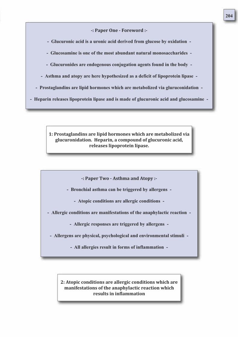
204
-: Paper One - Foreword :-
- Glucuronic acid is a uronic acid derived from glucose by oxidation -
- Glucosamine is one of the most abundant natural monosaccharides -
- Glucuronides are endogenous conjugation agents found in the body -
- Asthma and atopy are here hypothesized as a deficit of lipoprotein lipase -
- Prostaglandins are lipid hormones which are metabolized via gluruconidation -
- Heparin releases lipoprotein lipase and is made of glucuronic acid and glucosamine -
-: Paper Two - Asthma and Atopy :-
- Bronchial asthma can be triggered by allergens -
- Atopic conditions are allergic conditions -
- Allergic conditions are manifestations of the anaphylactic reaction -
- Allergic responses are triggered by allergens -
- Allergens are physical, psychological and environmental stimuli -
- All allergies result in forms of inflammation -
1: Prostaglandins are lipid hormones which are metabolized via
glucuronidation. Heparin, a compound of glucuronic acid,
releases lipoprotein lipase.
2: Atopic conditions are allergic conditions which are
manifestations of the anaphylactic reaction which
results in inflammation
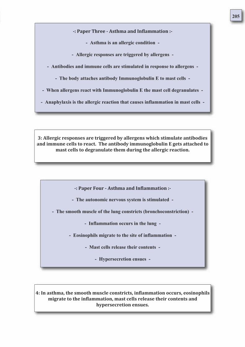
205
-: Paper Three - Asthma and Inflammation :-
- Asthma is an allergic condition -
- Allergic responses are triggered by allergens -
- Antibodies and immune cells are stimulated in response to allergens -
- The body attaches antibody Immunoglobulin E to mast cells -
- When allergens react with Immunoglobulin E the mast cell degranulates -
- Anaphylaxis is the allergic reaction that causes inflammation in mast cells -
-: Paper Four - Asthma and Inflammation :-
- The autonomic nervous system is stimulated -
- The smooth muscle of the lung constricts (bronchoconstriction) -
- Inflammation occurs in the lung -
- Eosinophils migrate to the site of inflammation -
- Mast cells release their contents -
- Hypersecretion ensues -
3: Allergic responses are triggered by allergens which stimulate antibodies
and immune cells to react. The antibody immunoglobulin E gets attached to
mast cells to degranulate them during the allergic reaction.
4: In asthma, the smooth muscle constricts, inflammation occurs, eosinophils
migrate to the inflammation, mast cells release their contents and
hypersecretion ensues.
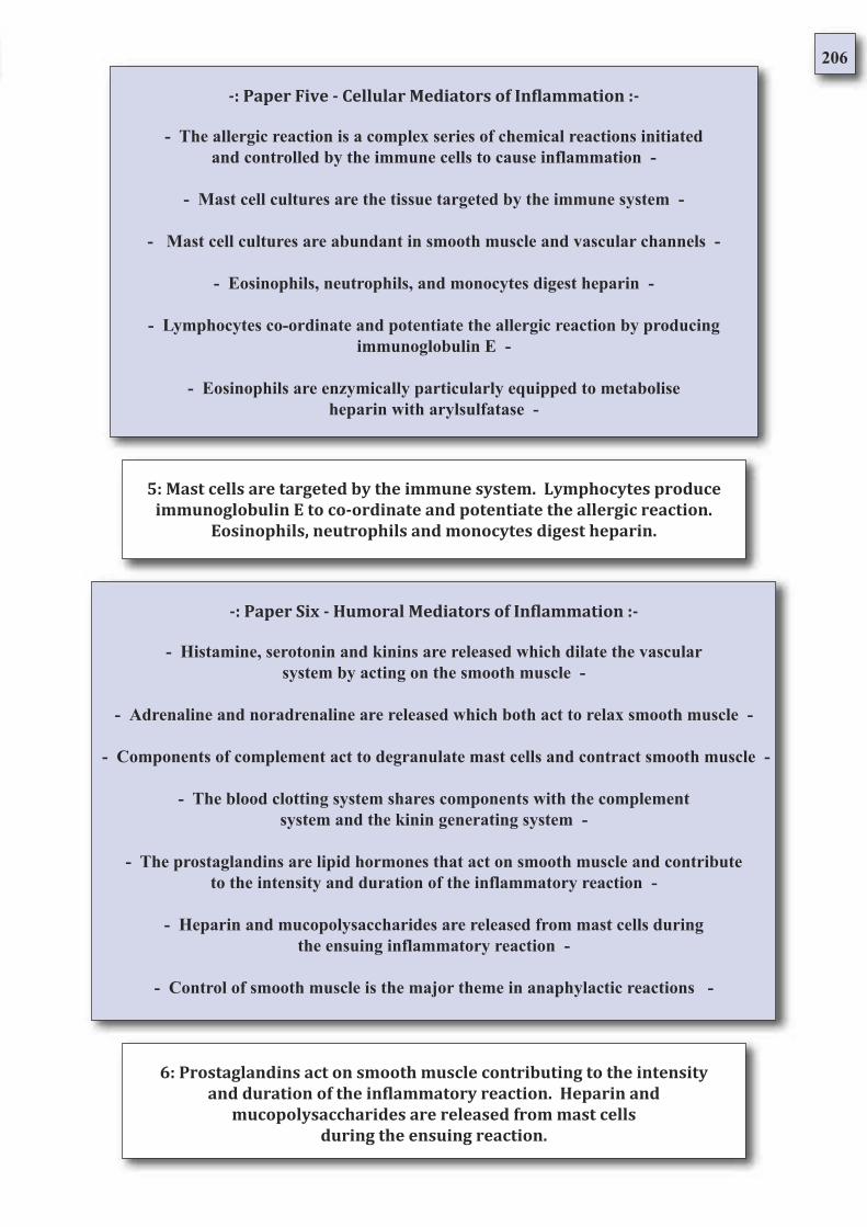
206
-: Paper Five - Cellular Mediators of Inflammation :-
- The allergic reaction is a complex series of chemical reactions initiated
and controlled by the immune cells to cause inflammation -
- Mast cell cultures are the tissue targeted by the immune system -
- Mast cell cultures are abundant in smooth muscle and vascular channels -
- Eosinophils, neutrophils, and monocytes digest heparin -
- Lymphocytes co-ordinate and potentiate the allergic reaction by producing
immunoglobulin E -
- Eosinophils are enzymically particularly equipped to metabolise
heparin with arylsulfatase -
-: Paper Six - Humoral Mediators of Inflammation :-
- Histamine, serotonin and kinins are released which dilate the vascular
system by acting on the smooth muscle -
- Adrenaline and noradrenaline are released which both act to relax smooth muscle -
- Components of complement act to degranulate mast cells and contract smooth muscle -
- The blood clotting system shares components with the complement
system and the kinin generating system -
- The prostaglandins are lipid hormones that act on smooth muscle and contribute
to the intensity and duration of the inflammatory reaction -
- Heparin and mucopolysaccharides are released from mast cells during
the ensuing inflammatory reaction -
- Control of smooth muscle is the major theme in anaphylactic reactions -
5: Mast cells are targeted by the immune system. Lymphocytes produce
immunoglobulin E to co-ordinate and potentiate the allergic reaction.
Eosinophils, neutrophils and monocytes digest heparin.
6: Prostaglandins act on smooth muscle contributing to the intensity
and duration of the inflammatory reaction. Heparin and
mucopolysaccharides are released from mast cells
during the ensuing reaction.
207
-: Paper 7 - Hypothesis of Biological Adaptation :-
- There is a hierarchical system in the biology of the organism. The brain occupies a
special position in this scheme. To illustrate this, brain gives priority to regulating its
own ATP concentration. It has been postulated that the peripheral energy supply is
only of secondary importance -
- The brain has two possibilities to ensure energy supply are allocation or intake.
‘Allocation’ refers to the allocation of energy resources between the brain and the
periphery -
- System control can be permanently displaced by extreme stress, starvation, exercise,
infectious diseases, hormones, drugs, substances of abuse, or chemical disruption of the
endocrine system -
- In the ‘Selfish Brain Theory’ a model newly describes a ‘principle of balance’ in
biological systems. The various organs and tissues must compete for allocation of a
limited number of resources -
-: Paper Eight - The Hormonal Pathology of Asthma :-
- Post mortem exam shows lungs over distended with
numerous tenacious mucous plugs -
- A dense exudate is found in the bronchial lumen -
- Eosinophils are found infiltrated in the bronchial lumen -
- Increased prostaglandins and leukotrienes
are central to the asthmatic reaction -
- Cysteinyl leukotrienes are found in increased levels in asthmatics -
- Edema produced by prostaglandins is not dose dependent -
7: There is a hierarchical system in the biology of the organism. In the
‘Selfish Brain Theory’ a model newly describes a ‘principle of balance’ in
biological systems. The various organs and tissues must compete for
allocation of a limited number of resources
8: Post mortem, asthmatic lungs are overdistended, eosinophil infiltrated
and have a dense exudate with mucous plugs. Increased prostaglandins and
leukotrienes are central to the asthmatic reaction
208
-: Paper Nine - The Metabolism of Prostaglandins :-
- Glucuronidation of fatty acids is a biotransformation
process which increases their solubility -
- Prostglandin glucuronidation is considered a
termination of their biological activity -
- Glucuronidation is the most effective detoxification process -
- Detoxification pathways may become depleted and respond poorly -
- Glucuronosyltrnasferase enzymes are suggested
to protect against prostaglandin accumulation -
- Heparin, a glycosaminoglycan, contains glucuronic acid
and is a structural precursor to lipoprotein lipase -
-: Paper Ten - Heparin and it’s Actions :-
- Heparin is both an anti inflammatory agent and an anticoagulant -
- Heparin causes the release of lipoprotein lipase -
- Heparin is a structural component of lipoprotein lipase -
- The sulfation pattern gives each glycosaminoglycan chain a unique structure -
- Heparin sulfate and related glycosmainoglycans are composed of glucuronic acid -
- Sulfatases hydrolyze a wide variety of sulfate esters -
10: Heparin is both an antiinflammatory agent and anticoagulant which
causes the release of lipoprotein lipase. Heparin is a structural component of
lipoprotein lipase and is a compound of glucuronic acid
9: Glucuronidation of prostaglandins represents a termination of their
biological activity. Glucuronidation is the most effective detoxification
process and may become depleted: Glucuronosyltransferase enzymes may
act to prevent prostaglandin accumulation
209
-: Paper Eleven - Glucuronidation :-
- The most important acid polysaccharides are those based
on glucuronic acid and acetyl galactosamine sulfate -
- Glucuronic acid conjugates with various chemicals
to allow their excretion as glucuronides -
- The amount of glucuronides in the ruine is increased
by intestinal putrifaction and by drugs -
- Glucuronic acid is known to conjugate alcoholic and phenolic hydroxyl groups -
- Glucuronic acid conjugates with the lipid female sex hormones for excretion -
- Various drugs such as antihistamines, phenothiazines and
certain antibiotics have a negative effect on heparin -
-: Paper Twelve - Hypothesis on Atopy’s Treatment :-
- Heparin is released from mast cells in response to exposure to allergens -
- Heparin inhibits all stages of cell migration -
- If heparin is injected into a subject, lipoprotein lipase is released into the bloodstream -
- Arylsulfatase is released byneutrophils, acts on aromatic and sulfur containing
comounds and is a strong inactivator of anaphylaactic events -
- Glucuronidation is main phase II metabolic pathway of drugs in humans -
- UDP-glucuronosyltransferases are enzymes which catalyze the binding of glucuronic
acid to hydroxyl, carboxyl, amino or thiol groups of chemically unrelated substances -
12: Heparin inhibits all stages of cell migration. Arylsulfatase is released by
neutrophils, acts on aromatic and sulfur containing compounds and is a
strong inactivator of anaphylactic events. UDP-glucuronosyltransferases are
enzymes which catalyze the binding of glucuronic acid to hydroxyl, carboxyl,
amine or thiol groups of chemically unrelated substances
11: Glucuronic acid conjugates various chemicals to allow their excretion as
glucuronides. The amount of glucuronides in the urine is increased by
intestinal putrifaction and by drugs
210
Synthesis
Prostaglandins are lipid hormones which are metabolized glucuronidation.
Heparin, a compound of glucuronic acid, releases lipoprotein lipase [1]. Atopic
conditions are allergic conditions which are manifestations of the anaphylactic
reaction which results in inflammation [2].
Allergic responses are triggered by allergens which stimulate antibodies and
immune cells to react. The antibody immunoglobulin E gets attached to mast
cells to degranulate them during the allergic reaction [3].
In asthma, the smooth muscle constricts, inflammation occurs, eosinophils
migrate to the inflammation, mast cells release their contents and hypersecretion
ensues [4]. Mast cells are targeted by the immune system. Lymphocytes produce
immunoglobulin E to co-ordinate and potentiate the allergic reaction.
Eosinophils, neutrophils and monocytes digest heparin [5].
Prostaglandins act on smooth muscle contributing to the intensity and duration of
the inflammatory reaction. Heparin and mucopolysaccharides are released from
mast cells during the ensuing reaction [6].
There is a hierarchical system in the biology of the organism. In the ‘Selfish
Brain Theory’ a model newly describes a ‘principle of balance’ in biological
systems. The various organs and tissues must compete for allocation of a limited
number of resources [7].
Post mortem, asthmatic lungs are overdistended, eosinophil infiltrated and have a
dense exudate with mucous plugs. Increased prostaglandins and leukotrienes are
central to the asthmatic reaction [8].
Glucuronidation of prostaglandins represents a termination of their biological
activity. Glucuronidation is the most effective detoxification process and may
become depleted: Glucuronosyltransferase enzymes may act to prevent
prostaglandin accumulation [9].
Heparin is both an antiinflammatory agent and anticoagulant which causes the
release of lipoprotein lipase. Heparin is a structural component of lipoprotein
lipase and is a compound of glucuronic acid [10].
Glucuronic acid conjugates various chemicals to allow their excretion as
glucuronides. The amount of glucuronides in the urine is increased by intestinal
putrifaction and by drugs [11].
Heparin inhibits all stages of cell migration. Arylsulfatase is released by
neutrophils, acts on aromatic and sulfur containing compounds and is a strong
inactivator of anaphylactic events. UDP-glucuronosyltransferases are enzymes
which catalyze the binding of glucuronic acid to hydroxyl, carboxyl, amine or
thiol groups of chemically unrelated substances [12].
211
Here we have a reduction of the deducible facts derived from the concorded
literature review which forms the basis of this study. From this set of data, a line
of inductive rationale behind the autoimmune conditions which manifest
anaphylactic inflammation, or type 1 hypersensitivity reactions.
It is proposed that through depletion of glucuronic acid and the poor response of
systems which require glucuronic acid, eicosanoid metabolism becomes
unregulated affecting, amongst other systems, smooth muscle in asthma for
example.
In an effort to resolve the local deficits brought about through increases in
demand, it is proposed that the immune system acts in a logistical capacity to
speedily requisit glucuronic acid resources from tissue through the aciton of
anaphylactic inflammation.
A chief resource of glucuronic acid is that of the maast cell. The mast cell is an
abundant source of sulfated glycosaminoglycans including heparin which is a
structural precursor to lipprotein lipase. Glycosaminoglycans, heparin and
lipoprotein lipase all represent resources of glucuronic acid.
Any increase in a demand for glucuronidation in a system deficient of glucuronic
acid, caused either by xenobiotics or endogenous lipid hormones, will require a
trade-off in opportunity costs between chemically inactivating foreign compounds
or chemically regulating endogenous hormones.
The body thus is forced to keep itself alive by the relatively violent action of
immune cells migrating to the site of inflammation to render glucuronic acid from
glycosaminoglycan molecules in the tissues. Mast cells are even specifically
tagged by immunoglobulin E produced by lymphocytes to facilitate anaphylaxis
as a course of action.
It is suggested that the body does this act of forced reallocation via use of large
amounts of arylsulfatase found in th emast cell, eosinophil and neutrophil.
Arylsulfatase acts to cleave aromatic and sulfur containing compounds, helping to
liberate the glucuronic aicd necessary to fill the metabolic demand.
Once enought glucuronic aicd has been released in its active form it is suggested
that the anaphylactic inflammation subsides having the resources to both regulate
any lipid hormones released or eliminate from the system xenobiotics.
According to this scheme of thought, an anaphylactic event can be brought about
(a) through endogenous production of eicosanoid hormones sufficient to outstrip
supply of glucuronic acid (exercise, emotion, cold) or (b) through toxic challenge
of a xenobiotic load sufficient to outstrip supply of glucuronic acid and impinge
on the balanced functioning of the eicosanoid hormones.
This scheme leaves adequate room to suggest that both the histamine reaction and
anaphylactic reaction in general are essential mechanisms of human biology.
Histamine seems to be integral to facilitating the migration of immune cells to the
tissue, and the immune reaction as a whole is proposed as an important self
preservation mechanism designed to keep the organism alive in states of deficit.
212
In a state of deficiency the body draws progressively upon more important stores
and resources of substances it requires to balance an immediate metabolic deficit;
The body draws upon biologically less important substances to manufacture from
them biologically more important and immediately required substances.
There is a hierarchical system in the biology of the organism. In the ‘Selfish
Brain Theory [7.1]’ a model newly describes a ‘principle of balance’ in biological
systems. The various organs and tissues must compete for allocation of a limited
number of resources. The principle is that the body will look after a more
important tissue before it looks after a less important tissue.
Importance in these terms refers to a relative scale of absolute requirement and
immediacy of need. The economics of alternative/opportunity costs and the
logistics biology thus must be taken as factors in the interpretation of biological
function.
The body draws upon the least important and most plentiful resources first. The
body prioritizes the functions, organs, tissues and chemical stores according to
how essential they are in the functioning of the body. In short there is a hierarchy
amongst the tissues of the body.
We are literally an assembly of what we eat, drink and breath. Ultimately the
body relies upon the diet for all its building blocks and fuel. A medical condition
manifests itself when the first metabolic action is impaired - that is to say - a
metabolically active substance is negatively affected and the function which it
supports is impeded forcing a biological adaptation of normal metabolism to
occur.
As deficiency progresses the body is forced to requisite resources so to supply
more vital functions with the components which are required to support the
biological action. An increasing amount of symptoms occur with an increasing
deficit of resources. When a biological function breaks down it is related as a
symptom.
The more active the substance that is requisited for reassignment of its
constituents - the more important the function that is affected in the running of the
body - the more dramatic is the symptom of deficit.
The greater the stage of deficiency the more important the function or tissue
which is affected. Deficit may be brought about by lack of the substance in the
diet, by increased utilization of the substance so that demand outstrips supply, or
by the destruction of the substance.
213
Bronchial Asthma and the Atopic Syndrome
Paper 14: Addendum - Extended Analysis
Now a deductive study of our data set has allowed the formulation of a
hypothesis through a process of inductive reasoning, it remains for some form
of falsification to be made of said hypothesis so to begin to test its rationale as a
reliable model. Various forms of falsification are available but for the purposes
of this project, a type of falsification method which is literature based is most
cogent with the overall framework ‘a study of the anatomy of a study’.
This paper represents the taking of a focused data set and applying the treatment
of language found in the concorded literature review which prefaces this paper.
As the treatment of language has been illustrated, this style of arranging and
reporting omits the concorded text but includes its sources for reference via
bibliography.
The style of anotation indicates the absolutely finite lineage of language back to
a specific piece of text. Where there is not found a reference number, it is an
authored statement. It is in the study of the common instrument of science, that
of the mode of communication and codification of information in such a way
that it not just holds information but also makes that information available thus
making communication and communicative endeavour possible; the basis of all
peer review and literature based learning.
It is in the collection of a further data set a form of falsification can be made not
only to get rid of incorrect assertions but to gain guidance as to whether the
interpretation of the available data is moving closer to a truthful representation
or further away. It is as much to rule out wasting time studying the wrong thing
as to answer the question in mind as it is to make a scrutinous study of the only
relevant factors.
The natural coaslescence of information is enough to make a prospective study
in something. Corroboration between data sets advances the study further down
the road towards reliable knowledge and performs the task of providing a
second landscape which must be conjunctive not only with the first data set in a
mutual verification and should also provide an articulated obstacle for any
hypothesis made before its collection.
214
Extended Analysis Index
Bronchial asthma
Allergic Challenge Asthma
Mast Cells
Allergic Response
Inflammation
Inositol
Histamine
Eosinophil
Arylsulfatase
Prostaglandins
M2 Acetylcholine Muscarinic Receptors
Heparin
Glycosaminoglycans
Eczema
Crohn’s disease
Arthritis
Hay Fever
Anaphylactic Shock
215
Asthma is a common chronic inflammatory condition of the lung airways whose cause is incompletely
understood. Symptoms are cough, wheeze, chest tightness and shortness of breath, often worse at night.
It has three characteristics: airflow limitation which is usually reversible spontaneously or with treatment;
airway hyperresponsiveness to a wide range of stimuli; inflammation of the bronchi with eosinophils,
T lymphocytes and mast cells with associated plasma exudation, oedema, marked smooth muscle
hypertrophy, mucus plugging and epithelial damage [32].
Bronchial Asthma
In chronic asthma, inflammation may be accompanied by irreversible airflow limitation.
The underlying pathology in pre-school children may be different, in that they may not
exhibit appreciable bronchial hyperreactivity. There is no evidence that chronic
inflammation is the basis for the episodic wheezing associated with viral infections [32].
In many countries the prevalence of asthma is increasing, particularly in the second decade
of life where this disease affects 10-15% of the population. There is also a geographical
variation, with asthma being common in more developed countries, some of the highest
rates being in New Zealand, Australia and the UK, but being much rarer in Far Eastern
countries such as China and Malaysia, and in Africa and Central and Eastern Europe [32].
Long-term follow-up in developing countries suggests that the disease may
become more frequent as individuals become more 'westernized'. Studies of
occupational asthma suggest that a high percentage of the workforce, perhaps
up to 20%, may become asthmatic if exposed to potent sensitizers [32].
Asthma can be divided into Extrinsic (implying a definite external cause) and Intrinsic or
Cryptogenic (when no causative agent can be identified) forms. Extrinsic asthma occurs
most frequently in atopic individuals who show positive skin-prick reactions to common
inhalant allergens. Positive skin-prick tests to inhalant allergens are shown in 90% of
children and 50% of adults with persistent asthma [32].
Childhood asthma is often accompanied by eczema. An overlooked cause of late-onset
asthma in adults is sensitization to chemicals or biological products in the workplace.
Intrinsic asthma often starts in middle age ('late onset'). Nevertheless, many patients with
adult-onset asthma show positive skin tests and on close questioning give a history of
respiratory symptoms compatible with childhood asthma [32].
216
There are two major factors involved in the development of asthma and many other stimuli that
can precipitate attacks. The term 'atopy' was used by clinicians at the beginning of the century
to describe a group of disorders, including asthma and hayfever, that appeared to run in families; to
have characteristic wealing skin reactions to common allergens in the environment; to have circulating
antibody in their serum that could be transferred to the skin of non-sensitized individuals [32].
The term is best used to describe those individuals who readily develop antibodies of IgE class
against common materials present in the environment. Such antibodies are present in 30-40% of
the UK population, and there is a link between serum Immunoglobulin E (IgE) levels and both
the prevalence of asthma and airway hyperresponsiveness [32].
Genetic and environmental factors affect serum IgE levels. The use of DNA microsatellite
markers to scan the entire genome has uncovered 22 chromosomal regions of interest
containing candidate genes. Some of these, in combination with environmental factors,
may turn out to play a key role in the development of asthma [32].
The genes controlling the production of the cytokines Interleukin 3 (IL- 3), Interleukin 4 (IL-4), Interleukin 5
(IL-5), Interleukin 9 (IL-9), Interleukin 13 (IL-13) and Granulocyte-Macrophage Colony Stimulating Factor
(GM-CSF) - which in turn affect mast and eosinophil cell development and longevity as well as
immunoglobulin E production - are present in a cluster on chromosome 5q31-33 (the IL-4 gene cluster) [32].
A gene on chromosome 2, PHF11, which controls IgE synthesis is strongly associated
with atopy whereas ADAM33 on chromosome 20 is more strongly associated with airway
hyperresponsiveness and the tissue changes of remodelling. Early childhood exposure to
allergens and maternal smoking have a major influence on IgE production [32].
Much current interest focuses on the role of intestinal bacteria and childhood in
shaping the immune system in early life. It has been suggested that growing up in a
relatively 'clean' environment may predispose towards an immunoglobulin E response
to allergens (the hygiene hypothesis) [32].
Non-atopic individuals may develop asthma in middle age from extrinsic causes
such as sensitization to occupational agents or aspirin intolerance, or because they
were given adrenoceptor-blocking agents for concurrent hypertension or angina.
Extrinsic causes must be considered in all cases of asthma [32].
217
Although the degree of hyperresponsiveness can itself be influenced by allergic mechanisms, its pathogenesis
involves a combination of airway inflammation and tissue remodelling. Over 200 materials encountered at the
workplace give rise to occupational asthma. The causes are recognized occupational diseases in the United
Kingdom, and patients in insurable employment are therefore eligible for statutory compensation provided they
apply within 10 years of leaving the occupation in which the asthma developed [32].
Asthma due to flour, organic dusts and other large protein molecules involves
specific immunoglobulin E antibodies. In contrast, reactive chemicals such as
isocyanates and acid anhydrides bond chemically to epithelial cells to activate
them as well as provide haptens recognized by T cells [32].
Cockroach allergy has been implicated in asthma in United States inner-city children, while allergens from furry
pets are also becoming increasingly common causes of asthma, rhinitis and urticaria. The fungal spores from
Aspergillus fumigatus give rise to a complex series of lung disorders, including asthma. Many allergens
including those from Aspergillus have intrinsic biological properties, e.g. proteolytic enzymes that facilitate
their passage through the airway epithelium to increase their sensitizing capacity [32].
Bronchial hyperresponsiveness can be demonstrated by asking the patient to inhale gradually increasing concen-
trations of either histamine or methacholine (bronchial provocation tests). This induces transient airflow limitation
in susceptible individuals (approximately 20% of the population); the dose of the agonist (provocation dose) neces-
sary to produce a 20% fall in FEVj is known as the PD20 FEVj (or PC20 FEV1) [32].
Patients with clinical symptoms of asthma respond to very low doses of methacholine; i.e. they have a low PC20 FEV1
(< 11 μmol). In general, the greater the degree of hyperreactivity, the more persistent the symptoms and the greater the
need for treatment. Some patients also react to methacholine but at higher doses and include those with attacks of asthma
only on extreme exertion; wheezing or prolonged periods of coughing following a viral infection; cough variant asthma;
seasonal wheeze in pollen season; allergic rhinitis, but not complaining of any lower respiratory symptoms until specifi-
cally questioned; some subjects with no respiratory symptoms [32].
Conversely, growing up in a 'dirtier' environment may allow the immune system to avoid
developing allergic responses. Components of bacteria (e.g. lipopolysaccharide endotoxin;
immunostimulatory CpG DNA sequences), viruses (e.g. DS RNA) and fungi (e.g. chiton, a cell
wall component) are able to stimulate receptors expressed on immune cells to direct the
immune and inflammatory response away from the allergic pathways [32].
Thus early life exposure to inhaled and ingested products of microorganisms may be critical in
helping shape the subsequent risk of a child becoming allergic and/or developing asthma. The
allergens involved in asthma are similar to those in rhinitis. Allergens from the faecal particles
of the house-dust mite are the most significant extrinsic cause of asthma world-wide [32].
218
It is thought that treatment with these drugs leads to an imbalance in the metabolism of arachidonic acid.
non-steroidal anti-inflammatory drugs inhibit arachidonic acid metabolism via the cyclo-oxygenase (COX)
pathway, preventing the synthesis of prostaglandins. It has been suggested that under these circumstances
there is a reduced production of prostaglandin E2 and an overproduction of cysteinyl leukotrienes by
eosinophils, mast cells and macrophages [32].
Non-Steroidal Anti-Inflammatory Drugs (NSAIDs), particularly aspirin and propionic acid de-
rivatives, e.g. indometacin, have a major role in the development and precipitation of attacks
in approximately 5% of patients with asthma. This effect is especially prevalent in those indi-
viduals who have both nasal polyps and asthma [32].
Increased intakes of fresh fruit and vegetables, or unprocessed diets have been shown to be protective.
Genetic variation in antioxidant enzymes is associated with more severe asthma. It is well known that
emotional factors may influence asthma, but there is no evidence that patients with the disease are any
more psychologically disturbed than their non-asthmatic peers [32].
Asthma exacerbations increase in both summer and winter air pollution episodes associ-
ated with climatic temperature inversions. Epidemics of the disease have occurred in the
presence of high concentrations of ozone, particulates and NO2 in the summer and particu-
lates, Nitrogen Dioxide (NO2) and Sulphur Dioxide (SO2) in the winter [32].
Many patients with asthma experience worsening of symptoms on contact with
cigarette smoke, car exhaust fumes, strong perfumes or high concentrations of dust
in the atmosphere. Major epidemics have been recorded when large amounts of al-
lergens are released into the air, e.g. soy bean epidemic in Barcelona [32].
The risk of developing some forms of occupational asthma increases in smokers. The proportion of employees
developing occupational asthma depends primarily upon the level of exposure. Proper enclosure of industrial
processes or appropriate ventilation greatly reduces the risk. Atopic individuals develop occupational asthma
more rapidly when exposed to agents causing the development of specific. Non-atopic individuals can also de-
velop asthma when exposed to such agents, but after a longer period of exposure [32].
The characteristic feature of bronchial hyperresponsiveness in asthma means that, as well as
reacting to specific antigens, the airways will also respond to a wide variety of non-specific
direct and indirect stimuli such as cold air and exercise. Most asthmatics wheeze after pro-
longed exercise. Typically, the attack does not occur while exercising but afterwards. The
inhalation of cold, dry air will also precipitate an attack [32].
Exercise-induced wheeze is driven by histamine and leukotrienes which are released
from mast cells when the epithelial lining fluid of the bronchi becomes hyperosmolar
owing to drying and cooling during exercise. The phenomenon can be shown by exer-
cise, cold air and hypertonic (e.g. saline or mannitol) provocation tests [32].
Following an immediate reaction many asthmatics develop a more prolonged and
sustained attack of airflow limitation (Dual and late-phase reactions) that responds
less well to inhalation of bronchodilator drugs such as salbutamol [32].
219
The pathogenesis of asthma is complex and not fully understood. It involves a number of
cells, mediators, nerves and vascular leakage that can be activated by several different
mechanisms, of which exposure to allergens is among the most significant [32].
Isolated late-phase reactions with no preceding immediate response can occur after the inhalation
of some occupational sensitizers such as isocyanates. The development of the late-phase reaction
is associated with an increase in the underlying level of airway hyperresponsiveness such that indi-
viduals may show continuing episodes of asthma on subsequent days [32].
The varying clinical severity and chronicity of asthma is dependent on an interplay between
airway inflammation and airway wall remodelling. The inflammatory component is driven
by Th2-type T lymphocytes which facilitate immunoglobulin E synthesis through production
of Interleukin-4 and eosinophilic inflammation through Interleukin-5 [32].
The experimental inhalation of allergen by atopic asthmatic individuals leads to the development of dif-
ferent types of reaction. Immediate asthma (early reaction): Airflow limitation begins within minutes of
contact with the allergen, reaches its maximum in 15-20 minutes and subsides by 1 hour [32].
Inhibition of this effect by b-adrenoceptor blocking drugs such as propranolol leads to bron-
choconstriction and airflow limitation, but only in asthmatic subjects. The so-called selective
b-adrenergic blocking drugs such as atenolol may still induce attacks of asthma; their use to
treat hypertension or angina in asthmatic patients is best avoided [32].
The airways have a direct parasympathetic innervation that tends to produce bronchoconstriction.
There is no direct sympathetic innervation of the smooth muscle of the bronchi, and antagonism of
parasympathetically induced bronchoconstriction is critically dependent upon circulating epinephrine
(adrenaline) acting through M2-receptors on the surface of smooth muscle cells [32].
In such patients there is evidence for polymorphisms involving the promoter region of the Leukotriene C4
synthase gene that controls the level of activity of this terminal enzyme of the leukotriene-generating path-
way. Interestingly, asthma in intolerant patients is not precipitated by COX-2 inhibitors, indicating that it
is blockade of COX-1 that is linked to impaired prostaglandin E2 production [32].
220
As defined by the management guidelines issued in 1999 by the National
Heart, Lung and Blood Institute standard asthma therapeutics include oral
and inhaled corticosteroids, leukotriene antagonists, short acting and long
acting ß-antagonists, cromolyn and nedocromil [12].
Elevated levels of heparin-like anticoagulants have been
demonstrated in atopic asthmatic patients, and have been
induced in some patients after antigen challenge [12].
Several studies in the 1960s first reported subjective improvement in asthmatic
symptoms with use of IV heparin. Bardana et al performed the first trial of in-
haled heparin reporting subjective but not objective improvements [12].
Asthma is one of the most common chronic diseases in industrialized countries, affecting 10% or more of
young children in some countries. An atopic disease with high IgE levels, asthma is induced by small
particles of antigen, which are able to penetrate deep into the lungs. About 80% of asthmatic children are
allergic to house mites. Animal danders are another major cause [21].
The incidence of asthma appears to be increasing in many modern societies, but
the reasons are unclear, and the increase doesn’t appear to be linked to air pollution.
A chronic atopic condition usually precedes an acute attack of asthma [21]
In addition to IgE and eosinophils there are excessive numbers of neutrophils with
high-affinity IgE receptors in the airway tissues. Prostaglandin D2 released from
mast cells may play a role in triggering an attack. Cytokines, nitric oxide, and nerve
growth factor may also participate in the response [21].
The presence of a high concentration of glutathione and of glutathione peroxidase, whose concen-
tration increases in asthmatic lungs, may reflect the action of the antioxidant system in combating
inflammation. Concentrations of surfactant proteins SP-A and SP-D are Ca2+- dependent lectins
which serve as regulators of the innate immune response increase in asthma [21].
221
Exercise induced Asthma
Inhaled heparin prevents bronchoconstriction response in exercise induced
asthmatic subjects [5][7]. Heparin prevents Exercise-Induced Asthma (EIA)
but not histamine induced bronchoconstriction. Heparin attenuates post exer-
cise decrease of the specific airway resistance [7].
Inhaled heparin attenuates acute bronchoconstrictor response to exercise. Heparin
completely, or partially inhibited exercise induced bronchoconstriction in 75% of
subjects. Inhaled low molecular weight heparin, enoxaparin attenuates exercise-in-
duced bronchoconstriction in a dose dependent manner [5].
Inhaled heparin inhibits early response to
exercise induced bronchoconstriction in
subject with asthma [6].
Bronchial asthma is recognized as an inflammatory disease which is character-
ized by bronchial hyperresponsiveness, excessive mucus production, mucosal
oedema and recruitment of inflammatory cells into the airways [10].
Heparins ability to inhibit airway responsiveness is unlikely to be related
solely to its highly anionic nature as similar effects were not observed
with the linear anionic molecule polyglutamic acid [10].
Aerosolized heparin may be helpful in alleviating symptoms of asthma although
no definitive bronchodilating activity has been observed [11a]. It is suggested that
heparin may be a natural "anti-asthmatic" molecule [7][11b].
The anti-asthmatic activity of heparin is thought not to be due to its anticoagulant properties as
the agent did not prolong the partial thromboplastin time measured one hour or three hours after
inhalation. The administration of heparin to sheep failed to change the partial thromboplastin
time for up to 12 hours after inhalation [11a].
Part of the immune response consists of the release from stimulated neutrophils, macrophages, and
other cells of Platelet Activating Factor (PAF: 1-O-alkyl-2-acetyl-sn-glycerophosphocholine), a material
that “activates” blood platelets. The principal interest in the platelet-activating factor has arisen
from its powerful effect in inducing inflammation. It is a remarkably potent compound, its effects
on platelets are observed at concentrations of 10–11 to 10–10 M [22].
222
Heparin prevents exercise-induced asthma without influencing histamine induced bronchoconstriction.
Various forms of exercise can induce post exercise bronchoconstriction in asthmatics attributed to large
volumes of inadequately conditioned air which leads to a loss of heat and water from respiratory mu-
cosa. The mechanism whereby airway cooling leads to bronchoconstriction is not clear [11a].
Mast cell stablilizing agents (i.e. cromolyn sodium) are known to attenuate exercise induced asthma. Inhaled heparin
prevents post-exercise bronchonconstriction in subjects with exercise induced asthma [11a]. Pharmacologic agents
including beta agonists, cromolyn sodium, atropine, furosemide and calcium-channel blockers offer varying degrees
of protection against exercise-induced asthma or asthma due to hyperventilation with cold dry air. Heparin was
found to be more effective than cromolyn in preventing post exercise bronchoconstriction [11a][11c].
Inhaled heparin preserves specific airway conductance (sGaw) in exercise in-
duced asthma better than did 20 mg inhaled cromolyn. Inhaled heparin was
superior in preserving specific airway conductance in exercise induced
asthma when administered up to 3 hours prior to exercise [12].
Mast cell mediators, including histamine and leukotrienes have been implicated in the pathogenesis of exer-
cise-induced asthma and H-histamine receptor antagonists as well as leukotriene D-receptor or
5-lipoxygenase inhibitors partly attenuate bronchoconstrictor responses induced by exercise or cold dry air
[11a]. Heparin prevents the bronchoconstrictor response in subjects with exercise induced asthma [11c].
Heparin inhibits exercise induced bronchoconstriction possibly by altering inositol
triphosphate (IP3) levels. Inhibition of Platelet Activating Factor (PAF)-induced air-
way hyperresponsiveness and cell infiltration by heparin cannot be explained by he-
parin acting merely as a platelet activating factor antagonist [10].
One effect of platelet activating factor on platelets is to induce a rapid (5–10s) cleavage of phosphatidylinositol
4,5-bisphosphate by phospholipase C to give diacylglycerol and inositol 1,4,5-trisphosphate. The subsequent ef-
fects of these two substances cause a rapid influx of Ca2+ and induce a series of secondary responses [22].
These compounds are also potent activators of platelets and cause a self-activating or
autocrine effect. While platelet activating factor has a beneficial function, it can
under some conditions contribute in a dangerous way to inflammation and to allergic
responses including anaphylaxis, asthma and cold-induced urticaria [22].
Among these responses are the release of the materials stored in the platelet’s granules. Platelet
activating factor also appears to inhibit adenylate cyclase and causes vasodilation, a property not
expected for a compound that stimulates clotting. Stimulated platelets release arachidonic acid
rapidly from their phospholipids, apparently as a result of activation of phospholipase A2. The re-
leased arachidonate can in turn be metabolized to endoperoxides and thromboxane A2 [22].
223
Use of a 5-lipoxygenase activating protein antagonist confirmed that leukotrienes significantly
contributed to the development of hyperventilation induced bronchoconstriction. Heparin attenu-
ates hyperventilation induced bronchoconstriction in asthmatics and acute antigen-induced bron-
choconstriction in sheep without modifying the effects of histamine [13].
Heparin tends to reduce the number of epithelial cells recovered in Broncho-Alveolar
Lavage Fluid (BALF) after dry air challenge. Inhaled heparin inhibits eicosanoid media-
tor production and release caused by hyperventilation with dry air and significantly at-
tenuates hyperventilation induced bronchoconstriction [13].
Mast cell degranulation of lung by conditions other than anaphylaxis, such as hy-
poxia or exercise in certain asthmatics may release Prostaglandin-generating fac-
tor of anaphylaxis (PGF-A) and lead to prostaglandin formation [20].
Exercise induced asthma is characterized by transient airway obstruction typically occurring
after hyperventilation with cold dry air. Airway Surface Fluid (ASF) osmolality increases dur-
ing hyperventilation and correlates with the magnitude of obstruction [13].
Reduction in hyperventilation induced airway obstruction caused by heparin is ac-
companied by a concomitant decrease in eicosanoid mediator concentrations. He-
parin inhibits hyperventilation induced bronchoconstriction via the inhibition of
either hyperventilation induced eicosanoid production or release [13].
Changes in airway surface fluid osmolality in canine peripheral airways are associated with bronchial mu-
cosal injury, mast cell degranulation and the elaboration of bronchoactive mediators. Inhaled heparin in-
hibits Hyperventilation Induced Bronchoconstriction (HIB) in humans. Numerous animal and human
studies suggest that a variety of eicosanoids contribute to the development of hyperventilation induced
bronchoconstriction. Inhalation of heparin attenuates hyperventilation induced bronchoconstriction [13].
A novel mediator of human lung anaphylaxis termed prostaglandin-generating factor of anaphylaxis (PGF-A)
has been described which may be partly responsible for prostaglandin generation accompanying allergic reac-
tions. PGF-A is an oligopeptide of 1,450 daltons that induces the production of PGFa, PGE, TXB2 and 5-,
12-, and 15-hydroxyeicosatetraenoic acid (HETE) from human lung parenchyma and airways [23].
Hyperventilation induced increases in Leukotriene C4 (LTC4), Leukotriene E4 (LTE4) and
Prostaglandin F2a (PGF2a) are inhibited and Prostaglandin D2 (PGD2) and Thromboxane B2
(TxB2) remain at baseline levels in heparin treated/dry air challenged airways [13].
Experimental evidence suggests that this factor is not preformed (like histamine),
but newly synthesized or rapidly generated by the anaphylactic event (like SRS-A).
Mediators other than histamine and PGF-A such as SRS-A and bradykinin have
also been shown to be capable of causing prostaglandin formation [23].
224
Allergic Challenge Asthma
Heparin decreased the response to vagally induced bronchoconstriction in sensitized antigen chal-
lenged animals [4]. Inhaled heparin attenuates antigen induced bronchoconstriction in allergic sheep
as well as prevents bronchoconstriction response to antigen in asthmatic human subjects [5].
Inhaled heparin prevents antigen induced bronchoconstriction and airway responsiveness
by modulation of mast cell mediator release [5]. Inhaled heparin inhibits early response to
allergen induced bronchoconstriction in subjects with asthma [6].
Heparin attenuates bronchoconstrictor response and immediate cutaneous re-
action to antigen in allergic sheep. Heparin inhibits the acute cutaneous reac-
tion and bronchoconstriction due to allergens in allergic subjects [7].
Heparin attenuated antigen induced bronchoconstriction in a dose dependent manner, and inhibited
bronchoconstriction responses to compound 48/80. Heparin also inhibits bronchoconstriction by in-
haled dust mite extract in allergic asthmatics [7]. In asthma, heparin in a single dose reduced early asth-
matic response to inhaled allergen (house dust mite extract) [11b].
Ether linked phospholipid platelet activating factor (PAF) plays an important role in the pathogenesis
of asthma as it can reproduce many of the features of the disease. Unfractionated heparin and the low
molecular weight heparinoid Org 10172 can inhibit airway hyperresponsiveness induced by
aerosolized platelet activated factor in neonatally immunized adult rabbits [10].
The ability of heparin and Org 10172 heparinoid to inhibit airway responsiveness to
histamine following Bovine Serum Albumen (BSA) challenge suggests an action
not attributable merely to the ability to bind histamine [10].
Both heparin and low molecular weight heparinoid, Org 10172, possess anti-inflammatory ac-
tivity in the lung and the ability to inhibit Platelet Activated Factor (PAF)-induced airway hy-
perresponsiveness in the rabbit. Heparin also inhibits platelet activated factor induced airway
hyperresponsiveness and eosinophil infiltration in guinea pigs [10].
Compound 48/80 is a polymer produced by the condensation of N-methyl-p-methoxyphenethylamine with
formaldehyde. It promotes histamine release. In biochemical research, compound 48/80 is used to pro-
mote mast cell degranulation. Heparin attenuates the effect of compound 48/80 and antigen-induced acute
bronchoconstrictor responses in sheep without modifying the effects of histamine [11a].
225
Inhaled heparin prevents allergic bronchoconstriction
in sheep and inhibits anti-IgE-mediated release of his-
tamine from mast cells in vivo [11a].
Heparin had no effect on histamine induced bronchoconstriction [11a]. Heparin does not
alter airway reactivity to histamine in human asthmatics or sheep. Heparin attenuates hy-
perventilation induced bronchoconstriction in asthmatics and acute antigen induced bron-
choconstriction in sheep without modifying the effects of histamine [13].
Heparin attenuates allergic bronchoconstriction in sheep, inhibits anti-IgE medi-
ated histamine release in isolated mast cells and prevents the bronchonconstrictor
response in subjects with exercise induced asthma [11c].
Analysis of the time course of the appearance of prostaglandins during human lung anaphylaxis
reveals a close and parallel relationship to histamine release. Histamine acts through an H1 re-
ceptor causing bronchial smooth muscle constriction which produces prostaglandin E [20].
A portion of the prostaglandin generation of anaphylaxis of human lung is due to histamine.
Addition of exogenous histamine generates about 50% of the prostaglandins produced dur-
ing anaphylaxis and H1 histamine receptor antagonists prevent about 50% [20].
Histamine and other mediators released from human mast cells un-
dergoing anaphylaxis are able to induce prostaglandin generation
and are found in the supernatant from lung anaphylaxis [20].
Rabbit antisera directed at human IgE molecules induce mediator release which in
the lung result in the release or formation of histamine, slow reacting substance of
anaphylaxis, eosinophil chemotactic factors of anaphylaxis, superoxide radicals,
prostaglandins F2a, E2, and thromboxane A2 and kallikrein of anaphylaxis [20].
A close direct relationship exists between the intensity of anaphylaxis induced as re-
flected by the concentration of histamine released from human lung during anaphylaxis
and the quantity of prostaglandin generating factor of anaphylaxis released [20].
226
Pre-treatment with 1000 U/kg heparin attenuated the increase in the lung resistance of previously
sensitized sheep by 91% on allergen exposure [12]. Inhaled heparin attenuates antigen-induced
bronchoconstriction and airway hyperresponsiveness in sheep [13]. An N-desulfated heparin failed
to prevent antigen induced bronchoconstriction [11a].
Anti-allergic actions of heparin may be related to the inhibition of mediator release from mast
cells. Heparin attenuates antigen induced bronchoconstriction in humans with asthma and can in-
hibit histamine release from isolated human mast cells. Heparin also inhibits histamine release
from blood cells induced by antigen, trypsin and other proteases [11a].
Platelets factor 4 (PF4) and related member of the IL-8 supergene family, RANTES
are chemoattractants for eosinophils. Heparin inhibits the actions of platelets factor
4, a cationic protein shown to be released following antigen challenge in sensitized
rabbits and human asthmatics [10].
Heparin restores M2 receptor function by binding to and neutralizing major basic
protein and polycationic substances [4]. Heparin reverses allergen induced in-
hibitory M2 receptor dysfunction [6]. Heparins inhibition of bronchoconstriction is
co-incident with return to normal of M2 receptor function [4].
Antigen induced hyperresponsiveness is associated with increased release of
acetylcholine from vagus nerves [4]. Inhibitory M2 muscarinic receptors control
release of acetylcholine from human pulmonary parasympathetic nerves.
In asthmatics the M2 acetylcholine muscarinic receptors do not function [4][6].
Heparin can modulate allergen airway hyperresponsiveness in guineapigs via a mechanism related
to reversing the effects of the eosinophil derived cationic protein Major Basic Protein (MBP) on M2
receptor function on airway ganglia [10]. Heparin failed to inhibit acetylcholine induced tracheal
smooth muscle contraction in tissue culture [7]. Inhaled heparin inhibits methacholine-induced bron-
choconstriction and is suggested to act directly on airway smooth muscle [7][13].
227
Mast cells
Stimulation of mast cells starts a series of cellular events and culminates
in mediator release. Mast cell stablilizing agents (i.e. cromolyn sodium) at-
tenuate exercise induced asthma. Both non immunologic and immuno-
logic stimuli can degranulate mast cells [11a].
Changes in Airway Surface Fluid (ASF) osmolality in canine peripheral
airways are associated with bronchial mucosal injury, mast cell degranu-
lation and the elaboration of bronchoactive mediators [13].
Mast cells produce Prostaglandin D2 (PGD2) and Prostaglandin Prostacyclin
(PGI2) but not the Prostaglandin F2a (PGF2a), Prostaglandin E (PGE), and Throm-
boxane A2 (TxA2) produced by lung anaphylaxis [20].
Histamine and other mediators released from human mast cells under-
going anaphylaxis are able to induce prostaglandin generation and are
found in the supernatant from lung anaphylaxis [20].
Mast cell degranulation of lung by conditions other than anaphylaxis, such as hypoxia or exercise in certain
asthmatics may release Prostaglandin Generating Factor of Anaphylaxis (PGF-A) and lead to prostaglandin
formation. Prostaglandin generation secondary to prostaglandin generating factor of anaphylaxis and other
mast cell derived mediators might contribute to alterations in lung function associated with variety of condi-
tions which involve mast cell degranulation [20].
Breakdown of inositol phospholipids leads to the generation of 1,4,5-inositol triphosphate which binds to
receptors on the endoplasmic reticulum and causes internal release of calcium in mast cells and many
other cells. Inositol triphosphate can degranulate mast cells and cause histamine release [11a].
Mast cells occur particularly often as single cells, but more frequently in small groups, in
the vicinity of small vessels. Mast cells (diameter 6–12μm) contain a rounded nucleus.
Their cytoplasm is loaded with basophilic, metachromatic granules. Paul Ehrlich (1877)
interpreted these as alimentary storage granules (Ehrlich’s mast cells) [24].
Mast cell activation stimulates the generation and release of newly formed mediators, in-
cluding prostaglandin D2 and the 5-lipooxygenase products leukotrienes C4 and D4 pre-
viously identified as slow acting substance of anaphylaxis [18].
Mast cells synthesize, store and extrude the acid and sulfatized glycosaminoglycan heparin, the
biogenic amine histamine, and also additional factors which play a role in anaphylactic reactions.
Histamine is released in large amounts during allergic reactions. It causes a widening of the capil-
laries [24]. Heparin inhibits anti-IgE mediated histamine release in isolated mast cells [11c] and can
inhibit histamine release from isolated human mast cells [11a].
228
Eosinophils have been mentioned as source of growth factors in diseases characterized by
chronic inflammation, such as asthma [2]. The safety of heparin and its capacity to inhibit
eosinophils may represent a therapeutic target in asthma [3].
Heparin inhibits Platelet Activating Factor (PAF) induced airway hyperresponsiveness and
eosinophil infiltration in guinea pigs. Eosinophils may not be a prerequisite for the induction of
airway hyperresponsiveness. Heparin can modulate allergen airway hyperresponsiveness in
guineapigs via a mechanism related to reversing the effects of the eosinophil derived cationic pro-
tein Major Basic Protein (MBP) on M2 muscarinic receptor function on airway ganglia [10].
Platelets factor 4 (PF4) and related members of the IL-8 supergene family, RANTES are chemoattrac-
tants for eosinophils. Heparin inhibits the actions of plateles factor 4, a cationic protein shown to be
released following antigen challenge in sensitized rabbits and human asthmatics [10].
Rabbit antisera directed at human IgE molecules induce mediator release which in the lung
result in the release or formation of histamine, Slow Reacting Substance of Anaphylaxis
(SRS-A), eosinophil chemotactic factors of anaphylaxis, superoxide radicals, prostaglandins
F2a, E2, and TxA2 and kallikrein of anaphylaxis [20].
Eosinophilic granulocytes occur not only in blood (1–4% of the leukocytes) but also in
connective tissue. Eosinophilic granulocytes measure about 12μm in diameter. That
makes them slightly larger than neutrophil cells. They have a bilobed nucleus (specta-
cle form). In the figure, the connection between the two lobes is not visible [25].
The characteristic marker for eosinophils are eosin-stained acidophilic or eosinophilic granules.
In electron microscopy, these show a remarkably regular and characteristic structure. The oval,
discus-shaped granules contain a central, oblong crystalloid body, termed internum. At higher
magnification, the internum reveals a lamellar structure. A layer of lesser electron density was
named externum or matrix. It surrounds the crystalloid inner body [25].
These acidophilic granules are modified lysosomes, which contain various acid hydrolases,
such as acid phosphatase, cathepsin, peroxidase, arylsulfatase and ribonuclease, among oth-
ers. There are few cytoplasmic organelles. Eosinophilic granulocytes recognize and phago-
cytose antigen-antibody complexes, and they show ameba-like mobility [25].
229
Eicosanoids include prostaglandins, thromboxanes, leukotrienes and epoxyeicosatrienoates.
Eicosanoids (Greek eicosa- = twenty carbon atoms) are derived in humans from the fatty acid arachi-
donic acid [28]. Numerous animal and human studies suggest that a variety of eicosanoids con-
tribute to the development of Hyperventilation Induced Bronchoconstriction (HIB) [13].
Inhaled heparin inhibits eicosanoid mediator production and release
caused by hyperventilation with dry air and significantly attenuates
hyperventilation induced bronchoconstriction [13].
A portion of the prostaglandin generation of anaphylaxis of human lung is due to histamine. Addition of
exogenous histamine generates about 50% of the prostaglandins produced during anaphylaxis and H1
histamine receptor antagonists prevent about 50%. Prostaglandins are generated during anaphylaxis in
human and guinea pig lung. Prostaglandin F2a, prostaglandin E, and thromboxane A2 are the predomi-
nant derivatives of arachidonic acid found [20].
Inflammatory cells and airway tissues may release prostaglandins
and other mediators which affect response to nerve stimulation [4].
Interruption of leukotriene cascade results in inhibition of exercise
induced bronchoconstriction some but not other patients [5].
Mast cell mediators, including histamine and leukotrienes have been implicated in the
pathogenesis of exercise-induced asthma. H-histamine receptor antagonists as well as
leukotriene D-receptor or 5-lipoxygenase inhibitors partly attenuate bronchoconstrictor
responses induced by exercise or cold dry air [11a].
Prostaglandin D2 and Prostacyclin (PGI2) were quantitatively predominant in
the anaphylactic reactions of human lung parenchyma and were 3 to 7 fold
greater than other arachidonic acid cyclooxygenase metabolites [19].
Reduction in hyperventilation induced airway obstruction caused by heparin is
accompanied by a concomitant decrease in eicosanoid mediator concentrations.
Heparin inhibits hyperventilation induced bronchoconstriction via the inhibition
of hyperventilation induced eicosanoid production or release [13].
Use of a 5-lipoxygenase activating protein antagonist confirmed that leukotrienes significantly contributes to the
development of hyperventilation induced bronchoconstriction. Hyperventilation induced increases in Leukotriene
C4 (LTC4) Leukotreine E4 (LTE4) and Prostaglandin F2a (PGF2a) are inhibited and Prostaglandin D2 (PGD2) and
Thromboxane B2 (TxB2) remain at baseline levels in heparin treated/dry air challenged airways [13].
230
Analysis of the time course of the appearance of prostaglandins during human lung anaphylaxis re-
veals a close and parallel relationship to histamine release. Histamine acts through an H1 receptor
causing bronchial smooth muscle constriction which produces prostaglandin E [20].
A close direct relationship exists between the intensity of anaphylaxis in-
duced as reflected by the concentration of histamine released from human
lung during anaphylaxis and the quantity of PGF-A released [20].
Anaphylaxis of human lung generates a novel mediator Prostaglandin Generating Factor
of Anaphylaxis (PGF-A), which is capable of causing prostaglandin synthesis.
Prostaglandin generating factor of anaphylaxis is a novel mediator distinct from other
molecules so far recognized as products of human lung anaphylaxis [20].
Anaphylaxis of guinea pig lung induces the release of Rabbit Aorta Contracting Substance-Releasing Fac-
tor (RCS-RF), whereas rat peritoneal anaphylaxis induces the appearance of arachidonic acid releasing fac-
tor. Prostaglandin formation can clearly be demonstrated during anaphylaxis of lung in vivo or in vitro.
Prostaglandins may be demonstrated to influence many lung functions including smooth muscle tone, vas-
cular permeability and tone, mucous secretion, mast cell mediator release and others [20].
Histamine and other mediators released from human mast cells undergoing anaphylaxis are
able to induce prostaglandin generation and are found in the supernatant from lung anaphy-
laxis. Mast cells produce prostaglandin D2 and prostacyclin but not the prostaglandin F2a,
prostaglandin E, and thromboxane A2 produced by lung anaphylaxis [20].
Prostaglandin generation secondary to PGF-A and other mast cell derived
mediators might contribute to alterations in lung function associated with
variety of conditions which involve mast cell degranulation [20].
Mast cell degranulation of lung by conditions other than ana-
phylaxis, such as hypoxia or exercise in certain asthmatics may
release PGF-A and lead to prostaglandin formation [20].
Rabbit antisera directed at human IgE molecules induce mediator release which in the lung result in
the release or formation of histamine, Slow Reacting Substance of Anaphylaxis (SRS-A) eosinophil
chemotactic factors of anaphylaxis, superoxide radicals, prostaglandins F2a, E2, and thromboxane
A2 and kallikrein of anaphylaxis [20].
231
Allergic Response
One in 10 persons, ~ 22 million people, in the United States have allergies. Ten million of these
suffer from the nasal discomfort of “hay fever” and six million from the more serious asthma.
Substantial numbers of people in the United States die of allergic reactions to insect stings (more
than 30 per year) or to injections of penicillin (300 per year in 1970) [30].
Foods, drugs, pollens, mold spores, mites in house dust, and even heat or cold can evoke seri-
ous allergic reactions. Among these eczema (atopic dermatitis) is very common. A major cause
of allergic reactions has been traced to molecules of immunoglobulins IgE, which bind to the
basophils in the blood and to the related mast cells of tissues [30].
Binding of an antigen to these IgE molecules activates them. These activated antibodies
bind to the α-subunits of the FcαRI, a transmembrane receptor on basophil or mast cell sur-
faces. If two or more IgE molecules bind to a mast cell, they may aggregate and activate the
mast cell to release its histamine-containing granules [30].
The granules also release cytokines and arachidonate, which is converted primarily into prostaglandin
D2 and into products of the 5-lipoxygenase pathway. The products include the chemotactic
leukotriene B4 and leukotrienes C4 and D4. The latter two constitute the slow-reacting substance of
anaphylaxis. The result is a rapid inflammatory response with dilation of blood vessels, increased
vascular permeability, infiltration of leukocytes, and destruction of tissues [30].
IgE is involved in killing of schistosomes, and elevated IgE levels
are seen in patients infected with various parasites. The killing of
schistosomes seems to be mediated by blood platelets as well as by
neutrophils and eosinophils with the help of mast cells [30].
Allergic persons often have an IgE level over ten times normal levels. This increase makes the individual es-
pecially sensitive to IgE-mediated reactions, a condition called atopy (meaning “strange disease”). Allergies
may also be accompanied by increased B cell levels. This can sometimes be responsible for the sudden and
sometimes fatal systemic reaction of anaphylaxis. T-cell responses may also cause anaphylaxis [30].
In addition to IgE and eosinophils there are excessive numbers of neutrophils with high-affinity IgE re-
ceptors in the airway tissues. Prostaglandin D2 released from mast cells may play a role in triggering an
attack. Cytokines, nitric oxide, and nerve growth factor may also participate in the response [30].
Most allergy-inducing antigens are proteins, but proteins vary widely in their antigenicity. Only a few natural proteins
are major allergens. Many of these are relatively small, with molecular masses of 5 – 50 kDa. Most are soluble, and
some are glycoproteins. Mites carry allergens that are among the most important causes of asthma and allergic dermati-
tis. The allergens are 125- to 129-kDa proteins crosslinked by three disulfide bridges. Cockroaches and other insects
also form many allergens. Among them are the hemoglobins of small flies of the chironomid family [30].
232
Heparin modulates allergic responses both in skin and in the respiratory system. Studies date
back to the 1910s reporting that heparin inhibits anaphylaxis [7]. Van de Carr and Williams
demonstrated in 1928 that heparin could modulate allergic responses in the skin and respiratory
system [10]. Observations of the effect of heparin in antigenic challenge are equivocal [7].
Proteins of cat saliva dry and flake off as dander, which contains major indoor allergens linked
to asthma. Dogs, horses, cattle, and other animals also provide several allergens, some of
which are lipocalins. Other allergens are provided by fungi that live on skin or nails [30].
Plants provide a host of allergens. Major allergens are found in pollen of rye-grass, of
many other grasses, of ragweed, and of olive trees. Natural rubber latex would appear to
be a harmless high polymer, but it contains antigenic proteins, which have been blamed for
1100 anaphylactic attacks with at least 15 deaths between 1988 and 1992 [30].
Diet is a major source of allergens and is often overlooked. Food allergies may be hard to
diagnose and symptoms such as headache, diarrhea, itching, and asthma may be attributed to
other causes. However, the occasional rapid death from anaphylactic shock, for example,
from exposure to peanuts, is a reminder that unrecognized food allergies exist [30].
About 100 – 200 persons die annually of food allergies. About 90% of recognized food allergies involve
milk, eggs, fish, crustacea, peanuts, tree nuts, soybeans, and wheat. No structural generalization can be
applied to all food allergens. The muscle protein tropomyosin is a well-known allergen whose aller-
genicity varies among different sources. Tropomyosin from beef, pork, and chicken is usually not highly
allergenic, but that from shrimp often is [30].
Asthma is one of the most common chronic diseases in industrialized countries, affecting 10% or
more of young children in some countries. An atopic disease with high IgE levels, asthma is in-
duced by small particles of antigen, which are able to penetrate deep into the lungs. About 80% of
asthmatic children are allergic to house mites. Animal danders are another major cause [30].
The incidence of asthma appears to be increasing in many modern societies, but
the reasons are unclear, and the increase doesn’t appear to be linked to air pollution.
A chronic atopic condition usually precedes an acute attack of asthma [30].
233
Many biological actions of heparin including the anti-allergic activity are
molecular weight dependent. Anti-allergic activity of fractionated heparins
is molecular weight dependent and an inverse relationship between molec-
ular weight and the anti-allergic activity was observed [5].
It has been suggested that the greater potency of enoxaparin is related
to the presence of a higher percentage of oligosaccharide chains pos-
sessing anti-allergic activity. The antiallergic activity of inhaled he-
parin is independent of its anticoagulant properties [5].
Prostaglandins E1, E2, F1a and F2a have been isolated from inflamed skin
of patients with allergic contact eczema [14]. Prostaglandin generating fac-
tors are an important phylogenetic concomitant of anaphylaxis although the
specific molecular form of the factors differs between species [20].
An increase in levels of endogenous "heparin-like material" have been found in the plasma of allergic pa-
tients plasma [7][10]. Perfusates from inflamed skin of allergic contact eczema contain nanogramme con-
centrations of prostaglandins. In contrast prostaglandins could not be detected by direct examination of
perfusates from normal skin though ethyl acetate extraction revealed the presence of trace amounts [14].
Anaphylaxis of guinea pig lung induces the release of Rabbit Aorta Contracting Substance-Releasing
Factor (RCS-RF), whereas rat peritoneal anaphylaxis has been found to induce the appearance of
arachidonic acid releasing factor. Rat periotoneal anaphylaxis induces the release of an arachidonic
acid releasing factor that is separable from Slow Reacting Substance of Anaphylaxis (SRS-A) [20].
Antigen induced hyperresponsiveness is associated with increased release of acetylcholine
from vagus nerves [4]. Heparins anti-allergic actions may be related to inhibition of mediator
release from mast cells [13]. Heparin inhibits types 1 hypersensitivity reactions in allergic
sheep, suggested to be via an inhibitory effect on mast cell degranulation [10].
Pollenosis is the best clinical example of the type 1 or the immediate hypersensitivity reaction.
In patients with allergic rhinitis provocation by allergens evokes both early and late reactions
characterized by the release of inflammatory mediators detectable in nasal lavage [18].
In atopic patients, exposure to high concentrations of aero-allergens
not only leads to the active sensation of mast cells but also increases
their numbers in the mucosa of the respiratory tract [18].
234
Histamine causes direct and reflex dilation of post capillary venules leading
to nasal obstruction, it also stimulates irritant receptors causing sneezing and
stimulates secretion from goblet cells and submucosal glands [18].
Experimental nasal provocation with allergens induces gross degranulation of mast cells both on
the mucosal surface and in the submucosa. The activation of mast cells by IgE leads to the re-
lease of preformed mediators including histamine, exoglycosides, the neutral protease tryptase, as
well as eosinophil and neutrophil chemotactic factors [18].
Mast cell activation stimulates the generation and release of newly formed
mediators, including prostaglandin D2 and the 5-lipooxygenase products
leucotrienes C4 and D4 which have been previously identified as Slow Act-
ing Substance of Anaphylaxis [18].
235
Although asthma often improves in children as they reach their teens, it is now realized
that the disease frequently returns in the second, third and fourth decades. In the past
the data indicating a natural decrease in asthma through teenage years have led to child-
hood asthma being treated as an episodic disorder [31].
High-affinity FcαRI on mast cells tightly bind locally produced allergen-specific IgE. On subsequent
allergen exposure, cross-linkage of the surface IgE molecules causes degranulation of the mast cell
and release of preformed (granule-derived) and newly formed (membrane derived) mediators [31].
The diagnosis is made by a typical clinical history and examination in conjunction with ei-
ther skin-prick testing (when a type I wheal and flare reaction is elicited by pricking the skin through
a solution of the test antigens) or by measuring specific IgE in the serum [31].
Allergens (antigens that evoke allergic responses), e.g. house-dust mite, pollens, ani-
mal danders or moulds, elicit IgE reactions in individuals who are said to be atopic.
Atopic diseases include, extrinsic asthma, some forms of eczema, allergic rhinitis/
conjunctivitis, food allergies, anaphylaxis and angio-oedema [31].
The type I reaction is an allergic response produced within 5-10 minutes of exposure to a specific
allergen. Type I reactivity is mediated by IgE, although later in the reaction other mechanisms of
inflammation including infiltration with eosinophils and lymphocytes may contribute [31].
Hypersensitivity reactions underlie a number of autoimmune and allergic conditions. The scheme
devised by Gell and Coombs is useful to group conditions with a similar underlying pathogenesis.
Allergic Disease is thus categorised Type I Reaction or Immediate Hypersensitivity Reactions [31].
Allergy or hypersensitivity was initially defined by Von Pirquet in 1906 as
'Specifically changed reactivity of an host to an agent on a second or subse-
quent occasion'. This definition would apply to all specific immune re-
sponses, and allergy is now taken to mean a damaging reaction [31].
However, airway inflammation is present continuously from an early age and usually persists even if
the symptoms resolve. Moreover, airways remodelling accelerates the process of decline in lung func-
tion over time. This has led to areappr aisal of the treatment strategy for asthma, mandating the early
use of controller drugs and environmental measures from the time asthma is first diagnosed [31].
These initiate the allergic response through increasing vascular permeability (causing
swelling of the tissue), inducing chemotaxis of neutrophils and eosinophils and later lym-
phocytes (inflammation) and increasing airways hyperactivity (bronchoconstriction) [31].
Inflammation
236
Eosinophils are posited as a source of growth factors in diseases characterized
by chronic inflammation such as asthma [2]. A range of glycosaminoglycans in-
hibit the trafficking of inflammatory cells into the airway in vivo induced by
both platelet activating factor (PAF) and antigen/antibody interactions [8].
Heparin possesses multiple noncoagulant properties including anti-inflammatory and
anti-complement activity, modulation of proteases and regulation of mast cell tryptase.
Heparin can inhibit lymphocyte activation, trafficking and delayed hypersensitivity re-
sponses which are lymphocyte driven [7].
Heparin and Org 10172 inhibits the infiltration of inflammatory cells into the airways
following platelet activating factor challenge however this action is not dependent on
the anti-inflammatory effect as polyglutamic acid also substantially inhibits cell infil-
tration without the associated airway hyperresponsiveness [10].
The recognition of the presence of high concentrations of heparin in preformed cytoplasmic
mast cell granules in endobronchial tissue first led to the speculation about its involvement
in airway inflammation. Heparin binds and inhibits a variety of cytotoxic and inflammatory
mediators including eosinophilic cation protein and peroxidase [12].
In patients with allergic rhinitis provocation by allergens evokes both early and late reactions char-
acterized by the release of inflammatory mediators detectable in nasal lavage. Chemotactic re-
cruitment of secondary effector cells such as eosinophils, neutrophils, and basophils to the sites of
mast cell degranulation, together with their activation, releases further mediators to compound the
inflammatory response which leads to a state of chronic nasal hyperreactivity [18].
Evidence indicates that prostaglandins participate in the pathogenesis of inflammation. Perfusates
from inflamed skin of allergic contact eczema contain nanogramme concentrations of prostaglandins.
In contrast prostaglandins could not be detected by direct examination of perfusates from normal skin
though ethyl acetate extraction revealed the presence of trace amounts [14].
Prostaglandin synthesis in inflammatory bowel disease has been seen in terms of genesis of mucosal in-
flammation and a global increase in the synthesis of prostanoids [15]. Rheumatoid arthritis is character-
ized by systemic and local inflammation resulting in cartilage and bone destruction. Prostaglandin E2 is
associated with some of the proinflammatory affects of rheumatoid arthritis specifically with the edema
and erosion of cartilage and juxta-articular bone [16].
The presence of a pharmacologically active fatty acid with prostaglandin
like properties have been identified in delayed cutaneous inflammation
due to exposure to ultraviolet radiation. It has been suggested that
prostaglandins are mediators of human cutaneous inflammation [14].
It has been suggested that the increased concentrations of inflamed skin could
be due to increased biosynthesis or alternatively prostaglandins could be re-
leased from cell membrane lipids after activation of tissue phospholipases [14].
237
Heparin has been studied as an anti-inflammatory agent [3]. Inhaled heparin possesses anti-
inflammatory and immunoregulatory properties [5][6][13]. As well as heparin having anti-in-
flammatory actions, it also demonstrates anticomplement activity. The anti-inflammatory
activity of heparin is not related to its anticoagulant properties [7].
Glycosaminoglycans can be anti-inflammatory. A range of glycosaminoglycans inhibit the
trafficking of inflammatory cells into the airway in vivo induced by both Platelet Activating
Factor (PAF) and antigen/antibody interactions. The anti-inflammatory effects of gly-
cosaminoglycans are as yet unexplained. Derivatives of heparin lacking anticoagulant activ-
ity inhibit inflammatory processes such as delayed hypersensitivity reactions [8].
Both heparin and low molecular weight heparinoid Org 10172 possess
anti-inflammatory activity in the lung and the ability to inhibit platelet
activating factor induced airway hyperresponsiveness in the rabbit [10].
Heparin possesses multiple non-anticoagulant properties which include modulation
of various proteases, anticomplement activity and anti-inflammatory action as well
as inhibition of cell growth [11a].
Heparin possesses anti-inflammatory properties
which are dependent on the dose, timing and route
of administration in animal studies [11b].
Inositol
Heparin (in vitro) binds to inositol triphosphate receptors and inhibits the inos-
itol triphosphate induced release of calcium in various tissues, including vas-
cular and airway smooth muscle, the cerebellum and the liver [11a].
Heparin is a specific blocker of inositol triphosphate mediated calcium release in various
cell types [13]. Heparin blocks inositol 1,4,5-triphosphate receptors and inositol triphosphate
mediated calcium release in vascular and airway smooth muscle [7].
Heparin binds to inositol triphosphate receptors and inhibits inositol triphosphate
induced release of calcium in various tissues including airway smooth muscle
[13]. Heparin inhibits both exercise and allergen-induced bronchoconstriction
possibly by altering inositol triphosphate levels [10].
238
Inositol triphosphate (IP3) can degranulate mast cells and cause histamine release.
Breakdown of inositol phospholipids leads to the generation of 1,4,5-inositol
triphosphate which binds to receptors on the endoplasmic reticulum and causes in-
ternal release of calcium in mast cells and many other cells [11a].
There are transmembrane receptors for which the reception of the signal and activation take place on the
inner side of the membrane. An example is the receptors for inositol triphosphate which are localized in the
membrane of Ca2+ storage organelles and also have the character of ligand-controlled ion channels [38].
Inositol triphosphate is an intracellular messenger substance that binds to the cytosolic
side of the corresponding receptor located in the membrane of cell organelles. Ligand
binding leads to opening of the ion channel via a conformational change and thus to in-
flux of Ca2+ ions from the storage organelle into the cytosol [38].
The process of platelet activation involves interaction of the stimulus (e.g. thrombin) with a
receptor, activation of G proteins, stimulation of phospholipase C, and liberation from
phosphatidylinositol bisphosphate of inositol triphosphate and diacylglycerol [39].
These two second messengers result in an elevation of intracellular Ca2+ and activation of protein
kinase C. In addition, activation of phospholipase A2 produces arachidonic acid that can be con-
verted to a variety of biologically active eicosanoids. The process of activation of neutrophils is es-
sentially similar. They are activated, via specific receptors, by interaction with bacteria, binding of
chemotactic factors, or antibody-antigen complexes [39].
The resultant rise in intracellular Ca2+ affects many processes in neutrophils, such as assembly of microtubules
and the actin-myosin system. These processes are respectively involved in secretion of contents of granules and
in motility, which enables neutrophils to seek out the invaders. The activated neutrophils are now ready to de-
stroy the invaders by mechanisms that include production of active derivatives of oxygen [39].
239
Histamine
In pollenosis, histamine causes direct and reflex dilation of post capillary venules lead-
ing to nasal obstruction, it stimulates irritant receptors causing sneezing and stimulates
secretion from goblet cells and submucosal glands. Histamine, leukotriene C4, and
THAME esterase are also generated during the late reaction [18].
Histamine, an important mediator (local signaling substance) and neurotransmitter, is mainly stored
in tissue mast cells and basophilic granulocytes in the blood. It is involved in inflammatory and
allergic reactions. “Histamine liberators” such as tissue hormones, type E immunoglobulins, and
drugs can release it [33].
Histamine acts via various types of receptor. Binding to H1 receptors promotes contraction of
smoothmuscle in the bronchia, and dilates the capillary vessels and increases their permeability.
Via H2 receptors, histamine slows down the heart rate and promotes the formation of HCl in the
gastric mucosa. In the brain, histamine acts as a neurotransmitter [33].
Analysis of the time course of the appearance of prostaglandins during human lung anaphylaxis re-
veals a close and parallel relationship to histamine release [20]. Arachidonic acid cyclooxygenase
metabolites are released in an antigen dose dependent manner and reach maximal release at antigen
concentrations lower than those required for maximal histamine release [19].
A close direct relationship exists between the intensity of anaphylaxis induced as reflected
by the concentration of histamine released from human lung during anaphylaxis and the
quantity of Prostaglandin Generating Factor of Anaphylaxis (PGF-A) released [20].
Rabbit antisera directed at human IgE molecules induce mediator release which in the
lung result in the release or formation of histamine, slow reacting substance of ana-
phylaxis, eosinophil chemotactic factors of anaphylaxis, superoxide radicals,
prostaglandins F2a, E2, and thromboxane A2 and kallikrein of anaphylaxis [20].
Prostaglandin generation/release has been considered a secondary event in anaphylaxis, and
mast cell derived histamine is visualized as stimulating surrounding tissues to induce
prostaglandin generation. Other mediators such as Slow Reacting Substance of Anaphylaxis
(SRS-A) and bradykinin are also capable of causing prostaglandin formation [20].
Histamine acts through an H1 receptor causing bronchial smooth muscle constriction which
produces Prostaglandin E. Peripheral lung is relatively free of airways and responds to his-
tamine with prostaglandin generation due to H1 stimulation. Addition of exogenous hista-
mine generates about 50% of the prostaglandins produced during anaphylaxis and H1
histamine receptor antagonists prevent about 50% [20].
240
Heparin can bind histamine. The ability of heparin and Org 10172 heparinoid to inhibit
airway responsiveness to histamine following Bovine Serum Albumin (BSA) challenge
suggests an action not attributable merely to the ability to bind histamine [10].
Heparin attenuates the effect of compound 48/80 and antigen-induced acute bronchoconstrictor responses
in sheep without modifying the effects of histamine. Heparin protects against the lethal effects of com-
pound 48/80 which releases histamine, and also prevents mast cell degranulation induced by this com-
pound in the subcutaneous tissue of mice [11a].
Heparin inhibits anti-IgE mediated histamine release in isolated mast cells [11c]. Heparin selectively
inhibited the anti-IgE-induced release of histamine from human uterine mast cells without altering the
effects of calcium ionophore A2318720. By increasing the preincubation period of heparin in vitro
from 20 to 60 minutes, the protective effect of heparin on anti-IgE-mediated histamine release and
mast cell membranes was increased by 50% [11a].
The activation of mast cells by IgE leads to the release of preformed mediators in-
cluding histamine, exoglycosides, the neutral protease tryptase, and eosinophil and
neutrophil chemotactic factors [18]. Heparin inhibits histamine release from blood
cells induced by antigen, trypsin and other proteases [11a].
Heparin can inhibit histamine release from isolated human mast cells
[11a][13]. Heparin inhibits anti-immunoglobulin E induced histamine
release from isolated human uterine mast cells [13].
Mast cell mediators, including histamine and leukotrienes have been implicated in
the pathogenesis of exercise-induced asthma. H1-histamine receptor antagonists
and leukotriene D-receptor or 5-lipoxygenase inhibitors partly attenuate bron-
choconstrictor responses induced by exercise or cold dry air [11a].
Heparin attenuates acute antigen-induced bronchoconstriction in sheep without modifying
the effects of histamine [13]. Heparin prevents exercise-induced asthma also without influ-
encing histamine induced bronchoconstriction [11a][7][13]. Inhaled heparin in sheep is known
to inhibit anti-IgE-mediated release of histamine from mast cells in vivo [11a].
A portion of the prostaglandin generation of anaphylaxis of human lung is due
to histamine. Histamine and other mediators released from human mast cells
undergoing anaphylaxis are able to induce prostaglandin generation and are
found in the supernatant from lung anaphylaxis [20].
241
Eosinophil
The human body contains five types of white blood cells: monocytes, neutrophils, basophils, eosinophils,
and lymphocytes. Each type of white blood cell plays a specific role in the body’s immune defense sys-
tem. Under a microscope, three kinds of white blood cells appear to contain granules within their cyto-
plasm. These three types are the neutrophils, basophils, and eosinophils [34].
Together, these three types of white blood cells are called the granular leukocytes.
The granules are specific chemicals released by these white blood cells during the
immune response. The other two types of white blood cells, the monocytes and
lymphocytes, do not contain granules [34].
Eosinophils, which comprise 2-4% of the total composition of white blood cells,
are believed to counteract the effects of histamine and other inflammatory
chemicals. They also phagocytize bacteria tagged by antibodies. All white
blood cells arise in the red bone marrow [34].
All cells expressing Heparin-binding EGF-like Growth Factor (HB-EGF; a member of the epidermal growth factor
family) mRNA are eosinophils and correlate in levels and number. Eosinophils have been posited as source of
growth factors in diseases characterized by chronic inflammation such as in the condition of asthma [2].
Eosinophil numbers and the state of their activation are related to symptoms. The safety of heparin and its ca-
pacity to inhibit eosinophils has been suggested to represent a therapeutic target in asthma [3].
Heparin affects eosinophils and neutrophils [7]. Heparin dose dependently in-
hibits eosinophil accumulation [3]. Eosinophils, Major Basic Protein (MBP)
and airway hyperresponsiveness are all positively correlated [4].
Experimental nasal provocation with allergens induces gross degranulation of mast cells both on
the mucosal surface and in the submucosa. The activation of mast cells by IgE leads to the release
of preformed mediators including histamine, exoglycosides, the neutral protease tryptase, and
eosinophil and neutrophil chemotactic factors [18].
Chemotactic recruitment of secondary effector cells such as eosinophils, neutrophils, and
basophils to the sites of mast cell degranulation, together with their activation, releases
further mediators to compound the inflammatory response which leads to a state of
chronic nasal hyperreactivity [18].
Cytological examination of nasal and conjunctival
secretions of patients with active pollenosis shows
many basophils and eosinophils [18].
Cationic peroxidases such as major basic protein and eosinophil peroxidase are neutralized by the
highly anionic nature of heparin [7]. Heparin may inactivate eosinophil cationic protein [3], a toxin se-
creted by activated human eosinophils that has anti-parasitic, antibacterial, neurotoxic activities and
also has ribonuclease activity as well as structural homology to other mammalian ribonucleases [35].
The unfractionated heparin preparation multiparin, the low molecular weight heparin preparation
Fragmin and the non-anticoagulant O-desulphated heparin inhibited the adhesion of polymor-
phonuclear leukocytes (PMNs) stimulated with formyl methionine leucyl phenylalanine (fMLP) to
unstimulated Human Umbilical Vein Endothelial Cells (HUVECs) [8].
Unfractionated heparin preparation inhibits adhesion of unstimulated Poly-Morpho-Nuclear leuko-
cytes (PMN) to endothelial cells stimulated with cytokines (IL-1ß or TNFa) or the bacterial product
lipopolysaccharide (LPS). Heparin in a polymorphonuclear leucocytes suspension reduced adhesion
of these cells to activated HUVECs stimulated with cytokines and lipopolysaccharide [8].
Heparin and two related compounds can inhibit the adhesion of polymorphonuclear leucocytes to
endothelial cells induced by a variety of stimuli via a mechanism which doesn't involve inhibition
of Intercellular Adhesion Molecule 1 (ICAM-1) or E-selectin expression on endothelial cells, in-
volvement of polyanionic nature, involvement of anticoagulant actions; and which involves inter-
ference with the adhesive processes of both neutrophils and endothelial cells [8].
242
Platelets factor 4 (PF4) and related member of the IL-8 supergene family,
RANTES are chemoattractants for eosinophils. Heparin inhibits the actions
of platelets factor 4, a cationic protein shown to be released following anti-
gen challenge in sensitized rabbits and human asthmatics [10].
In vivo and in vitro studies show heparin acts as a chemoattractant for monocytes and
neutrophils [13]. Aerosolized unfractionated heparin GM1060 significantly inhibits al-
lergen induced eosinophil infiltration into the airways of guinea pigs [11d].
Rabbit antisera directed at human IgE molecules induce mediator release which in the lung
result in the release or formation of histamine, Slow Reacting Substance of Anaphylaxis
(SRS-A), eosinophil chemotactic factors of anaphylaxis, superoxide radicals, prostaglandins
F2a, E2, and thromboxane A2 and kallikrein of anaphylaxis [20].
Heparin inhibits activation of the complement system, neutrophil chemotaxis and eosinophil
infiltration. Heparin protects tissues against damage induced by cationic leukocyte-derived
cationic mediators such as the specific eosinophil-derived mediators major basic protein,
eosinophilic cationic protein and eosinophil peroxidase [10].
Heparin inhibits Platelet Activating Factor (PAF) induced airway hyperresponsiveness and
eosinophil infiltration in guinea pigs. Eosinophils may not be a prerequisite for the induction
of airway hyperresponsiveness. Heparin can modulate allergen airway hyperresponsiveness in
guineapigs via a mechanism related to reversing the effects of the eosinophil derived cationic
protein major basic protein on M2 muscarinic receptor function on airway ganglia [10].
Heparin and some related glycosaminoglycans can inhibit allergen induced eosinophil
infiltration when directly administered to airways [11d]. Glycosaminoglycans affect en-
dothelial cell function and polymorphonuclear leucocyte function [8].
Heparin binds and inhibits a variety of cytotoxic
and inflammatory mediators including
eosinophilic cation protein and peroxidase [12].
Intravenous administration of various glycosaminoglycans, including heparin, causes an inhibition of leucocyte 'rolling'
(a step essential for subsequent adhesion and extravasation) in the mesenteric vasculature of the rabbit; sulfation was deemed
essential for this action. Negative charge is not a requisite for the inhibitory effect of glycosaminoglycans on leucocyte
'rolling'. Protamine sulphate, which is positively charged is also able to inhibit leucocyte 'rolling' [8].
243
Heparin binds the leucocyte adhesion molecule Mac-1. Heparin binds
directly to neural cell adhesion molecule (N-CAM) via a heparin bind-
ing domain located on the second immunoglobulin domain [8].
244
Arylsulfatase
Multiple sulfatase deficiency results in accumulation of sulfogalactosylceramide, steroid sul-
fates, and proteoglycans owing to a combined deficiency of arylsulfatases A, B, and C and
steroid sulfatase [37]. Arylsulfatase A and B are associated with mast cell rich portions of
human lung and inactivate Slow Reacting Substance of Anaphylaxis (SRS-A) in a time and dose
dependent fashion. Mast cells contain more arylsulfatase than eosinophils [1].
A selectively O-desulphated derivative of heparin lacks anticoagulant
activity. O-desulphated heparin inhibits allergen induced eosinophil
infiltration to the lungs of guinea pigs [8], however, an N-desulfated
heparin failed to prevent antigen induced bronchoconstriction [11a].
Heparin binds directly to Neural Cell Adhesion Molecule (N-CAM) via a heparin binding domain located on
the second immunoglobulin domain. Intravenous administration of various glycosaminoglycans including
heparin causes an inhibition of leucocyte 'rolling' (a step essential for subsequent adhesion and extravasation) in
the mesenteric vasculature of the rabbit. Sulfation was deemed essential for this action [8].
Mammalian arylsulfatases include a microsomal enzyme, arylsulfatase C, and two lysosomal enzymes, designated
arylsulfatases A and B, which are widely distributed in mammalian tissues. The arylsulfatase B enzymes have re-
ported apparent molecular weights in the range of 50,000 to 60,000 and are noncompetitively inhibited by sulfate
ions, whereas arylsulfatase A enzymes are of - 100,000 mol wt and are competitively inhibited by sulfate ions [36].
The eosinophil expresses the greatest arylsulfatase activity of the various leukocytes and the enzyme
has been characterized as being type B. Histochemical studies have localized arylsulfatase B to both
the large crystalloid- containing granule and to a smaller granule of the eosinophil. Human eosinophil
arylsulfatase B has been demonstrated to consist of four subunits, and shown to function enzymatically
as an associating-dissociating oligomeric enzyme [36].
Mast cell activation stimulates the generation and release of newly formed mediators, in-
cluding prostaglandin D2 and the 5-lipooxygenase products leukotrienes C4 and D4 pre-
viously identified as slow acting substance of anaphylaxis [18].
245
An O-desulphated derivative of heparin which lacks anticoagulant activity
inhibits polymorphonuclear leucocyte adhesion suggesting the inhibitory
effect of glycosaminoglycans on cell adhesion is not related to the well
know effect of heparin to inhibit the coagulation cascade [8].
SecretoryLeukocyte Protease Inhibitor (SLPI) functions to inhibit elastase,
cathepsin G and other proteases thereby protecting tissue from self-degradation
by these enzymes. Protease inhibition by secretory leukocyte protease inhibitor
is potentiated by the sulfated polysaccharides [9].
Exoglycosidases such as ß glucuronidase, ß hexoaminidase, ß galactosidase,
arylsulfatase and neutral proteases disrupt the integrity of the mucous mem-
brane through their specific actions on tissue ground substance [18].
The heparin fraction with highest affinity for secretory leukocyte protease
inhibitor contained an increased amount of undersulfated disaccharides aris-
ing from unsulfated iduronic or glucuronic residues [9].
246
Prostaglandins
The eicosanoids are a diverse group of extremely powerful hormone-like molecules produced in most
mammalian tissues. They mediate a wide variety of physiological processes. Examples include smooth
muscle contraction, inflammation, pain perception, and blood flow regulation. Eicosanoids are also im-
plicated in several diseases such as myocardial infarct and rheumatoid arthritis [29].
As they are generally active within the cell in which they are produced, the eicosanoids
are called autocrine regulators instead of hormones. Most eicosanoids are derived from
arachidonic acid, which is also called 5,8,11,14-eicosatetraenoic acid [29].
Production of eicosanoids begins after arachidonic acid is released from membrane phospholipid molecules
by the enzyme phospholipase A2. The eicosanoids, which include the prostaglandins, thromboxanes, and
leukotrienes, are extremely difficult to study because they are active for very short periods (often measured in
seconds or minutes). In addition, they are produced in very small amounts [29].
Prostaglandins are arachidonic acid derivatives that contain a cyclopentane ring with hydroxy groups at
C-11 and C-15. Molecules belonging to the E series of prostaglandins have a carbonyl group at C-9,
whereas the F series molecules have an hydroxyl group at the same position. The subscript number in a
prostaglandin name indicates the number of double bonds in the molecule [29].
Prostaglandins are involved in a wide range of regulatory functions. Prostaglandins promote in-
flammation, an infection-fighting process that produces pain and fever; they are also involved in
reproductive processes (e.g., ovulation and uterine contractions during conception and labor); and they
are also involved in digestion (e.g., inhibition of gastric secretion).
Prostaglandin metabolism is complex because there are many types of prostaglandins, the types and
amounts of prostaglandins are different in each tissue or organ, and certain prostaglandins have op-
posite effects in different organs, that is, their receptors are tissue-specific. For example, several E-
series prostaglandins cause smooth muscle relaxation in organs such as the intestine and uterus. The
same molecules promote contraction of the smooth muscle in the cardiovascular system [29].
The thromboxanes are also derivatives of arachidonic acid. They differ from other
eicosanoids in that their structures have a cyclic ether. Thromboxane A2 (TxA2), the
most prominent member of this group, is primarily produced by platelets. Once re-
leased, thromboxane A2 promotes platelet aggregation and vasoconstriction.
The leukotrienes are linear (non-cyclic) derivatives of arachidonic acid whose
synthesis is initiated by a peroxidation reaction. The leukotrienes differ in the
position of this peroxidation step and the nature of the thioether group attached
near the site of peroxidation [29].
247
The name leukotrienes stems from their early discovery in white blood cells (leukocytes) and the pres-
ence of a triene (three conjugated double bonds) in their structures. The term conjugated indicates that
carbon-carbon double bonds are separated by one carbon-carbon single bond. The subscript in a
leukotriene name indicates the total number of double bonds in the molecule [29].
Leukotrienes C4, D4, and E4 have been identified as components of slow-reacting substance of
anaphylaxis (SRS-A). Anaphylaxis is an unusually severe allergic reaction that results in respira-
tory distress, low blood pressure, and shock. During inflammation (a normal response to tissue
damage) these molecules increase fluid leakage from blood vessels into affected areas [29].
LTB4, a potent chemotactic agent, attracts infection-fighting white blood cells to damaged
tissue. Chemotactic agents are also referred to as chemoattractants. Other effects of
leukotrienes include vasoconstriction and bronchoconstriction (both caused by the contraction
of smooth muscle in blood vessels and the air passages in the lungs) and edema (increased capillary
permeability that causes fluid to leak out of blood vessels) [29].
Interruption of the leukotriene cascade results in inhibition of exercise induced
bronchoconstriction is some but not other patients [5]. Use of a 5-lipoxygenase ac-
tivating protein antagonist confirmed that leukotrienes significantly contributed to
the development of hyperventilation induced bronchoconstriction [13].
Standard asthma therapy, as defined by the management guidelines issued in 1999 by the Na-
tional Heart, Lung and Blood Institute includes oral and inhaled corticosteroids, leukotriene an-
tagonists, short acting and long acting ß-antagonists, cromolyn and nedocromil [12]. Evidence
indicates that prostaglandins participate in the pathogenesis of inflammation [14].
Heparin either inhibits or abolishes the release of
eicosanoid mediators in vivo [13]. Heparin does
not act as a leukotriene receptor antagonist [13].
Non-Steroidal Anti-Inflammatory Drugs (NSAIDs) act by inhibit-
ing cyclooxygenase (COX) activity and blocking downstream pro-
duction of prostanoids including PGE2 [16].
Heparin inhibits the increased vascular permeability induced by a variety of inflammatory media-
tors including histamine, bradykinin, prostaglandin E1, and cationic proteins [10]. Unfractionated
heparin inhibits adhesion of radiolabelled polymorphonuclear leucocytes to human umbilical
vein endothelial cells pre-stimulated with interleukin 1ß [8].
Rat periotoneal anaphylaxis induces the release of an arachidonic acid releasing factor
that is separable from Slow Reacting Substance of Anaphylaxis (SRS-A). Prostaglandin
generating factors are an important phylogenetic concomitant of anaphylaxis although the
specific molecular form of the factors differs between species [20].
248
Analysis of the time course of the appearance of prostaglandins during human lung anaphylaxis
reveals a close and parallel relationship to histamine release. Histamine acts through an H1 re-
ceptor causing bronchial smooth muscle constriction which produces prostaglandin E [20].
Anaphylaxis of human lung generates a novel mediator Prostaglandin Generating Factor of Anaphylaxis
(PGF-A), which is capable of causing prostaglandin synthesis. A close direct relationship exists between
the intensity of anaphylaxis induced as reflected by the concentration of histamine released from human
lung during anaphylaxis and the quantity of prostaglandin generating factor of anaphylaxis released [20].
Prostaglandin generation/release has been considered a secondary event in anaphylaxis, thus
mast cell derived histamine is visualized as stimulating surrounding tissues to induce
prostaglandin generation. Other mediators such as slow reacting substance of anaphylaxis
and bradykinin are also capable of causing prostaglandin formation [20].
Histamine and other mediators released from human mast cells undergoing anaphylaxis are
able to induce prostaglandin generation and are found in the supernatant from lung anaphy-
laxis. Addition of exogenous histamine generates about 50% of the prostaglandins produced
during anaphylaxis and H1 histamine receptor antagonists prevent about 50% [20].
Mast cell activation stimulates the generation and release of newly formed mediators, in-
cluding prostaglandin D2 and the 5-lipooxygenase products leukotrienes C4 and D4 pre-
viously identified as slow acting substance of anaphylaxis [18].
Prostaglandin generating factor of anaphylaxis is a novel mediator distinct
from other molecules hitherto recognized as products of human lung anaphy-
laxis. Bradykinin is susceptible to carboxypeptidase B degradation whereas
prostaglandin generating factor of anaphylaxis is resistant [20].
Prostaglandin generating factor of anaphylaxis has been described which may be partly responsible for
prostaglandin generation accompanying allergic reactions. It is an oligopeptide of 1,450 daltons that in-
duces the production of prostaglandin Fa (PGFa), prostaglandin E (PGE), thromboxane B2 (TxB2) and 5-,
12-, and 15-hydroxyeicosatetraenoic acid (HETE) from human lung parenchyma and airways [23].
Experimental evidence suggests that this factor is not preformed like histamine, but newly
synthesized or rapidly generated by the anaphylactic event like slow reacting substance of
anaphylaxis. Mediators other than histamine and PGF-A, such as SRS-A and bradykinin,
have also been shown to be capable of causing prostaglandin formation [23].
Prostaglandin generation secondary to prostaglandin generating factor of anaphylaxis
and other mast cell derived mediators might contribute to alterations in lung function
associated with variety of conditions which involve mast cell degranulation [20].
249
Prostaglandin D2 release is dependent on mast cell activation. Thromboxane A2,
prostaglandin D2 and Prostacyclin (PGI2) have potent effects on smooth muscle [19].
Prostaglandin D2 is a potent vasodilator causing vascular engorgement of the mucosa
while leukotriene C4 increases capillary permeability causing exudation [18].
Inflammatory cells and airway tissues may release prostaglandins and other mediators which affect response to
nerve stimulation [4]. Mast cell mediators, including histamine and leukotrienes have been implicated in the
pathogenesis of exercise-induced asthma. H-histamine receptor antagonists as well as leukotriene D-receptor or
5-lipoxygenase inhibitors partly attenuate bronchoconstrictor responses induced by exercise or cold dry air [11a].
Local prostaglandin synthesis has potent effects upon the functional character-
istics of intramucosal lymphocytes. There is an intimate relationship between
macrophage prostaglandin synthesis and lymphocyte function [15].
Mast cell activation stimulates the generation and release of newly formed mediators, in-
cluding prostaglandin D2 and the 5-lipooxygenase products leukotrienes C4 and D4 pre-
viously identified as slow acting substance of anaphylaxis [18].
Prostaglandin D2 is a relatively mast cell specific product [13]. Mast cells pro-
duce prostaglandin D2 and prostacyclin but not the Prostaglandin F2a,
prostaglandin E, and thromboxane A2 produced by lung anaphylaxis [20].
Unfractionated heparin (multiparin), low molecular weight heparin (fragmin) and the
non-anticoagulant O-desulphated derivative of heparin all inhibited polymorphonuclear
leuocyte adhesion to endothelial cells stimulated with interleukin 1ß, the bacterial prod-
uct lipopolysaccharide (LPS) or Tumor necrosis factor a [8].
Numerous animal and human studies suggest that a variety of eicosanoids contribute
to the development of Hyperventilation Induced Bronchoconstriction (HIB) [13].
Leukotriene inhibitors attenuate exercise induced bronchoconstriction [5].
Inhaled heparin inhibits eicosanoid mediator production and release
caused by hyperventilation with dry air and it significantly attenuates hy-
perventilation induced bronchoconstriction [13].
Reduction in hyper ventilation induced airway obstruction caused by heparin is accompanied by a concomitant
decrease in eicosanoid mediator concentrations. Hyperventilation induced increases in leukotriene C4 (LTC4),
leukotriene E4 (LTE4) and prostaglandin F2a (PGF2a) are inhibited and prostaglandin D2 (PGD2) and Throm-
boxane B2 (TxB2) remain at baseline levels in heparin treated/dry air challenged airways [13].
Prostaglandin formation can clearly be demonstrated during anaphylaxis of lung
and vivo or in vitro. Prostaglandins may be demonstrated to influence many lung
functions including smooth muscle tone, vascular permeability and tone, mucous
secretion, mast cell mediator release and others [20].
250
All arachidonic acid cyclooxygenase metabolites were released in an antigen
dose-dependent manner and reached maximal release at antigen concentrations
lower than those required for maximal histamine release [19].
Rabbit antisera directed at human IgE molecules induce mediator release which in the lung
result in the release or formation of histamine, slow reacting substance of anaphylaxis,
eosinophil chemotactic factors of anaphylaxis, superoxide radicals, prostaglandin F2a,
prostaglandin E2, and thromboxane A2 and kallikrein of anaphylaxis [20].
Anaphylaxis of guinea pig lung induces the release of Rabbit Aorta Contracting
Substance-Releasing Factor (RCS-RF), whereas rat peritoneal anaphylaxis in-
duces the appearance of arachidonic acid releasing factor [20].
Mast cell degranulation of lung by conditions other than anaphylaxis, such as hy-
poxia or exercise in certain asthmatics may release prostaglandin generating fac-
tor of anaphylaxis and lead to prostaglandin formation [20].
Antigen challenge of passively sensitized chopped human lung resulted in the generation of several
arachidonic acid cyclo-oxygenase metabolites thromboxane A2 as measured by its stable metabolite
thromboxane B2, prostaglandin D2, prostacyclin as measured by its stable metabolite 6-keto-PGF1a,
prostaglandin F2a and prostaglandin E [19].
Prostaglandin D2 and prostacyclin were predominant quantitatively in the anaphylactic re-
actions of human lung parenchyma and were 3 to 7 fold greater than other arachidonic
acid cyclooxygenase metabolites. Thromboxane B2 was generated in quantities compara-
ble to prostaglandin E and prostaglandin F2a [19].
The prostaglandins which are generated during anaphylaxis in human and guinea pig lung are
prostaglandin F2a, prostaglandin E, and thromboxane A2, and are the predominant derivatives of
arachidonic acid found. A portion of the prostaglandin generation of anaphylaxis of human lung
is due to histamine. Peripheral lung is relatively free of airways and responds to histamine with
prostaglandin generation due to H1 stimulation [20].
Evidence indicates that prostaglandins participate in the pathogenesis of inflammation. Perfusates
from inflamed skin of allergic contact eczema contain nanogramme concentrations of prostaglandins.
In contrast prostaglandins could not be detected by direct examination of perfusates from normal skin
though ethyl acetate extraction revealed the presence of trace amounts [14].
Prostaglandins E1, E2, F1a and F2a have been isolated from inflamed skin of patients with allergic
contact eczema. Prostaglandins are highly vasoactive in human skin, prostaglandin E being more
potent than prostaglandin F. Intradermal injections of prostaglandin E1 in concentrations as low as
10ng/ml caused pronounced erythema of a sustained quality [14].
251
It has been suggested that the increased concentrations of inflamed skin could be due to increased biosynthesis or
alternatively prostaglandins could be released from cell membrane lipids after activation of tissue phospholipases.
The presence of a pharmacologically active fatty acid with prostaglandin like properties has also been isolated
from delayed cutaneous inflammation due to exposure to ultraviolet radiation [14].
Non Steroidal Anti-Inflammatory Drugs (NSAIDs), inhibitors of prostaglandin E2 and other
prostanoids, are used to treat this disease. NSAIDs act by inhibiting cyclooxygenase (COX)
activity and blocking downstream production of prostanoids including prostaglandin E2 [16].
Prostaglandin E2 is associated with some of the proinflammatory effects of rheumatoid arthri-
tis specifically with the edema and erosion of cartilage and juxta-articular bone. There is also
an association between prostanoid EP4 receptors, interleukin 6, and arthritis. Interleukin 6 de-
ficient mice develop significantly less arthritis than do wild type controls [16].
Evidence suggests that prostaglandin E2 contributes to the pathogenesis of rheumatoid arthritis.
Synovial fluid from patients with rheumatoid arthritis shows increases in virtually all the
prostanoids. Pharmacological evidence from rheumatoid arthritis trials and preclinical models of
arthritis points to the importance of cox metabolites in the pathogenesis of the disease [16].
Several studies have shown increased synthesis of prostanoids such as prostaglandin E2,
prostaglandin F2a, thromboxane B2 and 6-keto PGF1a (a stable hydrolysis product of prostacyclin)
during relapse of ulcerative colitis. Fundamental differences from normal have not been shown
in remission. Peripheral blood mononuclear cells synthesize increased amounts of prostaglandin
E2 and thromboxane B2 during relapse of Crohn's disease [15].
There is enhanced thromboxane synthesis by macrophages in Crohn's disease.
Prostaglandin synthesis in inflammatory bowel disease has been seen in terms of genesis
of mucosal inflammation and a global increase in the synthesis of prostanoids [15].
Prostaglandin E was extracted from nasal secretions of individuals with hay fever
and from nasal washings of normal subjects [17]. Prostaglandin D2 and leukotrienes
C4 and B4 are released during the early reaction in parallel with histamine and a pro-
teolytic THAME esterase activity capable of generating bradykinin [18].
Histamine, leukotriene C4, and THAME esterase are also generated during the late reaction,
but the absence of prostaglandin D2 (whose release is specific for mast cells) suggests that ba-
sophils rather than mast cells are responsible for the secretion of mediators [18].
252
M2 Acetylcholine Muscarinic Receptors
In the central nervous system muscarinic acetylcholine receptors are more abundant than nicotinic
receptors. They consist of single-chain proteins of mass ~ 70 kDa. They are not ion channels but
are 7-helix receptors homologous in sequence with β-adrenergic receptors and with rhodopsin. Five
different subtypes (M1–M5) have been characterized [27].
The M1, M3, and M5 receptors are coupled to the Gq/G11 family of G proteins, and M2 and M4 are
coupled to Gi/Go proteins. Their effects are slower and of longer duration than those of the nicotinic
receptors. It has been difficult to assign functions to the individual types. Most regions of the brain
contain more than one type, but they are thought to be involved in locomotion, learning, memory, ther-
moregulation, and cardiac and pulmonary functions [27].
Many drugs, some of which are used in treatment of Parkinson and Alzheimer diseases,
epilepsy, and asthma, affect muscarinic receptors. The M2 receptors predominate in the
heart where they help to regulate the beating frequency and atrial contractility. Sudden
infant death may sometimes result from a defect in muscarinic receptors [27].
Knockout mice lacking M2 receptors also have problems with movement control, body tempera-
ture, and pain responses. Mice lacking M3 receptors are lean with very low levels of serum leptin
and insulin. Many of the muscarinic receptors activate adenylate cyclase, while others are cou-
pled to the phosphoinositide cascade. Some indirectly activate K+ channels [27].
In the lungs, release of acetylcholine from the vagus nerves is under the local control of in-
hibitory muscarinic autoreceptors on the postganglionic nerves. Acetylcholine released
from the vagus nerve stimulates both M3 muscarinic receptors on airway smooth muscle,
causing contraction and bronchoconstriction, and M2 muscarinic receptors on the nerves,
decreasing further release of acetylcholine [26].
These neuronal M2 receptors are present in most species studied, including humans. In the
guinea pig, the function of these inhibitory receptors is markedly impaired after acute viral in-
fection, acute ozone exposure, and antigen challenge of sensitized animals [26].
Loss of function of inhibitory M2 receptors is characterized by airway hyperresponsiveness to electrical stim-
ulation of the vagus nerves. Furthermore, airway hyperresponsiveness to histamine in antigen challenged
guinea pigs is due to increased vagally mediated reflex bronchoconstriction as a result of M2 receptor dys-
function. Airway M2 receptors are dysfunctional in some, but not all, patients with asthma [26].
253
Heparin restores M2 receptor function by binding to and neutralizing major basic pro-
tein and polycationic substances [4]. Heparin reverses allergen induced inhibitory M2
receptor dysfunction [6]. Heparins inhibition of bronchoconstriction is co-incident with
return to normal of M2 receptor function [4].
Antigen induced hyperresponsiveness is associated with increased release of
acetylcholine from vagus nerves [4]. Inhibitory M2 muscarinic receptors control
release of acetylcholine from human pulmonary parasympathetic nerves. In asth-
matics the M2 acetylcholine muscarinic receptors do not function [4][6].
Heparin can modulate allergen airway hyperresponsiveness in guineapigs via a mechanism related
to reversing the effects of the eosinophil derived cationic protein Major Basic Protein (MBP) on
M2 receptor function on airway ganglia [10]. Heparin failed to inhibit acetylcholine induced tra-
cheal smooth muscle contraction in tissue culture [7]. Inhaled heparin inhibits methacholine in-
duced bronchoconstriction and is suggested to act directly on airway smooth muscle [7][13].
Positive correlations have been made between post-hyperventilation Broncho-Alveolar Lavage Fluid
(BALF) prostanoid and epithelial cell concentrations. Hyperventilation induced bronchoconstriction is
prevented in the presence of eicosanoid and muscarinic receptor blockade [40].
Muscarinic cholinergic signaling plays an essential role in the control of the normal airway functions and
in the development of pulmonary pathologies including asthma. The airways of mice deficient in a
cAMP-specific phosphodiesterase (PDE4D) were markedly resistant to the development of methacholine
induced Airway Hyper-Responsiveness (AHR) because of reduced muscarinic receptor function. Airway
hyperreactivity that follows exposure to antigen is also abolished in PDE4D2/2 mice, despite an appar-
ently normal lung inflammatory infiltration [41].
The loss of cholinergic responsiveness was specific to the airway, not observed in the heart, and was
associated with a loss of signaling through muscarinic receptors with an inability to decrease cAMP
accumulation. Phosphodiesterase thus plays an essential role in cAMP homeostasis and cholinergic
stimulation of the airway. It also plays an important role in the development of hyperreactivity [41].
Many of the events and mechanisms involved in asthma pathogenesis are inhibited by
the activation of the cyclic nucleotide-signaling pathway. An increase in intracellular
cAMP interferes with lymphocyte, eosinophil, and mast cell activation, and blocks cy-
tokine production, cell replication, and cell chemotaxis to sites of inflammation [41].
Activation of the cAMP signaling pathway in airway smooth muscle cells promotes relaxation and blocks
smooth muscle cell replication, thus preventing the airway remodeling observed in chronic asthmatic pa-
tients. Genetic studies show that defective b2-adrenergic receptor signaling, which results in reduced
cAMP levels, is associated with increased airway hyperresponsiveness and nocturnal asthma [41].
Similarly, aberrant enhanced expression of phosphodiesterases (PDEs), which degrade and inactivate
cAMP, may also be associated with atopy, although this concept has been recently challenged. In line
with the inhibitory function of cAMP, inhibitors of phosphodiesterases block many of the symptoms of
asthma by increasing cAMP levels in the airway and in inflammatory cells [41].
254
Heparin
An increase in levels of endogenous "heparin-like material" has been found in allergic patients plasma [7][10]. He-
parin is an endogenous glycosaminoglycan widely known for its anticoagulant properties [12]. Heparin has multiple
non-anticoagulant properties which include interaction with various growth factors; regulation of cellular prolifera-
tion and angiogenesis, modulation of proteases and enzymes [5][7] including regulation of mast cell tryptase [7]. In-
haled heparin possesses anti-inflammatory and immunoregulatory properties [5].
Heparin has been studied as an anti-inflammatory agent [3][6][7][13]. Both heparin and low molecular
weight heparinoid Org 10172 possess anti-inflammatory activity in the lung and the ability to inhibit
Platelet Activating Factor (PAF)-induced airway hyperresponsiveness in the rabbit [10].
Derivatives of heparin lacking anticoagulant activity inhibit inflammatory
processes such as delayed hypersensitivity reactions [8]. The antiallergic activ-
ity of inhaled heparin is independent of its anticoagulant properties [5].
Heparin possesses anti-inflammatory properties which are dependent on the dose, timing and
route of administration in animal studies [11b]. Studies date back to the 1910s reporting that he-
parin inhibits anaphylaxis [7]. Van de Carr and Williams demonstrated in 1928 heparin could mod-
ulate allergic responses in the skin and respiratory system [10][7].
Heparin inhibits the increased vascular permeability induced by a wide range of ag-
onists acting via specific receptors located on the vascular endothelial cells [7]. He-
parin inhibits f-methionine-leucyl-phenylalanine (fMLP) activated neutrophil
adhesion to resting endothelial cells [8].
Heparin inhibits activation of the complement system,
neutrophil chemotaxis, modulate T cell function [13] and
eosinophil infiltration [10][12][7][13].
Heparin can modulate allergen airway hyperresponsiveness in guineapigs via a mech-
anism related to reversing the effects of the eosinophil derived cationic protein MBP
on M2 receptor function on airway ganglia [10].
It has been suggested that heparin is a natural "anti-asthmatic" molecule [7] and as a potential anti-asthma
therapy [11b]. Aerosolized heparin may be helpful in alleviating symptoms of asthma although no definitive
bronchodilating activity has been observed [11a]. Heparin has no bronchodilating effect suggesting it does
not act directly on baseline airway smooth muscle tone [13].
The effect of heparin on antigen challenge is reported to be equivocal [7]. He-
parin does not inhibit oedema formation [3]. Heparin may modulate smooth
muscle growth [13] and may also act directly on smooth muscle [7].
255
Many biological actions of heparin, are molecular weight dependent. Anti-allergic activity of fraction-
ated heparins is molecular weight dependent and an inverse relationship between molecular weight and
the anti-allergic activity was observed. Both the degree of sulfation and molecular chain length influ-
ence the anticoagulant, antiproliferative and elastase inhibitory activity of heparin [5].
The anti-inflammatory activity of heparin is not related to its
anticoagulant properties. Inhaled heparin has been shown not
to prolong plasma partial thromboplastin time [7].
A selectively O-desulphated derivative of heparin lacks anticoagulant activity. Partially desulphated deriva-
tives of heparin were able to inhibit non-activated neutrophil adhesion to endothelial cells stimulated by
Platelet Activating Factor (PAF) or thrombin. Inhibition was observed only when glycosaminoglycans were
present in the neutrophil suspension and not when added to the endothelial cells [8].
Heparin attenuated antigen induced bronchoconstriction in
a dose dependent manner [7]. Heparin's anti-inflammatory
activity is a time-dependent phenomenon [13].
Heparin is biosynthesized in mast cells as a proteoglycan consisting of a central core protein from
which multiple glycosaminoglycan chains extend. On isolation and purification from lung tissue,
heparin is released from its protein core and isolated as a glycosaminoglycan [9].
Glycosaminoglycan heparin is a polydisperse, highly sulphated, linear polysaccharide comprised of repeating
1-4 linked uronic acid and glucosamine residues with a molecular weight range of 5,000 - 40,000 and an aver-
age molecular weight of 14,000. Low molecular weight heparin has an average weight of 5,000 and is pres-
pared from heparin by chemical or enzymatic treatment as a clinical antithrombotic agent [9].
Heparin is found in the granules of mast cells which are present in substantial quantities in the human
lung [9]. Elevated levels of heparin-like anticoagulants have been demonstrated in atopic asthmatic pa-
tients, and have been induced in some patients after antigen challenge [12]. Heparin can bind many sub-
stances which can either inactivate or enhance their effects [3].
The anti-asthmatic activity of heparin is thought not to be due to its anticoagulant properties, as it did not prolong
the partial thromboplastin time measured one hour or three hours after inhalation. The administration of heparin
to sheep failed to change the partial thromboplastin time for up to 12 hours after inhalation [11a].
Heparin did not prolong the partial thrombin time, bleeding time or coagulation time during
experiments with Dry Air Challenge (DAC) [13]. It has been suggested the greater potency of
Enoxaparin (a low molecular weight heparin) is related to the presence of a higher percentage of
oligosaccharide chains possessing anti-allergic activity [5].
256
Heparin and two related compounds can inhibit the adhesion of Poly-Morpho-Nuclear leucocytes (PMNs)
to endothelial cells induced by a variety of stimuli via a mechanism which doesn't involve inhibition of in-
tercellular Adhesion Molecule 1 (ICAM-1) or E-selectin expression on endothelial cells, involvement of
polyanionic nature, involvement of anticoagulant actions and involves the interference with the adhesive
processes of both neutrophils and endothelial cells [8].
The effect of heparin on the K-alpha of the protease interaction, particularly human
lung elastase suggests its therapeutic application to elastase, based diseases of the
lung such as asthma. 10mg SLPI and 16.7mg heparin provided protection against
the development of hyperresponsiveness [9].
The heparin fraction with highest affinity of Secretory leukocyte protease inhibitor contained
an increased amount of undersulfated disaccharides arising from unsulfated iduronic or glu-
curonic acid residues. Heparin fractions binding most tightly to secretory leukocyte protease
inhibitor are enriched in unsulfated iduronic and glucuronic acid residues [9].
Intravenous administration of various glycosaminoglycans including heparin causes an
inhibition of leucocyte 'rolling' (a step essential for subsequent adhesion and extravasation) in
the mesenteric vasculature of the rabbit (sulfation was deemed essential) [8].
Heparin binds directly to Neural Cell Adhesion Molecule (N-CAM) via a
heparin binding domain located on the second immunoglobulin domain.
Heparin also binds the leucocyte adhesion molecule Mac-1 [8].
Heparins ability to inhibit airway responsiveness is unlikely to be related solely to its highly anionic nature
as similar effects were not observed with the linear anionic molecule polyglutamic acid. Heparin and Org
10172 inhibits the infiltration of inflammatory cells into the airways following Platelet Activating Factor-
challenge but this action is not dependent on this anti-inflammatory effect as polyglutamic acid substan-
tially inhibits cell infiltration without the associated airway hyperresponsiveness [10].
Heparin's flexible structure and high anionic charge allow heparin to interact with a variety of mole-
cules in vivo [12]. Heparin increases inhibition of chymotrypsin by Secretory Leukocyte Protease In-
hibitor (SLPI). Secretory leukocyte protease inhibitor functions to inhibit elastase, cathepsin G and
other proteases thereby protecting tissue from self degradation by these enzymes [9].
257
Heparin protects tissues against damage induced by cationic leukocyte-derived cationic
mediators such as the specific eosinophil-derived mediators Major Basic Protein (MBP),
eosinophilic cationic protein and eosinophil peroxidase [10].
Heparin can bind histamine. The ability of heparin and Org 10172 heparinoid to inhibit
airway responsiveness to histamine following Bovine Serum Albumin (BSA) challenge
suggests an action not attributable merely to the ability to bind histamine [10].
Heparin inhibits both exercise and allergen-induced bronchoconstriction possibly by altering inositol
triphosphate (IP3) levels [10]. Heparin is a specific blocker of inositol triphosphate (IP3) mediated cal-
cium release in various cell types. Heparin binds to inositol triphosphate receptors and inhibits inositol
triphosphate induced release of calcium in various tissues including airway smooth muscle [13].
Heparin regulates the activity of mast cell tryptase [7]. Heparin attenuates
antigen-induced bronchoconstriction in sheep and humans with asthma and
can inhibit histamine release from isolated human mast cells [11a].
Cationic peroxidases such as major basic protein and eosinophil peroxidase are
neutralized by the highly anionic heparin [7]. Heparin inhibits the increased vascu-
lar permeability induced by a variety of inflammatory mediators including hista-
mine, bradykinin, prostaglandin E1, and cationic proteins [10].
Heparin binds and inhibits a variety of cytotoxic and inflammatory mediators including eosinophilic cation
protein and peroxidase. Heparin increases the association rate of secretory leukocyte protease inhibitor
with human neutrophil elastase and cathepsin G reducing their activity [12].
Heparin selectively inhibits the anti-IgE-induced release of histamine from human uterine mast
cells without altering the effects of calcium ionophore A2318720. Heparin possesses multiple
non-anticoagulant properties which include modulation of various proteases, anticomplement
activity and anti-inflammatory action as well as inhibition of cell growth [11a].
Inhibition of platelet activating factor-induced airway hyperresponsiveness and cell infiltration by
heparin cannot be explained by heparin acting merely as a platelet activating factor antagonist [10].
Heparin was more effective than cromolyn in preventing post-exercise bronchoconstriction. In-
haled heparin does not act as an antagonist of mediator receptors [11a].
Heparin blocks Inositol 1,4,5-triphosphate (IP3) receptors and inositol triphoshate mediated calcium re-
lease in vascular and airway smooth muscle [7]. Heparin (in vitro) binds to inositol triphosphate recep-
tors and inhibits the inositol triphosphate induced release of calcium in various tissues, including
vascular and airway smooth muscle, the cerebellium and the liver [11a].
258
Heparin protects against the lethal effects of compound 48/80 which releases histamine,
and also prevents mast cell degranulation induced by this compound in the subcutaneous
tissue of mice [11a]. Heparin inhibits bronchoconstriction responses to compound 48/80 [7].
Heparin interacts and inhibits compound 48/80, antigen and bradykinin [3].
Heparin attenuates the effect of compound 48/80 and antigen in-
duced acute bronchoconstrictor responses in sheep without modify-
ing the effects of histamine. An N-desulfated heparin however
failed to prevent antigen induced bronchoconstriction [11a].
Heparins inhibition of bronchoconstriction is co-incident with return to normal of
M2 receptor function [4][6]. Heparin restores M2 receptor function by binding to
and neutralizing major basic protein and polycationic substances [4].
Inhaled heparin inhibits methacholine-induced bronchoconstriction and is suggested to
act directly on airway smooth muscle [7][13]. Heparin failed to inhibit acetylcholine in-
duced tracheal smooth muscle contraction in tissue culture [7].
Heparin can modulate allergen airway hyperresponsiveness in guineapigs
via a mechanism related to reversing the effects of the eosinophil derived
cationic protein Major Basic Protein on M2 acetylcholine muscarinic re-
ceptor function on airway ganglia [10].
Unfractionated heparin preparation inhibits adhesion of unstimulated polymorphonuclear
leukocytes to endothelial cells stimulated with cytokines (IL-1ß or TNFa) or the bacterial
product lipopolysaccharide (LPS). Heparin in a polymorphonuclear leucocytes suspension
reduced adhesion of these cells to activated Human Umbilical Vein Endothelial Cells (HU-
VECs) stimulated with cytokines and lipopolysaccharide [8].
Inhalation of heparin attenuates hyperventilation induced bronchoconstriction. Reduction in hyperventila-
tion induced airway obstruction caused by heparin is accompanied by a concomitant decrease in eicosanoid
mediator concentrations. Heparin inhibits hyperventilation induced bronchoconstriction via the inhibition
of either hyperventilation induced eicosanoid production or release [13].
Heparin does not act as a leukotriene receptor antagonist. Heparin either inhibits or abolishes the release of
eicosanoid mediators in vivo. Hyperventilation induced increases in Leukotriene C4 (LTC4), Leukotriene E4
(LTE4) and Prostaglandin F2a (PGF2a) are inhibited and Prostaglandin D2 (PGD2) and Thromboxane B2
(TxB2) remain at baseline levels in heparin treated/dry air challenged airways [13].
Inhaled heparin inhibits eicosanoid mediator production and release
caused by hyperventilation with dry air and significantly attenuates
Hyperventilation Induced Bronchoconstriction (HIB) [13].
259
Heparin is actively taken up by endothelial cells. Heparin displaces the binding of chemokines
to proteoglycans on endothelial surfaces [3]. Low molecular weight heparins have a lower bind-
ing affinity to endothelial cells and reduced non-renal cellular mechanism of clearance, resulting
in increased bioavailablility and prolonged in vivo biological activity [5].
Inhaled heparin prevents antigen-induced bronchoconstriction and airway
responsiveness by modulation of mast cell mediator release [5][6]. Heparin
attenuates anti-IgE induced mast cell degranulation in vitro [5].
The actions of heparin involve the interference with the adhesive processes of both neutrophils
and endothelial cells [8]. The recognition of the presence of high concentrations of heparin in pre-
formed cytoplasmic mast cell granules in endobronchial tissue first led to the speculation about
its involvement in airway inflammation [12].
Heparins inhibition of type 1 hypersensitivity reactions in allergic sheep is suggested to be via
an inhibitory effect on mast cell degranulation [10]. Heparin attenuates antigen-induced bron-
choconstriction in sheep and humans with asthma and also inhibits histamine release from iso-
lated human mast cells. Anti-allergic actions of heparin may be related to the inhibition of
mediator release from mast cells [11a].
Heparin selectively inhibited the anti-IgE-induced release of histamine from human uterine mast cells
without altering the effects of calcium ionophore A2318720. By increasing the preincubation period
of heparin in vitro from 20 to 60 minutes, the protective effect of heparin on anti-IgE-mediated hista-
mine release and mast cell membranes was increased by 50% [11a].
During incubation, heparin may bind to mast cell membranes and may then become internalized - an ac-
tive uptake of intratracheally administered heparin by mast cells has been observed [11a]. Heparin attenu-
ates allergic bronchoconstriction in sheep, inhibits anti-IgE mediated histamine release in isolated mast
cells and prevents the bronchoconstrictor response in subjects with exercise induced asthma [11c].
Heparin inhibits rat peritoneal mast cell degranulation in vitro. Heparin inhibits
anti-immunoglobulin E induced histamine release from isolated human uterine mast
cells. Heparin inhibits histamine release in vitro and prevents mast cell degranula-
tion. Heparins anti-allergic actions may be related to inhibition of mediator release
from mast cells [13].
260
Platelets factor 4 (PF4) and related member of the IL-8 supergene family, RANTES are chemoattractants
for eosinophils. Heparin inhibits the actions of platelets factor 4, a cationic protein shown to be released
following antigen challenge in sensitized rabbits and human asthmatics [10].
Intravenous administration of various glycosaminoglycans including
heparin causes an inhibition of leucocyte 'rolling' (a step essential for sub-
sequent adhesion and extravasation) in the mesenteric vasculature of the
rabbit; sulfation was deemed essential for the inhibition [8].
An O-desulphated derivative of heparin, which lacks anticoagulant activity, inhibits polymorphonu-
clear leucocyte adhesion suggests the inhibitory effect of glycosaminoglycans on cell adhesion is not
related to the well know effect of heparin to inhibit the coagulation cascade [8].
Heparin and two related compounds can inhibit the adhesion of polymorphonuclear leukocytes
(PMNs) to endothelial cells induced by a variety of stimuli. Heparin inhibits platelet activating
factor induced airway hyperresponsiveness and eosinophil infiltration in guinea pigs [10].
Heparin inhibits lymphocyte activation and limits the injurous effect of major
basic protein [6]. Heparin can inhibit lymphocyte activation, trafficking and de-
layed hypersensitivity responses which are lymphocyte driven [7][12][10].
The unfractionated heparin preparation multiparin, the low molecular weight heparin preparation Fragmin and
the non-anticoagulant O-desulphated heparin inhibited the adhesion of polymorphonuclear leukocytes (PMNs)
stimulated with formyl methionine leucyl phenylalanine (fMLP) to unstimulated human umbilical vein endothe-
lial cells (HUVECs). Unfractionated heparin inhibits adhesion of radiolabelled polymorphonuclear leucocytes to
human umbilical vein endothelial cells pre-stimulated with interleukin 1ß [8].
Heparin in a polymorphonuclear leucocyte suspension reduced adhesion of these
cells to activated HUVECs stimulated with cytokines and lipopolysaccharide. Co-
incubation of heparin with stimulated endothelial cells inhibits adhesion [8].
In vivo and in vitro studies show heparin acts as a chemoattractant for monocytes and
neutrophils [13]. Heparin effects eosinophils and neutrophils [7]. Heparin inhibits
neutrophil chemotaxis, smooth muscle growth and vascular tone [12].
Aerosolized unfractionated heparin GM1060 significantly inhibits allergen induced eosinophil
infiltration into the airways of guinea pigs. Glycosaminoglycans chondroitin sulphate A, chon-
droitin sulphate C and heparin sulphate influence the extent of allergen induced eosinophil infil-
tration into Broncho-Alveolar Lavage Fluid (BAL) [11d].
Heparin tends to reduce the number of epithelial cells recovered in bronchoalveolar
lavage fluid after dry air challenge [13]. Heparin and some related glycosaminoglycans
can inhibit allergen induced eosinophil infiltration when directly administered to air-
ways [11d]. Heparin can inhibit tumour metastasis [3].
261
Heparin decreased the response to vagally induced bronchoconstriction in sensitized,
challenged animals [4]. Inhaled heparin attenuates antigen-induced bronchoconstriction in
allergic sheep as well as prevents bronchoconstriction bronchoconstrictor response to ex-
ercise and antigen in asthmatic subjects [5].
Inhaled heparin attenuates acute bronchoconstrictor response to exercise. Heparin attenuates post exer-
cise decrease of the specific airway resistance [5][7]. Heparin attenuates hyperventilation induced bron-
choconstriction in asthmatics and acute antigen-induced bronchoconstriction in sheep without
modifying the effects of histamine [13]. Inhaled low molecular weight heparin, enoxaparin attenuates
exercise-induced bronchoconstriction in a dose dependent manner. Heparin completely, or partially in-
hibited exercise induced bronchoconstriction in 75% of subjects [5].
Heparin prevents exercise induced asthma but not histamine induced bronchoconstriction [7][11a][13]. In-
haled heparin inhibits early response to allergen and exercise induced bronchoconstriction in subjects
with asthma [6]. Inhaled heparin inhibits hyperventilation induced bronchoconstriction in humans [13].
Heparin attenuates bronchoconstrictor response and immediate cutaneous reaction
to antigen in allergic sheep [7]. Combination therapy of heparin and secretory
leukocyte protease inhibitor largely eliminates late phase bronchoconstriction in
sheep [9]. Heparin may act directly on smooth muscle [7].
Unfractionated heparin and the low molecular weight heparinoid Org 10172 can inhibit airway
hyperresponsiveness induced by aerosolized platelet activating factor in neonatally immunized
adult rabbits [10]. Inhaled heparin prevents allergic bronchoconstriction in sheep and inhibits
anti-IgE-mediated release of histamine from mast cells in vivo [11a].
Several studies in the 1960s first reported subjective improvement in asthmatic
symptoms with use of intravenous heparin. Bardana et al performed the first trial
of inhaled heparin reporting subjective but not objective improvements [12].
Heparin attenuates allergic bronchoconstriction in sheep, inhibits anti-IgE mediated histamine
release in isolated mast cells and prevents the bronchoconstrictor response in subjects with ex-
ercise induced asthma. Heparin offers greater protection against exercise induced asthma than
cromolyn. Heparin prevents exercise induced asthma for up to 3 hours [11c].
Heparin attenuates antigen-induced bronchoconstriction in sheep and humans with asthma
and can inhibit histamine release from isolated human mast cells. Heparin attenuates antigen
induced bronchoconstrition in sheep and human subjects with asthma and also inhibits hista-
mine release from blood cells induced by antigen, trypsin and other proteases.
262
Pre-treatment with 1000 U/kg heparin attenuated the increase in the lung resistance of previously sensitized sheep
by 91% on allergen exposure [12]. Inhaled heparin attenuates antigen-induced bronchoconstriction and airway hy-
perresponsiveness in sheep. Heparin attenuates methacholine-induced bronchoconstriction in asthmatics [13]. O-
desulphated heparin inhibits allergen induced eosinophil infiltration to the lungs of guinea pigs [8].
Inhaled heparin prevents post-exercise bronchoconstriction in subjects with exercise induced asthma.
Heparin was more effective than cromolyn in preventing post exercise bronchoconstriction [11a]. In-
haled heparin preserves specific airway conductance (sGaw) in exercise induced asthma better than did
20 mg inhaled cromolyn. Inhaled heparin was superior in preserving specific airway conductance in
exercise induced asthma when administered up to 3 hours prior to exercise [12]. Heparin inhibits bron-
choconstriction by inhaled dust mite extract in allergic asthmatics [7].
In asthma, heparin in a single dose reduced early asthmatic response
to inhaled allergen (house dust mite extract). Inhaled heparin reduces
late asthmatic response in asthmatic subjects [11b].
Heparin inhibits the acute cutaneous
reaction and bronchoconstriction due
to allergens in allergic subjects [7].
263
Glycosaminoglycans
Glycosaminoglycans can be anti-inflammatory [8]. The acetylated amino sugars N-acetyl-Dglucosamine
and N-acetyl-D-Galactosamine are often encountered as components of glycoproteins. N-acetylneuraminic
acid (sialic acid), is a characteristic component of glycoproteins. Other acidic monosaccharides such as
D-glucuronic acid, D-galacturonic acid, and liduronic acid, are typical constituents of the glycosaminogly-
cans found in connective tissue [44].
Glycosaminoglycans are made of disaccharide repeating units containing a derivative of an amino
sugar, either glucosamine or galactosamine. At least one of the sugars in the repeating unit has a
negatively charged carboxylate or sulfate group. Chondroitin sulfate, keratan sulfate, heparin, he-
paran sulfate, dermatan sulfate, and hyaluronate are the major glycosaminoglycans [42].
Glycosaminoglycans are usually attached to proteins to form proteoglycans. Heparin is synthesized in a
non-sulfated form, which is then deacetylated and sulfated. Incomplete modification leads to a mixture
of variously sulphated sequences. Some of them act as anticoagulants by binding specifically to an-
tithrombin, which accelerates its sequestration of thrombin. Heparan sulfate is like heparin except that it
has fewer N- and O-sulfate groups and more acetyl groups [42].
Proteoglycans resemble polysaccharides more than proteins in as much as the carbohy-
drate makes up as much as 95% of the biomolecule by weight. Proteoglycans function as
lubricants and structural components in connective tissue, mediate adhesion of cells to the
extracellular matrix, and bind factors that stimulate cell proliferation [42].
Glycoproteins contain oligosaccharides attached to the protein either through O-glycosidic link-
ages with hydroxyl groups of side chains of serine, threonine, hydroxyproline, or hydroxylysine
(O-linked) or via glycosylaminyl linkages to asparagine side chains (N-linked). The “core pro-
teins” of the proteoglycans carry long polysaccharide chains, which are usually O-linked and
are usually described as glycosaminoglycans [43].
Other glycosaminoglycans, including sulfate esters of chondroitin, dermatan, ker-
atan, heparan, and heparin, grow at their nonreducing ends. Their synthesis is usu-
ally initiated by the hydroxyl group of serine or threonine side chains at special
locations within several secreted proteins. These proteins are synthesized in the
rough Endoplasmic Reticulum (ER) and then move to the Golgi [43].
Addition of the first sugar ring begins in the endoplasmic reticulum with transfer of sin-
gle xylosyl residues to the initiating – OH groups. This reaction is catalyzed by the first
of a group of special glycosyltransferases of high specificity that form the special termi-
nal units, that anchor the alternating polysaccharide [43].
After transfer of the xylosyl residue from UDP-xylose to the – OH group in the protein, a second en-
zyme with proper specificity transfers a galactosyl group from UDP-galactose, joining it in β-1,4
linkage. A third enzyme transfers another galactosyl group onto the first one in β-1,3 linkage [43].
264
A fourth enzyme, with a specificity different from that used in creating the main chain, then trans-
fers a glucuronosyl group from UDP-glucuronic acid onto the chain terminus to complete the ter-
minal unit. Then two more enzymes transfer the alternating units in sequence to form the
repeating polymer with lengths of up to 100 or more monosaccharide residues [43].
Subsequent modifications of the polymers involve extensive formation of O-sulfate esters, N-
deacetylation and N-sulfation, and epimerization at C5. In some tissues almost all GluA is
epimerized. The modifications are especially extensive in dermatan, heparan sulfates, and
heparin. The modifications are not random and follow a defined order [43].
N-Deacetylation must precede N-sulfation, and O-sulfation is initiated only after N-sulfation of the entire
chain is complete. The modifications occur within the Golgi but not all of the glycosyltransferases, PAPS (3'-
phosphoadenosine 5'-phosphosulfate)-dependent sulfotransferases, and epimerases are present within a single
compartment. Nevertheless, an entire glycosaminoglycan chain can be synthesized within 1– 3 min [43].
The completed polymers are modified uniformly. One of the best known modifications
forms the unique pentasaccharide sequence, which is essential to the anticoagulant activity
of heparin. This sequence has been synthesized in the laboratory as have related longer he-
parin chains. A sequence about 17 residues in length containing an improved synthetic ver-
sion of the unique pentasaccharide binds tightly to both thrombin and antithrombin [43].
Heparan sulfate chains are found on proteoglycans throughout the body, but the highly modified heparin
does not circulate in the blood. It is largely sequestered in cytoplasmic granules within mast cells and is
released as needed. Heparin binds to many different proteins. Among them is the glycoprotein seleno-
protein P, which may impart antioxidant properties to the extracellular matrix [43].
Although glycosaminoglycans are most often attached to O-linked terminal units, chondroitin sulfate
chains can also be synthesized with N-linked oligosaccharides attached to various glycoproteins serving
as initiators. At least one form of keratan sulfate, found in the cornea, is linked to its initiator protein
via GlcNAc-Man to N-linked oligosaccharides of the type present in many glycoproteins [43].
At least 25 different proteins that are secreted into the extracellular spaces of the mam-
malian body carry glycosaminoglycan chains. Most of these proteins can be described as
small leucine-rich proteoglycans with 36- to 42-kDa protein cores and large modular pro-
teoglycans whose protein cores have molecular masses of 40 to 500 kDa [43].
The most studied of the large modular proteoglycans is aggrecan, a major component of cartilage. This 220-kDa
protein carries ~ 100 chondroitin chains, each averaging about 100 monosaccharide residues and ~ 100 negative
charges from the carboxylate and sulfate groups. Aggrecan has three highly conserved globular domains near the
N and C termini. The G1 domain near the N terminus is a lectin, which, together with a small link protein that is
structurally similar to the G1 domain, binds to a decasaccharide unit of hyaluronan [43].
One hyaluronan molecule of 500- to1000-kDa mass (~ 2500 – 5000 residues) may bind 100 aggrecan and link
molecules to form an ~ 200,000-kDa particle. These enormous highly negatively charged molecules, together
with associated counter-ions, draw in water and preserve osmotic balance. It is these molecules that keep our
joints mobile and which deteriorate by proteolytic degradation in the common osteoarthritis [43].
265
The keratan sulfate content of cartilage varies with age, and the level in serum and in synovial fluid is
increased in osteoarthritis. Keratan sulfate is also found in the cornea and the brain. Its content is
dramatically decreased in the cerebral cortex of patients with Alzheimer disease. Other modular core
proteins include versican of blood vessels and skin, neurocam and brevican of brain, perlecan of base-
ment membranes, agrin of neuromuscular junctions, and testican of seminal fluid [43].
A number of these have a broader distribution. The sizes vary from 44 kDa for testican to
greater than 400 kDa for perlecan. The numbers of glycosaminoglycan chains are smaller
than for aggrecan, varying from 1 to 30. Another of the chondroitin sulfate-bearing core pro-
tein is appican, a protein found in brain and one of the splicing variants of the amyloid pre-
cursor protein that gives rise to amyloid deposits in Alzheimer disease [43].
The core proteins of the leucine-rich proteoglycans have characteristic horseshoe shapes
and are constructed from ~ 28-residue repeats, each containing a β-turn and an α-helix.
A major function of these proteoglycans seems to be to interact with collagen fibrils,
which have distinct proteoglyan-binding sites, and also with fibronectin [43].
The small leucine-rich proteoglycans have names such as biglycan, decorin, fibromodulin, lumican,
keratoglycan, chondroadherin, osteoglycin, and osteoadherin. The distribution varies with the tissue
and the stage of development. For example, biglycan may function in early bone formation; decorin,
which has a high affinity for type I collagen, disappears from bone tissue as mineralization takes place.
Osteoadherin is found in mature osteoblasts. Phosphocan, another brain proteoglycan, has an unusu-
ally high content (about one residue per mole) of L-isoaspartyl residues [43].
Proteoglycans bind to a variety of different proteins and polysaccharides. For example, the large extracellular
matrix protein tenascin, which is important to adhesion, cell migration, and proliferation, binds to chondroitin
sulfate proteoglycans such as neurocan. Syndecan, a transmembrane proteoglycan, carries both chondroitin and
heparan sulfate chains, enabling it to interact with a variety of proteins that mediate cell–matrix adhesion [43].
The anti inflammatory effects of glycosaminoglycans are as yet unex-
plained [8]. As mentioned, heparin is an endogenous glycosaminogly-
can widely known for its anticoagulant properties [12].
Heparin is biosynthesized in mast cells as a proteoglycan consisting of a central core pro-
tein from which multiple glycosaminoglycan chains extend [9]. Two subpopulations of
mast cells located in the mucosa and connective tissue have different morphological, his-
tochemical, functional and pharmacological characteristics [18].
On isolation and purification from lung tissue, heparin is released from its protein core and isolated
as a glycosaminoglycan. Glycosaminoglycan heparin is a polydisperse, highly sulphated, linear
polysaccharide comprised of repeating 1- 4 linked uronic acid and glucosamine residues with a
molecular weight range of 5,000 - 40,000 and an average molecular weight of 14,000 [9].
266
The granule proteoglycan of mucosal mast cells differs from classical heparin in containing
glycosaminoglycan chains which are less sulphated (chondroitin sulphate di-B). The main
neutral protease of human connective tissue is tryptase and the main proteoglycan is a
smaller molecular weight species of heparin [18].
The activation of mast cells by IgE leads to the release of preformed mediators including histamine,
exoglycosides, the neutral protease tryptase, and eosinophil and neutrophil chemotactic factors [18].
Heparin possesses multiple non-coagulant properties including anti-inflammatory and anti-comple-
ment activity, modulation of proteases and regulation of mast cell tryptase [7].
Low molecular weight heparin has an average weight of 5,000 and is prepared from heparin by
chemical or enzymatic treatment as a clinical antithrombotic agent [9]. Exoglycosidases such as ß
glucuronidase, ß hexoaminidase, ß galactosidase, arylsulfatase and neutral proteases disrupt the
integrity of the mucous membrane through their specific actions on tissue ground substance, a
ubiquitous glycosaminoglycan [18].
A range of glycosaminoglycans inhibit the trafficking of inflammatory cells into the airway in vivo induced by
both Platelet Activating Factor (PAF) and antigen/antibody interactions. Heparin and partially desulphated de-
rivatives of heparin non-activated neutrophil adhesion to endothelial cells stimulated by platelet activating fac-
tor or thrombin. Inhibition was observed only when glycosaminoglycans were present in the neutrophil
suspension and not when added to the endothelial cells [8].
Glycosaminoglycans affect endothelial cell function and polymorphonuclear leucocyte function.
Intravenous administration of various glycosaminoglycans including heparin causes an inhibi-
tion of leucocyte 'rolling' (a step essential for subsequent adhesion and extravasation) in the mesen-
teric vasculature of the rabbit. Sulfation was deemed essential for this inhibition [8].
Negative charge is not a requisite for the inhibitory effect of gly-
cosaminoglycans on leucocyte 'rolling' as protamine sulphate, which is
positively charged is also able to inhibit leucocyte 'rolling' [8].
Heparin and some related glycosaminoglycans can inhibit allergen induced eosinophil infil-
tration when directly administered to airways. Glycosaminoglycans chondroitin sulphate A,
chondroitin sulphate C and heparin sulphate influence the extent of allergen induced
eosinophil infiltration into Broncho-Alveolar Lavage Fluid (BALF) [11d].
Numerous serine proteases, including trypsin-like enzymes called tryptases and
chymotrypsin like chymases, are found within tissues in which they are stored in
granules of mast cells, neutrophils, lymphocytes, and cytotoxic T cells [45].
Secretory granules of mast cells present in skin and other tissues contain high concentrations of
tryptase and chymase precursors which may be released as part of an inflammatory response.
Tryptase may be involved in asthma and other allergic responses. Many secreted proteins, as well as
smaller peptide hormones, are acted upon in the endoplasmic reticulum by tryptases and other serine
proteases. They often cut between pairs of basic residues such as KK, KR, or RR [45].
Eczema
267
The term 'eczema' derives from the Greek word for 'boiling', which reflects that the skin can be-
come so acutely inflamed that fluid weeps out or vesicles appear. It is synonymous with the term
dermatitis and the two words are interchangeable. In the developed world eczema accounts for a
large proportion of skin disease, both in hospital and community-based populations [46].
Strong detergents, chemicals and even woollen clothes can be irritant and exacerbate eczema.
Teething is another factor in young children. Severe anxiety or stress appear to exacerbate eczema in
some individuals. Cat and dog fur can certainly make eczema worse, possibly by both allergic and ir-
ritant mechanisms. There is some evidence that food allergens may play a role in triggering atopic
eczema and that dairy products may exacerbate eczema in a some infants [46].
The disease is significantly influenced by environmental factors. Infection either in the skin or
systemically can lead to an exacerbation, possibly by a superantigen effect. Paradoxically, lack
of infection (in infancy) may cause the immune system to follow a Th-2 pathway and allow
eczema to develop (the so-called 'hygiene hypothesis') [46].
If one parent has atopic disease the risk for a child of developing eczema is about 20-30%, and 50% if both parents
are affected. Genetic studies in atopy have so far shown linkage to several different loci where pathologically rele-
vant candidate genes exist: e.g. the isubunit of the high-affinity IgE receptor (Ilql3); Th2 cytokine genes
(5q31-33); the a-subunit of IL-4 (16ql2); and RANTES (17qll). Both eczema and psoriasis have been linked to chro-
mosome Iq21 and 17q25, suggesting common candidate genes controlling skin inflammation [46].
Atopic eczema is a genetically complex, familial disease with a strong maternal
influence. A positive family history of atopic disease is often present: there is a
90% concordance in monozygotic twins but only 20% in dizygotic twins [46].
The exact pathophysiology is not fully understood but there is a selective activation
of Th2-type CD4 lymphocytes in the skin which drives the inflammatory process.
In at least 90% of cases there is a raised serum total IgE level [46].
Atopic eczema This type of eczema (often called 'endogenous eczema') occurs in individuals
who are 'atopic'. It is common, occurring in up to 5% of the UK population. It is commoner
in early life, occurring at some stage during childhood in up to 10-20% of all children [46].
Histologically 'eczematous change' refers to a collection of fluid in the
epidermis between the keratinocytes ('spongiosis') and an upper dermal
perivascular infiltrate of lymphohistiocytic cells. In more chronic dis-
ease there is marked thickening of the epidermis ('acanthosis') [46].
Eczema is nearly always itchy. In subacute eczema the skin can be erythematous,
dry and flaky, oedematous, and crusted (especially if secondarily infected). Chronic
persistent eczema is characterized by thickened or lichenified skin. Vesicles or
bullae may appear in the acute stage if inflammation is intense [46].
It is estimated that 10% of people have some form of eczema at any one time,
and up to 40% of the population will have an episode of eczema during their
lifetime. All eczemas have some features in common and there is a spectrum
of clinical presentation from acute through to chronic [46].
268
Prostaglandins E1, E2, F1a and F2a were found in the inflamed skin of patients with allergic contact
eczema. Perfusates from inflamed skin of allergic contact eczema contain nanogramme concentrations
of prostaglandins. In contrast prostaglandins could not be detected by direct examination of perfusates
from normal skin though ethyl acetate extraction revealed the presence of trace amounts [14].
It has been suggested that the increased concentrations of inflamed skin could be due to increased
prostaglandin biosynthesis or alternatively prostaglandins could be released from cell membrane lipids
after activation of tissue phospholipases. Intradermal injections of prostaglandin E1 in concentrations
as low as 10ng/ml caused pronounced erythema of a sustained quality [14].
Atopic eczema can present as a number of distinct morphological variants. The commonest
presentation is of itchy erythematous scaly patch especially in the flexures such as in front
of the elbows and ankles, behind the knees and around the neck. In infants eczema often
starts on the face before spreading to the body [46].
General measures include avoiding known irritants (especially soaps or furry animals), wearing cotton clothes,
and not getting too hot. Manipulating the diet (e.g. dairy-free diet). Any change in diet should be done under
supervision, especially with growing children who may need supplements such as calcium [46].
The majority (90%) of children with early-onset atopic eczema will spontaneously improve and
'clear' before the teenage years, 50% being clear by the age of 6. A few will get a recurrence as
adults, even if just as hand eczema. However, if the onset of eczema is late in childhood or in
adulthood, the disorder follows a more chronic remitting/relapsing course [46].
The diagnosis of atopic eczema is normally clinical. Atopy is characterized by high serum
immunoglobulin E levels or high specific IgE levels to certain ingested or inhaled antigens.
The latter can be tested by radio-immunoabsorbent assay (RAST tests) of blood, or indirectly
by skin prick testing. A peripheral blood eosinophilia may also be evident [46].
Ocular complications of atopic eczema include conjunctival irritation and less commonly kera-
toconjunctivitis and cataract. Retarded growth may be seen in children with chronic severe
eczema; it is due to the disease itself and not the use of topical steroids [46].
Broken skin commonly becomes secondarily infected by bacteria. This is usually due to Staphylococcus
aureus although streptococci can colonize eczema, especially in macerated flexural areas such as the neck
and groin. Clinically this infection may appear as crusted, weeping impetigo-like lesions. Occasionally
Pseudomonas can be grown from skin swabs but this rarely causes a clinical problem [46].
Involvement of the nail bed may produce pitting and ridging of the nails. In some atopic individuals the skin
of the upper arms and thighs may feel roughened because of follicular hyperkeratosis ('keratosis pilaris'). The
palms may show very prominent skin creases ('hyperlinear palms'). There may be an associated dry 'fish-like'
scaling of the skin which is non-inflammatory and often prominent on the lower legs ('ichthyosis vulgaris') [46].
Very acute lesions may weep or exude and can show small vesicles. Scratching can produce excoriations, and re-
peated rubbing produces skin thickening (lichenification) with exaggerated skin markings. In patients with pig-
mented skin, eczema often shows a reverse pattern of extensor involvement. Also, the eczema may be papular or
follicul ar in nature and lichenification is common. A final problem in pigmented skin is of post-inflammatory
hyper- or hypopigmentation which is often very slow to fade after control of the eczema [46].
Crohn’s Disease
269
Two major forms of non-specific inflammatory bowel disease are recognized: Crohn's disease (Crohn’s disease),
which can affect any part of the GI tract, and ulcerative colitis (ulcerative colitis), which affects only the large bowel.
There is overlap between these two conditions in their clinical features, histological and radiological abnormalities;
in 10% of cases of colitis a definitive diagnosis of either ulcerative colitis or Crohn’s disease is not possible [47].
It is deemed necessary to distinguish between these two conditions because of differences in their
management. It is possible that these conditions represent two aspects of the same disease. Three
additional forms of non-specific inflammatory bowel disease are also recognized, namely micro-
scopic ulcerative, microscopic lymphocytic and microscopic collagenous colitis [47].
The incidence of Crohn's disease varies from country to country but is approximately 4-10 per 100 000 annually, with
a prevalence of 27-106 per 100 000. The incidence of ulcerative colitis is stable at 6-15 per 100 000 annually, with a
prevalence of 80-150 per 100 000. Both conditions have a world-wide distribution but are more common in the West.
The incidence is lower in the non-white races. Jews are more prone to inflammatory bowel disease than non-Jews,
and the Ashkenazi Jews have a higher risk than the Sephardic Jews [47].
Crohn's disease is slightly commoner in females (M : F = 1:1.2) and occurs at a younger age (mean
26 years) than ulcerative colitis (M : F = 1.2 : 1; mean 34 years). Although the aetiology of inflam-
matory bowel disease is unknown, although susceptibility involves, environment and host im-
mune response, with the environmental factors representing both the local microenvironment
(enteric microflora) and also the nutritional environment [47].
Both Crohn's disease (Crohn’s disease) and ulcerative colitis (ulcerative colitis) are more common
amongst relatives of patients than in the general population. Thus 6-10% of patients affected with
Crohn’s disease or ulcerative colitis have one or more relatives with the disease. The risk of Crohn’s
disease in first degree relatives of a Crohn’s disease patient is 10-14 times higher than in the general
population, with the risk of ulcerative colitis being about 8. In Crohn’s disease, but not ulcerative
colitis, affected patients are more likely to be siblings than first-degree relatives [47].
Good domestic hygiene has been shown to be a risk factor for Crohn’s disease but not for ulcerative colitis -
similarly Helicobacter pylori seroprevalence is reduced in Crohn’s disease but not in ulcerative colitis. In a
'clean' environment the intestinal immune system may not be exposed to pathogenic or non-pathogenic mi-
croorganisms, particularly helminthic parasites, and therefore be 'un-trained' to confront minor infections
without recruiting the full array of specific immune functions that lead to inflammation [47].
Helminth infections are associated with a type 2 helper T cell response (Th2), which would coun-
terbalance the type-1 helper T cell response (Thj) that is characteristic of Crohn’s disease. If such
a mechanism is operative, it would explain why there is a frequent association of a recent intes-
tinal infection with the first presentation and subsequent flare-ups of Crohn’s disease [47].
270
Many foods and food components have been suggested to play a role in the aetiopathogenesis of inflammatory bowel
disease. The results of numerous studies have been equivocal. The gut is colonized by 10 times more bacterial or-
ganisms than there are host cells, there being 300-400 distinct bacterial species. Inflammatory bowel disease is char-
acterized by an overaggressive immune response to luminal bacterial antigens and other products [47].
There is an alteration in the bacterial flora, with an increase in anaerobic bacteria in Crohn’s disease and an
increase in aerobic bacteria in ulcerative colitis. Bacteria may exert their proinflammatory influence by pro-
ducing toll-like receptor ligands such as peptidoglycan - polysaccharides (PG-PS), lipopolysaccharides
(LPS), bacterial DNA motifs or formylated oligopeptide s, e.g. N- formyl-methionyl leucylphenylalanine
(FMLP), which interact in the normal intestine with surface toll-like receptors (TLR) [47].
The disruption in toll-like receptors signalling could prevent the mucosa
withstanding bacterial insult. The intestinal wall in inflammatory bowel
disease patients is contaminated by adherent and invading bacteria [47].
Cationic antimicrobia l peptides normally protect the mucosa against adherent and invading bac-
teria. Evidence suggests a decrease in human P defensin- 1 (HBD-1) in both Crohn’s disease and
ulcerative colitis and lack of induction of HBD-2 and HBD-3 in Crohn’s disease [47].
Impaired mucosal barrier function may explain the presence of unusual and potentially pathogenic bacte-
ria, e.g. Mycobacterium paratuberculosis (MAP), Listeria, mucosal adherent E. coli. Their presence does
not necessarily imply causation of the disease. However, Mycobacterium paratuberculosis has recently
been found in the blood of patients with Crohn's disease and further studies are awaited [47].
Measles virus has been implicated as it was found in the vascular epithelium with as-
sociated vascular injury and focal enteritis. However, RT-PCR did not detect the
virus from intestinal biopsies from patients. There may be antigen mimicry [47].
Butyrate Sulphate-producing bacteria increase luminal levels of hydrogen sulphide (H2S),
which leads to a reduction of butyrate oxidation in colonic mucosa, producing an energy de-
ficient state and leading to mucosal inflammation. H2S and methane-ethiol may produce the
malodorous flatus that some patients complain of prior to a flare-up [47].
It is suggested that defective immunoregulation or barrier function/healing prevents
the appropriate downregulation of immune (antigen-specific) or antigen-non-specific
inflammatory responses to endogenous luminal antigens [47].
Specifically there is upregulation of macrophages and Thl lymphocytes in Crohn's disease. This
produces an excess of cytokines, interleukin-1/3 (IL-1/3), interleukin-1 receptor antagonist (IL-
1RA), interleukin-6, the chemokine interleukin-8 and Tumour Necrosis Factor a (TNF-α). Ul-
cerative colitis is a modified Th2 response with interleukin-5 and interleukin-10 [47].
There is also activation of other cells (eosinophils, mast cells, neutrophils and fibroblasts)
which leads to excess production of chemokines (lymphokines, arachidonic acid metabolites,
neuropeptides and free oxygen radicals), all of which can lead to tissue damage [47].
271
Crohn's disease is a chronic inflammatory condition that may affect any part of the gastrointestinal tract
from the mouth to the anus but has a particular tendency to affect the terminal ileum and ascending colon
(ileocolonic disease). The disease can involve one small area of the gut such as the terminal ileum, or mul-
tiple areas with relatively normal bowel in between (skip lesions). It may also involve the whole of the
colon (total colitis) sometimes without small bowel involvement [47].
Ulcerative colitis can affect the rectum alone (proctitis), can extend proximally to involve the sigmoid
and descending colon (left-sided colitis), or may involve the whole colon (total colitis). In a few of these
patients there is also inflammation of the distal terminal ileum (backwash ileitis) [47].
In Crohn's disease the involved small bowel is usually thickened and narrowed. There are deep ulcers and fissures in
the mucosa, producing a cobblestone appearance. Fistulae and abscesses may be seen in the colon. An early feature is
aphthoid ulceration, usually seen at colonoscopy. Later, larger and deeper ulcers appear in a patchy distribution, again
producing a cobblestone appearance. In ulcerative colitis the mucosa looks reddened, inflamed and bleeds easily. In
severe disease there is extensive ulceration with the adjacent mucosa appearing as inflammatory polyps [47].
In fulminant colonic disease of either type, most of the mucosa is lost, leaving a few islands
of oedematous mucosa (mucosal islands), and toxic dilatation occurs. On healing, the mucosa
can return to normal, although there is usually some residual glandular distortion [47].
In Crohn's disease the inflammation extends through all layers (transmural) of the bowel, whereas in ulcerative coli-
tis a superficial inflammation is seen. In Crohn's disease there is an increase in chronic inflammatory cells and
lymphoid hyperplasia, and in 50-60% of patients granulomas are present. These granulomas are noncaseating ep-
ithelioid cell aggregates with Langhans' giant cells. In ulcerative colitis the mucosa shows a chronic inflammatory
cell infiltrate in the lamina propria. Crypt abscesses and goblet cell depletion are also seen [47].
The differentiation between these two diseases can usually be made not only on the basis of clinical and radio-
logical data but also on the histological differences seen in the rectal and colonic mucosa obtained by biopsy.
It is occasionally not possible to distinguish between the two disorders, particularly if biopsies are obtained in
the acute phase, and such patients are considered to have an indeterminate inflammatory colitis. Serological
testing may be of value in differentiating the two conditions [47].
Joint complications are the commonest extragastrointestinal manifestations and the peripheral
arthropathies are now classified as type 1 (pauci-articular) and type 2 (polyarticular). Type 1 attacks
are acute, self-limiting (< 10 weeks) and occur with inflammatory bowel disease relapses; they are as-
sociated with other extraintestinal manifestations of inflammatory bowel disease activity [47].
Type 2 arthropathy lasts longer (months to years) , is independent of inflammatory bowel disease activity and
usually associated with uveitis. This is an association HLA DRBl*0103 with pauci-articular large joint
arthritis in ulcerative colitis and Crohn’s disease and small joint symmetrical arthritis with HLA-B44. HLA
B27 is associated with sacroileitis [47].
The major symptoms are diarrhoea, abdominal pain and weight loss. Constitutional symptoms of malaise,
lethargy, anorexia, nausea, vomiting and low-grade fever may be present and in 15% of these patients there
are no gastrointestinal symptoms. Despite the recurrent nature of this condition, many patients remain well
and have an almost normal lifestyle. However, patients with extensive disease often have frequent recur-
rences, necessitating multiple hospital admissions [47].
272
The clinical features are very variable and depend partly on the region of the bowel that
is affected. The disease may present insidiously or acutely. The abdominal pain can be
colicky, suggesting obstruction but it usually has no special characteristics and some-
times in colonic disease only minimal discomfort is present [47].
Diarrhoea is present in 80% of all cases and in colonic disease it usually contains blood, mak-
ing it difficult to differentiate from ulcerative colitis. Steatorrhoea can be present in small
bowel disease. Crohn's disease can present as an emergency with acute right iliac fossa pain
mimicking appendicitis. If laparotomy is undertaken, an oedematous reddened terminal ileum
is found. There are other causes of an acute ileitis (e.g. infections such as Yersinid) [47].
Up to 30% of patients presenting with acute ileitis turn out eventually to have Crohn's disease.
Crohn's disease can be complicated by anal and perianal disease and this is the presenting feature
in 25% of cases, often preceding colonic and small intestinal symptoms by many years. Enteric
fistulae, e.g. to bladder or vagina, occur in 20-40% of case s, equally divided between internal
and external fistulae; the latter usually occurring after surgery [47].
The major symptom in ulcerative colitis is diarrhoea with blood and mucus, sometimes ac-
companied by lower abdominal discomfort. General features include malaise, lethargy and
anorexia. Aphthous ulceration in the mouth is seen. The disease can be mild, moderate or
severe, and in most patients runs a course of remissions and exacerbations [47].
Ten per cent of patients have persistent chronic symptoms, while some patients may have only a
single attack. When the disease is confined to the rectum (proctitis), blood mixed with the stool, ur-
gency and tenesmus are common. There are normally few constitutional symptoms, but patients
are nevertheless greatly inconvenienced by the frequency of defecation [47].
In an acute attack of ulcerative colitis, patients have bloody diarrhoea, passing up to 10-20 liq-
uid stools per day. Diarrhoea also occurs at night, with urgency and incontinence that is se-
verely disabling for the patient. Occasionally blood and mucus alone are passed. The patient
is often very ill and needs urgent treatment in hospital [47].
In general there are no specific signs in ulcerative colitis. The abdomen
may be slightly distended or tender to palpation. The anus is usually nor-
mal. Rectal examination will show the presence of blood [47].
Rigid sigmoidoscopy is usually abnormal, showing an inflamed,
bleeding, friable mucosa. Very occasionally rectal sparing occurs,
in which case sigmoidoscopy will be normal [47].
273
Peripheral blood mononuclear cells are known to synthesize increased amounts of
prostaglandin E2 and thromboxane B2 during relapse of Crohn's disease. The synthesis
of prostaglandin E2 by inflamed rectal mucosa is enhanced in Crohn's disease. There is
also enhanced thromboxane synthesis by macrophages in Crohn's disease [15].
Prostaglandin synthesis in inflammatory bowel disease has been seen as the genesis of mucosal inflammation and
a global increase in the synthesis of prostanoids. Several studies have shown increased synthesis of prostanoids
such as prostaglandin E2, prostaglandin F2a, thromboxane B2 and 6-keto prostaglandin F1a occurs during re-
lapse of ulcerative colitis. Fundamental differences from normal have not been shown in remission [15].
The prevalence of collagenous colitis has been shown to be 15.7 /10 5 population with an annual in-
cidence of 1.8/105 population. There are a number of reports linking drugs to the development of
collagenous colitis (non-steroidal anti-inflammatory drugs, simvastatin, H2 - receptor antagonists) [47].
In microscopic collagenous colitis there is a thickened subepithelial collagen layer adjacent to
the basal membrane with increased infiltration of the lamina propria with lymphocytes and
plasma cells and surface epithelial cell damage. It is predominantly a disorder of middle-aged
or elderly females, and is associated with a variety of autoimmune disorders (arthritis, thyroid
disease, CREST syndrome and primary biliary cirrhosis) [47].
In microscopic lymphocytic colitis there is surface epithelial injury, prominent lymphocytic infil-
tration in the surface epithelium and increased lamina propria mononuclear cells. It affects males
and females equally and is associated with a high prevalence of antibiotic use [47].
There are three distinct forms of microscopic inflammatory colitis: microscopic ulcerative colitis;
microscopic lymphocytic colitis; microscopic collagenous colitis. In microscopic ulcerative coli-
tis, there is a chronic inflammatory cell infiltrate in the lamina propria, with deformed crypt archi-
tecture, and goblet cell depletion with or without crypt abscesses [47].
A third of patients with distal inflammatory proctitis due to ulcerative colitis will develop more
proximal disease, with 5-10% developing total colitis. A third of patients with ulcerative colitis will
have a single attack and the others will have a relapsing course. A third of patients with ulcerative
colitis will undergo colectomy within 20 years of diagnosis [47].
There is increased rectal mucosal release of cytokines and eicosanoids in ulcerative colitis, in proportion
to disease activity. In vitro studies showed that mucosal release of interleukin-1b (IL-1b), tumour necrosis
factor a (TNF-a), thromboxane B2 (TxB2), and prostaglandin E2 (PGE2) were significantly increased in
active ulcerative colitis (p=0.001) and correlated directly with disease activity (p=0.02) [49].
Concentrations of all eicosanoids in inflammatory bowel disease patients were significantly increased.
Leukotriene B4 in ulcerative colitis: mean 73.2 pg/ml, in Crohn’s disease: 96.4 pg/ml (both p<001 v con-
trols). Prostaglandin E2 in ulcerative colitis: 20.2 pg/ml, in Crohn’s disease: 43.4 pg/ml (p<0 01).
Thromboxane B2 in ulcerative colitis: 719.3 pg/ml, in Crohn’s disease: 180-6 pg/ml (both p<005) [50].
Arthritis
274
Rheumatoid arthritis is an autoimmune disease in which the joints are chronically inflamed. In autoimmune dis-
eases the immune system acts on the bodys own tissues. For reasons that are not understood, specific lymphocytes
are stimulated to produce antibodies, referred to as autoantibodies. These molecules bind to surface antigens on
the patient's own cells as if they were foreign. Then the immune system attacks affected cells [48].
In rheumatoid arthritis, several types of white blood cells infiltrate joint tissue as part of the inflammatory
process. The leakage of lysosomal enzymes from actively phagocytosing cell neutrophils and macrophages)
leads to further tissue damage. The inflammatory response is perpetuated by the release of eicosanoids by
white blood cells. A variety of eicosanoids have been implicated. For example, macrophages are known to
produce PGE4, TXA2, and several leukotrienes [48].
The eicosanoids are a diverse group of extremely powerful hormone-like molecules produced in most
mammalian tissues. They mediate a wide variety of physiological processes. Examples include smooth
muscle contraction, inflammation, pain perception, and blood flow regulation. Eicosanoids are also im-
plicated in several diseases such as myocardial infarct and rheumatoid arthritis [48].
Rheumatoid arthritis is a chronic symmetrical polyarthritis of unexplained cause. It is a sys-
temic disorder characterized by chronic inflammatory synovitis of mainly peripheral joints.
Its course is extremely variable and it is associated with nonarticular features [51].
The chronic synovial inflammation may be caused by ongoing T cell activation. Alternatively it may be main-
tained by the local production of rheumatoid factors and continuous stimulation of macrophages via immunoglob-
ulin G Fc receptors. Considering the extent of synovial inflammation and lymphocytic infiltration, there are only
minimal amounts of factors normally produced by T cells (interferon and interleukin -2 and -4) [51].
Conversely, the cytokines (IL-1, IL-8, TNF- a, granulocyte-macrophage colony-stimulating factor)
and chemokines produced by macrophages (macrophage inflammatory protein (MIP) and monocyte
chemoattractant protein (MCP)) and fibroblasts (producing IL-6) are abundant [51].
Activated mast cells which release histamine and TNF-a may also play a role.
CD4-specific antibodies, when used therapeutically, produce a specific helper T-
cell lymphopenia but do not significantly alter the disease, raising the possibil-
ity that T cells play a lesser role [51].
Antibodies to TNF-a, IL-1 or specific blocking agents produce marked short-term im-
provement in synovitis, indicating the pivotal role of these cytokines in the chronic syn-
ovitis. They also reduce the malaise felt in active rheumatoid arthritis [51].
Synovial fibroblasts have high levels of the adhesion molecule, vascular cell adhesion molecule (VCAM-1), a
molecule which supports B lymphocyte survival and differentiation, and of decay accelerating factor (DAF),
a factor that prevents complement -induced cell lysis. These molecules may facilitate the formation of ec-
topic lymphoid tissue in synovium. High affinity antibodies are not a feature of rheumatoid arthritis, unlike
other autoimmune diseases [51].
Bacterial or slow virus infections have been implicated but are unproven. It has been
suggested that an immune response to any pathogen is to produce autoantibodies by B
cell clonal expansion. In susceptible individuals such clones may persist [51].
275
Rheumatoid arthritis is typified by widespread persisting synovitis (inflammation of
the synovial lining of joints, tendon sheaths or bursae). The cause of this is unclear, but
the production of rheumatoid factors by plasma cells in the synovium and the local
formation of immune complexes play a part [51].
In rheumatoid arthritis, the normal synovium becomes greatly thickened to the extent that it is palpable
as a 'boggy' swelling around the joints and tendons. There is proliferation of the synovium into folds
and fronds, and it is infiltrated by a variety of inflammatory cells, including polymorphs, which transit
through the tissue into the joint fluid, and lymphocytes and plasma cells [51].
There are disorganized lymphoid follicles that are responsive to exogenous antigens. The normally
sparse surface layer of lining cells becomes hyperplastic and thickened. There is marked vascular
proliferation. Increased permeability of blood vessels and the synovial lining layer leads to joint ef-
fusions that contain lymphocytes and dying polymorphs [51].
The hyperplastic synovium spreads from the joint margins on to the cartilage surface.
This 'pannus' of inflamed synovium damages the underlying cartilage by blocking its
normal route for nutrition and by the direct effects of cytokines on the chondrocytes.
The cartilage becomes thinned and the underlying bone exposed [51].
Local cytokine production and joint disuse combine to cause juxta -articular osteoporosis during
active synovitis. Fibroblasts from the proliferating synovium also grow along the course of
blood vessels between the synovial margins and the epiphyseal bone cavity and damage the
bone. This is shown by MRI to occur in the first 3-6 months following onset of the arthritis, and
before the diagnostic, ill-defined juxta-articular bony 'erosions' appear on X-ray [51].
Low-dose steroids delay and anti-TNF-a agents halt or even reverse erosion formation. Ero-
sions lead to a variety of deformities and contribute to long-term disability. Rheumatoid fac-
tors These are circulating autoantibodies, which have the Fc portion of IgG as their antigen.
The nature of the antigen means that they self-aggregate into immune complexes and thus ac-
tivate complement and stimulate inflammation, causing chronic synovitis [51].
Transient production of rheumatoid factors is an essential part of the body's normal
mechanism for removing immune complexes, but in rheumatoid arthritis they show
a much higher affinity and their production is persistent and occurs in the joints.
They may be of any immunoglobulin class (IgM, IgG or IgA), but the most com-
mon tests employed clinically detect IgM rheumatoid factor [51].
Around 70% of patients with polyarticular rheumatoid arthritis have IgM rheumatoid factor in the serum. The term
seronegative rheumatoid arthritis is used for patients in whom the standard tests for IgM rheumatoid factor are persistently
negative. They tend to have a more limited pattern of synovitis. IgM rheumatoid factor is not diagnostic of rheumatoid
arthritis, nor does its absence rule the disease out; but it is a useful predictor of prognosis. A persistently high titre in early
disease implies more persistently active synovitis, more joint damage and greater disability eventually [51].
276
The most typical presentation of rheumatoid arthritis (approximately 70% of cases) begins
as a slowly progressive, symmetrical, peripheral polyarthritis, evolving over a period of a
few weeks or months. Women are affected three times more often than are men [51].
The patient is usually in their thirties to fifties, but the disease can occur at any age. Less commonly
(15%) a rapid onset can occur over a few days (or explosively overnight) with a severe symmetrical pol-
yarticular involvement. These patients often have a better prognosis. A worse than average prognosis
(with a predictive accuracy of about 80%) is indicated by being female, a gradual onset over a few months,
and a positive IgM rheumatoid factor, and/or anaemia within 3 months of onset [51].
In most cases many joints are involved, but 10% present with a monarthritis of the knee or
shoulder or with a carpal tunnel syndrome. The hips are rarely affected early in the disease.
The patient feels tired and unwell and the pain and stiffness are significantly worse in the
morning and may improve with gentle activity. Sleep is disturbed. The joints are usually
warm and tender with some joint swelling [51].
There is limitation of movement and muscle
wasting. Deformities develop as the disease
progresses. Nonarticular features develop [51].
The presentation and progression of rheumatoid arthritis is variable. Relapses and remissions occur ei-
ther spontaneously or in response to drug therapy. In some patients the disease remains active, producing
progressive joint damage. Rarely the process may cease ('burnt-out rheumatoid arthritis') [51].
A seronegative, limited synovitis initially affects the wrists more often than the fingers and has a less
symmetrical joint involvement. It has a better long-term prognosis, but some cases progress to severe
disability. This form can be confused with psoriatic arthropathy, which has a similar distribution.
There may be a family history of psoriasis or the patient may develop psoriasis later [51].
Palindromic rheumatism is unusual (5%) and consists of short lived (24 - 72 hours) episodes of acute
monarthritis. The joint becomes acutely painful, swollen and red, but resolves completely. Further at-
tacks occur in the same or other joints. About 50% go on to develop typical chronic rheumatoid syn-
ovitis after a delay of months or years. The rest remit or continue to have acute episodic arthritis. The
detection of IgM rheumatoid factor predicts conversion to chronic, destructive synovitis [51].
The majority of patients complain of pain and stiffness of the small joints of the
hands (metacarpophalangeal , MCP), proximal and distal interphalangeal (PIP, DIP)
and feet (metatarsophalangeal, MTP). The wrists, elbows, shoulders, knees and an-
kles are also affected [51].
277
Evidence suggests that prostaglandin E2 contributes to the pathogenesis of
rheumatoid arthritis. Non steroidal anti-inflammatory inhibitors of
Prostaglandin E2 and other prostanoids are used to treat this disease [16].
Interleukin 6 deficient mice develop significantly less arthritis
than do wild type controls. There is an association between
prostaglandin E receptor EP4, interleukin 6, and arthritis [16].
Rheumatoid arthritis is an inflammatory disorder which affects 1% of the adult population worldwide.
Rheumatoid arthritis is characterized by systemic and local inflammation resulting in cartilage and bone
destruction. Pharmacological evidence from rheumatoid arthritis trials and preclinical models of arthritis
points to the importance of cox metabolites in the pathogenesis of the disease [16].
Synovial fluid from patients with rheumatoid arthritis shows increases in
virtually all the prostanoids. Prostaglandin E2 is associated with some of
the proinflammatory effects of rheumatoid arthritis specifically with the
edema and erosion of cartilage and juxta-articular bone [16].
Hay Fever
The activation of mast cells by IgE leads to the release of preformed mediators including
histamine, exoglycosides, the neutral protease tryptase, and eosinophil and neutrophil
chemotactic factors. Immunoglobulin E binds via the C4 domain of its H chains with high
affinity to Fc receptors on the surface of mast cells and basophils [18].
278
Charles Blackley first described the association of nasal symptoms of sneezing, rhinorrhoea, and ob-
struction with seasonal changes in the pollen count and identified hay fever, seasonal allergic rhinitis
or pollenosis. Pollenosis is the best clinical example of the type 1 or immediate hypersensitivity reac-
tion. Soluble allergens derived from the pollen grains of trees, shrubs, grasses, and flowers can stimu-
late the systemic and mucosal synthesis of specific reaginic antibodies [18].
Mast cells are found in large numbers in the mucous membranes of the nose and
conjunctivae. Experimental nasal provocation with allergens induces gross degran-
ulation of mast cells both on the mucosal surface and in the submucosa [18].
Chemotactic recruitment of secondary effector cells such as eosinophils, neutrophils, and basophils to
the sites of mast cell degranulation, together with their activation, releases further mediators to com-
pound the inflammatory response which leads to a state of chronic nasal hyperreactivity [18].
Cytological examination of nasal and conjunctival secretions of patients with active
pollenosis shows many basophils and eosinophils. In patients with allergic rhinitis
provocation by allergens evokes both early and late reactions characterized by the
release of inflammatory mediators detectable in nasal lavage [18].
Histamine causes direct and reflex dilation of post capillary venules leading to nasal obstruction, it stimulates irri-
tant receptors causing sneezing and also stimulates secretion from goblet cells and submucosal glands. Exogly-
cosidases such as ß glucuronidase, ß hexoaminidase, ß galactosidase, arylsulfatase and neutral proteases disrupt the
integrity of the mucous membrane through their specific actions on tissue ground substance [18].
Mast cell activation stimulates the generation and release of newly formed mediators,
including prostaglandin D2 and the 5-lipooxygenase products leucotrienes C4 and D4
previously identified as slow acting substance of anaphylaxis [18].
Prostaglandin E was extracted from nasal secretions of individuals with hay fever and from nasal
washings of normal subjects [17]. Prostaglandin D2 is a potent vasodilator causing vascular engorge-
ment of the mucosa while leukotriene C4 increases capillary permeability causing exudation.
Prostaglandin D2 and leukotrienes C4 and B4 are released during the early reaction in parallel with
histamine and a proteolytic THAME esterase activity capable of generating bradykinin [18].
Histamine, leukotriene C4, and THAME esterase are also generated during the late reaction, but
the absence of prostaglandin D2 (whose release is specific for mast cells) suggests that basophils
rather than mast cells are responsible for the secretion of mediators [18].
279
Anaphylactic Shock
Type I anaphylactic reactions are mediated by IgE (i.e., antibody-mediated), which binds to antibody re-
ceptors on basophils and mast cells. When cross-linked by antigens, IgE triggers basophils and mast
cells to release their contents. Reaction occurs within minutes. Clinically, this type of reaction occurs in
a wide spectrum ranging from rashes and wheal-and-flare reactions to anaphylactic shock [54].
Type I reactions are common. On first contact, the allergen internalized by B cells is presented to
TH2 cells. The B cell then proliferates and differentiates into plasma cells, which release im-
munoglobulin E (IgE). The Fc fragment of IgE binds to mast cells and basophils [52].
On subsequent contact, the antigens bind to the already available IgE-linked mast cells. Due to
the rapid release of mostly vasoactive mediators of inflammation such as histamine, leukotrienes
and platelet-activating factor (PAF), an immediate reaction (anaphylaxis) occurs within seconds or
minutes: immediate type hypersensitivity [52].
This is the mechanism by which allergens breathed into the lungs
trigger hay fever and asthma attacks. The vasodilatory effect of a
generalized type I reaction can lead to anaphylactic shock [52].
Anaphylactic shock is characterized by elevated immunoglobulin-E (IgE) antibodies
that signal via the high affinity Fcεreceptor (FcεRI) to release inflammatory mediators.
A novel cytokine interleukin-33 (IL-33) potently induces anaphylactic shock in mice
and is associated with the symptom in humans [53].
IL-33 is a new member of the IL-1 family and the ligand for the orphan
receptor ST2. In humans, the levels of IL-33 are substantially elevated in
the blood of atopic patients during anaphylactic shock, and in inflamed
skin tissue of atopic dermatitis patients [53].
In murine experimental atopic models, IL-33 induced antigen-independent passive cutaneous and
systemic anaphylaxis, in a T cell–independent, mast cell–dependent manner. In vitro, IL-33 directly
induced degranulation, strong eicosanoid and cytokine production in IgE-sensitized mast cells [53].
The molecular mechanisms triggering these responses include the activation of phospho-
lipase D1 and sphingosine kinase 1 to mediate calcium mobilization, Nuclear factor–κB
activation, cytokine secretrion, eicosanoid secretion, and degranulation [53].
280
Bronchial Asthma and the Atopic Syndrome
Bibliography of References
Paper One: Foreword
Reference Number: 1.1Oxford Dictionary Of Biochemistry And Molecular Biology
Revised Edition 2000
Managing Editor Dr A D Smith University College London
General Editors: Professor S P Datta University College London
Dr G H Smith University College London
Professor P N Campbell (Chairman) University College London
Dr R Bentley University of Pittsburgh
Dr H A McKenzie Australian Defence Force Ac.
© The General Editors, 1997
Oxford University Press Inc., New York
ISBN 0 19 850673 2
Page 267; Glucuronic acid
Reference Number: 1.2Oxford Dictionary Of Biochemistry And Molecular Biology
Revised Edition 2000
Managing Editor Dr A D Smith University College London
General Editors: Professor S P Datta University College London
Dr G H Smith University College London
Professor P N Campbell (Chairman) University College London
Dr R Bentley University of Pittsburgh
Dr H A McKenzie Australian Defence Force Ac.
© The General Editors, 1997
Oxford University Press Inc., New York
ISBN 0 19 850673 2
Page 265; Glucosamine
Reference Number: 1.3Toxicological chemistry and biochemistry /
by Stanley E. Manahan.-- 3rd ed.
Includes bibliographical references and index.
ISBN 1-56670-618-1
© 2003 by CRC Press LLC
Lewis Publishers is an imprint of CRC Press LLC
Page 169; Section 7.4.1 Phase II Reactions of Toxicants;
Conjugation by Glucuronides
Reference Number: 1.4Delineation of hypothesis being posited
as the cause of allergic conditions
281
Reference Number: 1.5Glucuronidation of oxidized fatty acids and prostaglandins B1
and E2 by human hepatic and recombinant UDP glucuronosyltransferases
Joanna M. Little,* Mika Kurkela, † Julia Sonka,* Sirkku Jäntti, † Raimo Ketola, † Stacie Bratton,* Moshe Finel, †
and Anna Radominska-Pandya 1,
*Department of Biochemistry and Molecular Biology,* University of Arkansas for Medical Sciences, Little Rock,
AR; and Viikki Drug Discovery Technology Center,
† Faculty of Pharmacy, University of Helsinki, Helsinki, Finland
1To whom correspondence should be addressed.
e-mail: [email protected]
Manuscript received 15 March 2004 and in revised form 16 June 2004.
Published, JLR Papers in Press, July 1, 2004.
DOI 10.1194/jlr.M400103-JLR200
Copyright © 2004 by the American Society for
Biochemistry and Molecular Biology, Inc.
1694 Journal of Lipid Research Volume 45, 2004
Page 1696 Glucuronidation of lipid substrates
Reference Number: 1.6Journal of Lipid Research Volume 37,1996 p.693
Lipoprotein lipase and lipolysis: central roles in
lipoprotein metabolism and atherogenesis
Ira J. Goldberg
Department of Medicine, Columbia University
College of Physicians and Surgeons, 630 West 168th
Street, New York, NY 10032
Page 696; LPL hydrolysis of chylomicrons
Reference Number: 1.7Aggregate notes of paper 1
Paper Two: Asthma and Atopy
Reference Number: 2.1Oxford Reference Concise Medical Dictionary
Fourth Edition 1994
Oxford University Press
Concise definition of asthma
Page 53; asthma
Reference Number: 2.2Oxford Reference Concise Medical Dictionary
Fourth Edition 1994
Oxford University Press
Concise definition of status asthmaticus
Page 626; status asthmaticus
Reference Number: 2.3Oxford Reference Concise Medical Dictionary
Fourth Edition 1994
Oxford University Press
Concise definition of atopy
Page 55; atopy
282
Reference Number: 2.4Oxford Reference Concise Medical Dictionary
Fourth Edition 1994
Oxford University Press
Concise definition of allergy
Page 19; allergy
Reference Number: 2.5Oxford Reference Concise Medical Dictionary
Fourth Edition 1994
Oxford University Press
Concise definition of allergen
Page 19; allergen
Reference Number: 2.6Oxford Reference Concise Medical Dictionary
Fourth Edition 1994
Oxford University Press
Concise definition of hay fever
Page 295; hay fever
Reference Number: 2.7Oxford Reference Concise Medical Dictionary
Fourth Edition 1994
Oxford University Press
Concise definition of eczema
Page 206; eczema
Reference Number: 2.8Oxford Reference Concise Medical Dictionary
Fourth Edition 1994
Oxford University Press
Concise definition of dermatitis
Page 177; dermatitis
Reference Number: 2.9Oxford Reference Concise Medical Dictionary
Fourth Edition 1994
Oxford University Press
Concise definition of gastroenteritis
Page 266; gastroenteritis
Reference Number: 2.10Oxford Reference Concise Medical Dictionary
Fourth Edition 1994
Oxford University Press
Concise definition of diarrhoea
Page 182; diarrhoea
Reference Number: 2.11Oxford Reference Concise Medical Dictionary
Fourth Edition 1994
Oxford University Press
Concise definition of Crohn’s disease
Page 157; Crohn’s disease
283
Reference Number: 2.12Oxford Reference Concise Medical Dictionary
Fourth Edition 1994
Oxford University Press
Concise definition of anaphylaxis
Page 30; anaphylaxis
Reference: 2.13Aggregate notes of paper 2
Paper Three: Asthma and Inflammation
Reference Number: 3.1The Nutrition and Health Dictionary
Copyright 1995
Chapman & Hall
Definition of asthma
Page 33; asthma
Reference Number: 3.2Basic Pharmacology
Basic Principles in Therapeutics
Second edition 1978
Kenneth L. Melmon, M.D.
Howard F. Morrelli, M. D.
The Classic Drug Reaction:
The immunlogical response to an antigenic substance
Page 957 - 958
Reference Number: 3.3Joan F. Zilva
P.R. Pannall
Clinical Chemistry in Diagnosis and Treatment
Third edition 1979
Lloyd - Luke (Medical Books) Ltd
Function of the Immunoglobulins
Page 317 - 319 Immunoglobulin E
Reference Number: 3.4Kenneth L. Melmon, M.D.
Howard F. Morrelli, M.D.
Clinical Pharmacology
Basic Principles in Therapeutics
Second edition 1978
Bailliere, Tindall london
Cellular mediators of inflammation
Page 671 Mast cells
Reference Number: 3.5Kenneth L. Melmon, M.D.
Howard F. Morrelli, M.D.
Clinical Pharmacology
Basic Principles in Therapeutics
Second edition 1978
Bailliere, Tindall london
Clinical definition of anaphylaxis
Page 689 - 690 Inflammation
284
Reference Number: 3.6Aggregate notes of paper 3
Paper Four: Asthma and Inflammation
Reference Number: 4.1Kenneth L. Melmon, M.D.
Howard F. Morrelli, M.D.
Clinical Pharmacology
Basic Principles in therapeutics
Second edition 1978
Bailliere, Tindall london
Clinical definition of asthma
Page 483 - 484 Asthma
Reference Number: 4.2Oxford Reference
Concise Medical Dictionary
Fourth edition 1994
Oxford University Press
Concise definition of dyspnoea
Page 202 n. dyspnoea
Reference Number: 4.3Oxford Reference
Concise Medical Dictionary
Fourth edition 1994
Oxford University Press
Concise definition of oedema
Page 456 oedema n.
Reference Number: 4.4Oxford Reference
Concise Medical Dictionary
Fourth edition 1994
Oxford University Press
Concise definition of eosinophilia
Page 221 n. eosinophilia
Reference Number: 4.5Oxford Reference
Concise Medical Dictionary
Fourth edition 1994
Oxford University Press
Concise definition of eosinophil
Page 221 n. eosinophil
Reference Number: 4.6Oxford Reference
Concise Medical Dictionary
Fourth edition 1994
Oxford University Press
Concise definition of smooth muscle
Page 609 smooth muscle (involuntary muscle)
285
Reference Number: 4.7Oxford Reference
Concise Medical Dictionary
Fourth edition 1994
Oxford University Press
Concise definition of the autonomic nervous system
Page 59 autonomic nervous system
Reference Number: 4.8Oxford Reference
Concise Medical Dictionary
Fourth edition 1994
Oxford University Press
Concise definition of connective tissue
Page 146 connective tissue
Reference Number: 4.9Oxford Reference
Concise Medical Dictionary
Fourth edition 1994
Oxford University Press
Concise definition of mast cell
Page 392 mast cell
Reference Number: 4.10Aggregate notes of Paper 4
Paper Five: Cellular Mediators of Inflammation
Reference Number: 5.1Kenneth L. Melmon, M.D.
Howard F. Morrelli, M.D.
Clinical Pharmacology
Basic Principles in Therapeutics
Second edition 1978
Bailliere, Tindall london
Mediators of the inflammatory process
Page 660 Table 13 - 1
Reference Number: 5.2Kenneth L. Melmon, M.D.
Howard F. Morrelli, M.D.
Clinical Pharmacology
Basic Principles in Therapeutics
Second edition 1978
Bailliere, Tindall london
Interactions Between the Possible Mediators
Page 670 Interactions Between the Possible Mediators
Reference Number: 5.3Kenneth L. Melmon, M.D.
Howard F. Morrelli, M.D.
Clinical Pharmacology
Basic Principles in Therapeutics
Second edition 1978
Bailliere, Tindall london
Cellular mediators of inflammation
Page 671 Mast cells
286
Reference Number: 5.4Kenneth L. Melmon, M.D.
Howard F. Morrelli, M.D.
Clinical Pharmacology
Basic Principles in Therapeutics
Second edition 1978
Bailliere, Tindall london
Cellular mediators of inflammation
Page 671 Neutrophilic Polymorphonuclear Leukocytes
Reference Number: 5.5Kenneth L. Melmon, M.D.
Howard F. Morrelli, M.D.
Clinical Pharmacology
Basic Principles in Therapeutics
Second edition 1978
Bailliere, Tindall london
Cellular mediators of inflammation
Page 672 Eosinophilic Polymorphonuclear Leukocytes
Reference Number: 5.6Kenneth L. Melmon, M.D.
Howard F. Morrelli, M.D.
Clinical Pharmacology
Basic Principles in Therapeutics
Second edition 1978
Bailliere, Tindall london
Cellular mediators of inflammation
Page 672 Mononuclear cells
Reference Number: 5.7Kenneth L. Melmon, M.D.
Howard F. Morrelli, M.D.
Clinical Pharmacology
Basic Principles in Therapeutics
Second edition 1978
Bailliere, Tindall london
Cellular mediators of inflammation
Page 672 - 674 Lymphocytes
Reference Number: 5.8Aggregate notes of Paper 5
Paper Six: Humoral Mediators of Inflammation
Reference Number: 6.1Kenneth L. Melmon, M.D.
Howard F. Morrelli, M.D.
Clinical Pharmacology
Basic Principles in Therapeutics
Second edition 1978
Bailliere, Tindall london
Functional action of histamine
Page 660 Histamine
287
Reference Number: 6.2Kenneth L. Melmon, M.D.
Howard F. Morrelli, M.D.
Clinical Pharmacology
Basic Principles in Therapeutics
Second edition 1978
Bailliere, Tindall london
Functional action of serotonin
Page 661 Serotonin (5-hydroxytryptamine)
Reference Number: 6.3Kenneth L. Melmon, M.D.
Howard F. Morrelli, M.D.
Clinical Pharmacology
Basic Principles in Therapeutics
Second edition 1978
Bailliere, Tindall london
Functional action of catecholamines
Page 663 Catecholamines
Reference Number: 6.4Oxford Reference
Concise Medical Dictionary
Fourth edition 1994
Oxford University Press
Concise definition of adrenaline
Page 12 n. adrenaline
Reference Number: 6.5Oxford Reference
Concise Medical Dictionary
Fourth edition 1994
Oxford University Press
Concise definition of adrenaline
Page 449 n. noradrenaline
Reference Number: 6.6Kenneth L. Melmon, M.D.
Howard F. Morrelli, M.D.
Clinical Pharmacology
Basic principles in Therapeutics
Second edition 1978
Bailliere, Tindall london
Functioning action of peptides and proteins
Page 663 Peptides and Proteins
Reference Number: 6.7Kenneth L. Melmon, M.D.
Howard F. Morrelli, M.D.
Clinical Pharmacology
Basic principles in Therapeutics
Second edition 1978
Bailliere, Tindall london
Functioning action of peptides and proteins
Page 663 Kinins
288
Reference Number: 6.8
Kenneth L. Melmon, M.D.
Howard F. Morrelli, M.D.
Clinical Pharmacology
Basic principles in Therapeutics
Second edition 1978
Bailliere, Tindall london
Functioning action of peptides and proteins
Page 664 Activated Complement Components
Reference Number: 6.9
Kenneth L. Melmon, M.D.
Howard F. Morrelli, M.D.
Clinical Pharmacology
Basic principles in Therapeutics
Second edition 1978
Bailliere, Tindall london
Functioning action of peptides and proteins
Page 667 Components of the Blood Clotting System
Reference Number: 6.10
Kenneth L. Melmon, M.D.
Howard F. Morrelli, M.D.
Clinical Pharmacology
Basic principles in Therapeutics
Second edition 1978
Bailliere, Tindall london
Functioning action of prostaglandins
Page 667 Prostaglandins
Reference Number: 6.11
Percy J. Russell
Anita Williams
The Nutrition and Health Dictionary
Copyright 1995
Chapman & Hall
Definition of heparin
Page 213 Heparin
Reference Number: 6.12
E. A. Newsholme
A. R. Leech
Biochemistry For The Medical Sciences
Reprinted January 1992
John Wiley & Sons
Definition and function of Glycosaminoglycans
Page 596 Glycosaminoglycans
Reference Number: 6.13
Aggregate notes for Paper 6
Paper 7: Hypothesis of Biological Adaptation
Reference Number: 7.1
Neurosci Biobehav Rev. 2004 Apr;28(2):143-80.
The selfish brain: competition for energy resources.
Peters A, Schweiger U, Pellerin L, Hubold C, Oltmanns KM,
Conrad M, Schultes B, Born J, Fehm HL.
Department of Internal Medicine, University of Luebeck, Ratzeburger Allee 160,
D-23538 Germany. [email protected]
289
Paper Eight: The Hormonal Pathology of Asthma
Reference Number: 8.1Kenneth L. Melmon, M.D
Howard F. Morrelli, M.D
Clinical Pharmacology
Basic Principles in therapeutics
Second edition 1978
Bailliere, Tindall london
Clinical definition of asthma
Page 483 - 484 Asthma
Reference Number: 8.2J Clin Invest. 2006 May 1; 116(5): 1400–1409.
Copyright © 2006, American Society for Clinical Investigation
Airway smooth muscle prostaglandin-EP1 receptors directly modulate
β2–adrenergic receptors within a unique heterodimeric complex
Dennis W. McGraw,[1] Kathryn A. Mihlbachler,[2] Mary Rose Schwarb,[2] Fahema F.
Rahman,[1] Kersten M. Small,[1] Khalid F. Almoosa,[1] and Stephen B. Liggett[2]
1: Pulmonary Division, Department of Medicine,
University of Cincinnati College of Medicine,
Cincinnati, Ohio, USA.
2: Cardiopulmonary Genomics Program,
University of Maryland School of Medicine,
Baltimore, Maryland, USA.
Address correspondence to: Stephen B. Liggett, University of Maryland School of Medicine,
20 Penn Street, HSF-2, Room S-112, Baltimore, Maryland 21201, USA.
Phone: (410) 706-6256; Fax: (410) 706-6262; E-mail: sligg001/at/umaryland.edu.
Reference Number: 8.3Allergy. 1984 Aug;39(6):413-20
Leukotrienes and prostaglandins in asthma.
Bisgaard H.
Research Fellow, The Danish Medical Research Council,
Copenhagen, Denmark
Correspondence Address: Hans Bisgaard, Research Fellow,
Vester Søgade 20.6 tv, DK-1601 Copenhagen, Denmark
Copyright Munksgaard 1984
Reference Number: 8.4Science Vol 294 30 November 2001
Prostaglandins and Leukotrienes: Advances in Eicosanoid Biology
Colin D. Funk
www.sciencemag.org
Center for Experimental Therapeutics and Department
of Pharmacology, University of Pennsylvania,
Philadelphia, PA 19104, USA. E-mail: [email protected]
Reference Number: 8.5Kenneth L. Melmon, M.D.
Howard F. Morrelli, M.D.
Clinical Pharmacology
Basic principles in Therapeutics
Second edition 1978
Bailliere, Tindall london
Functioning action of prostaglandin's
Page 667 Prostaglandin's
290
Reference Number: 8.6Color Atlas of Physiology
5th edition, completely revised and expanded
Professor Agamemnon Despopoulos, M.D.
Formerly: Ciba Geigy, Basel
Professor Stefan Silbernagl, M.D.
Institute of Physiology, University of Wuerzburg
Wuerzburg, Germany
186 color plates by Ruediger Gay and Astried Rothenburger
Thieme: Stuttgart · New York
Despopoulos, Color Atlas of Physiology © 2003 Thieme
Page 269 Eicosanoids
Reference Number: 8.7Am J Respir Crit Care Med Vol 160. pp 1905–1909, 1999
Internet address: www.atsjournals.org
Induced Sputum Eicosanoid Concentrations In Asthma
Ian D. Pavord, Richard Ward, Gerrit Woltmann,
Andrew J. Wardlaw, James R. Sheller, and Ryszard Dworski
Department Of Respiratory Medicine And Thoracic Surgery,
Glenfield Hospital, Leicester, United Kingdom, And Vanderbilt University
School Of Medicine, Nashville, Tennessee
Correspondence and requests for reprints should be addressed to Dr. I. D. Pavord,
Department of Respiratory Medicine and Thoracic Surgery, Glenfield Hospital,
Leicester LE3 9QP, UK. E-mail: [email protected]
Reference Number: 8.8Biochemistry: the Molecular Basis of Life
by McKee, Trudy; McKee, James R.
Publisher: Oxford University Press, USA ISBN-13: 9780195305753
ISBN: 0195305752
Page 338 The Eicosanoids
Reference Number: 8.9Kenneth L. Melmon, M.D.
Howard F. Morrelli, M.D.
Clinical Pharmacology
Basic Principles in therapeutics
Second edition 1978
Bailliere, Tindall london
Clinical definition of asthma
Page 483 Asthma
Reference Number: 8.10Oxford Reference
Concise Medical Dictionary
Fourth Edition 1994
Oxford University Press
Concise definition of asthma
Page 53 asthma n.
Reference Number: 8.11Aggregate notes for Paper 8
291
Paper Nine: The Metabolism of Prostaglandins
Reference Number: 9.1Glucuronidation of oxidized fatty acids and prostaglandins B1and E2 by human hepatic and recombinant UDP
glucuronosyltransferases
Journal of Lipid Research
Volume 45, 2004; Pages 1694 -1703
Copyright © 2004 by the American Society for Biochemistry
and Molecular Biology, Inc.
Joanna M. Little,[1] Mika Kurkela, [2] Julia Sonka,[1] Sirkku Jäntti, [2] Raimo Ketola, [2] Stacie Bratton,[1] Moshe Finel,[2]
and Anna Radominska-Pandya,
[1]: Department of Biochemistry and Molecular Biology,
University of Arkansas for Medical Sciences, Little Rock, AR;
and Viikki Drug Discovery Technology Center,
[2]: Faculty of Pharmacy, University of Helsinki,
Helsinki, Finland
DOI 10.1194/jlr.M400103-JLR200
Reference Number: 9.2Linda Lazarides
The Nutritional Health Bible
First edition copyright 1997
Harper Collins Publishers
Page 93 Glucuronic Acid
Reference Number: 9.3Biochemical Pharmacology
Volume 49, Issue 1, 6 January 1995, Pages 65-68
Copyright © 1995 Published by Elsevier Science Inc.
Gábor Bánhegyi[1], István Mucha[2], Tamás Garzó[1],
Ferenc Antoni[1] and József Mandl[1],
[1]: 1st Institute of Biochemistry, Semmelweis University of Medicine, Budapest, Hungary
[2]: Institute of Isotopes of the Hungarian Academy of Sciences, Budapest, Hungary
Endotoxin inhibits glucuronidation in the liver:
An effect mediated by intercellular communication
Reference Number: 9.4Linda Lazarides
The Nutritional Health Bible
First edition copyright 1997
Harper Collins Publishers
Page 60 Glucuronidation
Reference Number: 9.5E. A. Newsholme
A. R. Leech
Biochemistry For The Medical Sciences
Reprinted January 1992
John Wiley & Sons
Definition and function of Glycosaminoglycans
Page 596 Glycosaminoglycans
Reference Number: 9.6John Daintith
Oxford Dictionary of Chemistry
Third edition 1996
Oxford University Press
Page 225 Glucuronic Acid
292
Reference Number: 9.7Aggregate notes from paper 9
Paper Ten: Heparin and its Actions
Reference Number: 10.1Kenneth L. Melmon, M.D.
Howard F. Morrelli, M.D.
Clinical Pharmacology
Basic principles in Therapeutics
Second edition 1978
Bailliere, Tindall london
Functioning action of peptides and proteins
Page 667 Components of the Blood Clotting System
Reference Number: 10.2Oxford Reference
Concise Science Dictionary
New edition 1991
Oxford University Press
Definition of heparin
Page 320 Heparin
Reference Number: 10.3Lipoprotein lipase and lipolysis: central roles in
lipoprotein metabolism and atherogenesis
Journal of Lipid Research Volume 37,1996 Page 696
Ira J. Goldberg
Department of Medicine, Columbia University
College of Physicians and Surgeons, 630 West 168th
Street, New York, NY 10032
Reference Number: 10.4Percy J. Russell
Anita Williams
The Nutrition and Health Dictionary
Copyright 1995
Chapman & Hall
Definition of lipoproteins
Page 249 Lipoproteins
Reference Number: 10.5Lipoprotein lipase and lipolysis: central roles in
lipoprotein metabolism and atherogenesis
Journal of Lipid Research Volume 37,1996 Page 697
Ira J. Goldberg
Department of Medicine, Columbia University
College of Physicians and Surgeons, 630 West 168th
Street, New York, NY 10032
Reference Number: 10.6E. A. Newsholme
A. R. Leech
Biochemistry For The Medical Sciences
Reprinted January 1992
John Wiley & Sons
Definition and function of Glycosaminoglycans
Page 596 Glycosaminoglycans
293
Reference Number: 10.7Lipoprotein lipase and lipolysis: central roles in
lipoprotein metabolism and atherogenesis
Ira J. Goldberg
Department of Medicine, Columbia University
College of Physicians and Surgeons, 630
West 168th Street, New York, NY 10032
Journal of Lipid Research Volume 37,1996 Page 696
Reference Number: 10.8The Heparin/Heparan Sulfate 2-O-Sulfatase from Flavobacterium heparinum
A Structural And Biochemical Study Of The Enzyme
Active Site And Saccharide Substrate Specificity
Received for publication, November 8, 2002, and in revised form, December 16, 2002
Published, JBC Papers in Press, January 7, 2003, DOI 10.1074/jbc.M211425200
Rahul Raman‡§, James R. Myette‡¶, Zachary Shriver‡, Kevin Pojasek‡,
Ganesh Venkataraman**, and Ram Sasisekharan‡ ‡‡
From the ‡Division of Biological Engineering and **
Division of Health Sciences and Technology,
Massachusetts Institute of Technology, Cambridge, Massachusetts 02139
Reference Number: 10.9Aggregate notes of paper 10
Paper Eleven: Glucuronidation
Reference Number: 11.1S. P. Datta
J. H. Ottaway
Biochemistry
Copyright 1965
Bailliere, Tindall and Cassell
Chemistry of the Carbohydrates
Page 25 Acid Polysaccharides
Reference Number: 11.2William Veale Thorpe
Biochemistry for Medical Students
Sixth edition 1955
J. & A. Churchill Ltd.
Organic acids in urine
Page 497 Glucuronic acid
Reference Number: 11.3William Veale Thorpe
Biochemistry for Medical Students
Sixth edition 1955
J. & A. Churchill Ltd.
Metabolism of foreign organic compounds
Page 334 Glucuronic acid
Reference Number: 11.4Kenneth L. Melmon, M.D.
Howard F. Morrelli, M.D.
Clinical Pharmacology
Basic principles in Therapeutics
Second edition 1978
Bailliere, Tindall london
Direct chemical interactions with heparin
Page 985 Direct Chemical or Physical Interactions
294
Reference Number: 11.5Aggregate notes of paper 11
Paper Twelve: Hypothesis on Atopy’s Treatment
Reference Number: 12.1Bench-to-bedside review: The role of glycosaminoglycans in respiratory disease
Alba B Souza-Fernandes [1], Paolo Pelosi [2] and Patricia RM Rocco [3]
1: Laboratory of Pulmonary Investigation, Carolos Chagas Filho Biophysics Institute,
Federal University of Rio de Janeiro, Ilha do Fundão, 21949-900,Rio de Janeiro, Brazil
2: Department of Ambient, Health and Safety, University of Insubria,
Viale Borri 57, 21100 Varese, Italy
3: Laboratory of Pulmonary Investigation, Carlos Chagas Filho Biophysics Institute,
Federal University of Rio de Janeiro, Ilha do Fundão, 21949-900, Rio de Janeiro, Brazil
Correspondence: Patricia RM Rocco, [email protected]
© 2006 BioMed Central Ltd
Reference Number: 12.2E. A. Newsholme
A. R. Leech
Biochemistry for the Medical Sciences
Reprinted January 1992
John Wiley & Sons
Effect of heparin on lipoprotein lipase production
Page 256 hydrolysis of triacylglycerol
Reference Number: 12.3Percy J. Russell
Anita Williams
The Nutrition and Health Dictionary
Copyright 1995
Chapman & Hall
Definition of heparin
Page 213 Heparin
Reference Number: 12.4E. A. Newsholme
A. R. Leech
Biochemistry For The Medical Sciences
Reprinted January 1992
John Wiley & Sons
Definition and function of Glycosaminoglycans
Page 596 Glycosaminoglycans
Reference Number: 12.5Kenneth L. Melmon, M.D.
Howard F. Morrelli, M.D.
Clinical Pharmacology
Basic Principles in Therapeutics
Second edition 1978
Bailliere, Tindall london
Cellular mediators of inflammation
Page 672 Eosinophilic Polymorphonuclear Leukocytes
295
Reference Number: 12.6
Glucuronidation of Drugs by Hepatic Microsomes Derived from
Healthy and Cirrhotic Human Livers1
Valerie Furlan, Sylvie Demirdjian, Olivier Bourdon, Jacques Magdalou, and Anne-Marie Taburet
Department of Clinical Pharmacy, Biceˆ tre Hospital, Assistance Publique,
Hoˆ pitaux de Paris, Paris, France (V.F., S.D., O.B., A.-M.T.);
and School of Medicine, UMR 7561, Centre National de la Recherche
Scientifique-University Henri Poincare´ , Nancy I, Nancy, France (J.M.)
Accepted for publication January 12, 1999
Reference Number: 12.7
Percy J. Russell
Anita Williams
The Nutrition and Health Dictionary
Copyright 1995
Chapman & Hall
Definition of Hyaluronic Acid
Page 219 Hyaluronic Acid
Reference Number: 12.8
Aggregate notes of paper 12
Chapter 14: Addendum – Extended Analysis Bibliography
Reference 1:
The Journal of Clinical Investigation Volume 60 September 1977-702-708
Regulation of Mammary and Adipose Tissue; Lipoprotein Lipase and Blood Triacylglycerol in Rats during
Late Pregnancy
Effect of Prostaglandins
Peter M. Spooner, Mary M. Garrison, and Robert Scow
From the Section on Endocrinology, Laboratory of Nutrition and Endocrinology, National Institute of
Arthritis, Metabolism, and Digestive Diseases, National
Institutes of Health, Bethesda, Maryland 20014
Reference 2:
Clinical & Experimental Allergy
Volume 34 Issue 8, Pages 1262 - 1269
Published Online: 4 Aug 2004
© 2009 Blackwell Publishing Ltd
The Official Journal of the British Society for Allergy & Clinical Immunology
Increase in urinary leukotriene B4 glucuronide concentration in patients with aspirin-intolerant asthma
after intravenous aspirin challenge
H. Mita, N. Higashi, M. Taniguchi, A. Higashi and K. Akiyama
Clinical Research Center, National Sagamihara Hospital, Kanagawa, Japan
Correspondence: Haruhisa Mita, Clinical Research Center, National Sagamihara Hospital, 18-1 Sakuradai,
Sagamihara, Kanagawa 228-8522, Japan.
E-mail address: [email protected]
Copyright © 2004 Blackwell Publishing Ltd
296
Reference 3:
The Journal of Immunology
CysteinylLeukotrienes and Their Receptors: Cellular Distribution
and Function in Immune and Inflammatory Responses
Yoshihide Kanaoka and Joshua A.Boyce
Departments of Medicine and †Pediatrics, Harvard University Medical School,
and ‡Division of Rheumatology, Immunology, and Allergy, Brigham and Women’s Hospital, and
PartnersAsthmaCenter,Boston,MA02115
ReceivedforpublicationMay18,2004.AcceptedforpublicationMay25,2004.
Address correspondence and reprint requests to Dr. Joshua A. Boyce, One Jimmy Fund Way, Smith Building
Room 626, Boston, MA02115.
E-mailaddress:[email protected]
Reference 4:
The simultaneous quantification of cytochrome P450 dependent linoleate
and arachidonate metabolites in urine by HPLC-MS/MS
John W. Newman, Takaho Watanabe, and Bruce D. Hammock
Department of Entomology and the UC Davis Cancer Center,
University of California, Davis, CA
Journal of Lipid Research Volume 43, 2002
Copyright © 2002 by Lipid Research, Inc.
Manuscript received 23 April 2002.
DOI 10.1194/jlr.D200018-JLR200
This article is available online at http://www.jlr.org
Reference 5:
Glucuronidation of Arachidonic and Linoleic Acid Metabolites by Human
UDP-glucuronosyltransferases.
David Turgeon∗, Sarah Chouinard ∗, Patrick Bélanger ∗, Serge Picard ϒ,
Jean-François Labbé ϒ, Pierre Borgeat ϒ and Alain Bélanger ∗.
∗Oncology and Molecular Endocrinology Research Center and ϒImmunology and
Rheumathology Research Center, CHUL Research Center, Laval University, Québec, Canada, G1V 4G2
JLR Papers In Press. Published on March 16, 2003 as Manuscript M300010-JLR200
To whom correspondence should be addressed: Dr. Alain Bélanger Oncology and Molecular Endocrinology
Research Center, 2705 Laurier blvd, Sainte-Foy, Québec, Canada, G1V 4G2
E-mail: [email protected]
Reference 6:
Color Atlas of Physiology
5th edition, completely revised and expanded
Professor Agamemnon Despopoulos, M.D.
Formerly: Ciba Geigy, Basel
Professor Stefan Silbernagl, M.D.
Institute of Physiology, University of Wuerzburg
Wuerzburg, Germany
186 color plates by Ruediger Gay and Astried Rothenburger
Thieme: Stuttgart • New York
Despopoulos, Color Atlas of Physiology © 2003 Thieme
Page 269 Eicosanoids
Reference 7:
Proc. Natl. Acad. Sci. USA
Vol. 75, No. 7, pp. 3065-3069, July 1978
Biochemistry
Structure-function relationships of heparin species
(purification of highly active heparin/partial sequence of heparin/chemical composition of heparin)
ROBERT D. ROSENBERG, GERARD ARMAND, AND LUN LAM
Sidney Farber Cancer Institute, Beth Israel Hospital, and Harvard Medical School, 44 Binney Street, Boston,
Massachusetts 02115
Communicated by K. M. Brlnkhous, April 3,1978
Reference 8:
297
Proc. Natl. Acad. Sci. USA
Vol. 76, No. 3, pp. 1218-1222, March 1979
Biochemistry
Correlation between structure and function of heparin
(mucopolysaccharide/anticoagulant function)
Robert D. Rosenberg And Lun Lam
The Sidney Farber Cancer Institute, Beth Israel Hospital; and Harvard Medical School,
44 Binney Street, Boston, Massachusetts 02115
Communicated by K. Frank Austen, January 2, 1979
Reference 9:
J. Oleo Sci., Vol. 50. No.7 (2001)
Effects of Oligosaccharides Having the Glucuronic Acid Residue
on Base-Catalyzed Prostaglandin E2 Dehydration and Isomerization
Noriyasu Hada, Tadahiro Takeda and Shoko Yokoyama
Kyoritsu College of Pharmacy
(1-5-30, Shibakoen, Minato-ku, Tokyo 105-8512, Japan
Edited by S. Osanai, Keio, Univ., and accepted March 10, 2001
Received for review January 18, 2001
Reference 10:
Glucuronidation and xenobiotics
UDP-Glucuronosyltransferases
Authors: King C.D.; Rios G.R.; Green M.D.; Tephly T.R.
Source: Current Drug Metabolism, Volume 1, Number 2, September 2000 , pp. 143-161(19)
Publisher: Bentham Science Publishers
DOI: 10.2174/1389200003339171
Reference 11:
Glucuronidation of arachidonic and linoleic acid
metabolites by human UDP-glucuronosyltransferases
David Turgeon,* Sarah Chouinard,* Patrick Bélanger,*
Serge Picard, † Jean-François Labbé, † Pierre Borgeat, †
and Alain Bélanger 1,
* Oncology and Molecular Endocrinology Research Center*
and Immunology and Rheumatology Research Center,
† CHUL Research Center, Laval University, Québec, Canada
Reference 12:
Glucuronidation of Fatty Acids and Prostaglandins
by Human UDP-Glucuronosyltransferases
Anna Radominska-Pandya, Joanna M. Little, and Arthur Bull
From: Methods in Pharmacology and Toxicology,
Drug Metabolism and Transport:Molecular Methods and Mechanisms
Edited by:L. Lash © Humana Press Inc., Totowa, NJ
Reference 13:
Glucuronidation of oxidized fatty acids and prostaglandins B1
and E2 by human hepatic and recombinant UDP-glucuronosyltransferases
Joanna M. Little,* Mika Kurkela,† Julia Sonka,* Sirkku Jäntti,† Raimo Ketola,
† Stacie Bratton,* Moshe Finel, † and Anna Radominska-Pandya
1,*Department of Biochemistry and Molecular Biology,*
University of Arkansas for Medical Sciences, Little Rock, AR;
and Viikki Drug Discovery Technology Center,
† Faculty of Pharmacy, University of Helsinki,
Helsinki, Finland
Reference 14:
Glucuronidation of prostaglandin F2 alpha
298
Eur Respir J 2008; 32:437-442
Copyright ©ERS Journals Ltd 2008
Increased urinary leukotriene E4 concentration in patients with eosinophilic pneumonia
E. Ono1,2, M. Taniguchi1, H. Mita1, N. Higashi1, Y. Fukutomi1, H. Tanimoto1, K. Sekiya1, C. Oshikata1, T.
Tsuburai1, N. Tsurikisawa1, M. Otomo1, Y. Maeda1, O. Matsuno2, E. Miyazaki2, T. Kumamoto2 and K.
Akiyama1
1 Clinical Research Centre for Allergy and Rheumatology, National Hospital Organization, Sagamihara
National Hospital, Sagamihara, and 2 Division of Third Dept of Internal Medicine, Faculty of Medicine, Oita
University, Yuhu, Japan.
Correspondence: E. Ono, Clinical Research Centre for Allergy and Rheumatology, Sagamihara National
Hospital, Sakuradai 18-1, Sagamihara, Kanagawa 228-8522, Japan. Fax: 81 427425314. E-mail: e-
ono{at}sagamihara-hosp.gr.jp
Reference 15:
Journal of Lipid Research, Vol. 45, 1694-1703, September 2004
Copyright © 2004 by American Society for Biochemistry and Molecular Biology
Glucuronidation of oxidized fatty acids and prostaglandins B1 and E2 by human hepatic and recombinant
UDP-glucuronosyltransferases
Joanna M. Little*, Mika Kurkela{dagger}, Julia Sonka*, Sirkku Jäntti{dagger}, Raimo Ketola{dagger}, Stacie
Bratton*, Moshe Finel{dagger} and Anna Radominska-Pandya1,*
* Department of Biochemistry and Molecular Biology, University of Arkansas for Medical Sciences, Little
Rock, AR
{dagger} Viikki Drug Discovery Technology Center, Faculty of Pharmacy, University of Helsinki, Helsinki,
Finland
1 To whom correspondence should be addressed. e-mail: [email protected]
Reference 16:
Heparin normalizes allergen-induced nitric oxide
deficiency and airway hyperresponsiveness
*,1 HarmMaarsingh, 1 JacobdeBoer, 2 HenkF.Kauffman, 1 JohanZaagsma& 1 HermanMeurs 1
Department of Molecular Pharmacology, University Centre for Pharmacy, University of Groningen, Antonius
Deusinglaan 1, 9713AVGroningen, The Netherlands and 2
Department of Allergology, University Hospital, Hanzeplein 1, 9700RBGroningen,
The Netherlands British Journal of Pharmacology (2004)142, 1293–1299 & 2004
Nature Publishing Group All rights reserved
*Author for correspondence; E-mail:[email protected]
Reference 17:
Lipoprotein lipase enhances the interaction of low density I i pop rote ins with artery-derived
extracellular
matrix proteoglycans
Iris J. Edwards,' Ira J. Goldberg: John S. Parks, Hongzhi Xu, and William D. Wagner
Department of Comparative Medicine, Bowman Gray School of Medicine,
Medical Center Boulevard,
Winston-Salem, NC 27157-1040, and Department of Medicine,'
Columbia University, New York,
NY 10032
Reference 18:
Heparin inhibition of anaphylactic shock
The Journal of Immunology, 1926, 12: 419-422.
Copyright © 1926 by The American Association of Immunologists, Inc.
Heparin Inhibition of Anaphylactic Shock
Preston Kyes and E. R. Strauser
From the Laboratory of Preventive Medicine, University of Chicago
Reference 19:
Biochem. J., (1980) 189, 625-633 625
Interaction of lipoprotein lipase with native and modified heparin-like polysaccharides
299
Gunilla Bengtsson And Thomas Olivecrona
Department of Chemistry, Section on Physiological Chemistry,
University of Umea, S-901 87 Umea, Sweden
and Magnus HOOK, Johan Riesenfeld And Ulf Lindahl
Department ofMedical and Physiological Chemistry,
Faculty of Veterinary Medicine, Swedish University of
Agricultural Sciences, The Biomedical Center, Box 575, S-751 23 Uppsala,
Sweden (Received 31 March 1980)
Reference 20:
Patent Title: Use Of Heparinoid Derivatives For The Treatment
And Diagnosis Of Disorders Which Can Be Treated With Heparinoids
Wolfgang Ulmer Hans-Paul Juretschke Christopher Kern
Agents: Andrea Q. Ryan;Sanofi-Aventis U.S. Llc
Assignees: Sanofi-Aventis Deutschland Gmbh
Origin: Bridgewater, Nj Us
Ipc8 Class: Ac07h2300fi
Uspc Class: 536121
Reference 21:
Lipoprotein lipase and lipolysis: central roles in
lipoprotein metabolism and atherogenesis
Ira J. Goldberg
Department of Medicine, Columbia University College of Physicians and Surgeons,
630 West 168th Street, New York, NY 10032
Journal of Lipid Research Volume 37,1996 693 – 707
Reference 22:
Bench-to-bedside review: The role of glycosaminoglycans in espiratory disease
Alba B Souza-Fernandes , Paolo Pelosi and Patricia RM Rocco 3
1 Laboratory of Pulmonary Investigation, Carolos Chagas Filho Biophysics Institute,
Federal University of Rio de Janeiro, Ilha do Fundão, 21949-900, Rio de Janeiro, Brazil
2 Department of Ambient, Health and Safety, University of Insubria,
Viale Borri 57, 21100 Varese, Italy
3Laboratory of Pulmonary Investigation, Carlos Chagas Filho Biophysics Institute,
Federal University of Rio de Janeiro, Ilha do Fundão, 21949-900, Rio de Janeiro, Brazil
Correspondence: Patricia RM Rocco, [email protected]
Published: 10 November 2006 Critical Care 2006, 10:237 (doi:10.1186/cc5069)
This article is online at http://ccforum.com/content/10/6/237
© 2006 BioMed Central Ltd
Reference 23:
Pall Corporation Datasheet
Heparin HyperD® M Affinity Chromatography Sorbent
Description
© Copyright Pall Corporation
Visit us on the Web at www.pall.com
Reference 24:
Clin Exp Allergy. 2009 Jan;39(1):5-7.
Increased production of cysteinyl leukotrienes and
prostaglandin D2 during human anaphylaxis.
Ono E, Taniguchi M, Mita H, Fukutomi Y, Higashi N,
Miyazaki E, Kumamoto T, Akiyama K.
Clinical Research Center for Allergy and Rheumatology,
National Hospital Organization, Sagamihara National Hospital,
Sagamihara, Kanagawa, Japan. [email protected]
Reference 25:
Clinical & Experimental Allergy Volume 39 Issue 1, Pages 72 - 80
Published Online: 28 Oct 2008 © 2009 Blackwell Publishing Ltd
300
The Official Journal of the British Society for Allergy & Clinical Immunology
Clinical Mechanisms in Allergic Disease
Increased production of cysteinyl leukotrienes and prostaglandin D2 during human anaphylaxis
E. Ono *† , M. Taniguchi * , H. Mita * , Y. Fukutomi * , N. Higashi * , E. Miyazaki † ,
T. Kumamoto † and K. Akiyama *
* Clinical Research Center for Allergy and Rheumatology, National Hospital Organization,
Sagamihara National Hospital, Sagamihara, Kanagawa, Japan and
† Third Department of Internal Medicine, Oita University Faculty of Medicine, Yuhu, Oita, Japan
Correspondence: Emiko Ono, Clinical Research Center for Allergy and Rheumatology, National Hospital
Organization, Sagamihara National Hospital, 18-1 Sakuradai,
Sagamihara, Kanagawa 228-8522, Japan.
E-mail: [email protected]
Copyright Journal compilation © 2009 Blackwell Publishing Ltd
Reference 26:
Quantification of leukotriene B4 glucuronide in human urine
doi:10.1016/j.prostaglandins.2006.09.010
Copyright © 2006 Elsevier Inc. All rights reserved.
Haruhisa Mita , a, , Naomi Turikisawaa, Tetsuo Yamadaa and Masami Taniguchia
aClinical Research Center, Sagamihara National Hospital, 18-1 Sakuradai, Sagamihara,
Kanagawa 228-8522, Japan
Received 31 July 2006;
revised 26 September 2006;
accepted 28 September 2006.
Available online 5 December 2006.
Reference 27:
Clinical & Experimental Allergy
Volume 34 Issue 8, Pages 1262 - 1269
Published Online: 4 Aug 2004
© 2009 Blackwell Publishing Ltd
Increase in urinary leukotriene B4 glucuronide concentration in patients
with aspirin-intolerant asthma after intravenous aspirin challenge
H. Mita, N. Higashi, M. Taniguchi, A. Higashi and K. Akiyama
Clinical Research Center, National Sagamihara Hospital, Kanagawa, Japan
Correspondence: Haruhisa Mita, Clinical Research Center, National Sagamihara Hospital, 18-1 Sakuradai,
Sagamihara, Kanagawa 228-8522, Japan.
E-mail address: [email protected]
Copyright © 2004 Blackwell Publishing Ltd
Reference 28:
Expression of the type 2 receptor for cysteinyl leukotrienes (CysLT2R)
by human mast cells: Functional distinction from CysLT1R
Elizabeth A .Mellor*,Nita Frank†, Dulce Soler†, Martin R .Hodge†,
Jose M. Lora†, K. Frank Austen*‡§, and Joshua A .Boyce*‡§
*Division of Rheumatology, Immunology, and Allergy, Brigham and Women’s Hospital,
and Departments of ‡Medicine and Pediatrics, Harvard Medical
School,Boston,MA02115;
§Partners Asthma Center, Boston, MA02115; and † Department of Immunobiology
and Inflammation, Millennium
Pharmaceuticals, Inc., Cambridge, MA02139
Contributed by K. Frank Austen , August 6 ,2003
Reference 29:
Lipoprotein lipase and lipolysis: central roles in lipoprotein metabolism and atherogenesis
Ira J. Goldberg
301
Department of Medicine, Columbia University College of Physicians and Surgeons
630 West 168th Street, New York, NY 10032
Journal of Lipid Research Volume 37, 1996 693 - 707
Reference 30:
Remnant lipoprotein metabolism: key pathways involving
cell-surface heparan sulfate proteoglycans and apolipoprotein E
Robert W. Mahley1,*,† and Zhong-Sheng Ji*
Gladstone Institute of Cardiovascular Disease,* Cardiovascular Research Institute,*
and Departments of Medicine and Pathology,
† University of California, San Francisco, CA 94141-9100
Reference 31:
Lipoprotein lipase and lipolysis: central roles in lipoprotein metabolism and atherogenesis
Ira J. Goldberg
Department of Medicine, Columbia University College of Physicians and Surgeons
630 West 168th Street, New York, NY 10032
Journal of Lipid Research Volume 37, 1996 693 - 707
Reference 32:
The Journal of the Argentine Chemical Society – Vol. 92 – No 4/6, 91-98 (2004)
O-sulfation of 4-o-substituted derivatives of d-glucuronic acid
Salmaso, N.; Castro, M.J.L.; Kovensky*,a, J.; Fernández Cirelli *,b , A.
Centro de Investigaciones en Hidratos de Carbono (Cihidecar, Conicet),
Departamento de Química Orgánica, Facultad de Ciencias Exactas
y Naturales, Universidad de Buenos Aires, Pabellón II,
Ciudad Universitaria, 1428 Buenos Aires, Argentina
Fax: +54 11 45258499, E-mail: [email protected]
Received October 04, 2004. In final form December 27, 2004.
* Corresponding authors.
A: Present addresses: Laboratoire des Glucides FRE 2779,
Université de Picardie Jules Verne, 33 rue St. Leu, 80039 Amiens,
France. Fax: +33 3 22 82 75 68. E-mail: [email protected] .
B: Centro de Estudios Transdisciplinarios de Agua,
Facultad de Ciencias Veterinarias, Universidad de Buenos
Aires, Av. Chorroarín 280, 1427 Buenos Aires, Argentina.
Reference 33:
William E. Connor and Mark L. Armstrong
Plasma Lipoprotein Lipase after Subcutaneous Heparin
Copyright © 1961 American Heart Association.
All rights reserved. Print ISSN:Dallas, TX 72514
Circulation is published by the American Heart Association.
7272 Greenville Avenue, 1961;24;87-93 Circulation
0009-7322. Online ISSN: 1524-4539
Reference 34:
Biochim Biophys Acta. 1988 Mar 25;959(2):106-17
Lipoprotein lipase in liver. Release by heparin and immunocytochemical localization.
Vilaró S, Ramírez I, Bengtsson-Olivecrona G, Olivecrona T, Llobera M.
Departament de Bioquímica i Fisiologia, Universitat de Barcelona, Spain.
Reference 35:
A Major Common Trisulfated Hexasaccharide Core Sequence,
Hexuronic Acid (2-Sulfate)- Glucosamine (N-Sulfate) – Iduronic
302
Acid-N-Acetylglucosamine-GlucuronicAcid-Glucosamine(N-Sulfate),
Isolated from the Low Sulfated Irregular Region of Porcine Intestinal Heparin*
(Received for publication, July 30, 1997, and in revised form, October 11, 1997)
Shuhei Yamada,Yukari Yamane, Hiromi Tsuda, Keiichi Yoshida‡ ,and Kazuyuki Sugahara §
From the Department of Biochemistry, Kobe Pharmaceutical University,
Higashinada-ku, Kobe658 and
‡Tokyo Research Institute of Seikagaku Corp., Higashiyamato-shi, Tokyo 207, Japan
The Journal Of Biological Chemistry Vol.273,
No.4, Issue of January 23 pp. 1863–1871, 1998
©1998 by The American Society for Biochemistry and Molecular Biology, Inc.
Reference 36:
Am J Physiol Lung Cell Mol Physiol
279: L779 – L789, 2000.
Structural determinants of antiproliferative activity of heparin on pulmonary artery smooth muscle cells
Harig. Garg, B. Taylor Thompson , and Charles A. Hales
Pulmonary/Critical Care Unit, Department of Medicine, Massachusetts General Hospital
And Harvard Medical School, Boston, Massachusetts 02114
Reference 37:
Prostacyclin glucuronides: Tissue distribution and biliary
excretion of prostacyclin metabolites in the rat
BM Taylor and FF Sun
Volume 214, Issue 1, pp. 24-30, 07/01/1980
Copyright © 1980 by American Society for Pharmacology and Experimental Therapeutics
Reference 38:
Allergy. 1984 Aug;39(6):413-20.
Leukotrienes and prostaglandins in asthma.
Bisgaard H.
PMID: 6208802 [PubMed - indexed for MEDLINE]
Reference 39:
Clin Exp Allergy. 2009 Jan;39(1):5-7.
Increased production of cysteinyl leukotrienes and prostaglandin D2 during human anaphylaxis.
Ono E, Taniguchi M, Mita H, Fukutomi Y, Higashi N, Miyazaki E, Kumamoto T, Akiyama K.
Clinical Research Center for Allergy and Rheumatology, National Hospital Organization, Sagamihara
National Hospital, Sagamihara, Kanagawa, Japan.
Reference 40:
Induced Sputum Eicosanoid Concentrations in Asthma
Iand.Pavord,Richardward,Gerritwoltmann,Andrewj.Wardlaw,Jamesr.Sheller,
And Ryszarddworski
Department of Respiratory Medicine and Thoracic Surgery, Glenfield Hospital, Leicester, United Kingdom,
and Vanderbilt University
School of Medicine, Nashville, Tennessee
Am J Respir Crit Care MedVol 160. pp 1905–1909, 1999
Internet address: www.atsjournals.org
(Received in original form March 23, 1999 and in revised form June 2, 1999)
Supported by Glenfield Hospital Research fund, by Astra Charnwood, and by
Grant GM 15431 from the National Institutes of Health.
Correspondence and requests for reprints should be addressed to
Dr. I. D. Pavord, Department of Respiratory Medicine and Thoracic Surgery,
Glenfield Hospital, Leicester LE3 9QP, UK.
E-mail: [email protected]
Reference 41:
Biochem. J. (1995) 307, 205-214
Structure of heparin fragments with high affinity for lipoprotein lipase and
inhibition of lipoprotein lipase binding to ;2-macroglobulin-receptor/low-
303
density-lipoprotein-receptor-related protein by heparin fragments
Anni Larnkjier,* Anders Nykjer,T Gunilla Olivecrona,T Henning Th0gersen§
And Per B. 0stergaard*Li
*Novo Nordisk A/S, Heparin Research Laboratory, DK-2820 Gentofte,
Denmark, tinstitute of Medical Biochemistry, University of Aarhus, DK-8000 Aarhus C, Denmark,
tDepartment of Medical Biochemistry and Biophysics, University of Umea, S-90187,
Umea, Sweden, and §Novo Nordisk A/S, Peptide Chemistry, DK-2760 Malov, Denmark
Reference 42:
Asthma: From Bronchoconstriction to Airways Inflammation and Remodeling
Jean Bousquet, Peter K.Jeffery, William W. Busse, Malcolm Johnson, and Antonio M. Vignola
Clinique des Maladies Respiratoires and INSERM U454, Hopital Arnaud de Villeneuve, Montpellier, France;
Imperial College School of Medicine,
National Heart and Lung Institute, London, United Kingdom; University of Wisconsin,
Department of Medicine, Madison, Wisconsin; Glaxo
Wellcome, United Kingdom; and Istituto di Fisiopatologia Respiratoria, CNR, Palermo, Italy
Am J Respir Crit Care MedVol 161. pp 1720–1745, 2000
Internet address: www.atsjournals.org
(Received in original form March 22, 1999 and in revised form September 3, 1999)
Correspondence and requests for reprints should be addressed to Pr. Jean Bousquet,
M.D., Ph.D., Clinique des Maladies Respiratoires, Hopital Arnaud de Villeneuve,
Centre Hospitalier Universitaire, 34295 Montpellier Cedex 5, France.
Reference 43:
Proteoglycans - Page 1154
Biochemistry: The Chemical Reactions of Living Cells; Second Edition
David E. Metzler
ISBN: 9780124925403
Elsevier Academic Press Inc
Publish Date: 3/1/2001
Reference 44:
Color Atlas of Biochemistry
Jan Koolman (Author), K. Rohm (Author)
Publisher: Thieme; 2 Rev Enl edition (December 7, 2004)
ISBN-10: 1588902471
ISBN-13: 978-1588902474
Page 38 Neuraminic acid
Reference 45:
Proteases – Page 610
Biochemistry: The Chemical Reactions of Living Cells; Second Edition
David E. Metzler
ISBN: 9780124925403
Elsevier Academic Press Inc
Publish Date: 3/1/2001
Reference 46:
Inflammatorymediatorsofasthma:Anupdate
Peterj.Barnes, A K.Fanchung,Andclivep.Page
Department of thoracic medicine (P.J.B.,K.F.C), National heart and lung institute,
Imperial college, And department of pharmacology
(C.P.P),Kingscollege,London,Unitedkingdom
This paper is available online at http://Www.Pharmrev.Org
Pharmacological Reviews Vol.50, No.4
Copyright©1998 by the American society for pharmacology and experimental therapeutics
Reference 47:
A Heparin – sensitive Phospholipase A2 and Prostaglandin
EndoperoxideSynthase-2 Are Functionally Linked in the
Delayed Phase of Prostaglandin D2 Generation in Mouse Bone Marrow – derived Mast Cells*
Clifton O. Bingham III, Makoto Murakami‡ ,Hiroshi Fujishima,
304
John E. Hunt§, K. Frank Austen, and Jonathan P. Arm
From the Department of Medicine, Harvard Medical School and
the Division of Rheumatology and Immunology,
Department of Medicine, Brigham and Women’s Hospital, Boston, Massachusetts 02115
The Journalof Biological Chemistry Vol.271,No.42, Issue of October 18, Pp. 25936–25944, 1996
©1996 by The American Society for Biochemistry and Molecular Biology, Inc.
Reference 48:
Science vol 294 30 november 2001 pp. 871-875
Prostaglandins and Leukotrienes: Advances in Eicosanoid Biology
Colin D.Funk
Lipid Biology Review
Center for Experimental Therapeutics and Department of Pharmacology,
University of Pennsylvania, Philadelphia,PA19104,USA.
E-mail:[email protected]
www.sciencemag.org
Center for Vascular Biology, Department of Physiology, University of Connecticut Health Center, Farmington,
CT06030Ð3501, USA.
*To whom correspondence should be addressed .E-mail:[email protected].
Reference 49:
Heparin Inhibits Eicosanoid Metabolism and Hyperventilation-induced Bronchoconstriction in Dogs
Ryoichisuzuki And Arthurn.Freed
Department of Environmental Health Sciences,
School of Hygiene and Public Health, Johns Hopkins Medical Institutions, Baltimore, Maryland
Am J Respir Crit Care MedVol 161. pp 1850–1854, 2000
Internet address: www.atsjournals.org
(Received in original form July 2, 1999 and in revised form November 11, 1999)
Supported by NIH National Heart, Lung, and Blood Institute Grant HL51930.
Correspondence and requests for reprints should be addressed to Arthur N.
Freed, Ph.D., Division of Physiology, 7006 SHPH, Johns Hopkins University, 615
North Wolfe Street, Baltimore, MD 21205. E-mail: [email protected]
Reference 50:
Biochem. J. (1978) 173, 565-568
Printed in Great Britain
Hyaluronic Acid in the Pulmonary Secretions of Patients with Asthma
By Saura Sahu And William S. Lynn
Departments of Biochemistry and Medicine, Duke University Medical Center,
Durham, NC 27710, U.S.A.
(Received 5 December 1977)
Reference 51:
Primary lipoprotein-lipase-activity deficiency: clinical investigation
of a French Canadian population
Claude Gagne, MD, L.-Daniel Brun, MD, Pierre Julien, PhD,
Sital Moorjani, PhD, Paul-J. Lupien, PhD, MD
From the Lipid Research Centre, Department of Medicine,
Centre hospitalier de l'universiteS Laval, Ste-Foy, PQ
Dr. Julien is a senior scholar of the Fonds de la recherche en
sante du Quebec.
Reprint requests to: Dr. Claude Gagne, Rm. S-102, Lipid
Research Centre, Centre hospitalier de luniversite Laval, 2705
Laurier Blvd., Ste-Foy, PQ Gl V 4G2
CMAJ, Vol. 140, February 15, 1989 405 – 411
Reference 52:
Regulation of Mammary and Adipose Tissue Lipoprotein Lipase and Blood
Triacylglycerol in Rats during Late Pregnancy; Effect Of Prostaglandins
Peter M. Spooner, Mary M. Garrison, And Robert 0. Scow
From the Section on Endocrinology, Laboratory of Nutrition and Endocrinology, National
Institute of Arthritis, Metabolism, and Digestive Diseases, National
305
Institutes of Health, Bethesda, Maryland 20014
A preliminary account ofthis work was presented at the 1976
Annual Meeting of the Endocrine Society, San Francisco,
Calif., and appeared in 1976. Endocrinology. 98: 183A.(Abstr.)
Received for publication 24 January 1977 and in revised form 10 May 1977.
The Journal of Clinical Investigation Volume 60 September 1977-702-708
Reference 53:
The Journal of Biological Chemistry
Vol. 254, No. 4, Issue of February 25, pp. 1305-1311, 1979
Development of Lipoprotein Lipase Activity and Accumulation of
Triacylglycerol in Differentiating 3T3-Ll Adipocytes
Effects Of Prostaglandin Fzo, L-Methyl-3-Isobutylxanthine, Prolactin, And Insulin*
(Received for publication, June 23, 1978)
Peter M. SpoonerS, Sidney S. Chernick, Mary M. Garrison, and Robert 0. Scow
From the Section on EndocrinoloPv. Laborator of Nutrition and Endocrinology, National Institute of
Arthritis, Metabolism CI”
and Digestiue Diseases, Bethesda, Maryland 26014
Reference 54:
2nd edition
High-Yield Histology
Professor Ronald W. Dudek, Ph.D.
Department of Anatomy and Cell Biology
East Carolina University School of Medicine
Greenville, North Carolina
Editor: Elisabeth Nicginski
Copyright © 2000 Lippincort Williams & Wilkins