Embed Size (px)
Citation preview

Brochure kindly sponsored by MSD


page 3
WELCOME
Dear Colleagues and Friends
It is once again my great pleasure as President of the Irish Society for Rheumatology towelcome you to our Autumn Conference in Trim. It promises to be a great occasion with avaried and interesting academic programme put together by our colleagues in St JamesHospital. Gaye, Barry and Michelle you have really done us proud.
I look forward to welcoming our international guests, Prof John Stone from Harvard Medical School who willpresent on “New insights into the Immune System” , and Prof Robert Inman from Toronto who will present on“Ankylosing Spondylitis”
From the University of Leeds we welcome Prof Philip Conaghan who will give us some “insights on imaging andtherapeutic directions in Osteoarthritis”. We also welcome Prof David Isenberg who will deal with “the rise ofBiologics in SLE”.
The home team will certainly not be let down. Prof Jim Lucey will emphasise the importance of anxiety anddepression in our patients and patients and there is no need to introduce our multi-talented ex-president RonanKavanagh who will present a fascinating talk on life-long learning. I do hope that you will enjoy thesepresentations and that you will find them interesting and stimulating.
I would again like to thank our friends in Industry for their ongoing support and I would ask that you visit thestands to show your appreciation.
This year it is with great pleasure that we honour Dr Eoin Casey with a “Life Time Achievement award” for hiscontribution to Rheumatology.
We are again pleased to have our colleagues in the IRHPS present with us this year, we hope that their meetingwill be fruitful and educational for them, we look forward to meeting with them during the two days.
During the past year the board of ISR has been very active in maintaining the status of ISR in difficult times. Asmost of you will know we have encountered problems in collecting membership subs. This has mainly been dueto the restructuring within the banking system which has been responsible for the direct debit falling through.We are actively looking at this and will review the overall situation when the new SEPA guidelines come in Feb2014.
Our 40th Anniversary Meeting was a huge success in Belfast last Autumn. The feedback from the meeting anddinner at the Titanic centre was extremely positive.
It has been my honour to serve you as President of our Society the past two years and I will hand over the chainto Prof David Kane next January. I would like to thank the Board, staff of ISR and members for your all support.
Enjoy the meeting and the opportunity of renewing old acquaintances and making some new ones.
Dr Gary Wright,ISR President
Dr Gary WrightDr Wright qualified from Queens University in 1987 and was appointed Consultant Rheumatologist at the RoyalVictoria Hospital and Musgrave Park Hospitals in Belfast in 1998. He is an Honorary Clinical lecturer at Queen’sUniversity Belfast.
He trained in Rheumatology in Belfast and spent a further year as Honorary Senior Registrar in Nottingham withProfessor Mike Doherty.
His Research interests include the genetics of osteoarthritis and crystal disease, early diagnosis and treatment ofinflammatory arthritis and musculoskeletal ultrasound in rheumatic disorders. He is the Royal College ofPhysicians of London Northern Ireland Regional Advisor for Training.

page 5
PROGRAMME ISR Autumn MeetingKnightsbrook Hotel, Trim, September 19th and 20th 2013
Thursday September 19th
9.00a.m: Registration / coffee / poster viewing
10.15a.m: Welcome from the President of the ISR: Dr Gary Wright
10.30a.m: Plenary Session 1Scientific Research Oral Presentations 1 - 4
11.30a.m: Talk 1: New Insights into the Immune System from IgG-Related DiseaseSpeaker: Professor John Stone, MD, MPH, Harvard Medical School, Boston, USA
12.15p.m: Lunch / poster viewing
1.30p.m: Plenary Session 2Clinical Research Oral Presentations 5 - 8
2.30p.m: Talk 2: Life-long learning using new technologiesSpeaker: Dr Ronan Kavanagh, MD, MRCP, Consultant Rheumatologist, Galway Clinic, Galway.
3.15p.m: Coffee / poster viewing
3.45p.m: Talk 3: Importance of recognising depression and anxiety in patients with rheumatic diseaseSpeaker: Professor Jim Lucey, MD, PhD, FRCPI, FRCPsych, Clinical Professor of Psychiatry, TCD
4.30p.m: Talk 4: The treatment of SLE – the rise of the new Biologicals’Speaker: Professor David Isenberg, MD, FRCP, FAMSArthritis Research UK Diamond Jubilee Professor of Rheumatology, University College London Medical School, UK
5.30p.m: ISR AGM
7.30p.m: Reception / Music
8.00pm: Conference Dinner
Friday 20th September
9.00a.m: Plenary Session 3Clinical Case Presentations(with Interactive audience participation)
10.00a.m: Talk 5: Osteoarthritis: insights on imaging and therapeutic directionsSpeaker: Professor Philip Conaghan, PhD FRACP FRCP, Professor of Musculoskeletal Medicine, University of Leeds, UK
10.45a.m. Coffee / poster viewing
11.15a.m. Talk 6: Current advances in Ankylosing spondylitisSpeaker: Professor Robert Inman, MD, Professor of Medicine and Immunology, University of Toronto, Canada
12.00p.m: Young Investigator Award Lecture
12.15p.m: Prize giving / close of meeting
12.45p.m: Lunch
page 7
IRHPS PROGRAMME Autumn MeetingKnightsbrook Hotel, Trim, September 19th and 20th 2013
Thursday 19th September 2013
09.00 Registration OpensCoffee & Poster Viewing
10.15 IRHPS ProgrammeChairs: Derek Deely, Susan Somerville
10.15 Welcome byIRHPS Chairperson; Rhona Galway
Working with Arthritis: An Occupational Therapy led Vocational Rehabilitation service for people with arthritis in the Border, Western and Midland Region of Ireland.Dr. Katie Robinson, Course Director MSc Occupational Therapy, University of Limerick
10.30 Impact of Fatigue and Activity Management Education (FAME) on activity participation of individuals with Systemic Lupus Erythematosus.Dr Deirdre Connolly, Head of Discipline of Occupational Therapy, Trinity Centre for Health Sciences, Dublin
11.00 Fatigue in inflammatory arthritis: A symptom in its own rightDr. Patricia Minnock, Advanced Nurse Practitioner Rheumatology Rehabilitation, Our Lady’s Hospice and Care services, Harold’s Cross
11.30 ISR Programme
12.15 Lunch & Poster viewing
13.30 IRHPS programmeChairs: Rhona Galway, Bindu Irudayaraj
Oral Presentations:
The effectiveness of education and aerobic exercise in ‘high functioning’ patients with fibromyalgia: evaluation of a new service.Catherine Cullinane, Physiotherapist, Waterford Regional Hospital
Physiotherapy Awareness of Inflammatory Back Pain / Ankylosing Spondylitis Martina Fitzpatrick, Physiotherapist, St. Vincent’s University Hospital, Dublin
14.30 ISR Programme
17.30 IRHPS AGM
19.30 Drinks Reception
20.00 Gala Dinner
Friday 20th September 2013
08.30 Registration
09.00 ISR Programme
10.45 Coffee & Poster Viewing
11.15 ISR Programme
12.15 Prize Giving & Close of Meeting
12.45 Lunch
page 8
The Irish Society forRheumatology wishes to expressits gratitude to all its sponsorsand in particular to the following
‘Major Exhibitors’
AbbVie Ltd
MSD Ireland Ltd
Pfizer Healthcare Ireland
Roche Products (Ireland) Ltd
A.Menarini Pharmaceuticals Ltd
Actelion Pharmaceuticals UK Ltd
Amgen Ireland Ltd
Arthritis Ireland
Bristol-Myers Squibb Pharmaceuticals
Eli-Lilly & Co. (Ireland) Ltd
Hospira Ireland Ltd
Pro Bono Bio Ltd
Servier Laboratories (Ireland) Ltd
Swedish Orhpan Biovitrum Ltd
UCB (Pharma) Ireland Ltd
The Pharmas listed above have all supported this meeting through a payment to exhibit a stand. They have had no
involvement in any other aspect of this meeting.
Irish Society for Rheumatology Board Members
PRESIDENTDr Gary Wright
Consultant RheumatologistMusgrave Park Hospital
Belfast
HONORARY SECRETARYDr Sinéad Harney
Consultant RheumatologistCork University Hospital
Cork
HONORARY TREASURERProfessor David Kane
Consultant RheumatologistAdelaide and Meath Hospital, Tallaght
Dublin 24
BOARD MEMBERProf Gaye Cunnane
Consultant Rheumatologist St. James’s Hospital
Dublin 8
BOARD MEMBERDr Alexander Fraser
Consultant Rheumatologist Mid-Western Regional Hospital
DooradoyleLimerick
BOARD MEMBERDr Donough Howard
Consultant Rheumatologist Beaumont Hospital
Dublin 9
BOARD MEMBERDr Frances Stafford
Consultant Rheumatologist Blackrock Clinic
Co. Dublin
BOARD MEMBERDr Suzanne Donnelly
Consultant RheumatologistMater Hospital
Dublin 7
BOARD MEMBERDr Miriam O’Sullivan
SpR Representative

page 9
SpR Update
This year the Royal College of Physicians has been busy signingoff five rheumatology specialist registrars from the trainingscheme. Congratulations to Drs Ausaf Mohammad, BernadetteLynch, Grainne Murphy, Joanne Kitchen and Peter Browne oncompleting their rheumatology and GIM training this year. Theyhave all recently either taken up a consultant post or will be doingso in the near future in Ireland and the UK.
Also, congratulations to this year’s winners of the 3 rheumatologyeducational bursaries; Dr Lorraine O’Neill received the MSDfellowship to support her research project, Dr Richard Conwaywas awarded the Bresnihan Molloy fellowship sponsored byAbbvie and Dr Fahd Adeeb Ashraf received the Pfizer bursary forhis research.
Finally, the SpRs would like to welcome Dr Carl Orr, who joinedthe rheumatology scheme this year. Carl has started his trainingin a research post in St Vincents University Hospital.
ISR Young Investigator Award 2013
Muhammad Haroon MB, MMedSc, MRCPI
Muhammad Haroon graduated inMedicine from King Edward MedicalCollege Lahore in 1999. Hecompleted his basic specialisttraining in Ireland before embarkingon higher specialist training inRheumatology. He also received hisMasters in Sports and Exercise Medicine from UniversityCollege Cork. He undertook a period of research at StVincent’s University Hospital Dublin under the supervisionof Prof Oliver FitzGerald, and developed a special interestin clinical and genetic research in Psoriatic Arthritis andSpondyloarthropathies. He has authored so far, 33 peer-reviewed articles as a first author, and has presented >50abstracts as first author in different national andinternational rheumatology meetings. He has recentlytaken up the post of Consultant Rheumatologist at KerryGeneral Hospital and South Infirmary-Victoria UniversityHospital, Cork.
Life Time Achievement 2013
Dr Eoin Casey
Dr Eoin B Casey is a consultant rheumatologist with a vastexperience in the field of musculoskeletal disorders. He receivedhis medical degree from Cork University Hospital in 1962 and afterspending his Intern year at St Finbarr’s Hospital, Cork, he went tothe UK to undertake his general and specialist education in Derbyand Sheffield. He pursued higher training in Neurology in Londonand was subsequently awarded his MD for research in ClinicalElectrophysiology. He then spent several years in Rheumatology /Rehabilitation in London, moving to Dublin as a Consultant inthese dual specialties in 1974.
Dr Casey was a single-handed Rheumatologist in the Trinity-associated hospitals for nearly 30 years, cycling his bicyclebetween St James’s Hospital, Dr Steevens’s Hospital and theAdelaide and Meath hospitals, the latter two subsequentlymerging into Tallaght hospital in the 1990s. He retained close linkswith Trinity College, holding the honorary title of Senior Lecturerand pursuing collaborative research with his colleagues,particularly in the fields of Immunology, Dermatology andGastroenterology.
Dr Casey has a life-long interest in teaching and has inspiredseveral generations of young physicians. He continues to provideguidance at the weekly Medical Update meetings in St James’sand was famous for the pre-MRCPI tutorials for Senior HouseOfficers which took place in his own home several times a year.
Dr Casey was President of the Irish Society for Rheumatologyfrom 1990 – 1992. The ISR is delighted to honour him with the2013 Lifetime Achievement Award for services to teaching,research and clinical work in the field of Rheumatology.

page 10
Welcome from Academic Team atSt. James’s Hospital
Welcome to the 2013 AGM of the Irish Society for Rheumatology.We are thrilled to have an exciting and stimulating programmewith key speakers from Ireland and around the world presentingthe latest information on rheumatic diseases and relatedsubjects. Professor John H Stone (Boston) has a special interestin vasculitis and has won several awards both for his work in thisfield and for excellence in teaching. Dr Ronan Kavanagh (Galway)will update us on the use of new technologies which can help toeducate and improve communication with our patients and eachother. Professor Jim Lucey (Dublin) will speak about theimportance of mental well-being in those with chronic arthritis,while Professor Isenberg (London) will deliver his expertise onthe treatment of SLE. The subject of new therapies forosteoarthritis will be discussed by Professor Philip Conaghan(Leeds) and we will hear about recent research into AnkylosingSpondylitis from Professor Robert Inman (Toronto). In addition,we are looking forward to the presentations by our youngphysicians, scientists and health professionals. We applaud all ofthe work that has gone into the production of abstracts on a widevariety of clinical and scientific subjects in the field ofRheumatology. We hope you enjoy our programme and lookforward to seeing you in Trim.
Professor Gaye Cunnane, Dr Michele Doran and Dr Barry O’Shea, Academic Organisers
Academic Organising TeamBiographical Sketches
Professor Gaye CunnanePhD, MB, FRCPI
Professor Gaye Cunnane is a Clinical Professor of Rheumatologyand Consultant Rheumatologist at Trinity College / St James’sHospital, Dublin. She received her medical degree from TrinityCollege and her subsequent PhD from University College Dublin.Her Fellowship training took place at the University of California,San Francisco, USA, after which she joined the University ofLeeds, UK as Senior Lecturer in Rheumatology before taking upher current post in Dublin.
In addition to her clinical duties, Professor Cunnane is the InternTutor and Director of Post-graduate Education at St James’sHospital/Trinity College where she also runs a researchprogramme which focuses on cardiovascular risk factors ininflammatory arthritis. She has held a number of recent nationalroles in Irish Rheumatology, including National Specialty Directorfor Rheumatology Training (2005 – 2012). She is the immediatepast President of the Irish Society for Rheumatology.
Dr Michele DoranMD, FRCPI
Dr. Michele Doran is a ConsultantRheumatologist/Trinity College Lecturer inMedicine since 2003. She graduated fromUniversity College Dublin and completedher initial training between St Vincent’s andthe Mater Misericordiae University Hospitals. She thencommenced her specialist training in Rheumatology and GeneralMedicine and completed this over subsequent years betweenIreland, UK, and USA. She spent 2 years working as a ResearchFellow at the Mayo Clinic, USA, from which she obtained a Doctorof Medicine Degree by thesis and a Masters Degree in ClinicalResearch. Her areas of interest include early inflammatoryarthritis, teaching and the epidemiology of rheumatic diseases.
Dr Barry O’SheaMB, MRCPI
Dr Barry O’Shea is a ConsultantRheumatologist in St James’s Hospital,Dublin. During his specialist training inRheumatology he worked in St Vincent’sHospital, Waterford Regional Hospital, StJames’s Hospital and the Mater Hospital. Hewas the inaugural recipient of the Irish Society for Rheumatology/ Wyeth Travelling Fellowship award. This facilitated thecompletion of his training in the University of Toronto and TorontoWestern Hospital, Canada. He went on to undertake a ResearchFellowship in Toronto with Dr Robert Inman. Specifically he wasinvolved in coordinating and running the Spondylitis Clinic inToronto Western Hospital. Particular areas of research includedimaging of the sacroiliac joint and spine in back pain andankylosing spondylitis (AS), as well as comparative studiesbetween juvenile and adult onset AS. He has presented at theAmerican College of Rheumatology Annual Meeting on this work.Since his return to St James’s Hospital he has established adedicated multi-disciplinary AS Clinic. He is an active member ofASAS (Assessment of SpondyloArthritis international Society), aninternational group of experts in the field of AS. He is a co-founder and principal investigator of ASRI – the recently createdAnkylosing Spondylitis Registry of Ireland, a national database ofpatients with AS from across Ireland.

page 11
Invited SpeakersBiographical Sketches
Professor John StoneMD. MPH,
Professor John H Stone is Professor ofMedicine at the prestigious Harvard MedicalSchool and Director of ClinicalRheumatology at the MassachusettsGeneral Hospital (MGH), Boston. He hashad a career-long interest in teaching, research, and clinical careof patients with rheumatic diseases. His research has focused onthe systemic vasculitides and, more recently, on IgG4-relateddisease (IgG4-RD).
Prof Stone graduated from Harvard Medical School, after whichhe completed his internal medicine training at Johns HopkinsUniversity, Baltimore, and then moved to the University ofCalifornia-San Francisco as a Rheumatology Fellow. Hesubsequently returned to Johns Hopkins as Faculty memberwhere he co-founded and directed the Vasculitis Center at JohnsHopkins University, the first of its kind in the United States.
In 2008, he was recruited as Director of Clinical Rheumatology atthe Massachusetts General Hospital (MGH), where he hascontinued his extensive research in vasculitis, publishing manyhighly-cited articles, including those as first author in the NewEngland Journal of Medicine. He is the Section Editor for Vasculitison Up-to-Date. He has written and edited a new book entitled AClinician’s Pearls & Myths in Rheumatology (Springer). His workhas significantly changed clinical practice in relation to themanagement of vasculitis and facilitated the use of new drugs forthe treatment of these diseases. He is the global PrincipalInvestigator for a new clinical trial of interleukin-6 receptorblockade in giant cell arteritis that will enroll its first patients in2013.
Professor Stone has won many awards for his work, including 2Department of Medicine Teaching Awards. He has conducted“Meet the Professor” or “Curbside Consultation” sessions eachyear at the American College of Rheumatology meetings since2002. He has lectured on this work in the United States, Europe,and Asia. He gave the Sir James Cameron Lecture at the RoyalCollege of Physicians (Edinburgh) in 2003, the Dunlop-DottridgeLecture at the Canadian Rheumatology Association (2007), theWoodbury Lecture at Dalhousie University (2010), the CogenLecture at Maine Medical Center (2011), and the DworkinMemorial Lecture at McGill University in 2012.
Professor Jim LuceyMD (Dub), PhD (Lond), FRCPI, FRCPsych
Medical Director, St. Patrick`s UniversityHospital, Dublin and Clinical Professor ofPsychiatry, TCD
Clinical Expertise: Prof. Jim Lucey is MedicalDirector of St. Patricks University Hospital since 2008. He hasmore than 25 years experience in psychiatry. In addition tomedical management he maintains his clinical practice at St.Patrick`s where he works on the assessment, diagnosis andmanagement of obsessive compulsive (OCD) and other anxietydisorders.
Career: Prof. Lucey was educated at St. Michaels College and atthe Royal College of Surgeons in Ireland (RCSI) where hequalified in medicine in 1977. He trained in psychiatry at St.Patrick`s Hospital in Dublin and at the Maudsley Hospital inLondon, graduating with an MD from Trinity College, Dublin anda PhD from the University of London. He is a fellow of the RoyalCollege of Physicians of Ireland and of the Royal College ofPsychiatrists in London, as well as a member of the College ofPsychiatry of Ireland.
In 1993 he was appointed Consultant Psychiatrist, and Directorof Psychiatric Intensive Care in St. Bartholomew`s Hospital inLondon. He returned to Ireland in 1997 to become a ConsultantPsychiatrist with at Connolly Hospital. In 2002 he was appointedto St. Patrick`s where he became the Director of the AnxietyDisorders Service and also Consultant Psychiatrist withresponsibility for electro-convulsive therapy. He has been a SeniorLecturer in Psychiatry at the University of London (1993-1997) atSt. Bartholomew`s Hospital, London, and at The Royal College ofSurgeons in Ireland (1998-2002) and is currently ClinicalProfessor of Psychiatry at Trinity College Dublin.
Other Activities: Dr. Lucey`s research includes studies into thebiology of OCD. Dr Lucey provides psychiatric assessmentservices to undergraduate students at The Royal College ofSurgeons in Ireland (RCSI). He teaches medical undergraduatesand post graduate students of psychiatry at TCD. He gives publiclectures and is a regular broadcaster on mental health matters onRTE radio.


page 13
Ronan Kavanagh MD, MRCPI
Dr Ronan Kavanagh is a ConsultantRheumatologist at the Galway Clinic. Hisspecial interests include the management ofinflammatory arthritis and osteoporosis. Inaddition, he runs a clinic for musicians withmusculoskeletal disorders.
He has expertise in the use of new technologies (including socialmedia and blogging) to help keep himself (and his patients) up todate with current developments in rheumatology.
Dr Kavanagh also acts as a medical advisor to a number ofmedical technology companies (Clear.MD, Full Health andHealthsnap) and runs an annual medical innovation meeting(DotMed), the next one of is scheduled for 6th December 2013.
He is a former president and secretary of the Irish Society forRheumatology and a founding member of Performing ArtsMedicine Ireland and a member of the British Association ofPerforming Arts Medicine, Performing Arts Medicine Association(USA) and the American College of Rheumatology.
Professor David IsenbergMD, FRCP, FAMS
Professor Isenberg is the Arthritis ResearchUK Diamond Jubilee Professor ofRheumatology, University College LondonMedical School, UK. He graduated from theUniversity of London in 1973, after whichhe pursued his clinical training at University College Hospital(UCH), London. He undertook the Jules Thorn Scholarship) inRheumatology & Haematology in UCH, after which he became aResearch Fellow in Haematology / Oncology at Tufts University,Boston, USA. He returned to the UK in 1983 as a Senior Registrarin Rheumatology at UCH and shortly afterwards was offered aConsultant Rheumatologist post. He has been Professor ofRheumatology since 1992. He has an extensive publication recordand has been honoured on multiple occasions for his research inSLE and other rheumatic diseases. He received the Evelyn Hessprize award in 2010 from The Lupus Foundation of America for‘outstanding contribution to research and treatment of Lupus’. Hewas awarded the Roger Demers award in 2012 from theLaurentian Conference of Rheumatology for ‘Unique Contributionto International Rheumatology’.
Professor Robert InmanMD
Dr. Inman completed his undergraduatedegree at Yale University, USA and hismedical degree at McMaster University,Canada. He did his training in InternalMedicine at Vanderbilt University and hisfellowship in Rheumatology at Cornell University, based at theHospital for Special Surgery, USA. He worked as a research fellowat the Hammersmith Hospital in London, UK and was thenappointed Assistant Professor of Medicine at Cornell University,USA. He moved to the University of Toronto where he wasappointed Professor in the Departments of Medicine andImmunology. He was Director of Rheumatology at the Universityof Toronto 1991-2002. He is currently Director of the ArthritisCenter of Excellence at the University Health Network, Director ofthe Spondylitis Program at Toronto Western Hospital, and DeputyPhysician in Chief, Research at University Health Network. He hasheld many national and international appointments including themember of the Board of Directors of the American College ofRheumatology, Chair of the Medical and Scientific Board of theSpondylitis Association of America, and member of the AdvisoryBoard of the Assessment of Spondyloarthritis InternationalSociety (ASAS).
Professor Philip ConaghanMBBS PhD FRACP FRCP
Professor Philip Conaghan holds the Chairof Musculoskeletal Medicine at theUniversity of Leeds, and is a ConsultantRheumatologist for the Leeds TeachingHospitals NHS Trust. He is a SeniorInvestigator for the UK NIHR and is Deputy Director of the NIHRLeeds Musculoskeletal Biomedical Research Unit. His researchcovers a spectrum from translational studies through to largeclinical trials. His major research interests are in understandingpathogenesis and therapeutic response in arthritis, with a specialfocus on the role of imaging biomarkers. Nationally he is Chair ofthe NICE OA Guidelines Development Group and of the ArthritisResearch UK Osteoarthritis Clinical Studies Group; internationallyhe is co-Chair of the international outcomes group OMERACT. Hewas President of the International Society for MusculoskeletalImaging in Rheumatology and inaugural Chair of the EULARStanding Committee on Musculoskeletal Imaging. He is co-editorof the Oxford Textbook of Rheumatology, is on a number ofjournal editorial boards and has authored over 300 publications.


page 15
Oral Presentations – Autumn Meeting 2013September 19th and 20th 2013
Abstract No Abstract Title Author(s) Day Time
PLENARY SESSION 1: SCIENTIFIC ORAL PRESENTATIONS
1 13A121 Interleukin-34 regulates angiogenesis in Inflammatory Arthritis Emese Balogh Thurs 10.30
2 13A136 Hypoxia and STAT3 signalling interactions regulate pro-inflammatory pathways in Rheumatoid Arthritis Wei Gao Thurs 10.45
3 13A161 TLR-2 induces pro-inflammatory/angiogenic mechanisms in GCA Temporal artery explant cultures ex vivo Aoife Maher Thurs 11.00
4 13A171 Characterising monocyte activation in Caucasian SLE Patients Eoghan M. McCarthy Thurs 11.15
PLENARY SESSION 2: CLINICAL ORAL PRESENTATIONS
5 13A105 Defining hand arthritis in SLE, from an Ultrasound, MRI & antibody status perspective Elisabeth Ball Thurs 1.30
6 13A151 Declining incidence of co-morbidites in rheumatoid arthritis inpatients: 6yr analysis of nationwide data Len Harty Thurs 1.45
7 13A158 Does rheumatoid arthritis disease activity correlate with weather conditions? Eimear Savage Thurs 2.00
8 13A179 Major cost savings associated with reduced biologic dosing frequency in Inflammatory Arthritis-2 year data Claire-Louise Murphy Thurs 2.15
PLENARY SESSION 3: CLINICAL CASE ORAL PRESENTATIONS
9 13A133 Case 1 (Abstract 9) Claire Benson Friday 9.00
10 13A138 Case 2 (Abstract 10) Ali Taha Friday 9.15
11 13A160 Case 3 (Abstract 11) Kieran Murray Friday 9.30
12 13A168 Case 4 (Abstract 12) Surabhi Waghmare Friday 9.45
PLENARY SESSION 3: YOUNG INVESTIGATOR AWARD
13 13A132 A novel evidence-based detection of undiagnosed Spondyloarthritis in patients presenting with Acute Anterior Uveitis: the DUET (Dublin Uveitis Evaluation Tool) Muhammad Haroon Friday 12.00

page 17
PLENARY SESSION 1
SCIENTIFIC RESEARCH ORAL PRESENTATIONS
Abstract 1 (13A121) Oral Presentation
Interleukin-34 regulates angiogenesis in InflammatoryArthritis
Emese Balogh, Monika Biniecka, Mary Connolly, JenniferMcCormick, Douglas J. Veale, U. Fearon
Rheumatology, Translational Research Group, Dublin AcademicMedical Center, St. Vincent’s University Hospital, Dublin, Ireland
Introduction:Aims/Background: IL-34 is a cytokineimplicated in angiogenesis and osteoclastogenesis ininflammatory arthritis(IA), we investigated its role in theregulation of angiogenesis and hypoxia in IA.
Method: IA patients (n=23) were recruited with a subgroup of(n=6) pre-post TNFi therapy. Knee synovial tissue (ST) wascollected and in vivo ST tpO2 was measured. Synovial IL-34,VEGF, Ang-2, Tie-2 and Ki67 expressions were measured byimmunohistology, colocalisation by immunofluorescence. IAfibroblasts(IASFC) and human endothelial cells (HMVECs) wereexamined by proliferation/tube formation assays. VEGFexpression was measured by ELISA, IAFSC and osteoarthritic(OASFCs) mRNA levels were compared by RT-PCR. MMPexpression was examined by zymography, PBMC adhesion byadhesion assay.
Results: Baseline mean tpO2 level was hypoxic(25.94 mmHg).Highest IL-34 expression was observed in the vascular region. IAsynovial IL-34 expression was higher compared tocontrol(p<0.05), and correlated with VEGF(r=0.60, p=0.011),Tie2(r=0.50, p=0.021), Ang2(r=0.70, p=0.013) andKi67(r=0.56, p=0.025) and macroscopic vascularity(r=0.47,p=0.043). IL-34 expression decreased in the sublining/vascularlayers(p=0.039, p=0.026) posttherapy, tpO2 increased(20.9->23.2 mmHg). IL-34 colocalised with actin/vimentin at baseline.Basal IL-34 mRNA expression was higher in IASFCs than inOASFCs(p<0.05), the previous was potentiated by TNF?stimulation(p<0.05). IL-34 induced proliferation and tubeformation(all p<0.05), that was potentiated by hypoxia(p<0.05).IL-34 induced VEGF expression in IASFCs and stimulated PBMCadhesion to HMVECs, also facilitated MMP-2/MMP-9 expression.
Conclusions: IL-34 is strongly associated with synovialinflammation and promotes synovial angiogenesis and cellproliferation, an effect that is potentiated by hypoxia.
References:Interleukin 34 expression is associated with synovitis severity inrheumatoid arthritis patients. Chemel M, Le Goff B, Brion R, CozicC, Berreur M, Amiaud J, Bougras G, Touchais S, Blanchard F,Heymann MF, Berthelot JM, Verrecchia F, Heymann D. Ann RheumDis. 2012 Jan;71(1):150-4.Redox balance dynamically regulates vascular growth andremodeling. Shyamal C. Bir, Gopi K Kolluru, Kai Fang, ChristopherG. Kevil. Semin Cell Dev Biol. 2012 Sep; 23(7):745-57.
Abstract 2 (13A136) Oral Presentation
Hypoxia and STAT3 signalling interactions regulate pro-inflammatory pathways in Rheumatoid Arthritis
Wei Gao, Jennifer McCormick, Mary Connolly, Emese Balogh,Douglas J. Veale, Ursula Fearon
Rheumatology Department, St. Vincent’s University Hospital,Dublin
Introduction: Signal Transducer and Activator of Transcription3 (STAT3), plays a crucial role in the pathogenesis of RheumatoidArthritis.
Aims/Background: This study was to examine the effect ofhypoxia on STAT3-induced pro-inflammatory pathways in RA.
Method: Expression of pSTAT3 was assessed by IH/IF. PrimaryRASFC and synoviocyte cell lines (K4IM) were cultured under 3%hypoxia and normoxia conditions +/-Stat3-siRNA, HIF-siRNA orWP1066. HIF1�, p-STAT3 and Notch-1IC protein expressionwere analyzed by Western blot. Functional mechanisms werequantified by invasion chamber, matrigel and wound repairassays. IL-6, IL-8, IL-10 and MMP-3 were quantified by ELISA.Notch-1 receptor, its DLL-4 ligand and downstream target geneswere quantified by Real-time PCR. Finally the effect of WP1066on spontaneous secretion of pro/anti-inflammatory cytokines wasexamined in RA synovial explants ex-vivo.
Results: Increased p-STAT3 expression was demonstrated in RAsynovium compared to healthy control and 3% hypoxia inducednuclear translocation of p-STAT3 in RASFC. Hypoxia induced p-STAT3 and HIF1� expression, an effect blocked by Stat3-siRNAand WP1066. Hypoxia-induced cell invasion, migration andcytokine production were inhibited by Stat3-siRNA and WP1066.While HIF1� siRNA inhibited hypoxia-induced p-STAT3expression, Stat3-siRNA also inhibited hypoxia-induced HIF1�.Furthermore hypoxia-induced Notch-1IC, DLL4, hrt-1 and -2expression were significantly inhibited by STAT3 blockade.Finally, in RA synovial explant cultures ex-vivo, STAT3 blockadedecreased spontaneous secretion of IL-6, IL-8 and MMP3, andinduced IL-10.
Conclusions: This is the first study to provide evidence of afunctional link between HIF1�, STAT3 and Notch-1 signalling inthe regulation of pro-inflammatory mechanisms in RA.
Abstract 3 (13A161) Oral PresentationTLR-2 induces pro-inflammatory/angiogenicmechanisms in GCA Temporal artery explant cultures exvivo
A. Maher, D. Molloy, J. McCormick, L. O’Neill, D. Veale, C. Murphy,U. Fearon, E. Molloy
Dublin Academic Medical Centre, St. Vincent’s University Hospitaland Royal Victoria Eye and Ear Hospital, Dublin
Introduction: Giant cell arteritis (GCA) is a common form ofprimary vasculitis characterised by dysfunctional vessels andinflammatory infiltration leading to luminal occlusion. Toll-Likereceptor 2(TLR2) has been implicated in the pathogenesis ofGCA.
page 18
Aims/Background: This study examines the mechanistic roleof TLR2 activation in regulating the pro-inflammatorymechanisms in GCA.
Method: Temporal artery (TA) sections from patients with GCAwere assessed for TLR2 expression by immunohistology. Ex-vivoTA explant cultures were stimulated with Pam3CSK4 (TLR2agonist)(1ug/ml) for 24 hrs. VEGF, Ang2, IL-6, IL-8 and MMP2/9expression were quantified in cultured supernatants by ELISA andgelatin zymography. The effect of Pam3CSK4-induced GCA TA-explant conditioned media (CM) on endothelial cell (dHMVEC)function was assessed by angiogenic assays. GCA TA-explantmyofibroblast outgrowth was assessed using matrigel assays1.The effect of GCA TA-explant CM on HEK-TLR2 cells wasquantified by NF�B-luciferase reporter assays.
Results: TLR2 expression was higher in positive TA-sections.Pam3CSK4 significantly increased VEGF, IL-6 and IL-8 expression,with differential effects observed for Ang2 and MMP2/9 in TAexplant cultures. Pam3CSK4-induced GCA TA CM promoteddHMVEC tube formation. Pam3CSK4-induced myofibroblastoutgrowths from GCA TA-explants. GCA TA CM induced TLR2activation through induction of NF-?B activation in HEK-TLR2cells(p<0.05), confirming the presence of endogenous ligands forTLR2 at the site of inflammation.
Conclusions: Using ex-vivo TA-explant cultures, TLR2 activationinduced angiogenic and invasive functional mechanisms.Furthermore activation of TLR2 by GCA TA CM, suggests thatTLR2 may represent a potential therapeutic target for GCA.
References: 1. Lozano E, Segarra M, García-Matínez A,Hernández-Rodríguez J, Cid MC. (2008) Imatinib mesylate inhibitsin vitro and ex vivo biological responses related to vascularocclusion in giant cell arteritis. Ann Rheum Dis., 67(11): 1581-8.
Abstract 4 (13A171) Oral Presentation
Characterising monocyte activation in Caucasian SLEPatients
Eoghan M. McCarthy1,2, Joan Ní Gabhann2, Siobhán Smith2,Suzanne Donnelly3 , Eamonn Molloy4, Ann-Barbara Mongey4,Donough Howard1, Paul O’Connell1,Caroline A. Jefferies2,Grainne Kearns1
1Beaumont Hospital, Dublin2Molecular and Cellular Therapeutics, Royal College of Surgeonsin Ireland, Dublin 3Mater Misericordiae University Hospital, Dublin4St. Vincent’s University Hospital, Dublin
Introduction: Monocytes play a significant role in thedysregulated immune response seen in SLE, with members of theToll like receptor (TLR) family also identified as key players.
Aims/Background: To characterise the activation state of SLEmonocytes in an Irish population.
Method: CD14+ SLE monocytes was characterised in theresting start and following TLR stimulation by flow cytometryusing the following markers: CD80, CD86, HLA-DR. Sampleswere stimulated with TLR 3, 4, 7 and 9.Results: 25 Patients were recruited. Resting state lupus patientmonocytes have higher expression of surface CD86 and HLA-DR
compared to controls [(CD86 85.32% v 64.42 %, p=0.018)(HLA-DR(68.5% v 46.5%, p=.03)]. Both inactive and active patientsexpressed more CD86 in their resting state than controls.Following stimulation with each of the TLR ligands a significantincrease in monocyte CD80 expression from the resting state wasobserved in healthy controls. No significant increase wasobserved in SLE patient monocytes.SLE patient monocytes failed to upregulate HLA-DR expressionfollowing TLR3 stimulation in comparison to healthy controlmonocytes,the reverse effect being observed following TLR9stimulation, with SLE monocytes upregulating HLA-DR expressionwhereas no increase was observed in healthy control monocytesin response to TLR9.A significant relationship being observed between %CD80 and%HLA-DR cell surface expression and IFN- � production [(CD 80r=0.69,p=0.016)(HLA-DR r=0.623, p=0.03)]. Monocyte HLA-DRlevels also demonstrated significant correlation with serum IL-6levels (r=0.837, p=.001).
Conclusions: SLE patient monocytes are hyperactivated in theirresting state and appear less responsive to TLR stimulation thancontrols in part due to this baseline hyperactivated state.
PLENARY SESSION 2
CLINICAL RESEARCH ORAL PRESENTATIONS
Abstract 5 (13A105) Oral Presentation
Defining hand arthritis in Systemic Lupus Erythematosus,from an Ultrasound, MRI and antibody status perspective
Elisabeth MA Ball, Arthur Grey, Gunter Steiner, Ai Lyn Tan, EijiFukuba, Dennis McGonagle, Aubrey L Bell, Madeleine M Rooney
Queen’s University Belfast/Musgrave Park Hospital,Belfast/Chapel Allerton Hospital, Leeds/Medical UniversityHospital, Vienna
Introduction: Musculoskeletal involvement in systemic lupuserythematosus is common but poorly characterised.
Aims/Background: Erosive disease in SLE associated withrheumatoid factor (RF) or anti-CCP antibody (ACPA) is oftenreferred to as ‘rhupus’[1].
Method: 50 SLE patients with joint symptoms and 40 RApatients had a detailed US scan (Grey-scale (GS) and PowerDoppler (PD)) of dominant hand/wrist [2,3]. 34 of these SLEpatients had a contrast enhanced MRI of their hand which wasscored according to the OMERACT RAMRIS system. Extendedantibody analysis (including anti-RA33 antibody and ACPA) wasperformed.
Results: 61.8% of SLE patients had MRI determined MCP jointerosion compared to 100% of RA patients. 93.3% had at leastone erosion at the wrist with erosions in 45% of the total number(n = 240) of SLE carpal bones. There was a strong negativecorrelation with anti-RA33 and total MRI MCP and PIP boneoedema (p = 0.013 and p = 0.019). Five SLE patients fulfilledcriteria for ‘rhupus’ (a positive ACPA or RF in the presence oferosive disease).There was good correlation of MRI synovitis scores at the MCPjoints with MCP ultrasound GS and PD (p = 0.003 and p < 0.001).
page 19
Conclusions: This is the largest study to date using MRI inlupus and the first to combine US and MRI in SLE arthritis. Wehave shown that erosive lupus arthritis is independent of RF orACPA status, challenging the use of the term ‘Rhupus’ as the onlymanifestation of erosive arthritis in SLE. The association of anti-RA33 with a more favourable outcome has been reported beforein RA but not in relation to lupus arthritis.
References:1. Fernández, G. Quintana, F. Rondón et al., “Lupus arthropathy:a case series of patients with rhupus,” Clinical Rheumatology2006;25 (2); 164–167.2. Wakefield RJ et al. Musculoskeletal ultrasound includingdefinitions for ultrasonographic pathology. J Rheum 2005;32(12):2485-87.3. Szkudlarek M et al. Interobserver agreement inultrasonography of the finger and toe joints in rheumatoidarthritis. A & R 2003;48(4):955-62.
Abstract 6 (13A151) Oral Presentation
Declining incidence of co-morbidites in rheumatoidarthritis inpatients: 6yr analysis of nationwide data
Harty L, FitzGerald O
Department of Rheumatology, St. Vincent’s University Hospital,Dublin 4
Introduction: 83% TNFi treated RA patients now reach LDAreducing the systemic burden of RA(1). It is unclear howeffective suppression of synovitis has impacted upon RAcomorbidities.
Aims/Background: To evaluate comorbidities in RA inpt’s from2005-2010.
Method: HIPE was evaluated from 57 hospitals from 2005-10.Annual national TNFi prescription was discovered. Values aregiven as total annual numbers in 2005 and 2010 and the averagepercentage reduction from 2005-10. Linear regression wasemployed to assess correlation of TNFi prescription withoutcomes. p<0.05 was significant.
Results: 13,902 inpt records were reviewed; F:M 2:1, mean age66 (Std.Dev 15). 851 RA pts were admitted for non-traumaticrheumatic complaints in 2005, decreasing by 12%pa to 492 in2010 (r2=0.97, p<0.01). Pts admitted for anaemia decreased by12%pa from 40 to 20 (r2=0.95, p<0.01) and GORD decreased by9%pa from 27 to 22. The number of OGD’s decreased by 3%paalong with a 6%pa reduction in blood transfusions from 40 to 35.Fractures increased by 4%pa from 55 to 69 whereas the relativeincidence of RA inpt events for DM, CVA, MI, Cancer and infectionremained stable compared to the general population.
Conclusions: TNFi’s effects on RA may be both direct andindirect. TNFi prescription negatively correlates with admissionsfor arthritis and anaemia in RA pts possibly due to reduced NSAIDand steroid usage. Despite wider TNFi use, cancer and infectionrates remain stable as do DM, cardio & cerebrovascular incidents.Fractures in RA pts continue to increase possibly related togreater longevity or enhanced locomotion.
References: 1. Smolen JS, et al. Maintenance, reduction, orwithdrawal of etanercept after treatment with etanercept andmethotrexate in patients with moderate rheumatoid arthritis(PRESERVE): a randomised controlled trial. Lancet. 2013 Mar16;381(9870):918-29.
Table 1. Principal reason for admission and procedures inRA inpts 2005-10
Abstract 7 (13A158) Oral Presentation
Does rheumatoid arthritis disease activity correlate withweather conditions?
Savage EM, McCormick D, McDonald S, Moore O, Stevenson M,Cairns A
Department of Rheumatology, Musgrave Park Hospital, BelfastHealth and Social Care Trust, Belfast
Aims/Background: Patients with Rheumatoid Arthritis (RA)often report increasing joint pain with changing weatherconditions. Previous studies examining weather impact on painseverity have yielded contradictory results(1,2). The relationshipbetween disease activity in RA patients on Anti-TNF and weathervariance has not previously been examined.
Method: A retrospective analysis of 133 patients attendingMusgrave Park Hospital, with a diagnosis of RA on anti-TNF wasperformed. Data collected at 5 time points included TJC, SJC,VAS, ESR, CRP, and DAS-28. This was correlated withmaximum/minimum temperature, hours of sunshine, rainfall,relative humidity, pressure and wind-speed from a local weatherstation on day of attendance. A linear regression analysis wasused to determine relationship between weather components,disease activity and pain.
Results: The weather-based components were extracted aftera global factor analysis using data from all time-points revealedthree components from the seven quantitative variables. Thesewere; temperature component, sunny/dry component, wet/windycomponent. All components were calculated from z-scores.Using DAS-28 as an outcome variable, increased hours ofsunshine and low humidity resulted in a lower das-28 score(p=0.001). Sunny and dry conditions resulted in a DAS-28reduction of 0.143 (95% CI -0.230, -0.057) p=0.001.Temperature component resulted in a DAS-28 reduction of 0.048(95% CI -0.129, 0.032), p = 0.23. Wet and windy conditions ledto a DAS-28 increase (0.013 (95%CI -0.098, 0.123) p=0.82.
Conclusions: While DAS-28 scores tended to be higher in timesof low temperature, dull, wet and windy weather, our studyhighlights statistically significant lower DAS-28 scores in sunnyand dry conditions.
References: 1. Drane D, Berry G, Bieri D, McFarlane AC, BrooksP. The association between external weather conditions and pain

page 21
Welcome to Trim for the Annual Scientific Meeting of the Irish Rheumatology Societyand the Irish Rheumatology Health Professionals Society.
2013 has again been a busy year for the IRHPS!!
At EULAR in Madrid we were ratified to EULAR membership. EULAR Health Professionals StandingCommittee is very keen to work collaboratively with us. At congress there are study groups andyou can meet and discuss areas of common interest. There are also benefits IRHPS members inthe opportunity to apply for educational travel bursaries, research grant funding and travelbursaries when abstracts have been accepted for congress. They have a twice a year newsletterwhich will be displayed on www.irhps.ie.
Thanks again to the Pharma companies for their continued support, without which a valuableeducational opportunity would be lost. Thanks must go to MSD and Roche. New this year is theAbbvie bursary which goes to the two top abstract submissions chosen for oral presentation –this is financial support for an educational visit and also the “Professor Barry Bresnihan goldmedal to the top abstract and the IRHPS silver medal to the second place abstract. Please takethe opportunity to have a look at the large number of posters we have received again this yearand remember to vote for the “Peoples’ choice” poster!
The IRHPS committee’s support and dedication over the year has been invaluable. Many Thanksto all!
Finally remember if you come across an interesting speaker or any developments in your areaplease let us know on [email protected].
Best Wishes
Rhona GalwayIRHPS Chairperson

page 22
and stiffness in women with rheumatoid arthritis. JRheumatology 1997; 24:1309–16.2. Aikman H. The association between arthritis and the weather.Int J Biometeorol 1997; 40:192–9.
Abstract 8 (13A179) Oral Presentation
Major cost savings associated with reduced biologicdosing frequency in Inflammatory Arthritis-2 year data
Claire-Louise Murphy, Miriam O’Sullivan, Sohail Awan, ClaraBannon, Linzi Martin, Chavrimootoo Shawn, Trevor Duffy, EithneMurphy, Maurice Barry
Department of Rheumatology, Connolly hospital,Blanchardstown, Dublin 15
Introduction: Biologic agents are highly effective inInflammatory Arthritis but are extremely expensive. A sustainedreduction in biologic dosing frequency would lead to major costsavings.
Aims/Background: The purpose of this study was to explorewhether patients with Inflammatory Arthritis would remain inremission following a reduction in biologic dosing frequency andtherefore lead to cost savings.
Method: This prospective non-blinded non-randomised studycommenced in 2010.
Patients with Inflammatory Arthritis (RA, PsA or AS) being treatedwith a biologic agent were screened for disease activity. A cohortof those (DAS28<2.6, BASDAI<4) was offered a reduction in thedosing frequency of two biologic therapies (etanercept 50mgonce per fortnight instead of weekly, adalimumab 40mg once permonth instead of fortnightly). Patients were assessed for diseaseactivity at 3, 6, 12, 18 and 24 months following reduction indosing frequency. Cost saving was calculated over two years.
Results: 79 patients with inflammatory arthritis in remissionwere recruited. 57% had RA (n=45), 13% PsA (n=10) and 30%AS (n=24). 57% (n=45) were taking etanercept and 43% (n=34)adalimumab. The percentage of patients in remission at 24months was 56% (n=44). Using paired sample t-tests inSPSSv20, no significant difference in measures of DAS28, HAQ orBASDAI scores was identified from baseline to 24 months in thosewho remained in remission. This resulted in an actual saving tothe state of approximately 600,000 euro over 2 years.
Conclusions: This study suggests reduction in biologic dosingfrequency is feasible and results in considerable cost savings at 2years. The potential for major cost savings in biologic usageshould be pursued further.
References:1. Raffeiner B, Botsios C et al. The effects of low dose etanercepton disease control and radiographic progression in moderate tosevere rheumatoid arthritis. Arthritis Rheum 2010;62:S120.2. Van der Maas A, Kievit W, Van den Bemt B, Van den Hoogen F,Van Riel P, den Broeder A. Down-titration and discontinuation ofinfliximab in rheumatoid arthritis patients with stable low diseaseactivity and stable treatment: an observational cohort study. AnnRheum Dis 2012;71:1849-1854.3. Cantini F, Niccoli L, Cassara E et al. Sustained maintenance of
clinical remission after adalimumab dose reduction in patientswith early psoriatic arthritis: a long term follow-up study.Biologics 2012;6:201-6.4. Taniguchi T, Noda S, Takahasi N et al. An observational,prospective study of monthly adalimumab therapy for diseasemaintenance in psoriasis patients: a possible new therapeuticoption for good responders to the initial induction treatment. JEur Acad Dermatol Verereol. 2012 Jun 15. doi: 10.1111/j.1468-3083.2012.04610.x.5. Den Broeder AA, Creemers MC, van Gestel AM, van Riel PLDose titration using the Disease Activity Score (DAS28) inrheumatoid arthritis patients treated with anti-TNF-alpha.Rheumatology (Oxford) 2002 Jun;41(6):638-42.6. Navarro-Compan V, Moreira V et al. Low doses of etanerceptcan be effective in ankylosing spondylitis patients who achieveremission of the disease. Clin Rheumatol. 2011 Jul;30(7):993-6.7. Tada M, Koike T, Okano T et al. Comparison of joint destructionbetween standard and low-dose etanercept in rheumatoidarthritis from the Prevention of Cartilage by Etanercept(PRECEPT) study. Rheumatology(Oxford) 2012Dec;51(12):2164-9.8. Moghimi J, Sheikhvatan M et al. The use of low-doseetanercept as an alternative therapy for treatment of ankylosingspondylitis: a case series. Rheumatology International 2012Aug;32(8):2271-4.9. Smolen JS, Nash P, Durez P, Hall S, Ilivanova E, Irazoque-Palazuelos F, Miranda P, Park MC, Pavelka K, Pedersen R, SzumskiA, Hammond C, Koenig AS, Vlahos B. Maintenance, reduction, orwithdrawal of etanercept after treatment with etanercept andmethotrexate in patients with moderate rheumatoid arthritis(PRESERVE): a randomised controlled trial. Lancet. 2013 Mar16;381(9870):918-29. doi: 10.1016/S0140-6736(12)61811-X.Epub 2013 Jan 17.
PLENARY SESSION 3
page 23
YOUNG INVESTIGATOR AWARD: 2013
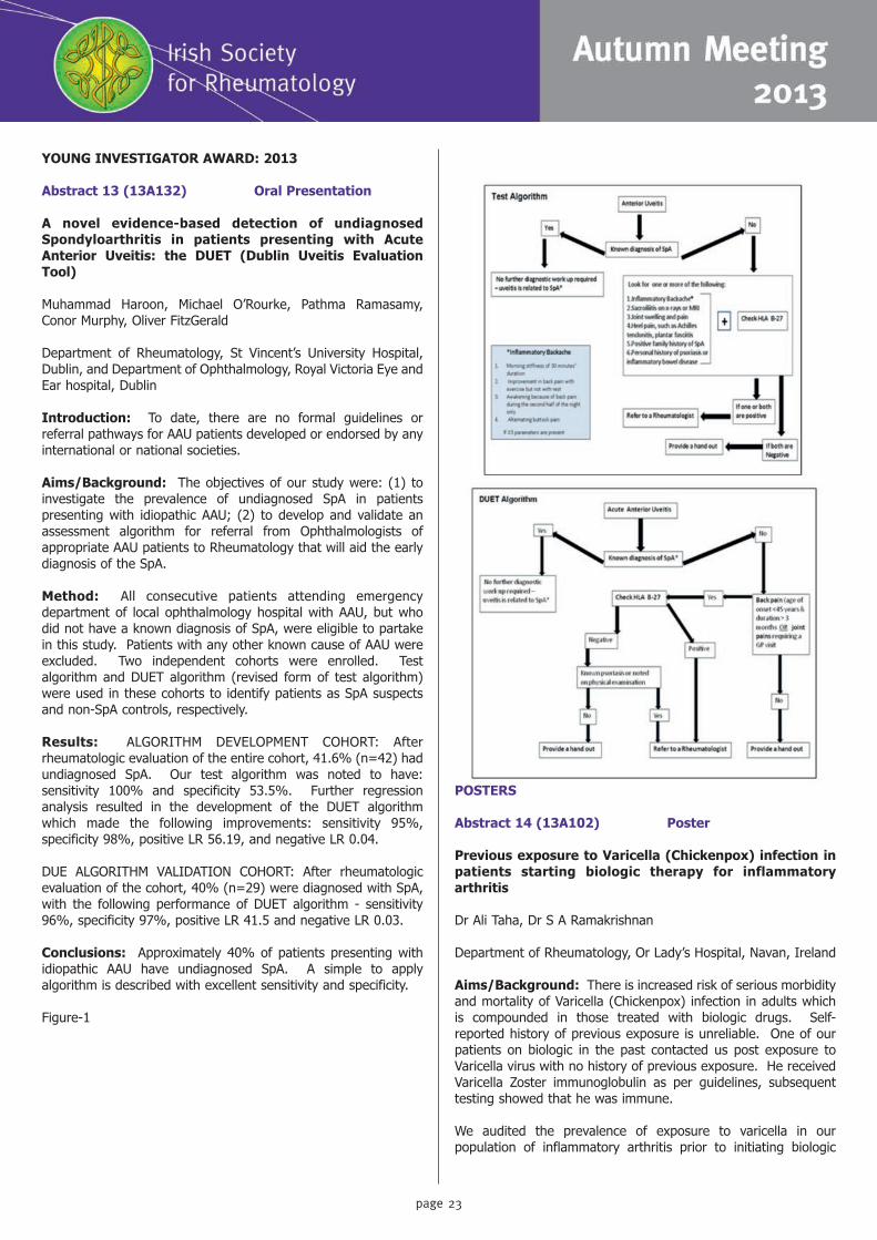
Abstract 13 (13A132) Oral Presentation
A novel evidence-based detection of undiagnosedSpondyloarthritis in patients presenting with AcuteAnterior Uveitis: the DUET (Dublin Uveitis EvaluationTool)
Muhammad Haroon, Michael O’Rourke, Pathma Ramasamy,Conor Murphy, Oliver FitzGerald
Department of Rheumatology, St Vincent’s University Hospital,Dublin, and Department of Ophthalmology, Royal Victoria Eye andEar hospital, Dublin
Introduction: To date, there are no formal guidelines orreferral pathways for AAU patients developed or endorsed by anyinternational or national societies.
Aims/Background: The objectives of our study were: (1) toinvestigate the prevalence of undiagnosed SpA in patientspresenting with idiopathic AAU; (2) to develop and validate anassessment algorithm for referral from Ophthalmologists ofappropriate AAU patients to Rheumatology that will aid the earlydiagnosis of the SpA.
Method: All consecutive patients attending emergencydepartment of local ophthalmology hospital with AAU, but whodid not have a known diagnosis of SpA, were eligible to partakein this study. Patients with any other known cause of AAU wereexcluded. Two independent cohorts were enrolled. Testalgorithm and DUET algorithm (revised form of test algorithm)were used in these cohorts to identify patients as SpA suspectsand non-SpA controls, respectively.
Results: ALGORITHM DEVELOPMENT COHORT: Afterrheumatologic evaluation of the entire cohort, 41.6% (n=42) hadundiagnosed SpA. Our test algorithm was noted to have:sensitivity 100% and specificity 53.5%. Further regressionanalysis resulted in the development of the DUET algorithmwhich made the following improvements: sensitivity 95%,specificity 98%, positive LR 56.19, and negative LR 0.04.
DUE ALGORITHM VALIDATION COHORT: After rheumatologicevaluation of the cohort, 40% (n=29) were diagnosed with SpA,with the following performance of DUET algorithm - sensitivity96%, specificity 97%, positive LR 41.5 and negative LR 0.03.
Conclusions: Approximately 40% of patients presenting withidiopathic AAU have undiagnosed SpA. A simple to applyalgorithm is described with excellent sensitivity and specificity.
Figure-1
POSTERS
Abstract 14 (13A102) Poster
Previous exposure to Varicella (Chickenpox) infection inpatients starting biologic therapy for inflammatoryarthritis
Dr Ali Taha, Dr S A Ramakrishnan
Department of Rheumatology, Or Lady’s Hospital, Navan, Ireland
Aims/Background: There is increased risk of serious morbidityand mortality of Varicella (Chickenpox) infection in adults whichis compounded in those treated with biologic drugs. Self-reported history of previous exposure is unreliable. One of ourpatients on biologic in the past contacted us post exposure toVaricella virus with no history of previous exposure. He receivedVaricella Zoster immunoglobulin as per guidelines, subsequenttesting showed that he was immune.
We audited the prevalence of exposure to varicella in ourpopulation of inflammatory arthritis prior to initiating biologic


page 25
therapy.
Method: To ascertain serological immunity to Varicella inconsecutive patients starting biologics for various inflammatoryarthritides from June 2011 to March 2012, samples were sent forvaricella titres along with usual other screening. Previous historyof varicella exposure was recorded.
Results: 52 consecutive patients were tested. Patients wereaged between 18 and 82 years. 31 females, 21 males. All testedpositive with Varicella Zoster IgG > 100 mIU/ML. 29 hadRheumatoid Arthritis, 15 Spondyloarthropathy, 4 Psoriaticarthropathy and 4 undifferentiated arthritis.40 patients reported exposure to Varicella (Chickenpox) and 12patients were not aware. Of our patients 4 were easternEuropeans, 3 non Caucasians (two African decent and one Asian)and 45 were Irish.
Conclusions: Previous Varicella zoster infection is the norm inour patients starting biologic drugs. Given that the history ofprevious exposure is unavailable in many cases, documentedimmunity is very valuable for post exposure advice. Wecontinued testing for varicella immunity for all patients startingBiologic therapy.
References: 1. Smitten AL, Choi HK, Hochberg MC, et al. Therisk of herpes zoster in patients with rheumatoid arthritis in theUnited States and the United Kingdom. Arthritis Rheum2007;57:1431–8.2. Galloway JB, Mercer LK, Moseley A, et al. Risk of skin and softtissue infections (including shingles) in patients exposed to anti-tumour necrosis factor therapy: results from the British Societyfor Rheumatology Biologics Register. Ann Rheum Dis2013;72:229–34.3. McDonald JR, Zeringue AL, Caplan L, et al. Herpes zoster riskfactors in a national cohort of veterans with rheumatoid arthritis.Clin Infect Dis 2009;48:1364–71.4. Dworkin RH, Johnson RW, Breuer J, et al. Recommendationsfor the management of herpes zoster. Clin Infect Dis 2007;44(Suppl 1):S1–26. 5. Harpaz R, Ortega-Sanchez IR, Seward JF. Prevention of herpeszoster: recommendations of the Advisory Committee onImmunization Practices (ACIP).MMWR Recomm Rep 2008;57(RR-5):1–30.
Abstract 15 (13A108) Poster
Patient characteristics and reason for referral for BoneMineral Densitometry in a community based hospital
Aoife McPartland, Bernie McGowan, Carmel Silke, Bryan Whelan
The North Western Rheumatology Unit, Our Lady’s Hospital,Manorhamilton, Co Leitrim
Introduction: Dual-energy X-ray absorbtiometry (DXA) is themost accurate and widely used method to identify patients withlow bone mineral density (BMD), however it is expensive and haslimited availability across the country.
Aims/Background: To identify the reasons for referral to DXAin patients presenting to the Bone Densitometry Service, Our
Lady’s Hospital, Manorhamilton, Co. Leitrim.
Method: Reasons for referral for DXA were examined in 283patients aged > 40 years, referred in 2012 to the The NorthWestern Rheumatology Unit. Age, sex, clinical risk factors,fracture history and bone mineral density (BMD) T scores wererecorded.
Results: Mean age of the study population was 63.2 yrs (Std +11.9), females 87%, males 13%. In total 36.5% of the patientshad normal BMD levels, a further 33% were in the osteopenicgroup and 30% were either osteoporotic or severely osteoporotic.Seventy percent of the patients referred (N=192) had a clinicalrisk factor documented and the most common reason for referralwas advanced age (35%) and history of a fracture (27%). Intotal 75 patients (64%) with a history of fracture did not haveosteoporosis (27% normal BMD, 37% osteopenic). There was astatistically significant correlation between the presence of aclinical risk factor and BMD level p<.05 and also with the numberof clinical risk factors and BMD level p<.05. BMD levels werefound to decrease with age.
Conclusions: BMD levels alone cannot accurately predictfracture probability and the challenge for health careprofessionals in primary and secondary care continues to bescreening and accurately identifying and treating patients whoare at increased risk of sustaining a fragility fracture.
Abstract 16 (13A110) Poster
Major and minor discordance in the diagnosis ofosteoporosis among Irish men and women using hip andspine dual-energy X-ray absorptiometry
Bernie McGowan, Aoife McPartland, Carmel Silke, Bryan Whelan
The North Western Rheumatology Unit, Our Lady’s Hospital,Manorhamilton, Co Leitrim
Introduction: Diagnostic discordance for osteoporosis is thepresence of different categories of T-scores in 2 different skeletalsites, falling into 2 different diagnostic categories as identified bythe World Health Organization classification system (1).Discordances between hip and spine areal density T-score valuesare common and incompletely understood.
Aims/Background: To determine discordance in the diagnosisof osteoporosis among patients referred for DXA scan at TheNorth Western Rheumatology Unit using hip and spine Dual-energy X-ray Absorptiometry.
Method: The study included men and women who underwentbone mineral densitometry (BMD) for suspected osteoporosis atThe North Western Rheumatology Unit. The BMD measures atthe hip and spine were used to derive T-scores and to determinethe prevalence of discordance. Factors potentially associatedwith discordance were explored in univariate and a multivariateregression model.
Results: The mean age of the 276 patients in the study was63.2 ± 11.92 years (males 35 (13%), females 241 (87%).

page 26
Results of T-Score Concordance was identified in 128 patients(51.2%), minor discordance in 101 patients (36.5%) and majordiscordance was seen in 21 patients (7.6%). Independent t-testof age, BMI, presence of co-morbidities, fracture history,identified age as the only risk factor (P<.05) which had asignificant effect on T-score discordance.
Conclusions: At least 40% of patients tested by DXA willdemonstrate T-score discordance between spine and total hipmeasurement sites. T-score discordance has been shown tooccur for a variety of reasons related to physiologic andpathologic patient factors as well as the performance or analysisof DXA itself (2).
References: 1. Faulkner KG, von Stetten E, Miller P. Discordancein patient classification using T-scores. J Clin Densitom 1999; 2343-350.2. Mounach MD, Mouinga A, Ghazi M, et al. Discordance betweenHip and Spine Bone Mineral Density Measurement Using DXA:Prevalence and Risk Factors. Bone 2009; 38, 467-471.
Abstract 17 (13A111) Poster
A discrete-choice experiment to elicit the preferences ofIrish patients for osteoporosis drug treatment
McGowan B 1, Carmel Silke1, Bryan Whelan1, Hiligsmann M 2,Dellaert B 3, Dirksen C 2, van der Weijden T 2, Watson V 4,Boonen A 2
1 The North Western Rheumatology Unit, Our Lady’s Hospital,Manorhamilton, Co Leitrim
Aims/Background: To evaluate the preferences of patientswith osteoporosis for medication attributes and to establish howthey trade between these attributes.
Method: A discrete-choice experiment was designed in whichpatients were asked to choose, from a series of hypotheticalscenarios, between two drug alternatives which vary in fiveattributes: efficacy in fracture risk reduction, type of side-effects,mode and frequency of administration and costs. An efficientexperimental design was used to construct the sets of treatmentoptions and a mixed logit panel data model was employed toestimate patients’ preferences and their trade-offs betweenattributes.
Results: 200 patients with osteoporosis completed theexperiment. Patients preferred a drug treatment with a highereffectiveness and a lower cost. They also preferred 6-monthsubcutaneous injection and yearly intravenous above weekly oraltablets and favoured weekly oral tablet over 3-monthsintravenous injections. No significant difference in preferencewas observed between weekly oral tablets, monthly oral tabletsand 3 monthly subcutaneous injections. Patients disliked beingat risk of gastro-intestinal disorders more than being at risk ofskin reactions and flu-like symptoms. Significant heterogeneityfor the preferences was present among nearly all attributes.
Conclusions: This study revealed that, at the group level,osteoporotic Irish patients preferred 6-monthly subcutaneous
injections and yearly intravenous injection and compared to otherpotential side-effects, gastro-intestinal effects were the leastfavoured. Moreover, they are willing to pay a personalcontribution or to trade efficacy for such outcomes. Preferenceheterogeneity suggests the need to incorporate individualpreferences into clinical decision-making to improve osteoporosiscare.
Abstract 18 (13A112) Poster
Plasma IL-6 levels correlate with clinical and ultrasoundmeasures of disease activity in Lupus Arthritis
Elisabeth Ball, David Gibson, Aubrey Bell, Madeleine Rooney
Queen’s University, Belfast
Introduction: The role of specific cytokines in lupus arthritishas not been elucidated (1).
Aims/Background: To analyse the cytokine profile in SLEpatients with erosive, non-erosive arthritis and arthralgia.
Method: 50 SLE patients, and 40 RA patients had an US scanof their hand (2,3). US scores were expressed per joint and as atotal ‘ultrasound activity’ score (sum of Power Doppler (PD) andGrey Scale Synovial Hypertrophy scores in all joints) and a totalerosion score. SLE disease activity was assessed. Plasma levelsof IL-6, TNF-alpha and BLyS were measured (Quantikine® ELISAkits, R & D).
Results: On the basis of the ultrasound results the SLE patientswere divided into erosive arthritis (n = 24), non-erosive arthritis(n = 14) and those with a normal ultrasound scan (n = 12). CRPand IL-6 levels between the erosive lupus group and the RAgroup were not significantly different (p = 0.3 and p = 0.2).Across the SLE groups plasma IL-6 levels correlated with CRP ( p< 0.001), hand deformity scores ( p = 0.005), swollen joint count(p = 0.002) and BILAG musculoskeletal score (p = 0.009); alsowith wrist PD score ( p = 0.01), extensor tenosynovitis (p =0.008) and total ultrasound activity score ( p < 0.001) (whichremained constant when corrected for total BILAG score (p <0.01)).
Conclusions: The most significant finding is that IL-6 levelscorrelated with both clinical and ultrasound measures of arthritisdisease activity. There is preliminary evidence that IL-6 blockademay be a potential treatment for SLE(4) and if arthritis specificoutcome measures were included in future clinical trials morerobust evidence could be generated.
References: 1.Jacob N & Stohl W. Cytokine disturbances inSystemic Lupus Erythematosus. Arthritis Research & Therapy2011; 13 (4): 244.2. Wakefield RJ et al. Musculoskeletal ultrasound includingdefinitions for ultrasonographic pathology. J Rheum 2005;32(12):2485-87.3. Szkudlarek M et al. Interobserver agreement inultrasonography of the finger and toe joints in rheumatoidarthritis. A & R 2003;48(4):955-62.4. Illei G et al. Tocilizumab in systemic lupus erythematosus: dataon safety...... A & R 2010;62(2):542-52.

page 27
Abstract 19 (13A113) Poster
An audit of the vaccination status of patients withautoimmune anti-inflammatory rheumatic diseaseattending The North Western Rheumatology Unit
Dr Sarah Nicholson1, Dr Carmel Silke1, Dr Bryan Whelan1, DrCathriona Walsh2, Mary McGovern1 , Bernie McGowan1
1The North Western Rheumatology Unit, Our Lady’s Hospital,Manorhamilton, Co Leitrim2HSE Community Services West (Sligo/Leitrim), Sligo
Introduction: Patients with immune modulated illnesses andthose undergoing treatment with immunosuppressant therapiesare more likely to develop vaccine-preventable disease than thegeneral population(1). EULAR guidelines state that vaccinationstatus should be assessed in the initial workup of those withautoimmune inflammatory rheumatic disease (AIIRD)(2). TheNational Immunisation Advisory Committee (NIAC) in Irelandrecommends influenza and pneumococcal vaccine in thosepersons with chronic illness and immunosuppression(3).
Aims/Background: To assess the vaccination status of patientswith AIIRD attending The North Western Rheumatology Unit(NWRU).
Method: This was a cross sectional retrospective study in whichan anonymised questionnaire was distributed to patients (N=66 )with AIIRD over a 3 week period. The data was analysed usingexcel and JMP® 8.0.2 data analysis software. The study focusedon the influenza and pneu mococcal vaccines.
Results: In total, 67% of respondents had received the fluvaccine, 24% were unsure and 8% had not received the vaccine.A further 11% of respondents had received the pneumococcalvaccine, 53% were unsure and 35% had not received it. 53%(n=35) had a diagnosis of rheumatoid arthritis. In total, 86% ofthe patients were taking immunosuppressant medications.
Conclusions: The results of this study show that even thoughuptake of the flu vaccine (67%) is better than pneumococcal(11%), both vaccinations have a poor uptake in this groupcompared to the WHO target of 75% uptake in at risk groups(5).The poor uptake of vaccines may be due to a lack of educationabout the risks and benefits of the flu and pneumococcalvaccines. It is imperative that patients are educated about therisks of vaccine-preventable disease
References: 1. Rahier JF, Moutschen M, Van Gompel A, VanRanst M, Louis E, Segaert S, et al. Vaccinations in patients withimmune-me diated inflammatory diseases. Rheumatology2010;49:1815-27.2. EULAR recommendations for vaccination in adult patients withautoimmune infl ammatory rheumatic diseases.Van Assen, S., etal. Ann Rheum Dis 2011;70:414–4223. Risk Groups and Uptake of Influenza and PneumococcalVaccine in Ireland. Available at http://www.hpsc.ie/hpsc/EPI-Insight/Volume92008/File,2887,en.PDF4. National immunisation news, November 2012: available athttp://www.immunisation.ie/en/Publications/ImmunisationNewsletters/PDFFile_17148_en.pdf5. Influenza vaccination uptake monitoring on behalf of the
Department of Health. Available at http://www.hpa.org.uk/web/HPAweb&HPAwebStandard/HPAweb_C/1195733756886#r1
Abstract 20 (13A114) Poster
Muscle Function in Patients with Stable RheumatoidArthritis
Elaine Walsh1, McGowan B2, Carmel Silke2, Bryan Whelan2
1Department of Life Sciences, Sligo Institute of Technology, Sligo2The North Western Rheumatology Unit, Our Lady’s Hospital,Manorhamilton, Co Leitrim
Introduction: Rheumatoid arthritis patients are less physicallyactive than the general population(1). Patients with rheumatoidarthritis show lower cardiorespiratory fitness than normalsubjects(2). The American College of Rheumatologyrecommends strengthening and aerobic conditioning regimens inits guidelines for the management of rheumatoid arthritis(3).
Aims/Background: The aim of the study was to assessbaseline muscle function in patients with stable rheumatoidarthritis.
Method: Functional assessments were conducted on 30 stablerheumatoid arthritis patients and compared to normative valuesof a healthy population. Assessments consisted of hand gripdynamometer, 2 minute step test, sit and reach flexibility test andsingle leg balance test.
Results: In total 30 stable rheumatoid arthritis patients (meanage 61.2, 10.6 SD) were tested and the results were comparedto the age/gender specific norms of a healthy population. For the2 minute step test, there was a statistically significant difference(p<0.001, 95% CI -28.96 to -10.71) between the results of thepatient study group and the normative values. The results of thesit and reach test identified that in total (78.6%) of males and allfemales (100%) rated in the poor category. In the single legbalance test, only 20% reached the maximum score. In total43.7% of females and 50% of males reached the age/genderspecific norms of the hand grip dynamometer.
Conclusions: The results show that the rheumatoid arthritispatients show lower levels of muscle function than the normalpopulation. Efforts should be made to include physical training inthe treatment of rheumatoid arthritis.
References: 1. Sokka, T., Häkkinen, A., Kautiainen, H.,Maillefert, J. F., Toloza, S. et al. (2008) Physical inactivity inpatients with rheumatoid arthritis: Data from twenty-onecountries in a cross-sectional, international study, Arthritis Careand Research, 59 (1), 42-50.2. Cimen, B., Deviren, S. D., Yorganclo�lu, Z. R. (2001)Pulmonary function tests, aerobic capacity, respiratory musclestrength and endurance of patients with rheumatoid arthritis,Clinical Rheumatology, 20 (3) 168-73.3. American College of Rheumatology Subcommittee (2002)Rheumatoid Arthritis Guidelines: Guidelines for the managementof rheumatoid arthritis, Arthritis and Rheumatism, 46, 328–346.
Abstract 21 (13A115) Poster
page 28
To identify adherence to EULAR guidelines for anti TNFtreatment in patients with a diagnosis of psoriaticarthritis
Haithem Mohammed, Bernie McGowan, M Khalifa, Carmel Silke,Bryan Whelan
The North Western Rheumatology Unit, Our Lady’s Hospital,Manorhamilton, Co Leitrim
Introduction: The course of Psoriatic arthritis (PsA) is variableand unpredictable ranging from a non-destructive disease to asevere debilitating erosive arthropathy. Erosive and deformingarthritis occurs in 40–60% of PsA patients and is progressive fromwithin the first year of diagnosis (1,2). The classification of PsAis an area of ongoing international discussion. The fivesubgroups proposed by Moll and Wright(3) are still frequentlyused, although considerable overlap between these groups isnow recognized.
Aims/Background: To check adherence to EULAR guidelinesfor anti TNF treatment in psoriatic arthritis.
Method: 78 patients’ medical records were reviewed, andretrospectively audited against EULAR guidelines. Patients weresubgrouped as RA pattern, oligoarthritis, enthesopathy , axialdisease and arthritis mutilans PsA. Diagnosis was madeaccording to the CASPAR criteria (5). Information recordedincluded assessment for subtype of arthritis, duration of disease& pharmacological treatment used.
Results: Rheumatoid arthritis pattern was the most commonlyoccurring sub-type in 56% (n=44), followed by oligoarthriticvariant in 40% (n=31). Enthesitis and axial disease accountedfor 8% and 4%, respectively. Methotrexate was the first lineDMARD in 59 patients (77%) of the patients, followed bySulphasalazine in 17patients (22%) and Leflonamide in onepatient (1.2%). In total 29 patients (37%) were changed fromDMARD1 to DMARD2, the majority of whom were changed tomethotrexate (55%, n=17). 97.4% of the patients werecommenced on an anti TNF.
Conclusions: All patients in the study received biologic DMARDsappropriately. Subclassification of psoriatic arthritis & PsARCneed to be recorded at baseline so the escalation of treatmentand choice of pharmacological approach will be instituted in atimely manner and response to same would accurately bemonitored.
References:1. Gladman DD, Stafford-Brady F, Chang CH, Lewandowski K,Russell ML. Longitudinal study of clinical and radiological progres-sion in psoriatic arthritis. J2. 4-Rheumatol 1990;17:809–12. 4. McHugh NJ, Balakrishnan C,Jones SM. Progression of peripheral joint disease in psoriaticarthritis. Rheumatology 2003; 42:778–83.3. Taylor W, Gladman D, Helliwell P, et al. Classification criteria forpsoriatic arthritis: development of new criteria from a largeinternational study. Arthritis Rheum 2006; 54:2665.
Abstract 22 (13A116) Poster
Audit of intra-articular steroid injection approach in IrishSociety of Rheumatology members
David McCormick, Andrew Cairns
Rheumatology Department, Musgrave Park Hospital, Belfast
Introduction: Intra-articular steroid injections are a key aspectin the management of rheumatology patients. There are,however, recognised potential risks that patients should beinformed of.A recently updated patient information leaflet produced by theArthritis Research Council (ARC) for intra-articular steroidinjections suggested checking (i) blood pressure and (ii) bloodglucose prior to injection due to potential derangement. Aliterature review did not reveal any evidence to warrant thisstatement. This could result in a delay which lengthens patientsuffering and clinic waiting times. This was not routine practicein Belfast so an audit sought to establish the practices ofmembers of the Irish Society of Rheumatology.
Results: A questionnaire was completed by 22 ISR membersand results showed that 32% used the ARC leaflet. Only 9%consented for effect on blood pressure but 73% discussed raisedblood glucose in diabetics.14% checked blood pressure prior to injection and nonerecommended follow up after injection. None checked bloodglucose prior to injection, however, 14% recommendedmonitoring of blood glucose levels for 48 hours after injection.Information was also gained on aseptic technique and injectionapproach which showed a wide variation in results.
Conclusions: Audit results confirmed that it was not commonnational practice to check blood glucose and blood pressure priorto steroid injection. Findings were communicated with theauthors of the ARC leaflets who have agreed with our suggestionand future versions will have this statement removed therebyhopefully reducing any unnecessary delay and any furtherpotential joint destruction.
References: Note- data on aseptic technique and injectionapproach etc. would be available as well as data on bloodpressure/blood glucose.
Abstract 23 (13A117) Poster
Does Fibrinogen have a role in the assessment of patientswith Giant Cell Arteritis?
O’ Neill L, McCarthy E, Madigan A, Veale D, Murphy C, Fearon U,McCarthy G, Molloy E
St. Vincent’s University Hospital, Dublin, Ireland. MaterMisericordiae University Hospital, Dublin. Royal Victoria Eye andEar Hospital, Dublin
Introduction: Considerable difficulties exist in the evaluation ofpatients with suspected GCA. Temporal artery biopsy is negativein a substantial minority of patients. The traditional biomarkersESR and CRP are non-specific and can also be negative.Therefore development of better disease biomarkers is needed

page 29
for diagnosis in GCA.
Aims/Background: The aim of this study was to evaluate theutility of plasma Fibrinogen in the assessment of patients withGCA.
Method: Patients presenting with suspected new-onset GCAwere prospectively enrolled. Plasma Fibrinogen, ESR and CRPwere assayed at baseline and 3 months following initiation ofsteroid therapy.Biomarkers were also assayed in 25 age and sex matchedcontrols attending with osteoarthritis (OA).
Results: Plasma Fibrinogen levels were significantly elevated inpatients with GCA when compared with OA controls. (Mean 4.9g/L vs. 3.05 g/L, p = 0.0017).Fibrinogen levels demonstrated a response to therapy. (Mean of4.9 g /L at baseline vs. 3.5 g / L at 3 months, p = 0.04).Baseline Fibrinogen levels correlated with baseline ESR (r =0.6946, p < 0.0001) and CRP (r= 0.6951, p < 0.0001. ROCanalysis revealed Fibrinogen to be a less sensitive but morespecific marker of GCA than CRP (Sensitivity of 67% vs. 82% andSpecificity of 100% vs. 80% respectively) with comparablespecificity to ESR above a cut off for ESR >30 mm/hr.Values above the upper limit of normal for Fibrinogen (4 g/L)were associated with a positive likelihood ratio of 15.18 for GCA.
Conclusions: Plasma fibrinogen levels are elevated in patientswith GCA, respond to glucocorticoid therapy and correlate withserum ESR and CRP levels.
Abstract 24 (13A118) Poster
Lower Tissue Expression Of IL 6 In Patients With GiantCell Arteritis Presenting With Cranial IschaemicComplications
O’ Neill L, McCormick J, Molloy D, Veale D, McCarthy G, MurphyC, Fearon U, Molloy E
St. Vincent’s University Hospital, Mater Misericordiae UniversityHospital, Royal Victoria Eye and Ear Hospital
Introduction: The majority of the morbidity associated withGiant Cell Arteritis is related to its ischaemic complications ofacute visual loss and stroke. It has previously been suggestedthat patients with cranial ischaemic complications (CICs) due toGCA have lower tissue expression of IL 6.
Aims/Background: 1. To compare the spontaneous release ofIL 6 from cultured temporal artery explants of patients with GCAand CICs to those without.2. To investigate the association between spontaneous release ofIL 6 from the explant cultures and findings on histopathology.
Method: Patients presenting with suspected GCA wereprospectively enrolled.Patients underwent a temporal artery biopsy and had serumbiomarkers assayed at baseline.TAB sections were sent to histopathology for diagnostic purposes.Additional sections were cultured and IL 6 expression in thesupernatants quantified.
Results: 11 of the 55 (20%) patients included in the analysispresented with an ischaemic complication attributable to GCA.Patients with CICs had significantly lower levels of of IL 6 inexplant supernatants when compared to those without CICs(Mean 8.66 ng/ml/mg biopsy weight vs 18 .92 ng/ml/mg biopsyweight, p <0.01).This lower tissue IL 6 production was significantly associated withboth intimal hyperplasia (p < 0.03) and luminal occlusion (p <0.01) on histopathology.
Conclusions: The study confirms the association between lowerlevels of IL 6 expression in patients with CICs than those without.
References:Cid et al, Circulation 2003
Abstract 25 (13A119) Poster
Treatment with the GLP-1 Analogue Liraglutide isassociated with Inflammatory Arthritis DAS28 Reductionin patients with concommitant T2DM
Catherine Sullivan1, Michele Doran2, David Kane1, DonalO’Shea3, Ronan H. Mullan1
1Department of Rheumatology, Bone & Joint Unit, TallaghtHospital, Dublin 242Department of Rheumatology, St. James’s Hospital, Dublin 83St. Vincent’s University Hospital, Dublin 4
Introduction: Recent evidence suggests that Glucagon-likepeptide-1 (GLP-1) analogues, used for the treatment of type 2diabetes (T2DM), possess novel anti-inflammatory effectsincluding the amelioration of TNF inflammatory responses.
Aims/Background: To investigate the amelioration ofinflammatory arthritis disease activity in a cohort of patientsundergoing liraglutide therapy for T2DM.
Method: Patients with T2DM and either RA (n=11) or PsA (n=4) were commenced on liraglutide 1.2mg s/c od. DAS28 scores,weight, HbA1C, were recorded at baseline and weeks 6, 12 and24. DAS28 outcomes were defined by EULAR good, or moderatevs non-responders.
Results: Patients had active arthritis (DAS28 4.38+0.4) andinadequate glycaemic control Hba1C (55 + 5.1) at baseline. Ninepatients achieved DAS28 response (DAS28=4.2+0.8 pre, 2.7+0.5post) vs (DAS28=4.7+0.8 pre, 5.0+2.7post, n=6). Significantweight loss was seen in DAS28 responders (94+ 5 pre,90.6+5.2kg, p=0.008) but not non-responders (93.8+3.3kg pre,95+2.8kg post, p=0.79). A significant fall in HbA1C was seen inDAS28 responders (60.5+6.3 pre, 45.5+2.1 post, p=0.012) butnot non-responders (47.1+9.5 pre vs 39.4+13, p=0.144). Asignificant fall in SJC28 was seen in DAS28 responders (3.3+0.9pre, 1.2+0.6, p=0.027) but not non-responders (4.2+2.8 pre,4.8+3.2 post, p=0.66). Weight loss following liraglutidesignificantly associated with achieving a DAS28 response by ChiSquare (p=0.044). No differences were observed in baselineDAS28, weight, HbA1C, SJC, ESR or CRP according to subsequentDAS28 response grouping.
Conclusions: Liraglutide lowering effects for HbA1C and BMI,significantly associated with a DAS28 reduction. Further studies

page 30
may lead to novel therapeutic strategies in the treatment ofinflammatory arthritis.
Abstract 26 (13A122) Poster
The Ankylosing Spondylitis Registry of Ireland (ASRI)
Martina Fitzpatrick1, Phil Gallagher1, Catherine Molloy2, OliverFitzGerald1, Finbar O’Shea3
1St. Vincent’s University Hospital, Dublin2Cork University Hospital, Cork3St. James’ Hospital, Dublin
Introduction: In 2007, rheumatologists from Scotland andNorthern Ireland created the Scotland and Ireland Registry forAnkylosing Spondylitis. As part of this collaboration,rheumatologists in the Republic of Ireland intend to create asimilar registry of patients - The Ankylosing Spondylitis Registryof Ireland (ASRI).
Aims/Background: The primary objectives of ASRI are toprovide basic descriptive epidemiological data on the ASpopulation in Ireland, and to establish a registry for potentialfuture studies of genetics, aetiology and therapeutics.Specifically, we will measure the burden of AS in the populationand identify early predictors of a poor outcome. The secondaryaim is to collect biological samples (serum and DNA) fromparticipants. These samples will be used to investigate if geneticand serological factors predict poor outcomes.
Method: This is a prospective cohort study and will include adultpatients who have been diagnosed with AS. A standardisedclinical assessment of patients will be performed and entered ina web-based database. Results of relevant laboratory parametersand imaging studies will be documented. Specific measures ofdisease activity (BASDAI), function (BASFI and HAQ), and qualityof life (ASQoL) will be obtained. Samples for serum and DNA willbe stored for future use to correlate with clinical data and tofacilitate genetic studies.
Results: Recruitment has recently commenced and is open toany centre in Ireland.
Conclusions: This is a unique opportunity for the IrishRheumatology community to generate a valuable resource thatwill ultimately improve the delivery of care to Irish AS patients.
Abstract 27 (13A123) Poster
Ultrasound is as sensitive as nerve conduction studies butmore cost effective and patient acceptable in thediagnosis of carpal tunnel syndrome
Cara McDonagh, Michael Alexander, David Kane
Rheumatology Department and Neurophysiology Department,Tallaght hospital, Dublin 24
Introduction: Carpal tunnel syndrome (CTS) is the commonestentrapment neuropathy affecting 9% of women(1, 2). Sensitivity
of ultrasound to diagnose carpal tunnel syndrome has beenreported to be as high as 97.9%(3), while nerve conductionstudies (NCS) have a sensitivity of >85%.(4).
Aims/Background: To document the sensitivity of ultrasoundto diagnose CTS and to establish a new pathway for diagnosis ofCTS using ultrasound.
Method: Patients with clinically confirmed idiopathic CTS wererecruited from the rheumatology, orthopaedic and neurologyoutpatient clinics and the neurophysiology department in atertiary referral teaching hospital. They were assessed clinically,by ultrasound and nerve conduction studies. Clinical diagnosiswas the reference standard(5).
Results: 29 patients (40 symptomatic wrists) have beenrecruited into the study. Of the 40 wrists at baseline 32 hadultrasound confirmation of CTS and 34 had positive NCS. Thesensitivity of ultrasound to diagnose CTS when compared toclinical assessment was 80% and the sensitivity of NCS was 85%.The cost of ultrasound is £64 and EMG/NCS costs £256,potentially yielding a 75% saving if ultrasound is used as the firstline test(6). No patient reported pain during ultrasoundexamination and all subjects agreed to follow-up. Ultrasoundadditionally identified structural causes of CTS such as bifidmedian nerve, arterial anomaly and tenosynovitis, and ultrasoundwas used to guide injection avoiding nerve damage.
Conclusions: Ultrasound is a sensitive, more cost effective,highly patient acceptable modality for the diagnosis of CTS. Anew paradigm is proposed where ultrasound is used as the initialtest for CTS and NCS are reserved for those with a negativeultrasound or atypical symptoms.
References: 1. de krom M, Knipschild P, Kester A, Thijs C,Boekkooi P, Spaans F. Carpal tunnel syndrome: prevalence in thegeneral populatio n. J Clin Epidemiol. 1992;45:373-6.2. Bongers F, Schellevis F, van den Bosch W, van der Zee J. Carpaltunnel syndrome in general practice (1987 and 2001): Incidenceand the role of occupational and non occupational factors. Br JGen Pract. 2007;57:36-9.3. El Miedany Y, Aty S, Ashour S. Ultrasonography versus nerveconduction study in patients with carpal tunnel syndrome:substantive or complementary tests? Rheumatology.2004;43(7):887-95.4. Jablecki C, Andary M, So Y, Wilkins D, Williams F. Literaturereview of the usefulness of nerve conduction studies andelectromyography for the evaluation of patients with carpaltunnel syndrome. AAEM Quality Assurance Committee. Muscleand Nerve. 1993;16:1392-414.5. Neurology AAo. Practice parameter for carpal tunnelsyndrome(Summary Statement). Neurology. 1993;43:2406-9.6. University College London Hospitals NHS Foundation Trust:Provider to Provider Services 2013-2014 Tariff.
Abstract 28 (13A124) Poster

page 31
The prognosis of Scleroderma Renal Crisis in RNA-polymerase III antibody (ARA) positive compared to ARAnegative patients
Bernadette M. Lynch, Henry Penn, Jennifer Harvey, Aine Burns,Christopher P. Denton
Department of Rheumatology, The Royal Free Hospital, PondStreet, London NW3 2QG
Introduction: Scleroderma renal crisis (SRC) usually presentingwith accelerated hypertension and acute kidney injury (AKI) isone of the most severe complications of Systemic Sclerosis (SSc).
Aims/Background: The presence of RNA-polymerase III auto-antibodies (ARA) is recognized as a strong risk factor for SRC butstudies have not explored long-term outcomes in ARA positivecases compared to ARA negative cases.
Method: Of more than 2000 SSc patients attending ourinstitution between 1990-2013, 150 patients had a confirmedSRC. 80% patients had diffuse cutaneous SSc (dcSSc) and 20%patients had limited cutaneous SSc (dcSSc). ARA was measuredby commercial ELISA or radio-immuno-precipitation. Patientswere divided into two groups: ARA positive or ARA negative.Demographic and clinical parameters were compared betweengroups using Student’s t-test or Chi-squared analyses whereappropriate.
Results: 61/150 (41%) patients were A RA positive andsignificantly more likely to have dcSSc (88.3% vs 73.8%,p=0.032) than lcSSc compared to ARA negative patients. Therewas no significant difference in age of onset of SRC (51.2 vs 51.9years) or the number of females (73% vs 79%) between the twogroups. 50.8% of ARA positive patients required dialysiscompared to 29.2% of ARA negative patients (p=0.07). Themean time to recovery of renal function was significantly longerin ARA positive patients (14.25 vs 8.27 months, p=0.032).Significantly more ARA positive patients were able to discontinuedialysis compared to ARA negative patients (53.3% vs 25.5%,p=0.01). ARA positive patients had a significantly better survivaloutcome.
Conclusions: In SRC, although more ARA positive patientsrequired dialysis they had significantly greater capacity for long-term recovery and survival compared to ARA negative patients.
Abstract 29 (13A125) Poster
ISR Presidents
Dr Gary Wright 2012 - PresentBelfast
Prof. G. Cunnane 2010 – 2012Dublin
Dr. R. Kavanagh 2008-2010Galway
Dr. J. Lee 2006-2008Craigavon
Dr. P. O’Connell 2004-2006Dublin
Prof. O. Fitzgerald 2002-2004Dublin
Dr. A. Taggart 2000-2002Belfast
Dr. D. Raman 1998-2000Sligo
Dr. A. Bell 1996-1998Belfast
Prof. B. Bresnihan 1994-1996Dublin
Prof. M. Molloy 1992-1994Cork
Dr. E. Casey 1990-1992Dublin
Dr. S. Roberts 1988-1990Belfast
Dr. C. Barry 1985-1987Dublin
Dr. D. Roden 1983-1985Dublin
Dr. W. Boyd 1981-1983Belfast
Dr. T. Gregg 1979-1981Dublin
Dr. J. Molony 1977-1979Dublin
Dr. M. McMahon 1975-1977Cork
Dr. T. O’Reilly 1973-1975Dublin

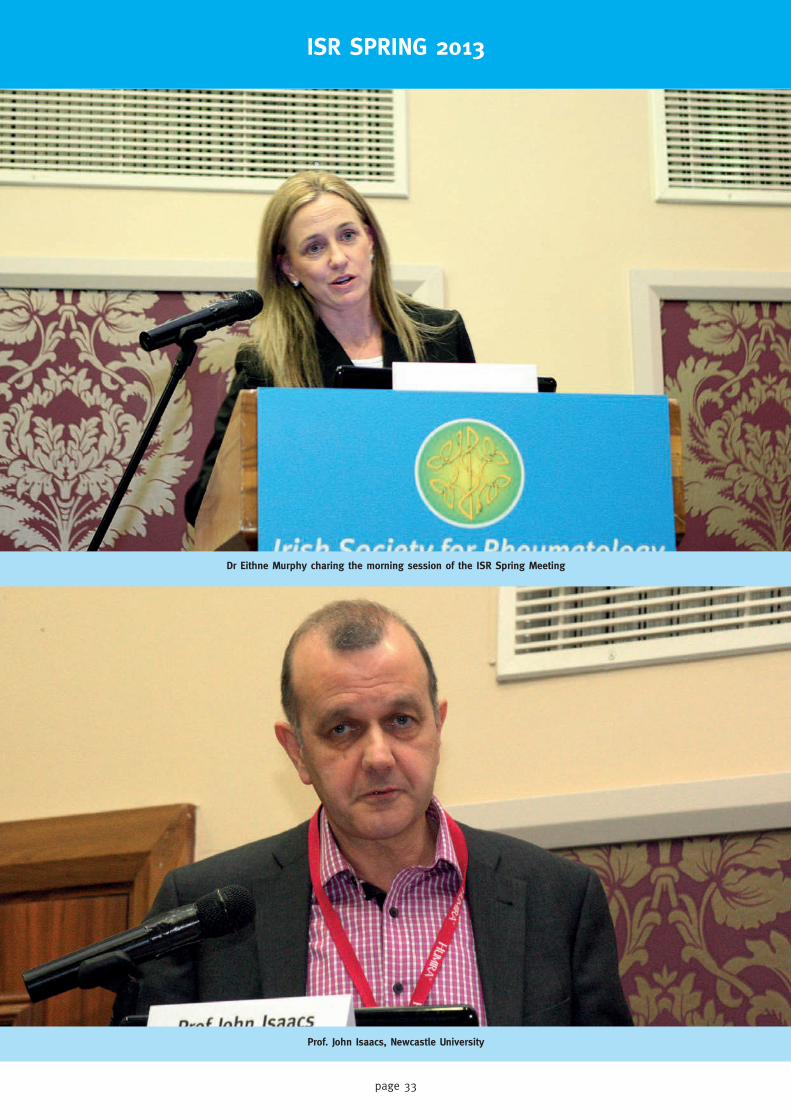
ISR SPRING 2013
Dr Eithne Murphy charing the morning session of the ISR Spring Meeting
Prof. John Isaacs, Newcastle University
page 33

page 34
Scleroderma Renal Crisis and Pulmonary ArterialHypertension are rare in the same patient
Bernadette M Lynch, Vincent Sobanski, Clive E Handler, BenjaminE Schreiber, John G Coghlan, Voon Ong, Aine Burns, ChristopherP Denton
Department of Rheumatology and Pulmonary Hypertension, TheRoyal Free Hospital, Pond Street, London NW3 2QG
Introduction: Scleroderma renal crisis (SRC) and pulmonaryarterial hypertension (PAH) have similar histological features andshared pathogenic mechanisms are plausible. Pulmonaryhypertension (PH) after SRC is frequently observed but generallydue to post capillary mechanisms.
Aims/Background: To explore the association of PAH withSRC.
Method: Of more than 2000 SSc patients attending ourinstitution between 1990-2013, 150 patients had a confirmeddiagnosis of SRC of which six (4%) patients had a diagnosis ofPAH following right heart catherisation. Demographic, clinicaland haemodynamic parameters are expressed as mean ± SD.
Results: Two patients had diffuse and 4 patients had limitedcutaneous SSc. 5/6 had anti-RNA polymerase III (ARA) reactivity.All patients with SRC and PAH were female. PAH was diagnosedafter SRC in all patients. 4/6 patients required dialysis. 2/6 whodid not require dialysis had an eGFR of 26 and 44mls/min/1.73m2. 2 patients recovered renal function at 6 and 14months after the diagnosis of SRC. The mean mPAP (meanpulmonary arterial pressure) was 42 ± 12mmHg. 3/6 patientsdied with a mean survival from date of diagnosis of SRC of 112months and a mean survival from date of diagnosis of PAH of 47months.
Conclusions: The presence of SRC and PAH in the same patientis very rare and PAH occurred after SRC in all patients at a meanof 5 years later. Temporal separation of SRC and PAH suggeststhat these complications may occur independently, but thefrequent association with ARA suggests common susceptibilityfactors may be relevant.
Abstract 30 (13A126) Poster
Reduced eGFR in Systemic Sclerosis associatedPulmonary Arterial Hypertension is a marker of severity
Bernadette M. Lynch1, Vincent Sobanski1, Clive E. Handler2,Benjamin E. Schreiber2, John G. Coghlan2, Aine Burns3,Christopher P. Denton1
1Department of Rheumatology, The Royal Free Hospital, London 2Department of Pulmonary Hypertension, The Royal FreeHospital, London3Department of Nephrology, The Royal Free Hospital, London
Introduction: The prognosis of Systemic Sclerosis associatedPulmonary Arterial Hypertension (SSc-PAH) is determined by theseverity of PAH and other organ involvement.
Aims/Background: To examine the prevalence of reducedeGFR in SSc-PAH and its association with clinical demographics,
pulmonary function tests and haemodynamics.
Method: We included 865 SSc patients from our centre between1996-2010. In 580 patients who underwent a right heartcatherisation (RHC) and eGFR prior to RHC, 290 patients hadPAH. 209 patients had limited cutaneous SSc (lcSSc) and 81patients had diffuse cutaneous SSc (dcSSc). Patients weredivided into two groups: eGFR <60 or ?60ml/min/1.73m2.Demographic, clinical and haemodynamic parameters werecompared between groups using Student’s t-test, Mann-WhitneyU test or Chi-Squared analyses where appropriate.
Results: 77/290 (27%) patients had an eGFR <60. There weremore females (92% vs 77%, p<0.01) in this group who wereolder (63.5 vs 56.8 years, p<0.01), more likely to have lcSSc(91% vs 65%, p<0.01) than dcSSc compared to those with aneGFR ?60. DLCO was significantly lower (42% vs 38%, p<0.05)in the eGFR <60 group but there was no significant difference inforced vital capacity (FVC) (87% vs 73%, p=0.37). The meanpulmonary artery pressure (mPAP) (42 vs 36mmHg, p<0.01) andpulmonary vascular resistance (PVR) (730 vs 524dyn.s.cm-5,p<0.01) were significantly higher in the eGFR <60 group.
Conclusions: Reduced eGFR in SSc-PAH is more frequent inlimited cutaneous SSc, female and older patients and isassociated with a reduced DLCO and preserved FVC. A highermPAP and PVR reflects more severe pulmonary vascular disease.
Abstract 31 (13A127) Poster
High prevalence of Metabolic syndrome is associatedwith the severity of underlying Psoriatic Arthritis
Muhammad Haroon, Phil Gallagher, Eric Heffernan, Prof OliverFitzGerald
Department of Rheumatology and Diagnostic Imaging, StVincent’s University Hospital, Dublin, Ireland
Aims/Background: The objectives of our study were: 1) toinvestigate the prevalence of metabolic syndrome (MetSyn) in anethnically homogenous cohort of established psoriatic arthritis(PsA), 2) to identify clinical associations of MetSyn in patientswith PsA.
Method: A cohort of 283 PsA patients, fulfilling CASPAR criteria,attending rheumatology clinics was included. Following informedconsent, patients underwent detailed skin and rheumatologicassessments, along with cardiovascular risk factor evaluation.Moreover, an extensive medical record review was performed.Severe PsA was defined as the presence of PsA-relatedradiographic damage - peripheral joint erosions, osteolysis andsacroiliitis - along with their PsA requiring TNFi therapy.
Results: The demographics and clinical characteristics of thecohort was as follows: mean age 54.6±12 years; 52% female;mean PsA duration of 19±9 years; 60% of patients requiring TNFifor PsA, mean maximum PASI of 5.7±5.2. MetSyn was presentin 44% of the entire studied cohort. On univariate analysis,MetSyn patients were less educated, had more type-2 psoriasis,later PsO and PsA age of onset, shorter time from psoriasis toarthritis development, worse EQ-5D scores and high smokingpack years. No significant association was noted with PsA

page 35
duration, inflammatory markers and units of alcohol consumed.However, on multiple regression analysis, a significant associationof MetSyn was noted with more severe PsA (OR 4.47, p=<0.001),higher smoking pack years (OR 1.03, p=0.02), and worse EQ5Dscores (OR 1.28, p=0.02).
Conclusions: Among PsA patients, MetSyn is highly prevalent,and is associated with the severity of underlying PsA.
Table-1 Univariate and multivariate (adjustedsimultaneously for variables shown) associations ofdifferent clinical variables with the development ofMetabolic syndrome in patients with Psoriatic arthritis(n=283)
(Univariate Model-1 Model-2 Model-3
analysis) (adjusted* OR) (adjusted* OR) (adjusted* OR)
Age 1.04 (1.02-
1.06), <0.001
PsA Duration 0.99 (0.95-1.00),
0.20
Smoking pack years 1.02 (0.99-1.04), 1.02 (1.00-1.05), 1.02 (1.00-1.05), 1.03 (1.00-1.06),
0.056 0.03 0.052 0.02
Low Education Status 1.94 (1.08-3.48),
0.02
Type-2 Psoriasis 2.35 (1.25-4.42),
0.008
PASI max 1.02 (0.98-1.07),
0.25
PsO age of onset 1.03 (1.01-1.05,
<0.001
PsA age of onset 1.02 (1.00-1.04),
0.006
Time from PsO to 0.97 (0.95-1.00),
arthritis development 0.052
Deformed joints 1.54 (0.93-2.54),
0.08
HAQ 1.45 (0.96-2.20),
0.07
EQ5D 1.16 (1.02-1.32), 1.22 (1.00-1.05), 1.28 (1.03-1.59),
0.19 0.050 0.025
Definition-1 of 1.20 (0.74-1.94), 2.36 (1.03-5.41),
severe PsA 0.45 0.04
(defined as PsA
requiring TNFi)
Abstract 32 (13A128) Poster
Diagnostic delay of even 6 months contributes to poorradiographic and functional outcome in Psoriatic Arthritis
Muhammad Haroon, Phil Gallagher, Prof Oliver FitzGerald
Department of Rheumatology, St. Vincent’s University Hospital,Dublin, Ireland
Aims/Background: The objectives of this study were: 1) toinvestigate the demographic and clinical characteristics
contributing to the delay from symptom onset to the first visitwith a rheumatologist; 2) to compare clinical and radiographicfeatures and patient-reported outcome measures in those whosaw a rheumatologist early in their disease course compared tothose who diagnosed later, in a long-term follow-up cohort of PsApatients.
Method: All PsA patients, fulfilling CASPAR criteria, with anaverage disease duration of >10 years were invited for detailedclinical evaluation. Patients were classified as early consulters orlate consulters depending on whether they were seen by arheumatologist within or beyond 6 months of symptom onset.Following informed consent, patients underwent a detailed skinand rheumatologic assessment.
Results: A total of 283 PsA patients were studied. Thirtypercent (n=86) of the cohort was seen by a rheumatologistwithin 6 months of the disease onset. On univariate analysis,late consulters had significantly more erosions (OR 4.58,p=<0.001), osteolysis (OR 3.6, p=0.01), sacroiliitis (OR 2.28,p=0.01), arthritis mutilans (OR 10.6, p=0.02), number ofdeformed joints (OR 1.06, p=0.006), more DMARDs/TNFi failures(OR 1.47, p=0.007), less patients achieving drug free remission(OR 0.42, p=0.01), and worse functional disability as reflected bythe HAQ scores (OR 2.17, p=0.003). On multiple step-wiseregression analysis, the model predicted significant association oflate consulters with the development of peripheral joint erosions(OR 4.25, p=0.001) and worse HAQ scores (OR 2.2, p=0.004).
Conclusions: Even a 6 month delay from symptom onset to thefirst visit with a rheumatologist contributes to the development ofperipheral joint erosions and worse long-term physical function.
Abstract 33 (13A129) Poster
Clinical phenotype of patients with Arthritis Mutilans hasimportant differences compared to other patients withPsoriatic Arthritis
Muhammad Haroon, Phil Gallagher, Prof Oliver FitzGerald
Department of Rheumatology, St Vincent’s University Hospital,Dublin, Ireland
Introduction: Arthritis mutilans (AM), a severe form of psoriaticarthritis (PsA). It is still unclear whether AM is simply the end-stage of polyarticular PsA, or whether it is a unique entity withdifferent pathogenic mechanisms.
Aims/Background: The objectives of our study were 1) toinvestigate the prevalence of AM in an ethnically homogenousconsecutive cohort of established PsA, 2) to identify the clinicalphenotype of AM and its effect on quality of life, functionalimpairment and fatigue scores compared to those PsA patientswho do not have AM.
Method: A cohort of 283 consecutive PsA patients, fulfillingCASPAR criteria, was included. Following informed consent,patients underwent detailed skin and rheumatologic assessmentsincluding disease activity measures.
Results: Among this cohort, 8% (n=23) of patients were foundto have AM, with 65% female. On univariate analysis, longer PsA

page 36
duration (OR 1.06, p=<0.001), longer diagnostic delay (OR 1.10,p=0.01), more tender joint counts (OR 1.07, p=0.009), moreswollen joint counts (OR 1.09, p=0.009), PsA requiring TNFi (OR3.37, p=0.03), oral corticosteroids usage (OR 9.2, p=<0.001),lower CRP maximum (OR 0.98, p=0.07), lower ESR maximum(OR 0.98, p=0.13) and higher HAQ score (OR 1.07, p=0.10),MCS-SF36 (OR 1.02, p=0.16) and PCS-SF36 (OR 1.00, p=0.80).On multivariate regression analysis, it was noted that heavieralcohol intake (OR 1.08, p=0.002), lower CRP rise (0.96,p=0.007), greater oral corticosteroids usage (OR 7.6, p=0.003)and longer duration of PsA (OR 1.08, p=0.001) all remainedsignificantly associated with the diagnosis of AM.Conclusions: The clinical profile of patients with AM whencompared to other PsA patients suggests that non-inflammatorypathogenic mechanisms, possibly related to bone turnover, mayexplain this unique clinical phenotype.
Abstract 34 (13A130) Poster
HLA-B*0801 is strongly associated with asymmetricalsacroiliitis and HLA-B*27 with symmetrical involvementin psoriatic arthritis: results of a long-term follow-upstudy examining clinical and genetic
Muhammad Haroon1, Agnes Szenpetery1, Prof RobertWinchester2, Prof Oliver FitzGerald1
1Department of Rheumatology, St Vincent’s University Hospital,Dublin, Ireland 2Columbia University Medical Centre, New York, NY, USA
Aims/Background: The objectives of our study were: 1) Toinvestigate the prevalence of SI in an ethnically homogenouscohort of established PsA, 2) to identify clinical and geneticpredictors of SI in patients with PsA, 3) to describe differentradiographic patterns of SI and their potential associations withclinical and genetic characteristics.
Method: A cohort of 283 PsA patients, fulfilling CASPAR criteria,was included. Following informed consent, patients underwent adetailed skin and rheumatologic assessment. In addition, HLA-B*27 and B*080101 status was recorded, which we have recentlyshown are the key genetic markers of radiographic SI.
Results: 70 patients (25%) had radiographic SI; all either hadpresent or past history of backache. Mean age of patients withSI was 51.6±11 years, and 53.5% were male. Unilateral SI waspresent in 14 patients (27%); bilateral SI in 38 patients (73%).Of those with bilateral involvement, SI was asymmetrical in 28(73%) patients. HLA-B*0801 was significantly associated withasymmetrical SI, which included those with unilateral SI orasymmetrical bilateral involvement (p=0.001), and in strikingcontrast, HLA-B*2705 was significantly associated with bilateralsymmetrical SI (p=<0.001). On backward step-wise multipleregression analysis, model predicted significant association ofperipheral joint erosions (OR 1.91, p=0.03), PASI maximum (OR1.05, p=0.03), younger PsA age of onset (OR 0.93, p=<0.001),presence of HLA-B*0801 (OR 2.9, p=0.001) and HLA-B*2705 (OR2.39, p=0.02) with SI.
Conclusions: PsA developing at younger age, severe skin PsO,peripheral joint erosions, HLA-B*0801 and HLA-B*2705 are
clinical and genetic predictors for the development of SI. Wereport for the first time that there are two separate principalpatterns of HLA antigens explaining 2 clinically distinct sub-typesof radiographic SI.
Abstract 35 (13A131) Poster
Clinical and patient-reported outcome measures aspredictors of poor functional outcome in PsoriaticArthritis
Muhammad Haroon, Naomi Petty-Saphon, Phil Gallagher, ProfOliver FitzGerald
Department of Rheumatology, St Vincent’s University Hospital,Dublin, Ireland
Aims/Background: To identify predictors of poorer physicalfunction in established psoriatic arthritis (PsA).
Method: A consecutive cohort of 283 PsA patients attendingrheumatology clinics at St. Vincent’s University Hospital, Dublinwas included. Following informed consent, patients underwent adetailed skin and rheumatologic assessment including diseaseactivity measures [PASI, Body Surface Area (BSA) for Psoriasis(Ps); DAS 28 CRP], CRP and ESR, patient-reported outcomemeasures (HAQ, Dermatology Life Quality Index (DLQI), BristolRheumatoid Arthritis Fatigue Numeric Rating Scale (BRAF-NRS),EuroQol questionnaire (EQ5D), and radiographs were taken forinvolved joints along with hands, feet and sacroiliac joints.
Results: A total of 283 PsA patients were studied. The mean ±SD HAQ score of the cohort was 0.57± 0.57 (median 0.50). Onunivariate analysis, significant association of HAQ score wasnoted with female gender (p=<0.001), older age (p=<0.001),longer disease duration (p=0.003), smoking (p=0.02), age of Psonset (p=0.01), disease duration prior to diagnosis (p=0.001),radiographic osteolysis (p=0.012), number of deformed joints onexamination (p=0.001), maximum tender and maximum swollenjoint counts (p=<0.001), DLQI (p=<0.001), BRAF (p=<0.001),EQ5D (p=<0.001). On multiple linear regression analysis, themodel predicted significant increases in HAQ related to smoking(p=0.009), disease duration prior to diagnosis (p=0.013), femalegender (p=<0.001), number of deformed joints on examination(p=<0.001), BRAF (p=0.001), EQ5D (p=<0.001), and later PsAage of onset (p=0.007).
Conclusions: Smoking, delay to diagnosis, older age at thediagnosis, female gender, clinically deformed joints, and worsefatigue and quality of health-related life scores are associatedwith poor functional outcome in a cohort of established patientswith PsA.
Abstract 36 (13A134) Poster

page 37
Performance and benefits of replacing Mantoux test withQuantiferon in screening for latent TB in patients prior toanti TNF therapy
Eileen O’Flynn, Muhammad Haroon, Madeline O’ Neill, OliverFitzGerald
Departments of Rheumatology, Dublin Academic Medical Centre,St. Vincent’s University Hospital and RheumatologyRehabilitation, Our Lady’s Hospice and Care Services, Dublin
Introduction: Evidence shows an increased infection risk, inparticular reactivation of latent TB, in patients receiving TNFinhibitor treatment(1). Our earlier study revealed Quantiferontesting may identify additional patients with latent TB in Mantouxnegative patients, indicating a potential benefit for Quantiferontesting as part of TB screening. In our modified screeningprotocol Mantoux testing was replaced with Quantiferon;however, chest x-ray and risk history remain part of the protocol.
Aims/Background: In this study, we sought to evaluateperformance and benefits of incorporating Quantiferon into a TBscreening protocol.
Method: 109 patients with inflammatory arthropathies werescreened for latent TB over a twelve month period. Risk historyassessment was taken, chest x-ray and Quantiferon. C.T thoraxwas performed if indicated by the radiologist.
Results: Mean age was 50.5 years, 55% female. Diagnosis:50.5%, 27% and 21% had RA, PsA and AS, respectively, 8% (9patients) had positive Quantiferon, all received LTBichemoprophylaxis. Chest x-ray was suggestive of latent TB in2.8% (3), all subsequently had normal CT- thorax. Moreover,these 2.8% (3) had negative Quantiferon testing. 7% (8) hadhigh TB risk history, only one patient tested Quantiferon positive.To date, no patient from this cohort has developed active TB aftera mean follow-up of one year.
Conclusions: Mantoux testing requires two patient visits,Quantiferon just one, with substantial benefits for the patient andthe rheumatology nurse specialist identified in terms of time andindirect cost.Replacing Mantoux testing with Quantiferon has to date proven asafe and effective strategy for latent TB screening.
References:dbKeane J. (2005) TNF blocking agents and tuberculosis; newdrugs illustrate an old topic. Rheumatology (Oxford) 44, (10),1205-1206
Abstract 37 (13A135) Poster
Interleukin 10 negatively correlates with GlycoproteinVI-related platelet activation in rheumatoid arthritis(RA): a novel potential target of cardiovascular morbidityin RA
Bell L, Madigan A, MacMullan PA, Dunne E, Bagaglia PM, DurcanLJ, Kenny D, McCarthy GM
Mater Misercordiae University Hospital, Dublin & Royal College ofSurgeons Ireland, Dublin
Introduction: Patients with rheumatoid arthritis (RA) dieprematurely of cardiovascular disease (CVD). Platelet mediatedthrombosis is a major cause of CVD. Platelets amplifyinflammation in RA via the collagen receptor, glycoprotein (GP)VI. When platelets are activated, the GPVI receptor is shed andpresent in plasma as soluble GPVI (sGPIV).
Aims/Background: We investigated whether sGPVI differs inpatients with active or stable RA. Since cytokines associated withdisease activity in RA may effect platelet activation, we alsoassessed the relationship between sGPVI and plasma cytokinelevels in this cohort.
Method: We assayed blood samples from healthy donors (n=5),patients with active RA (n=13), and controlled RA (n=10).Disease activity was assessed using the DAS-28 score. Plateletactivation was assessed by measuring sGPVI. Plasma wascentrifuged and sGPVI, tumour necrosis factor (TNF)�,interleukin (IL)1�, IL6, IL8, IL10, IL12p70 and interferon(IFN)� were measured by ELISA.
Results: Mean plasma sGPVI was significantly higher in patientswith active RA (14 ±7ng/ml;(mean±SEM) p<0.005) compared tostable RA (5±1ng/ml) and controls (5±2 ng/ml). Mean plasmaIL-10 was significantly higher in stable RA patients (39±31 pg/ml;p <0.005) compared to both active RA and controls (15±8 and2.24±0.33pg/ml respectively). There was no association withmeasured cytokine and sGPVI levels apart from IL-10, which wasnegatively correlated with sGPVI in these 28, matched plasmasamples (-0.432; p ?0.045).
Conclusions: sGPVI is a marker for platelet activation in activeRA. The anti-inflammatory cytokine IL10 may negatively regulateplatelet activation in active RA and could potentially be exploitedin the management of RA.
Abstract 38 (13A139) Poster
page 38
Incident Vertebral Fractures on Routine Chest X-ray – AMissed Opportunity!
Taha A, Chavrimootoo S
Rheumatology Department, Our Lady’s Hospital, Navan, Co.Meath
Introduction: Vertebral fractures are the most commonosteoporotic fracture. Women with vertebral fractures have afive-fold increased risk of developing a new vertebral fracturecompared to a two-fold increased risk of developing a hipfracture.
Aims/Background: To determine the utility of chestradiographs in detecting vertebral fractures, establish their ratesof documentation and evaluate osteoporosis management.
Method: We conducted a retrospective study in a randomsample of 206 patients older than 60 years who presented to ouremergency department over a 3 month period and underwentchest radiography for any indication. These lateral chest x-rayswere independently reviewed by two clinicians to grade vertebralfractures and discrepant interpretations were subsequentlyresolved by consensus. Official radiology reports were thenreviewed to determine the prevalence of reported fractures. Inthese patients, we compared their clinical notes to establish thefrequency with which fractures were documented andappropriate, osteoporosis-preventing medications prescribed.
Results: Mean age was 73.2 years and 48.5% were women.The prevalence of moderate-to-severe vertebral fracturesaccording to clinician interpretation was 9.2%. Of these 19patients, 57.9% had a vertebral fracture mentioned in the officialradiology report and even amongst these 11 patients, only 4 hadfractures documented in their clinical notes while 3 were ontreatment for osteoporosis.
Conclusions: Relatively few patients with evidence of clinicallyimportant vertebral fractures on chest x-ray had these recognisedand documented. As up to two-thirds of these fractures areasymptomatic, this represents a missed opportunity to offerpatients at high risk of future fracture appropriate treatment forosteoporosis.
References: 1. O’Neill, T.W., et al., The prevalence of vertebraldeformity in european men and women: the European VertebralOsteoporosis Study. J Bone Miner Res, 1996. 11(7): p. 1010-8.2. Genant, H.K., et al., Vertebral fracture assessment using asemiquantitative technique. J Bone Miner Res, 1993. 8(9): p.1137-48.
Abstract 39 (13A140) Poster
Hydrotherapy results in reduced pain and fatigue inpatients with arthritis
Catherine O’Connor1, Una Dunne Shannon2, Bernie Mc Gowan1,Bryan Whelan1, Carmel Silke1
1Department of Rheumatology, Our Lady’s Hospital,Manorhamilton, Co.Leitrim 2Department of Physiotherapy, Our Lady’s Hospital,Manorhamilton, Co.Leitrim
Introduction: Hydrotherapy, involving physiotherapy in aheated pool with the temperature ranging from 32º-36º C, isthought to give rise to decreased pain, sensation, decreasedstiffness of the musculoskeletal system, and causes musclerelaxation.
Aims/Background: To establish the effectiveness ofhydrotherapy in rheumatology patients with chronic pain.
Method: Short-tern effects of hydrotherapy on fatigue and painwere studied, using the FACIT Fatigue Scale (version 4) and aQuadruple Visual Analogue Scale (current/typical/best/worstpain). Surveys were given to 24 rheumatology inpatients beforeand after 3 days of hydrotherapy. Patients’ diagnoses includedosteoarthritis, rheumatoid arthritis, degenerative disc disease,psoriatic arthritis and lupus. A literary review of existingrandomised controlled trials or quasi-randomised clinical trialsusing the Cochrane Library was also carried out.
Results: Of the 24 patients a 17% (p.004) reduction in fatigueand a 9% (p 0.13) reduction in pain was obtained. Randomisedcontrol studies showed that a statistically significant improvementin physical function, pain, mental health and quality of life, whencompared to a control group but in a six month follow-uprandomized control trial reporting on this lasting effectiveness,benefits were not shown to last more than 6 months, suggestingthat hydrotherapy may be better used on more of a continuumthan a once off treatment(1).
Conclusions: In conclusion, hydrotherapy is an excellent baseon which to start physiotherapy before commencing land-basedprogrammes and may be continued to preserve improvementsseen at the end of initial hydrotherapy. Further studies looking atthe longer term impact of hydrotherapy intervention are planned.
References: (1) Cochrane T, Davey RC, Matthes Edwards SM.Randomised controlled trial of the cost-effectiveness of water-based therapy for lower limb osteoarthritis. Health TechnologyAssessment 2005;9(31):iii-xi
Abstract 40 (13A143) Poster
The Prevalence and Pattern Of Self Reported Joint

page 39
Symptoms In Cystic Fibrosis
Carl Orr1, Cormac McCarthy1, Johan Meurling1, Paul G.O’Connell2, Cedric Gunaratnam1,2 and Noel G McElvaney1
1Royal College of Surgeons in Ireland2Departments of Rheumatology and Respiratory Medicine,Beaumont Hospital, Dublin
Introduction: Arthritis in cystic fibrosis (CF) can beincapacitating, and is mainly of three types: CF arthritis,hypertrophic osteoarthropathy, and arthritis due to co-existentconditions and drug reactions[1]. Episodic arthritis occurring inpatients with CF has been recognised[2]. However, althoughjoint manifestations are common in children with cystic fibrosis(CF), they have received little attention in adults[3].
Aims/Background: The prevalence of joint pain in the CFpatients has been reported variously as: 12.9% [4], 2-8.5% of patients [3] and 4.5% in patients agedover 10 [5].
Method: 55 adult patients with CF (age 18 to 63 years) wereassessed by questionnaire and phone interview designed to elicitthe pattern of joint problems, and to determine features ofdefinite inflammatory problems.
Results: 63.6% (35/55) reported having experienced joint pain,but only 37.1% of those who reported pain at any point said thatit had affected important activities in their lives. 16.4% (9/55)reported more than one hour of morning stiffness in their joints.30.9% (17/55) reported joint swelling or difficulty making a fist.In those that reported pain, 60% (21/35) reported wrist pain,with 42.9% (9/21) of these patients reporting wrist swelling.16.4% (9/55) of the total cohort had experienced both wrist painand swelling.In those that reported pain, 57.1% (20/35) reported hand pain,with 60% (12/20) reporting swelling and 65% (13/20) havingdifficulty making a fist.Knee 80% (28/35), ankle 60% (21/35) and foot 31.4% (11/35)pain were cited by those reporting pain.Symmetrical disease in time was reported in 48.6% (17/35)patients.
Conclusions: The prevalence of joint symptoms by self-reportin our CF cohort far exceed that reported in previous studies, withup to 30% having inflammatory symptoms/signs.No definite pattern of involvement could be identified in thisstudy.
References: 1. Parasa, R.B. and N. Maffulli, Musculoskeletalinvolvement in cystic fibrosis. Bulletin-Hospital for Joint Diseases,1999. 58(1): p. 37-44.2. Johnson, S. and A. Knox, Arthropathy in cystic fibrosis.Respiratory medicine, 1994. 88(8): p. 567-570.3. Botton, E., et al., Musculoskeletal manifestations in cysticfibrosis. Joint Bone Spine, 2003. 70(5): p. 327-335.4. Koch, A.-K., et al., Musculoskeletal manifestations andrheumatic symptoms in patients with cystic fibrosis (CF)—noobservations of CF-specific arthropathy. The Journal ofRheumatology, 2008. 35(9): p. 1882-1891.5. Pertuiset, E., et al., Cystic fibrosis arthritis. A report of five
cases. Rheumatology, 1992. 31(8): p. 535-538.
Abstract 41 (13A144) Poster
An examination of the potential to change the NSAIDprescription to paracetemol in primary care
Carl Orr, Caroline O’Connor, Kilian McGrogan
Department of Medicine RCSI, Mercer Medical Centre
Introduction: The NSAID prescription is prevalent in primarycare(1), but new evidence suggests that there is a significantincrease in the risk of death or myocardial infarction (MI), inpatients with previous MI when given even short term treatmentwith NSAIDs(2). Although NSAIDs have been shown to besuperior to paracetemol in their analgesic effect, this difference issmall(3).
Aims/Background: We studied the prescribing of NSAIDs byduration of use, indication and the concomitant prescribing ofparacetemol in a large primary care facility. The principal aimwas to determine if patients could have been given a trialprescription for regular paracetemol instead of an NSAID.
Method: The clinics active patient list (n=10,000) was studiedby interrogating the practice management software, to identifypatients over 50 years who had been prescribed NSAIDs for anyduration, over a two month period in 2012. A record was kept ofthe indication for treatment and the duration of prescription, aswell as whether the patient was also prescribed or known to betaking paracetemol.
Results: 108 (45 male) patients were prescribed NSAIDs duringthe period of the study, whose ages ranged from 50-87 years.29/108 (26.9%) were concomitantly prescribed paracetemol.This number increased for those prescribed NSAIDs for longerthan one month to 17/108 (29%).
Figure 1 displays the indications for NSAID
Conclusions: Given the indications for the prescription ofNSAIDs in this study, nearly all patients would likely benefit fromeither a replacing the NSAID with paracetemol, or the addition of


page 42
paracetemol to their NSAID prescription.Recent recommendations in treating osteoarthritis do notadvocate NSAIDs over paracetemol(4).
References: 1. Baum C, Kennedy DL, Forbes MB. Utilization ofnonsteroidal antiinflammatory drugs. Arthritis & Rheumatism.2005;28(6):686-92.2. Olsen AMS, Fosbøl EL, Lindhardsen J, Folke F, Charlot M,Selmer C, et al. Duration of Treatment With Nonsteroidal Anti-Inflammatory Drugs and Impact on Risk of Death and RecurrentMyocardial Infarction in Patients With Prior MyocardialInfarctionClinical Perspective. Circulation. 2011;123(20):2226-35.3. Towheed T, Maxwell L, Judd M, Catton M, Hochberg MC, WellsG. Acetaminophen for osteoarthritis. Cochrane Database SystRev. 2006;1(1).4. Hochberg MC, Altman RD, April KT, Benkhalti M, Guyatt G,McGowan J, et al. American College of Rheumatology 2012recommendations for the use of nonpharmacologic andpharmacologic therapies in osteoarthritis of the hand, hip, andknee. Arthritis Care & Research. 2012;64(4):465-74.
Abstract 42 (13A145) Poster
An assessment of the prevalence of NSAID prescriptionsin primary care in those with ischaemic heart disease(IHD) or risk factors for IHD
Carl Orr, Caroline O’Connor, Judith Kavanagh
Department of Medicine RCSI, Mercer Medical Centre, Dublin
Introduction: Nonsteroidal anti-inflammatory drugs (NSAIDs)are commonly prescribed in primary care(1), but little is knownabout NSAID treatment duration and its implications forcardiovascular risk(2, 3). New evidence suggests that there is asignificant increase in the risk of death or myocardial infarction(MI), in patients with previous MI when given even short termtreatment (<1 week) with NSAIDs(4).
Aims/Background: We set out to address whether there is aneed for prescribing guidelines on the use of NSAIDs in thosewith IHD.
Method: All patients over 50 years, who had been prescribedNSAIDs for any duration, over a two month period in 2012 wereincluded. Those with documented IHD as well as those withdiabetes mellitus and/or hypertension were identified.
Results: 108 patients were prescribed NSAIDs during the periodof the study. 39/108 (36%) patients (17 male) had establishedischaemic heart disease or risk factors for cardiovascular diseasewhen they were prescribed NSAIDs.Diclofenac was the NSAID prescribed in 56% of cases.The mean duration of treatment in the 39 patients was 265 days.22 of 39 (56%) were prescribed NSAIDs for longer than 1 month,and 6 of the 39 (15%) were prescribed NSAIDs for a year orlonger.
Conclusions: It is disconcerting that diclofenac is the mostcommonly prescribed NSAID. The increased risk of death and MIbecomes apparent immediately with it(4). Diclofenac was
prescribed as the preferred agent in 53.9% of (NSAID)prescriptions filled in Ireland in 2010, making it the mostcommonly prescribed NSAID(5).A critical component of new guidelines should be arecommendation to switch from using diclofenac as the NSAID ofchoice, to safer alternatives.
References: 1. Baum C, Kennedy DL, Forbes MB. Utilization ofnonsteroidal antiinflammatory drugs. Arthritis & Rheumatism.2005;28(6):686-92.2. Lagakos SW. Time-to-event analyses for long-term treatments-the APPROVe trial. New England Journal of Medicine.2006;355(2):113.3. Hammad TA, Graham DJ, Staffa JA, Kornegay CJ, Dal Pan GJ.Onset of acute myocardial infarction after use of non�steroidalanti�inflammatory drugs. Pharmacoepidemiology and drugsafety. 2008;17(4):315-21.4. Olsen AMS, Fosbøl EL, Lindhardsen J, Folke F, Charlot M,Selmer C, et al. Duration of Treatment With Nonsteroidal Anti-Inflammatory Drugs and Impact on Risk of Death and RecurrentMyocardial Infarction in Patients With Prior MyocardialInfarctionClinical Perspective. Circulation. 2011;123(20):2226-35.5. Primary Care Reimbursement Service: Statistical analysis ofclaims and payments 2010 Accessed 15/01/13. Available from:http://www.hse.ie/eng/staff/PCRS/PCRS_Publications/claimsandpayments2010.pdf.
Abstract 43 (13A147) Poster
Adherence to Osteoporosis medications is better inpatients who also have Rheumatoid Arthritis due toawareness of consequences of untreated disease &increased satisfaction with healthcare providers
Anne-Marie Sweeney, Bernie McGowan, Elaine Walsh, AoifeMcPartland, Sinead McDermott, Bryan Whelan, Carmel Silke
Department of Medicine, NUI Galway; North WesternRheumatology Unit, Our Lady’s Hospital Manorhamilton; LifeSciences Department, Sligo IT
Introduction: Rheumatoid Arthritis (RA) and Osteoporosis (OP)are chronic diseases requiring long-term medication. Non-adherence is a well recognised problem in treating theseconditions¹,². Patients’ overall perception of their condition andits management could have an impact on their adherence³.
Aims/Background: To identify patients’ perceptions of livingwith RA and/or OP and the overall management of theircondition. There was a particular focus on medication adherenceand factors that may influence it.
Method: 33 patients with RA and/or OP were identified throughchart review and invited to participate in an interview. 15individual interviews and 3 focus groups were conducted.Transcripts were analysed and the process of open data codingwas used to identify themes emerging from conversations.Prevailing themes were agreed upon and statements wereselected to illustrate each theme.
Results: Patients with RA reported better adherence for RA andOP medications than patients with OP alone. Reasons for thiswere regular follow up and confidence in healthcare

page 43
professionals. Patients with RA were confident that medicationconcerns would be dealt with. “I’m very pleased that they’retrying different things, they don’t make you feel that you’re anuisance when one drug doesn’t work.” Patients with OP alonereported less contact with health professionals in relation to theircondition and were less concerned about their disease.
Conclusions: Regular contact with the healthcare providerimproves patients’ satisfaction with their disease managementand in turn has a positive impact on medication adherence.Improved support and education may help to improve adherencein OP patients.
References: 1. Silverman S. Adherence to Medications for theTreatment of Osteoporosis. Rheum Dis Clin N Am. 2006;32:721–731.2. Van den Bemt BJ, Van den Hoogen FH, Benraad B, Hekster YA,Van Riel PL, Van Lankveld W. Adherence rates and associationswith nonadherence in patients with rheumatoid arthritis usingdisease modifying antirheumatic drugs. J. Rheumatol. 36(10),2164–2170 (2009).3. Treharne GJ, Lyons AC, Kitas GD. Medication adherence inrheumatoid arthritis: Effects of psychosocial factors
Abstract 44 (13A148) Poster
Orthopaedic Surgeons’ views of Physiotherapy ExtendedScope Practitioners in Ireland
Sinéad O’Sullivan1,Catherine Doody2
1Physiotherapy Department, St. Vincent’s University Hospital,Dublin 42School of Public Health, Physiotherapy and Population Science,University College Dublin
Introduction: Physiotherapy Extended scope practitioners(ESPs) are defined as Clinical Specialists with an extended scopeof practice(1). ESP posts have resulted in reduced patient waitingtimes and costs in the United Kingdom(2). The Health ServiceExecutive has recently introduced ESP-led musculoskeletal clinicsin Ireland. Research suggests that successful clinics depend ongood relationships between Doctors and ESPs(3).
Aims/Background: The primary aim of this survey was todetermine Irish Orthopaedic Surgeons’ views of ESPs and theirlevel of support for this initiative.
Method: A cross-sectional, self-reported survey of 141Consultant and Specialist Registrar members of the Irish Instituteof Trauma and Orthopaedic Surgery (IITOS) was conducted usingonline and postal formats. Data were analysed using descriptivestatistics and Spearman’s rank correlation coefficients using SPSSv20.
Results: A response rate of 43% (60/141) was achieved (male88%, Consultant level 70%). The majority (79.3%) supportedthe introduction of ESPs. Respondents agreed that benefitsincluded waiting list reduction (66%), and freeing up OrthopaedicSurgeons’ time for alternative tasks (73%). 71% agreed thatESPs are competent to formulate accurate diagnoses however
some disagreed that ESPS would be competent to interpret theresults of MRIs (63%) and x-rays (45%). No significantcorrelations were found between the level of support for ESPintroduction and respondents’ demographic details.
Conclusions: In the first survey of its kind in Ireland, a highlevel of support was expressed by IITOS members for the ESProle. However members expressed concerns regarding aspectsof competence and training. There is a need for further largerscale study as ESPs become established posts in Ireland.
References: 1. The Chartered Society of Physiotherapy.Information paper PA 29. Chartered physiotherapists working asextended scope practitioners. London: The Chartered Society ofPhysiotherapy; 2002.2. Stanhope J, Grimmer-Somers K, Milanese S, Kumar S, Morris J.Extended scope physiotherapy, roles for orthopaedic outpatients:an updated review of the literature. J. Multidiscip. Healthc. 2012;5: p. 37-45.3. Dawson L, Ghazi F. The experience of physiotherapy extendedscope practitioners in orthopaedic outpatient clinics.Physiotherapy. 2004; 90: p. 210-16.
Abstract 45 (13A152) Poster
An Audit of concurrent Proton Pump Inhibitor (PPI)usage in Rheumatoid Arthritis patients who are on Non-Steroidal Anti-Inflammatory (NSAID) medications
Khan S, Mohammed A, Camon A, O’ Rourke KP
Rheumatology, Midlands Regional Hospital, Tullamore
Introduction: There is a well established relationship betweenNSAID usage and gastrointestinal injury. The incidence of gastricand duodenal ulcer and their complications such as haemorrhageand perforation is much higher in patients taking NSAIDS, ascompared to placebo, the risk is five times in patients above 60yrs.
Aims/Background: To determine how many of ourRheumatoid Arthritis (RA) patient cohort are taking NSAIDs andif PPIs are prescribed correctly in particular high risk patientsubgroups(ACG Guidelines) to protect against adverse events.ACG Guidelines: Patients prescribed NSAIDs, that are in a highrisk subgroups should be co-prescribed a PPI.High Risk includes: 1) Age>60, 2) Prior history of a duodenal orgastric ulcer, 3) Concurrent use of steroids , Aspirin oranticoagulant, 4) Helicobacter Pylori infection.
Method: A retrospective study, in which the electronic out-patient health records of all RA patients attending therheumatology outpatients in Tullamore Hospital were reviewed.Patients currently using an NSAID were noted. It was thendetermined if they were in a high risk category. Finally it wasclarified if they were co-prescribed a PPI.
Results: 450 RA patients were analysed for data collection, withan age range from 21 to 90.
Conclusions: We have identified that there are RA patients who
page 44
are taking NSAIDs who fall into high risk group for developingadverse gastrointestinal events, that are currently not on a PPI.At outpatient review, RA patients that are on NSAIDs should bescrutinized to determine if they fall into a high risk group so thatsubsequent co-prescription of a PPI can be added to their drugregime.
References: 1. Cryer B, Feldman M. 2. Soll AH, Weinstein WM,Kurata J et al.
Abstract 46 (13A153) Poster
Hydroxychloroquine and retinal screening, are weadhering to the guidelines
Helbert L, Durcan L, Geraghty L, Kavanagh P, Donnelly S
Rheumatology Department, Mater University Hospital
Introduction: Hydroxychloroquine (HCQ) is a commonlyprescribed medication in rheumatic conditions. HCQ is associatedwith retinal toxicity. The guidelines from The American Academyof Ophthalmology (AAO) in 2011 recommend annual screeningfor retinal toxicity after 5 years of exposure. The majority ofcases of retinal toxicity have occurred after this timeframe. Inour department at present there is no consensus on the best timefor referral to ophthalmology.
Method: We screened the first fifty patients found to beprescribed HCQ. We recorded diagnosis, duration of therapy,maintenance dose and time of screening. Those who had notbeen screened accordingly were highlighted and a referral toophthalmology was sent.
Results: Twenty seven (54%) had RA, 14 (28%) had SLE and9 (18%) had other inflammatory conditions. Ten (20%) weretaking 200mg per day and 40 (80%) were taking 400mg per day.The mean length of exposure was 5.34 years. Twenty four (48%)
individuals had in excess of 5 years exposure. Of these 6 (25%)had not been screened for retinal toxicity. In contrast to this 14(53.8%) individuals with less than 5 years exposure have beenscreened. None of the screened patients were found to have anytoxicity.
Conclusions: We found that a significant proportion of ourpatients taking long term HCQ therapy are not beingappropriately screened, while in other cases patients are beingreferred prematurely. We have updated our patient informationleaflet to reflect the AAO guidelines and have arranged aquarterly shared ophthalmology meeting as a best practiceinitiative. We now specifically educate patients on the need foran eye assessment after 5 years.
References: The American Academy of Ophthalmology (AAO)2011, www.aao.org.
Abstract 47 (13A154) Poster
Certain Class I HLA Alleles and Haplotypes Appear to Playa Role in Variations of the Psoriatic Arthritis Phenotype
Muhammad Haroon, Jon T Giles, Prof Robert Winchester, ProfOliver FitzGerald
Department of Rheumatology, St Vincent’s University Hospital,Dublin, Ireland and Columbia University medical Centre, NewYork, NY, USA
Aims/Background: Psoriatic arthritis (PsA) is associated withthe inheritance of several different class-I alleles. This study wasbased on the hypothesis that this genetic heterogeneitycontributes to the clinical heterogeneity of PsA.
Method: A cohort of 282 PsA patients was assessed for clinicalrisk factor and disease features. HLA-B and HLA-C genotypeswere determined by sequence-based typing. We exploredunivariate associations of these alleles and haplotypes withspecific clinical characteristics and with a propensity score thatranked patients according to the number of distinctive PsAfeatures present (enthesitis, dactylitis, sacroiliitis, joint deformity,joint fusion, erosion, and osteolysis). To explore the potentialgenotypic effects of pairwise combinations of different HLA-B andC alleles/haplotypes on the propensity score, we created a seriesof allele/haplotype risk scores combining single alleles/haplotypesthat were separately associated with the highest Propensityscore.
Results: In univariate analysis enthesitis was stronglyassociated with the inheritance of HLA-B*27:05-C*01:01 and itsconstituent alleles. The more severe PsA phenotypecharacterized by the greatest propensity score was characterizedby three haplotypes: B*2705-C*02:02 haplotype, or itsconstituent alleles, but not B*27:05-C*01:01; haplotypeB*08:01:01-C*07:01:01 and the B*37:01-C*06:02 haplotype,but not B*57:01-C*06:02. In contrast, B*44:02:01:01-C*05:01:01:01 haplotype was associated with milder disease.
Conclusions: Multiple individual HLA alleles and haplotypes areseparately associated with particular clinical features present inthe overall phenotype of PsA such as enthesitis, osteolysis ordactylitis. Additionally, a more severe or a milder disease

page 45
phenotype is conferred in an apparent additive manner bydifferent combinations of alleles.
Figure
Abstract 48 (13A156) Poster
Detection of Pulmonary Hypertension in Patients withConnective Tissue Disease
C Kennedy, R Cusack, D Fitzgerald, J Ryan
Department of Rheumatology, Cork University Hospital
Introduction: Pulmonary complications are a leading cause ofmorbidity and mortality in patients with systemic sclerosis andother connective tissue diseases.
Aims/Background: Right heart catheterization (RHC) is theinvestigation of choice for the diagnosis and evaluation ofpulmonary hypertension. Accurate patient selection for thisinvasive test is important.
Method: We identified all patients with connective tissuedisease who underwent RHC in the previous five years andgathered an extensive clinical dataset on these patients. Wevalidated a recently published formula for non-invasive estimationof pulmonary pressures in patients with connective tissuedisease. mPAP = 136 – SpO2 – 0.25 x DLCO% predicted
Results: We identified 20 patients with connective tissuedisease who underwent RHC due to clinical suspicion ofpulmonary hypertension; the majority (n=16) had systemicsclerosis. 12 patients were diagnosed with pulmonaryhypertension on the basis of RHC findings; 8 had normalpulmonary pressures.
When the formula for non-invasive detection of pulmonaryhypertension was applied, those with a predicted mPAP of >30mmHg (n=9) were at high risk of having RHC confirmedpulmonary hypertension.
Conclusions: We validated an easily applied formula thatidentifies a subgroup with a high prevalence of pulmonaryhypertension. This information could improve the selection ofpatients for RHC.
References: Improving the Detection of PulmonaryHypertension in Systemic Sclerosis Using Pulmonary FunctionTests. Benjamin E. Schreiber, Christopher J. Valerio, Gregory J.Keir, Clive Handler, Athol U. Wells, Christopher P. Denton and JohnG. Coghlan. ARTHRITIS & RHEUMATISM. Vol. 63, No. 11,November 2011, pp 3531–3539.
Abstract 49 (13A159) Poster
The effect of exercise on sleep and fatigue in rheumatoidarthritis: a randomised controlled study
Durcan L, Wilson F, Cunnane G
St James’s Hospital, Dublin 8. Department of Physiotherapy,Trinity College Dublin
Introduction: Sleep disturbance and chronic fatigue arecommon in rheumatoid arthritis (RA) and contribute to disability,symptomatology and healthcare utilization. It has long beenrecognised in other populations that exercise can improve sleepand diminish fatigue. The effect of exercise on sleep quality andfatigue in RA has not been evaluated.
Aims/Background: This is a randomised controlled study in RAto determine the effect on an exercise programme on sleepquality and fatigue.
Method: RA patients were randomised to either a 12 weekhome-based exercise intervention or usual care. The programmeconsisted of specific exercises to target deficiencies identifiedusing the health assessment questionnaire (HAQ) withcardiovascular work as per the guidelines. Full evaluation wascarried out at baseline and at 12 weeks. Fatigue was measuredusing the Fatigue Severity Index. Sleep quality was evaluatedusing the Pittsburgh Sleep Quality Index.
Results: Forty patients were randomised to the interventionwith 38 controls. In the exercise intervention group there was astatistically significant improvement in HAQ, pain, stiffness, andperceptions regarding exercise benefits, sleep quality and fatigue.In our control group there was a statistically significantimprovement demonstrated in their overall perceptions of thebenefits of exercise but none of the other parameters (Table 1.).
Conclusions: This is the first study evaluating the effect ofexercise on sleep quality and fatigue in RA. It demonstratesclinically important and statistically significant improvements infatigue, sleep quality, symptomatology and functional disability.This gives further weight to the theory that in RA exercise shouldbe encouraged, prescribed and could have disease modifyingproperties.
Intervention Group

page 46
Control Group
Table 1. Differences in self reported outcomes in theintervention and control groups at baseline and following12 weeks. *indicates statistical significanceHAQ= Health assessment questionnaireEBBS= Exercise benefits and barriers scalePSQI= Pittsburgh sleep quality indexFSS= Fatigue severity index
Abstract 50 (13A162) Poster
Role of Nox2 in Angiogenesis in the Hypoxic Joint ofInflammatory Arthritis
Monika Biniecka1, Mary Connolly1, Chin T Ng1, Emese Balogh1,Martina Gogarty2, Lelani Santos2, Evelyn Murphy2, DavidBrayden2, Douglas J Veale1, Ursula Fearon1
1Translation Research Group, St. Vincent’s University Hospital,Dublin, Ireland2School of Veterinary Medicine, University College Dublin, Ireland
Introduction: Angiogenesis is an early and fundamentalcomponent of synovial inflammation and oxygen metabolism isrecognized as important mediator of joint vascular remodeling.
Aims/Background: To investigate if in vivo synovial hypoxia(tpO2) and TNF blocking therapy alters synovial vascularexpression of Nicotinamide Adenine Dinucleotide PhosphateOxidase (Nox2), and how this regulates angiogenic mechanisms.
Method: Patients with active inflammatory arthritis wererecruited pre/post-TNFi therapy and underwent arthroscopy andsynovial tissue oxygen (tpO2) measurements. Mice were injectedwith collagen to induce arthritis. Human MicrovascularEndothelial Cells (HMVECs) were stimulated with Nox2 activators(TNF-alpha and 4-HNE) and inhibitor (DPI) under normoxia or3% hypoxia and pro-angiogenic effects were assessed.
Results: We demonstrated increased Nox2 expression in IA andCIA joints compared to controls. Nox2 expression was higher inpatients with synovial tpO23%, and correlated with in vivomacroscopic/microscopic scores of angiogenesis. Decrease inNox2 expression paralleled by an increase in in vivo tpO2 wasfound only in those patients who were defined as TNFiresponders. In vitro Nox2 activators and 3% hypoxia promotedangiogenic tube-formation, cell migration and secretion of pro-
angiogenic mediators, effects that were blocked by Nox2inhibitor.
Conclusions: Hypoxia activates expression of Nox2 protein andNox2-induced oxidative stress may be an initiating factor indriving angiogenesis.
Abstract 51 (13A165) Poster
Periarticular Bone Gain in Early Psoriatic Arthritis but notin Rheumatoid Arthritis Following Anti-RheumaticTreatment as Measured by Digital X-RayRadiogrammetry
Agnes Szentpetery1, Muhammad Haroon1, Phil Gallagher1, EricHeffernan2, Oliver FitzGerald1
1Department of Rheumatology, St. Vincent’s University Hospital,Dublin, Ireland2Department of Radiology, St. Vincent’s University Hospital,Dublin, Ireland
Introduction: Hand bone loss is an early feature in both RA andPsA, but there is less data available on bone gain followingtreatment. Digital-X-ray-radiogrammetry (DXR) is a sensitivemethod for quantifying early changes in periarticular bonemineral density (DXR-BMD).
Aims/Background: To investigate DXR-BMD changes in earlyPsA and RA prior to and 3, 12 months after introducing an anti-rheumatic-drug.
Method: Recent-onset, active, treatment-naïve PsA and RApatients were selected. Hand BMD was measured by DXR at 3timepoints. Mean DXR-BMD (mg/cm2) values of both hands anddelta-DXR-BMD (mg/cm2/month) were calculated.
Results: 64 patients were included. 95% were commenced ona DMARD, 11.7% on a TNFi.DXR-BMD decreased in both diseases at 3 months and wassignificantly lower in RA at 12 months compared to baseline. Incontrast DXR-BMD increased in PsA over the study being higherat both 3 and 12 months compared to RA.DXR-BMD loss was significantly higher in RA compared to PsAfrom baseline to 12 and 3 to 12 months. Among all patients withelevated BMD loss (>-0.25mg/cm2/month), change in DXR-BMDwas significantly less marked in PsA compared to RA frombaseline to 3 and 3 to 12 months.
Conclusions: This is the first prospective study showing handbone loss as early as 3 months measured by DXR in both PsA andRA despite intervention of appropriate anti-rheumatic-drug. After1 year of treatment we observed cortical bone gain in PsA butfurther bone loss in RA supporting the hypothesis of differentpathomechanisms being involved in hand bone remodelling inPsA.
Abstract 52 (13A166) Poster
Serum CTX-I Predicts Systemic Bone Loss at the Hip over1 Year in Patients with Early Psoriatic Arthritis
page 47
Agnes Szentpetery1, Mark Kilbane2, Myra P. O’Keane2,Muhammad Haroon1, Phil Gallagher1, Susan van der Kamp3,Malachi J. McKenna1,3, Oliver FitzGerald1
1Department of Rheumatology, St. Vincent’s University Hospital,Dublin, Ireland2Metabolism Laboratory, St. Vincent’s University Hospital, Dublin,Ireland3DXA Unit, St. Vincent’s University Hospital, Dublin, Ireland
Introduction: There is a growing interest in bone biomarkersthat could be used predicting structural damage in inflammatoryarthritis.
Aims/Background: To study changes in bone biomarkers andbone mineral density (BMD) in early PsA and RA prior to and afterintroducing an anti-rheumatic-drug and to investigate if bonebiomarkers predict systemic bone loss.
Method: Recent-onset, treatment-naive patients were recruited.We measured serum bone-specific-alkaline-phosphatase (bone-ALP), a bone formation marker, and C-terminal-cross-linking-telopeptide (CTX-I), a degradation marker at baseline 3 and 12months by immunoassay. ALP/CTX-1 ratios were calculated.BMD was obtained at left hip and lumbar spine by DXA.
Results: 64 patients were included. 95% were commenced ona DMARD, 11.7% on a TNFi.CTX-I levels decreased significantly after 3 months in RA and inthe entire group remaining lower at 12 months. Bone ALP/CTX-I ratio was higher at 1 year compared to baseline in the entiregroup reflecting improvement in bone remodelling balance.Hip BMD decreased in both diseases, spine BMD decreased in RAbut increased in PsA. Baseline CTX-I levels correlated with delta-BMD of the hip over 12 month.Multiple logistic regression analysis revealed significantassociations of baseline CTX-I levels with delta-BMD of the hipover 12 months in PsA and in the entire cohort.
Conclusions: The improvement in bone remodelling balanceseen in early PsA and RA patients is most likely due to decreasein bone resorption after 1 year of appropriate anti-rheumatic-therapy. High baseline levels of serum CTX-I may predictsystemic bone loss at the hip over 1 year in patients with PsA.
Abstract 53 (13A167) Poster
Chemokine Expression in Systemic Lupus Erythematosus
Eoghan M. McCarthy1,2, Ruth Z. Lee1, Joan Ní Gabhann2,Siobhán Smith2, Gaye Cunnane3, Michele F. Doran3, DonoughHoward1, Paul O’Connell1,Caroline A. Jefferies2,Grainne Kearns1
1Department of Rheumatology, Beaumont Hospital, Dublin2Molecular and Cellular Therapeutics, Royal College of Surgeonsin Ireland3Department of Rheumatology, St. James’s Hospital, Dublin
Introduction: Chemokines which are normally involved inleucocyte chemotaxis may be associated with tissue injury in SLEvia promoting leucocyte infiltration.
Aims/Background: To investigate the role chemokines play indisease in SLE patients.
Method: Serum levels of CXCL10, CXCL13, CCL17 and CXCL8were determined. Disease activity as per SLEDAI and damagescores (SLICC) at 5 year follow-up were recorded. Active diseasewas defined as a SLEDAI score> 6.
Results: 45 patients were recruited. Serum levels of allchemokines assayed were higher in SLE patients than controls(Table1). Higher levels of CXCL10 were observed earlier indisease course as well as in those patients with active disease(493.5pg/ml v 94.2 pg/ml,p=0.005) and those who suffereddamage over the follow up period (407.1pg/ml v94.2pg/ml,p=0.006). CCL17 and CXCL8 were also higher inpatients with active disease with a significant associationobserved between SLEDAI and CCL17 expression[CCL17(211.7pg/ml v 108.2pg/ml, p<0.0001), CXCL8(9.784pg/ml v 5.576pg/ml, p=0.02)].Regarding clinical involvement CXCL10 levels were higher inthose with CNS involvement (649.7pg/ml v 151.7pg/ml, p=0.02)whilst higher levels of CCL17 were observed in those with renalinvolvement (146.8pg/ml v113.4pg/ml, p=0.04) and serositis(166.8pg/ml v 108.8pg/ml, p=0.04). CXCL10 and CXCL8 levelswere elevated with immunological involvement.Finally CXCL10 and CXCL13 levels correlated strongly inpatients(Spearman r = 0.711, p<0.001).
Conclusions: This is the first report to describe a relationshipbetween CCL17 and both disease activity and renal involvementin SLE. High CXCL10 levels are associated with damage accrual.
Abstract 54 (13A169) Poster
Methotrexate and alcohol: a multi-centre studyevaluating patient’s practices and opinions
Durcan L, Madigan A, Daly M, Harney S, McCarthy G
Mater Misericordiae University Hospital, Cork University Hospital
Introduction: Methotrexate(MTX) is commonly prescribed inrheumatic conditions. It is necessary to monitor liver function(LFTs) in those taking MTX due to potential hepatotoxicity.Alcohol consumption with MTX increases the risk ofhepatotoxicity. Patients prescribed MTX are advised to limit theiralcohol consumption.
Aims/Background: This study was undertaken to evaluate theadvice being given to patients and their understanding of thepotential side-effects of MTX.
Method: A questionnaire was given to patients taking MTX in
Table1. Serum cytokine levels in SLE Patients and Controls Patient(pg/ml)IQR range Control(pg/ml)IQR
range P-Value
CXCL10 234.6 [85.49;499] 84.76 [46.59;107.3] 0.001 CXCL13 311.6 [131.4;545.8] 64.99 [43.49;444.4] 0.01 CCL17 117.2 [69.21;181.3] 32.93 [18.49;171.2] 0.0097 CXCL8 6.13 [4.52;10.43] 1.7 [1.27;1.985] 0.0001 Cytokine levels in SLE patients(n=45) and age-matched controls(n=20) were measured by ELISA
page 48
two large teaching hospitals. This evaluated their dose, durationof therapy, drinking opinions and habits and history of liverabnormalities. Analysis was performed using SPSS version 18.0.
Results: We evaluated 248 patients taking MTX with a meanage of 55.9(SD 13.7) years. They had a mean dose of 14.7mg(SD 4.3) and mean duration of treatment was 54.7(SD43.8)months. The average alcohol consumption was 4.5units/week(SD 8.7), 41(16.5%) individuals were consuming in excess of 10units/week and 9 in excess of 20 units. 41(16.5%) have a historyof abnormal LFTs. In those who had a history of abnormal LFTsthe mean consumption was 5.9units/week. In the remainder themean alcohol consumption was 4.3units/week. This was non-significant. 14(5.6%) were not aware that liver function can beinfluenced by MTX. See Table for opinions.
Conclusions: There is a wide variation in practices and opinionsregarding alcohol and MTX. The mean alcohol consumption inthe group was 4.5units which is above that recommended by theACR and BSR. A significant proportion of patients continue todrink to excess while taking MTX.
Patients opinion on alcohol and MTX (patients could pick more than one option) N (%)
Advised against alcohol altogether 72 (29%)
No alcohol day before, day of, day after 84 (32.6%)
One to two units per week 152 (58.9%)
Can drink without restriction 15 (5.8%)
Table1. Patients opinions regarding MTX and alcohol
Abstract 55 (13A173) Poster
Resting and activated NK Cell function in SLE patients
Eoghan M. McCarthy1,2, Joan Ní Gabhann2, Siobhán Smith2,Suzanne Donnelly3, Eamonn Molloy4, Ann-Barbara Mongey4,Donough Howard1, Paul O’Connell1,Caroline A.Jefferies2,Grainne Kearns1
1Beaumont Hospital, Dublin2Molecular and Cellular Therapeutics, Royal College of Surgeonsin Ireland3Mater Misericordiae University Hospital, Dublin4St. Vincent’s University Hospital, Dublin
Introduction: Systemic Lupus Erythematosus(SLE) ischaracterised by complex interactions between both innate andadaptive immune systems. Natural Killer (NK) cells areincreasingly recognised to play a significant role in thedysregulated immune response seen in SLE along with membersof the Toll like receptor (TLR) family.
Aims/Background: To characterise the activation state of SLENK cells.
Method: SLE patients surface activation of CD56+CD3-(NK)cells was characterised in the resting start and following TLRstimulation by flow cytometry using the following markers: CD69and CD25. All samples were stimulated with TLR 3, 4, 7 and 9.
Results: 25 Patients with SLE were recruited. In the restingstate SLE patients NK cells have higher expression of surfaceCD69 compared to controls (30.1% v 20.3%, p=.013). SLEpatients on Hydroxychloroquine were observed to havesignificantly lower resting CD69 surface expression than thosenot on this medication. In addition a significant difference for CD25 expression was seen in SLE patients NK cells compared tocontrols. No association was seen between disease activity andCD69 or CD25 expression.Regarding responses to TLR ligands, TLR 7 stimulation resulted ina significant increase in NK cell surface expression of both CD69and CD25 from the resting state in patients. Stimulation with theremaining TLR ligands resulted in increased NK Cell CD25expression in SLE patients as compared to stimulated healthycontrols.
Conclusions: Our results suggest that SLE patient NK cells arehyperactivated in their resting state compared to healthycontrols. Despite this baseline hyperactivated state SLE patientsdemonstrate increased responsiveness to TLR ligands.
Abstract 56 (13A174) Poster
Optimal Management of Foot and Ankle Disease inRheumatoid Arthritis: Evaluation of SynovialInflammation and Joint Erosion in the Foot and Ankle inAnti TNF inhibitors/Biologics Naïve RA patients usingultrasoundA Saeed, S Lee, R Mullan, D Kane
Rheumatology, Adelaide and Meath Hospital, Tallaght, Dublin 24
Introduction: Erosive foot and ankle disease can be presentingfeature in 30-50% of RA patients. Musculoskeletalultrasonography (MSKUS) is helpful in detecting synovitis withgreater sensitivtiy and specificity than clinical examination.
Aims/Background: To comprehensively evaluate foot andankle disease using clinical and functional scores, biomechanicalassessment and to compare with MSKUS in a cohort of diagnosedRA patients’ prior to and 6 months after biological treatment.
Method: 18 patients with biologic naïve established RA, wererecruited. Baseline and 6 month post biological treatment,assessments with ESR, CRP, DAS28, assessment of ankle, sub-talar, all mid-tarsal, metatarsophalangeal and interphalangealjoints by Rheumatology nurse specialist (SL), Leeds Foot ImpactScale (LFIS) and MSKUS examination of feet and ankles(performed by an experienced physician (AS)).
Results: This preliminary data include 9 patients with completedfollow-up. patients demographics shown in table 1. Following sixmonths of therapy, marked reductions were observed in ESR,CRP, swollen and tender ankle and foot joints count with slightreductions in LFIS (table 1). DAS28CRP did show improvementsin all patients when performed (4 patients). Baseline ultrasoundfindings did correlate well with disease activity but alsodemonstrated ongoing grey scale (GS) and Power Doppler (PD)synovitis in 3 patients with normal CRPs. At 6 months, 5 patientswith clinical improvement still demonstrated persistant GSsynovitis. Interestingly one patient (patient 2, Table 1) no CRP

page 49
available did show worsening disease on MSKUS imaging (GS andPD synovitis at multiple joints).
Conclusions: RA foot and ankle disease has huge impact onpatient QoL. MSKUS of ankle and foot joints aid in betterobjective monitoring of disease activity.
Table: Demographics and Results
BL= Baseline, mo= months, NA= not available
Abstract 57 (13A175) Poster
Anti-TNF therapy reduces platelet reactivity and isassociated with improved insulin sensitivity in patientswith inflammatory arthritis
Mac Mullan PA1, Madigan AM1, Durcan L1, Egan K2, BagagliaPM1, Kenny D2, McCarthy GM1
1Department of Rheumatology, Mater Misericordiae UniversityHospital, Dublin2Cardiovascular Biology Lab, RCSI
Introduction: Patients with inflammatory arthritis (IA) dieprematurely from cardiovascular disease (CVD).
Aims/Background: To assess the influence of anti-TNF therapyon platelet function, LDL cholesterol, and insulin metabolism inpatients with IA.
Method: Patients with a diagnosis of IA were recruited andevaluated on 2 separate occasions, before commencing an anti-TNF agent and after 4 months. Disease activity assessmentcomprised of serological markers, patient measures, evaluatorglobal assessment, and the DAS-28 score. Patients wereclassified as responders by reduction of at least 1 diseasecategory in DAS-28. Samples of fasting LDL, glucose, and insulinwere obtained. Platelet responses to multiple concentrations ofseveral agonists were measured simultaneously using amodification of light transmission aggregometry.
Results: Data from 19 patients were analysed (n=15responders [mean DAS 28 score 5.1 vs 3.18, p<0.01], n=4 non-responders (mean DAS-28 score 4.51 v 4.55]). Post treatmentplatelet responses to ADP were significantly reduced inresponders only (EC50 1.97 vs 1.17, p<0.001, while in non-responders, pre and post treatment values were similar. (EC501.89 vs 1.94). Also, there were no differences in pre/post plateletresponses to any of the other agonists for either group.Responders also demonstrated reduced insulin resistance (mean
[95%CI] HOMA-IR 1.995 [1.4-2.58] vs 1.19 [0.86-1.52] pre andpost treatment, p<0.01) compared to non-responders (meanHOMA-IR 1.9 vs 2.05, pre and post treatment, respectively),while mean LDL values were similar across all subjects.
Conclusions: These data are a prospective demonstration ofdecreased platelet reactivity and improved insulin sensitivity inpatients with active IA who respond to anti-TNF therapy, and mayrepresent potential mechanisms by which anti-TNF therapyreduces CVD events in this high risk population.
Abstract 58 (13A176) Poster
Immunology Testing in a Rheumatology Consult Service
Richard Conway, Niall Conlon, Finbar D O’Shea, Gaye Cunnane,Michele F Doran
Departments of Rheumatology and Immunology, St. James’sHospital, Dublin, Ireland
Introduction: When inpatients complain of symptomssuggestive of a rheumatic condition, there are a wide range ofpossible investigations that may be ordered, but the relevance ofthese largely depends on the presenting clinical features. Therequesting of ‘screening’ tests when placing a rheumatologyconsult, especially those tests performed in the immunologylaboratory that are time intensive or expensive, can place asignificant burden on laboratory services.
Aims/Background: To assess the number of immunology testsordered, number of positive results, and the relationship todiagnostic grouping, in patients reviewed by a rheumatologyconsult service in a university teaching hospital.
Method: We reviewed all consults sent to our rheumatologyservice over a year. Final diagnosis and immunology testsordered within a two week period of the consult were recorded.Descriptive statistics were used to evaluate the total number oftests ordered, number of positive results, and the relationship tothe diagnostic grouping.
Results: Over the 12 month study period 219 patients wereseen as consults by the rheumatology service. Full details of theimmunology tests performed are shown in Table 1. Immunologytests requested included ANA in 67 patients (14 positive, 7 withtitre>1/160); 45 ENA (4 positive, all anti-Ro); and 38 ANCA (4positive, all PR3/MPO negative). In the majority of cases, thepatients did not have evidence of inflammatory disease.
Conclusions: A significant number of immunology tests areordered when requesting a consult from the rheumatologyservice at St. James’s Hospital. The time and cost burden ofthese tests warrants further assessment of their necessity.
Table 1: Immunology Testing in Consult Cohort
No age Duration months
BL ESR
ESR 6
mo
BL CRP
CRP 6
mo
feet & ankles SJC BL
feet & ankles SJC 6
mo
feet &
ankle TJC BL
feet &
ankle TJC 6mo
DAS CRP BL
DAS CRP
6 mo
LFIS BL
LFIS 6 mo
1 49 32 27 14 11.8 1 2 0 3 1 3.95 1.76 5 8 2 57 4 NA NA NA NA 9 5 13 8 NA NA 46 30 3 28 1 22 42 10.5 2.5 3 9 8 10 5.09 5.1 35 27 4 48 20 63 10 1.1 1.9 8 3 11 3 6.38 2.44 31 22 5 54 360 38 44 17.5 1 2 2 3 2 NA NA 34 37 6 65 18 41 31 2.7 10.5 0 0 3 3 4.5 2.56 44 45 7 45 58 101 22 94.7 1 6 6 6 6 NA NA 36 36 8 67 11 63 78 26.4 48 4 2 8 3 NA NA 32 40 9 27 10 5 57 1 28.9 6 4 8 6 NA NA 39 36
mean 49 57.1 40 33.1 20.5 10.5 4.4 3.4 7 4.6 4.98 2.96 33 31
page 50
Abbreviations: (+), positive results; CTD, connective tissuedisease; IA, inflammatory arthritis; LKS, liver kidney smoothmuscle; TTG, tissue transglutaminase; APL, antiphospholipidantibody.*Inflammatory Arthritis defined as rheumatoid arthritis, psoriaticarthritis, spondyloarthritis, sarcoid arthritis, viral arthritis andundifferentiated inflammatory arthritis** 3 samples rejected as incorrect temperature
Abstract 59 (13A177) Poster
Audit of musculoskeletal ultrasound in department ofrheumatology, Musgrave Park Hospital
Surabhi Waghmare,D McCormick, C Masih, K Aston, S Khan, EBall, A Taggart, T Wazir, A Pendleton, G Wright, A.P.Cairns
Department of Rheumatology, Musgrave Park Hospital, Belfast
Introduction: Musculoskeletal ultrasound (US) is anindispensible tool in current rheumatological practice
Aims/Background: We conducted a clinical audit evaluating itscurrent use and compared this with a previous audit wepresented at ISR 10 years ago.
Method: Data was collected over one month using paperproformas at all our Rheumatology clinics and wards. Results arepresented as n (%) and compared with previous data from theunit.
Results: 130 proformas were completed by 12 operators. Meanpatient age was 57 (sd 15, range 20-90) years. 89/130 (68%)were females. Data source were joint injection clinics (50%),OPD (36%), day-ward (6%) and others (8%). 182 regions werescanned: 37 (20%) shoulder, 35 (19%) wrist including carpeltunnel, 29 (16%) knee, 23 (13%) hand, 24 (13%) foot includingplantar fascia, 24 (13%) ankle/Achilles, 7 (4%) hip and 3 (2%)elbow. Findings were 32 (25%) synovitis, 33 (25%) effusion, 33(25%) bursitis, 15 (12%) osteoarthritis, 12 (9%) crystaldeposition, 12 (9%) erosion, 20 (15%) tenosynovitis and othertendinopathy 3 (2%), double contour sign, 5 (4%) median nervethickening and 19 (15%) no abnormality. US was useful inconfirming suspected diagnosis (84/65%), judging diseaseseverity (14/11%), therapy optimisation (17/13%), identifyingsites of disease activity (13/10%), aiding US guided procedures(25/19%), localisation (44/34%), avoiding steroid injection (16/12%) and changing diagnosis (8/6%). 97% operators(compared to 70% previously) felt the US had an impact onpatient management and that if US was unavailable 48% wouldhave requested other imaging (37% previously). Operator
satisfaction with images obtained and in image interpretation wasreported in 100%.
Conclusions: US remains an essential tool for therheumatologist.
Reference: The integration of musculoskeletal ultrasound intoroutine rheumatological practice. ISR 2003, oral presentation.Cairns A.P., Wright G, Bell A, Rooney M, Wright S, Meenagh G,Matthews C, Taggart A.
Abstract 60 (13A178) Poster
NSAID Use and Knowledge in Patients with AnkylosingSpondylitis
Candice Low, Richard Conway, Fathelrahman Ibrahim, Michele FDoran, Gaye Cunnane, Finbar D O’Shea
Department of Rheumatology, St. James’s Hospital, Dublin,Ireland
Introduction: Patient education on the use and adverse eventprofile of prescribed medications is an important aspect of clinicalpractice. Significant effort and time is devoted to educationaround biologic agents. Less time is devoted to other drugs, inparticular Non-Steroidal Anti-Inflammatory Drugs (NSAIDs), oftenon the presumption that patients are familiar with these products.
Aims/Background: To assess NSAID usage, as well asknowledge of potential adverse events among patients attendinga dedicated ankylosing spondylitis clinic.
Method: Patients attending our ankylosing spondylitis clinic overa six month period completed a questionnaire regarding theirusage and perception of the risks of NSAIDs.
Results: Thirty-eight patients completed the questionnaire. Sixpatients (16%) were using NSAIDs on a regular basis and 27(71%) on an as required basis. Fourteen patients (37%) wereprescribed biologic agents. Seven patients (21%) were co-prescribed a PPI. Eleven patients (33%) were unaware of anypotential side effects of their medication, 25 patients (76%) wereaware of the possibility of gastric ulceration, 23 patients (70%) ofthe possibility of renal dysfunction, 22 (67%) of hepaticdysfunction, while only 15 patients (45%) were aware of theincreased risk of myocardial infarction and stroke. Six patients(18%) had had significant adverse events as a consequence ofNSAID use – all gastric ulceration. Sixteen patients (48%) wereprescribed alternative analgesia. Twenty-one patients (64%) hadattempted NSAID cessation, 12 (36%) succeeded.
Conclusions: Knowledge regarding potential adverse events ofNSAIDs is suboptimal, particularly for cardiovascular adverseevents. This indicates the need for further patient education inthe rheumatology outpatient setting.Abstract 61 (13A180) Poster
Audit of trend in cholesterol levels in patients initiated onTocilizumab
Kingston, C. Benson, C
Test Number Positive Type CTD n (+)
IA* n (+)
Mechanical /Other n(+)
ANA 67 14 7 titre>1/160 11 (5+) 16 (2+) 40 (7+) ENA 45 4 4 anti-Ro 11 (3+) 12 (0+) 22 (1+) ANCA 38 4 3 pANCA
1 atypical 4 (1+) 11 (1+) 23 (2+)
dsDNA 5 0 - 2 (0+) 0 (0+) 3 (0+) RF 48 13 11 >40IU/ml 3 (1+) 18 (10+) 27 (2+) Anti-CCP 42 9 9 >40IU/ml 3 (1+) 18 (7+) 21 (1+) LKS 21 5 3 Parietal
2 SMA 4 (1+) 6 (2+) 11 (2+)
C3/C4 22 3 2 low C3 (1 also low C4)
5 (1+) 6 (0+) 11 (1+)
Cryos 9** 0 - 4 (0+) 2 (0+) 3 (0+) TTG 21 0 - 4 (0+) 5 (0+) 12 (0+) APL 10 0 - 3 (0+) 2 (0+) 5 (0+)
page 51
Musgrave Park Hospital
Introduction: Tocilizumab is an IL-6 inhibitor, licensed inrheumatoid arthritis patients, who have failed standard diseasemodifying drugs.
Aims/Background: A rise in cholesterol levels is a recognisedside effect of tocilizumab therapy. The mechanism by which IL-6blockade raises cholesterol is not yet clear.
Method: An audit was performed of all patients whocommenced tocilizumab therapy in the last 3 years at ourinstitution. All patients who received 3 doses or more oftocilizumab were included. A retrospective chart review wasperformed and baseline lipid profile was recorded. Frequencyand results of subsequent lipid profiles were documented, alongwith the dates of tocilizumab infusion. Patients were contacteddirectly to obtain a full history of prescribed lipid lowering agents.These results were compared to local guidelines.
Results: 56 patients received at least 8 weeks of tocilizumabtherapy. Average duration of therapy was 383 days. Of these 56patients, 38% had raised total cholesterol at baseline, rising to50% at 8 weeks. Of the 30 patients who completed the moredetailed survey, 18 had been on tocilizumab for 1 year or more.At 1 year 57% of this group were on a statin compared to 37%at baseline. At the 1 year mark 30% of patients with highcholesterol were not yet on a statin.
Conclusions: Hypercholesterolaemia remains an under-recognised comorbidity in our rheumatoid arthritis patients. Asignificant number of people required statin treatment, followinginitiation of tocilizumab. There is wide variation in the statintherapy used to treat raised cholesterol. Strict local guidelinesneed to be in place to ensure statin therapy is escalated asrequired.
Abstract 62 (13A183) Poster
Clinical characteristics associated with infection duringfirst year of biological therapy
Daly M, Morley D, Harty L, Harney S, Ryan J, Molloy C.Department of Rheumatology Cork University Hospital
Introduction: Biological agents significantly increase risk ofinfection especially in the first twelve months of treatment.
Aims/Background: The objective of this study was to assesscharacteristics associated with infection (serious and non serious)in patients with rheumatoid arthritis (RA) and ankylosingspondylitis (AS) during the first year of treatment with a biologicalagent.
Method: Data on 63 patients receiving biological treatment forRA or AS was collected. A manual review of patients charts wasperformed and the following data were extracted: demographicdata, underlying rheumatological condition, history of priorexposure to biological agents, the number and type of prevalentinfections as documented in patient charts during the first year of
treatment.
Results: 63 patients were included in this study (35 males(56%), median age 50.8 years (IQR 41 – 60)). 42 patients(67%) had a diagnosis of RA; 21 (33%) of AS. 50 (79%) were onanti-TNF agents (etanercept 27 (43%), adalimumab 13 (21%),golimumab 8 (13%), certulizumab 2 (3%)). Other agentsincluded abatacept 7 (11%), tocolizumab 4 (6%), and rituximab2 (3%).21 (33%) had a documented infection during the first year oftherapy. The infection group was older with a mean age of 55years versus 49 years in non-infection group (p=0.08). 8 (38%)of the group who developed infection had prior exposure to otherbiological agents compared to 14 (33%) in the non-infectiongroup (RR 1.14, 95% CI 0.6-2.3). 16 (76%) had a diagnosis ofRA compared with 26 (62%) in the group that didn’t developinfection (RR 1.2, 95% CI 0.9-1.7).
Conclusions: Patient characteristics including age, underlyingdiagnosis and prior treatment with biological therapy mayincrease infection risk with biological agents during the first yearof treatment.
References: 1. Long-term anti-TNF therapy and the risk ofserious infections in a cohort of patients with rheumatoidarthritis: Comparison of adalimumab, etanercept and infliximab inthe GISEA registry Fabiola Atzeni et al,Autoimmunity Reviews 12(2012) 225–229.2. Evaluation of the RABBIT Risk Score for serious infections. ZinkA, Manger B et al.Ann Rheum Dis. 2013 Jun 28.3. Choice of Biologic Therapy for Patients with RheumatoidArthritis: The Infection Perspective. Filip De Keyser. CurrRheumatol Rev. 2011 February; 7(1): 77–87.
Abstract 63 (13A184) Poster
Monitoring for adverse events in patients on long-termglucocorticoid therapy
Bell L, Donnelly S
Mater Misercordiae University Hospital, Dublin
Introduction: Glucocorticoid (GC) therapy is beneficial in manyinflammatory and rheumatic diseases, reducing disease activityand pain. Their use is restrained by the occurrence of adverseevents (AEs).
Aims/Background: As the literature data was incomplete,EULAR published consensus-based recommendations onmonitoring for GC-related AEs in daily practice. We investigatedwhether the daily practice in our out-patient departmentincorporated all these EULAR recommendations.
Method: We reviewed the charts of 50 patients who had beenon GCs for over three months. We recorded GC dosage, durationand if the reasons for GCs were still valid. EULAR recommendsthe recording of blood pressure, ischaemic cardiovascular diseaserisks, examining for peripheral oedema at the start of GC therapy,questioning for peptic ulcer disease, fasting blood glucose levels,weight and height measurements at the start of GC therapy,DEXA scanning and, if there are risk factors for glaucoma, EULAR


Dr Carmel Silke, Roger Towey & Joe Murnane of Amgen
Theresa Atkinson & Gerard Walshe of RocheKevin McDonagh & Karine Daly of Actelion
Prof. David Kane & Dr Maurice BarryAudience participation
page 53
ISR SPRING 2013
page 54
recommends ophthalmological evaluation prior tocommencement of GCs. We recorded if the EULARrecommendations were met in these 50 patients.
Results: The most common conditions were rheumatoidarthritis (n=12) and temporal arteritis (n=12). Mean initial GCdosage was 35.6 (±19.1) mg and mean duration of GC therapywas 38 months (range of 5 to 156 months). For all 50 patientsthe reasons for GC remained valid. Only 12 patients wereexamined for peripheral oedema and no patients were assessedfor glaucoma risk factors. Cardiovascular, peptic ulcer disease,endocrine and osteoporosis monitoring recommendations wereall met.
Conclusions: We need to expand our daily practice to includemonitoring for peripheral oedema and glaucoma in patients onlong-term GC therapy.
References:Monitoring adverse events in glucocorticoid therapy: EULARrecommendations for clinical trials and daily practice. Ann RheumDis 2010; MC van der Goes et al
Abstract 64 (13A185) Poster
A one year audit of the role of the Rheumatology ClinicalNurse Specialist stable patient review service
Miriam Molloy, Eileen O’Flynn, Douglas Veale, Eamonn Molloy,Ann-Barbara Mongey, Shafiq Alraqi and Oliver FitzGerald
St. Vincent’s University Hospital, Elm Park, Dublin 4
Introduction: The current pathway of care for patients withchronic rheumatic disease does not include a reliable system ofmonitoring efficacy and safety of treatments in a primary caresetting. As a consequence, many patients with stable rheumaticdisease on on-going treatment continue to attend secondary careout-patient clinics when with support, they could equally bemanaged in a primary care setting.
Aims/Background: In early 2011, a pilot CNS-led outreachservice was established. With much of the effort in the first yearfocused on the development of the service with clinics beginningin Arklow March 2012 and later in Newtownmountkennedy.
Method: Between March 2012 and February 2013 an audit wascarried out on the 464 patients who were reviewed by the CNS-led service. Detailed data was captured including demographics,medications, problems identified, actions taken, patientsatisfaction and effects of service on secondary care waiting listsand OPD activity.
Results: 42% attended the CNS-led clinic at SVUH, 24% werereviewed by telephone and 34% were reviewed in the community(Arklow 20%, Newtownmountkennedy 14%).- For Patients: - Reviewed in their community.- Reduced travel time, no parking charges, less interruption indaily lives, reduced work absence – resulting in a 90% +satisfaction rate -For Secondary care:
- 80% of patients were reviewed without the need for any othermedical/OPD intervention -Improved new:return ratio at outpatient clinics. During this 1year period, the new: return ration fell from 1:7 To 1:4- Increase in new-patient attendances. More urgent return slots
Conclusions: In the pilot year studied above, 371 outpatientvisits were not required as a result of the CNS-led communityservice.
Abstract 65 (13A186) Poster
A study of radiation exposure in Yttrium radiosynoviorthesis - is it safe?
David McCormick, Adrian Pendleton
Rheumatology Department, Musgrave Park Hospital, Belfast
Introduction: Radiosynoviorthesis is an option in patients whofail more conventional forms of treatment for chronic synovitis ofa single joint. The radioisotope, yttrium-90, is commonly used forrecurrent knee synovitis with good responses and potentiallydelays the need for surgical synovectomy.Yttrium is injected aseptically alongside steroid via an intra-articular approach to the knee joint. In Musgrave Park Hospitala plaster of Paris is used post-procedure for 3 days to immobilisethe joint (this is not standard practice in all centres). This alsopotentially gives protection against the radioactivity of yttrium.
Aims/Background: After a recent yttrium synovectomysession in Musgrave Park Hospital, there were concerns regardingthe potential radiation exposure to healthcare staff from treatedpatients, especially in those who may be pregnant.
Method: Advice was sought from the Radiation ProtectionService in Belfast Trust
Results: Radiation levels were measured on 6 patients who hadreceived individual administrations of up to 180MBq of Yttrium-90. The maximum measured exposure levels were <20µSvhr-1at 50 cm.As a comparison, the average radiation exposure received in theUK from background radiation is approximately 6µSv per day.
Conclusions: These results show that since the yttrium-90 willbe retained in the patients knee the exposure to staff or patient’srelatives in the wards is negligible. This is further reduced by theuse of plaster of Paris post-procedure.Thankfully the data shows the radiation exposure from yttriumtreatment is reassuringly low and will allow for optimal patientcare to be continued without concern.
Abstract 66 (13A187) Poster
C5orf30 a novel mediator of joint damage in rheumatoidarthritis
M Muthana, H Davies, S Khetan, A G Wilson
page 55
Infection & Immunity, Sheffield University
Introduction: A SNP (rs26232) in the 1st intron of theuncharacterised gene, C5or30, has been associated at genome-wide significance levels with susceptibility to RA (1). In addition,in three European RA populations we have have recently reportedan allele dose association with the severity of radiological joint(2). C5orf30 encodes an uncharacterised protein of 206 aminoacids.
Aims/Background: The aim was to determine the functionalactivities of C5orf30 in the rheumatoid joint.
Method: Immunohistochemistry was used to determine cellularexpression of C5orf30 in RA and OA synovium, & confocalmicroscopy determine intracellular localisation. Biologicalactivities of synovial fibroblasts (SF) was examined using siRNAtechnology to target C5orf30 in synovial fibroblasts. Bindingpartners were determined using immunoprecipitation and massspectrometry.
Results: C5orf30 is highly expressed in synovial fibroblasts andmacrophages and is expressed in both the nucleus andcytoplasm. Knockdown of C5orf30 in SF resulted in increasedinvasiveness into Matrigel and reduced expression of modulatorsof tissue destruction including TIMP3.
Conclusions: Using genetics approaches we have identifiedC5orf30 to be a novel mediator of tissue destruction in RA. Ourfunctional data suggest that it has anti-inflammatory and anabolicproperties in the synovium. Further functional studies in differentcell types are ongoing.
References: 1. Stahl EA, et al. (2010) Genome-wide associationstudy meta-analysis identifies seven new rheumatoid arthritis riskloci. Nat. Genet. 42(6):508-514.2. Teare MD, et al. (2013) Allele dose association of the C5orf30rs26232 variant with joint damage in rheumatoid arthritis. A&R(accepted May 2013)
CASE POSTERS
Abstract 67 (13A104) Case Poster
Relapsing polychondritis and tracheomalacia
David McCormick, Andrew Cairns
Musgrave Park Hospital, Belfast
A 64 year old female with a history of chronic obstructivepulmonary disease presented with a mild seronegativeinflammatory arthritis which improved with azathioprine afterfailure of methotrexate and sulfasalazine. Inflammation of hernasal bridge was noted with subsequent indentation and saddleshaped deformity. Recurring inflammation of pinna 20 years priorhad resulted in a deformity of right pinna. These featuressuggested relapsing polychondritis.
Worsening exertional breathlessness developed with a harsh non-productive cough and reduced exercise tolerance despite
maximally treated chronic obstructive pulmonary disease.Pulmonary function tests showed an intra and extrathoracicobstructive pattern and flow volume loops were flattened.
Dynamic expiratory phase of CT chest showedtracheobronchomalacia with widespread airway collapse and airtrapping in keeping with a history of relapsing polychondritis.Bronchoscopy was abandoned on safety grounds due to markedsubglottic stenosis distal to the vocal cords and biopsy was notattained.
This lady has since been treated with high dose intravenoussteroids and now oral maintenance prednisolone. She has alsobeen treated with Infliximab infusions 5mg/kg with limitedbenefit but no further clinical worsening. The cardiothoracicteam have been involved, however, stenting is not felt to befeasible due to the extent of airway involvement. We arecurrently discussing with the patient the risks and benefits ofproceeding with further infliximab as well as exploring any otherpossible options.
- images of clinical photos, pulmonary function tests,bronchoscopy and ct are all available and show some impressivefindings.
Abstract 68 (13A120) Case Poster
Refractory PMR with Aortitis: Life-Saving Treatment withAnti-IL6 Monoclonal Antibody (Tocilizumab) and SurgicalReconstruction of the Ascending Aorta
Fahd Adeeb Mohamed Ashraf, Shakeel Anjum, Orla NiMhuircheartaigh, Joe Devlin, Alexander Fraser
Department of Rheumatology, University Hospital Limerick,Ireland
Introduction: Aortitis is uncommon but well described inpatients with polymyalgia rheumatica (PMR)(1,2,3). Whileglucocorticoid remains the mainstay therapy for large-vesselvasculitis, there have been cases where tocilizumab therapy ledto clinical and serologic improvement in patients with relapsing orrefractory disease(4,5,6). We report a case of life-threateningPMR with aortitis in the absence of manifestations related to GCA,which, having failed to respond to corticosteroid therapy wassuccessfully treated with tocilizumab and emergencyreconstruction of the ascending aorta. This case adds to theliterature supporting the potential value of IL-6 inhibition in rarerheumatologic conditions such as inflammatory aortitis.
Conclusions: The case described here emphasizes theimportance and the need of evaluation of the aorta as arecommendation for patients with PMR presented with cardiacsymptoms.• Early diagnosis and treatment of aortitis is crucial for improvingsurvival and preventing postoperative complications.• This case adds to the literature supporting the potential valueof IL-6 inhibition in rare rheumatologic conditions such as in non-infectious inflammatory aortitis.
References: 1. Narvaez JA, Nolla JM, Sirvent E, Reina D,Valverde J. Giant cell arteritis and polymyalgia rheumatica:

page 56
usefulness of vascular magnetic resonance imaging studies in thediagnosis of aortitis. Rheumatology (Oxford). 005;44:479–83.2. Kataoka H, Atsumi T, Hashimoto T, Horita T, Yasuda S, Koike T.Polymyalgia rheumatica as the manifestation of unclassifiedaortitis. Mod Rheumatol. 2008;18:105–8.3. Milchert M, Brzosko M. Comment on “Polymyalgia rheumaticaas the menifestation of unclassified aortitis”. Mod Rheumatol(2008) 18:427-4284. Schafer VS, Zwerina J. Biologic treatment of large-vesselvasculitides. Curr Opin Rheumatol. 2012 Jan;24 (1):31-75. Nishimoto N, Nakahara H, Yoshio-Hoshino N, Mima T.Successful treatment of a patient with Takayasu arteritis using ahumanized anti-interleukin-6 receptor antibody. Arthritis andRheumatism. Vol 58, No. 4, April 2008:1197-12006. Unizony S, Arias-Urdaneta L, Miloslavsky E et al. Tocilizumabfor the treatment of large-vessel vasculitis (giant cell arteritis,Takayasu arteritis) and polymyalgia rheumatica. Arthritis Care &Research Vol. 64, No. 11, November 2012,pp 1720-1729.
Abstract 69 (13A137) Case Poster
Successful treatment of acute necrotising myocarditis ina patient with mixed connective tissue disease
Claire Benson, Kerry Aston, Carol Wilson
Department Rheumatology, Belfast Health and Social Care Trustand Department of Cardiology, Belfast Health and Social CareTrust
Introduction: A 53 year old gentleman with an eight yearhistory of mixed connective tissue disease (MCTD), onhydroxychloroquine 200mg daily, was admitted with a six hourhistory of chest tightness. He had had flu like symptoms for thepreceding 48 hours.Results: His troponin T was over 7000 ng/l on admission andhe received thrombolysis for a possible myocardial infarction. Heclinically deteriorated with pulmonary oedema and wastransferred for coronary catheterisation. No significant coronaryartery disease was found. Echocardiogram showed severe leftventricular impairment and an intra-aortic balloon pump wasinserted. Myocardial biopsy showed an acute necrotisinglymphocytic myocarditis. His myocardial biopsy and serum werenegative for viral PCR. His CRP was elevated at 328 mg/l and hisESR was 40 mm/hr. His complement was normal. Hesubsequently developed acute renal impairment with proteinuriaand microscopic haematuria.He had an acute flare of his skin disease. He was commenced onprednisolone 40mg daily and given intravenous immunoglobulin(0.4g / kg) for 5 days. He made a dramatic response, his cardiacfunction improved and by day 6 his balloon pump was removed.His renal function normalised. By day 10 his ECHO showednormal left ventricular function. He was discharged on reducingdose of oral prednisolone and commenced on mycophenolatemofetil. Four months later he remains very well.
Conclusions: Despite negative viral PCR an underlying viralaetiology cannot be excluded. This case illustrates the safe andsuccessful treatment of necrotising myocarditis, in a patient withMCTD, with prednisolone and immunoglobulin.
Abstract 70 (13A141) Case Poster
Between a Rock and a Hard Place – DisseminatedMorphoea Profunda Successfully Treated with anti-CTLA4Mono-Clonal Antibody
Shakeel Anjum, Fahd Adeeb Mohamed Ashraf, C.B. Sabu, JoeDevlin, Alexander Fraser
Rheumatology Department, University Hospital Limerick
Introduction: Morphoea Profunda (MP) is a vanishingly rareautoimmune disease associated with diffuse severe skin sclerosisand thickening. It is progressive and highly destructive leavingpatients with total body rock like skin, joint contractures,restrictive breathing deficits, severe pain and ultimately in theworst cases early death. To date no treatments have beenreported to be successful. Recent research has implicated TH-17cells in skin sclerosis so T cell directed therapies may beindicated. Recently 2 cases of successful treatment of MP withAbatacept were reported in Denmark.
Aims/Background: To report a case of DisseminatedMorphoea Profunda Successfully Treated with abatacept.
Method: A 55 yo lady presented with history of skin swelling,itch and pain. She had whole body skin thickening with rock likeskin of her legs and knee contractures. In conjunction with theMunster Dermatology a diagnosis of MP was made. Her Rodnanskin score was 34/60 which is exceptionally high. She underwentcomprehensive disease staging with skin biopsies, high resolutiondermal microscopy, high resolution dermal ultrasound and wholebody MRI. Following recent reports from Denmark and bearingin mind this lady’s extreme disease, we decided to treat her withAbatacept together with modest dose corticosteroid andMethotrexate.
Results: At 6 months her disease has dramatically improved.Skin scores, whole body MRI, HRUS and miscroscopy have allimproved. Her pain and joint contractures have resolvedcompletely.
Conclusions: This case further offers support for the use ofAbatacept in cases of severe MP, a dreadful, disfiguring and lifethreatening disease with apparently no effective treatment untilnow.
References:Stausbol – Gron et al. Acta Derm Venerol 2011 Oct; 91(6):686-8
Abstract 71 (13A146) Case Poster
Small bowel perforation with Churg-Strauss Vasculitis
Kerry Aston

page 57
Musgrave Park Hospital, Belfast
Introduction: This case reports a 55 year old lady with adiagnosis Churg-Strauss Vasculitis. Her disease was initiallytreated with azaithioprine and had been quiescent for manyyears. Immunosuppression had been stopped in 2007. Herdisease flared in August 2012 with symptoms of widespreadarthralgia and paraesthesia in her hands and feet and she wascommenced on mycophenolate. Bloods at this stage showedpANCA titres had risen from 20 to 80, associated with a MPO titregreater than 8.0.She was reviewed a few months later. At this stage shecomplained of nausea and abdominal bloating. It was feltsymptoms were secondary to her medication and it wasdiscontinued.Several weeks later she presented with a peripheral neuropathyand increased abdominal pain. CT angiogram of abdomen wasperformed which showed no acute abnormalities. She wastreated with high dose methylprednisolone. A few days later shebecame acutely unwell with signs of abdominal perforation. Shewas transferred to the surgical unit and underwent emergencylaparotomy. Multifocal ischaemia was visible intra-operativelyand she underwent double barrell ileosotomy for perforated smallbowel. Pathology the small bowel shows focal ischaemia whichis arising as a consequence of widespread vasculitis.Treatment with high dose methylprednisolone andcyclophosphamide was started. She complained of on goingparaesthesia and burning sensation in hands and feet andsubsequently developed a right sided foot drop after severalweeks of treatment.She remains on cyclophosphamide and reducing dose of steroids.Her disease appears to be in remission with falling pANCA titres.
Abstract 72 (13A149) Case Poster
Aortitis in relapsing Polychondritis
Gavin Sugrue1, Leann Bell1, Laura Durcan1, Ferdia Bolster2, EoinKavanagh2, Suzanne Donnelly1
1Department of Rheumatology, Mater Misericordiae UniversityHospital , Eccles St, Dublin 72Department of Radiology, Mater Misericordiae UniversityHospital, Eccles St, Dublin 7
Introduction: Relapsing Polychondritis (RP) is a rareinflammatory disease affecting cartilaginous tissues. This casehighlights RP complicated by Aortitis despite intensiveimmunosuppressant regimes. This is the third reported case oftreatment of Aortitis in RP with anti-TNF therapy1,2. It is thefirst, however, that has utilised serial PET-CT scans as a clinicaltool to guide immunosuppressant therapies.
Aims/Background: A 51 year old male presented with aprodrome of arthralgia, intermittent oral ulceration and raisedinflammatory markers for one year. He had been fully worked upby Infectious Diseases and Rheumatology, however no diagnosiscould be established. Eventually, he represented with earswelling and ear biopsy confirmed auricular chondritis. CT Thoraxto evaluate tracheal cartilage demonstrated incidental aorticdilatation. ECHO revealed aortic incompetence and PET-CTshowed increased FDG accumulation in the ascending aorta,
leading to a diagnosis of Aortitis secondary to RP.Initial therapy was with Prednisone 1mg/kg and Methotrexate(25mg weekly). However, inflammatory markers failed to settleand Infliximab (5mg/kg 6 weekly) was added. Interval PET CTscans at 18 months demonstrated progression of Aortitis despitePrednisolone, Methotrexate and Infliximab. Infliximab wasincreased to 10mg/kg 6 weekly and after 12 months CRP hasfallen to the normal range, allowing a decrease in Prednisolone to7.5mg.
Conclusions: This case adds weight to the growing evidence tosuggest that biological targeted therapies are effective treatmentoptions in vasculitis secondary to RP when conventional therapieshave failed3. Further, the case demonstrates the clinical utility ofPET-CT in the surveillance of large vessel vasculitis.
References: 1. M. W. Seymour, D. M. Home, et al. Prolongedresponse to anti-tumour necrosis factor treatment withadalimumab (Humira) in relapsing polychondritis complicated byaortitis. Rheumatology (2007) 46 (11):1738-1739.2. M Isabelle, Laure Lahaxe1, Séverine Josse1 et al. Sustainedresponse to infliximab in a patient with relapsing polychondritiswith aortic involvement Rheumatology (2009) 48 (10): 1328-13293. Schäfer VS, Zwerina J. Biologic treatment of large-vesselvasculitides. Curr Opin Rheumatol. 2012 Jan;24(1):31-7.
Abstract 73 (13A150) Case Poster
Macrophage Activation Syndrome Secondary to AdultOnset Still’s Disease; An Important Sepsis Mimic
Arneill M, Savage EM, Maiden N
Department of Rheumatology, Craigavon Area Hospital. SouthernHealth and Social Care Trust, Northern Ireland
Introduction: Adult Onset Still’s disease (AOSD) is a systemicinflammatory disorder of unknown aetiology(1). Rarely, it may becomplicated by Macrophage Activation Syndrome (MAS), amultisystem inflammatory syndrome caused by massive cytokinerelease from activated lymphocytes and macrophages.
Aims/Background: We report the case of a 31-year-old femalewho presented with a 3-week history of arthralgia, myalgia, feverand sore throat.
Method: Blood tests revealed a serum ferritin>2000ug/L, ESR55mm/hr and C-reactive protein 205mg/L. Blood cultures,rheumatoid factor, ANA and ANCA were negative. AOSD wasdiagnosed and oral prednisolone commenced. One-week laterthe patient was admitted with a 2-day history of diarrhoea,fatigue and pyrexia. She was haemodynamically unstable withderanged LFT’s and grossly elevated serum ferritin (9000ug/L).Haemoglobin and platelet count fell acutely with associatedhypofibrinogenaemia and coagulopathy. Bone marrow biopsydemonstrated haemophagocytosis. MAS was diagnosed and IVmethylprednisolone commenced. The patient was transferred toICU due to persistent hypotension despite fluid resuscitation. Shereceived IV immunoglobulins, packed red-cells, cryoprecipitateand FFP transfusion. Cyclosporine was commenced and shestabilised after 5-days. Anakinra was introduced for treatment of

page 58
AOSD and marked improvement was noted over the followingweeks.
Results: Complete resolution of symptoms noted at four-monthfollow-up, with normal FBC, LFTs, coagulation-screen andinflammatory markers.
Conclusions: This case highlights the fact that active AOSD andMAS can mimic sepsis. MAS is rare, and is most commonly seensecondary to underlying inflammatory disease. It is importantthat MAS is promptly recognised and treated, given its highmortality rate, despite treatment(2).
References: 1. Hakim A, Clunie G, Haq I. Oxford Handbook ofRheumatology. New York. Oxford University Press. 2011. 3rd ed.Pg. 257. 2. Henter JI, Samuelsson-Horne A, Arico M, Egeler RM, Elinder G,Filipovich AH, Gadner H, Imashuku S, Komp D, Ladisch S, WebbD, Janka G. Treatment of hemophagocytic lymphohistiocytosiswith HLH-94 immunotherapy and bone marrow transplantation.Blood 2002; 100: 2367-2373.
Abstract 74 (13A155) Case Poster
Gleich Syndrome – A Case Report
C Kennedy, A Mumtaz, C Gleeson, J Ryan
Department of Rheumatology, Cork University Hospital
Introduction: Rheumatologists are often involved in themanagement of patients with hypereosinophilia (loosely definedas peripheral eosinophil count >1500/mm3 with tissue damage).The diagnostic work up of a hypereosinophilic disorder beginswith exclusion of secondary/reactive causes. Thereafter thepossibility of a clonal eosinophilia should be considered. Finally,evaluation for one of the primary hypereosinophilias, which varyin clinical severity and significance, should be performed.
Method: A 34 year old female presented with episodic arthralgiaand rash associated with oedema of the affected area. Thefrequency and severity of episodes had recently increased and itwas now troubling her approximately once per month. She hada marked eosinophilia and an extensive work-up for ahypereosinophilic disorder ensued.
Results: There was no evidence of a secondary eosinophilicprocess or a malignant hypereosinophilia. A skin biopsy wasconsistent with eosinophilic cellulitis (Well’s Syndrome). Adiagnosis of Gleich Syndrome was made. Corticosteroids wereinitiated with rapid resolution of all symptoms and rash.
Conclusions: Gleich Syndrome is a rare disorder that typicallypresents with episodic oedema, fever and urticaria. Eosinophiliaand hypergammaglobulinaemia (usually IgM) are characteristiclaboratory findings. It is thought to be a T-helper (TH) cell drivenprocess with increased interleukin-5 (IL-5) production.As there is no visceral involvement, the prognosis in GleichSyndrome is believed to be benign. Corticosteroids remain themainstay of treatment. This case highlights the importance of athorough diagnostic evaluation in the setting of hypereosinophiliathat enables focused treatment, prognostication and prediction of
organ involvement.
References: Gleich GJ, Schroeter AL, Marcoux JP, et al. Episodicangioedema associated with eosinophilia. N Engl J Med. 1984 Jun21;310(25):1621-6.
Abstract 75 (13A157) Case Poster
Inflammatory Pancreatic Mass Secondary to TemporalArteritis
Savage EM, Liggett N
Rheumatology Department, Craigavon Area Hospital, SouthernHealth and Social Care Trust, Northern Ireland
Introduction: A 68-year-old female was admitted with “achesand pains”. ESR and CRP were elevated, 114 and 238,respectively. A diagnosis of Polymyalgia Rheumatica was madeand Prednisolone 15mg od commenced.
Aims/Background: Despite one week of treatment, symptomspersisted and ESR remained elevated. No active infections noted.An ECHO showed no evidence of endocarditis. Urinary Bence-Jones protein was negative. A CT Chest, Abdomen and pelvisrevealed a 3.1 x 2.5 x 2.6cm smooth walled, well-defined cystwithin the head of the pancreas.
Method: This lady had on-going myalgia and new headachesdespite 15mg prednisolone od. A temporal artery biopsy wasnon-diagnostic. A working diagnosis of Temporal Arteritis wasmade and prednisolone was up-titrated. Given the presence of apancreatic cyst on CT, Ca19-9 levels were checked (16 (0-37)).Endoscopic ultrasound was performed and it was felt that thiswas a non-complex simple pancreatic cyst. Aspiration wasunsuccessful. MRCP confirmed these findings.
Results: Follow-up MRI 6 months later showed reduction in sizeof the cystic abnormality, measuring 2.0cm x 1.6 cm. Repeat MRIat 12 months showed the cystic mass had resolved. Over thisperiod Prednisolone dose was tapered to 10mg po od.Methotrexate was added as a steroid-sparing agent.
Conclusions: Full resolution of pancreatic cyst was noted withtreatment of underlying temporal arteritis. It is most likely thatthis was secondary to the underlying inflammatory process, andresolution occurred following treatment with steroids. Pancreaticcysts in the setting of Temporal Arteritis have not previously beenreported, however, similar cases have been reported in thesetting of Systemic Lupus Erythematosus(1).
References:1. Campos CF, Scrignoli JA, Almeida LP, Ferreira BL, Ribeiro SL, deLima DS, Passos LF. Acute pancreatitis and spontaneous ruptureof pancreatic pseudocyst. Atca Rheumatol Port. 2010; 35(2):236-40.
Abstract 76 (13A163) Case Poster
Granulomatosis with Polyangiitis Presenting as SuddenBlindness, a case report
Hussein O, Anjum S, Sebastian, A, Devlin J, Fraser A

page 59
Rheumatology Department, Mid-Western Regional Hospital,Limerick
Introduction: Granulomatosis with ployangiitis (GPA) is a smallvessels vasculitis. Ocular manifestations are less commoncompared to kidneys and respiratory tract features.
Aims/Background: We present a case of unusual GPApresentation with sudden onset unilateral blindness in a middleaged patient. The patient was found to have branch retinal arteryocclusion.
Method: The Case: A 65 year old man presented toophthalmology clinic with sudden onset right side blindness,which started as complete blindness then partially improved atthe time of assessment with residual right superior hemianopia.Fundoscopy revealed branch retinal artery occlusion. He admittedhistory of nasal discharge and blockage, hearing impairment andgeneralized joint pain in the preceding three weeks.
Results: His CRP was 257, ESR 92 and WCC 18. Urgentechocardiogram was negative for Subacute BacterialEndocarditis. Urgent vasculitis screen was performed. His cANCAwas positive at 1:320 and PR3 was elevated at 356. Accordinglythe patient was considered as having GPA complicated by branchretinal artery occlusion. He received methylprednisolone followedby high dose of prednisolone in addition to methotrexate andaspirin. Thoracic CT showed right lung infiltrate with nocavitation. Audiometry revealed bilateral conductive deafness.The patient showed great improvement after one week with nearnormal visual acuity. CRP was 7 on discharge.
Conclusions: Retinal artery occlusion is an uncommon featureof GPA. The diagnosis needs high index of clinical suspicion withdetailed medical history. Immediate intensiveimmunesuppression is required to improve the prognosis. Ourpatient was diagnosed and received methyleprednisolone within4 hours of presentation to the hospital.
References: 1. Lazono-Lopez V, Rodriquez-Lozano B, Losada-Castillo MJ, Delgado-Frias E, Dopazo-Luque D, Serrano-Garcia M.Central retinal artery occlusion in Wegener’s granulomatosis: adiagnostic dilemma. J Ophthalmic Inflamm Infect 2011Jun;1(2):71-5. doi: 10.1007/s12348-010-0003-9. Epub 2011 Feb24.2. Khurma V, Appen R, Wolf MD, Hansen KE. Wegenergranulomatosis presenting as bilateral loss of vision. J ClinRheumatol 2005 Oct;11(5):267-9.3. P. Lamprecht, C. Lerin�Lozano, E. Reinhold�Keller, B. Nölle 1and W. L. ross Retinal artery occlusion in Wegener’sgranulomatosis. Oxford Journals Medicine Rheumatology Volume39, Issue 8.
Abstract 77 (13A164) Case Poster
Life Threatening Antiphospholipid Syndrome – Responseto B Cell Depletion Therapy
Hussein O, Anjum S, Sebastian A, Devlin J, Fraser A
Rheumatology Department, Mid-Western Regional Hospital,Limerick
Introduction: Antiphospholipid syndrome (Hughes’ Syndrome)is an autoimmune disorder which is also known as autoimmunecoagulopathy. It is characterized by a thrombophilic state withpregnancy complications associated with presence of circulatingantiphospholipids antibodies, which include anticardiolipin, lupusanticoagulant, and /or anti �2 glycoprotein.
Aims/Background: We present a case of limb threatening APSin a young man who developed severe thrombosis with gangrenewhile fully anticoagulated
Method: The Case: A previously healthy 44 year old male wasreferred to the hospital by his general practitioner for left legswelling, which was preceded by rash of his legs. Initially he wasdiagnosed as thrombo-phlebitis. The patient represented for thesecond time for right leg swelling which was found to be deepvenous thrombosis and he was started on anticoagulation.Despite adequate warfarinisation he developed massivepulmonary embolism. Surviving this he some weeks later, againfully anti-coagulated, developed severe gangrene of his right footto the mid-foot. Full malignancy screening was negative butthrombi in femoral arteries were noted at arteriography.
Results: Lupus anticoagulant was positive and then a positiveanticardiolipin ab was detected together with ANA 1/800. Thepatient is diagnosed with severe antiphospholipid syndrome.Following discussion with Haematology and literature review thispatient was treated with Rituximab APP. Consequently he hashad no further clotting episodes and appears likely to retain hisleg and foot which initially seemed unlikely.
Conclusions: We present this case of limb and life threateningAPS progressing rapidly despite aggressive anti-coagulation,treated apparently successfully to date with B cell depletingmono-clonal antibody therapy.
References: 1. Horacio Bermana, Ignasi Rodríguez-Pintóa,Ricard Cerveraa, Nathalie Morelb, e, Nathalie Costedoat-Chalumeaub, e, Doruk Erkanc, Yehuda Shoenfeldd, GerardEspinosaa. Rituximab use in the catastrophic antiphospholipidsyndrome: Descriptive analysis of the CAPS registry patientsreceiving rituximab, Autoimmun Review.
2. B. Routy,1 T. Huynh,2 R. Fraser,3 and C. Séguin1. VascularEndothelial Cell Function in Catastrophic AntiphospholipidSyndrome: A Case Report and Review of the Literature. CaseReports in Hematology.
Abstract 78 (13A170) Case Poster
Novel therapy and novel therapeutic pathways in gout –how to “cure” advanced gout?
A Saeed, O Buckley, M Alexander, R Mullan, D Kane
page 60
Rheumatology, Adelaide and Meath Hospital, Tallaght, Dublin 24
Aims/Background: A severe case of gout is presented as aclinical therapeutic challenge to rheumatologists and as achallenge to our understanding of conventional mechanisms ofgout pathogenesis.
Method: A 70-year-old male presented with confirmed chronictophaceous gout for 33 years. He developed polyarticular diseaseover the period but never experienced any pain in the left hand.He had a past history of chronic residual weakness in the left armand leg due to a forceps injury, high alcohol consumption andstable chronic renal failure. Current medications were Allopurinol300 mg daily, Colchicine 0.5 mg daily. On examination he hadpolyarthritis with multiple tophi and a non-healing ulcer over alarge tophus on the left first metatarsophalangeal joint.Neurological evaluation confirmed grade 4/5 power in the leftupper limb with 3/5 power on hand grip. He had grade 4/5power in the left lower limb with slightly reduced touch sensationin left hand and foot. A normal neurological examination on rightside except slightly reduced grip due to painful joints.
Results: Serum urate of 346 umol/L. Plain radiographyconfirmed destructive gouty arthritis with relative sparing of lefthand. Hand ultrasound demonstrated bilateral double contoursign of uric acid crystal deposition and multiple tophi in handsdespite the fact he never experienced pain in his left hand. Dualenergy CT scan showed severe symmetrical tophaceous gout anderosions.
Conclusions: A novel therapy was administered. Patient handfunction has been restored and the patient lives independently. Anovel therapeutic pathway regulating the clinical phenotype ofgout is presented.
References: 1. Kane D, Lockhart JC, Balint PV, Mann C, FerrellWR, McInnes IB. Protective effect of sensory denervation ininflammatory arthritis (evidence of regulatory neuroimmunepathways in the arthritic joint). Ann Rheum Dis. 2005Feb;64(2):325-7. PMID: 15155371.2. Veale D, Farrell M, Fitzgerald O. Mechanism of joint sparing ina patient with unilateral psoriatic arthritis and a longstandinghemiplegia. Br J Rheumatol 1993;32:413-6. PMID: 7684307.3. Mulherin D, Bresnihan B, FitzGerald O. Digital denervationassociated with absence of nail and distal interphalangeal jointinvolvement in psoriatic arthritis. J Rheumatol 1995;22:1211-2.
Abstract 79 (13A172) Case Poster
A rare cause of stress fractures in a young female
A Saeed, R Mullan, D Kane
Rheumatology, Adelaide and Meath Hospital, Tallaght, Dublin 24
Introduction: Osteoporosis (OP) is common in Ireland.Secondary screening is important in evaluating OP patients <50years.
Method: A 37 year old female presented to her GP with 3 weekshistory of spontaneous right foot pain after vigorous physicalexercise. She had an MRI scan of the foot after a normalradiograph and persistent pain, which showed fractures of right
2nd and 3rd metatarsal shafts. She also had plaque psoriasis andwas receiving fumaric acid 240mgs tds from dermatology. A DXAscan showed T score of -2.5 at L1-L4 ( -2.7 at L2) and T score -1.4 at neck of femur. She attended rheumatology for ongoingsymptoms and advice on osteoporosis management. Onexamination she had ribcage tenderness and minimal psoriasisbut no active joint disease.
Results: She had a normal secondary screen for osteoporosis.Further investigations revealed raised alkaline phosphatase 208mmol/l, reduced serum phosphate 0.49 mmol/l (0.8-1.4), normalurinary calcium, phosphate and markedly elevated Urinary aminoacids. She also had proteinuria, glycosuria and elevated urinaryPCR and was awaiting nephrology review. Her bone scan raiseda concern of metastatic disease due to multiple asymmetricalareas of increased uptake with subsequent negative CT TAP formalignancy. A diagnosis of acquired Fanconi syndrome withproximal renal tubule dysfunction secondary to fumaric acidtreatment was made. Fumaric acid esters were discontinued.Within 4 weeks, she had marked clinical improvement withnormalization of serum phosphate and urinalysis.
Conclusions: Fanconi syndrome is a rare side effect of fumaricacid. Stress fractures in a young patient require cautiousinvestigations prior to treatment.
Abstract 80 (13A181) Case Poster
A case of retinal vein thrombosis in a patient with Anti-Phospholipid Antibody Syndrome (APS) and SystemicLupus Erythematosus (SLE) following commencement ofRivaroxiban
Waldron C., O’Connell P., Howard D., Kearns G.
Rheumatology Department, Beaumont Hospital, Dublin
Introduction: A 33 year-old woman with Rothmund-Thomsonsyndrome, Systemic Lupus Erythematosus (SLE) and Anti-Phospholipid antibody Syndrome (APLS) attended theRheumatology clinic in Beaumont Hospital with sudden loss ofvision in her right eye.
Aims/Background: Rothmund-Thomson syndrome is a rareautosomal recessive condition. The affected gene is located onchromosome 81. Features of the syndrome include bone, ocularand skin abnormalities(1,2,3).
Method: At 25 years of age this patient was diagnosed with lowgrade SLE. She was intolerant of Hydroxychloroquine andAzathioprine and was managed successfully on intermittent shortcourses of steroids.She had a history of several miscarriages and one full termpregnancy. In 2012 she was diagnosed with APS when shepresented with an extensive right lower limb deep veinthrombosis requiring stenting and the insertion of an inferior venacaval filter. She was commenced on Warfarin and maintained anINR of approximately three. Unfortunately due to extensive hairloss attributable to Warfarin she was switched to Rivaroxibanthree weeks before presentation.
Results: Right central retinal vein occlusion secondary tothrombosis. She was restarted on Warfarin and Aspirin.
Conclusions: This young lady developed complete and
page 61
permanent loss of vision in one eye three weeks after switchingfrom Warfarin to Rivaroxiban. This highlights the devastatingconsequences of suboptimal anticoagulation. In Atrial Fibrillationstudies have shown that Rivaroxiban is inferior to Warfarin for theprevention of embolization or stroke4. Perhaps the neweranticoagulation agents should not be used in patients with APSuntil randomized controlled trials demonstrate efficacy.
References: 1. Kitao S, Shimamoto A, Goto M, et al. Mutationsin RECQL4 cause a subset of cases of Rothmund-Thomsonsyndrome. Nat Genet. May 1999;22(1):82-4.2. Lindor Noralane M., Furuichi Yasuhiro, Kitao Saori, ShimamotoAkiro, Arndt Carola, Jalal Syed. Rothmund-Thomson syndromedue to RECQ4 helicase mutations: Report and clinical andmolecular comparisons with Bloom syndrome and Wernersyndrome. American Journal of Medical Genetics Volume 90,Issue 3 pages 223–228, 31 January 2000.3. Wang Lisa L, Levy, Moise L, Lewis Richard A., ChintagumpalaMurali M., Lev Dorit, Rogers Mauree and. Plon Sharon E. Clinicalmanifestations in a cohort of 41 Rothmund-Thomson syndromepatients. American Journal of Medical Genetics. Vol 102, Issue1 pages 11–17, 22 July 2001.4. Manesh R., Patel, Kenneth W. Mahaffey, Jyotsna Garg, GuohuaPan, Daniel E. Singer, Werner Hacke, Günter Breithardt, JonathanL. Halperin, Graeme J. Hankey, Jonathan P. Piccini, M.D., RichardC. Becker, M.D., Christopher C. Nessel, John F. Paolini, Scott D.Berkowitz, M.D., Keith A.A. Fox, M.B., Ch.B., Robert M. Califf,M.D., and the ROCKET AF Steering Committee for the ROCKET AFInvestigators. Rivaroxaban versus Warfarin in Nonvalvular AtrialFibrillation. N Engl J Med 2011; 365:883-891September 8,2011DOI: 10.1056/NEJMoa1009638.
Abstract 81 (13A182) Case Poster
A Life Threatening Complication of Treatment forSystemic Vasculitis
Dr M Leith, Dr D McCormick, Dr A Taggart
Musgrave Park Hospital, Belfast
Introduction: A 66 year old lady presented to rheumatologyservices in September 2012 with a 4 month history of fevers,lethargy and myalgia. Elevated inflammatory markers and P-ANCA (at 320 with MPO >8.0) suggested a p-ANCA vasculitiswhich responded well to 3 pulses of IV methylprednisolone andsubsequent rituximab 1g. Mycophenolate mofetil was thencommenced as a steroid sparing agent and she managed well onreducing doses of oral prednisolone until January 2013 when shedeveloped persistent diarrhoea. Stool cultures were negative andCMV titres were 79000. She was managed conservatively on amedical ward and discharged on prednisolone 40mg andmycophenolate 1g BD. She was then readmitted a few weekslater with worsening diarrhoea and signs of sepsis. Bloodcultures and MSSU grew coliforms and IV Tazocin and gentamicinwere commenced. Repeat CMV titres serially increased to90500000. During subsequent ganciclovir treatment she becameacu tely unwell and developed an acute abdomen. CT scan ofabdomen confirmed pneumoperitoneum and pelvic fluid inkeeping with perforated viscus. Laparotomy revealed 3 sigmoidcolonic perforations, necessitating a Hartmann’s procedure.Pathology showed a CMV colitis with shallow and deeppenetrating ulceration lined by granulation tissue with multipleCMV antibody positive cytoplasmic and nuclear inclusions in
endothelial cells. After 4 days in ICU and 3 weeks of ganciclovirtreatment, CMV titres fell to 32400. She was discharged onreducing doses of oral prednisone and mycophenolate hasremained on hold with no evidence of recurrence of vasculitisthus far.
Abstract 82 (13A188) Case Poster
Anti-Ro 52 positive Dermatomyositis Presenting asRapidly Progressive Interstitial Lung Disease
Richard Conway, Rory O’Donnell, Ruairi Fahy, Michele F Doran,Gaye Cunnane, Finbar D O’Shea
Departments of Rheumatology and Respiratory Medicine, St.James Hospital, Dublin, Ireland
Introduction: Dermatomyositis is frequently accompanied byinterstitial lung disease. This varies greatly in severity, from anasymptomatic imaging finding to a fatal rapidly progressivedisease.
Aims/Background: We present a successfully treated case ofrapidly progressive anti-Ro 52 positive dermatomyositis relatedinterstitial lung disease.
Method: A 48-year-old man presented with a six week historyof dry cough, exertional dyspnoea, generalised rash, fevers and15kg weight loss. Examination revealed a generalisederythematous rash affecting the face, torso and limbs. Therewere bibasal lung crepitations and subtle muscle weakness in theproximal upper limb muscles. Erythrocyte sedimentation ratewas 65 mm/hr, C-reactive protein 48 mg/L, creatine kinase 272units/L.
Results: Antinuclear antibodies were negative however anti-Ro52 antibody was strongly positive. Chest radiograph revealedwidespread inflammatory change in both lungs. CT thoraxdemonstrated ground glass changes throughout both lungs. Skinbiopsy was consistent with dermatomyositis. A diagnosis ofdermatomyositis with interstitial lung disease was made andtreatment with methylprednisolone and monthly intravenouscyclophosphamide instituted.
Conclusions: His clinical condition deteriorated rapidly over twoweeks with the development of respiratory failure necessitatingintubation and admission to the ICU. Treatment withmethylprednisolone and cyclophosphamide was continued. Aturbulent clinical course ensued with superimposed hospitalacquired pneumonia, pulmonary oedema and pulmonary embolisuccessfully treated. A tracheostomy and a prolonged period ofrehabilitation were needed due to myopathy secondary to acombination of dermatomyositis and critical illness myopathy. Hewas discharged home five months later on tapering prednisoloneto continue monthly outpatient cyclophosphamide infusions.Follow-up chest radiograph demonstrated complete resolution ofall changes.Abstract 83 (13A189) Case Poster
Myositis Ossificans following Brain Stem Infarction
Richard Conway, Finbar D O’Shea, Michele F Doran, JosephHarbison, Gaye Cunnane
Departments of Rheumatology and Medical Gerontology, St.

James Donohue & colleagues of HospiraSinéad Bailey, Mike Arnold,
Business Manager Ireland/Scotland & Eugene O’Connor all of UCB
ISR SPRING 2013
page 63
Dr Gary Wright & Dr Eithne Murphy
page 64
James Hospital, Dublin, Ireland
Introduction: A 39 year old man developed right hip pain 6months after a brain stem stroke following basilar arterythrombosis. His inpatient stay had been complicated by aprolonged ICU admission following small bowel infarction.
Aims/Background: He complained of right hip pain limiting hismovement. Clinical examination revealed diffuse tenderness ofthe right buttock and thigh with decreased range of movement ofthe right hip and knee. Plain radiography and computedtomography (CT) revealed a large area of calcificationsurrounding the right hip. Isotope bone scan demonstratedintense tracer uptake at the right hip suggestive of ongoingmetabolic activity.
Method: A diagnosis of myositis ossificans secondary toprolonged immobility was made. Surgical resection,extracorporeal shock wave lithotripsy and radiotherapy wereconsidered but not performed due to the size of the involvedarea. Intravenous zoledronic acid was administered to attempt toreduce progression.
Results: While there was no clinical improvement, CT showedno progression in the ossification and repeat isotope bone scanrevealed a dramatic decrease in metabolic activity. Heprogressed slowly with physiotherapy and was discharged to arehabilitation facility.
Conclusions: Myositis ossificans is a rare benign conditioncharacterised by heterotopic bonelike deposits in muscle. Thiscondition of unknown pathogenesis most commonly occursfollowing trauma however an association with prolongedimmobility and acute brain injury has been reported. Surgicalresection is the definitive treatment, extracorporeal shock wavelithotripsy, NSAIDs and radiotherapy have also been utilised. Theuse of bisphosphonates is theoretically attractive and has beenreported in a small number of cases.
Abstract 84 (13A190) Case Poster
Hyperferritinaemia, Osteopenia, Polydactyly andSyndactyly – A Proposed Role for the WNT-signallingPathway
Richard Conway, Finbar D O’Shea, Michele F Doran, GayeCunnane
Department of Rheumatology, St. James Hospital, Dublin, Ireland
Introduction: A 39 year old man was referred with unexplainedhyperferritinaemia and a distant history of joint pains.
Aims/Background: He reported arthralgia in his hands andwrists five years previously while working in construction. Therewas no history of joint swelling and he had no joint symptoms inthe intervening five years. He had a background of polydactylywith removal of a toe from each foot, two of his children hadidentical abnormalities. He was a carrier of alpha-1-antrypsindeficiency with the MZ genotype, a heterozygote for the C282Ymutation of haemochromatosis and had 4 cousins with confirmedhaemochromatosis. He consumed 15 units of alcohol weekly.
Method: On examination he had ankylosis of the thumb
interphalangeal joints with syndactyly and pes planus in his feetbilaterally. The remainder of his joints were normal.Investigations revealed an elevated ferritin of 1560µg/L, theremainder of his routine bloods including transferrin saturationwere normal. Plain radiographs demonstrated diffuse osteopenia,ankylosis of the thumb interphalangeal joints and congenitalshortening of the first metatarsal bilaterally with pes cavus. Liverbiopsy revealed haemosiderosis but no fibrosis.
Results: Polydactyly is frequently associated with other skeletalabnormalities and genetic disorders. Many of the geneticdisorders associated with polydactyly develop liver fibrosis. Therehas been a suggested role for ciliary dysfunction as an underlyingpathogenic mechanism. The WNT-signalling pathway isimportant in ciliary function and also in bone metabolism.
Conclusions: We hypothesise a common pathogenesis for thisman’s skeletal abnormalities, abnormal iron metabolism andosteopenia involving the WNT-signalling pathway and ciliarydysfunction.
Abstract 85 (13A191) Case Poster
Hickam’s Dictum or Occam’s Razor: Searching for a sign
Len Harty, Deirdre Morley, John O’Grady, Joanne Maher, SineadHarney, Catherine Molloy, John Ryan
Department of Rheumatology, Cork University Hospital, Wilton,Cork.
Introduction: Antibodies to Signal Recognition Peptide(SRP)are present in 4-6% necrotising myopathy patients, arepathogenic and rarely associated with other autoimmuneconditions.(1,2) Myositis in SRP myopathy is greater than inlupine or other myopathies unrelated to SRP (mean CK 6782 v1535 U/L, p<0.001).(3)
Aims/Background: Steroid monotherapy is ineffective; MTX,cyclophosphamide, ciclosporin, IVIg, rituximab(RTX) and plasmaapheresis have all been used with varying results.(3-6) Wedescribe successful plasma apheresis of anti-SRP myopathyoccurring contemporaneous to renal lupus.
Method: A 53yr old man presented with bilateral leg cramps,fatigue and anorexia of 2wks duration. Clinical examinationrevealed upper and lower limb bilat. proximal muscularweakness(2/5). Lower limb oedema ensued. History andexamination were otherwise unremarkable. Abnormal tests areshown in table-1. Nephrotic syndrome, class V lupus nephritisand necrotising myopathy were confirmed. CT TAP outruledparaneoplastic syndrome. MMF and prednisolone 1mg/kg werecommenced following IV MP 250mg. RTX 375mg/m2 x 2 wasgiven without effect. 6wks after presentation thrice weeklyplasma apheresis over 3wks was commenced.
Results: After 3 plasma apheresis, proximal powerreturned(4/5) and CK decreased to 1247. No significant adversereactions were encountered and the patient recovered with bothhis necrotising myositis and lupus nephritis remitting.
Conclusions: Anti-SRP necrotising myopathy is a difficult totreat, debilitating and potentially fatal condition. Identification ofanti-SRP in myopathic patients guides therapy, suggesting theneed for plasma apheresis which as we have shown was an
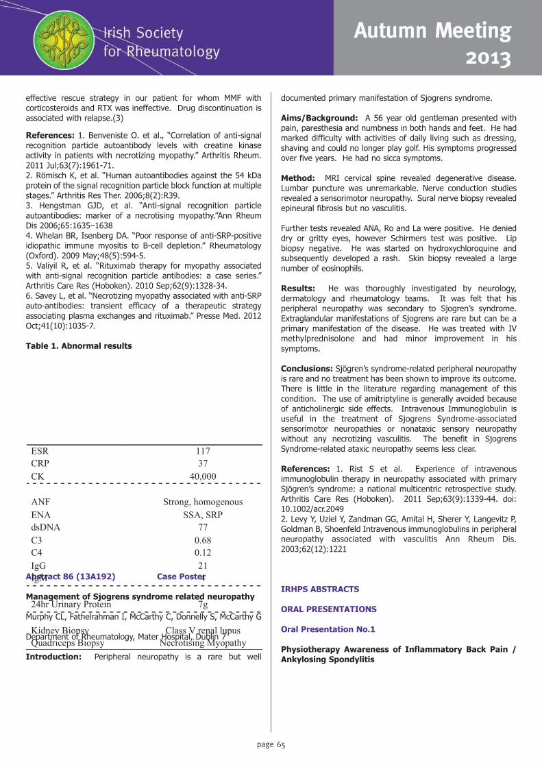
page 65
effective rescue strategy in our patient for whom MMF withcorticosteroids and RTX was ineffective. Drug discontinuation isassociated with relapse.(3)
References: 1. Benveniste O. et al., “Correlation of anti-signalrecognition particle autoantibody levels with creatine kinaseactivity in patients with necrotizing myopathy.” Arthritis Rheum.2011 Jul;63(7):1961-71.2. Römisch K, et al. “Human autoantibodies against the 54 kDaprotein of the signal recognition particle block function at multiplestages.” Arthritis Res Ther. 2006;8(2):R39.3. Hengstman GJD, et al. “Anti-signal recognition particleautoantibodies: marker of a necrotising myopathy.”Ann RheumDis 2006;65:1635–16384. Whelan BR, Isenberg DA. “Poor response of anti-SRP-positiveidiopathic immune myositis to B-cell depletion.” Rheumatology(Oxford). 2009 May;48(5):594-5.5. Valiyil R, et al. “Rituximab therapy for myopathy associatedwith anti-signal recognition particle antibodies: a case series.”Arthritis Care Res (Hoboken). 2010 Sep;62(9):1328-34.6. Savey L, et al. “Necrotizing myopathy associated with anti-SRPauto-antibodies: transient efficacy of a therapeutic strategyassociating plasma exchanges and rituximab.” Presse Med. 2012Oct;41(10):1035-7.
Table 1. Abnormal results
Abstract 86 (13A192) Case Poster
Management of Sjogrens syndrome related neuropathy
Murphy CL, Fathelrahman I, McCarthy C, Donnelly S, McCarthy G
Department of Rheumatology, Mater Hospital, Dublin 7
Introduction: Peripheral neuropathy is a rare but well
documented primary manifestation of Sjogrens syndrome.
Aims/Background: A 56 year old gentleman presented withpain, paresthesia and numbness in both hands and feet. He hadmarked difficulty with activities of daily living such as dressing,shaving and could no longer play golf. His symptoms progressedover five years. He had no sicca symptoms.
Method: MRI cervical spine revealed degenerative disease.Lumbar puncture was unremarkable. Nerve conduction studiesrevealed a sensorimotor neuropathy. Sural nerve biopsy revealedepineural fibrosis but no vasculitis.
Further tests revealed ANA, Ro and La were positive. He denieddry or gritty eyes, however Schirmers test was positive. Lipbiopsy negative. He was started on hydroxychloroquine andsubsequently developed a rash. Skin biopsy revealed a largenumber of eosinophils.
Results: He was thoroughly investigated by neurology,dermatology and rheumatology teams. It was felt that hisperipheral neuropathy was secondary to Sjogren’s syndrome.Extraglandular manifestations of Sjogrens are rare but can be aprimary manifestation of the disease. He was treated with IVmethylprednisolone and had minor improvement in hissymptoms.
Conclusions: Sjögren’s syndrome-related peripheral neuropathyis rare and no treatment has been shown to improve its outcome.There is little in the literature regarding management of thiscondition. The use of amitriptyline is generally avoided becauseof anticholinergic side effects. Intravenous Immunoglobulin isuseful in the treatment of Sjogrens Syndrome-associatedsensorimotor neuropathies or nonataxic sensory neuropathywithout any necrotizing vasculitis. The benefit in SjogrensSyndrome-related ataxic neuropathy seems less clear.
References: 1. Rist S et al. Experience of intravenousimmunoglobulin therapy in neuropathy associated with primarySjögren’s syndrome: a national multicentric retrospective study.Arthritis Care Res (Hoboken). 2011 Sep;63(9):1339-44. doi:10.1002/acr.20492. Levy Y, Uziel Y, Zandman GG, Amital H, Sherer Y, Langevitz P,Goldman B, Shoenfeld Intravenous immunoglobulins in peripheralneuropathy associated with vasculitis Ann Rheum Dis.2003;62(12):1221
IRHPS ABSTRACTS
ORAL PRESENTATIONS
Oral Presentation No.1
Physiotherapy Awareness of Inflammatory Back Pain /Ankylosing Spondylitis
ESR 117 CRP 37 CK 40,000 ANF Strong, homogenous ENA SSA, SRP dsDNA 77 C3 0.68 C4 0.12 IgG 21 IgM 4 24hr Urinary Protein 7g Kidney Biopsy Class V renal lupus Quadriceps Biopsy Necrotising Myopathy

page 66
Martina Fitzpatrick1, Maria Flynn2, Sinéad O’Malley2, CaitrionaCunningham2
1Physiotherapy Department, St. Vincent’s University Hospital,Dublin, Ireland2School of Public Health, Physiotherapy and Population Science,University College Dublin, Dublin, Ireland
Aim/Introduction: Inflammatory back pain (IBP) andAnkylosing Spondylitis (AS) are associated with long diagnosticdelays. This study aims to investigate awareness and knowledgeof features of IBP and AS, initial physiotherapy management andrelated educational preferences among physiotherapists inIreland.
Method: An online survey, based on the Assessment ofSpondyloarthritis International Society (ASAS) and Berlincriteria1,2, was distributed to all members of the private practiceand community care employment groups of the Irish Society ofChartered Physiotherapists. Questions regarding IBP features,associated rheumatologic features, initial physiotherapymanagement and educational preferences were included. Datawere analysed with descriptive statistics using the statisticalpackage for the social sciences (SPSS v. 20).
Results: The survey was completed by 100 physiotherapists,representing a 20 % response rate. Only 11% of respondentslisted 4 features of inflammatory back pain corresponding withthe Berlin and ASAS criteria, while only 7.8 % of respondentsidentified all the Berlin back pain elements from a random list.Forty per cent did not believe peripheral joint symptoms wereassociated with inflammatory back pain and 4.6 % were ‘unsure’about the significance of enthesitis. Group exercise and Selfmanagement were commonly cited management approaches. Ofthe respondents, 80% were interested in further education withmost favouring written information or online materials.
Conclusion: Private practice and community care physiotherapyrespondents demonstrated a lack of awareness and knowledgeregarding diagnostic criteria for inflammatory back pain andAnkylosing Spondylitis, indicating the need for related education.
References: 1. Rudwaleit M, van der Heijde D, Landewé R,Listing J, Akkoc N, Brandt J, Braun J, Chou CT, Collantes-EstevezE, Dougados M, Huang F, Gu J, Khan MA, Kirazli Y,MaksymowychWP, Mielants H, Sørensen IJ, Ozgocmen S, Roussou E, Valle-Oñate R,Weber U, Wei J, Sieper J. The development ofAssessment of SpondyloArthritis international Societyclassification criteria for axialspondyloarthritis (partII): validationand final selection. Ann Rheum Dis. 2009 Jun;68(6):777-83.doi:10.1136/ard.2009.108233. Epub 2009 Mar 17. PubMed PMID:19297344.2. Rudwaleit M, Metter A, Listing J, Sieper J, Braun J.Inflammatory back pain in ankylosing spondylitis: a reassessmentof the clinical history for application as classification anddiagnostic criteria. Arthritis Rheum. 2006 Feb;54(2):569-78.PubMed PMID: 16447233.
Oral Presentation No.2
The effectiveness of education and aerobic exercise in‘high functioning’ patients with fibromyalgia: evaluationof a new service
Catherine Cullinane1, Dr. Claire Sheehy1, Bindu Irudayaraj1,Joseph G McVeigh2
Waterford Regional Hospital1
University of Ulster2
Introduction/Aim: Fibromyalgia syndrome (FMS) ischaracterised by chronic widespread pain, fatigue and reducedphysical function. Exercise has positive effects on physicalfunction, fatigue and well-being in FMS1. However, patients findit difficult to engage in exercise and often have low self-efficacyfor exercise. The aim of this study was to evaluate a new earlyaccess service consisting of a 6-week education and aerobicexercise intervention in patients with FMS, who were classified as‘high functioning.’
Method: Participants (n=32) were referred from WRHrheumatology department and attended a 6-week exercise andeducation intervention delivered by physiotherapist andoccupational therapist. Participants completed the FibromyalgiaImpact Questionnaire (FIQ), Hospital Anxiety and DepressionScore (HAD) Six-Minute-Walk Test (6MWT) and Exercise Self-Efficacy Scale (ESES). Follow up was carried out post and 3months post programme.
Results: Participants who completed the intervention had asignificant improvement in mean (SD) pain from baseline 7.01(1.8) to the end of the programme 5.6 (2.9), p=0.03. Mean (SD)6MWT improved from 386m (96) to 416m (57) at the end of theprogramme (p=0.05), this was maintained at 3 month follow-up.There was no significant change in total FIQ score or the HADscale, however, participants’ exercise self-efficacy significantlyimproved from 58.2 (18.6) to 68 (11.6), p=0.01.
Conclusion: Improvements were recorded post programme inexercise capacity, exercise self-efficacy and VAS pain.Improvement was maintained at 3 months follow up. Anticipatedservice developments are use of FIQ to subgroup2, longer termfollow up, vocational rehabilitation and pedometer use toevaluate exercise adherence.3
Hauser, W., Klose, P., Langhorst, J., Moradi, B., Steinbach, M.,Schiltenwolf, M. & Busch, A. (2010) Efficacy of different types ofaerobic exercise in fibromyalgia syndrome: a systematic reviewand meta-analysis of randomized controlled trials. ArthritisResearch & Therapy, 12(3), R79.De Souza, (2009) Fibromyalgia Subgroups: Profiling distinctsubgroups using the Fibromyalgia Impact Questionnaire. APreliminary Study. Rheumatology Int.29:509-515Tudor-Locke C., Craig C., Aoyagi Y., (2011) .How many steps/dayare enough? For older adults and special populations. A literatureReview.International Journal of Behavioural Nutrition and PhysicalActivity 8:80
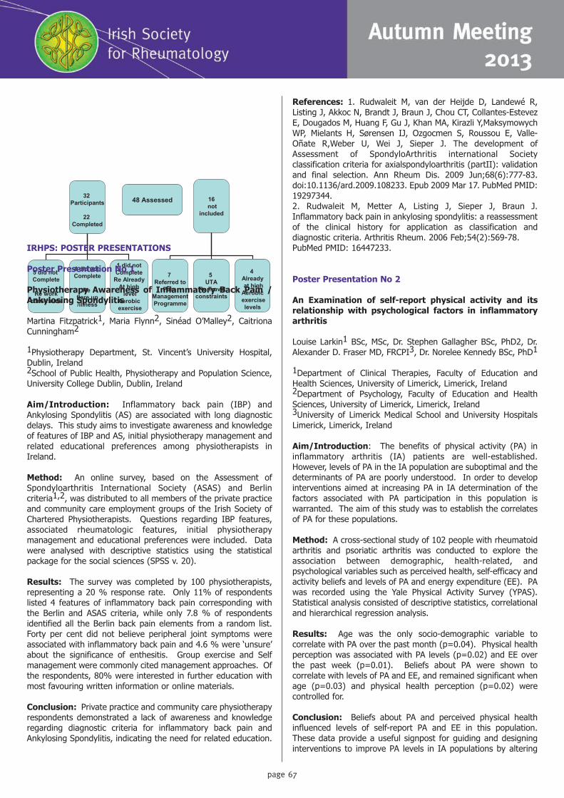
page 67
IRHPS: POSTER PRESENTATIONS
Poster Presentation No 1
Physiotherapy Awareness of Inflammatory Back Pain /Ankylosing Spondylitis
Martina Fitzpatrick1, Maria Flynn2, Sinéad O’Malley2, CaitrionaCunningham2
1Physiotherapy Department, St. Vincent’s University Hospital,Dublin, Ireland2School of Public Health, Physiotherapy and Population Science,University College Dublin, Dublin, Ireland
Aim/Introduction: Inflammatory back pain (IBP) andAnkylosing Spondylitis (AS) are associated with long diagnosticdelays. This study aims to investigate awareness and knowledgeof features of IBP and AS, initial physiotherapy management andrelated educational preferences among physiotherapists inIreland.
Method: An online survey, based on the Assessment ofSpondyloarthritis International Society (ASAS) and Berlincriteria1,2, was distributed to all members of the private practiceand community care employment groups of the Irish Society ofChartered Physiotherapists. Questions regarding IBP features,associated rheumatologic features, initial physiotherapymanagement and educational preferences were included. Datawere analysed with descriptive statistics using the statisticalpackage for the social sciences (SPSS v. 20).
Results: The survey was completed by 100 physiotherapists,representing a 20 % response rate. Only 11% of respondentslisted 4 features of inflammatory back pain corresponding withthe Berlin and ASAS criteria, while only 7.8 % of respondentsidentified all the Berlin back pain elements from a random list.Forty per cent did not believe peripheral joint symptoms wereassociated with inflammatory back pain and 4.6 % were ‘unsure’about the significance of enthesitis. Group exercise and Selfmanagement were commonly cited management approaches. Ofthe respondents, 80% were interested in further education withmost favouring written information or online materials.
Conclusion: Private practice and community care physiotherapyrespondents demonstrated a lack of awareness and knowledgeregarding diagnostic criteria for inflammatory back pain andAnkylosing Spondylitis, indicating the need for related education.
References: 1. Rudwaleit M, van der Heijde D, Landewé R,Listing J, Akkoc N, Brandt J, Braun J, Chou CT, Collantes-EstevezE, Dougados M, Huang F, Gu J, Khan MA, Kirazli Y,MaksymowychWP, Mielants H, Sørensen IJ, Ozgocmen S, Roussou E, Valle-Oñate R,Weber U, Wei J, Sieper J. The development ofAssessment of SpondyloArthritis international Societyclassification criteria for axialspondyloarthritis (partII): validationand final selection. Ann Rheum Dis. 2009 Jun;68(6):777-83.doi:10.1136/ard.2009.108233. Epub 2009 Mar 17. PubMed PMID:19297344.2. Rudwaleit M, Metter A, Listing J, Sieper J, Braun J.Inflammatory back pain in ankylosing spondylitis: a reassessmentof the clinical history for application as classification anddiagnostic criteria. Arthritis Rheum. 2006 Feb;54(2):569-78.PubMed PMID: 16447233.
Poster Presentation No 2
An Examination of self-report physical activity and itsrelationship with psychological factors in inflammatoryarthritis
Louise Larkin1 BSc, MSc, Dr. Stephen Gallagher BSc, PhD2, Dr.Alexander D. Fraser MD, FRCPI3, Dr. Norelee Kennedy BSc, PhD1
1Department of Clinical Therapies, Faculty of Education andHealth Sciences, University of Limerick, Limerick, Ireland2Department of Psychology, Faculty of Education and HealthSciences, University of Limerick, Limerick, Ireland3University of Limerick Medical School and University HospitalsLimerick, Limerick, Ireland
Aim/Introduction: The benefits of physical activity (PA) ininflammatory arthritis (IA) patients are well-established.However, levels of PA in the IA population are suboptimal and thedeterminants of PA are poorly understood. In order to developinterventions aimed at increasing PA in IA determination of thefactors associated with PA participation in this population iswarranted. The aim of this study was to establish the correlatesof PA for these populations.
Method: A cross-sectional study of 102 people with rheumatoidarthritis and psoriatic arthritis was conducted to explore theassociation between demographic, health-related, andpsychological variables such as perceived health, self-efficacy andactivity beliefs and levels of PA and energy expenditure (EE). PAwas recorded using the Yale Physical Activity Survey (YPAS).Statistical analysis consisted of descriptive statistics, correlationaland hierarchical regression analysis.
Results: Age was the only socio-demographic variable tocorrelate with PA over the past month (p=0.04). Physical healthperception was associated with PA levels (p=0.02) and EE overthe past week (p=0.01). Beliefs about PA were shown tocorrelate with levels of PA and EE, and remained significant whenage (p=0.03) and physical health perception (p=0.02) werecontrolled for.
Conclusion: Beliefs about PA and perceived physical healthinfluenced levels of self-report PA and EE in this population.These data provide a useful signpost for guiding and designinginterventions to improve PA levels in IA populations by altering
page 68
beliefs about PA and health perceptions.
Poster Presentation No 3
Physical Activity and Fatigue in Fibromyalgia
Dr. Norelee Kennedy & Sarah O’ Connell
Department of Clinical Therapies, University of Limerick
Aim/Introduction: Fibromyalgia (FM) is a chronic conditionassociated with widespread pain and fatigue. Fatigue is oftendescribed as the most troublesome symptom of FM, resulting inreduced levels of physical activity (PA) (Ryan 2011). The aim ofthe study is to describe fatigue, self-report PA and fibromyalgiaimpact in a population with FM through the use of an onlinequestionnaire. To determine if correlations exist between self-report PA and fatigue levels.
Method: A quantitative cross-sectional study design was used.Male and female participants over 18, with a diagnosis of FMwere recruited through Arthritis Ireland. Participants completedan online survey consisting of a demographics questionnaire, theFibromyalgia Impact Questionnaire, the Short Questionnaire toAssess Health Enhancing Physical Activity (SQUASH) and theFatigue Severity Scale. SPSS version 20 was used to analyze thedata using descriptive and correlational statistical tests -Spearman’s Rho, Mann-Whitney U, Kruskal-Wallis
Results: Overall response rate was 31.28% (n=61). Of therespondents 50.82% (n=31) fully completed the survey. Anoverall low negative correlation between self-report PA andfatigue (rho=-0.206) was found, but this was not statisticallysignificant (p=0.226). A strong, statistically significant negativecorrelation (rho=-0.857, p=0.007) was found between self-reportPA and fatigue for the SQUASH 4th quartile results.
Conclusion: This pilot study found high levels of fatigue and lowPA levels in participants. A significant negative correlationbetween higher levels of PA and fatigue was found. Furtherresearch with larger sample sizes is required to further explorethese findings.
Poster Presentation No 4
To Investigate the correlation between physical activity,fatigue and self-efficacy in rheumatoid arthritis
Norelee Kennedy1, Stella McGlennon1, Alexander Fraser2
1Department of Clinical Therapies, University of Limerick2Department of Rheumatology, University Hospital Limerick
Aim/Introduction: Physical Activity (PA) levels in rheumatoidarthritis (RA) have been shown to be lower than internationalrecommendations. Fatigue is a signficant, debilitating symptomin RA. Psychological traits such as self-efficacy may be amediator in the relationships between fatigue and physicalactivity in this population. The aim of the study is to explore thecorrelations between self-reported PA, fatigue and self-efficacy inRA.
Method: A cross-sectional survey study design was used.People with RA attending rheumatology out-patient clinics overan eight week period were surveyed using the InternationalPhysical Activity Questionnaire (IPAQ), Bristol RA Fatigue-Multidimensional Questionnaire (BRAF MDQ) and the RA Self-Efficacy Questionnaire (RASE). Data was anlaysed usingdescriptive statistics in SPSS.
Results: Seventeen (15 female) people participated in thesurvey. The mean time since diagnosis was 5.48 years (±SD4.7). The mean total self-report PA score was moderate(2,263.92 MET-minutes/week, ±SD 3,160.34). Mean fatiguescore was moderate (30.8 ± 19.59). Mean SE was high (99.21 ±12.64). Moderate negative correlations were found between PAand fatigue (r = -0.487, p = 0.128) and between fatigue and SE(r = -0.327, p = 0.276). A small positive correlation between PAand SE (r = 0.023, p = 0.947) was found.
Conclusion: This pilot study identified trends in the associationbetween fatigue, physical activity and self-efficacy in people withRA. People with higher levels of PA had higher self-efficacy andlower fatigue scores. Larger studies are needed to furtherexplore the relationships between these variables.
Poster Presentation No 5
Early diagnosis of incomplete atypical femoral fracturesin patients on prolonged bisphosphonate therapy usingDXA
Susan van der Kamp, Eric Heffernan, Conor Hurson, MalachiMcKenna, Oliver FitzGerald
Osteoporosis Centre, DXA Unit, St. Vincent’s University Hospital,Dublin 4
Aim/Introduction: Atypical femoral fractures (AFF) areassociated with prolonged bisphosphonate therapy. A feature ofincomplete AFF is a localized periosteal reaction. It has beensuggested that extending the length of the femur image at thetime DXA may diagnose an incomplete AFF.
Method: In patients over 50 years old on bisphosphonatetherapy for more than 5 years, we extended femur length at timeof routine DXA. Patients with abnormal DXA images werereferred for pelvis X-ray with views of lateral femurs.
Results: We studied 257 consecutive patients who meet theselection criteria. Abnormal DXA images were suggested in 19 of257 (7.4%). On X-ray, 7 (2.7%) showed no abnormality, 7 (2.7%)showed evidence of AFF, and 5 (2.0%) showed an unrelatedradiographic abnormality. Of the 7 cases with X-ray evidence ofAFF, 5 had a periosteal flare and 2 had a visible fracture line, bothof whom needed insertion of an intramedullary nail. Of the 7cases with X-ray evidence of AFF, 5 had a periosteal flare and 2had a visible fracture line, both of whom needed insertion of anintramedullary nail.
Conclusion: We demonstrated that it is feasible to detectincomplete AFF early using extended femur length imaging withprevalence in our sample of 2.7% (95% CI, 1.7 to 3.7%). We are

page 69
now exploring the feasibility of single-energy imaging of theentire length of each femur as means of improving the diagnosticutility because this gives much higher resolution thatapproximates the quality of plain X-ray.
Poster Presentation No 6
Review of an advanced practice Physiotherapist’sexperience in a musculoskeletal ultrasound clinic(MSKUS) within a Rheumatology setting
Grainne Cussen, Clinical Specialist Physiotherapist inRheumatology, Dr D. O’Gradaigh, Dr C. Sheehy
Physiotherapy Dept, Waterford Regional Hospital, IrelandRheumatology Dept, Waterford Regional Hospital, Ireland
Aim/Introduction: To evaluate an advanced practicePhysiotherapist’s experience in a MSKUS and injection clinicwithin a Rheumatology service from January 2011 to May 2013.Ultrasound is widely recognised as a tool to complement clinicaldiagnosis of both musculoskeletal (MSK) and inflammatoryconditions [1,2,3]. Physiotherapists working in advanced roleshave acquired this skill to enhance patient management,including triaging to the appropriate service following USevaluation.Method: Patients were referred to the MSKUS clinic fromRheumatology, Primary Care (PCCC) Physiotherapists and MSKTriage Physiotherapists. Patients were assessed clinically andthen examined with MSKUS by the advanced practicePhysiotherapist.
Results: Of 261 patients referred, 188 (72%) were fromRheumatology, 55 (21%) were from PCCC Physiotherapists and18 (7%) were from MSK Triage physiotherapists.The joint areas referred were: Shoulder 177(68%), foot andankle 40 (15%), wrist and hand 25 (10%), hip 11 (4%), elbow 5(2%) and knee 3 (1%).Background history of the patients included: MSK 154 (59%),inflammatory arthritis 78 (30%) and a query of inflammatorydisease 29(11%).Patients were subsequently referred to the following pathways:Injection by the Physiotherapist and follow up Physiotherapy104(40%), US guided injection with the Consultant 23(8%),Physiotherapy 50(19%),Orthopaedic Consult 39(15%), consultwith the Rheumatologist 66(25%), further investigation 5(0.1%),of which 3 were MRI and 2 CT. 2 patients did not attend.
Conclusion: Physiotherapists working in advanced roles withthe support of a Rheumatologist can enhance patientmanagement; and reduce the need for MRI which is costly anddifficult to access.
References:Cooperberg PL, Tsang I, Truelove L, Knickerbocker WJ. Gray scaleultrasound in the evaluation of rheumatoid arthritis of the knee.Radiology 1978;126(3):759-63.
De Flaviis L, Scaglione P, Nessi R, Ventura R, Calori G.Ultrasonography of the hand in rheumatoid arthritis. Acta Radiol1988;29(4):457-60.Karim Z, Wakefield RJ, Conaghan PG et al. The impact of
ultrasonography on diagnosis and management of patients withmusculoskeletal conditions. Arthritis Rheum 2001;44(12):2932-3
Poster Presentation No 7
Differential Diagnosis Algorithm for a Painful, SwollenKnee in the Absence of Trauma
Patricia Kavanagh (CNS), Dr Barry Sheane, Dr Conor McCarthy
Department of Rheumatology Mater University Hospital
Aim/Introduction: Monoarthritis of the knee is a commonpresentation to a rheumatology clinic. A definitive diagnosis isessential so that treatment can be commenced immediately andimproved outcomes achieved. If not diagnosed promptly and ifappropriate treatment is not initiated rapidly the condition may bemore difficult to treat. Joint pain can be associated with a varietyof different conditions and it is necessary to establish if the causeof pain is from within the joint or adjacent soft tissues. In orderto correctly diagnose, the exact position of pain and swelling isessential. The aim of the algorithm is to benefit overall practiceand to serve as a pathway to stimulate thought processes for theadvanced nurse practitioner.
Method: Adapting a problem based framework can serve toenhance judgement and advanced decision making in clinicalpractice. Potentially this algorithm can be utilised in diagnosingother rheumatologic conditions.
Results: On designing the algorithm a logical approach hasbeen adopted with all differential diagnosis falling within theremit of rheumatology clinical practice: septic arthritis, gout,osteoarthritis and rheumatoid arthritis in the absence of traumathe four differential diagnoses could present as a monoarthritis.
Conclusion: A considered and informed approach has beenadopted in reaching as accurate a diagnosis as possible and outruling possible diagnoses. A problem solving logical approach hasbeen applied. Evidence based practice and clinical experiencehas contributed to determining a most accurate diagnosis aspossible.

page 70
Poster Presentation No 8
The accuracy of a physiotherapist in recognising anddiagnosing inflammatory joint disease while working in anew patient rheumatology clinic
Paul Kirwan and Trevor Duffy
Rheumatology Department, Connolly Hospital, Blanchardstown,Dublin 15
Aim/Introduction: Physiotherapists working in triage roles arewell established in orthopaedic and spinal clinics, but there is lessliterature available on their role in rheumatology. There is adearth of research investigating the accuracy of a physiotherapistin diagnosing inflammatory joint conditions. The aim of this auditwas to assess the accuracy of a physiotherapist in recognisingand diagnosing inflammatory joint disease while working in a newpatient rheumatology clinic.
Method: Data was collected consecutively on all patientsassessed by the Physiotherapist at the Rheumatology NewPatient Clinic from December 2010 to June 2013. Patients with asuspected inflammatory diagnosis were assessed and appropriateinitial diagnostic tests were ordered. These patients had followup appointments scheduled with a consultant rheumatologist inorder to confirm or refute the initial diagnosis. Medical chartswere reviewed to ascertain if the Physiotherapist’s initialdiagnosis concurred with that of the Consultant Rheumatologist.
Results: A total of 210 patients were assessed over the timeperiod. Forty six patients were suspected of having aninflammatory arthritis. Thirty six of the 46 patients (78%) wereconfirmed by the Rheumatologist as having an inflammatoryarthritis, 11% were still pending diagnosis. Eleven percent of thepatients were given alternative diagnoses upon review by theConsultant Rheumatologist.
Conclusion: The data indicate that a physiotherapist withspecialist training in rheumatology can work effectively and safelyin a rheumatology new patient clinic. It also highlights the highnumber of non-inflammatory conditions seen in a rheumatologynew patient clinic, confirming the efficacy of a physiotherapistworking in this capacity.
Poster Presentation No 9
‘Reviewing the Impact of a Multi-Disciplinary GroupEducation Programme for Patients with Fibromyalgia’
Katie McCausland (OT) & Petrina Donohue (PT)
Rehabilitation Dept, Our Lady’s Hospital, Navan
Aim/Introduction: A MDT group education programme isprovided to Fibromyalgia patients from the North-East region inconjunction with pharmacological management. The impact ofthis programme was reviewed as part of a quality initiative, usingthe FIQ and Patient Satisfaction Questionnaire.
Method: An analysis of the database (n=66) was conductedincluding percentage of participation in the programme,geographical distribution, severity of FM and patient satisfaction.
Results: There was a high level of satisfaction feedback from allgroup participants but only a proportion of patients had clinicallysignificant improvements in symptoms and QOL. It was foundthat there was equal participation among the participantsbetween the mid, moderate and severe groups. Among the‘severe’ group there were very high levels of self-reported anxietyand depression.
Conclusion: The group education format for patients withFibromyalgia is an efficient use of resources for a busyRheumatology MDT. However, despite high rates of patientsatisfaction, the programme does not always provide clinicallysignificant changes of patient symptoms. A high incidence ofdepression and anxiety among the patients (as indicated by FIQQ. 9 & 10) indicates a need for psychological interventions as partof their treatment. There is a need for ongoing review of currentpractice and evidence-based research to provide an effective andefficient treatment programmes for people with FM.
Poster Presentation No 10
A Qualitative Exploration of the Impact of Fibromyalgiaon Meaningful Occupational Engagement
Claire O’Brien (BSc in Occupational Therapy), Eimear Lyons (MScClinical Therapies; BSc Occupational Therapy)
Rheumatology Unit Our Lady’s Hospice and Harold’s Cross
Aim/Introduction: Current literature suggests that living withfibromyalgia can have detrimental effects on participation in dailylife and cause disruption to global health status (Campos &Vazquez, 2012; Creek & Hughes, 2008; Sim & Madden, 2008).There is a growing body of qualitative research regarding theconsequence of fibromyalgia on overall health and well-being,however there is a paucity of research on the perceived impact offibromyalgia on occupational participation. This study aims togain an understanding of the impact that fibromyalgia can haveon meaningful occupational engagement.
Method: This is a descriptive study based on a qualitativeresearch design. Semi-structured one to one interviews werecompleted in an in-patient rheumatology service with a group ofwomen (n=6) women living with fibromyalgia. The interviewswere audio-recorded and transcribed verbatim by the researcher.Thematic analysis was used to analyse and code the data intoprevailing themes.
Results: Findings are represented by five main themes:

page 71
disruption to daily routine, role change, impact on self-identity,coping methods and future care recommendations. Diminishedengagement in valued occupations was evident for allparticipants.
Conclusion: These findings describe the negative effects thatfibromyalgia can have on meaningful occupational engagementfor affected individuals. This knowledge may lead to an increasedunderstanding among health professionals working with thisclient group and encourage a holistic approach to practice. Thesefindings also have potential to guide resource allocation, systemplanning and development of care pathways for this client group.
References: Campos, R. P., & Vazquez, M. I. (2012). Health-related quality of life in women with fibromyalgia: clinical andpsychological factors associated. Clinical Rheumatology, 31, 347-355. Creek, J., and Hughes, A. (2008). Occupation and health: Areview of selected literature. The British Journal of OccupationalTherapy, 71(11), 456-468. Sim, J., & Madden, S. (2008). Illness experience in fibromyalgiasyndrome: A metesynthesis of qualitative studies. Social Scienceand Medicine, 67(1), 57-67.
Poster Presentation No 11
“Working with Arthritis”: Study Protocol of a CohortStudy to Determine the Outcomes of an OccupationalTherapy Led Vocational Rehabilitation Programme forPeople with Arthritis
Eimear Lyons (MSc. Clinical Therapies; BSc. OccupationalTherapy),Dr. Katie Robinson, PhD (Lecturer/ Course Director MScOccupational Therapy, University of Limerick)Miriam Noonan (BSc. Occupational Therapy)
Department of Clinical Therapies, University of Limerick
Aim/Introduction: Work disability is pervasive among peoplewith arthritis (Bevan et al 2009). Vocational Rehabilitation (VR)can reduce work disability and associated economic, social andhuman costs (Waddell et al 2008). Here we report the studydesign of ‘Working with Arthritis: Strategies & Solutions’ an OTled VR programme aiming to reduce work disability among peoplewith arthritis.
Method: Study design will be a prospective cohort study ofworking age people with arthritis, in receipt of illness/disabilitypayments, living in the Border, Western and Midland regions ofIreland. The program aims to recruit 250 people with arthritisover a 2 year period. It is designed to overcome the barrierspeople with arthritis face when accessing, remaining in, orreturning to work. Psychosocial, environmental and occupationalfactors influencing work ability will be identified and evaluated.Data will be collected at three time points; programmecommencement, conclusion and at six month follow up.
Results: The primary outcomes of the study are work/education participation. Secondary outcomes include symptomexperience and quality of living. Economic evaluation of theprogram will also be completed.
Conclusion: Study findings will contribute to the evidence base
for VR programmes and will inform social policy regarding the keydeterminants of work ability and participation for those witharthritis of working age.
References:Bevan, S., McGee, R. and Quadrello, T. (2009b) Fit For Work?Musculoskeletal Disorders and the Irish Labour Market, London:The Work FoundationWaddell, G., Burton, A. and Kendall, N. (2008) ‘Vocationalrehabilitation: what works, for whom, and when’, London: TheStationery Office
Poster Presentation No 12
The development of a Rheumatology communityoutreach programe
Eileen O’Flynn, Miriam Molloy, Oliver FitzGerald
Department of Rheumatology, St. Vincent’s University HospitalDublin
Aim/Introduction: The current care pathway for patients withchronic rheumatic disease does not include a reliable system ofmonitoring efficacy and safety of treatments in primary caresettings. Many patients with stable rheumatic disease continueto attend outpatient clinics when with support, they could equallybe managed in a primary care setting.
Method: A prospective sample of patients was referred to theCNS-led service by a consultant rheumatologist at a point wherethe patients had stable disease but required ongoing out-patientreview to assess efficacy and safety of continued medication.During a twelve month period a total of 464 patients werereviewed by the service. 42% attended SVUH, 24% werereviewed by telephone and 34% were reviewed in the community(Arklow 20%, Newtownmountkennedy14%).
Results: In relation to outcomes 73% had stable disease; 16%required a medical review appointment; 4% required admissionto Harold’s Cross rheumatology rehabilitation unit; 3% and 4%respectivaly were referred for physiotherapy and single jointinjection. Thus, 80% or 371 patients were reviewed by the CNS-led service, clinical outcome measures obtained, drug safetyissues reviewed and repeat prescriptions provided with noadditional outpatient visits required. Feedback ascertainedfrom 100 patients rated the service very good or excellent.
Conclusion:• 464 patients were reviewed in the community by the CNS-led
service over a twelve month period• It facilitated more timely access for new patients and urgent
returns to the rheumatology outpatient clinics.• New : return ratio fell from1:7 to 1:4 at outpatients clinics.
Poster Presentation No 13
Abatacept at Home, The Sunny South East Experience
Una Martin, Paula Dreelan, Shane Brennan
Waterford Regional Hospital, VHI Home Care Service
Aim/Introduction: A combination of technology and economic

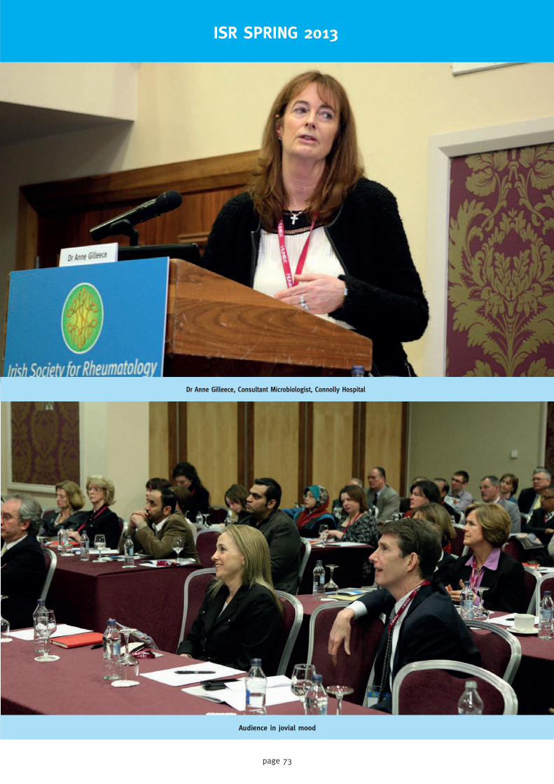
page 73
Audience in jovial mood
ISR SPRING 2013
Dr Anne Gilleece, Consultant Microbiologist, Connolly Hospital
page 74
pressures has imposed pressure for health care resources to bedelivered in a more timely and proficient manner. Patients canhave the choice of home infusion service for delivery ofAbatacept. In our experience this service has brought numerouspersonal benefits to the patient, advantages to the rheumatologydepartment as well as the organisation as a whole.
Method: We have availed of the home infusion service tomanage our patients since 2008 and to date over 18 patientshave availed of the service and 15 of these patients are continueto use the service.
Results: Of the 15 patients that are receiving the service, 9patients are female and 6 male. The average age is 53 with theaverage female age of 50 and male 60. The age range isbetween the age of 36 and 82. Thirteen of the 15 patients liveoutside the county of Waterford 2 of the female patients areengaged in full time employment. The advantages to the patientinclude minimal disruption to work, family and lifestyle as well asa reduction in the cost of travelling and other associated costs.This service has created increased capacity within our owninfusion service to treat other patients in addition to added costsavings to the hospital.
Conclusion: A patient satisfaction survey supports the successof the programme. In our experience the success of theprogramme can be attributed to the collaboration between thenursing staff, selection of appropriate patients and well definedpolicies and transferable standards of care.
Poster Presentation No 14
Quality of Life and Juvenile Idiopathic Arthritis (JIA) inIreland
Dr. Mary O’Hara¹, Dr. Margaret Hodgins²
¹School of Nursing & Midwifery, National University of Ireland,Galway²Discipline of Health Promotion, National University of Ireland,Galway
Introduction: JIA is an autoimmune inflammatory diseasediagnosed during childhood before the age of 16 with anincidence of 1 in 10000¹. Characterized by persistent synovialinflammation which can cause functional impairment, pain andactivity limitation; these may affect quality of life. Treatmentmodalities are not curative and aim to control the inflammatoryprocess. Parents and clinicians discussed the impact of thiscondition.
Method: Fourteen parents of young people aged 12-18 yearswith JIA and four clinicians from one geographical area in Irelandwere interviewed, using semi-structured and in-depthinterviewing techniques. The impact of the condition and itstreatment; service provision, education and future prospectswere considered. Interviews were transcribed verbatim andanalyzed.
Results: In general terms, parents considered that their youngpeople had a good quality of life. Parental recognition of thepractical difficulties that children were encountering and pride in
the efforts of the young people to adapt to difficult situations wasa major theme. Service provision and service organization werefrequently encountered as problematic. Safety considerationsrelating to the long-term effects of medications and the transitionto adult services were significant parental concerns. Socialsupport and an easily accessible and supportive healthcare teamwere found to be protective factors for the young people andtheir families in buffering the impact of juvenile idiopathicarthritis.
Conclusion: Person-centred care from the onset of JIA isneeded to meet the needs of young people across their lifespan,to optimize their life opportunities and quality of life.
Reference: ¹Beresford, M.W. (2011) ‘Juvenile idiopathicarthritis, new insight into classification, measures of outcome andpharmacotherapy’, Pediatric Drugs, 13, (3), pp. 161-173.
Poster Presentation No 15
Respiratory problems in patients on Etanercept
McKee H. and Whitehead E.
Rheumatology Department, Antrim Hospital
Aim/Introduction: Etanercept use is established. Interstitiallung disease in listed as an “uncommon” side effect. We describefour rheumatoids with respiratory problems within six months ofcommencing etanercept.
Method: Information from patients notes.
Results:Patient 158yr old female non smoker, on leflunomide. 3 months afterstarting etanercept 50mg weekly , presented with hypoxia sats86%. CT scan showed ground glass changes predominantlyupper lobe and a right lower lobe pulmonary embolus. There wascomplete resolution after steroids and drug withdrawal.Patient 267yr female. Mild asthma,.non smoker, on methotrexate 17years. CXR mild chronic R basal changes. Commencedetanercept 50mg weekly, 6 months later presented withincreasing shortness of breath. CT scan showed bilateral patchyinfiltrates and profound hypoxia. Improved with prednisoloneand drug withdrawal . Patient 358yr female, smoker, diabetic, not on methotrexate(intermittently abnormal LFTs). with a history of a previoushospital admission with a chest infection and some R sided basalchanges. 3 months after starting etanercept with increasingshortness of breath.and hypoxia. Chest x ray showed lung “whiteout”. Symptoms improved with drug withdrawal, steroids andantibiotics.Patient 461yo female, smoker. Normal CXR. On methotrexate 15mgweekly. Commenced etanercept 50mg weekly. Presented 9weeks later with cough and pleuritic pain. Symptoms resolved ondrug withdrawal CT chest showed no acute changes.
Conclusion: These patients, all female, presented with acute

page 75
respiratory symptoms within 6 months of starting etanercept.None had evidence of TB. All improved with drug withdrawal.
Poster Presentation No 16
An evaluation of a home occupational therapyprogramme for adults with rheumatoid arthritisprescribed Cimzia® within the Irish healthcare system
Trish Fitzgerald¹ and Oriel Corcoran²
¹Ashfield Healthcare²Dept of Rheumatology, Waterford Regional
Aim/Introduction: Given the incidence of rheumatoid arthritis(RA), the evidence for best practice supporting early occupationaltherapy intervention and the Irish health service advocatingaccessible services, there is a pressing need for early andaccessible occupational therapy services to be available to adultswith RA. This evaluation aims to identify the outcomes of a 12week home occupational therapy programme for adultsdiagnosed with RA prescribed Cimzia® at 5 designatedrheumatology sites.
Method: Adults with RA prescribed Cimzia® were referred tothe service by a member of their rheumatology team andreceived three sessions of Occupational Therapy (OT). Data wascollected during the initial and at final OT sessions using thefollowing outcome measures; Multi-Dimensional HealthAssessment Questionnaire, Arthritis Impact Measurement scale 2– short form, Joint protection knowledge questionnaire, GripStrength and Grip Ability Test, Arthritis Self Efficacy Scale ShortVersion, Canadian Occupational Performance Measure andpatient satisfaction questionnaire.
Results: Participants (n_21: 16 women; 6 men). Meanage_50.6 (SD 16.4) years; RA mean duration_6.4 (SD 8.9) years;Mean duration prescribed Cimzia®_4.7 (SD 8.0) months.There are a further 7 participants currently engaging in theprogramme and results awaiting analysis.Preliminary results suggest significant improvement inparticipant’s performance in everyday activities, self-efficacy andknowledge of self-management techniques.
Conclusion: This project is due for completion October 2013,thus data analysis is ongoing with a view to determining clinicaland statistical significance. This evaluation is important as it isthe first Irish home based OT rheumatology programmeproviding local and early access to OT in areas whererheumatology OT resources are limited in primary care.
Poster Presentation No 17
Patient satisfaction of Advanced PracticePhysiotherapists (APPs) working in an extended role inRheumatology and Orthopaedic clinics
Ashton J*, Breen R^, Callanan E∞, FitzGerald O�
*Physiotherapy Department, Beaumont Hospital, Beaumont,
Dublin 9^ Royal College of Physicians of Ireland, Dublin 2∞ Physiotherapy Department, Galway University Hospitals,Galway� Rheumatology dept, St. Vincent’s University Hospital, ElmPark, Dublin 4
Aim: i) Measure patient satisfaction with the APP assessment ina Rheumatology or Orthopaedic clinic using a standardised tool(VSQ 9) (Kennedy et al 2010).As part of the Rheumatology and Orthopaedic clinical careprogrammes, APPs were employed nationally from 2011 as partof a waiting list reduction initiative. Physiotherapists assessedpatients previously seen by Consultants therefore it wasimperative to evaluate the patient experience. It has previouslybeen shown that patient satisfaction can be reduced if theirexpectations are not met (Jackson and Kroenke 2001 cited byKennedy et al 2010).
Method: A sample of convenience was recruited of consecutivepatients that agreed to complete the questionnaire from Januaryto June 2013 across 10 hospital sites nationally. Questionnaireswere anonymously completed post clinic visit and given to theadministrative staff to reduce any bias. 340 questionnaires werecollected. Questionnaires were collated, inputted and analysedcentrally by an independent researcher using Microsoft Excel2007.
Results: Mean scores (95% confidence intervals) are detailed intable 1 and showed that all questions scored over 85/100. Interms of overall satisfaction with the APP assessment, 93% ofpatients rated it as excellent or very good; there were no poorresponses.
Conclusion: Overall satisfaction was mostly excellent or verygood with all aspects of the patient’s visit with the APP. Furtheranalysis is needed to compare satisfaction with Consultants andPhysiotherapists.
Reference:Kennedy D, Robarts S and Woodhouse L (2010) Patients areSatisfied with Advanced Practice Physiotherapists in a roletraditionally performed by orthopaedic surgeons. PhysiotherCan; 62: 298- 305
Table 1: Summary of results for satisfactionquestionnaires
page 76
APP Advance practice physiotherapist
Poster Presentation No 18
The Musculoskeletal Triage Service - Naas GeneralHospital and Tallaght Hospital: Audit of 1 Years Data
A.Brennan1, G.Kelly1, F.MacLeid1, E.Barker1, E.McGrane2,R.Mullan4, D.Moore3, D.Kane4.
Departments of Physiotherapy1, Orthopaedics3, Rheumatology4
Adelaide and Meath incorporating the National Children’sHospital, Tallaght, Dublin 24Department of Physiotherapy2, Naas General Hospital, Co.Kildare
Introduction/Aim: In 2012, three Extended ScopePhysiotherapists (ESP) were appointed, through the ClinicalStrategy and Programmes Directorate of the HSE, to provideorthopaedic and rheumatology triage services in Naas GeneralHospital (NGH) and Tallaght Hospital (TH). The aims were toreduce the waiting time for outpatient consultation fororthopaedic and rheumatology patients. To establish a diagnosisand triage patients along the most appropriate care pathwayaccording to their diagnosis.
Method: Suitability Criteria for inclusion for triage services wereestablished and agreed. Separate orthopaedic and rheumatologytriage waiting lists were established. All triage clinics ranalongside consultant clinics which offered a one stop shop forpatients, support with clinical diagnosis and with the ordering ofinvestigations and with management of patients as appropriate.Patients were assessed by the ESP and if required by theOrthopaedic or Rheumatology Consultants or Registrars on theday.
Results: From January to December 2013 1753 patients wereseen by ESP’s in the Musculoskeletal Triage Service between NGHand TH. Of this 1287 (73%) patients were assessed anddischarged at their clinic visit; 367 (21%) patients requiredmedical input on the day of their clinic visit; 142 (8%) patientsrequired a return visit to the ESP clinic and 347 (20%) patientsrequired a return visit at either the medical orthopaedic orrheumatology clinics.
Conclusion: Appropriately trained and supported ESP’s canassist in the management of both Orthopaedic and Rheumatologywaiting lists. Further investigation is required to ensure that
patients at these clinics are appropriately managed according tobest practice guidelines.
Poster Presentation No 19
An Investigation into the Use and Benefit of PermanentCustom Made Orthotics Prescribed by Physiotherapists -A Pilot Study
A. Brennan, G. Kelly
Department of Physiotherapy1, Tallaght Hospital, Tallaght, Dublin24
Aim/Introduction: In Tallaght Hospital PhysiotherapyDepartment, permanent custom made orthotics (PCMO) areprovided to patients for biomechanical anomalies. For patientswith Medical cards, these are funded by the General Medical CardScheme (GMS). The aim of this study was to investigate the useand benefit of PCMO in patients who received their PCMO throughGMS funding.
Method: A questionnaire regarding the use and benefit of PCMOwas devised by two Physiotherapists. In February 2013, thirty-eight questionnaires were sent to a sample of patients who hadreceived PCMO funded by the GMS between January 2010 andDecember 2012. Patients were asked to complete thequestionnaire anonymously and return to the PhysiotherapyDepartment, via stamped addressed envelopes provided.
Results: Seventeen completed questionnaires were returned(response rate 45%). Seventy-six percent (n=13) reportedwearing their PCMO full time. The reasons outlined for notwearing the PCMO full time were; that they did not fit in theirfootwear (18%; n=3) and that they were not helpful (6%; n=1).Seventy-six percent of respondents (n=13) reported that thePCMO prescribed provided full resolution of their symptoms.Maximum benefit of PCMO was observed to be between 0-6months in 35% (n=6) and >6 months in 12% (n=2) ofrespondents.
Conclusion: PCMO prescribed by Physiotherapists providebenefit to patients with biomechanical anomolies. It may beimportant to review patients following provision of funded PCMOto ensure cost effectiveness of this funding. This pilot has shownthat it may be necessary to provide a service to review patients6 months following receiving their PCMO.
Poster Presentation No 20
A pilot of a new Fibromyalgia Physiotherapy ManagementPathway in Tallaght Hospital
Sarah O’Driscoll
Physiotherapy Department, Tallaght Hospital, Tallaght, D24
APP Mean score
(95% confidence interval) Q1 Getting through to the outpatient clinic by phone
86.4 (83.48-89.3289)
Q2 Length of time waiting once you arrived for your appointment
86.4 (83.88-88.92)
Q3 Time spent with the healthcare provider you saw
86.4 (82.57-90.23)
Q4 Answers to your questions 86.4
(82.58-90.22) Q5 Explanation of the results of the assessment
86.4 (82.58-90.22)
Q6 Advice and information about the management of your condition
86.4 (82.74-90.06)
Q7 The technical skills of the healthcare provider you saw
86.4 (82.32-90.48)
Q8 The personal manner of the healthcare provider you saw
86.4 (81.65-91.15)
Q9 The visit overall 87.6
(83.50-91.70)

Prof Viet van Riel, University Medical Centre Nijmegen
page 77
Aim/Introduction: A standardised Fibromyalgia PhysiotherapyManagement Pathway (FPMP) was introduced in Tallaght Hospitalin October 2012. The aim of the pathway was to: streamline careof FMS patients in line with EULAR guidelines (EULAR, 2007),improve patient education and self management.
Method: In October 2012, all FMS patients referred to TallaghtHospital physiotherapy department were assessed. Outcomemeasures including Fibromyalgia Impact Questionnaire (FIQ) anda timed repeated sit-stand test were taken. Followingassessment, patients were enrolled into the FPMP. This includedan education class and 6 week course of hydrotherapy. The classincluded information on FMS, exercise management, pacing,exercise diaries, sleep and stress management and stretchingexercises. Following the FPMP, outcome measures were retakenand patients completed a satisfaction questionnaire.
Results: Forty patients were referred to the FPMP from October2012 to June 2013 (female 39, male 1). Thirty patients (75%)attended the education class. Fourteen patients completedsatisfaction questionnaires (35%) and 11 (28%) completed alloutcome measures. All outcome measures improved postattendance of FPMP (average FIQ: pre 1.48, post: 1.1, averagesit to stand pre: 19.4 seconds, post: 14.29 seconds). Patientsatisfaction scores (n=14) revealed 100% of patients rated theFMS education class as good/excellent. Ninety-three percent (n=13) reported they were very satisfied with the amount of helpreceived.
Conclusion: The FPMP streamlined patient care in line withEULAR 2007 guidelines. It showed positive changes re FIQ, sit tostand tests and patient satisfaction. Further research isnecessary with a large profile of patients.
Poster Presentation No 21
Pregnancy and Inflammatory arthritis: A descriptivestudy of clinical practice and patient experience relatedto medication management and care
Louise Moore1, Patricia Minnock1, Joan Lalor2, Douglas Veale3
1Rheumatology Rehabilitation, Our Lady’s Hospice and CareServices, Harold’s Cross, Dublin 6W2School of Nursing and Midwifery, Trinity College Dublin, Dublin 23St Vincent’s University Hospital, Elm Park, Dublin 4
Aim: To investigate approaches to prescribing practice and care,from the perspective of both rheumatology clinicians and femalepatients who had a pregnancy wish or a pregnancy within the lastfive years.
Method: A questionnaire (adapted with permission) wasemployed to collect data from members of the Irish Society ofRheumatology and Irish Rheumatology Nursing Forum (n = 114).In addition six interviews were undertaken with women who hadreceived care in rheumatology clinics to explore their experienceof the clinicians’ response to their health care needs in relation tomedication management and overall care whilst pregnant.
Results: A response rate of 62% (n = 71) was achieved.Variations existed with prescribing practice and advice with
respect to synthetic DMARDs (excluding methotrexate andleflunomide) and all biological DMARDs most notably in the pre-conception phase. Ninety per cent (n = 62) were in favour of anational register to record pregnancy outcomes for women withinflammatory arthritis. The main theme that emerged from thequalitative enquiry was that women need support, knowledgeand education on the potential impact of their arthritis on theirpregnancy and on medication management during all stages ofpregnancy from all clinicians involved in their care.
Conclusion: Findings confirm a discrepancy betweenknowledge of prescribing recommendations among clinicians.This led to dissatisfaction among women with the care cliniciansoffered. The findings from this research process will be utilisedto develop a specific care pathway for patients during all stagesof pregnancy incorporating support, medication and diseasemanagement and health promotion.
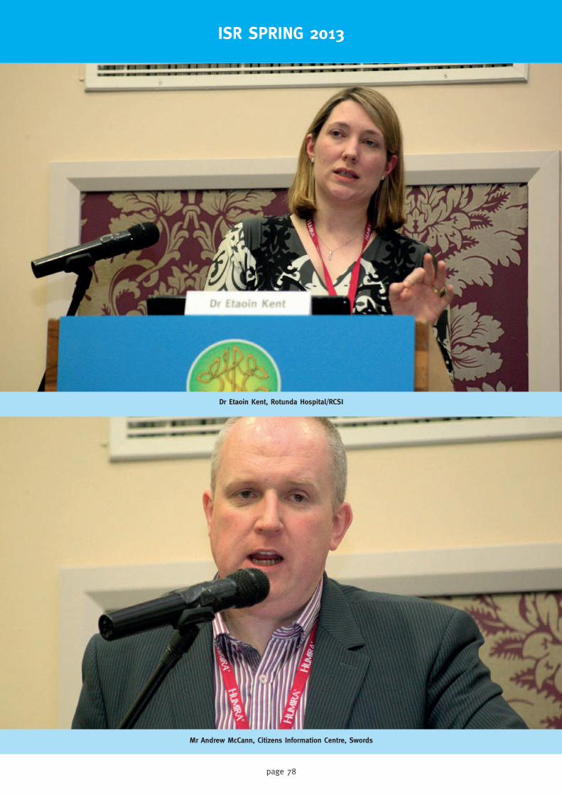
page 78
ISR SPRING 2013
Dr Etaoin Kent, Rotunda Hospital/RCSI
Mr Andrew McCann, Citizens Information Centre, Swords


![[XLS]minsvyaz.ruminsvyaz.ru/common/upload/docs/2007061917193vq.xls · Web viewINTRALINK IDR-38 INTRALINK ISR-71 INTRALINK ISR-81 INTRALINK ISR-13 INTRALINK ISR-15 INTRALINK ISR-18](https://img.pdfslide.us/doc/110x75/5b1d3ffb7f8b9acf678b6c15/xls-web-viewintralink-idr-38-intralink-isr-71-intralink-isr-81-intralink-isr-13.jpg)