Embed Size (px)
Citation preview
www.thelancet.com/infection Published online March 12, 2015 http://dx.doi.org/10.1016/S1473-3099(15)70006-X 1
Review
Lancet Infect Dis 2015
Published OnlineMarch 12, 2015http://dx.doi.org/10.1016/S1473-3099(15)70006-X
Department of Microbiology, Royal Brompton Hospital, London, UK (S Schelenz PhD); Department of Microbiology, University Hospital, Cardiff , UK (Prof R A Barnes MD); Mycology Reference Centre, Leeds General Infi rmary, Leeds, UK (R C Barton PhD); Department of Radiology, Royal Free Hospital, London, UK (J R Cleverley FRCR); Departme nt of Histopathology, St Thomas’s Hospital, London, UK (Prof S B Lucas FRCPath); Centre for Medical Microbiology, University College London, London, UK (Prof C C Kibbler FRCPath); and National Aspergillosis Centre, University Hospital of South Manchester and University of Manchester, Manchester, UK (Prof D W Denning FRCP)
Correspondence to:Dr Silke Schelenz, Department of Microbiology, Royal Brompton Hospital, Sydney Street, London SW3 6NP, [email protected]
British Society for Medical Mycology best practice recommendations for the diagnosis of serious fungal diseasesSilke Schelenz, Rosemary A Barnes, Richard C Barton, Joanne R Cleverley, Sebastian B Lucas, Christopher C Kibbler, David W Denning, on behalf of the British Society for Medical Mycology
Invasive fungal diseases are an important cause of morbidity and mortality in a wide range of patients, and early diagnosis and management are a challenge. We therefore did a review of the scientifi c literature to generate a series of key recommendations for the appropriate use of microbiological, histological, and radiological diagnostic methods for diagnosis of invasive fungal diseases. The recommendations emphasise the role of microscopy in rapid diagnosis and identifi cation of clinically signifi cant isolates to species level, and the need for susceptibility testing of all Aspergillus spp, if treatment is to be given. In this Review, we provide information to improve understanding of the importance of antigen detection for cryptococcal disease and invasive aspergillosis, the use of molecular (PCR) diagnostics for aspergillosis, and the crucial role of antibody detection for chronic and allergic aspergillosis. Furthermore, we consider the importance of histopathology reporting with a panel of special stains, and emphasise the need for urgent (<48 hours) and optimised imaging for patients with suspected invasive fungal infection. All 43 recommendations are auditable and should be used to ensure best diagnostic practice and improved outcomes for patients.
IntroductionInvasive fungal diseases are a worldwide health problem, not only in immunocompromised patients and those undergoing intensive-care treatment, but also increasingly in patients with chronic disorders such as chronic lung diseases.1–5 Despite the discovery of new antifungal agents and formulations, the morbidity and mortality of invasive fungal diseases is high.1,6,7 Therefore, early recognition and diagnosis of mycoses have become a major focus for improvement of the management and outcome of these infections.5,8,9
In 2003, a working group of the British Society of Medical Mycology (BSMM) proposed quality-of-care standards for patients with invasive fungal infections.10 These standards attempted to provide guidance for microbiology and histopathology laboratories and radiology and clinical specialists for improved use of available diagnostic tests for the management of invasive fungal diseases. Subsequent audits of these standards identifi ed areas for improvement.11,12
Inclusion of antigen testing and radiology within the consensus defi nitions for invasive fungal infections by the European Organization for Research and Treatment of Cancer (EORTC) and Mycoses Study Group (MSG) lead us to include best practice guidance for the use of antigen and molecular testing, and more detailed radiology diagnostics, in this document.9,13 The European guidelines for diagnosis and management of candida diseases were likewise considered, whereas guidance for antifungal treatment was omitted since clinical guidelines have been published.14 We defi ne best practice recommendations for the microbiological (panel 1), histopathological (panel 2), and radiological (panel 3) diagnostic investigation for diagnosis of serious fungal diseases, including serology, molecular diagnostics, and susceptibility testing.
Microbiology best practiceDirect microscopy, for many sample types, provides an important diagnostic benefi t that is greater than culture alone. Another advantage of microscopy is the rapid availability of results—often within 2–4 h of a specimen’s arrival in the laboratory.15 Rapid processing and reporting is important, because delayed diagnosis of an invasive fungal infection can be lethal.
Microscopy can distinguish whether an infection is caused by a septate mould (Aspergillus spp) or non-septate mould of the order Mucorales (members of the families Mucoraceae, Cunninghamellaceae, Saksenaeaceae, Mortierellaceae, and Syncephalastraceae), which aff ects the choice of antifungal treatment. Direct microscopy is especially important for diagnosis of non-septate fungi, because these fungi are poorly recovered by culture, partly as a result of damage during refrigeration or homogenisation of tissues. Many other distinctive fungi can be provisionally identifi ed by direct microscopy (capsule of Cryptococcus spp, spherules of Coccidioides spp, or small intracellular yeasts of Histoplasma capsulatum or Talaromyces [Penicillium] marneff ei). Direct visualisation of fungi in diagnostic sterile fl uids or tissues likewise aids confi rmation of cases of invasive fungal disease when a fungal growth alone could be a result of culture contamination.9 Centrifugation and staining (Gram stain, India ink, or optical brighteners) of liquid specimens concentrates fungal elements and increases the probability of detection. Microscopy can be useful for several specimens, which are discussed below.
First, blood cultures should be done in all cases in which systemic fungal infections are suspected, with a large volume of blood and recommended commercial systems and media.16,17 Gram stain of routine blood cultures is suitable for detection of the most prevalent
2 www.thelancet.com/infection Published online March 12, 2015 http://dx.doi.org/10.1016/S1473-3099(15)70006-X
Review
fungaemia pathogens (Candida spp and Cryptococcus spp), but is unsuitable for speciation and identifi cation of moulds.18 The clinical usefulness of microscopy can be
improved by the use of peptide nucleic acid fl uorescent in-situ hybridisation (PNA-FISH), which can identify Candida species that are likely to be fl uconazole-resistant
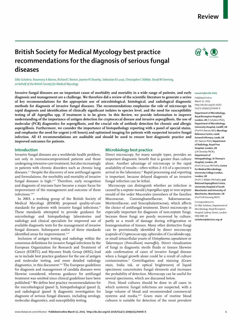
Panel 1: Microbiology best practice recommendations
Microscopy and stains• Fluids from usually sterile sites and bronchoalveolar lavage
(BAL) from patients with suspected infection should be examined by direct microscopy with suitable methods for fungal detection*
• Adequate tissue for histology and culture should be ensured before direct microscopy is done on the rest of the sample
• Optical brighteners are recommended for microscopy on all samples from immunocompromised patients
• Direct fl uorescent-antibody staining, PCR, or both is recommended for patients with suspected pneumocystis infection
• India ink staining of cerebrospinal fl uid samples from immunocompromised patients is recommended in addition to Gram staining if cryptococcus capsule antigen (CRAG) testing is not available on site
Culture and identifi cation• Bronchoscopy fl uids should be cultured in suitable media to
support fungal growth*• Yeasts cultured from urine samples should be identifi ed to
species level and reported for all critical care and immunocompromised patients*
• All clinical isolates of aspergillus from patients who will receive antifungal treatment should be identifi ed to species complex level, by referral to a specialist laboratory if necessary*
• All fungi (yeasts and moulds) obtained from sterile sites, including blood and continuous ambulatory peritoneal dialysis fl uids, and intravenous line tips should be identifi ed to species complex level by referral to a specialist laboratory if necessary. Bronchoscopy fl uid and paranasal sinus material is regarded as sterile in this context for all fungi except Candida spp*
• All aspergillus isolates from patients who have allergic bronchopulmonary aspergillosis, aspergilloma, or acute or chronic aspergillosis should be susceptibility tested for antifungals used for treatment (eg, voriconazole or itraconazole) if therapy is initiated; isolates should be stored for 6 months in case additional susceptibility testing is needed at a later date
• Any amount of fungi cultured from vascular-device tips should be identifi ed to species level and reported
Respiratory specimens• At least three sputum samples should be obtained for
detection of respiratory fungi, unless a validated PCR method is used
• BAL fl uid is recommended for diagnosis of invasive fungal disease in patients with haematological disease
• Respiratory and fl uid samples should be concentrated by
centrifugation at 1000 g or greater for at least 10 min or with the cytocentrifuge before microscopy
• Respiratory samples should be liquifi ed, especially for detection of Pneumocystis jirovecii
• Isolation of Aspergillus spp from respiratory samples should be reported with interpretative comments according to patient risk group and likelihood of invasive, chronic, or allergic disease
Cerebrospinal fl uid (CSF) specimens• All CSF specimens that are from immunocompromised
patients, from those with sarcoidosis or cancer, or who show abnormal concentrations of glucose, protein, or leucocytes without an adequate explanation should be cultured and antigen tested for Cryptococcus neoformans; all bacterial plates should be incubated for a minimum of 5 days and fungal media incubated at 30°C for up to 21 days
Fungal serological and molecular testing• Serum samples from immunocompromised patients with
presentations consistent with cryptococcal meningitis for whom a CSF specimen is not available (eg, cases in which lumbar puncture is contraindicated) should be tested for Cryptococcus spp antigen (CRAG)
• Galactomannan screening of serum (two times per week) from patients with haematological malignancies at high risk of invasive aspergillosis should be considered in those not receiving mould-active prophylaxis; optical density (OD) index threshold of 0·5 has a high negative predictive value, enabling invasive aspergillosis to be excluded
• Galactomannan testing of BAL from patients at high risk of invasive aspergillosis should be considered, although the current OD index cutoff of 0·5 might change
• β-D-glucan screening of serum from patients at high risk of invasive fungal disease should be considered; a negative result has a high negative predictive value, enabling invasive fungal disease to be excluded
• PCR screening of serum for aspergillus from patients at high risk of invasive fungal disease should be considered; a negative result has a high negative predictive value, enabling invasive fungal disease to be excluded
• Combination testing with aspergillus PCR plus another antigen test improves the positive predictive value and diagnosis of invasive fungal disease
• Patients with pulmonary cavities of uncertain cause (with or without an aspergilloma) should have serum samples tested for antibodies to aspergillus
• Patients with suspected allergic bronchopulmonary aspergillosis should have serum samples tested for total IgE and aspergillus-specifi c IgE
(Continues on next page)
www.thelancet.com/infection Published online March 12, 2015 http://dx.doi.org/10.1016/S1473-3099(15)70006-X 3
Review
in 100% of cases within 2 hours of a positive Gram stain.19–21 Another rapid technique for identifi cation of fungi directly from blood cultures is matrix-assisted laser desorbtion/ionisation time-of-fl ight mass spectrometry (MALDI-TOF), but MALDI-TOF is less sensitive and specifi c than PNA-FISH.22
Second, discrimination between colonisation and infection is diffi cult when yeasts are seen in respiratory samples, since colonisation is common with candida, and pulmonary infection is rare. For moulds, sensitivity of sputum microscopy can be increased with larger sample volumes and repeated sampling, and usually represents disease in at-risk patients.23,24 Overall sensitivity of culture and microscopy of bronchoalveolar lavage (BAL) fl uid in invasive aspergillosis is about 50% in high-risk patients with haematological disease. Sensitivity of fungal culture is increased in patients with severe and advanced infection.23,25–28 BAL is the preferred specimen for Pneumocystis jirovecii microscopy in patients without AIDS, whereas in those with AIDS, induced or routine sputum might be as sensitive as BAL.29–32 Microscopy of respiratory samples is also helpful for diagnosis of cryptococcosis and infections with endemic fungi. For diagnosis of fungal sinusitis, microscopy of sinus biopsy tissue can be done rapidly with a wet potassium hydroxide preparation, which has a sensitivity of 78%, when compared with histological examination in a study of aspergillus sinusitis.33
In a review of cryptococcal meningitis, India ink staining of CSF gave an overall sensitivity of 60% in HIV-positive and HIV-negative immunocompromised patients.34 For candida meningitis, microscopy gives a positive result in about 40% of Gram-stained CSF.35 Microscopy of undiluted, centrifuged urine can rapidly detect the presence of fungi and tubular casts.36 However, the importance of urine microscopy for guidance of the management of invasive fungal diseases is unclear in at-risk patients. Because the presence of fungus in tissue is
diagnostic for proven fungal infection, biopsy of infected tissue should be attempted whenever possible.9 Tissue samples should be kept moist during transport, processed promptly, and cultured in fungal media (up to 14 days or longer if positive on microscopy or evidence of possible endemic fungal infection exists).14 Formal histopathological examination is preferable to rapid smears.33,37,38
Rapid and easily reproducible staining methods typically used in routine microbiology or cytology laboratories
Panel 2: Histopathology best practice recommendations
Specialised stainsSpecialised stains should be done in parallel with standard stains if mycosis or another infection is to be assessed or excluded*• Standard stain: haematoxylin and eosin (H&E) on
histopathology slides; Giemsa or Papanicolaou on smears• Triple set of stains: Ziehl–Neelsen stain for acid-fast
organisms; Gram stain for bacteria, fungi, and others; Grocott silver stain, or periodic acid–Schiff , to highlight fungi
Reporting of resultsReport fungal morphology (yeast or hyphae), including the following• Whether a yeast is small, medium, or large• Whether a yeast has cross walls or septa (ie, is splitting
rather than budding)• Whether a hyphal form has usual width, or has a dilated,
bizarre shape• Whether H&E-stained fungi are pigmented and brown, or
are unpigmented and colourless or pale blue
Positive results should be telephoned to clinicians immediately*
*Previously published BSMM standards10
Antifungal drug-susceptibility testing• Isolates of Candida spp from sterile sites, or from patients not
responding to therapy at a minimum should have their susceptibility tested against fl uconazole
• Isolates of Aspergillus fumigatus should have their susceptibility tested against antifungal agents used locally for treatment (eg, itraconazole and voriconazole) if antifungal treatment is given
Therapeutic drug monitoring• No indications for therapeutic drug monitoring of
amphotericin B or the echinocandins; measurement of fl uconazole concentrations is rarely necessary
• Therapeutic drug monitoring of itraconazole, voriconazole, and posaconazole is usually needed. Specifi cally, voriconazole monitoring is needed in most patients, and certainly in children, including repeat monitoring after dose changes and
shift from intravenous to oral treatment; dose optimisation during long-term therapy needs such monitoring
• Flucytosine monitoring is recommended for all patients receiving treatment
Clinical requests and reporting• All test requests should state whether the patient is
immunocompromised*• All intravascular devices should be removed promptly if
clinically feasible after diagnosis of candidaemia• All new fungaemia, positive results of microscopy on sterile
tissues or fl uids, and positive cryptococcal antigen and galactomannan results should be telephoned or actively communicated by laboratory staff to clinicians within 2 h of their availability
*Previously published BSMM standards10
4 www.thelancet.com/infection Published online March 12, 2015 http://dx.doi.org/10.1016/S1473-3099(15)70006-X
Review
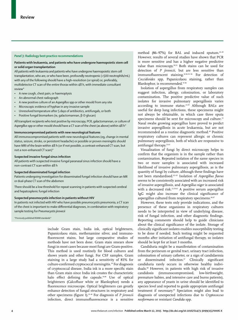
include Gram stain, India ink, optical brighteners, Papanicolaou stain, methenamine silver, and immuno-fl uorescent stains, but large comparative studies of methods have not been done. Gram stain smears show fungi in most cases because most fungi are Gram-positive. This method is used routinely for blood cultures and shows yeasts and other fungi. For CSF samples, Gram staining in a large study had a sensitivity of 85% for culture-confi rmed cryptococcal meningitis.39 For diagnosis of cryptococcal disease, India ink is a more specifi c stain than Gram stain since India ink creates the characteristic halo eff ect defi ning the capsule.34,40 Use of optical brighteners (Calcofl uor white or Blankophor) needs a fl uorescence microscope. Optical brighteners can greatly enhance detection of fungal structures in respiratory and other specimens (fi gure 1).41–44 For diagnosis of P jirovecii infection, direct immunofl uorescence is a sensitive
method (86–97%) for BAL and induced sputum.45–49 However, results of several studies have shown that PCR is more sensitive and has a higher negative predictive value than microscopy.50,51 Both stains can be used for detection of P jirovecii, but are less sensitive than immunofl uorescent staining.42,46,52–56 For detection of Coccidioides spp, Papanicolaou staining, rather than Blankophor, is recommended.57,58
Isolation of aspergillus from respiratory samples can suggest infection, allergy, colonisation, or laboratory contamination. The positive predictive value of such isolates for invasive pulmonary aspergillosis varies according to immune status.23,59 Although BALs are useful for deep lung infections, these specimens might not always be obtainable, in which case three sputa specimens should be sent for microscopy and culture.23 Nasal swabs growing aspergillus have proved to predict invasive aspergillosis in acute leukaemia, but are not recommended as a routine diagnostic method.60 Positive respiratory cultures can represent allergic or chronic pulmonary aspergillosis, both of which are responsive to antifungal therapy.23,61
Visualisation of fungi by direct microscopy helps to confi rm that the organism is in the sample rather than contamination. Repeated isolation of the same species in two or more samples is associated with increased likelihood of invasive pulmonary aspergillosis, as is the quantity of fungi by culture, although these fi ndings have not been standardised.62–65 Isolation of Aspergillus fl avus seems to be consistently associated with an increased risk of invasive aspergillosis, and Aspergillus niger is associated with a decreased risk.23,59,65 A positive serum aspergillus IgG might also increase the clinical signifi cance of aspergillus cultured from respiratory specimens.66,67
However, these tests only provide indications, and the presence of these organisms in respiratory cultures needs to be interpreted in view of underlying disease, risk of fungal infection, and other diagnostic fi ndings. Reporting comments should help to guide clinicians about the clinical signifi cance of the isolate. Storage of clinically signifi cant isolates enables susceptibility testing to be done if needed. Such testing might be requested months after initiation of antifungal therapy, so isolates should be kept for at least 3 months.
Candiduria might be a manifestation of contamination from the perineum or genital tract, urinary tract infection, colonisation of urinary catheter, or a sign of candidaemia or disseminated infection.67 Clinically signifi cant candiduria rarely occurs in otherwise healthy indivi-duals.68 However, in patients with high risk of invasive candidosis (immunocompromised, low-birth weight, premature babies, and intensive care and burns patients), any appearance of yeasts in urine should be identifi ed to species level and reported to guide appropriate antifungal treatment if necessary.69 Speciation might also lead to diagnosis of unexpected infections due to Cryptococcus neoformans or resistant Candida spp.
Panel 3: Radiology best practice recommendations
Patients with leukaemia, and patients who have undergone haemopoietic stem cell or solid organ transplantationAll patients with leukaemia and patients who have undergone haemopoietic stem cell transplantation, who are, or who have been, profoundly neutropenic (<500 neutrophils/mL) with any of the following should have a high-resolution (or spiral) or, preferably, multidetector CT scan of the entire thorax within 48 h, with immediate consultant review*• A new cough, chest pain, or haemoptysis• An abnormal chest radiograph• A new positive culture of an Aspergillus spp or other mould from any site• Microscopic evidence of hyphae in any invasive sample• Unresolved temperature after 5 days of antibiotics, antifungals, or both• Positive fungal biomarkers (ie, galactomannan, β-D glucan)
All transplant recipients who test positive by microscopy, PCR, galactomannan, or culture of Aspergillus spp or other mould should have a CT scan of the chest (as above) within 48 h*
Immunocompromised patients with new neurological featuresAll immunocompromised patients with new neurological features (eg, change in mental status, seizure, stroke, or persistent headache) or possible or proven meningitis should have MRI of the brain within 48 h (or if not possible, a contrast-enhanced CT scan, but not a non-enhanced CT scan)*
Suspected invasive fungal sinus infectionAll patients with suspected invasive fungal paranasal sinus infection should have a non-contrast CT scan within 48 h
Suspected disseminated fungal infectionPatients undergoing investigation for disseminated fungal infection should have an MR or dual-phase CT scan of the abdomen
There should be a low threshold for repeat scanning in patients with suspected cerebral and hepatosplenic fungal infection
Suspected pneumocystis infection in patients without HIVIn patients not infected with HIV who have possible pneumocystis pneumonia, a CT scan of the chest is important to make diff erential diagnoses, in combination with respiratory sample testing for Pneumocystis jirovecii
*Previously published BSMM standards10
www.thelancet.com/infection Published online March 12, 2015 http://dx.doi.org/10.1016/S1473-3099(15)70006-X 5
Review
To assess the clinical signifi cance of candiduria in catheterised patients, re-culture of urine after catheter removal is advisable. If the repeat urine culture is negative, antifungal treatment is usually not warranted.69 By contrast, persistent candiduria can mean upper urinary tract infection, and further renal imaging is needed to exclude renal obstructions.
Diagnosis of fungal meningitis such as cryptococcosis is well established and discussed in the serology and microscopy sections.34,70,71 Fungal media should be kept for long-term CSF culture (up to 21 days) at 30°C.72 Candida meningitis yeasts can be seen with Gram stain and cultured on Sabouraud agar. Rare causes of fungal meningitis include Coccidioides spp (best test is CSF antibody), H capsulatum (best test is histoplasma antigen), and Aspergillus spp (best tests are galactomannan and PCR).73,74
Two key advances in the diagnosis of invasive fungal diseases are antigen testing and molecular techniques. International study groups have published and revised consensus diagnostic criteria for invasive fungal diseases, including serological testing.9,13 Although intended for use in clinical research, these defi nitions have clear potential to aff ect patient management, and might contribute to improved outcomes.75–77 C neoformans and Cryptococcus gattii shed a capsule antigen (CRAG) that can be detected in the CSF and serum of infected patients with commercial assays.70,71,78–81 The simple lateral-fl ow device for serum, plasma, urine, and CSF CRAG detection is used in many laboratories for screening and diagnosis.80 In symptomatic patients with HIV, CRAG detection is highly sensitive and specifi c.70 The usefulness of monitoring CRAG titres in serum in patients without HIV continues to be debated, but is unreliable in HIV-positive patients.34,82 However, increasing or persistently high CRAG titres might herald therapeutic failure or relapse, and might be useful to distinguish immune reconstitution from persistent cryptococcal infection.83
Galactomannan (aspergillus antigen) detection in body fl uids is more sensitive than culture for diagnosis of invasive aspergillosis. In serum, the sensitivity is variable (17–100%), having a 0·5 cutoff ratio with the highest sensitivity in patients with haematological disease who are not on antimould prophylaxis.84,85 For screening, the key factors that aff ect accuracy of aspergillus antigen testing are the prevalence of invasive aspergillosis and eff ect of antifungal prophylaxis. The positive predictive value of the test increases from 31% in a population with 5% prevalence to 69% when the prevalence is 20%.86 As a result, galactomannan screening is unlikely to be benefi cial and cost eff ective if the probability of invasive aspergillosis before testing is low, and should be reserved for high-risk populations (patients undergoing allogeneic-stem-cell transplantation, those with acute myeloid leukaemia, and those undergoing aggressive chemo-therapeutic regimens for relapsed disease). A
meta-analysis87 supports the 0·5 cutoff and shows that overall sensitivity is 78% and specifi city is 81% in neutropenic patients. This moderate-quality evidence supports the recommendation that serial screening of blood specimens from high-risk patients is appropriate when invasive aspergillosis prevalence exceeds 7% and no antimould prophylaxis is given. Additionally, galactomannan detection is widely used in BAL specimens with evidence that galactomannan values of OD index of 0·5–1·0 have decreased predictive values compared with results of greater than 1·0.88 The test has diagnostic merit in patients who are undergoing lung transplantation or who are in intensive care.89–91
Cross-reactivity and false positives arise as a result of dietary and medical factors, reducing the specifi city of the test in serum and BAL specimens. Piperacillin–tazobactam was a source of false positives, but the problem has been resolved.92,93
1,3-β-D-glucan (BDG) is a carbohydrate moiety in the cell walls of many fungi, and is produced in vivo during infection by several important fungal organisms (Aspergillus spp, Candida spp, and P jirovecii, but not by Cryptococcus spp or species of the order Mucorales). The BDG test is indicated for the presumptive diagnosis of invasive fungal disease, and seems to be sensitive with a good negative predictive value (ie, excludes infection).94,95 This test might have a role in some care pathways, making use of the high negative predictive value, but positive results will always necessitate further investigations. False positive results can occur, resulting from gauze dressings, dialysis, and some bacteria.95,96
Figure 1: Calcofl uor white staining of Aspergillus spp hyphaeFluorescence microscopy (x100) showing Calcofl uor white staining of centrifuged bronchoalveolar lavage from a patient with acute myeloid leukaemia presenting with febrile neutropenia and cough. Bright fl uorescent dichotomous branching septate hyphae can be seen, which were later confi rmed by culture to be Aspergillus fumigatus.
6 www.thelancet.com/infection Published online March 12, 2015 http://dx.doi.org/10.1016/S1473-3099(15)70006-X
Review
The test is useful for diagnosis of pneumocystis pneumonia, especially when a respiratory sample cannot be obtained.95
Commercially available ELISAs for detection of candida antigen (mannan) and anti-mannan antibodies are available for diagnosis of candida infection, but little clinical assessment has been done, and testing might not detect some candida species.97 On the basis of the poor quality of evidence, we do not recommend testing for mannan and anti-mannan antibodies at present, although other guidelines provide some indication for use.14
Detection of aspergillus antibodies (mainly Aspergillus fumigatus) is useful for diagnosis of several forms of aspergillosis in immunocompetent patients. Absence of comparative studies of methods restricts highly specifi c recommendations; in UK laboratories, passive diff usion, counter immunoelectrophoresis, in-house and com-mercial ELISAs, and semiautomated fl uorescent immune assay systems are all used.98 Despite these caveats, increased concentrations of IgG against aspergillus (often called aspergillus precipitins) are useful to con fi rm chronic pulmonary aspergillosis and aspergilloma. Patients with allergic bronchopulmonary aspergillosis in asthma and cystic fi brosis and those with aspergillus bronchitis might likewise have increased concentrations of IgG antibodies. A raised total serum IgE (>1000 IU/mL), an increased concentration of A fumigatus IgE, or both are essential criteria for diagnosis of allergic bronchopulmonary aspergillosis.99
Molecular or nucleic acid amplifi cation tests have the potential to improve diagnosis of invasive fungal diseases, but are not implemented in most diagnostic laboratories. Scarcity of standardisation and absence of fully assessed commercial systems mean that PCR testing was not included in the 2002 or revised 2008 EORTC and MSG diagnostic criteria. However, publication of standards for aspergillus and candida assays and protocols for whole blood and serum ensure effi cient DNA extraction, amplifi cation, and standardised methods.100–103 Agreement about international standards and the availability of external quality-control schemes have enabled robust validation of analytical performance, clinical use has been partly assessed, and prospective controlled trials clearly show screening of some patient groups to be useful.104,105 The usefulness of PCR has been reviewed for diagnosis of candidosis and invasive aspergillosis in meta-analyses.106,107 As a result, moderate evidence supports use of molecular assays for blood specimens for diagnosis of candida and aspergillus infections in immunocompromised patients. Data for pan-fungal assays are scarce, and these assays are still being assessed. Aspergillus PCR has also been applied to BAL fl uid and sputum specimens, but insuffi cient data exist to recommend PCR as a sole diagnostic technique.108–110
For diagnosis of pneumocystis pneumonia, PCR in-house assays have been used for more than 20 years, and
commercial assays are available.51,111 Pneumocystis PCR is more sensitive than staining methods and, when used on deep respiratory specimens, showed excellent diagnostic value and was suffi cient to confi rm or exclude diagnosis of disease in high-risk patients. More data for performance in other specimens (sputa or blood) and in diff erent patient populations are needed.
Galactomannan, BDG, and aspergillus PCR all have a high negative predictive value and are ideally suited for screens to exclude diagnosis of invasive aspergillosis. Positive predictive values are suboptimum since disease prevalence is low. A combination of biomarkers increases confi dence in a diagnosis, and results of a multicentre randomised trial105 showed that a combination of galactomannan testing and PCR increased sensitivity of invasive aspergillosis diagnosis and enabled more rational use of antifungal agents. These fi ndings have been supported by real-life observational studies reporting that a combination of galactomannan and PCR leads to accurate detection of invasive aspergillosis infection, and that diagnosis precedes development of overt disease, enabling earlier initiation of antifungal treatment.112,113 Results of retrospective studies114,115 also suggest that the combination of a molecular and an antigen-based test is best for diagnosis, but which commercial antigen assay is best is uncertain.
Culture, microscopy, PCR, and antigen-test positivity are reduced by eff ective antifungal therapy, such that screening assays are not appropriate for patients receiving adequate prophylactic or empirical antifungal therapy.116 Persistently positive tests in a patient receiving ostensibly appropriate therapy might suggest resistance, low antifungal concentrations, inadequate source control, or a sequestered site (ie, intravascular device or aspergilloma). Blood-culture sensitivity for Candida spp, especially Candida albicans, is reduced by fl uconazole, and presumably by other eff ective agents. In serum, aspergillus antigen detection is adversely aff ected by itraconazole and other drugs, but is aff ected to a lesser extent in BAL fl uids.116–119 Aspergillus PCR sensitivity in BAL fl uids is reduced by dual antifungal therapy, but pneumocystis PCR does not seem to be diminished within the fi rst 7 days of therapy, although more data are needed.120,121 Antifungal therapy takes years to reduce the sensitivity of aspergillus IgG or IgE testing, and takes days or weeks to reduce circulating BDG.122,123
Antimicrobial susceptibility testing is clinically useful if resistance is associated with therapeutic failure, the test is done in a timely manner, and resistance is suffi ciently common to warrant testing. The method has a major eff ect on the test result, and validated and standardised assays for antifungal susceptibility testing such as the European Committee for Antimicrobial Susceptibility Testing (EUCAST) or Clinical and Laboratory Standards Institute (CLSI) reference methods are therefore recommended.124 Some commercial assays, including Etest (bioMerieux SA, Marcy L’Etoile, France) and Sensititre YeastOne system
www.thelancet.com/infection Published online March 12, 2015 http://dx.doi.org/10.1016/S1473-3099(15)70006-X 7
Review
(Trek Diagnostic Systems Ltd, East Grinstead, UK), match the EUCAST and CLSI criteria for several antifungal agents, but other tests might have variable reliability.124 The automated VITEK2 system (bioMerieux SA, Marcy L’Etoile, France) for analysis of candida is not concordant with the EUCAST azoles breakpoints, and might not identify echinocandin susceptibility reliably for some Candida glabrata.125,126 Genotypic resistance might be a better marker of resistance than phenotypic testing, especially for echinocandin resistance in candida. Our recommendations for susceptibility testing are fl uconazole and fl ucytosine for Candida spp, and itraconazole and voriconazole for A fumigatus, but testing should also be guided by local epidemiological data for antifungal resistance.127–129 Routine testing of all isolates is unnecessary unless periodical surveillance of susceptibility is done as part of antifungal stewardship.14 Only isolates from severe infection, isolates that need long-term therapy, or isolates cultured during antifungal therapy need susceptibility testing.
Therapeutic monitoring of antifungal agentsThe pharmacokinetics of antifungal agents can vary between patients for various reasons (unpredictable absorption, compliance, metabolism, elimination, or drug–drug interaction), leading to inconsistent serum concentrations. Therapeutic drug monitoring guidance has been published by the BSMM and is recommended for some antifungal agents to monitor therapeutic serum concentrations (for itraconazole and posaconazole) or to avoid toxicity (for fl ucytosine and voriconazole).130–132 Indications for therapeutic drug monitoring of amphotericin B or echinocandins do not exist, because little variation between individuals occurs, and plasma concentrations do not relate to tissue concentrations, effi cacy, or toxicity.132
Requests for, and reporting of, laboratory testsWith increasing demand for hospital diagnostic services and the importance of appropriate and timely management of immunocompromised patients, suffi cient clinical details should crucially be provided with the request for investigations.133,134 The requester should state whether the patient is immunocompromised for laboratory and radiology departments to prioritise, assist with appropriate test selection, and rapidly communicate positive results. All new fungaemia, positive fungal microscopy (sterile tissues or fl uids), and cryptococcal antigen and galactomannan results should be provided to the clinical team within 2 hours of their availability.135
Histopathology best practiceThese recommendations (panel 2) apply to all relevant samples received by cellular pathology departments. Detection of fungi in tissue (surgical or fi breoptic scope biopsy specimens, needle aspiration specimens, dab imprints, or autopsy tissues) often provides defi nitive diagnosis of invasive fungal disease. Speed is crucial for
an early diagnosis of invasive fungal disease, and assessment of haematoxylin and eosin (H&E) stains of tissues before deciding whether to use specialised fungal stains frequently results in delays for patients. Specialised stains should be done in parallel with standard stains. Hyphae and yeasts are often invisible on standard sections stained with H&E stain or Gram stain alone.136
Screening of tissues for fungal and other infectionsIf an infection is to be assessed or excluded, a triple set of histochemical stains should be done in addition to initial standard stains (H&E on histopathology slides; Giemsa or Papanicolaou on smears) to optimise identifi cation of infectious agents. This triple set of histochemical stains consists of: Grocott (methenamine) silver (GMS) stain or periodic acid–Schiff (PAS), to highlight fungi; Ziehl–Neelsen stain for acid-fast organisms; and Gram stain for bacteria and fungi. These infection stains should be used routinely in samples from immunocompromised patients (HIV; gluco corti-coid therapy; malignant disease, including leukaemia; cancer chemotherapy; solid organ or bone marrow transplantation; congenital immuno defi ciency; and immunosuppressive agents such as anti-tumour necrosis factor immunomodulators or methotrexate).
For visualisation of fungi in tissue, GMS stain is more sensitive than PAS, but has a signal-to-noise recognition problem because it stains tissue reticulin and the lysosomes of infl ammatory cells. Additionally, the morphology of the tissue adjacent to the fungi can be better visualised with PAS than with GMS. For dif-ferentiation of some fungi, mucicarmine (cryptococcus capsule) and Fontana–Masson stain (dematiaceous fungi) are useful.137 Moulds of the order Mucorales might need long staining times, but other fungi can become overstained, so good control sections are needed. In one study, diff erent stains were assessed in an animal model of aspergillus keratitis, and GMS stain with an H&E counterstain gave the best results.138 At least one specialised stain for fungi is recommended for tissues from immunocompromised patients concurrently with H&E and other relevant stains.
Recognition of common histological reactions to fungal infectionsFungi induce varied reactions in tissues in immuno-com petent and immunocompromised hosts. None-theless, general clues suggest fungal infections (their presence, and sometimes the type). Both yeast and fi lamentous fungi can produce mixed granu lomatous reaction purulent centres within granulomas. Several yeasts (Histoplasma spp and Coccidioides spp) charac-teristically induce granulomas with caseation necrosis, mimicking mycobacterial infections. Moulds of the order Mucorales, and candida, and aspergillus species are typical, but not exclusive, causes of vascular invasion and secondary thrombosis with infarction and
8 www.thelancet.com/infection Published online March 12, 2015 http://dx.doi.org/10.1016/S1473-3099(15)70006-X
Review
haemorrhage. In skin, epithelial hyperplasia, mixed infl ammation, and abscesses within the epidermis are characteristic of cuta neous mycoses. An absence of host cell reaction in immuno compromised patients is characteristic of pulmonary pneumocystis and disseminated cryptococcus infections.
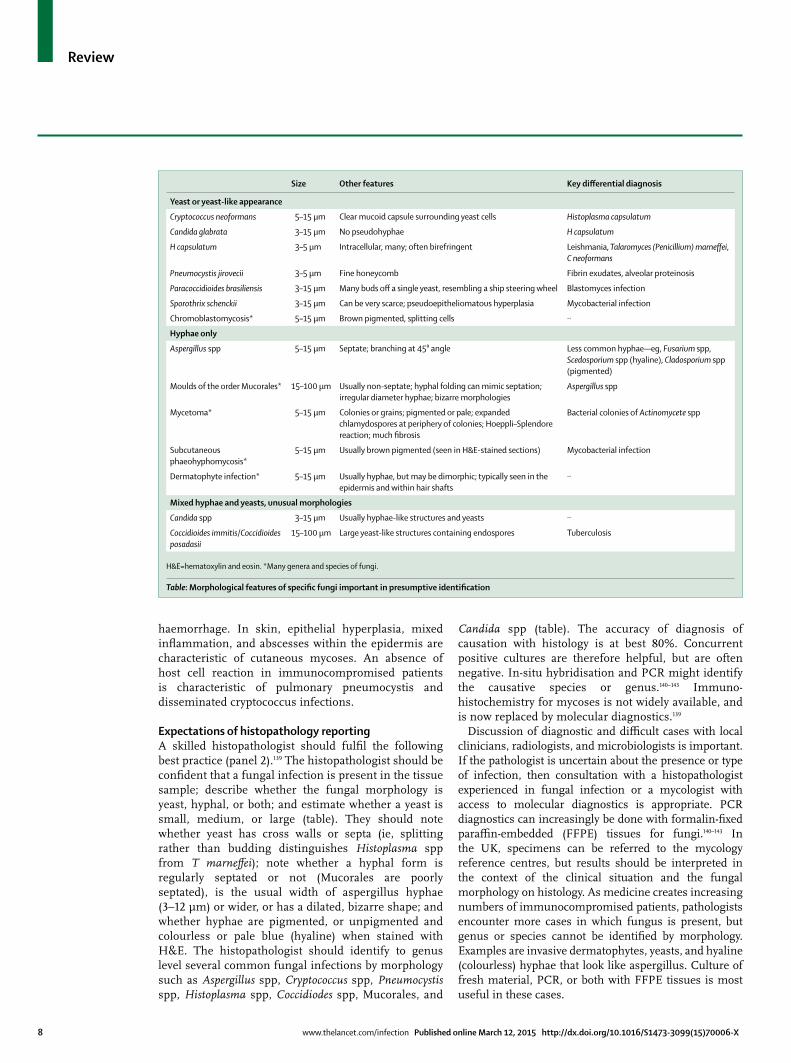
Expectations of histopathology reportingA skilled histopathologist should fulfi l the following best practice (panel 2).139 The histopathologist should be confi dent that a fungal infection is present in the tissue sample; describe whether the fungal morphology is yeast, hyphal, or both; and estimate whether a yeast is small, medium, or large (table). They should note whether yeast has cross walls or septa (ie, splitting rather than budding distinguishes Histoplasma spp from T marneff ei); note whether a hyphal form is regularly septated or not (Mucorales are poorly septated), is the usual width of aspergillus hyphae (3–12 µm) or wider, or has a dilated, bizarre shape; and whether hyphae are pigmented, or unpigmented and colourless or pale blue (hyaline) when stained with H&E. The histopathologist should identify to genus level several common fungal infections by morphology such as Aspergillus spp, Cryptococcus spp, Pneumocystis spp, Histoplasma spp, Coccidiodes spp, Mucorales, and
Candida spp (table). The accuracy of diagnosis of causation with histology is at best 80%. Concurrent positive cultures are therefore helpful, but are often negative. In-situ hybridisation and PCR might identify the causative species or genus.140–143 Immuno-histochemistry for mycoses is not widely available, and is now replaced by molecular diagnostics.139
Discussion of diagnostic and diffi cult cases with local clinicians, radiologists, and microbiologists is important. If the pathologist is uncertain about the presence or type of infection, then consultation with a histopathologist experienced in fungal infection or a mycologist with access to molecular diagnostics is appropriate. PCR diagnostics can increasingly be done with formalin-fi xed paraffi n-embedded (FFPE) tissues for fungi.140–143 In the UK, specimens can be referred to the mycology reference centres, but results should be interpreted in the context of the clinical situation and the fungal morphology on histology. As medicine creates increasing numbers of immunocompromised patients, pathologists encounter more cases in which fungus is present, but genus or species cannot be identifi ed by morphology. Examples are invasive dermatophytes, yeasts, and hyaline (colourless) hyphae that look like aspergillus. Culture of fresh material, PCR, or both with FFPE tissues is most useful in these cases.
Size Other features Key diff erential diagnosis
Yeast or yeast-like appearance
Cryptococcus neoformans 5–15 µm Clear mucoid capsule surrounding yeast cells Histoplasma capsulatum
Candida glabrata 3–15 µm No pseudohyphae H capsulatum
H capsulatum 3–5 µm Intracellular, many; often birefringent Leishmania, Talaromyces (Penicillium) marneff ei, C neoformans
Pneumocystis jirovecii 3–5 µm Fine honeycomb Fibrin exudates, alveolar proteinosis
Paracoccidioides brasiliensis 3–15 µm Many buds off a single yeast, resembling a ship steering wheel Blastomyces infection
Sporothrix schenckii 3–15 µm Can be very scarce; pseudoepitheliomatous hyperplasia Mycobacterial infection
Chromoblastomycosis* 5–15 µm Brown pigmented, splitting cells ··
Hyphae only
Aspergillus spp 5–15 µm Septate; branching at 45⁰ angle Less common hyphae—eg, Fusarium spp, Scedosporium spp (hyaline), Cladosporium spp (pigmented)
Moulds of the order Mucorales* 15–100 µm Usually non-septate; hyphal folding can mimic septation; irregular diameter hyphae; bizarre morphologies
Aspergillus spp
Mycetoma* 5–15 µm Colonies or grains; pigmented or pale; expanded chlamydospores at periphery of colonies; Hoeppli–Splendore reaction; much fi brosis
Bacterial colonies of Actinomycete spp
Subcutaneousphaeohyphomycosis*
5–15 µm Usually brown pigmented (seen in H&E-stained sections) Mycobacterial infection
Dermatophyte infection* 5–15 µm Usually hyphae, but may be dimorphic; typically seen in the epidermis and within hair shafts
··
Mixed hyphae and yeasts, unusual morphologies
Candida spp 3–15 µm Usually hyphae-like structures and yeasts ··
Coccidioides immitis/Coccidioides posadasii
15–100 µm Large yeast-like structures containing endospores Tuberculosis
H&E=hematoxylin and eosin. *Many genera and species of fungi.
Table: Morphological features of specifi c fungi important in presumptive identifi cation
www.thelancet.com/infection Published online March 12, 2015 http://dx.doi.org/10.1016/S1473-3099(15)70006-X 9
Review
Radiology best practiceImaging has a crucial role in diagnosis and management of patients with suspected invasive fungal disease who are immunosuppressed (panel 3). Invasive fungal diseases should be considered with any new broad-spectrum antibiotic, persistent fever, new pulmonary symptoms, or infi ltrates during substantial immuno suppression. Pulmonary infection is most common, but such patients are likewise susceptible to sinus, cerebral, hepatic, splenic, renal, bone, and disseminated fungal infection. The threshold for imaging should be very low when the patient has persistent fever and symptoms and signs of pulmonary infection (cough, shortness of breath, chest pain, bronchial breathing, pleural rub, or pulmonary decompensation), or when biomarkers are positive (panel 3).
Findings of a chest radiographs might seem normal or non-specifi c in neutropenic patients with invasive pulmonary aspergillosis.144 The presence of nodules on the chest radiographs is the most specifi c fi nding for invasive fungal infection.145 Overall, chest radiographs are inadequate for investigation of pulmonary invasive fungal diseases in this patient population, except for pneumocystis infection. Pneumocystis pneumonia can seem normal on a radiograph at presentation, followed by typical bilateral, perihilar, diff use granular, or hazy (ground-glass) opacifi cation, which becomes denser with worsening infection, progressing to areas of consolidation.146
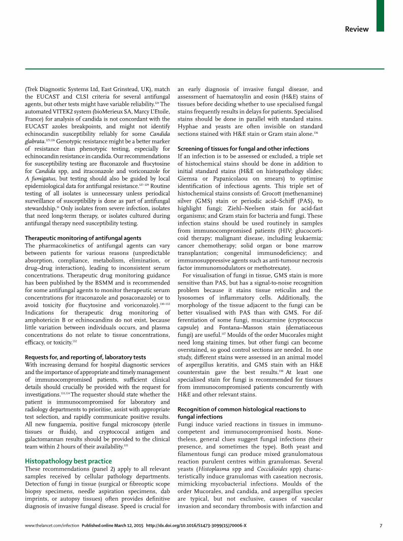
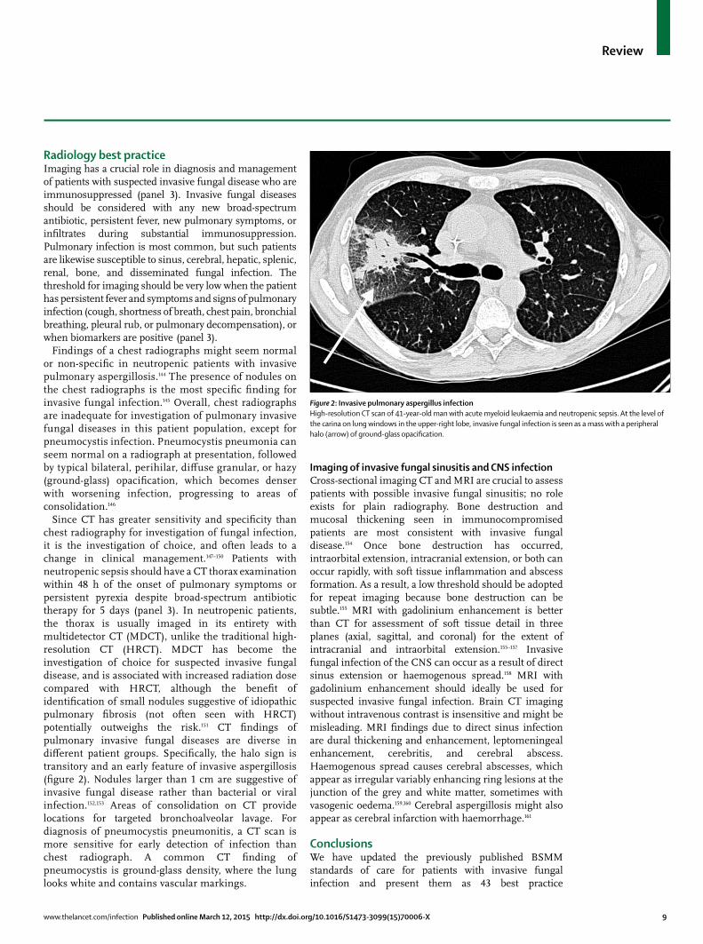
Since CT has greater sensitivity and specifi city than chest radiography for investigation of fungal infection, it is the investigation of choice, and often leads to a change in clinical management.147–150 Patients with neutropenic sepsis should have a CT thorax examination within 48 h of the onset of pulmonary symptoms or persistent pyrexia despite broad-spectrum antibiotic therapy for 5 days (panel 3). In neutropenic patients, the thorax is usually imaged in its entirety with multidetector CT (MDCT), unlike the traditional high-resolution CT (HRCT). MDCT has become the investigation of choice for suspected invasive fungal disease, and is associated with increased radiation dose compared with HRCT, although the benefi t of identifi cation of small nodules suggestive of idiopathic pulmonary fi brosis (not often seen with HRCT) potentially outweighs the risk.151 CT fi ndings of pulmonary invasive fungal diseases are diverse in diff erent patient groups. Specifi cally, the halo sign is transitory and an early feature of invasive aspergillosis (fi gure 2). Nodules larger than 1 cm are suggestive of invasive fungal disease rather than bacterial or viral infection.152,153 Areas of consolidation on CT provide locations for targeted bronchoalveolar lavage. For diagnosis of pneumocystis pneumonitis, a CT scan is more sensitive for early detection of infection than chest radiograph. A common CT fi nding of pneumocystis is ground-glass density, where the lung looks white and contains vascular markings.
Imaging of invasive fungal sinusitis and CNS infectionCross-sectional imaging CT and MRI are crucial to assess patients with possible invasive fungal sinusitis; no role exists for plain radiography. Bone destruction and mucosal thickening seen in immunocompromised patients are most consistent with invasive fungal disease.154 Once bone destruction has occurred, intraorbital extension, intracranial extension, or both can occur rapidly, with soft tissue infl ammation and abscess formation. As a result, a low threshold should be adopted for repeat imaging because bone destruction can be subtle.155 MRI with gadolinium enhancement is better than CT for assessment of soft tissue detail in three planes (axial, sagittal, and coronal) for the extent of intracranial and intraorbital extension.155–157 Invasive fungal infection of the CNS can occur as a result of direct sinus extension or haemogenous spread.158 MRI with gadolinium enhancement should ideally be used for suspected invasive fungal infection. Brain CT imaging without intravenous contrast is insensitive and might be misleading. MRI fi ndings due to direct sinus infection are dural thickening and enhancement, leptomeningeal enhancement, cerebritis, and cerebral abscess. Haemogenous spread causes cerebral abscesses, which appear as irregular variably enhancing ring lesions at the junction of the grey and white matter, sometimes with vasogenic oedema.159,160 Cerebral aspergillosis might also appear as cerebral infarction with haemorrhage.161
ConclusionsWe have updated the previously published BSMM standards of care for patients with invasive fungal infection and present them as 43 best practice
Figure 2: Invasive pulmonary aspergillus infectionHigh-resolution CT scan of 41-year-old man with acute myeloid leukaemia and neutropenic sepsis. At the level of the carina on lung windows in the upper-right lobe, invasive fungal infection is seen as a mass with a peripheral halo (arrow) of ground-glass opacifi cation.
10 www.thelancet.com/infection Published online March 12, 2015 http://dx.doi.org/10.1016/S1473-3099(15)70006-X
Review
recommendations. These recommendations provide the opportunity for microbiologists, histopathologists, radio-logists, and clinicians to implement, assure, and audit best practice for the management of serious fungal diseases. The recommendations emphasise the role of microscopy in rapid diagnosis and identifi cation of clinically signifi cant isolates to species level, and the need for susceptibility testing of all Aspergillus spp, if treatment is to be given. We provide information to improve understanding of the importance of antigen detection for cryptococcal disease and invasive aspergillosis, use of molecular (PCR) diagnostics for aspergillosis, and the crucial role of antibody detection for chronic and allergic aspergillosis, and we emphasise the need for urgent (<48 h) and optimised imaging for patients with suspected invasive fungal infection. All 43 recommendations are auditable and should be used to ensure best diagnostic practice and improved outcomes for patients.ContributorsSS led the British Society of Medical Mycology (BSMM) working group. All authors worked collectively on development of the recommendations described in this manuscript, reviewed the evidence for recommendations, and contributed to writing of the manuscript. All authors have read and approved the fi nal version.
Declaration of interestsSBL declares no competing interests. SS has received honoraria from Pfi zer. RAB has served on advisory boards, and has received sponsorship from Merck, Astellas, Gilead, and Pfi zer. RAB has received sponsorship from Merck Sharp and Dohme (MSD), Pfi zer, and Gilead. RCB received sponsorship from MSD. JRC received an educational honorarium from MSD. CCK has received honoraria from Astellas, MSD, Pfi zer, and Gilead. DWD holds founder shares in F2G Ltd, and has grant support from the National Institute of Allergy and Infectious Diseases, National Institute of Health Research and European Union. DWD has been an advisor to F2G, T2 Biosystems, Pfi zer, Merck, Nektar, Astellas, and Gilead. He has received honoraria from Merck, Astellas, GlaxoSmithKline, Novartis, Merck, Dainippon, and Pfi zer.
AcknowledgmentsThe recommendations contained in this report have been endorsed by the BSMM committee. We thank Richard Hobson and Ibrahim Hassan (UK Clinical Mycology Network), William Hope, and Ruth Ashbee (BSMM), the Royal College of Pathologists, London, and the Royal College of Radiologists, London, who all provided comments on the best practice recommendations.
Search strategy and selection criteria
An agreement about the scope of the recommendations was achieved by the authors, on the basis of knowledge, experience, discussion, and advice from expert colleagues. For each recommendation, relevant published evidence was collected from database searches. References in English, French, and German were identifi ed through PubMed searches for articles published from Jan 1, 1971, to Aug 1, 2014. Search terms varied depending on the precise point to be referenced, but several terms were used to ensure comprehensiveness. In subject areas for which many publications exist, systematic reviews and meta-analyses were sought. A summary of the referenced evidence for key recommendations is provided as an appendix. See Online for appendix
References1 Pfaller MA, Diekema DJ. Epidemiology of invasive mycoses in
North America. Crit Rev Microbiol 2010; 36: 1–53.2 Lortholary O, Gangneux JP, Sitbon K, et al. Epidemiological trends
in invasive aspergillosis in France: the SAIF network (2005–2007). Clin Microbiol Infect 2011; 17: 1882–89.
3 Schelenz S. Management of candidiasis in the intensive care unit. J Antimicrob Chemother 2008; 61 (suppl 1): i31–34.
4 Guinea J, Torres-Narbona M, Gijon P, et al. Pulmonary aspergillosis in patients with chronic obstructive pulmonary disease: incidence, risk factors, and outcome. Clin Microbiol Infect 2010; 16: 870–77.
5 Hayes GE, Denning DW. Frequency, diagnosis and management of fungal respiratory infections. Curr Opin Pulm Med 2013; 19: 259–65.
6 Nivoix Y, Velten M, Letscher-Bru V, et al. Factors associated with overall and attributable mortality in invasive aspergillosis. Clin Infect Dis 2008; 47: 1176–84.
7 Dignani MC. Epidemiology of invasive fungal diseases on the basis of autopsy reports. F1000Prime Rep 2014; 6: 81.
8 Perfect JR. Fungal diagnosis: how do we do it and can we do better? Curr Med Res Opin 2013; 29 (suppl 4): 3–11.
9 De Pauw B, Walsh TJ, Donnelly JP, et al. Revised defi nitions of invasive fungal disease from the European Organization for Research and Treatment of Cancer/Invasive Fungal Infections Cooperative Group and the National Institute of Allergy and Infectious Diseases Mycoses Study Group (EORTC/MSG) Consensus Group. Clin Infect Dis 2008; 46: 1813–21.
10 Denning DW, Kibbler CC, Barnes RA. British Society for Medical Mycology proposed standards of care for patients with invasive fungal infections. Lancet Infect Dis 2003; 3: 230–40.
11 Schelenz S, Barnes RA, Kibbler CC, Jones BL, Denning DW. Standards of care for patients with invasive fungal infections within the United Kingdom: a national audit. J Infect 2009; 58: 145–53.
12 Hassan IA, Critten P, Isalska B, Denning DW. Audit of laboratory mycology services for the management of patients with fungal infections in the northwest of England. J Clin Pathol 2006; 59: 759–63.
13 Ascioglu S, Rex JH, de Pauw B, et al. Defi ning opportunistic invasive fungal infections in immunocompromised patients with cancer and hematopoietic stem cell transplants: an international consensus. Clin Infect Dis 2002; 34: 7–14.
14 Cuenca-Estrella M, Verweij PE, Arendrup MC, et al. ESCMID* guideline for the diagnosis and management of Candida diseases 2012: diagnostic procedures. Clin Microbiol Infect 2012; 18 (suppl 7): 9–18.
15 Yuen KY, Woo PC, Ip MS, et al. Stage-specifi c manifestation of mold infections in bone marrow transplant recipients: risk factors and clinical signifi cance of positive concentrated smears. Clin Infect Dis 1997; 25: 37–42.
16 Plorde JJ, Tenover FC, Carlson LG. Specimen volume versus yield in the BACTEC blood culture system. J Clin Microbiol 1985; 22: 292–95.
17 Alexander BD, Pfaller MA. Contemporary tools for the diagnosis and management of invasive mycoses. Clin Infect Dis 2006; 43 (suppl 1): S15–27.
18 Kosmin AR, Fekete T. Use of fungal blood cultures in an academic medical center. J Clin Microbiol 2008; 46: 3800–01.
19 Stone NR, Gorton RL, Barker K, Ramnarain P, Kibbler CC. Evaluation of PNA-FISH yeast traffi c light for rapid identifi cation of yeast directly from positive blood cultures and assessment of clinical impact. J Clin Microbiol 2013; 51: 1301–02.
20 Farina C, Perin S, Andreoni S, et al. Evaluation of the peptide nucleic acid fl uorescence in situ hybridisation technology for yeast identifi cation directly from positive blood cultures: an Italian experience. Mycoses 2012; 55: 388–92.
21 Shepard JR, Addison RM, Alexander BD, et al. Multicenter evaluation of the Candida albicans/Candida glabrata peptide nucleic acid fl uorescent in situ hybridization method for simultaneous dual-color identifi cation of C. albicans and C. glabrata directly from blood culture bottles. J Clin Microbiol 2008; 46: 50–55.
22 Gorton RL, Stone NARH, Ramnarain P, Barker K, Mchugh TD, Kibbler CC. A comparison of three rapid identifi cation techniques for the identifi cation of yeasts from positive blood cultures: Gram’s stain, PNA-FISH and MALDI-TOF. Mycoses 2011; 54: 78.
www.thelancet.com/infection Published online March 12, 2015 http://dx.doi.org/10.1016/S1473-3099(15)70006-X 11
Review
23 Horvath JA, Dummer S. The use of respiratory-tract cultures in the diagnosis of invasive pulmonary aspergillosis. Am J Med 1996; 100: 171–78.
24 McCarthy DS, Pepys J. Allergic broncho-pulmonary aspergillosis. Clinical immunology. 2. Skin, nasal and bronchial tests. Clin Allergy 1971; 1: 415–32.
25 Reichenberger F, Habicht J, Matt P, et al. Diagnostic yield of bronchoscopy in histologically proven invasive pulmonary aspergillosis. Bone Marrow Transplant 1999; 24: 1195–99.
26 Einsele H, Quabeck K, Muller KD, et al. Prediction of invasive pulmonary aspergillosis from colonisation of lower respiratory tract before marrow transplantation. Lancet 1998; 352: 1443.
27 Maschmeyer G, Beinert T, Buchheidt D, et al. Diagnosis and antimicrobial therapy of lung infi ltrates in febrile neutropenic patients: guidelines of the infectious diseases working party of the German Society of Haematology and Oncology. Eur J Cancer 2009; 45: 2462–72.
28 McWhinney PH, Kibbler CC, Hamon MD, et al. Progress in the diagnosis and management of aspergillosis in bone marrow transplantation: 13 years’ experience. Clin Infect Dis 1993; 17: 397–404.
29 Hargreaves NJ, Kadzakumanja O, Phiri S, et al. Pneumocystis carinii pneumonia in patients being registered for smear-negative pulmonary tuberculosis in Malawi. Trans R Soc Trop Med Hyg 2001; 95: 402–08.
30 Turner D, Schwarz Y, Yust I. Induced sputum for diagnosing Pneumocystis carinii pneumonia in HIV patients: new data, new issues. Eur Respir J 2003; 21: 204–08.
31 Kovacs JA, Hiemenz JW, Macher AM, et al. Pneumocystis carinii pneumonia: a comparison between patients with the acquired immunodefi ciency syndrome and patients with other immunodefi ciencies. Ann Intern Med 1984; 100: 663–71.
32 Vargas SL, Ponce CA, Gallo M, et al. Near-universal prevalence of Pneumocystis and associated increase in mucus in the lungs of infants with sudden unexpected death. Clin Infect Dis 2013; 56: 171–79.
33 Gupta AK. Postgraduate institute management protocol for invasive Aspergillus fl avus sinusitis: is it eff ective? Int J Infect Dis 2009; 13: 134–49.
34 Antinori S, Radice A, Galimberti L, Magni C, Fasan M, Parravicini C. The role of cryptococcal antigen assay in diagnosis and monitoring of cryptococcal meningitis. J Clin Microbiol 2005; 43: 5828–29.
35 Sanchez-Portocarrero J, Perez-Cecilia E, Corral O, Romero-Vivas J, Picazo JJ. The central nervous system and infection by Candida species. Diagn Microbiol Infect Dis 2000; 37: 169–79.
36 Navarro EE, Almario JS, King C, Bacher J, Pizzo PA, Walsh TJ. Detection of Candida casts in experimental renal candidiasis: implications for the diagnosis and pathogenesis of upper urinary tract infection. J Med Vet Mycol 1994; 32: 415–26.
37 Yeghen T, Kibbler CC, Prentice HG, et al. Management of invasive pulmonary aspergillosis in hematology patients: a review of 87 consecutive cases at a single institution. Clin Infect Dis 2000; 31: 859–68.
38 Roden MM, Zaoutis TE, Buchanan WL, et al. Epidemiology and outcome of zygomycosis: a review of 929 reported cases. Clin Infect Dis 2005; 41: 634–53.
39 Dunbar SA, Eason RA, Musher DM, Clarridge JE 3rd. Microscopic examination and broth culture of cerebrospinal fl uid in diagnosis of meningitis. J Clin Microbiol 1998; 36: 1617–20.
40 Jarvis JN, Meintjes G, Williams A, Brown Y, Crede T, Harrison TS. Adult meningitis in a setting of high HIV and TB prevalence: fi ndings from 4961 suspected cases. BMC Infect Dis 2010; 10: 67.
41 Bakare N, Rickerts V, Bargon J, Just-Nubling G. Prevalence of Aspergillus fumigatus and other fungal species in the sputum of adult patients with cystic fi brosis. Mycoses 2003; 46: 19–23.
42 Ng VL, Yajko DM, McPhaul LW, et al. Evaluation of an indirect fl uorescent-antibody stain for detection of Pneumocystis carinii in respiratory specimens. J Clin Microbiol 1990; 28: 975–79.
43 Gopinathan U, Garg P, Fernandes M, Sharma S, Athmanathan S, Rao GN. The epidemiological features and laboratory results of fungal keratitis: a 10-year review at a referral eye care center in South India. Cornea 2002; 21: 555–59.
44 Weinberg JM, Koestenblatt EK, Tutrone WD, Tishler HR, Najarian L. Comparison of diagnostic methods in the evaluation of onychomycosis. J Am Acad Dermatol 2003; 49: 193–37.
45 Alvarez F, Bandi V, Stager C, Guntupalli KK. Detection of Pneumocystis carinii in tracheal aspirates of intubated patients using calcofl uor-white (Fungi-Fluor) and immunofl uorescence antibody (Genetic Systems) stains. Crit Care Med 1997; 25: 948–52.
46 Procop GW, Haddad S, Quinn J, et al. Detection of Pneumocystis jirovecii in respiratory specimens by four staining methods. J Clin Microbiol 2004; 42: 3333–35.
47 Cregan P, Yamamoto A, Lum A, VanDerHeide T, MacDonald M, Pulliam L. Comparison of four methods for rapid detection of Pneumocystis carinii in respiratory specimens. J Clin Microbiol 1990; 28: 2432–36.
48 Lautenschlager I, Lyytikainen O, Jokipii L, et al. Immunodetection of Pneumocystis carinii in bronchoalveolar lavage specimens compared with methenamine silver stain. J Clin Microbiol 1996; 34: 728–30.
49 Ng VL, Virani NA, Chaisson RE, et al. Rapid detection of Pneumocystis carinii using a direct fl uorescent monoclonal antibody stain. J Clin Microbiol 1990; 28: 2228–33.
50 McTaggart LR, Wengenack NL, Richardson SE. Validation of the MycAssay Pneumocystis kit for detection of Pneumocystis jirovecii in bronchoalveolar lavage specimens by comparison to a laboratory standard of direct immunofl uorescence microscopy, real-time PCR, or conventional PCR. J Clin Microbiol 2012; 50: 1856–69.
51 Lu Y, Ling G, Qiang C, et al. PCR diagnosis of Pneumocystis pneumonia: a bivariate meta-analysis. J Clin Microbiol 2011; 49: 4361–63.
52 Naryshkin S, Daniels J, Freno E, Cunningham L. Cytology of treated and minimal Pneumocystis carinii pneumonia and a pitfall of the Grocott methenamine silver stain. Diagn Cytopathol 1991; 7: 41–47.
53 Nassar A, Zapata M, Little JV, Siddiqui MT. Utility of refl ex Gomori methenamine silver staining for Pneumocystis jirovecii on bronchoalveolar lavage cytologic specimens: a review. Diagn Cytopathol 2006; 34: 719–23.
54 Tregnago R, Xavier RG, Pereira RP, Prolla JC. The diagnosis of Pneumocystis carinii pneumonia by cytologic evaluation of Papanicolaou and Leishman-stained bronchoalveolar specimens in patients with the acquired immunodefi ciency syndrome. Cytopathology 1993; 4: 77–84.
55 Schumann GB, Swensen JJ. Comparison of Papanicolaou’s stain with the Gomori methenamine silver (GMS) stain for the cytodiagnosis of Pneumocystis carinii in bronchoalveolar lavage (BAL) fl uid. Am J Clin Pathol 1991; 95: 583–86.
56 Homer KS, Wiley EL, Smith AL, et al. Monoclonal antibody to Pneumocystis carinii. Comparison with silver stain in bronchial lavage specimens. Am J Clin Pathol 1992; 97: 619–24.
57 Saubolle MA, McKellar PP, Sussland D. Epidemiologic, clinical, and diagnostic aspects of coccidioidomycosis. J Clin Microbiol 2007; 45: 26–30.
58 Sarosi GA, Lawrence JP, Smith DK, Thomas A, Hobohm DW, Kelley PC. Rapid diagnostic evaluation of bronchial washings in patients with suspected coccidioidomycosis. Semin Respir Infect 2001; 16: 238–41.
59 Yu VL, Muder RR, Poorsattar A. Signifi cance of isolation of Aspergillus from the respiratory tract in diagnosis of invasive pulmonary aspergillosis. Results from a three-year prospective study. Am J Med 1986; 81: 249–54.
60 Martino P, Raccah R, Gentile G, Venditti M, Girmenia C, Mandelli F. Aspergillus colonization of the nose and pulmonary aspergillosis in neutropenic patients: a retrospective study. Haematologica 1989; 74: 263–65.
61 Chakrabarti A, Sethi S, Raman DS, Behera D. Eight-year study of allergic bronchopulmonary aspergillosis in an Indian teaching hospital. Mycoses 2002; 45: 295–99.
62 Weiland D, Ferguson RM, Peterson PK, Snover DC, Simmons RL, Najarian JS. Aspergillosis in 25 renal transplant patients. Epidemiology, clinical presentation, diagnosis, and management. Ann Surg 1983; 198: 622–29.
63 Nalesnik MA, Myerowitz RL, Jenkins R, Lenkey J, Herbert D. Signifi cance of Aspergillus species isolated from respiratory secretions in the diagnosis of invasive pulmonary aspergillosis. J Clin Microbiol 1980; 11: 370–76.
64 Treger TR, Visscher DW, Bartlett MS, Smith JW. Diagnosis of pulmonary infection caused by Aspergillus: usefulness of respiratory cultures. J Infect Dis 1985; 152: 572–76.
12 www.thelancet.com/infection Published online March 12, 2015 http://dx.doi.org/10.1016/S1473-3099(15)70006-X
Review
65 Greub G, Bille J. Aspergillus species isolated from clinical specimens: suggested clinical and microbiological criteria to determine signifi cance. Clin Microbiol Infect 1998; 4: 710–16.
66 Bulpa P, Dive A, Sibille Y. Invasive pulmonary aspergillosis in patients with chronic obstructive pulmonary disease. Eur Respir J 2007; 30: 782–800.
67 Felton TW, Baxter C, Moore CB, Roberts SA, Hope WW, Denning DW. Effi cacy and safety of posaconazole for chronic pulmonary aspergillosis. Clin Infect Dis 2010; 51: 1383–91.
68 Sobel JD, Fisher JF, Kauff man CA, Newman CA. Candida urinary tract infections—epidemiology. Clin Infect Dis 2011; 52 (suppl 6): S433–36.
69 Pappas PG, Kauff man CA, Andes D, et al. Clinical practice guidelines for the management of candidiasis: 2009 update by the Infectious Diseases Society of America. Clin Infect Dis 2009; 48: 503–35.
70 Hamilton JR, Noble A, Denning DW, Stevens DA. Performance of cryptococcus antigen latex agglutination kits on serum and cerebrospinal fl uid specimens of AIDS patients before and after pronase treatment. J Clin Microbiol 1991; 29: 333–39.
71 Goodman JS, Kaufman L, Koenig MG. Diagnosis of cryptococcal meningitis. Value of immunologic detection of cryptococcal antigen. N Engl J Med 1971; 285: 434–36.
72 Kwong-Chung KJB, Bennett, JE. Cryptococcosis. In: Kwong-Chung KJB, Bennett JE, eds. Medical Mycology. Philadelphia: Lea & Febiger, 1992: 397–466.
73 Gray GC, McCarthy T, Lebeck MG, et al. Genotype prevalence and risk factors for severe clinical adenovirus infection, United States 2004–2006. Clin Infect Dis 2007; 45: 1120–31.
74 Wheat LJ, Musial CE, Jenny-Avital E. Diagnosis and management of central nervous system histoplasmosis. Clin Infect Dis 2005; 40: 844–52.
75 Maertens J, Theunissen K, Verhoef G, et al. Galactomannan and computed tomography-based preemptive antifungal therapy in neutropenic patients at high risk for invasive fungal infection: a prospective feasibility study. Clin Infect Dis 2005; 41: 1242–50.
76 Girmenia C, Micozzi A, Gentile G, et al. Clinically driven diagnostic antifungal approach in neutropenic patients: a prospective feasibility study. J Clin Oncol 2010; 28: 667–74.
77 Upton A, Kirby KA, Carpenter P, Boeckh M, Marr KA. Invasive aspergillosis following hematopoietic cell transplantation: outcomes and prognostic factors associated with mortality. Clin Infect Dis 2007; 44: 531–40.
78 Gade W, Hinnefeld SW, Babcock LS, et al. Comparison of the PREMIER cryptococcal antigen enzyme immunoassay and the latex agglutination assay for detection of cryptococcal antigens. J Clin Microbiol 1991; 29: 1616–19.
79 Babady NE, Bestrom JE, Jespersen DJ, et al. Evaluation of three commercial latex agglutination kits and a commercial enzyme immunoassay for the detection of cryptococcal antigen. Med Mycol 2009; 47: 336–38.
80 Jarvis JN, Percival A, Bauman S, et al. Evaluation of a novel point-of-care cryptococcal antigen test on serum, plasma, and urine from patients with HIV-associated cryptococcal meningitis. Clin Infect Dis 2011; 53: 1019–23.
81 Lindsley MD, Mekha N, Baggett HC, et al. Evaluation of a newly developed lateral fl ow immunoassay for the diagnosis of cryptococcosis. Clin Infect Dis 2011; 53: 321–25.
82 Lin TY, Yeh KM, Lin JC, Wang NC, Peng MY, Chang FY. Cryptococcal disease in patients with or without human immunodefi ciency virus: clinical presentation and monitoring of serum cryptococcal antigen titers. J Microbiol Immunol Infect 2009; 42: 220–26.
83 Lortholary O, Fontanet A, Memain N, Martin A, Sitbon K, Dromer F. Incidence and risk factors of immune reconstitution infl ammatory syndrome complicating HIV-associated cryptococcosis in France. AIDS 2005; 19: 1043–49.
84 Marr KA, Balajee SA, McLaughlin L, Tabouret M, Bentsen C, Walsh TJ. Detection of galactomannan antigenemia by enzyme immunoassay for the diagnosis of invasive aspergillosis: variables that aff ect performance. J Infect Dis 2004; 190: 641–49.
85 Wingard JR, Carter SL, Walsh TJ, et al. Randomized, double-blind trial of fl uconazole versus voriconazole for prevention of invasive fungal infection after allogeneic hematopoietic cell transplantation. Blood 2010; 116: 5111–18.
86 Pfeiff er CD, Fine JP, Safdar N. Diagnosis of invasive aspergillosis using a galactomannan assay: a meta-analysis. Clin Infect Dis 2006; 42: 1417–27.
87 Leefl ang MM, Debets-Ossenkopp YJ, Visser CE, et al. Galactomannan detection for invasive aspergillosis in immunocompromized patients. Cochrane Database Syst Rev 2008; 4: CD007394.
88 D‘Haese J, Theunissen K, Vermeulen E, et al. Detection of galactomannan in bronchoalveolar lavage fl uid samples of patients at risk for invasive pulmonary aspergillosis: analytical and clinical validity. J Clin Microbiol 2012; 50: 1258–63.
89 Pasqualotto AC, Xavier MO, Sanchez LB, et al. Diagnosis of invasive aspergillosis in lung transplant recipients by detection of galactomannan in the bronchoalveolar lavage fl uid. Transplantation 2010; 90: 306–11.
90 Luong ML, Clancy CJ, Vadnerkar A, et al. Comparison of an Aspergillus real-time polymerase chain reaction assay with galactomannan testing of bronchoalvelolar lavage fl uid for the diagnosis of invasive pulmonary aspergillosis in lung transplant recipients. Clin Infect Dis 2011; 52: 1218–26.
91 Meersseman W, Lagrou K, Maertens J, et al. Galactomannan in bronchoalveolar lavage fl uid: a tool for diagnosing aspergillosis in intensive care unit patients. Am J Respir Crit Care Med 2008; 177: 27–34.
92 Orlopp K, von Lilienfeld-Toal M, Marklein G, et al. False positivity of the Aspergillus galactomannan Platelia ELISA because of piperacillin/tazobactam treatment: does it represent a clinical problem? J Antimicrob Chemother 2008; 62: 1109–12.
93 Mikulska M, Furfaro E, Del Bono V, et al. Piperacillin/tazobactam (Tazocin) seems to be no longer responsible for false-positive results of the galactomannan assay. J Antimicrob Chemother 2012; 67: 1746–48.
94 Karageorgopoulos DE, Vouloumanou EK, Ntziora F, Michalopoulos A, Rafailidis PI, Falagas ME. β-D-glucan assay for the diagnosis of invasive fungal infections: a meta-analysis. Clin Infect Dis 2011; 52: 750–70.
95 Onishi A, Sugiyama D, Kogata Y, et al. Diagnostic accuracy of serum 1,3-β-D-glucan for Pneumocystis jiroveci pneumonia, invasive candidiasis, and invasive aspergillosis: systematic review and meta-analysis. J Clin Microbiol 2012; 50: 7–15.
96 Pickering JW, Sant HW, Bowles CA, Roberts WL, Woods GL. Evaluation of a (1->3)-β-D-glucan assay for diagnosis of invasive fungal infections. J Clin Microbiol 2005; 43: 5957–62.
97 Fujita S, Takamura T, Nagahara M, Hashimoto T. Evaluation of a newly developed down-fl ow immunoassay for detection of serum mannan antigens in patients with candidaemia. J Med Microbiol 2006; 55: 537–43.
98 Barton RC, Hobson RP, Denton M, et al. Serologic diagnosis of allergic bronchopulmonary aspergillosis in patients with cystic fi brosis through the detection of immunoglobulin G to Aspergillus fumigatus. Diagn Microbiol Infect Dis 2008; 62: 287–91.
99 Agarwal R, Chakrabarti A, Shah A, et al. Allergic bronchopulmonary aspergillosis: review of literature and proposal of new diagnostic and classifi cation criteria. Clin Exp Allergy 2013; 43: 850–73.
100 White PL, Barton R, Guiver M, et al. A consensus on fungal polymerase chain reaction diagnosis?: a United Kingdom–Ireland evaluation of polymerase chain reaction methods for detection of systemic fungal infections. J Mol Diagn 2006; 8: 376–84.
101 White PL, Linton CJ, Perry MD, Johnson EM, Barnes RA. The evolution and evaluation of a whole blood polymerase chain reaction assay for the detection of invasive aspergillosis in hematology patients in a routine clinical setting. Clin Infect Dis 2006; 42: 479–86.
102 White PL, Bretagne S, Klingspor L, et al. Aspergillus PCR: one step closer to standardization. J Clin Microbiol 2010; 48: 1231–40.
103 White PL, Mengoli C, Bretagne S, et al. Evaluation of Aspergillus PCR protocols for testing serum specimens. J Clin Microbiol 2011; 49: 3842–48.
104 Barnes RA, White PL, Bygrave C, Evans N, Healy B, Kell J. Clinical impact of enhanced diagnosis of invasive fungal disease in high-risk haematology and stem cell transplant patients. J Clin Pathol 2009; 62: 64–69.
www.thelancet.com/infection Published online March 12, 2015 http://dx.doi.org/10.1016/S1473-3099(15)70006-X 13
Review
105 Morrissey CO, Chen SC, Sorrell TC, et al. Galactomannan and PCR versus culture and histology for directing use of antifungal treatment for invasive aspergillosis in high-risk haematology patients: a randomised controlled trial. Lancet Infect Dis 2013; 13: 519–28.
106 Nguyen MH, Peacock JE Jr., Tanner DC, et al. Therapeutic approaches in patients with candidemia. Evaluation in a multicenter, prospective, observational study. Arch Intern Med 1995; 155: 2429–35.
107 Mengoli C, Cruciani M, Barnes RA, Loeffl er J, Donnelly JP. Use of PCR for diagnosis of invasive aspergillosis: systematic review and meta-analysis. Lancet Infect Dis 2009; 9: 89–96.
108 Baxter CG, Jones AM, Webb K, Denning DW. Homogenisation of cystic fi brosis sputum by sonication—an essential step for Aspergillus PCR. J Microbiol Methods 2011; 85: 75–81.
109 Denning DW, Park S, Lass-Florl C, et al. High-frequency triazole resistance found In nonculturable Aspergillus fumigatus from lungs of patients with chronic fungal disease. Clin Infect Dis 2011; 52: 1123–9.
110 Tuon FF. A systematic literature review on the diagnosis of invasive aspergillosis using polymerase chain reaction (PCR) from bronchoalveolar lavage clinical samples. Rev Iberoam Micol 2007; 24: 89–94.
111 Hauser PM, Bille J, Lass-Florl C, et al. Multicenter, prospective clinical evaluation of respiratory samples from subjects at risk for Pneumocystis jirovecii infection by use of a commercial real-time PCR assay. J Clin Microbiol 2011; 49: 1872–78.
112 Barnes RA, Stocking K, Bowden S, Poynton MH, White PL. Prevention and diagnosis of invasive fungal disease in high-risk patients within an integrative care pathway. J Infect 2013; 67: 206–14.
113 Rogers TR, Morton CO, Springer J, et al. Combined real-time PCR and galactomannan surveillance improves diagnosis of invasive aspergillosis in high risk patients with haematological malignancies. Br J Haematol 2013; 161: 517–24.
114 Springer J, Morton CO, Perry M, et al. Multicenter comparison of serum and whole-blood specimens for detection of Aspergillus DNA in high-risk hematological patients. J Clin Microbiol 2013; 51: 1445–50.
115 White PL, Parr C, Thornton C, Barnes RA. Evaluation of real-time PCR, galactomannan enzyme-linked immunosorbent assay (ELISA), and a novel lateral-fl ow device for diagnosis of invasive aspergillosis. J Clin Microbiol 2013; 51: 1510–16.
116 Marr KA, Laverdiere M, Gugel A, Leisenring W. Antifungal therapy decreases sensitivity of the Aspergillus galactomannan enzyme immunoassay. Clin Infect Dis 2005; 40: 1762–69.
117 Kami M, Machida U, Okuzumi K, et al. Eff ect of fl uconazole prophylaxis on fungal blood cultures: an autopsy-based study involving 720 patients with haematological malignancy. Br J Haematol 2002; 117: 40–46.
118 McCulloch E, Ramage G, Rajendran R, et al. Antifungal treatment aff ects the laboratory diagnosis of invasive aspergillosis. J Clin Pathol 2012; 65: 83–86.
119 Nguyen MH, Leather H, Clancy CJ, et al. Galactomannan testing in bronchoalveolar lavage fl uid facilitates the diagnosis of invasive pulmonary aspergillosis in patients with hematologic malignancies and stem cell transplant recipients. Biol Blood Marrow Transplant 2011; 17: 1043–50.
120 Reinwald M, Hummel M, Kovalevskaya E, et al. Therapy with antifungals decreases the diagnostic performance of PCR for diagnosing invasive aspergillosis in bronchoalveolar lavage samples of patients with haematological malignancies. J Antimicrob Chemother 2012; 67: 2260–67.
121 Tsolaki AG, Miller RF, Wakefi eld AE. Oropharyngeal samples for genotyping and monitoring response to treatment in AIDS patients with Pneumocystis carinii pneumonia. J Med Microbiol 1999; 48: 897–905.
122 Koo S, Baden LR, Marty FM. Post-diagnostic kinetics of the (1-->3)-β-D-glucan assay in invasive aspergillosis, invasive candidiasis and Pneumocystis jirovecii pneumonia. Clin Microbiol Infect 2012; 18: E122–27.
123 Baxter CG, Denning DW, Jones AM, Todd A, Moore CB, Richardson MD. Performance of two Aspergillus IgG EIA assays compared with the precipitin test in chronic and allergic aspergillosis. Clin Microbiol Infect 2013; 19: E197–204.
124 EUCAST. Antifungal agents. Breakpoint tables for interpretation of MICs. http://www.eucast.org/fi leadmin/src/media/PDFs/EUCAST_fi les/AFST/Antifungal_breakpoints_v_4.1.pdf (accessed Jan 19, 2015).
125 Farina C, Manso E, Andreoni S, et al. Interlaboratory evaluation of VITEK2 system and Sensititre YeastOne(R) for antifungal susceptibility testing of yeasts isolated from blood cultures against four antifungal agents. New Microbiol 2011; 34: 195–201.
126 Astvad KM, Perlin DS, Johansen HK, Jensen RH, Arendrup MC. Evaluation of caspofungin susceptibility testing by the new Vitek 2 AST-YS06 yeast card using a unique collection of FKS wild-type and hot spot mutant isolates, including the fi ve most common candida species. Antimicrob Agents Chemother 2013; 57: 177–82.
127 Pfaller MA. Antifungal drug resistance: mechanisms, epidemiology, and consequences for treatment. Am J Med 2012; 125 (suppl 1): S3–13.
128 Larsen RA, Bauer M, Pitisuttithum P, et al. Correlation of susceptibility of Cryptococcus neoformans to amphotericin B with clinical outcome. Antimicrob Agents Chemother 2011; 55: 5624–30.
129 Verweij PE, Howard SJ, Melchers WJ, Denning DW. Azole-resistance in Aspergillus: proposed nomenclature and breakpoints. Drug Resist Updat 2009; 12: 141–47.
130 Pascual A, Calandra T, Bolay S, Buclin T, Bille J, Marchetti O. Voriconazole therapeutic drug monitoring in patients with invasive mycoses improves effi cacy and safety outcomes. Clin Infect Dis 2008; 46: 201–11.
131 Tan K, Brayshaw N, Tomaszewski K, Troke P, Wood N. Investigation of the potential relationships between plasma voriconazole concentrations and visual adverse events or liver function test abnormalities. J Clin Pharmacol 2006; 46: 235–43.
132 Ashbee HR, Barnes RA, Johnson EM, Richardson MD, Gorton R, Hope WW. Therapeutic drug monitoring (TDM) of antifungal agents: guidelines from the British Society for Medical Mycology. J Antimicrob Chemother 2013; 69: 1162–76.
133 Baricchi R, Zini M, Nibali MG, et al. Using pathology-specifi c laboratory profi les in clinical pathology to reduce inappropriate test requesting: two completed audit cycles. BMC Health Serv Res 2012; 12: 187.
134 Negrini G, Liverani S, De Biasio V, et al. Quality of the documents requesting and reporting CT scans: a retrospective study in four Italian hospitals. Ig Sanita Pubbl 2010; 66: 9–32 (in Italian).
135 Royal College of Pathologists. Key performance indicators in pathology. http://www.rcpath.org/clinical-eff ectiveness/kpi (accessed Jan 19, 2015).
136 Ramos AM, Sales Ade O, de Andrade MC, Bittencourt JF, Ramos CC. A simple method for detecting subcutaneous phaeohyphomycosis with light-colored fungi. A study of eight cases. American J Surg Pathol 1995; 19: 109–14.
137 Lazcano O, Speights VO, Strickler JG, Bilbao JE, Becker J, Diaz J. Combined histochemical stains in the diff erential-diagnosis of Cryptococcus-neoformans. Modern Pathol 1993; 6: 80–84.
138 Fons A, Garcia-de-Lomas J, Nogueira JM, Buesa FJ, Gimeno C. Histopathology of experimental Aspergillus fumigatus keratitis. Mycopathologia 1988; 101: 129–31.
139 Guarner J, Brandt ME. Histopathologic diagnosis of fungal infections in the 21st century. Clin Microbiol Rev 2011; 24: 247–80.
140 Paterson PJ, Seaton S, McLaughlin J, Kibbler CC. Development of molecular methods for the identifi cation of aspergillus and emerging moulds in paraffi n wax embedded tissue sections. Mol Pathol 2003; 56: 368–70.
141 Rickerts V, Mousset S, Lambrecht E, et al. Comparison of histopathological analysis, culture, and polymerase chain reaction assays to detect invasive mold infections from biopsy specimens. Clin Infect Dis 2007; 44: 1078–83.
142 Rickerts V, Khot PD, Myerson D, Ko DL, Lambrecht E, Fredricks DN. Comparison of quantitative real time PCR with sequencing and ribosomal RNA-FISH for the identifi cation of fungi in formalin fi xed, paraffi n-embedded tissue specimens. BMC Infect Dis 2011; 11: 202.
143 Lass-Florl C, Follett SA, Moody A, Denning DW. Detection of Aspergillus in lung and other tissue samples using the MycAssay Aspergillus real-time PCR kit. Can J Microbiol 2011; 57: 765–68.
144 Hauggaard A, Ellis M, Ekelund L. Early chest radiography and CT in the diagnosis, management and outcome of invasive pulmonary aspergillosis. Acta Radiol 2002; 43: 292–98.
14 www.thelancet.com/infection Published online March 12, 2015 http://dx.doi.org/10.1016/S1473-3099(15)70006-X
Review
145 Leung AN, Gosselin MV, Napper CH, et al. Pulmonary infections after bone marrow transplantation: clinical and radiographic fi ndings. Radiology 1999; 210: 699–710.
146 Goodman PC. Pneumocystis carinii pneumonia. J Thorac Imaging 1991; 6: 16–21.
147 Barloon TJ, Galvin JR, Mori M, Stanford W, Gingrich RD. High-resolution ultrafast chest CT in the clinical management of febrile bone marrow transplant patients with normal or nonspecifi c chest roentgenograms. Chest 1991; 99: 928–33.
148 Heussel CP, Kauczor HU, Heussel G, Fischer B, Mildenberger P, Thelen M. Early detection of pneumonia in febrile neutropenic patients: use of thin-section CT. AJR Am J Roentgenol 1997; 169: 1347–53.
149 Heussel CP, Kauczor HU, Heussel GE, et al. Pneumonia in febrile neutropenic patients and in bone marrow and blood stem-cell transplant recipients: use of high-resolution computed tomography. J Clin Oncol 1999; 17: 796–805.
150 Schueller G, Matzek W, Kalhs P, Schaefer-Prokop C. Pulmonary infections in the late period after allogeneic bone marrow transplantation: chest radiography versus computed tomography. Eur J Radiol 2005; 53: 489–94.
151 Webb WR. Airway diseases. In: Webb WR, Muller NL, Naidich DP, editors. High-resolution of CT of the lung, 4th ed. Lippincott Williams & Wilkins, 2009: 492–554.
152 Escuissato DL, Gasparetto EL, Marchiori E, et al. Pulmonary infections after bone marrow transplantation: high-resolution CT fi ndings in 111 patients. AJR Am J Roentgenol 2005; 185: 608–15.
153 Bruno C, Minniti S, Vassanelli A, Pozzi-Mucelli R. Comparison of CT features of Aspergillus and bacterial pneumonia in severely neutropenic patients. J Thorac Imaging 2007; 22: 160–65.
154 Aribandi M, McCoy VA, Bazan C 3rd. Imaging features of invasive and noninvasive fungal sinusitis: a review. Radiographics 2007; 27: 1283–96.
155 Gillespie MB, O’Malley BW Jr, Francis HW. An approach to fulminant invasive fungal rhinosinusitis in the immunocompromised host. Arch Otolaryngol Head Neck Surg 1998; 124: 520–26.
156 Gruden JF, Ouanounou S, Tigges S, Norris SD, Klausner TS. Incremental benefi t of maximum-intensity-projection images on observer detection of small pulmonary nodules revealed by multidetector CT. J Roentgenol 2002; 179: 149–57.
157 Sonnet S, Buitrago-Tellez CH, Tamm M, Christen S, Steinbrich W. Direct detection of angioinvasive pulmonary aspergillosis in immunosuppressed patients: preliminary results with high-resolution 16-MDCT angiography. Am J Roentgenol 2005; 184: 746–51.
158 Tempkin AD, Sobonya RE, Seeger JF, Oh ES. Cerebral aspergillosis: radiologic and pathologic fi ndings. Radiographics 2006; 26: 1239–42.
159 Ashdown BC, Tien RD, Felsberg GJ. Aspergillosis of the brain and paranasal sinuses in immunocompromised patients: CT and MR imaging fi ndings. Am J Roentgenol 1994; 162: 155–59.
160 Yuh WT, Nguyen HD, Gao F, et al. Brain parenchymal infection in bone marrow transplantation patients: CT and –fi ndings. Am J Roentgenol 1994; 162: 425–30.
161 DeLone DR, Goldstein RA, Petermann G, et al. Disseminated aspergillosis involving the brain: distribution and imaging characteristics. Am J Neuroradiol 1999; 20: 1597–604.