Embed Size (px)
Citation preview
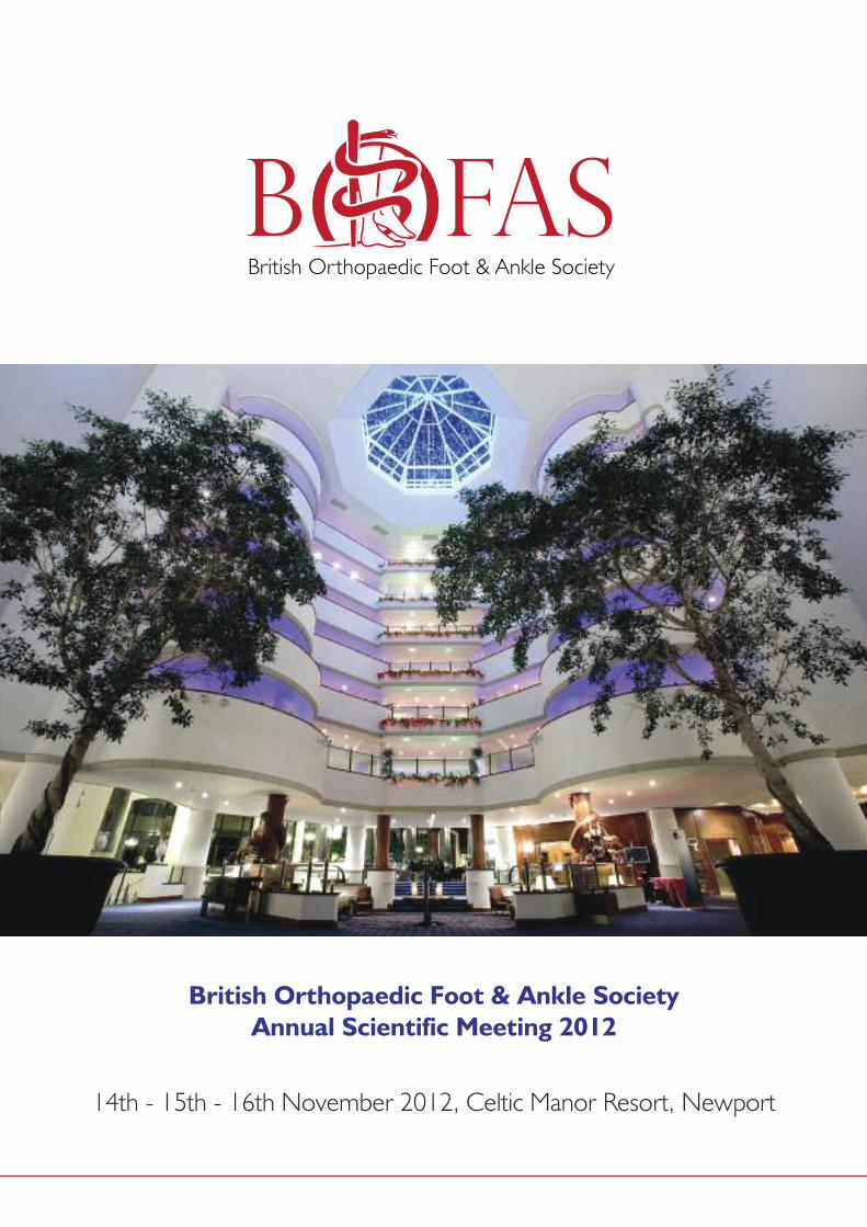
British Orthopaedic Foot & Ankle SocietyAnnual Scientific Meeting 2012
14th - 15th - 16th November 2012, Celtic Manor Resort, Newport
1
Dear delegate,
Croeso y Cymru
A warm Welsh welcome to the Celtic Manor Resort for the Annual Scientific Meeting of BOFAS,which is being held in Wales for the very first time in its long history.
The Education Committee has put together an exciting and stimulating programme and I am sureyou will enjoy the varied and novel topics that will be covered over the next 3 days.
We have a distinguished international and national faculty who have been extremely kind in offeringtheir time, expertise and knowledge and I am sure you will enjoy the robust debates and questionsthat the topics are bound to bring up!
The Allied Health Professions Programme is on the Thursday morning and covers sports injuries ofthe foot and ankle. This session is a unique inter-face between AHPs and Orthopaedic surgeons andis very popular every year! In addition, on the Thursday morning, there are industry workshops aswell as the Difficult Cases session. Please do make the most of them!
A large trade show is also available and I do hope you will make the effort during the breaks in theprogramme to visit our trade partners who, as always, have been ever so generous in sponsoringthe meeting and making it possible.
The annual dinner on Thursday evening will, I am sure, be a memorable one and I would love to seeas many of you as possible for a great evening of camaraderie, friendship and banter.
I do hope that you have an enjoyable and memorable stay in Newport. I am personally lookingforward to seeing you all here and am sure that you will enjoy flavours of the famous hospitality thatWales is well known for.
There are so many people I would like to thank for their help and support in making this meetinghappen but in particular, the members of the various Committees, members of Council and ofcourse our Executive Assistant Ms. Rosemarie Maio for all of their hard work and dedication.I am ever so grateful to all of you for attending and making this meeting a grand success.
Diolch yn fawrWith best wishes
Kartik Hariharan President BOFAS 2012-13
2
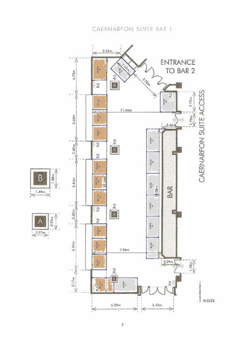
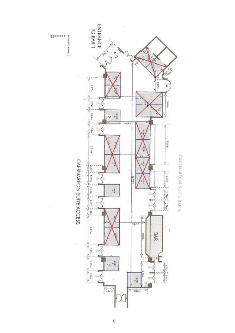
BOFAS Exhibition Caernarfon Suite BAR 1 & 2
PLATINUM SPONSORS:BIOMET 6 & 7WG HEALTHCARE 15 & 16ORTHOSOLUTIONS 3 & 4
GOLD PACKAGES:INTEGRA 13 & 14WRIGHT MEDICAL 11 & 12STRYKER 9 & 10
SILVER PACKAGES:DJO 1BIOCOMPOSITE 8OTSIS 19 NORTHSTAR 20OSTEOTEC 26SPECTRUM TECHNOLOGY 21ARTHREX 22
BRONZE PACKAGES:BIO-VATION UK Ltd 28OSSUR 29CORIN 30NOVEL 31QUATRO MEDICAL 32SYNTHES DE PUY 33LAVANDER 34MARQUARDT 35MEDARTIS 36VERTEC 37ACUMED 38SONOSITE 23C2UK IMPLANTS 24
3
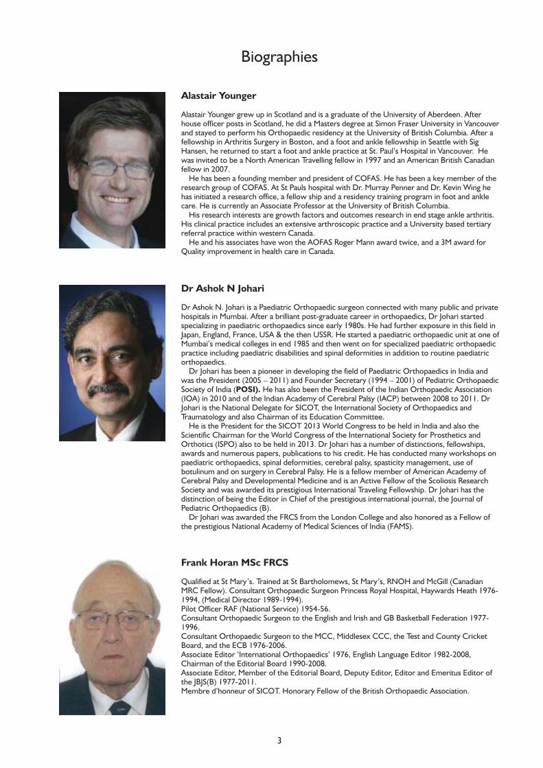
Alastair Younger
Alastair Younger grew up in Scotland and is a graduate of the University of Aberdeen. Afterhouse officer posts in Scotland, he did a Masters degree at Simon Fraser University in Vancouverand stayed to perform his Orthopaedic residency at the University of British Columbia. After afellowship in Arthritis Surgery in Boston, and a foot and ankle fellowship in Seattle with SigHansen, he returned to start a foot and ankle practice at St. Paul’s Hospital in Vancouver. Hewas invited to be a North American Travelling fellow in 1997 and an American British Canadianfellow in 2007.
He has been a founding member and president of COFAS. He has been a key member of theresearch group of COFAS. At St Pauls hospital with Dr. Murray Penner and Dr. Kevin Wing hehas initiated a research office, a fellow ship and a residency training program in foot and anklecare. He is currently an Associate Professor at the University of British Columbia.
His research interests are growth factors and outcomes research in end stage ankle arthritis.His clinical practice includes an extensive arthroscopic practice and a University based tertiaryreferral practice within western Canada.
He and his associates have won the AOFAS Roger Mann award twice, and a 3M award forQuality improvement in health care in Canada.
Dr Ashok N Johari
Dr Ashok N. Johari is a Paediatric Orthopaedic surgeon connected with many public and privatehospitals in Mumbai. After a brilliant post-graduate career in orthopaedics, Dr Johari startedspecializing in paediatric orthopaedics since early 1980s. He had further exposure in this field inJapan, England, France, USA & the then USSR. He started a paediatric orthopaedic unit at one ofMumbai’s medical colleges in end 1985 and then went on for specialized paediatric orthopaedicpractice including paediatric disabilities and spinal deformities in addition to routine paediatricorthopaedics.
Dr Johari has been a pioneer in developing the field of Paediatric Orthopaedics in India andwas the President (2005 – 2011) and Founder Secretary (1994 – 2001) of Pediatric OrthopaedicSociety of India (POSI). He has also been the President of the Indian Orthopaedic Association(IOA) in 2010 and of the Indian Academy of Cerebral Palsy (IACP) between 2008 to 2011. DrJohari is the National Delegate for SICOT, the International Society of Orthopaedics andTraumatology and also Chairman of its Education Committee.
He is the President for the SICOT 2013 World Congress to be held in India and also theScientific Chairman for the World Congress of the International Society for Prosthetics andOrthotics (ISPO) also to be held in 2013. Dr Johari has a number of distinctions, fellowships,awards and numerous papers, publications to his credit. He has conducted many workshops onpaediatric orthopaedics, spinal deformities, cerebral palsy, spasticity management, use ofbotulinum and on surgery in Cerebral Palsy. He is a fellow member of American Academy ofCerebral Palsy and Developmental Medicine and is an Active Fellow of the Scoliosis ResearchSociety and was awarded its prestigious International Traveling Fellowship. Dr Johari has thedistinction of being the Editor in Chief of the prestigious international journal, the Journal ofPediatric Orthopaedics (B).
Dr Johari was awarded the FRCS from the London College and also honored as a Fellow ofthe prestigious National Academy of Medical Sciences of India (FAMS).
Frank Horan MSc FRCS
Qualified at St Mary’s. Trained at St Bartholomews, St Mary’s, RNOH and McGill (CanadianMRC Fellow). Consultant Orthopaedic Surgeon Princess Royal Hospital, Haywards Heath 1976-1994, (Medical Director 1989-1994).Pilot Officer RAF (National Service) 1954-56. Consultant Orthopaedic Surgeon to the English and Irish and GB Basketball Federation 1977-1996.Consultant Orthopaedic Surgeon to the MCC, Middlesex CCC, the Test and County CricketBoard, and the ECB 1976-2006. Associate Editor ‘International Orthopaedics’ 1976, English Language Editor 1982-2008,Chairman of the Editorial Board 1990-2008.Associate Editor, Member of the Editorial Board, Deputy Editor, Editor and Emeritus Editor ofthe JBJS(B) 1977-2011. Membre d’honneur of SICOT. Honorary Fellow of the British Orthopaedic Association.
Biographies
4
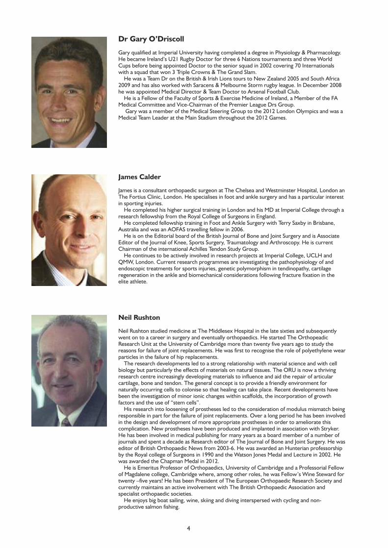
Dr Gary O’Driscoll
Gary qualified at Imperial University having completed a degree in Physiology & Pharmacology.He became Ireland’s U21 Rugby Doctor for three 6 Nations tournaments and three WorldCups before being appointed Doctor to the senior squad in 2002 covering 70 Internationalswith a squad that won 3 Triple Crowns & The Grand Slam.
He was a Team Dr on the British & Irish Lions tours to New Zealand 2005 and South Africa2009 and has also worked with Saracens & Melbourne Storm rugby league. In December 2008he was appointed Medical Director & Team Doctor to Arsenal Football Club.
He is a Fellow of the Faculty of Sports & Exercise Medicine of Ireland, a Member of the FAMedical Committee and Vice-Chairman of the Premier League Drs Group.
Gary was a member of the Medical Steering Group to the 2012 London Olympics and was aMedical Team Leader at the Main Stadium throughout the 2012 Games.
James Calder
James is a consultant orthopaedic surgeon at The Chelsea and Westminster Hospital, London anThe Fortius Clinic, London. He specialises in foot and ankle surgery and has a particular interestin sporting injuries.
He completed his higher surgical training in London and his MD at Imperial College through aresearch fellowship from the Royal College of Surgeons in England.
He completed fellowship training in Foot and Ankle Surgery with Terry Saxby in Brisbane,Australia and was an AOFAS travelling fellow in 2006.
He is on the Editorial board of the British Journal of Bone and Joint Surgery and is AssociateEditor of the Journal of Knee, Sports Surgery, Traumatology and Arthroscopy. He is currentChairman of the international Achilles Tendon Study Group.
He continues to be actively involved in research projects at Imperial College, UCLH andQMW, London. Current research programmes are investigating the pathophysiology of andendoscopic treatments for sports injuries, genetic polymorphism in tendinopathy, cartilageregeneration in the ankle and biomechanical considerations following fracture fixation in theelite athlete.
Neil Rushton
Neil Rushton studied medicine at The Middlesex Hospital in the late sixties and subsequentlywent on to a career in surgery and eventually orthopaedics. He started The OrthopeadicResearch Unit at the University of Cambridge more than twenty five years ago to study thereasons for failure of joint replacements. He was first to recognise the role of polyethylene wearparticles in the failure of hip replacements.
The research developments led to a strong relationship with material science and with cellbiology but particularly the effects of materials on natural tissues. The ORU is now a thrivingresearch centre increasingly developing materials to influence and aid the repair of articularcartilage, bone and tendon. The general concept is to provide a friendly environment fornaturally occurring cells to colonise so that healing can take place. Recent developments havebeen the investigation of minor ionic changes within scaffolds, the incorporation of growthfactors and the use of “stem cells”.
His research into loosening of prostheses led to the consideration of modulus mismatch beingresponsible in part for the failure of joint replacements. Over a long period he has been involvedin the design and development of more appropriate prostheses in order to ameliorate thiscomplication. New prostheses have been produced and implanted in association with Stryker.He has been involved in medical publishing for many years as a board member of a number ofjournals and spent a decade as Research editor of The Journal of Bone and Joint Surgery. He waseditor of British Orthopaedic News from 2003-6. He was awarded an Hunterian professorshipby the Royal college of Surgeons in 1990 and the Watson Jones Medal and Lecture in 2002. Hewas awarded the Chapman Medal in 2012.
He is Emeritus Professor of Orthopaedics, University of Cambridge and a Professorial Fellowof Magdalene college, Cambridge where, among other roles, he was Fellow’s Wine Steward fortwenty –five years! He has been President of The European Orthopaedic Research Society andcurrently maintains an active involvement with The British Orthopaedic Association andspecialist orthopaedic societies.
He enjoys big boat sailing, wine, skiing and diving interspersed with cycling and non-productive salmon fishing.
5
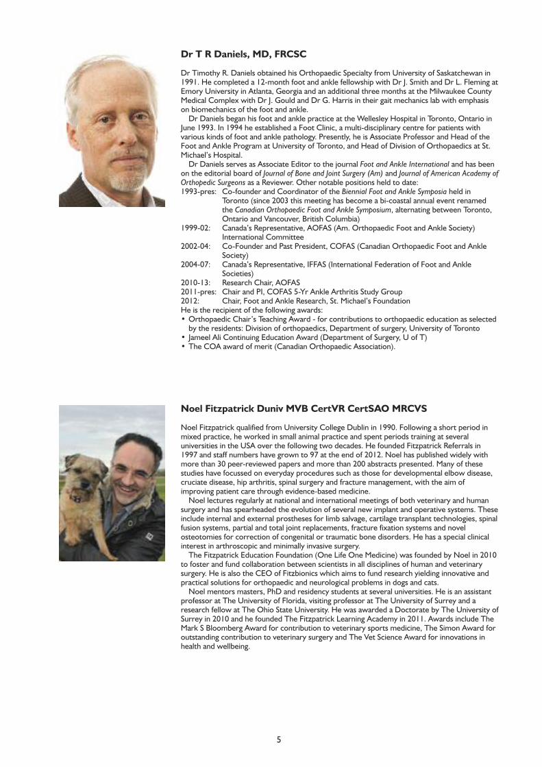
Dr T R Daniels, MD, FRCSC
Dr Timothy R. Daniels obtained his Orthopaedic Specialty from University of Saskatchewan in1991. He completed a 12-month foot and ankle fellowship with Dr J. Smith and Dr L. Fleming atEmory University in Atlanta, Georgia and an additional three months at the Milwaukee CountyMedical Complex with Dr J. Gould and Dr G. Harris in their gait mechanics lab with emphasison biomechanics of the foot and ankle.
Dr Daniels began his foot and ankle practice at the Wellesley Hospital in Toronto, Ontario inJune 1993. In 1994 he established a Foot Clinic, a multi-disciplinary centre for patients withvarious kinds of foot and ankle pathology. Presently, he is Associate Professor and Head of theFoot and Ankle Program at University of Toronto, and Head of Division of Orthopaedics at St.Michael’s Hospital.
Dr Daniels serves as Associate Editor to the journal Foot and Ankle International and has beenon the editorial board of Journal of Bone and Joint Surgery (Am) and Journal of American Academy ofOrthopedic Surgeons as a Reviewer. Other notable positions held to date:1993-pres: Co-founder and Coordinator of the Biennial Foot and Ankle Symposia held in
Toronto (since 2003 this meeting has become a bi-coastal annual event renamedthe Canadian Orthopaedic Foot and Ankle Symposium, alternating between Toronto,Ontario and Vancouver, British Columbia)
1999-02: Canada’s Representative, AOFAS (Am. Orthopaedic Foot and Ankle Society)International Committee
2002-04: Co-Founder and Past President, COFAS (Canadian Orthopaedic Foot and AnkleSociety)
2004-07: Canada’s Representative, IFFAS (International Federation of Foot and AnkleSocieties)
2010-13: Research Chair, AOFAS 2011-pres: Chair and PI, COFAS 5-Yr Ankle Arthritis Study Group 2012: Chair, Foot and Ankle Research, St. Michael’s Foundation He is the recipient of the following awards: • Orthopaedic Chair’s Teaching Award - for contributions to orthopaedic education as selected
by the residents: Division of orthopaedics, Department of surgery, University of Toronto • Jameel Ali Continuing Education Award (Department of Surgery, U of T)• The COA award of merit (Canadian Orthopaedic Association).
Noel Fitzpatrick Duniv MVB CertVR CertSAO MRCVS
Noel Fitzpatrick qualified from University College Dublin in 1990. Following a short period inmixed practice, he worked in small animal practice and spent periods training at severaluniversities in the USA over the following two decades. He founded Fitzpatrick Referrals in1997 and staff numbers have grown to 97 at the end of 2012. Noel has published widely withmore than 30 peer-reviewed papers and more than 200 abstracts presented. Many of thesestudies have focussed on everyday procedures such as those for developmental elbow disease,cruciate disease, hip arthritis, spinal surgery and fracture management, with the aim ofimproving patient care through evidence-based medicine.
Noel lectures regularly at national and international meetings of both veterinary and humansurgery and has spearheaded the evolution of several new implant and operative systems. Theseinclude internal and external prostheses for limb salvage, cartilage transplant technologies, spinalfusion systems, partial and total joint replacements, fracture fixation systems and novelosteotomies for correction of congenital or traumatic bone disorders. He has a special clinicalinterest in arthroscopic and minimally invasive surgery.
The Fitzpatrick Education Foundation (One Life One Medicine) was founded by Noel in 2010to foster and fund collaboration between scientists in all disciplines of human and veterinarysurgery. He is also the CEO of Fitzbionics which aims to fund research yielding innovative andpractical solutions for orthopaedic and neurological problems in dogs and cats.
Noel mentors masters, PhD and residency students at several universities. He is an assistantprofessor at The University of Florida, visiting professor at The University of Surrey and aresearch fellow at The Ohio State University. He was awarded a Doctorate by The University ofSurrey in 2010 and he founded The Fitzpatrick Learning Academy in 2011. Awards include TheMark S Bloomberg Award for contribution to veterinary sports medicine, The Simon Award foroutstanding contribution to veterinary surgery and The Vet Science Award for innovations inhealth and wellbeing.
6
Lt Col David Standley FRCS RAMC
Biosketch AAOS EWILieutenant Colonel David Standley joined the Royal Army Medical Corps in 1988 while at medical school in London. In 1996,following basic medical and surgical training in London and Germany, he started his specialist orthopaedic training in the Southwest ofEngland.
He became a consultant in Trauma and Orthopaedics (T&O) in 2003 and was immediately posted to Operation TELIC. For theinitial stages of the Iraq war he headed orthopaedic surgery at 22 and 33 Field Hospitals. Later that year he served in the NATOhospital in Bosnia. He has deployed 4 times to Afghanistan, to both Role 2 and Role 3 surgical facilities, most recently in September2011.
In 2004 he moved his clinical practice to the Princess Elizabeth Orthopaedic Centre, Exeter, where he is the Clinical Director oforthopaedics.
His training background includes being part of the faculty for the Military Operational Surgical Training course, which is the mainclinical course in pre-deployment and is hosted by the Royal College of Surgeons of England, London.
David Standley also holds a seat on the Specialist Advisory Committee, which advises the Royal College of Surgeons on all mattersof orthopaedic training in the UK.
On 1st January 2012 he started a new position as the Consultant Advisor (T&O) to the Director General Army Medical Services.
Lt Col David Standley
Preparing for Deployed operationsSince 2009 the Military Operational Surgical Training (MOST) Course has formed a major part of pre-deployment training within theDefence Medical Services of the UK.
The course was developed by the Defence Professor of Surgery, Surgeon Captain Mark Midwinter, and is hosted by the RoyalCollege of Surgery of England, London. From the start MOST has involved the wider surgical team and this allows a unifiedunderstanding of the unit’s goals for treatment and surgical strategies that will be employed.
The course is over one week and is largely practical-based, with formal lectures kept to a minimum. As well as “hands on”cadaveric surgery there are anatomy demonstrations; Emergency Department exercises within a simulator suite; group discussionsinclude onward care at UK Role 4, the role of the Deployed Medical Director and the team management of challenging cases.
MOST is resource-intensive with the last course requiring 57 faculty, 8 cadavers, surgical sets and equipment, as well as the RavenDepartment of Education at the Royal College of Surgeons.
To date 7 courses have been run with nearly 400 delegates being trained. As well as the UK delegates, teams have been sent from3 other countries prior to their tours in the Role 3 hospital at Camp Bastion.
The course continues to progress and will soon increase the involvement of Emergency Medicine, so bringing together the wholeresuscitation stage within a deployed medical facility. With the Defence Medical Services moving towards a “contingency planningMOST is expected to become the method of maintaining lessons-learned as well as eliminating any “learning curves”.
Lt Col Michael Butler MA FRCS(Tr&Orth) RAMC
Biosketch BOFAS 2012Lieutenant Colonel Michael Butler joined the Royal Army Medical Corps in 1993 while at Cambridge University before moving to theLondon Hospital Medical College. In 2003, following basic medical and surgical training he started his specialist orthopaedic trainingin the Southwest of England.
He became a consultant in Trauma and Orthopaedics after Foot and Ankle training in Exeter, Bristol and Oxford. Militarily,Michael has served in Belize, Kenya, Germany and has completed 3 Northern Ireland tours and in 2003 deployed to the Iraq warunder Lt Col David Standley. Since becoming a consultant, he has completed 2 tours to Afghanistan returning most recently inAugust 2012.
His consultant practice is at The Royal Cornwall Hospital in Truro as colleague to Steve Parsons whose registrar he had been in2005.
His training background includes being part of the faculty for the Military Operational Surgical Training course, hosted by the RoyalCollege of Surgeons of England, organising the Instructional Course for the Combined Services Orthopaedic Society and is keenlyinvolved in the Peninsula Deanery Training Programme.
Mike conducts military clinics in Germany 2-3 times per year and is a reviewer for Foot and Ankle Surgery, The Knee and Injury.
Lt Col Michael Butler
Emergency Care of the Trauma patient- from Point of Wounding to Definitive careMilitary Surgeons are currently seeing and gaining experience in high-energy trauma due to ballistic and blast injury which are oftendevastating to the extremities- which are the most frequently injured anatomical zones.
The remit of the military medical chain is to save life primarily and then to save limb and sight where possible and this has lead to arapid evolution of emergency healthcare from point of wounding through to transport back to definitive healthcare and subsequentreconstruction and rehabilitation.
The emphasis in the pre-hospital phase is of life-saving interventions to prevent and halt life-threatening haemorrhage with anumber of treatments and adjuncts. In hospital treatment is characterised by a team-based approach to facilitate rapid assessmentand investigation of all injuries and expeditious transfer to the operating facility.
Surgery is team-based with the aim of life and limb saving surgery to facilitate a safe and rapid transfer to the Role 4 facility. TeamWork and Communication are a critical part of this process and the military facilities have been seen to lead the world in advancedpatient care in the severely traumatised patient.
7
8
9
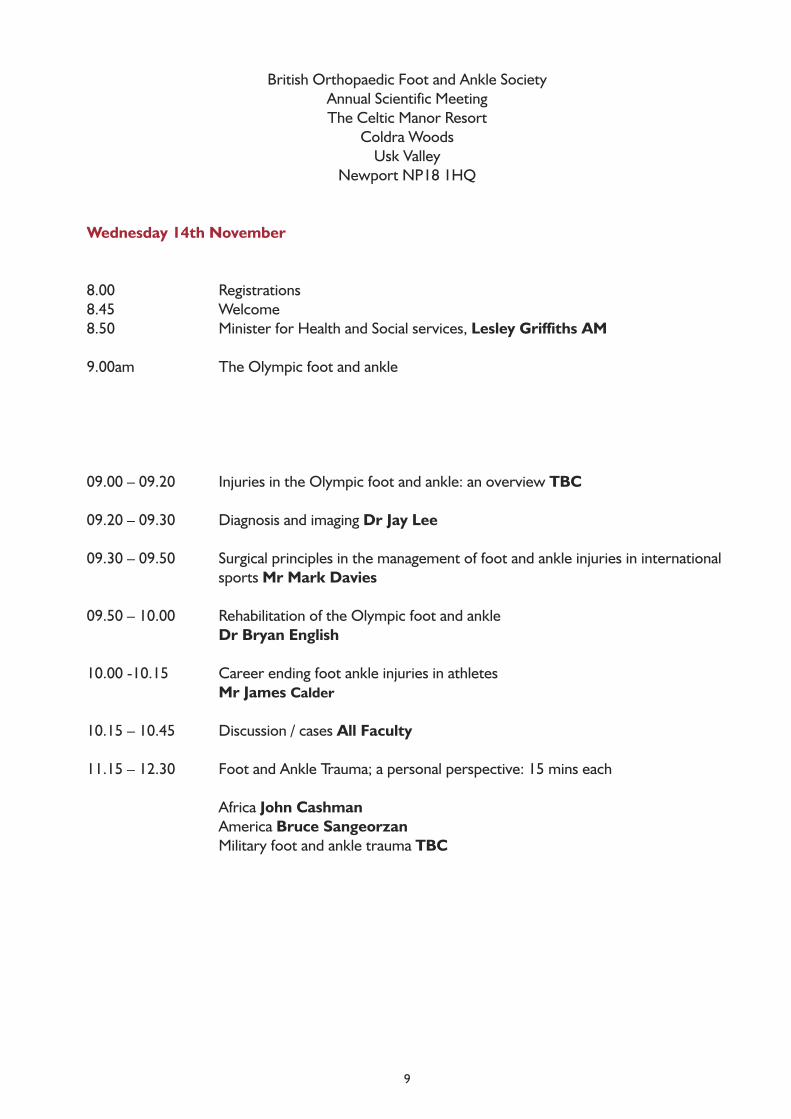
British Orthopaedic Foot and Ankle Society Annual Scientific MeetingThe Celtic Manor Resort
Coldra WoodsUsk Valley
Newport NP18 1HQ
Wednesday 14th November
8.00 Registrations 8.45 Welcome8.50 Minister for Health and Social services, Lesley Griffiths AM
9.00am The Olympic foot and ankle
09.00 – 09.20 Injuries in the Olympic foot and ankle: an overview TBC
09.20 – 09.30 Diagnosis and imaging Dr Jay Lee
09.30 – 09.50 Surgical principles in the management of foot and ankle injuries in internationalsports Mr Mark Davies
09.50 – 10.00 Rehabilitation of the Olympic foot and ankle Dr Bryan English
10.00 -10.15 Career ending foot ankle injuries in athletes Mr James Calder
10.15 – 10.45 Discussion / cases All Faculty
11.15 – 12.30 Foot and Ankle Trauma; a personal perspective: 15 mins each
Africa John CashmanAmerica Bruce SangeorzanMilitary foot and ankle trauma TBC
10
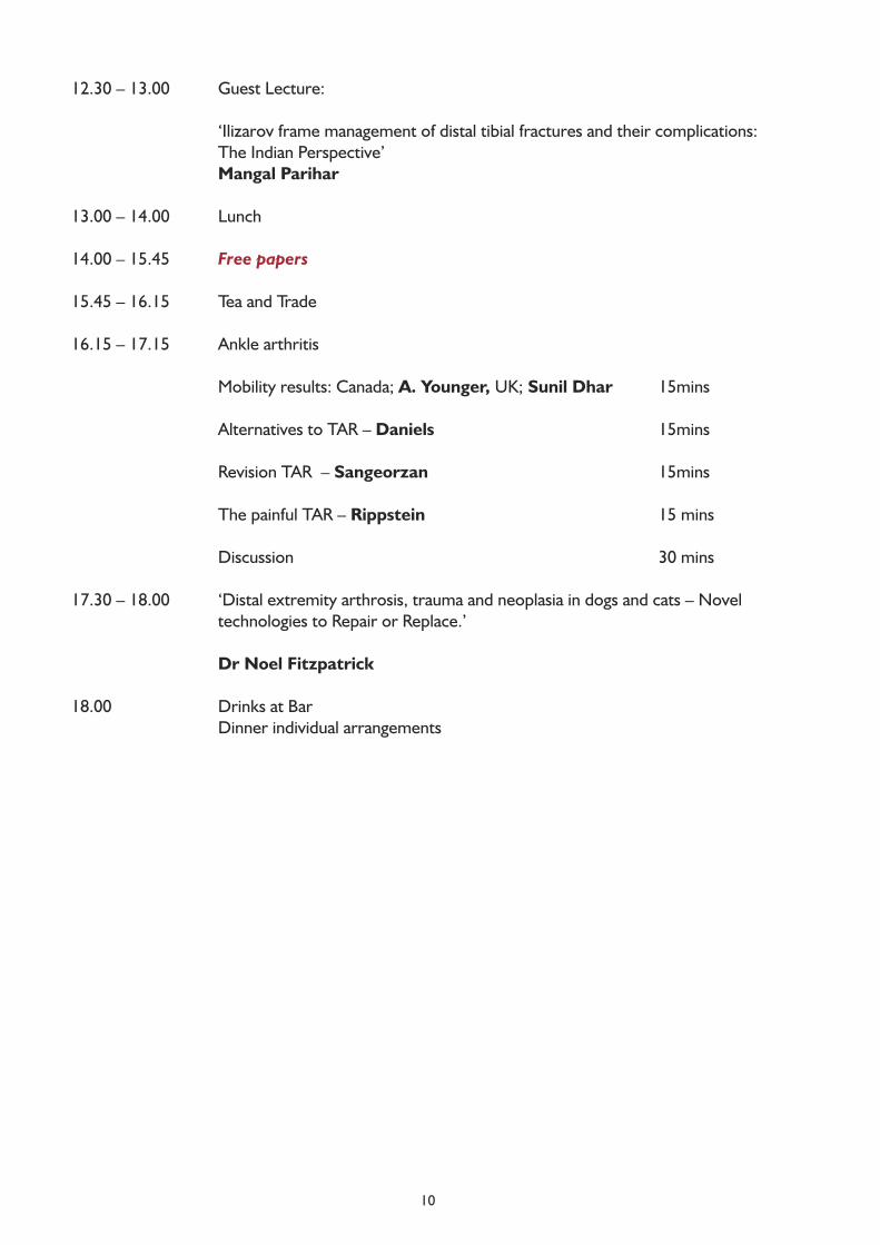
12.30 – 13.00 Guest Lecture:
‘Ilizarov frame management of distal tibial fractures and their complications:The Indian Perspective’Mangal Parihar
13.00 – 14.00 Lunch
14.00 – 15.45 Free papers
15.45 – 16.15 Tea and Trade
16.15 – 17.15 Ankle arthritis
Mobility results: Canada; A. Younger, UK; Sunil Dhar 15mins
Alternatives to TAR – Daniels 15mins
Revision TAR – Sangeorzan 15mins
The painful TAR – Rippstein 15 mins
Discussion 30 mins
17.30 – 18.00 ‘Distal extremity arthrosis, trauma and neoplasia in dogs and cats – Noveltechnologies to Repair or Replace.’
Dr Noel Fitzpatrick
18.00 Drinks at BarDinner individual arrangements
11
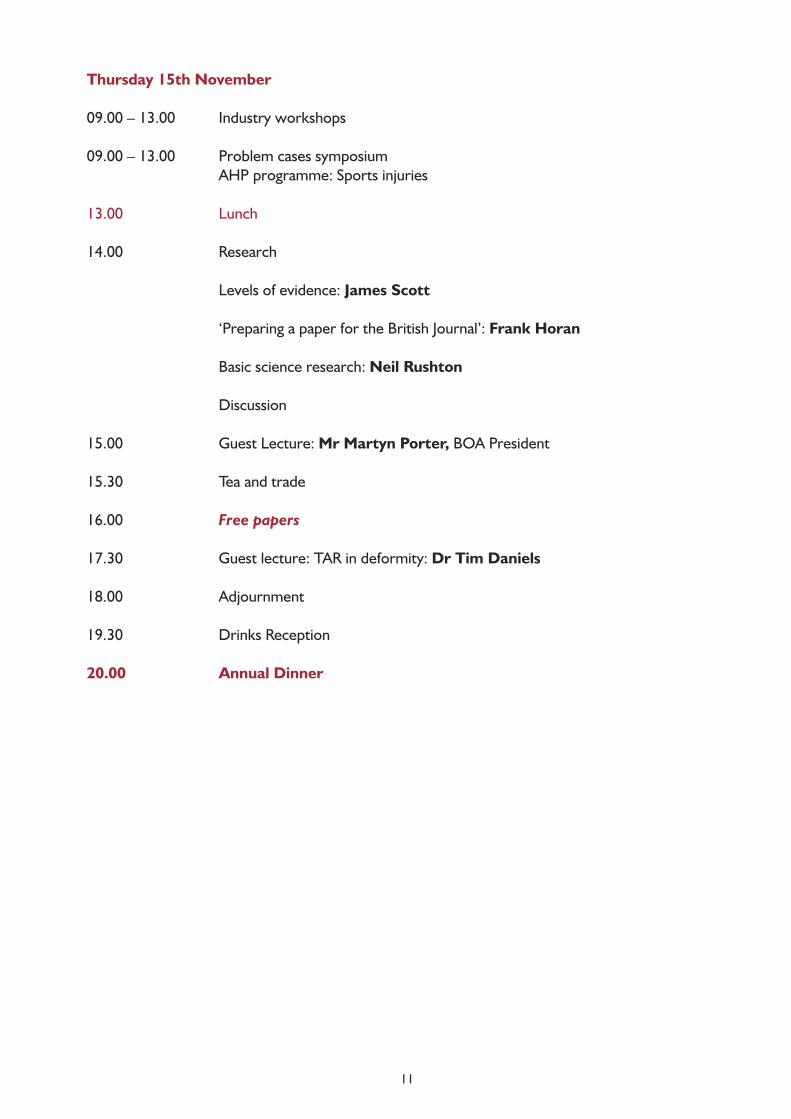
Thursday 15th November
09.00 – 13.00 Industry workshops
09.00 – 13.00 Problem cases symposiumAHP programme: Sports injuries
13.00 Lunch
14.00 Research
Levels of evidence: James Scott
‘Preparing a paper for the British Journal’: Frank Horan
Basic science research: Neil Rushton
Discussion
15.00 Guest Lecture: Mr Martyn Porter, BOA President
15.30 Tea and trade
16.00 Free papers
17.30 Guest lecture: TAR in deformity: Dr Tim Daniels
18.00 Adjournment
19.30 Drinks Reception
20.00 Annual Dinner
12
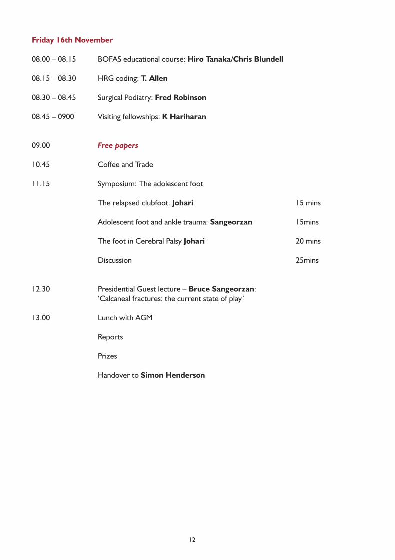
Friday 16th November
08.00 – 08.15 BOFAS educational course: Hiro Tanaka/Chris Blundell
08.15 – 08.30 HRG coding: T. Allen
08.30 – 08.45 Surgical Podiatry: Fred Robinson
08.45 – 0900 Visiting fellowships: K Hariharan
09.00 Free papers
10.45 Coffee and Trade
11.15 Symposium: The adolescent foot
The relapsed clubfoot. Johari 15 mins
Adolescent foot and ankle trauma: Sangeorzan 15mins
The foot in Cerebral Palsy Johari 20 mins
Discussion 25mins
12.30 Presidential Guest lecture – Bruce Sangeorzan:‘Calcaneal fractures: the current state of play’
13.00 Lunch with AGM
Reports
Prizes
Handover to Simon Henderson
13
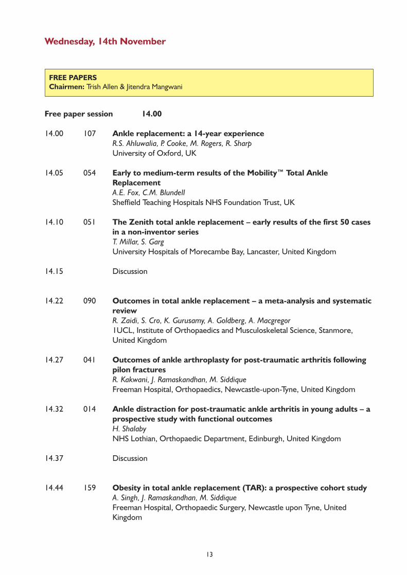
Wednesday, 14th November
Free paper session 14.00
14.00 107 Ankle replacement: a 14-year experience R.S. Ahluwalia, P. Cooke, M. Rogers, R. Sharp University of Oxford, UK
14.05 054 Early to medium-term results of the Mobility™ Total AnkleReplacement
A.E. Fox, C.M. Blundell Sheffield Teaching Hospitals NHS Foundation Trust, UK
14.10 051 The Zenith total ankle replacement – early results of the first 50 casesin a non-inventor series
T. Millar, S. Garg University Hospitals of Morecambe Bay, Lancaster, United Kingdom
14.15 Discussion
14.22 090 Outcomes in total ankle replacement – a meta-analysis and systematicreview
R. Zaidi, S. Cro, K. Gurusamy, A. Goldberg, A. Macgregor 1UCL, Institute of Orthopaedics and Musculoskeletal Science, Stanmore,
United Kingdom
14.27 041 Outcomes of ankle arthroplasty for post-traumatic arthritis followingpilon fractures
R. Kakwani, J. Ramaskandhan, M. Siddique Freeman Hospital, Orthopaedics, Newcastle-upon-Tyne, United Kingdom
14.32 014 Ankle distraction for post-traumatic ankle arthritis in young adults – aprospective study with functional outcomes
H. Shalaby NHS Lothian, Orthopaedic Department, Edinburgh, United Kingdom
14.37 Discussion
14.44 159 Obesity in total ankle replacement (TAR): a prospective cohort study A. Singh, J. Ramaskandhan, M. Siddique Freeman Hospital, Orthopaedic Surgery, Newcastle upon Tyne, United
Kingdom
FREE PAPERSChairmen: Trish Allen & Jitendra Mangwani
14
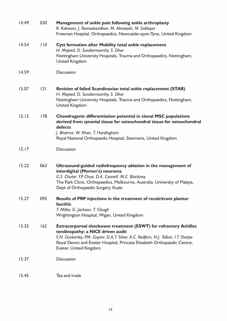
14.49 030 Management of ankle pain following ankle arthroplasty R. Kakwani, J. Ramaskandhan, M. Almaiyah, M. Siddique Freeman Hospital, Orthopaedics, Newcastle-upon-Tyne, United Kingdom
14.54 110 Cyst formation after Mobility total ankle replacement H. Majeed, D. Sundarmoorthy, S. Dhar Nottingham University Hospitals, Trauma and Orthopaedics, Nottingham,
United Kingdom
14.59 Discussion
15.07 121 Revision of failed Scandinavian total ankle replacement (STAR) H. Majeed, D. Sundarmoorthy, S. Dhar Nottingham University Hospitals, Trauma and Orthopaedics, Nottingham,
United Kingdom
15.12 178 Chondrogenic differentiation potential in clonal MSC populationsderived from synovial tissue for osteochondral tissue for osteochondraldefects
J. Bhamra, W. Khan, T. Hardingham Royal National Orthopaedic Hospital, Stanmore, United Kingdom
15.17 Discussion
15.22 063 Ultrasound-guided radiofrequency ablation in the management ofinterdigital (Morton’s) neuroma
G.S. Chuter, Y.P. Chua, D.A. Connell, M.C. Blackney The Park Clinic, Orthopaedics, Melbourne, Australia. University of Malaya,
Dept of Orthopaedic Surgery, Kuala
15.27 095 Results of PRP injections in the treatment of recalcitrant plantarfasciitis
T. Millar, G. Jackson, T. Clough Wrightington Hospital, Wigan, United Kingdom
15.32 162 Extracorporeal shockwave treatment (ESWT) for refractory Achillestendinopathy: a NICE driven audit
S.N. Dunkerley, P.M. Guyver, D.A.T. Silver, A.C. Redfern, N.J. Talbot, I.T. Sharpe Royal Devon and Exeter Hospital, Princess Elizabeth Orthopaedic Centre,
Exeter, United Kingdom
15.37 Discussion
15.45 Tea and trade
15
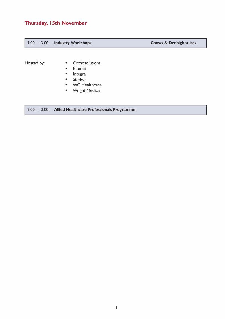
Thursday, 15th November
Hosted by: • Orthosolutions • Biomet • Integra • Stryker • WG Healthcare • Wright Medical
9.00 – 13.00 Industry Workshops Conwy & Denbigh suites
9.00 – 13.00 Allied Healthcare Professionals Programme
16
Thursday 15th November
Free paper session 16.00
16.00 070 Lesser toe plantar anatomy – considerations for minimally invasivesurgeryD. Loveday, A. RobinsonNorfolk & Norwich University NHS Trust, Orthopaedics, Bury St Edmund’s,UK
16.05 003 Our initial experience using minimally invasive Chevron and Akinosteotomies for Hallux Valgus correction: short term resultsN. Hossain, M. BudgenYork Teaching Hospital, Trauma & Orthopaedics, York, United Kingdom
16.10 085 Minimally invasive arthrodesis of the first metatarsophalangeal jointR.N. Fanous, S. Horriat, S. Ridgers, A.H. SottEpsom & St Helier University Hospital NHS Trust, Foot and Ankle Unit,London, United Kingdom
16.15 Discussion
16.22 080 Patient-reported outcomes (MOxFQ) and satisfaction eight yearsfollowing Hallux valgus surgeryJ. Dawson, M. Rogers, G. Lavis, R. Sharp, P.H. Cooke1University of Oxford, Oxford, United Kingdom,2Nuffield OrthopaedicCentre, Oxford, United Kingdom
16.27 138 Outcome of first metatarso-phalangeal total joint replacement(ToeFit): a clinical outcome and survival-ship analysisM. Al-Maiyah, P. Rice, T. SchneiderMelbourne Orthopaedic Group, Melbourne, Australia
16.32 125 Long term outcome of first MTPJ replacement using ceramicprosthesis with press fit designM.T. Nagy, C.R. Walker, S.P. SirikondRoyal Liverpool University Hospital, Orthopaedic Surgery – Foot and AnkleUnit, Liverpool, United Kingdom
16.37 Discussion
FREE PAPERSChairmen: Mr Andy Molloy & Anthony Perera
17
16.44 057 An assessment of how bone stress across the first metatarsophalangealjoint varies in different foot structures, utilising finite elementalanalysisR. Russell, R. Mootanah, A. Truchetet, S. Rao, H.J. HillstromBroomfield Hospital, Essex, United Kingdom. Anglia Ruskin University, Essex,United Kingdom. New York University, New York, United States. Hospital forSpecial Surgery, New York, United States
16.49 086 The hallucal metatarso-sesamoid articulation – a three dimensionalquantitive analysisB. Jamal, A. Pillai, Q.A. Fogg, S. KumarSouthern General Hospital, Department of Trauma & Orthopaedics, Glasgow,United Kingdom. University of Glasgow, Department of Human Anatomy,Glasgow, United Kingdom. Glasgow Royal Infirmary, Department of Trauma &Orthopaedics, Glasgow, United Kingdom
16.54 044 Can modification of the Weil osteotomy reduce the risk of a floatingtoe deformity – a biomechancial cadaveric analysisA. Perera, O. Helguera- Mendoza, M. MyersonUniversity Hospital of Wales, Trauma and Orthopaedics, Cardiff, UnitedKingdom. Hospital Puerto de Hierro, Orthopaedics and Trauma, Guadalajara,Mexico. Institute of Foot and Ankle Reconstruction, Baltimore, United States
16.59 Discussion
17.06 117 Lengthening Scarf osteotomy for recurrent Hallux valgusB. Rose, N. Bowman, H. Edwards, A. SkyrmeEastbourne District General Hospital, Eastbourne, United Kingdom
17.11 088 Intraoperative radiography for Scarf osteotomies of the firstmetatarsalP. Holland, A.P. MolloyAintree University Hospital, Department of Orthopaedics, Merseyside, UnitedKingdom. Aintree University Hospital, Department of Orthopaedics,Liverpool, United Kingdom
17.16 149 The Stainsby procedure for rheumatoid forefoot arthroplasty: 5 yearprospective follow up studyK.S. Rankin, A. Singh, J. Jalali, P. BriggsFreeman Hospital, Orthopaedic Surgery, Newcastle upon Tyne, UnitedKingdom
17.21 Discussion
18
Friday 16th November
Free paper session 0900
09.00 188 Bone marrow oedema syndrome of foot and ankleA. Ferrero, N. Cullen, D. SinghRoyal National Orthopaedic Hospital, Foot and Ankle Unit, Stanmore, UnitedKingdom
09.05 169 Simple stress fractures? Are they under-investigated?G. Chami, N. HarrisLeeds Teaching Hospitals, Leeds, United Kingdom
09.10 072 Do nerve conduction studies help with the diagnosis and managementof patients who present with non specific foot pain?J. Stevenson, A. Tong, Y. Joshi, P.W. Laing, N. Makwana1Wrexham Maelor Hospital, Trauma and Orthopaedics, Wrexham, UnitedKingdom,2The Robert Jones and Agnes Hunt Orthopaedic Hospital, Traumaand Orthopaedics, Oswestry, United Kingdom
09.15 Discussion
09.22 059 The home therapy ankle pathway – a novel and cost effective methodof initial management of ankle fracturesN. Baraza, S. Lever, G. Waight, V. DhukaramUniversity Hospital Coventry and Warwickshire, Trauma and Orthopaedics,Coventry, United Kingdom
09.27 104 Fifth metatarsal fractures – is routine follow-up necessary?J.D. Bone, L.A. Rymaszewski, C.S. Kumar, N.J. MadeleyGlasgow Royal Infirmary, Department of Trauma and Orthopaedics, Glasgow,United Kingdom. University of Glasgow, School of Medicine, Glasgow, UnitedKingdom
09.32 017 The restoration of anatomy following ankle fractures. Does grade ofsurgeon influence radiographic outcome?J. Jackson, M. Parry, S. MitchellBristol Royal Infirmary, Bristol, United Kingdom
09.37 Discussion
FREE PAPERSChairmen: Mr William Harries & Derek Robinson
19
09.44 049 The IED blast foot: outcomes from foot and ankle blast injuriesA. Ramasamy, S. Masouros, R. Phillip, I. Gibb, A. Bull1, J. ClasperImperial College London, The Royal British Legion Centre for Blast InjuryStudies, London, United Kingdom. Defence Medical Rehabilitation Centre,Epsom, United Kingdom. Centre for Defence Imaging, Gosport, UnitedKingdom
09.49 039 Clinical, radiological and functional outcomes after gradual correctionof stiff equino-cavo-varus deformity with the V-osteotomy and theIlizarov techniqueH. Shalaby, A. Wood, A. Keenan, C. ArthurNHS Lothian, Orthopaedic Department, Edinburgh, United Kingdom
09.54 140 Surgical outcomes of Lisfranc fixation – a single surgeon seriesJ.R. Eyre, S. Gudipati, G. Chami, R. MonkhouseLeeds Teaching Hospitals, Department of Orthopaedic and Trauma Surgery,Leeds, United Kingdom
09.59 Discussion
10.06 152 Management of middle facet tarsal coalitions with concomitant severeflat foot. Would resection alone suffice?O. Akilapa, H. PremBirmingham Children’s Hospital, Orthopaedics, Birmingham, United Kingdom
10.11 093 Arthroscopic fixation of displaced calcaneal fractures – percutaneousarthroscopic calcaneal osteosynthesisP. Harnett, P. RosenfeldImperial College, London, United Kingdom
10.16 050 An anatomical and cadaveric study examining the risk of sural nerveinjury in percutaneous Achilles tendon repair using the Achillon deviceK.J. Porter, P. Karia, M. Szarko, A. AminSt George’s Hospital, Trauma and Orthopaedics, London, United Kingdom. StGeorge’s Hospital, Medical School, London, United Kingdom. St George’sHospital, Anatomy, London, United Kingdom
10.21 Discussion
10.28 043 The role of venous stasis and endothelial damage in VTE in traumapatients treated with a cast- where does the clot occur?H. Jones, B. Hickey, A.G. Ghaffar, A. PereraUniversity Hospital of Wales, Trauma and Orthopaedics, Cardiff, UnitedKingdom. University Hospital of Wales, Cardiff, United Kingdom
20
10.33 116 Venous thromboembolism in ankle ORIF – the case againstpharmacological prophylaxis, a 5 year retrospective reviewC.L. Cozon, M.J. Welck, P.S. RayBarnet General Hospital, London, United Kingdom
10.38 Discussion
10.45 Coffee And trade
21
2012 Annual Scientific Meeting
Abstracts of Podium Presentations
22
Ankle replacement: a 14-year experience
R.S. Ahluwalia1,2, P. Cooke1,2, M. Rogers1,2, R. Sharp1,2
1University of Oxford, Nuffield Department of Orthopaedic, Rheumatology and MusculoskeletalSciences, Oxford, United Kingdom, 2Abertawe Bro Morgannwg University Health Board,Orthopaedic Department, Swansea, United Kingdom
Introduction: Ankle replacement is now common in the UK.In a tertiary referral NHS practice, between 1997-2011 we implanted two types of cementlessmobile bearing total ankle replacements (TAR).
Methods: We reviewed our operative database and electronic patient records and confirmed thenumber of prosthesis with our theatre records. All case notes and radiographs were reviewed.Failure was taken as revision, and patients were censored due to death or loss to follow-up. Thesurvivorship was calculated using a life table (the Kaplan-Meier method), with 95% confidenceintervals.
Results: We found a total of 358 NHS patients had a TAR from Jan 1997 to April 2012 total anklereplacements; the mean follow up was 76 months. The principle indications for surgery includedprimary OA (n=146) and inflammatory arthritis in (n=79). Overall survival was 90.9% (94-84) at10 years. A complication requiring revision developed in 42 ankles and 36 were revised or fused. Thirty-twoTAR’s underwent further hind foot fusions which were not attributed as a failure of the prosthesis.We arthroscoped 6 TAR’s for hetrotrophic calcification. When we separated the implants we found the STAR (implanted from 1997-2004) had a 5-yearsurvival of 95.2% (98-91) and the Mobility (implanted from 2004-11) of 92.6% (96-88). We foundearly failures (within 2 years of implantation) were higher within 2 years of introduction of TAR andon changing our prosthesis.
Conclusion: In a study of TAR undertaken at one centre principally by an experienced surgeon andteam, we have shown a learning curve. Cementless mobile bearing total ankle replacements (TAR) conducted on a routine basis withcareful patient selection has a 90.9% survivorship over a 10-year period. The difference in survivalfor two implants is not statistically significant.
Wednesday, 14th November

23
Early to medium-term results of the Mobility™ Total Ankle Replacement
A.E. Fox1, C.M. Blundell11Sheffield Teaching Hospitals NHS Foundation Trust, Foot & Ankle Unit, Sheffield, United Kingdom
Introduction: The Mobility™ prosthesis [Depuy] is the most extensively used TAR in the UK,though there are few published results. We present our complete experience of the Mobilityprosthesis in a diverse population.
Methods: From March 2005 to December 2009, 84 consecutive Mobility ankle replacements wereperformed by the senior author, in 79 patients (28 female, 51 male) with mean age 64.5 years (43-80). This complete cohort included the first and last cases with this implant. Mean follow-up was50.1±18.2 months (range 14-86). Patients with ankle replacements in situ, were reviewed clinically and radiologically. Clinicaloutcome measures were: AOFAS score, MOXFQ (adapted for the ankle), and VAS for pain. Post-operative radiographs were reviewed to assess component position and examine for zones oflucency.
Results: At final review, 1 patient had died (unrelated), 13 had been revised as follows: Arthrodesis 7 Further TAR 2 Talus only revised 1 Tibia only revised 1 Amputation 2 (one for an unrelated problem) Exchange of bearing had been carried out in 4. Intra-operative malleolar fractures occurred in 4.8% and were internally fixed. 62 patients attended for clinical review and 8 completed postal questionnaires. At follow up: Mean AOFAS hindfoot score was 72.4±17.5 (0-100). Mean MOXFQ scores were: Walking/Standing 40.8±28.4 Pain 31.6±20.8 Social 23.1±23.0. Mean VAS 2.7±2.3. Survival of the implant was: 91.7 (CI 83.4-96.0) at 2 years 89.2 (CI 80.2-94.2) at 3 years 84.1 (CI 73.4-90.8) at 4 years 84.1 (CI 73.4-90.8) at 5 years 78.9 (CI 62.6-88.7) at 6 years
Conclusion: This study is a complete review and our failure rate is comparable to otherpublications. Early failures included some poor case selections with large pre-operative deformityand reflects the initial period of the learning curve of TAR. Longer term follow up is needed toevaluate for ongoing failures and monitor progressive radiolucency.
24
The Zenith total ankle replacement – early results of the first 50 cases in a non-inventor series
T. Millar1, S. Garg1
1University Hospitals of Morecambe Bay, Lancaster, United Kingdom
Introduction: Total ankle replacement (TAR) surgery remains a reasonable alternative toarthrodesis in a select group of patients with end stage ankle joint arthritis. We describe the earlyresults of a prospective study of the first 50 Zenith total ankle replacements performed by a singlesurgeon (SKG).
Methods: Demographic details, Visual Analogue Score (VAS) for pain (0, no pain; 10, worstpossible pain), AOFAS scores, ‘would have surgery again’ and satisfaction levels were collated, pre-operatively and at their most recent outpatient review. Any post-operative complications werenoted. Radiographs were also assessed for evidence of loosening, progressive osteolysis, subsidenceand overall alignment of the implant.
Results: One patient died at 25 months following surgery from unrelated causes. No patients havebeen lost to follow up. A review of 50 patients (35 males, 15 females; mean age 65 years, range 44-88 years) with a mean follow up of 30 months (range 11-48) included 48 patients withosteoarthritis and two patients with rheumatoid arthritis. There was one medial malleolar fractureat the time of surgery which required fixation and one fracture of the lateral malleolus which waspicked up at the six week review. At their latest review the VAS and AOFAS score had improvedsignificantly and 46 patients were satisfied and 4 patients unsatisfied with the outcome of surgery. One patient has cyst formation around the tibial component but is pain free with a stable implantand does not wish further intervention. The components were satisfactorily aligned in the vastmajority of patients.
Conclusion: This non-inventor series of the Zenith TAR has shown excellent results in the shortterm. We feel that the instrumentation allows for more reproducible cuts which appear to betechnically easier than with some other designs. However, studies looking at long term results willbe necessary.
25
Outcomes in total ankle replacement – a meta-analysis and systematic review
R. Zaidi1, S. Cro2, K. Gurusamy3, A. Goldberg1, A. Macgregor1
1UCL, Institute of Orthopaedics and Musculoskeletal Science, Stanmore, United Kingdom, 2MRC,Clinical Trials Unit, London, United Kingdom, 3UCL, Division of Surgery and Interventional Science,London, United Kingdom
Introduction: Surgeons, commissioners and patients are increasingly seeking more in depth detailson outcomes of total ankle replacement (TAR). We set out to perform a detailed and up to datemeta-analysis of the outcomes of TAR, with a focus on PROMS.
Methods: We searched MEDLINE, Cochrane, EMBASE, CINAHL and the Science Citation Indexdatabases using the terms ‘’total’’; ‘’ankle’’; ‘’arthroplasty’’ or ‘’replacement’’ to April 2012. Weincluded all languages; series with greater than 20 TAR; minimum 2 years follow-up. We excludedpapers on revisions; prostheses no longer marketed; and kin studies. We worked with theCochrane Collaboration to adopt their methodology including the creation of a risk profile assessingall forms of bias.
Results: Of 1841 papers identified, 51 remained for analysis, with a pool of 6719 patients. Themean patient age was 59.3(17-95) and mean BMI was 28.8(19.4-44). 53% of patients were male.The most common indication was posttraumatic osteoarthritis. The majority of the studies werelevel IV and more than half the studies had several forms of bias. Intraoperative complication rate was 9%, with medial malleolar fracture (4.4%) being the mostcommon. The pooled mean pre-op VAS was 7.6 which improved to 1.5 at 4-5 years. The mean pre-opAOFAS was 39.7, improving to 79.9 for up-to 10 years. Range-of-motion increased after TAR from 22.8° preoperatively to 33.6° postoperatively. Radiographic abnormalities were found in 22% of cases with a mean follow up of 53 months, ofwhich 7.9% were re-operated upon. Gait velocity, cadence, stride length and power all improve following TAR. Survival at 8-10 years was 89.4%, with a cumulative failure rate of 1.9%.
Conclusion: This is the most comprehensive meta-analysis carried out on TAR to date. TARprovides patients with an increased range of motion and improvement postoperative PROMSmaintained up to 10 years.
26
Outcomes of ankle arthroplasty for post-traumatic arthritis following pilon fractures
R. Kakwani1, J. Ramaskandhan2, M. Siddique2
1Freeman Hospital, Orthopaedics, Newcastle-upon-Tyne, United Kingdom, 2Freeman Hospital,Newcastle-upon-Tyne, United Kingdom
Aim: A prospective cohort of patients undergoing total ankle arthroplasrty for arthritis followingpilon fractures was included in the present study. This group of patients generally have poor softtissue envelope and have had previous surgical interventions prior to the ankle arthroplasty, makingthe arthroplasty more difficult as well as prone to complications.
Methods: The data collected included patient demographics, American Orthopaedic Foot andAnkle Score (AOFAS) and patient reported outcomes (FAOS, SF-36, patient satisfaction) The datawas collected preoperatively and at 1 & 2 years postoperatively. The minimum follow-up periodwas 2 years post-operatively.
Results: A total of 167 total ankle arthroplasties were performed by the senior author between Jan2006 and June 2010. Of this cohort, the indication for 12 arthroplasties was arthritis following pilonfractures of the distal tibia. The average of the patients at the time of the surgery was 64.2yrs. Theaverage number of previous surgeries prior to the ankle arthroplasty was 1.5. There weresignificant improvements in the AOFAS scores from an average of 18 to 75 at final review. TheWOMAC scores improved from 31 to 71 for pain, stiffness improved from 31 to 60 and functionimproved from 33 to 63. The improvement of the SF36 and patient satisfaction score is similar tothe ones for primary ankle osteoarthritis. The complications were: 1 case of superficial woundinfection which settled with antibiotics, one fracture of medial malleolus and one case ofundisplaced distal tibial fracture treated conservatively to union.
Conclusion: The Indications for TAR can be safely broadened to include younger patients witharthritis following pilon fractures of the tibia. The Outcomes after TAR for patients with arthritisfollowing pilon fractures are comparable to those for primary osteo arthritis of the ankle.
27
Ankle distraction for post-traumatic ankle arthritis in young adults – a prospective study with functional outcomes
H. Shalaby1
1NHS Lothian, Orthopaedic Department, Edinburgh, United Kingdom
Aim: Young patients with ankle arthritis that remains symptomatic in spite of conservativetreatment and following arthroscopic debridement are usually offered either ankle fusion or anklereplacement. Both these options are far from ideal in this age group. The aim of this study was toevaluate functional outcomes following ankle distraction to determine whether it is a reliablealternative for the treatment of ankle arthritis in young adults.
Material and methods: Data was collected prospectively for 15 patients (9 males and 6 females,mean age 31.9 years) with “stage 2” ankle arthritis who failed conservative treatment andcontinued to be symptomatic following arthroscopic ankle debridement. Distraction of 8 mm wasdone using a dynamic constrained ankle circular frame and all patients were allowed full weightbearing all through the distraction process. The subjective functional evaluation was based on theAmerican Orthopaedic Foot and Ankle Score (AOFAS), the Foot Disability Index (FADI) and theVisual Analogue Score (VAS). In 10 patients the Manchester Oxford Foot questionnaire (MOXFQ)and the Short Form (SF) 12 patient satisfaction questionnaire were also filled preoperatively and atfinal follow up.
Results: At a minimum follow-up of 24 months (mean 34.4) none of the patients required fusion orreplacement. There was a significant improvement in all the functional outcome scores. There wasalso a significant improvement in the ankle joint space on weight bearing x-rays.
Conclusion: Based on these results the use of ankle distraction can be considered a useful optionfor the treatment of symptomatic “stage 2” ankle arthritis in young adults. Longer-term follow-upand comparison with alternative techniques will be required to evaluate the true effectiveness ofthis treatment option.
28
Obesity in total ankle replacement (TAR): a prospective cohort study
A. Singh1, J. Ramaskandhan1, M. Siddique1
1Freeman Hospital, Orthopaedic Surgery, Newcastle upon Tyne, United Kingdom
Aim: We aimed to study the effect of BMI on clinical and patient-reported outcomes in patientswith TAR with a minimum follow-up of three years.
Method: Patients who underwent a TAR between March 2006 and May 2009 were invited to takepart in the hospital patient registry. Patients were divided into two groups based on BMI (Group A –BMI < 30 and Group B – BMI >30). Patient demographics, co-morbidities, clinical (AOFAS),patient reported outcomes (FAOS, SF-36, patient satisfaction) and complications were collectedpre-operatively and at 1, 2 and 3 years and comparison made between groups.
Results: There were 50 patients in group A and 31 in group B. There was no significant differencebetween age (Mean 64.5 (A) and Mean 61.3 (B), gender and side of operation between the groups(p>0.05). Group A had higher percentage of patients with OA and RA compared to Group B(p=0.027). Group B (1.16) reported higher co-morbidites than Group A (1.81); p=0.012. Therewas no difference in AOFAS scores, FAOS scores (for pain, function and stiffness) and SF-36 scoresreported between groups at 1, 2 and 3 years post-operatively (p>0.05). At 3 years, Group Breported less patient satisfaction with return to ADL 84.6 %(A) vs. 50.1%(B) and recreationalactivities 73.1 %(A) vs. 43.8 %(B); p< 0.05. There was no significant difference in overallsatisfaction and pain relief and also in the number of reported complications between Groups A andB (p>0.05).
Conclusion: Patients with BMI > 30 reported higher co-morbidities pre-operatively and lesspatient satisfaction (return to ADL and recreation) at 3 years when compared to patients with BMI< 30. There was no difference in clinical, complications and patient reported outcomes betweenthese groups.
29
Management of ankle pain following ankle arthroplasty
R. Kakwani1, J. Ramaskandhan2, M. Almaiyah2, M. Siddique2
1Freeman Hospital, Orthopaedics, Newcastle-upon-Tyne, United Kingdom, 2Freeman Hospital,Newcastle-upon-Tyne, United Kingdom
Introduction: Postoperative pain following the 3 component ankle arthroplasty (AA) (Mobility TM)is a recognised problem without any apparent cause. This study aimed to determine pattern ofpostoperative pain following Total Ankle Arthroplasty (TAA) and its management options.
Materials and methods: In prospective observational study 167 patients who had (AA) andminimum follow-up of 24 months were included. FAOS ankle score, patients’ satisfaction, SF36 anddiagrammatic mapping of postoperative pain among other parameters were collectedpreoperatively and postoperatively at 3 months, 6 months and the annually. 20 Patients (12%) hadmoderate to severe postoperative ankle pain following the ankle arthroplasty.
Results: Most of patients with mild pain and low AOFAS score during first year improved by the 2year review. The pain was localised to the medial aspect of the ankle in 10 patients, lateral side in 8patients, and both medial and lateral side in 1 patient and global in 1 patient with complex regionalpain syndrome. 8 patients with medial or lateral pain needed a re-operation. 5 patients with medialpain were treated by complete release of deltoid ligament along with bony decompression of themedial compartment. None of the above implants were loose intra-operatively. 2 AA with lateralpain needed subtalar arthrodesis. 1 patient needed removal of metalwork from the calcaneum forrelief of symptoms. A significant improvement of pain and AOFAS scores was observed in 3 out ofthe 5 patients who underwent medial compartment decompression and both patients whounderwent subtalar arthrodesis.
Conclusion: There are 10- 13 % of low AOFAS scores following Ankle Arthroplasty due to pain. Inour series, the pain did not co-relate to implant loosening. Our treatment protocol of mapping ofpain and re-do surgery could improve the long term outcome in a significant proportion of thepatients.
30
Cyst formation after Mobility total ankle replacement
H. Majeed1, D. Sundarmoorthy1, S. Dhar1
1Nottingham University Hospitals, Trauma and Orthopaedics, Nottingham, United Kingdom
Introduction: Periprosthetic cyst formation following ankle replacement, requiring revisionsurgery, has previously been reported. The exact pathogenesis of cyst formation is unclear butconsidered to be due to a combination of biological and mechanical factors. Our objective was toreview the incidence of periprosthetic cyst formation following Mobility ankle replacement andtheir outcome.
Patients and methods: We reviewed all the Mobility ankle replacements performed by the seniorauthor from Oct 2005 till May 2012. Serial radiographs were reviewed to identify the presence ofcystic lesions in the tibia or the talus.
Results: 124 Mobility ankle replacements were performed in 116 patients during our study period.Average age was 65 years (22 to 88) with male to female ratio of 2:1. Average follow-up was 32months (7 to 73). Radiographic review of the most recent available radiograph showed cysticchanges in the distal tibia in 10 patients (8%). One patient had cystic appearance pre-operativelywhich was not found to be progressive after replacement. Seven patients were asymptomatic. Three patients presented with ankle pain, which was thoughtto be due the cyst. One of the symptomatic patients had undergone revision of tibial componentand bone grafting of the cyst 32 months after primary surgery. The second patient is awaitingsurgery for exploration and possible bone graft (40 months after primary surgery). The 3rd patientis awaiting CT scan for further evaluation of the cyst.
Conclusion: Our study shows that cystic changes were present in 8% of TAR at medium termreview. 70% (7 patients) were asymptomatic and 30% required intervention for beingsymptomatic. Regular review of the TAR patients is essential to identify the patients who developcyst formation.
31
Revision of failed Scandinavian total ankle replacement (STAR)
H. Majeed1, D. Sundarmoorthy1, S. Dhar1
1Nottingham University Hospitals, Trauma and Orthopaedics, Nottingham, United Kingdom
Introduction: With increasing numbers of primary total ankle replacements being performed, thenumber of revision ankle surgeries is expected to rise also. We present the results of the revisionprocedures for failed Scandinavian total ankle replacements.
Patients and methods: We retrospectively reviewed all the Scandinavian TAR done by the seniorauthor from March 1999 till Jan 2006. Patients who underwent revision surgery were identified andtheir data was collected including indications for revision surgery, procedure performed, symptomsand the overall outcome.
Results: 25 patients underwent revision of Scandinavian TARs between April 2000 and April 2012out off a total of 213 primary STARs (11%). Average age was 68 years (45 to 82), with male tofemale ratio of 4:1. The causes of failure of primary implants included broken polyethylene insertsin 12 patients, aseptic loosening in 6 and ankle instability in 7 patients. No septic loosening wasfound in any of our patients. Revision procedures which were performed in these patients included exchange of inserts in 13patients, revision of all components in 2, revision of tibial component in 3, talar component in 2 andankle arthrodesis with hindfoot nail in 4 and with ilizarov frame in 1 patient. The average time fromthe primary procedure to revision surgery was 78 months (12 to 156). The average follow up afterrevision surgery was 26.5 months (2 to 57). Four patients have died. Two patients weresymptomatic with mild pain and stiffness while the rest are asymptomatic after their revisionsurgery.
Conclusion: In our study the mechanical failure was found to be the most common cause of failureof Scandinavian TARs. The outcome of revision surgery has been found to be satisfactory andcomparable to other series that is reported in the literature.
32
Chondrogenic differentiation potential in clonal MSC populations derived from synovial tissue for osteochondral defects
J. Bhamra1, W. Khan1, T. Hardingham1
1Royal National Orthopaedic Hospital, Stanmore, United Kingdom
Introduction: Mesenchymal stem cells (MSCs) are a potential source of cells for the repair ofarticular cartilage and osteochondral defects (OCD) in the ankle. Synovial tissue has been shown tobe a rich source of MSCs with the ability to undergo chondrogenic differentiation. Although thesecells represent a heterogenous population, clonal populations have not been previously studied.
Methods: MSCs were isolated from synovial tissue of a patient undergoing joint arthroplasty andexpanded in culture. Six clonal populations were also isolated and expanded. The cells from themixed parent population and the derived clonal populations were characterised for stem cellsurface epitopes, and then cultured in chondrogenic mediums. Various assays were determined toanalyse for features of differentiation.
Results: Cells from the mixed parent population and the derived clonal populations stainedstrongly for markers of adult mesenchymal stem cells including CD44, CD90 and CD105, and theywere negative for the haematopoietic marker CD34 and for the neural and myogenic markerCD56. Interestingly, a variable number of cells were also positive for the pericyte marker 3G5 bothin the mixed parent and clonal populations. The clonal populations exhibited a variablechondrogenic response.
Conclusion: Pericytes are a candidate stem cell in many tissues and our results show that all sixclonal populations derived from the heterogenous synovium population express the pericytemarker 3G5. The chondrogenic potential of synovial tissue could be optimised by the identificationof clonal populations with a propensity to differentiate down particular differentiation pathways.Our study demonstrates a role for MSCs in of osteochondral defects (OCDs) and areas of focalcartilage damage in the ankle joint.
33
Ultrasound-guided radiofrequency ablation in the management of interdigital (Morton’s) neuroma
G.S. Chuter1, Y.P. Chua2, D.A. Connell3, M.C. Blackney1
1The Park Clinic, Orthopaedics, Melbourne, Australia, 2University of Malaya, Dept of OrthopaedicSurgery, Kuala Lumpur, Malaysia, 3Monash University, Dept of Medicine, Melbourne, Australia
Introduction: Up to 70% of patients with symptomatic Morton’s neuroma proceed to surgeryhaving failed non-operative management. The success of surgical excision is up to 85% but carrieswith it significant morbidity. Radiofrequency ablation (RFA) is a less invasive alternative.
Methods: We studied a consecutive cohort of patients with Morton’s neuroma that had failed non-operative treatment. Instead of undergoing surgical excision, these patients were referred for RFA.Under a local anaesthetic nerve block, RFA was performed under ultrasound-guidance, as an out-patient procedure, by a single radiologist. The procedure was repeated after 4 weeks if necessary.We followed patients for a minimum of 6 months to assess their change in visual analogue painscores (VAS), overall symptom improvement, complications and progression to surgical excision.
Results: 30 feet in 25 patients were studied. There were 4 males and 21 females with an averageage of 55 years (range 33-73y). All had tried previous methods of non-operative management. 40%presented with 2nd space neuromas and 60% with 3rd space. The average number of treatmentsessions was 1.6 (range 1-3, mode 1). Prior to treatment, all patients had pain on activity (VASaverage: 6.0, range 3-9). At 6 months post treatment, there was a statistically significant reductionin pain scores (post RFA VAS average: 1.7, range 0-8, p< 0.001). The average overall symptomimprovement was 76%. There was one minor complication of temporary nerve irritation. 3neuromas (10%) have progressed to surgical excision. 1 patient has ongoing, unchanged pain withno obvious cause. At 6 months, 26 out of 30 feet had a satisfactory outcome.
Conclusion: RFA has potentially reduced the need for surgical excision of Morton’s neuromas by>85%.
34
Results of PRP injections in the treatment of recalcitrant plantar fasciitis
T. Millar1, G. Jackson1, T. Clough1
1Wrightington Hospital, Wigan, United Kingdom
Introduction: Whilst most cases of plantar fasciitis can be resolved with existing conservativeestablished treatment options, a few intractable cases can be difficult to resolve. New biologictreatments have been proposed for a variety of soft tissue tendon problems. We evaluated theresults of PRP in the treatment of recalcitrant chronic cases of plantar fasciitis.
Methods: Patients with plantar fasciitis that had not responded to a minimum of 8 months standardconservative management (eccentric stretching, physiotherapy, cortisone injection, night splints)were offered PRP therapy. The injection into the tender spot at the proximal plantar fascialinsertion was performed in theatre as a day case. Roles Maudsley (RM) scores, Visual analoguescores (VAS) for pain, AOFAS scores and �would have injection again� were collated pre-operatively, at three and six months.
Results: Prospective data was collected on 39 patients (44 heels – 15 males, 24 females; mean age51 years, range 25-79 years). No complications were noted. At six months review RM scoreimproved from 3.8 to 2.5 (p< 0.001), VAS improved from 7.7 to 4.2 (p< 0.001) and AOFASimproved from 61 to 82 (p< 0.001). 21 patients had complete relief of symptoms on 3 monthsreview. 25 patients were very satisfied with the clinical improvement and would have the injectionagain. Whilst there was a slight improvement in scores from 3 to 6 months, this was not significant.3 patients with bilateral injections on the same sitting did not improve, though 2 patients withbilateral injections on separate sittings did improve.
Conclusion: In this series of chronic intractable cases, PRP injection produced a 64% satisfactionrate from patients. The procedure was safe with no reported complications. The authors feel PRPfor plantar fasciitis may have some role in treatment and merits further study with a prospectiverandomised trial.
35
Extracorporeal shockwave treatment (ESWT) for refractory Achilles tendinopathy: a NICE driven audit
S.N. Dunkerley1, P.M. Guyver1, D.A.T. Silver1, A.C. Redfern1, N.J. Talbot1, I.T. Sharpe1
1Royal Devon and Exeter Hospital, Princess Elizabeth Orthopaedic Centre, Exeter, United Kingdom
Introduction: Achilles tendinopathy is chronic degeneration of the Achilles tendon, usuallysecondary to injury or overuse. It involves a triad of pain, swelling and impaired function. Primarytreatment is rest, analgesia, corticosteroid injections and physiotherapy (eccentric training and heelpads to correct gait). Some patients remain symptomatic and further treatment options needconsidering.
Methods: NICE produced a document from the Interventional Procedures Advisory Committee in2009 which reviewed the literature and evidence for extracorporeal shockwave treatment (ESWT).Low energy shock wave treatment (SWT) is thought to stimulate soft tissue healing, inhibit painreceptors and promote angiogenesis. NICE guidance was that ESWT could be used in refractoryAchilles tendinopathy if used for clinical governance, audit or research. Patients with refractory Achilles tendinopathy were enrolled between October 2010 and 2011.They received three sessions of ESWT over three week. Patients completed visual analogue scale(VAS) scores for pain at rest and on activity and the Victorian Institute of Sport Assessment-Achilles(VISA-A) questionnaire pre-treatment. These outcome measures and a six-point Likert satisfactionscale (six points, high is worsening) were reassessed at 6 and 16 weeks post treatment.
Conclusion: 51 patients completed follow up. The mean age was 56 (34-80) years and mean lengthof symptoms 34 (4-252) months. There was a significant improvement (p< 0.05) in VAS scoresobserved from baseline and 16 weeks post treatment. This was also the case in the VISA-A scores.The mean Likert score was 3 (somewhat improved) at 16 weeks but there was no statisticalsignificance. This study suggests that ESWT improves subjective and objective outcomes in patients withrefractory Achilles tendinopathy. Patients over 60 possibly have a worse outcome along with patientwho had symptoms for over 25 months. Follow up scores at one year are due to be collected andthe data will be submitted to NICE.
36
Lesser toe plantar anatomy – considerations for minimally invasive surgery
D. Loveday1, A. Robinson2
1Norfolk & Norwich University NHS Trust, Orthopaedics, Bury St Edmund’s, United Kingdom,2Addenbrooke’s Hospital, Orthopaedics, Cambridge, United Kingdom
Introduction: The aim of this study is to better understand the anatomy of the forefoot tominimise surgical complications following minimally invasive forefoot surgery.
Methods: The study examines the plantar anatomy of the lesser toes in ten cadaver feet. Thetendons, nerves and bony anatomy are recorded.
Results: The anatomy of the flexor tendons reveals the short flexor tendon bifurcates to allow thelong flexor tendon to pass through it reliably at the level of the metatarsophalangeal joint (MTPJ) inthe lesser rays. The division of the intermetatarsal nerves to digital nerves relative to the MTPJ ismore variable. This nerve division is more consistently related to the skin of the web between thetoes. In the first webspace the division is on average 3cm proximal to the skin at the deepest part ofthe cleft. In the second, third and fourth webspaces this distance is reduced to 1cm. The level ofthe deepest part of the webspace to the MTPJ is also variable.
Discussion: Surgical release of the flexor tendons is recommended just proximal to the MTPJ forreleasing both tendons and distal to the proximal interphalangeal joint for the long flexor tendon.The webspace skin and MTPJ’s are easily identifiable landmarks clinically and radiologically.Awareness of the intermetatarsal nerve division will help to reduce nerve injuries with minimallyinvasive surgery to the plantar forefoot.
Thursday, 15th November
37
Our initial experience using minimally invasice chevron and akin osteotomyfor Hallux valgus correction: short term results
N. Hossain1, M. Budgen1
1York Teaching Hospital, Trauma & Orthopaedics, York, United Kingdom
Minimally invasive chevron and akin osteotomy are being used in a few centres in the UK. Thepurpose of our study was to analyse our early results and present our early experience of minimallyinvasive chevron and akin osteotomy (MICA) for the correction of mild to moderate hallux valgus.This study assessed the radiological and clinical measurements, American Orthopaedic Foot andAnkle Society (AOFAS) scores, pain scores and patient satisfaction associated with performance ofthe MICA, for the treatment of hallux valgus.Between September 2010 and April 2012, 96 consecutive patients (122 feet) who underwentMICA were assessed.The overall satisfaction rate was over 90%. The mean total AOFAS score was89.7 points. VAS for pain reduced from a mean of 7.4 to less than 1 point. On weightbearinganterior-posterior foot radiographs there was a significant improvement in the mean IMA and HVA.Complications included 2 episodes of superficial infection (1.6%), 1 fracture (0.8%), 4 incidence ofnerve injury (3.3%) (numbness) and 9 patients requiring removal of screw (7.4%). However, thesescrew removals occurred early on in the study and diminished after a slight modification in surgicaltechnique.Based on our findings we concluded that MICA is an effective procedure with good patientsatisfaction in the treatment of mild to moderate hallux valgus.
38
Minimally invasive arthrodesis of the first metatarsophalangeal joint
R.N. Fanous1, S. Horriat1, S. Ridgers1, A.H. Sott1
1Epsom & St Helier University Hospital NHS Trust, Foot and Ankle Unit, London, United Kingdom
First metatarsophalangeal joint (MTPJ) arthrodesis plays a significant role in the management ofsymptomatic hallux rigidus. Several open and very few percutaneous techniques have been describedin the literature. The authors present a new minimally invasive technique along with patient reportedoutcome, radiological parameters and a discussion on this novel technique.A total of fifteen cases of first MPTJ arthrodesis were included in this prospective continuous seriesfrom September 2011 to June 2012. Mean age was 56 years and the indication for surgery was halluxrigidus in 13 of 15 cases. All patients underwent the same minimally invasive procedure by the sameprimary surgeon (AHS), 13 of 15 as day cases. Clinical outcome and patient satisfaction were assessedby the Manchester-Oxford Foot Questionnaire (MOXFQ) preoperatively and at most recent followup. Radiographic and clinical evaluation of fusion was also assessed. No patients were lost to followup and all scores were collected by an independent surgical practitioner to avoid bias. Mean followup was six months (range 1 to 9 months).The MOXFQ score for cases where fusion was achieved (n=14) improved from a mean 40/64preoperatively to a mean 15/64 at last follow up (p=0.001). Patient satisfaction was overall very good. This minimally invasive technique for first MTPJ arthrodesis is simple and can achieve results similaror better than open techniques in experienced hands. Further cases are needed to expand this seriesand evaluate for further complications.
39
Patient-reported outcomes (MOxFQ) and satisfaction eight years following Hallux valgus surgery
J. Dawson1, M. Rogers2, G. Lavis2, R. Sharp2, P.H. Cooke2
1University of Oxford, Oxford, United Kingdom, 2Nuffield Orthopaedic Centre, Oxford, UnitedKingdom
Background: Evaluation of outcomes and satisfaction following hallux valgus (HV) surgery is usuallyretrospective and rarely uses patient-reported outcome measures (PROMs).
Design: Prospective Cohort Study. Postal evaluation survey of patients who had provided pre-operative PROMs data.
Methods: Consecutively recruited patients completed the Manchester-Oxford Foot Questionnaire(MOxFQ) prior to surgery. Of 91 patients (124 feet) proceeding to one-stage HV surgery, 69 of 88eligible patients (78%; 95/124=77% feet) returned a postal follow-up questionnaire including theMOxFQ and a standard satisfaction rating for surgical outcome around 8 years (range 7.4 to 8.9) later.
Results: Of the 69 respondents, patients’ mean pre-operative age was 49.8 (SD 12.5) years; 66(95.7%) were female. Of the 95 feet, 78 (82.1%) patients were ‘Very pleased’ or ‘Fairly pleased’ withthe outcome, with 17/95 (17.9%) ‘Not very pleased/very disappointed’.Change in all 3 MOxFQ scalesshowed a significant linear relationship with satisfaction ratings (ANOVA p< 0.001) with MOXFQpain change scores for the ‘very pleased’ response in particular being significantly different from thoseof other response groups.
Conclusions: At around 8 years following HV surgery, the majority of patients were pleased with theoutcome. Change in the MOXFQ pain scale is particularly important in interpreting patients’satisfaction with surgery.
40
Outcome of first metatarso-phalangeal total joint replacement (ToeFit): a clinical outcome and survival-ship analysis
M. Al-Maiyah1, P. Rice1, T. Schneider1
1Melbourne Orthopaedic Group, Melbourne, Australia
Introduction: Hallux Rigidus affects 2-10% of population, usually treated with cheilectomy orarthrodesis, however, for the subclass of patients who refuse to undergo fusion, Arthroplasty is analternative solution, it maintain some degree of motion and provide pain relief. Toefit; is one of theprostheses being used. It is a total joint replacement with polyethylene insert. The aim of this study is to find clinical and radiological outcomes of Toefit arthroplasty.
Method: A prospective study. Ethical committee approval was obtained. Patient who have receivedToefit Arthroplasty with at least 12 months follow-up and were willing to participate in the study wereincluded. Patients were reviewed by independent surgeon. Questionnaires were completed followedby clinical examination. This followed by radiographic assessment. Patients, who were willing to takepart in the study but could not attend a clinical review, were invited to participate in telephonequestionnaire. Pre and postoperative AOFAS scores were compared, patients’ satisfaction and clinicaland radiological outcome were assessed using descriptive statistics, t-test and survivalship analysiswere done.
Results: 180 patients had Toefit (September 2004- June 2011). 160 patients participated in the study(170 prostheses), 87% were females. Age range (38-89) year. AOFAS improved significantly from 38to 83, with average arc of movement of 37 degrees. Patient satisfaction was high, VAS score1. Failure rate of 4.9%, there was high rate of revision of 29% due to sesamoid pain or stiffness in theinitial group of patients, decreased to 8% in the second group. Radiological review showedasymptomatic aseptic loosening of 20%, mainly of the proximal phalanx components.
Conclusion: First MTP joint replacement can provide pain relief and maintain good range ofmovement. However, this study highlighted high rate of revision and aseptic loosening. Long termreview is required.
41
Long term outcome of first MTPJ replacement using ceramic prosthesis with press fitdesign
M.T. Nagy1, C.R. Walker1, S.P. Sirikonda1
1Royal Liverpool University Hospital, Orthopaedic Surgery – Foot and Ankle Unit, Liverpool,United Kingdom
Introduction: There are a number of options available for surgical management of hallux rigidus.Ceramic implants of the first metatarsophalangeal joint (MTPJ) have been available for years; howeverthere are no published long-term results existing.
Methods: We performed a retrospective review of all consecutive first MTPJ replacements carriedout for later stage hallux rigidus using second generation MOJE ceramic implant with press-fit design.Two specialised foot and ankle surgeons performed these operations at a tertiary referral centre.Patient underwent regular follow ups including clinical review, functional scoring (AOFAS and FFI) andassessment of radiographs. Kaplan Meyer Survival analysis was performed.
Results: Our study included 31 prostheses in 24 female patients. Average age at operation was 55.3years and average follow up time was 80 months. No patients were lost until follow up. Complicationsincluded one case of superficial infection and five cases of revision, reasons being fracture of theprostheses (1), unexplained pain (1), subluxation (1) and loosening/sinkage of the implant (2).Prosthesis survival rate was 85.2% at seven years. Assessment of the radiographs showed considerablesinkage of the prosthesis in 43%, tilting in 33% and loosening of the implant in 40.9%. Averagepostoperative AOFAS score was 71.6 and the average FFI was 27.7. 84% of the patients were satisfiedwith the results of their operation.
Conclusion: Surgery has failed to preserve the function and increase the range of movement in mostcases in the long duration. From the patients perspective however the satisfaction with the proceduresuggests a success of the implant. Due to poor radiological results and high revision rate we do notrecommend the routine use of this prosthesis and all patients that have this type of prosthesis needregular follow up consultations at least yearly with radiographs to assess the position of the implant.
42
An assessment of how bone stress across the first metatarsophalangeal joint varies indifferent foot structures, utilising finite elemental analysis
R. Russell1, R. Mootanah2, A. Truchetet2, S. Rao3, H.J. Hillstrom4
1Broomfield Hospital, Essex, United Kingdom, 2Anglia Ruskin University, Essex, United Kingdom,3New York University, New York, United States, 4Hospital for Special Surgery, New York, UnitedStates
Introduction: Osteoarthritis commonly affects the first metatarsophalangeal joint. Stress across thisjoint has been postulated to increase the incidence of osteoarthritis. Certain foot structures have beenassociated with a higher incidence of osteoarthritis of the big toe. Utilizing finite elemental analysis,bone stress across the first metatarsophalangeal joint was calculated during mid stance phase of gaitand compared in different foot structures.
Method: A geometrically accurate three dimensional model of the first metatarsophalangeal jointwas created utilising a high resolution 7 tesla MRI and Mimics v14 imaging software. Planus, rectusand cavus feet were simulated by varying the metatarsophalangeal declination angle to 10.1, 20.2 and30.7 degrees, respectively . A non-manfold computer aided design technique in Mimics v14.2 andfinite element method in ANSYS v12 FE were utilised to create the boundary conditions, representingthe double support stance phase of gait. Using information from 61 asymptomatic patients withdifferent foot types walking over a Novel emed-x plantar pressure measuring system, plantar loadingconditions were applied. Finite elemental analysis was used to predict stress in the firstmetatarsophalangeal joint in the different foot types.
Results: The peak stresses in the distal first metatarsophalangeal joint cartilage were 1.1x10(6) Pa,6.0x10(5) Pa and 9.7x10(5) Pa for planus ,rectus and cavus foot types, respectively. This correspondsto 83.3 percent and 61.6 percent increases in first metatarsophalangeal joint contact stress for theplanus and cavus feet relative to the rectus foot.
Conclusion: The results suggest there is a higher contact stress of the first metatarsophalangeal jointin patients with pes planus and pes cavus compared to the rectus foot. This may account for theincrease risk of first metatarsophalangeal joint osteoarthritis in patients with pes planus.Further work has been initiated utilising this model to measure first metatarsophalangeal joint stresswith different hindfoot loading.
43
The hallucal metatarso-sesamoid articulation – a three dimensional quantitive analysis
B. Jamal1, A. Pillai2, Q.A. Fogg2, S. Kumar3
1Southern General Hospital, Department of Trauma & Orthopaedics, Glasgow, United Kingdom,2University of Glasgow, Department of Human Anatomy, Glasgow, United Kingdom, 3GlasgowRoyal Infirmary, Department of Trauma & Orthopaedics, Glasgow, United Kingdom
Introduction: The anatomy of the first metatarsophalangeal (MTP) joint and, in particular, themetatarsosesamoid articulation remains poorly understood. The movements of the sesamoids inrelation to the metatarsal plays a key role in the function of the first MTP joint. Although thedisorders affecting the sesamoids are described well, the movements of the metatarsosesamoidjoints and the pathomechanics of these joints have not been described. We have performed acadaver study detailing and quantifying the three dimensional movements occurring at these joints.
Methods: Fresh frozen cadaveric specimens without evidence of forefoot deformity weredissected to assess the articulating surfaces throughout a normal range of motion. The dissectionswere digitally reconstructed in positions ranging from 10 degrees of dorsiflexion to 60 degrees ofplantarflexion using a MicroScribe, enabling quantitative analyses in a virtual 3D environment.
Results: The sesamoids demonstrated excursion both in the sagittal and coronal plane. The tibialsesamoid had a mean saggital excursion of 14.2 mm; the mean excursion of the fibular sesamoidwas 8.7 mm. The mean coronal excursion of the tibial sesamoid was 2.8 mm while that of thefibular sesamoid was 3.2 mm. We also describe the mean saggital and coronal excursion of thesesamoids during smaller, incremental motions of the MTP joint.
Conclusion: There appears to be differential tracking of the hallucal sesamoids. The tibial sesamoidhas comparatively increased longitudinal excursion whilst the fibular sesamoid has comparativelygreater lateral excursion.
Clinical relevance: The greater excursion of the tibial sesamoid could explain the higher incidenceof pathology in this bone. The differential excursion of the sesamoids is also a factor that should beconsidered in the design and mechanics of an effective hallux MTP joint arthroplasty.
44
Can modification of the Weil osteotomy reduce the risk of a floating toe deformity – a biomechancial cadaveric analysis
A. Perera1, O. Helguera- Mendoza2, M. Myerson3
1University Hospital of Wales, Trauma and Orthopaedics, Cardiff, United Kingdom, 2HospitalPuerto de Hierro, Orthopaedics and Trauma, Guadalajara, Mexico, 3Institute of Foot and AnkleReconstruction, Baltimore, United States
Background: The Weil osteotomy is successful in the management of metatarsalgia and toedeformity. The aim is to achieve controlled shortening whilst avoiding plantarflexion. Recognisedcomplications of the Weil osteotomy include a ‘floating toe’ in up to 20% or more of cases.
Aim: Can modification of the Weil osteotomy avoid the metatarsal head plantarflexion andsubsequent dorsal subluxation of the interosseous muscle that is implicated in the development of a‘floating toe’.
Methodology: The 2nd, 3rd and 4th rays were dissected out en bloc with all tendons attachedfrom 6 pairs of fresh frozen cadavers. The ray was fixed to a board and the tendons physiologicallybalanced via low-friction pulleys and light weights. This model is highly sensitive to alterations inmoment arm of the tendons. Once the MTP joint was balanced these weights were not altered. After standard release of the dorsal capsule a Maceira or ‘triple-cut’ osteotomy was performed.This osteotomy was developed in order to permit shortening without plantar flexion of themetatarsal head. The osteotomy was performed using a image intensifier in order to maintain uniformity of theosteotomy cut. The neck was shortened by removing a 2mm slice of bone made with parallel cuts.The ray was photographed and x-rayed pre and post ‘surgery’ and the relationships of theinterosseous tendon andcentre of rotation of the metatarsal head, tendon balance and toe posturewere recorded.
Results: The Maceira osteotomy can avoid plantarflexion of the metatarsal head The line of pull ofthe interosseous tendon can maintains it’s normal relationship with the centre of rotation of themetatarsal head. None of the toes developed dorsiflexion. In fact, plantarflexion at the MTP jointcould be seen. PIP ‘fusion’ resulted in dorsiflexion at the MTP joint in some toes.
45
Lengthening scarf osteotomy for recurrent Hallux valgus
B. Rose1, N. Bowman1, H. Edwards1, A. Skyrme1
1Eastbourne District General Hospital, Eastbourne, United Kingdom
Introduction: Hallux valgus surgical correction has a variable but significant risk of recurrence.Symptoms result from an iatrogenic first brachymetatarsia following the index surgical procedure.First metatarsal shortening has been shown to correlate with the onset of transfer metatarsalgia.We describe the use of the scarf osteotomy to both correct the recurrent deformity and lengthenthe shortened first metatarsal.
Methods: 36 lengthening scarf osteotomies were undertaken in 31 patients. Clinical (AOFAS andSF12 scores) and radiographic measures (IMA, HVA) were taken pre- and post-operatively. Themaximum theoretical lengthening was 10mm, to prevent first MTP joint stiffness post-operatively.The actual lengthening was determined and measured intra-operatively.
Results: There were 28 female and three male patients, with mean age at presentation 53.4 years.The mean follow-up was 3.9 years. Four cases were lost to follow-up. The mean first metatarsallengthening achieved was 4.9mm (range 1 – 8mm). All of the osteotomies united withoutcomplication. The mean IMA reduction was 4.0° (p< 0.001) and HVA 13.0° (p< 0.001). The meanAOFAS score increase was 33.8 (p< 0.001). There was no correlation between change in IMA andAOFAS score (r=-0.13) or between improvement in HVA and AOFAS score (r=-0.02). There wasa positive trend but no correlation (r=0.28) between amount of metatarsal lengthening and changein AOFAS score. The inter- and intra-observer correlation was excellent. The SF12 physical sub-domains improved more than the mental sub-domains.
Conclusion: We describe the largest series of lengthening scarf osteotomies for recurrent halluxvalgus and symptomatic iatrogenic first brachymetatarsia. The significant improvement in bothclinical and radiographical measures suggests the procedure is successful, with a low complicationrate. Lengthening did not reduce the MTPJ range of movement. We hypothesise that restoring boththe length and alignment enables greater weight-bearing under the first metatarsal head, reducingbiomechanical transfer metatarsalgia.
46
Intraoperative radiography for scarf osteotomies of the first metatarsal
P. Holland1, A.P. Molloy2
1Aintree University Hospital, Department of Orthopaedics, Merseyside, United Kingdom, 2AintreeUniversity Hospital,Department of Orthopaedics, Liverpool, United Kingdom
Introduction: When performing scarf osteotomies some surgeons use intraoperative radiographyand others do not. Our experience is that when using intraoperative radiography we often changethe osteotomy position to improve the correction of the hallux valgus angle and sesamoid position.We report the results of a single surgeon series of 62 consecutive patients who underwent a scarfosteotomy for hallux valgus. The first 31 patients underwent surgery without the use ofintraoperative radiographs and the subsequent 31 patients underwent surgery with the use ofintraoperative radiographs, this reflects a change in the surgeons practice. Hallux valgus angle,intermetatarsal angle, distal metatarsal articular angle and sesamoid position using the HardyClapham grading system were recorded. All patients had measurements recorded from weightbaring radiographs taken pre operatively as well as at 6 and 12 weeks post operatively.Intraoperative measurements were also recorded for all patients in the intraoperative radiographygroup.The mean hallux valgus angle preoperatively was 28.5° in the control group and 30.5° in theintraoperative radiography group. The mean hallux valgus angle in the control group at 6 weeks was12.4° and at 12 weeks was 12.6°. The mean hallux valgus angle in the intraoperative radiographygroup at 6 weeks was 10.5° and at 12 weeks was 9.8°.The median sesamoid position pre operatively was 4 for both groups. At 6 and 12 weeks thesesamoid position improved by a median of 1 position in the control group and 2 positions in theintraoperative radiography group (p< 0.05).We recommend that surgeons who do not routinely use intraoperative radiography undertake atrial of this. We have found that the use of intraoperative radiography improves the correction ofhallux valgus angle and sesamoid position. These have been shown to increase patient satisfactionand reduce recurrence.
47
The Stainsby procedure for rheumatoid forefoot arthroplasty: 5 year prospective follow up study
K.S. Rankin1, A. Singh1, J. Jalali1, P. Briggs1
1Freeman Hospital, Orthopaedic Surgery, Newcastle upon Tyne, United Kingdom
Introduction: Excision of prominent metatarsal heads for severe rheumatoid forefoot deformity iswell established in clinical practice but results may deteriorate with time. The Stainsby forefootarthroplasty however, recognises the pathological anatomy of the deformity and is designed topreserve the metatarsal heads by repositioning the plantar plates and forefoot fat pad underneaththem.
Design: A prospective case series observing the clinical outcomes and changes in pedobarographpatterns in rheumatoid arthritis patients with severe deformity undergoing Stainsby forefootarthroplasty.
Materials and methods: Twelve patients (21 operated feet) were reviewed at 5 years. AOFASscores and pedobarographs were recorded pre-operatively and at 5 years post-operatively.
Results: AOFAS scores improved significantly from 21 ± 15 pre-operatively to 61 ± 12 at 5 years(p< 0.0001) with most of the improvement occurring in the pain score. Pre-operative pedobarographs showed a concentration of forefoot loading, either under the 1stmetatarsal or metatarsals 2 and 3, in 38% of patients. Post-operatively there was a significantreduction of peak loading under the 1st metatarsal and metatarsals 2 and 3 and a more evendistribution of loading under the forefoot.The duration of the forefoot stance phase as a percentage of total stance phase improved from24% pre-operatively to 31% post-operatively. Conclusions: Patient reported outcomes indicate the Stainsby procedure provides increasedfunction and lasting pain relief. This is further supported by pedobarograph data showingimprovement in the pressure distribution and stance times during gait. We therefore recommendthis procedure for management of severe rheumatoid forefoot deformity.
48
Bone marrow oedema syndrome of foot and ankle
A. Ferrero1, N. Cullen1, D. Singh1
1Royal National Orthopaedic Hospital, Foot and Ankle Unit, Stanmore, United Kingdom
Introduction: Bone Marrow Oedema Syndrome (BMOS) is an unusual and poorly understoodcondition. It commonly affects the hips and knees and is reported to have a tendency to recur. Thefoot and ankle are less frequently involved but nevertheless patients can be severely impaired. Onlycase reports of BMOS of the foot and ankle have been published.The aim of this study is to evaluate the sites of occurrence, risk factors, efficacy of immobilisation,response to intravenous biphosphonates and local or remote recurrence over the following years.
Methods: A retrospective review of 25 adult patients who have been diagnosed with BMOS havebeen followed-up for an average of 5.8 years (range: 2-11).Six patients have not been traced.
Results: There were 6 males and 13 females with an average age at presentation of 45.8 and 58.8years respectively. No statistically significant risk factors could be identified. The talus was the commonest affected bone (68% of the cases) followed by the intermediatecuneiform and the adjacent second metatarsal (21%) and the cuboid with the adjacent third andfourth metatarsals (15%).All 19 patients were treated initially with a pneumatic walker: 5 patients had a resolution of theirsymptom at an average time of 7 weeks . Of the 14 patients who had not improved byapproximately 8 weeks, nine received intravenous zolendronate along with the same pneumaticwalker and 5 continued with the pneumatic walker alone. The 9 patients who received a singledose of zolendronate experienced significant improvement within an average of 3.6 weeks. Eightpatients out of the 19 continue to have pain.
Conclusion: BMOS is often a diagnosis of exclusion. The talus is the most affected bone in the footand ankle. The majority of patients in this study have improved with a brace and or intravenouszolendronate.
Friday, 16th November
49
Simple stress fractures? Are they under-investigated?
G. Chami1, J. Eyre2, N. Harris1
1Leeds Teaching Hospitals, Leeds, United Kingdom, 2FRCS (Tr and Orth), Leeds, United Kingdom
Introduction: Stress fractures in the foot are common; the common practice is to look for anyfactor in the history or for any foot deformity that could cause the fracture. Once found, it iscommon to treat the fractures without further investigations. The aim of this study is to assess if weare missing any underlying metabolic disorder associated with such injury.
Materials and methods: We studied 34 sequential cases referred for chronic foot pain. Stressfractures were confirmed either by classic x-ray’s features or MRI. Clinical examination and furthertests were performed for Vitamin D levels, Thyroid function, PTH, DEXA scan, Biochemical andbone profile. All stress fractures were treated conservatively.
Results: Mean age was 50 (13-72) (22 Females, 9 Males), 29 patients were fit and healthy ASA 1, 5cases were ASA 2. 12 cases presented with a precipitating event. 14 cases had a minor footdeformity (such as hallux valgus of minor pes planus). mean BMI 27.6.The blood tests newly diagnosed: 17 Vitamin D deficiencies cases, 2 hyperparathyroidism cases,one case of Cushing’s Syndrome secondary to adrenal adenoma and one case of delay in puberty inaddition to 3 osteoporosis cases. All fractures responded to conservative means of treatment andnone needed surgical interventions.
Discussion: The study shows that the incidence of endocrinological disorders is patients presentingwith stress fractures is much higher than the incidence in the general population; for example theincidence of Cushing ‘s syndrome is 1 per 130000 individuals while in our series was 1 per 34. Thisstudy raises the question if all patients presetting with stress fractures should be investigated forendocrinological disorders.
50
Do nerve conduction studies help with the diagnosis and management of patients who present with non specific foot pain?
J. Stevenson1, A. Tong1, Y. Joshi1, P.W. Laing1,2, N. Makwana1,2
1Wrexham Maelor Hospital, Trauma and Orthopaedics, Wrexham, United Kingdom, 2The RobertJones and Agnes Hunt Orthopaedic Hospital, Trauma and Orthopaedics, Oswestry, UnitedKingdom
Introduction: Patients who present with atypical foot pain in a non specific sensory distributionmay benefit from having nerve conduction studies (NCS). The aim of this study was to confirmwhether NCS is a useful tool.
Methods: Between July 2005 and March 2011, 78 patients had NCS to investigate foot pain. Themanagement following NCS was compared with the initial management plan.
Results: Complete data was available for 60 patients, of whom 31 were male. The mean age atpresentation was 54.4 years (range 18 – 89.7). Eighteen patients had bilateral symptoms. Thepredominant symptom was pain in 47 patients. Aching, burning, throbbing, shooting, pins andneedles, proximal/distal radiation, numbness, and paraesthesia were also described in 23 patients.Twenty patients had a history of trauma and five developed symptoms following elective lower limbsurgery. Eight patients had a cavovarus foot. There were 22 normal results. The NCS diagnosed peripheral nerve (13) / nerve root (8)pathology, Charcot-Marie-Tooth (1), amyotrophic lateral sclerosis (1), tarsal tunnel syndrome (3),and nonspecific neuropathy (12).Surgery was initially considered for 9 patients. Following a normal result, 4 out of 7 had surgery.One patient with an abnormal result proceeded to surgery. The NCS was abnormal in 70.1%(36/51) of patients who were for nonoperative management, 4 of whom proceeded to havesurgery. None of the patients who had a normal NCS had an operation.
Conclusion: NCS provided a diagnosis for 63.3% of the patients. Following NCS, the managementplan changed in 44% of patients who were initially considered for surgery (4/9) and in 7.8% ofpatients who were initially considered for nonoperative management (4/51). This investigation is auseful adjunct in guiding the management of patients who present with atypical non specific footpain.
51
The home therapy ankle pathway – a novel and cost effective method of initialmanagement of ankle fractures
N. Baraza1, S. Lever2, G. Waight2, V. Dhukaram2
1University Hospital Coventry and Warwickshire, Trauma and Orthopaedics, Coventry, UnitedKingdom, 2University Hospital Coventry and Warwickshire, Coventry, United Kingdom
Introduction: Operative fixation of ankle fractures is often deferred due to swelling to avoid therisk of wound problems. The routine practice is to admit the patient and operate once the swellinghas subsided. We introduced a new pathway to manage these ankle fractures at homepreoperatively to improve service efficiency. We studied the impact of home therapy on length ofinpatient stay and associated problems.
Methods: A control group was studied from December 2009 to March 2010, where patients weretreated normally. The home therapy ankle pathway was then introduced in August 2010. Patientspresenting with excess ankle swelling were placed in a back slab following reduction of ankle to asatisfactory position. The patients were provided limb care advice, thromboprophylaxis, anemergency contact number and discharged home on crutches with a predetermined operative slot,usually 6 days following injury. Patients were also contacted by a member of staff to ensure theywere coping with the injured limb at home. Patients who are unsafe to be discharged on hometherapy were admitted. This cohort of patients was studied between August 2010 and December2011.
Results: In the control group, 49 ankle fractures required operative intervention. The mean pre-operative length of stay and post op length of stay were 5 days and 2.88 days respectively. BetweenAugust 2010 and December 2011, following implementation of the pathway, 195 ankle fracturesrequired operative treatment. Of these, 107 patients were eligible for home therapy. The averagepre-op length of stay was 0.17 days. Home therapy was carried out for an average of 6.63 days.There were no soft tissue or home therapy complications. The average post op length of stay was1.72 days (P< 0.001).
Conclusion: The home therapy ankle pathway is a safe and cost-effective method of initialmanagement of ankle fractures.
52
Fifth metatarsal fractures – is routine follow-up necessary?
J.D. Bone1,2, L.A. Rymaszewski1, C.S. Kumar1, N.J. Madeley1
1Glasgow Royal Infirmary, Department of Trauma and Orthopaedics, Glasgow, United Kingdom,2University of Glasgow, School of Medicine, Glasgow, United Kingdom
Introduction: Fifth metatarsal fracture is a common injury. Current practice supports conservativemanagement, with surgery in the event of non-union. Early fracture clinic review is not perceived toimprove patient experience or increased detection of non-union. A new protocol standardisestreatment to symptom level and discharges patients from ED with advice but without any routinefollow-up arranged. A leaflet advises on management, prognosis and helpline details and there is anopen-access policy for those whose symptoms persist to investigate potential non-union.
Method: A prospective audit evaluated the protocol, surveying patients at 8-weeks and 6-monthspost-injury. A minor injuries unit continued to refer to fracture clinic and was the control group.During 6-months 46 acute fractures were recorded in the new protocol(group 1) and 47 in thecontrol(group 2). 1 patient in each group was known to experience non-union. 31 of group 1 and22 of group 2 responded to at least one survey.
Results: Satisfaction with information provided at initial presentation was high for both protocols.87%(27/31) satisfied in group 1 and 90%(20/22) group 2(p =1.0000).At 8-weeks 82%(19/23) were satisfied with their progress following the new protocol compared to89%(17/19) in the control(p=0.6729). At 6-months 88%(22/25) were satisfied compared to76%(13/17) respectively(p=0.4133). The new protocol is not associated with a significant symptoms increase. At 8 weeks 22%(5/23)described their pain severity as >5/10 reducing to 8%(2/25) by 6-months. Compared to10%(2/19) of the control at 8-weeks and 6%(1/17) at 6-months.Patients rating fracture management overall as ‘good or excellent’ were comparable between bothnew, 67%(21/31) and traditional, 77%(17/22) protocols(p=0.5441).
Conclusion: No significant difference in patient satisfaction and symptom levels between the oldand new protocol was found. The new protocol reduces unnecessary patient appointments. Thisreduces demands on fracture clinic while maintaining a safe, resource efficient service.
53
The restoration of anatomy following ankle fractures. Does grade of surgeon influenceradiographic outcome?
J. Jackson1, M. Parry1, S. Mitchell11Bristol Royal Infirmary, Bristol, United Kingdom
Introduction: Post-traumatic arthritis is the commonest cause of arthritis of the ankle.Development of arthritis is dependent on the restoration of pre-injury anatomy. To assess the effectof grade of lead surgeon on the accuracy of surgical reduction, we performed a retrospectiveradiographic analysis of all ankle fractures undergoing open reduction and internal fixation, in asingle institution.
Method: All patients treated by surgical intervention in an 11 month period (January to November2011) were included, with the grade of lead surgeon performing the operation recorded.105patients, 48 males and 53 females, were included with a mean age of 41 years (range: 17 – 89 ).Standard antero-posterior (AP) and mortise views were analysed for tibiofibular overlap, ankleclear space and talocrural angle and compared to standardised values from the literature. Leadsurgeon grade was stratified as either, trauma consultant, senior registrar (years 4+) or juniorregistrar (years 1 -3).
Results: Radiographic reduction within accepted margins was achieved in 78% of ankles on the APradiograph and 81% on the mortise view. Trauma consultants achieved the highest rate ofanatomical reduction, followed by senior registrars, with junior registrars achieving the lowest rate;the rates of anatomical reduction on the mortise view were 83.3%, 79.2% and 75%, respectively.However, senior registrars performed the majority of cases (70%).
Conclusion: Radiographic reduction in this institute is comparable to that in the literature. Themajority of cases are performed by senior trainees who are able to restore to anatomical reductionradiographically. Junior registrars achieved the lowest rate of anatomical reduction, which mayreflect their level of experience and a greater need for supervision in the early years of specialtytraining.
54
The IED blast foot: outcomes from foot and ankle blast injuries
A. Ramasamy1, S. Masouros1, R. Phillip2, I. Gibb3, A. Bull1, J. Clasper1
1Imperial College London, The Royal British Legion Centre for Blast Injury Studies, London, UnitedKingdom, 2Defence Medical Rehabilitation Centre, Epsom, United Kingdom, 3Centre for DefenceImaging, Gosport, United Kingdom
Background: The conflict in Afghanistan has been epitomised by the emergence of the ImprovisedExplosive Device (IEDs). Improvements in protection and medical treatments have resulted inincreasing numbers of casualties surviving with complex lower extremity injuries. To date, there hasbeen no analysis of foot and ankle blast injuries as a result of IEDs. Therefore the aims of this studyare to report the pattern of injury and determine which factors were associated with a poor clinicaloutcome.
Methods: Using a prospective trauma registry, UK Service Personnel who sustained lower leginjuries following an under-vehicle explosion between Jan 2006 and Dec 2008 were identified.Patient demographics, injury severity, the nature of lower limb injury and clinical management wasrecorded. Clinical endpoints were determined by(i) need for amputation and (ii) need for ongoing clinical output at mean 33.0 months follow-up.
Results: 63 UK Service Personnel (89 injured limbs) were identified with lower leg injuries fromexplosion. 50% of casualties sustained multi-segmental injuries to the foot and ankle complex.26(29%) limbs required amputation, with six amputated for chronic pain 18 months followinginjury. Regression analysis revealed that hindfoot injuries, open fractures and vascular injuries wereindependent predictors of amputation. Of the 69 limbs initially salvaged, the overall infection rate was 42%, osteomyelitis 11.6% and non-union rates was 21.7%. Symptomatic traumatic osteoarthritis was noted in 33.3% salvaged limbs.At final follow-up, 66(74%) of injured limbs had persisting symptoms related to their injury, withonly 9(14%) fit to return to their pre-injury duties.
Conclusions: This study demonstrates that foot and ankle injuries from IEDs are frequentlyassociated with a high amputation rate and poor clinical outcome. Although, not life-threatening,they remain a source of long-term morbidity in an active population. Primary prevention of theseinjuries remain key in reducing the injury burden.
55
Clinical, radiological and functional outcomes after gradual correction of stiff equino-cavo-varus deformity with the V-osteotomy and the Ilizarov technique
H. Shalaby1, A. Wood1, A. Keenan1, C. Arthur1
1NHS Lothian, Orthopaedic Department, Edinburgh, United Kingdom
Introduction: Longstanding complex muliplanar foot deformities represent a significant challenge.The traditional surgical techniques involve excessive dissection and excision of large bony wedgesor modifications of the triple fusion to correct the deformity. The majority of the reports in theliterature present collective data on different deformity patterns and also mix paediatric and adultpatients, even with multiple correction techniques. The aim of this study was to evaluate theclinical, radiological and functional outcomes of the gradual correction of a single commondeformity pattern of equino-cavo-varus using a single correction technique of the V-osteotomy andthe Ilizarov frame.
Material and methods: We present prospectively collected data on 40 feet in 35 adult patientswith stiff longstanding equino-cavo-varus deformity. All patients had a V-osteotomy and gradualcorrection using an Ilizarov frame, with a mean follow-up of 20 months. We collected the AmericanOrthopaedic Foot and Ankle Scocity score (AOFAS), the Foot and Ankle Disability Index (FADI)and a Visual Analogue Pain score (VAS) for all ptients preoperatively and between 1 and 2 yearsfollowing frame removal.
Results: In 33 patients (38 feet) a stable plantigrade foot was achieved with significantimprovement in the gait and the foot alignment. The mean equinus, heel varus and metatarsusadductus improved significantly as measured on x-rays. The mean AOFAS score improved from38.2 to 73.2, the mean FADI improved from 51.1 to 70.6 and the mean VAS improved from 4.5 to0.5. Pin-site infection was encountered in 7 feet, premature consolidation in 2 feet andundercorrection in 4 feet. In 2 patients the correction had to be stopped.
Conclusion: The results of this report on a single deformity pattern of equino-cavo-varus supportthe use of this technique for the management of these challenging cases, as a safe, versatile andpowerful tool with predictable outcome.
56
Surgical outcomes of Lisfranc fixation – a single surgeon series
J.R. Eyre1, S. Gudipati1, G. Chami1, R. Monkhouse1
1Leeds Teaching Hospitals, Department of Orthopaedic and Trauma Surgery, Leeds, UnitedKingdom
Background: Lisfranc / midfoot injuries are complex injuries with a reported incidence of 1 in55000 in literature and frequently overlooked. But, recently they are becoming more commonlydiagnosed with advent of CT scan and examination under anaesthesias (EUA) for suspicionfractures. Here we present a case series results of a single surgeons experience over the last 6years.
Methods: Retrospective review of 68 patients treated by a single surgeon over the last 6 years.Injuries were diagnosed on plain Xrays, clinic examination. Any suspicious injury were furtherassessed by a CT scan, all injuries were confirmed by EUA and treated with open reduction andinternal fixation within 4 weeks of injury. Post-operative immobilisation in full cast for 6 weeks thena removal boot with non-weight bearing for a total of 3months. They were followed up regularlyinitially at 3, 6 and 12months. At final review the following data was collected: clinical examination,plain x-ray looking for : late deformity, signs of OA in Lisfranc joint, Auto fusion rate, rate of metalwork failure. The x-rays findings were correlated with: (1) type of fixation. (2) The following scores:FAOS, AOFAS-M, specially designed new foot and ankle score.
Results: 43 males: 25 females. 37 right: 31 left sided injuries. 90% were fully weight bearing withminimal discomfort after 6months. In 12 months all of them returned to their normal daily lifeactivities. Wound complications: 2 of them had initial wound complications which were treatedsuccessfully with 2 weeks of oral antibiotics, 2 had lateral scar tenderness. One had loosened metalwork, revised to fusion.
Conclusion: Early operative intervention with good anatomical reduction can minimise thepotential chronic disability associated with these injuries. This is a largest series of Lis-franc injuriesof a single surgeon with good clinical outcome following surgical fixation.
57
Management of middle facet tarsal coalitions with concomitant severe flat foot. Wouldresection alone suffice?
O. Akilapa1, H. Prem1
1Birmingham Children’s Hospital, Orthopaedics, Birmingham, United Kingdom
Introduction: Historically, surgeons have focused on isolated simple coalition resection insymptomatic tarsal coalition with concomitant rigid flat foot. However, a review of literaturesuggests that coalitions with severe preoperative planovalgus malposition treated with resectionalone are associated with continued disability and deformity. We believe that concomittant severeflatfoot should be considered as much as a pathological component and pain generator as thecoalition itself. Our primary hypothesis is that simple resection of middle facet tarsal coalitions andsimultaneous flat foot reconstruction can improve clinical outcomes.
Methods: Thirteen consecutively treated patients (eighteen feet) were retrospectively reviewedfrom the senior author’s practice. Clinical examination, American Orthopaedic foot and AnkleSociety (AOFAS) hindfoot scores, and radiographic assessments were evaluated after resection ofmiddle facet tarsal coalitions with simultaneous flat foot reconstruction.
Results: All patients with resection and simultaneous flat foot reconstruction (calcaneallengthening, medial cuneiform osteotomy) were satisfied and would have the same procedureagain. Most patients were able to return to a higher level of sporting activity compared withpreoperative ability. None of the patients had a fair or poor outcome as adjudged by their AOFASscores.
Conclusion: Our study shows that concomittant flatfoot reconstruction in patients withsymptomatic middle facet tarsal coalition increased hindfoot motion, corrected malalignment andsignificantly improved pain. We believe that coalition resection and concomitant flatfootreconstruction is better option than surgical resection alone or hindfoot fusion in this cohort ofpatients. Triple arthrodesis should be reserved as a salvage procedure.
58
Arthroscopic fixation of displaced calcaneal fractures – percutaneous arthroscopiccalcaneal osteosynthesis
P. Harnett1, P. Rosenfeld1
1Imperial College, London, United Kingdom
Introduction: We present a consecutive series of 19 patients with 22 intra-articular calcanealfractures treated by percutaneous arthroscopic fixation (percutaneous arthroscopic calcanealosteosynthesis “PACO”). Traditional open reduction and fixation regularly has significant woundcomplications. PACO has the advantage of direct visualization of the joint surface reduction withthe benefit of minimal soft tissue trauma and wound complications.
Methods: Between July 2010 & April 2012, 39 isolated closed intra-articular calcaneal fractureswere admitted to St Mary’s Hospital. All Sanders type 2 and type 3 fractures were included.Undisplaced fractures (13) were treated non-operatively and comminuted type 4 fractures (4)were treated with primary arthroscopic fusion. Surgery was performed on the next list with nodelay for swelling. All patients had pre and post op CT scans. Patients were discharged in atemporary cast with routine follow up at 2, 6 and 12 weeks.
Technique: Arthroscopy was performed in the lateral position with a 4.0mm arthroscope, usingtwo sinus tarsi portals and a posterolateral portal. The fracture fragments were reducedpercutaneously, held with wires before definitive fixation.
Results: There were 10 three-part fractures (Sanders 3AB) and 12 two part (Sanders 2A/2B).Bohler’s angle improved from 9.9 (7 – 18) to 27.7 (23 – 32) P < 0.001. The mean time to surgerywas 4 days (1-7), mean post-op stay was 1.9 days. Mean articular step of 0.9mm (0.4-1.9mm) onpost-op CT. There were no deep wound infections.
Conclusions: PACO is an accurate and reliable technique for fixation of calcaneal fractures, with alow complication rate, and minimal pre and post-operative delay.
59
An anatomical and cadaveric study examining the risk of sural nerve injury inpercutaneous Achilles tendon repair using the Achillon device
K.J. Porter1, P. Karia2, M. Szarko3, A. Amin1
1St George’s Hospital, Trauma and Orthopaedics, London, United Kingdom, 2St George’s Hospital,Medical School, London, United Kingdom, 3St George’s Hospital, Anatomy, London, UnitedKingdom
Introduction: Minimally invasive Achilles tendon repair has recently gained popularity amongstfoot and ankle surgeons. This study aims to quantify the risk of sural nerve injury when using theAchillon device (Integra), as well as delineate its anatomical relationship to the Achilles tendon.
Methods: In 15 cadaveric specimens, the Achilles tendon was transected through a 2cm transverseincision made 4cm proximal to the palpable Achilles tendon insertion point. The Achillon devicewas inserted beneath the paratenon both proximally and distally and six needle passers mountedwith sutures were introduced percutaneously into the tendon (x3 proximal and x3 distal). Wedissected around the Achillon jig to determine whether the needle and suture had punctured thesural nerve. We also documented the position of the sural nerve in relation to the Achilles tendon.
Results: The mean horizontal distance from the Achilles tendon insertion to the sural nerve was22.5mm (15.9mm – 30.2mm). The mean vertical distance from the Achilles tendon insertion to thepoint where the sural nerve crosses the lateral border of the tendon was 96.1mm (77.4mm –134.9mm).In 4 out of 15 cadaveric specimens (27%) the sural nerve was punctured. In total, the sural nervewas punctured 6 times (twice in 2 specimens) in 90 needle passes (6.7%).Five out of the 6 punctures occurred when the Achillon device was inserted into the distal tendonportion with the most proximal hole being responsible for 3 of the punctures.
Conclusions: The sural nerve displays a highly variable anatomical course and our findings highlighta significant risk of puncture during percutaneous Achilles tendon repair using the Achillon device.More studies are needed to clarify whether this risk equates to a significant clinical problem andwhether a change in technique or instrumentation can decrease this risk.
60
The role of venous stasis and endothelial damage in VTE in trauma patients treated witha cast- where does the clot occur?
H. Jones1, B. Hickey2, A.G. Ghaffar2, A. Perera1
1University Hospital of Wales, Trauma and Orthopaedics, Cardiff, United Kingdom, 2UniversityHospital of Wales, Cardiff, United Kingdom
Background: Despite the suggestion by Virchow in 1856 that thrombosis was the result of venousstasis, endothelial dysfunction and hypercoagulability there are some fundamental questions whichremain to be answered. The published studies fail to provide specific details such as cast type andanatomical location of the thrombosis, but instead focus on the incidence of VTE and whichchemical thromboprophylaxis is most effective. Previous studies of VTE in trauma patients haveinvolved small numbers of patients and have not look at the risk medium to long term risk. Mostimportantly they have not looked at the site of the VTE.This makes interpretation of the linkbetween cast and VTE even more complex.
Methodology: We analysed 1479 consecutive trauma cast applications and the incidence ofsymptomatic VTE in the six months following the injury. The diagonosis, cast type and site of theVTE was recorded.
Results: The overall incidence of DVT was 2.5% (2.2% distal and 0.3% proximal), 50% occuredinthe first 3 weeks, the rest were between 6-13 weeks. The incidence of PE was 0.7%, there was1 death due to PE. Achilles tendon injury was a statistically significant risk factor, there were no other conditions with aspecific risk. There was no difference between above and below knee cast immobilisation.However all symptomatic DVTs occured in the casted leg.
Discussion: This is the first study to look at long term VTE risk and the site of thrombosis, thesefindings have implications for VTE prophylaxis. It would appear that the risk of developingsymptomatic VTE extends beyound the currently advised treatment period. It also suggests thatvenous stasis and endothelial damage are more important that hypercoagulability in thedevelopment of VTE after cast treatment for trauma. We recommend a programme of exerciseswithin the cast to reduce this risk.
61
Venous thromboembolism in ankle ORIF – the case against pharmacological prophylaxis,a 5 year retrospective review
C.L. Cozon1, M.J. Welck1, P.S. Ray1
1Barnet General Hospital, London, United Kingdom
Introduction: Venous thromboembolism (VTE) represents a major cause of morbidity, mortalityand financial burden to the NHS. Acquired risk factors are well documented, includingimmobilisation, lower limb plaster cast and surgery. NICE guidance on VTE prophylaxis withinorthopaedics currently excludes operative ankle fracture fixation (ankle ORIF).
Aims: Ascertain the local incidence of VTE; compare our local VTE rates with published data fromother institutions; review guidelines, scientific literature and other hospitals policies; formulate alocal policy for VTE prophylaxis.
Method: Retrospective analysis of records of all patients undergoing ankle ORIF in our hospitalover a continuous 5 year period, identifying cases of VTE, individual risk factors and surgicalduration.
Results: 380 patients underwent ankle ORIF; 3 developed VTE; no mortality. VTE incidence 0.79%(0.26%DVT; 0.53%PE). Operative duration 88+/-34mins (mean+/- 1S.D); in those with VTE,duration was 35, 90&85min. There is no statistically significant difference (p=0.18) observedbetween our local and national VTE incidence rates. Operative duration was not a significant factorin those developing VTE. Additional risk factors were identified in one patient with VTE.
Discussion: The incidence of heparin induced thrombocytopenia is 0.5%, its associated mortality10% (i.e. 1:2000). To prevent one fatal PE in foot & ankle surgery, 10,000 must receive VTEprophylaxis. Therefore, heparin associated mortality exceeds VTE associated mortality in foot &ankle surgery.
Conclusion: Our local VTE rates are comparable to national rates. Risk of pharmacologicalprophylaxis exceeds benefit; therefore routine use not justified. Individual risk should be assessed;higher risk patients may benefit.
63
2012 Annual Scientific Meeting
Winning Abstracts for Podium Presentation
64
ABSTRACT 1
Long term (10-15 year) results of silastic hinge arthroplasty for Hallux rigidus in patientsunder 65 years of age
R. Kakwani1, S. Asaad2
1Northumbria Healthcare, Orthopaedics, Newcastle-upon-Tyne, United Kingdom, 2NorthumbriaHealthcare, Newcastle-upon-Tyne, United Kingdom
Aim: A consecutive series of patients aged under 65 yrs, undergoing silastic hinge arthroplasty forfirst metatarso-phalangeal joint arthritis by the senior author from 1997-2002 were included in thepresent study.
Methods: The data collected retrospectively included patient demographics, foot and ankledisability index and patient reported outcomes (patient satisfaction). The minimum follow-upperiod was 10 years post-operatively.
Results: 55 patients underwent the silastic hinge arthroplasty from 1997-2002. The average age ofthe patients was 54 years, range (41-65). There are 11 males and 44 females. 3 females and 1 malehas bilateral first mtp joint replacements.The mean Foot & ankle disability index at the time of the final review was 81.7. The improvementin the scores was statistically significant: paired t-test, p< 0.005.None of the patients had revision surgery. 3 patients had metatarsalgia treated conservatively withinsoles
Conclusion: The long term (10 – 15 years) review in our series was comparable to publishedresults using other implants and arthrodesis of the first metatarso-phalangeal joint.
65
ABSTRACT 2
Replacement of the first metatarsophalangeal joint using a metallic hemi-arthroplasty
I. Ahmed1, K. Chrysostomou1, C. Thomson1, J. Mckinley1, H. Shalaby1
1Royal Infirmary of Edinburgh, Department of Trauma and Orthopaedic Surgery, Edinburgh, UnitedKingdom
Introduction: There is a paucity of published studies reporting on the long-term outcomesfollowing a first metatarsophalangeal joint (MTPJ) hemi-arthroplasty. The purpose of this study wasto evaluate the functional outcomes following this procedure to determine whether it is a reliableoption for the treatment of hallux rigidus in selected patients.
Methods: Between 2008 and 2011 we implanted metallic first MTPJ hemiarthroplasties using theBioPro implant (BioPro, Inc., Port Huron, M I) in 72 patients. The cohort included 19 men and 53women with a mean age of 55.4 years (range 35 to 74 years). The subjective functional evaluationwas based on the Manchester Oxford Foot questionnaire (MOXFQ) and the Short Form (SF) 12patient satisfaction questionnaire.
Results: At a minimum follow-up of 1 year (mean 1.7 years, range 1.1 to 3.5 years) 68 patientswere available for follow up. One patient required a revision hemiarthroplasty and 2 patients wereconverted to an arthrodesis for ongoing pain. Complications included superficial wound infection in7 patients which was treated successfully with antibiotics and one patient required an excisionarthroplasty for chronic infection. There was a significant improvement in the MOXFQ score (p<0.001) and both the physical and mental components of the SF12 scores (p< 0.001).
Discussion: Based on these results the use of a metallic hemiarthroplasty can be considered auseful option for the treatment of hallux rigidus. Longer term follow-up and comparison withalternative techniques will be required to evaluate the true effectiveness of this surgical option.
66
ABSTRACT 3
1st hemiartroplasty for Hallux rigidus. Just another way to fuse the joint?
D. Gheorghiu1, J. Ballester2
1St Helens and Knowsley Hospital Trust, Whiston Hospital, Trauma and Orthopaedics, Prescot,United Kingdom, 2St Helens and Knowsley Hospital Trust, Whiston Hospital, Whiston, UnitedKingdom
Introduction: Hallux rigidus is a condition resulting from degenerative change at the firstmetatarsophalangeal joint (MTPJ). Some studies have shown good results in treatment withhemiarthroplasties however arthrodesis remains the gold standard of treatment yielding the bestpatient satisfaction. This study is aimed to assess the clinical outcome and patient satisfactionspecifically for the HemiCap® hemiarthroplasty at a mean follow up of 5 years.
Methods/results: Retrostepctive study of 11 patients (12 feet) with Grade 2 Hallux Rigidus. Allpatients underwent a first MTPJ hemiarthroplasties using the HemiCap® prosthesis. Patientsatisfaction was assessed with the AFOAS and FADI scoring system and the patients were clinicallyand radiologically examined by a clinician at their last follow up appointment. Mean time of follow up 5 years (range 4-5). Mean AFOAS score 66.5 (range 22-92). Mean FADIscore 46.9 (range 26.9-98.1). Mean subsidence 2.71 (range 1-4). All patients had severe restrictionof movement of the 1st MTPJ.
Discussion: Previous studies showed variable results with most studies reporting good patientsatisfaction following 1st MTPJ hemiarthroplasty at short term follow up.To our knowledge this is the first study with a mean follow up of 5 years.Although patient satisfaction was reasonable, 1st MTPJ movement was severely restricted.None of the patients had more movement than 5 degrees in the sagital pane and we thereforequestion whether the 1st MTPJ hemiartroplasty is just another way to fuse the joint?
67
ABSTRACT 4
A prospective audit of surgical coding and HRG payments for Hallux valgus surgery
Z. Khan1, M. Jones1, R. Natarajan1
1Northampton General Hospital, Trauma & Orthopaedics, Northampton, United Kingdom
Introduction: Hallux Valgus is a deformity of the foot in which the great toe deviates towards thelesser toes. It is more common in females and causatively linked with footwear. It is one of thecommonest presentations to the foot and ankle surgeons. Pain arising from the prominent medialeminence warrants surgery, which includes a variety of osteotomies and soft tissue procedures.Multiple operative procedures required to carry out hallux valgus correction can generate problemsfor clinical coders potentially affecting the overall income to the hospital. An audit was conducted toinvestigate whether the same sequence of codes were uniformly awarded to hallux valgusoperations of similar nature.
Methodology: Twenty consecutive and similar cases were included in this study where patientshad undergone a soft tissue release, SCARF and AKIN osteotomy. The clinical coding and financeteams were contacted to obtain the HRG codes provided and tariff.
Results: A series of 20 consecutive patients who were matched for type of surgery, anesthesia,post operative management and length of inpatient stay were included in this study. No immediateper operative or post operative complications were noted. This completely matched series wasbroken down into minor, intermediate and major foot surgery cases by the coding department.Based on this the tariff ranged from GBP 822 to 5409 with an average of 1760.
Conclusion: The results of the study were fed back to the clinical coding and local financedepartments for explanation. The variation in coding between similar procedures was the result ofnationally agreed sequencing rules for procedure clusters. Although nationally agreed conventionsfor coding hallux valgus surgery are designed to lower the financial award for this operation, regulardialogue with the coding department and regular audits of the codes awarded are necessary toensure appropriate payment award for this surgery.
68
ABSTRACT 5
A comparative study of metatarsal shortening and bone loss with use of a burr versus sawblades in corrective surgery for Hallux valgus
G. Phillips1, P. Lee1, M. Shahid1, S. Evans1, R. Thomas1
1Cardiff University, Cardiff, United Kingdom
Introduction: The aim of this study was to assess the thickness of the cut made and bone losswhilst using a burr for percutaneous hallux valgus correction by comparison with that of moretraditional saw cuts in open procedures.
Methods: Twelve blocks of ‘Sawbones’ were used. Cuts of same depth were made in each blockwith two different saw blades and one burr. Each block was assessed for bone loss and thickness ofthe cut, and hence the potential for metatarsal shortening.
Results: There was statistically significant (p< 0.05) increased bone loss and thickness of the cutswith use of the burr as compared to use of two different saw blades. The use of a burr resulted in3-fold increase of loss of bone material and 4-fold increased thickness of the cut as compared touse of two different saw blades.
Conclusion: While the benefits of minimal access are widely acknowledged elsewhere, metatarsalshortening, and hence metatarsalgia, is a risk factor in percutaneous hallux valgus surgery with useof a burr osteotomy.
69
ABSTRACT 6
Outcomes following scarf osteotomy for Hallux valgus correction
E.C. Matthews1, O. Aiyenuro1, A.J.L. Jowett1
1Queen Alexandra Hospital, Trauma and Orthopaedics, Portsmouth, United Kingdom
Introduction: Hallux valgus is a common condition often leading to significant symptoms, howeverits correction has recently been suggested by some to be a procedure of limited clinical value.There is belief that it may lead to pathology and symptoms elsewhere in the foot. Scarf osteotomyis one of the most commonly performed operations for hallux valgus correction. Althoughtechnically demanding, it is powerful in its capacity to correct the hallux valgus deformity andsufficiently robust with internal fixation to allow early weight bearing.
Methods: We prospectively collected data for consecutive scarf osteotomies performed by fourfellowship trained foot and ankle consultant surgeons, one foot and ankle fellow and specialistregistrars between 2008 and 2011. Pre operative and 6 week post operative assessment was made using radiographic measurements(HVA (hallux valgus angle) and IMA (inter metatarsal angle).The modified AOFAS (American Footand Ankle Society) hallux clinical scores were collected pre operatively and at most recent followup (average 18 weeks post operation).
Results: We evaluated 130 scarf osteotomies, 117 female, 13 male (mean age 53). 60 Left, 70 Rightsided.The mean HVA improved from 29.5 pre-operatively to 12.6 post correction (16.9 degreereduction). The mean IMA improved from 12.4 pre-operatively to 8.1 post correction, (4.3 degreereduction mean average).The AOFAS hallux scores improved from an average of 55 pre op to 79 post operation.
Conclusion: The results suggest that hallux valgus correction does have clinical value and that scarfosteotomy is a reproducible procedure, with a generally good to excellent results in the short term.We believe that if newer minimally invasive techniques are to be used to correct similar deformitiesthe results should be compared to those using this technique as the gold standard.
70
ABSTRACT 7
Long term outcomes of open plantar fascial release
S.C. Roberts1, A. MacInnes1, A. Pillai11NHS Tayside, Department of Orthopaedics, Dundee, United Kingdom
Introduction: Plantar fasciitis is thought to be a self limiting condition best treated by conservativemeasures, but despite this many patients have a prolonged duration of symptoms and surgery maybe indicated. Partial plantar fascial release is reported to have a short term success rate of up 80%,but anecdotally this was not thought to represent local experience.
Methods: An audit of long term patient reported outcomes following surgery was performed. Atotal of 26 patients (29 feet) were identified retrospectively and case notes were reviewed. Patientswere asked to complete two validated patient reported outcome score questionnaires (foot andankle visual analogue scale (VAS) and MOXFQ).
Results: Mean time from operation to questionnaire feedback was 65 months (range 14-128). Theaverage age was 42.4(range 28-61) for males and 46.2 (range 33-60) for female patients, with afemale:male ratio of 2.7:1. The average duration of conservative treatment prior to surgicalintervention was 3.1 years (range 1-5). The average MOXFQ score was 52.5 (range 0-100), whilethe average VAS score was 57.9 (range 24.2-100). Only 4 (15.4%) patients achieved an excellentresult, 7 (26.9 %) a satisfactory result and 15 (57.7 %) a poor result. The best outcomes wereachieved in excess of 80 months post operatively, which questions whether the operation alters thenatural history of the disease.
Conclusion: We conclude that the results from open partial plantar fascial release are poor and it isa technique of dubious clinical value.
71
ABSTRACT 8
The economic outcomes of crythotherapy on isolated acute ankle fracture’s
I. Gill1, M. Greenfield1, J. Quayle1, A. Wee1
1Frimley Park Hospital, Trauma and Orthopaedics, Frimley, United Kingdom
Introduction: Ankle fractures regularly require operative fixation. Fixation is often delayed due tosoft tissue swelling. We introduced continuous cryotherapy combined with an Aircast Boot ® toattempt to reduce preop delay due to swelling, benefiting patients and generate savings.
Methods: A retrospective audit was undertaken to establish current practice in a large hospital.The audit loop was completed with a change of practice that was introduced from the orthopaedicdepartment in tandem with the emergency department. An algorithm of accepted fracturespatterns was implemented along with additional training in the application of the cryocuffcryotherapy and Aircast boot® in the emergency department. A prospective audit wasimplemented with the primary aim of time to theatre and secondary aims of loss of position orassociated complications.
Results: During the initial audit an average pre-op delay 3.3 days (range 0-14, median 2) wasnoted. For the prospective audit 25 cases were recorded. 6 patients were subsequently placed intoa moulded back slab. In each case the boot was applied correctly but the reduction was inadequate,a repeat attempt should have been undertaken. 19 patients had an average1.6 days pre-operatively(range 0-5, median 1). An unpaired t test demonstrated the reduction of 1.7 days was statisticallysignificant (p 0.01).
Conclusions: The cost of a trauma bed is estimated at £225 per day. The difference in pre-operative hospital stay between the 2 groups was 1.7 days. When this is offset against the extra costof £60 per boot, the saving would be reduced to £32250 based upon 100 fixations per year.The cryoboot reduced time to fracture fixation. However, this system was associated with loss offracture reduction and skin complications in 24% (6/25). Therefore, this system cannot berecommended for use in reducing hospital stay until these problems are resolved.
72
ABSTRACT 9
Recreating blast injuries to the foot and ankle complex
A. Ramasamy1, S. Masouros1, N. Newell1, T. Bonner1, A. Bull1, J. Clasper1
1Imperial College London, The Royal British Legion Centre for Blast Injury Studies, London, UnitedKingdom
Introduction: Current military conflicts are characterised by the use of the Improvised ExplosiveDevice (IED). Improvements in personal protection, medical care and evacuation logistics haveresulted in increasing numbers of casualties surviving with complex musculoskeletal injuries, oftenleading to life-long disability. Thus, there exists an urgent requirement to investigate the mechanismof extremity injury caused by these devices in order to develop mitigation strategies. In addition,the wounds of war are no longer restricted to the battlefield; similar injuries can be witnessed incivilian centres following a terrorist attack. Key to mitigating such injuries is the ability todeconstruct the complexities of an explosive event into a controlled, laboratory-basedenvironment.
Method: A unique anti-vehicle underbelly injury simulator (AnUBIS), capable of recreating in thelaboratory the impulse from an anti-vehicle (AV) explosion (impacts up to 25 m/s), is presented andcharacterised. Tests were then conducted to assess the simulator’s ability to interact with humancadaveric legs. Two mounting conditions were assessed, simulating a typical seated and standingvehicle passenger using instrumented cadaveric lower limbs.Based on finite element modelling and experimental characterisation, an operational map wasdeveloped, so that a 43kg projectile could be accelerated up to velocities exceeding 20 m/s within5ms. At a velocity of 8.3 m/s, no injuries were noted in the seated position. However at the samevelocity, multisegmental injuries to the foot and ankle complex was noted in both standing cadavericspecimens.
Results: Our initial data suggests that limb orientation plays a significant role in injury creation fromblast. This internationally unique test bed will allow researchers (a) to gain comprehensive understanding of the load-transfer mechanisms through the lower limb, (b) to characterize the dissipating capacity of mitigation technologies, and (c) to assess the biofidelity of surrogates as well as validate numerical models of the lower limbwithin the blast environment.
73
ABSTRACT 10
Cadaveric analysis of the structures at risk during calcaneal fracture plate fixation –which screw holes are the riskiest?
A.M. Perera1, G. Pereira2
1University Hospital of Wales, Trauma and Orthopaedics, Cardiff, United Kingdom, 2UniversityCalifornia Davis, Orthopaedics and Trauma, Sacromento, United States
Aim: We present two cases of tarsal tunnel syndrome following calcaneal fracture fixation.Investigations demonstrated medial neural irritation from the screws. We aimed to define thestructures at risk during a calcaneal fracture ORIF. Identify the safe zone and determine whichscrew holes pose greatest risk.
Methodology: We performed a sham calcaneal ORIF on 20 fresh frozen cadavers. The fixation wasperformed from the lateral side using an extended lateral incision as we would in routine practice.The plate was fixed onto the lateral wall and then medial hindfoot was dissected. 8 Synthes plates, 6Depuy plates and 6 Orthohelix plates were used.The skin was excised and deep dissection through the fat was performed with cotton buds andsaline to minimise soft-tissue disruption. Metallic strips were placed over the nerves of the tarsaltunnel and the foot was x-rayed from the lateral side to demonstrate the anatomy of the tarsaltunnel in relation to the screw holes.
Results: The danger zone of the calcaneum can be accurately and reproducibly demarcated. Theband is a 1.5cm wide area whose posterior margins start 1cm posterior to the superior end of theposterior facet. This then arcs inferiofly with an apex at the bisector of a line drawn from theinferior calcaneal tubercle to the anterior end of the posterior facet and a line drawn from theinferior end of the calcaneocuboid joint to the superior calcaneal tuberosity. It ends 1.5 cmposterior to the inferior end of the calcaneocuboid joint. The band extends forward for 1.5cm. The superior and middle landmarks were entirely consistent but occasionally the inferior landmarkwas more posterior.
Discussion: This has particular implications for the subchondral screws, care needs to be takenwhen drilling and to ensure that the screws are not too long when inserted.
74
ABSTRACT 11
Radiosynovectomy in chronic recurrent haemophilic haemarthrosis: the northeastexperience
M. Khan1, P. Briggs1, B. Buzzard1, K. Talks1, J. Hanley1
1Newcastle upon Tyne Hospitals NHS Foundation Trust, Newcastle upon Tyne, United Kingdom
Introduction: Radiosynovectomy is used to manage recurrent haemophilic haemarthrosis. Theprimary aim of this study was to observe the effectiveness of radiosynovectomy in reducing theincidence haemarthrosis in the ankle joint. The secondary aims were to assess benefit in painreduction, joint movement, and duration of improvement, and complications related to theprocedure.
Material and methods: We performed 13 radiosynovectomies in 12 patients with recurrenthaemarthroses. Intra-articular injection with Rhenium was performed under local or generalanaesthetic. All the patients were males with an age range from 11 to 29 years (median 17). Elevenpatients had haemophilia-A and one had haemophilia-B. Eleven patients had severe haemophilia andone had mild disease. All of the patients were receiving alternate to twice weekly prophylaxis offactor replacement. One of the patients had circulating inhibitor. One of the patients had anklearthrodesis two years after the radiosynovectomy.
Results: The rate of bleeding episodes in the target-joint was 9 per person per year prior to theradiosynovectomy. This reduced by 80% to 1.83 after treatment, which was maintained up to fiveyears later (77%) (Fig.1). Improvement was found in ankle plantar flexion (5º) and dorsiflexion (7º).One of the patients had repeat radiosynovectomy at 3 years, and adequate reduction in bleedingrate was achieved. One patient had ankle arthrodesis for painful advanced degenerative changestwo years after radiosynovectomy. No complications of radiosynovectomy were observed in thisstudy.
Conclusion: Radiosynovectomy appears to be a safe and effective procedure in the reducing rateof haemophilic hemarthrosis and joint pain, and improves joint function. There was a reduced needfor therapeutic factor replacement. Longer term benefits in terms of joint preservation remain tobe demonstrated, but the short term benefits justify continuation of radiosynovectomy for thetarget joints in haemophilia.
75
ABSTRACT 12
Ewing’s Sarcoma of the foot: experiences of a bone tumour unit
E. Gillott1, S. Kahane1, R. Pollock1, J. Skinner1, S. Cannon1, W. Aston1, T.W. Briggs1
1Royal National Orthopaedic Hospital, Bone Tumour Unit, Stanmore, United Kingdom
Introduction: The accepted surgical treatment of Ewing�s Sarcoma of the foot was until veryrecently, limb ablation. We present the outcomes of The Bone Tumour Unit, Royal NationalOrthopaedic Hospital (Stanmore) in relation to those patients diagnosed with Ewing’s Sarcoma ofthe foot within the past 10 years and treated with limb salvage surgery.
Methods: Retrospective study of the cases identified from the pathology database. Notes reviewedfor presentation, treatment and follow up. TESS (Toronto Extremity Salvage Score) and MSTS(Musculoskeletal tumour score) were calculated.
Results: 6 patients identified with positive diagnosis of Ewing’s Sarcoma of the Foot. Male:Femaleratio of 5:1. Age range 15-31 (Mode 25). 4 cases skeletal, 2 extra skeletal. All cases reviewed bysupra-regional MDT and received adjuvant and neo-adjuvant chemotherapy. All except one patientunderwent limb-salvage surgery. MDT decision for remaining patient was amputation as only viablesurgical option (patient and parents requested radiotherapy without surgical treatment). Mean survival 40 months (15-107 months). All patients survive at time of submission. MeanMSTS/TESS scores 93% (80-100%) and 94.6% (85-100) respectively.
Discussion: All patients reported a delay between first presentation and referral to the sarcomaunit. This experience is common across the literature for this rare pathology. Lowest scores submitted by the two patients who had amputation of their great toe. All patientshappy with their outcome and decision to salvage their limb. All patients scored themselves as “notat all disabled” and two stated this would not have been their response if they had lost their foot.
Conclusion: Amputation is psychologically difficult to accept and patients are more receptive tolimb salvage surgery. Our patients demonstrate good functional outcome. Our experience atStanmore suggests that limb salvage surgery with adequate MDT surveillance for Ewing’s Sarcomaof the Foot can be a viable alternative to amputation.
76
ABSTRACT 13
Why do daycases stay overnight? Audit & reaudit of an intervention to improve foot &ankle daycase throughput by early communication with the multidisciplinary team
H.G. Burnand1, S. Clint1
1Gloucestershire Hospitals NHS Trust, Trauma and Orthopaedics, Cheltenham, United Kingdom
Aims: Daycase surgery has numerous advantages for patients, clinicians and trusts. The BestPractice Tariff uplift is £200 per Minor Foot Procedure performed as a dances. Foot & Ankledaycases in Cheltenham General Hospital require physiotherapy assessment & frequently anorthotic aid before discharge. This audit analysed length of stay of daycase patients on a Foot andAnkle list. The standard was 100% of daycase patients to be discharged on the same day as surgery.
Methods: A consecutive series of patients was generated from all daycase procedures in a threemonth period from October 2010 to December 2010. Length of stay was calculated using PatientAdmissions System (PAS) data and formulated on a Microsoft Excel spreadsheet. This data wascompared with a second three month period from October 2011 to December 2011 after anintervention comprising a weekly multidisciplinary bulletin from the Orthopaedic Consultant tohighlight post-operative weight-bearing instructions and orthotic requirements for forthcomingdaycase patients to physiotherapists, nursing staff and junior doctors.
Results: The first series included 38 listed daycases. 61% (23 patients) were discharged the sameday. The second series comprised 41 listed daycases. This group received pre-operativephysiotherapy assessment and provision of required orthotic aids. 85% (35 patients) of the secondgroup were discharged the same day. Data analysis using Fisher’s exact test reveals this interventionhad a statistically significant impact on the number of patients discharged the same day (p <0.0207).
Conclusions: The financial implications are increased revenue for Best Practice Tariff with an upliftof £1800 over the period and an associated reduction in the estimated cost of 20 unnecessaryovernight stays of £4640 over the 3 month period. Improved multidisciplinary communication via aweekly team bulletin can significantly improve the patient experience, impact on bed occupancyand cost of care.
77
ABSTRACT 14
Tourniquet use in ankle arthroscopy: time for a re-think
K. Hasan1, A. Sharma1, R. Zaidi1, A. Ferraro1, N. Cullen1, D. Singh1, A. Goldberg1
1Royal National Orthopaedic Hospital, London, United Kingdom
Introduction: It is standard practice to use a tourniquet during ankle arthroscopy. However, thereis no literature proving the superiority of using a tourniquet. The main perceived advantage of usinga tourniquet during ankle arthroscopy would be to provide a ‘bloodless’ visual field. The theoreticaldisadvantages of a tourniquet application include an increased risk of nerve palsy, vascular injury,muscle damage, post-op swelling and stiffness. This study proposes that an ankle arthroscopy can be performed just as effectively without atourniquet.
Methods: We conducted a prospective non-inferiority study to assess complications as the primaryend-point and time of procedure for ankle arthroscopies without tourniquet compared to historiccontrols of ankle arthroscopies with tourniquet. All parametric data were analysed using unpaired t-tests and the non-parametric data using the Chi-Square test. A p-value < 0.05 was consideredstatistically significant.
Results: There were 31 patients in the tourniquet group (n=31) and 32 in the non-tourniquetgroup (n=32). The mean follow-up, in the tourniquet group was 57.9 weeks (SE=7.9) and in thenon-tourniquet group 36 weeks (SE=5.34), p=0.02 (sig.). The mean duration of surgery was 52.10mins (SE=3.75) and 57.19 mins (SE=9.51) in the tourniquet and non-tourniquet group respectively,p=0.29 (ns). There was a single complication in the tourniquet group with one patient developing asuperficial peroneal nerve neuroma in close proximity to the anterolateral portal. This did not reachstatistical significance with a p-value=0.99.
Conclusion: This study demonstrates that it is perfectly feasible to carry out ankle arthroscopies inthe absence of a tourniquet. A larger study in the form of a randomised controlled trial betweentourniquet vs no tourniquet would allow for better control of patient variables.
78
ABSTRACT 15
Outcome of the primary closure of diabetic foot amputations
N. Shaikh1, P. Vaughan2, A. Robinson2
1Addenbrooke’s Hospital, Foot and Ankle Unit, Orthopaedics Dept, Cambridge, United Kingdom,2Addenbrooke’s Hospital, Cambridge, United Kingdom
Introduction: Limited amputation can be used to treat diabetic foot ulcers complicated byosteomyelitis. The wounds can be closed primarily or left to heal by secondary intention. Wepresent a retrospective analysis of a series of patients who had undergone a limited foot amputationwith primary closure.
Methods: All patients had diabetic foot ulcers complicated by osteomyelitis and were operated onunder a single surgeon. Amputations were recorded as either minor (toe) or major (ray, metatarsalor transmetatarsal).Patient demographics, co-morbidities and the duration of post-operative stay, antibiotic therapy andwound healing were recorded. The outcome was recorded as healed, re-ulceration, re-amputationor death. All patients were followed-up in the multidisciplinary diabetic foot clinic.
Results: 75 patients with a mean age of 67 were included. 38 (51%) patients had minoramputations and 37 (49%) had major amputations. Post-operatively the mean length of stay was 24days, length of antibiotic therapy was 34 days and all the wounds healed at a mean of 45 days. Following initial amputation, 52 (70%) of patients did not require a second operation and did notdevelop re-ulceration. Six of these patients died at a mean of 379 days.
23 patients developed re-ulceration at a mean time of over 11 months (353 days, range 42-1084days). Half of these patients were managed conservatively and half required a second amputation. Conclusion: Diabetic foot ulcers with underlying osteomyelitis can be successfully treated withamputation and primary closure. Approximately 1:3 patients will re-ulcerate and 1:6 will requirefurther surgery. Post-operatively these patients may require prolonged inpatient stay and IVantibiotics for approximately 1 month. Follow-up should be continued for several years to ensurethat re-ulceration is not missed.
79
ABSTRACT 16
Incidence of symptomatic subtalar nonunion following tibiotalocalcaneal arthrodesis usingretrograde intramedullary nail with preparation of the subtalar joint
Z. Higgs1, G. Hooper1, C.S. Kumar1
1Glasgow Royal Infirmary, Glasgow, United Kingdom
Introduction: Tibiotalocalcaneal (TTC) arthrodesis using a retrograde nail is a common salvageprocedure for a range of indications. Previous work has suggested subtalar joint preparation isunnecessary to achieve satisfactory results but more recently some authors have acknowledged thesymptomatic subtalar joint to be a significant complication following this procedure. We examinethe incidence of symptomatic subtalar nonunion following tibiotalocalcaneal fusion in a series ofpatients, all of whom had full preparation of the subtalar joint.
Methods: We performed a retrospective review of all patients who underwent TTC arthrodesisfrom 2004-2010. All fusions were performed by the same surgeon with full preparation of bothtibiotalar and subtalar joints.
Results: 61 tibiotalocalcaneal arthrodeses were performed in 55 patients (mean age = 59 years)using an intramedullary retrograde nail. Mean follow-up was 18 months (6-48 months). Fifty-sixankles (92%) achieved satisfactory union. Four patients (7%) had symptomatic non-union of thesubtalar joint, with 3 patients undergoing revision subtalar arthrodesis. One patient (2%) had asymptomatic nonunion of the tibiotalar joint. Nine patients required removal of the calcaneal screw(16%) – all had evidence of isolated subtalar nonunion prior to metalwork failure. Eight of thesepatients achieved asymptomatic union following screw removal.
Conclusion: Symptomatic subtalar nonunion is becoming increasingly recognized as a complicationof TTC arthrodesis using an intramedullary nail. This has resulted in changes to nail design in recentyears, attempting to address this by increasing stability across the subtalar joint. Our resultsdemonstrate a favourable rate of symptomatic subtalar nonunion and this would suggest that fullpreparation of the subtalar joint should also be considered a key factor in reducing the incidence ofthis problem.
80
ABSTRACT 17
Follow up results of patients treated with extra corporeal shock wave treatment forplantarfascitis
A. Ved1, F. Mohammed1, M. Hussain1, N. Makwana1, P. Laing1
1Wrexham Maelor Hospital, Trauma and Orthaopaedics, Wrexham, United Kingdom
Background: Plantar fasciitis is a common cause of heel pain, affecting 10% of the generalpopulation. Extracorporeal shock wave therapy has been recommended as treatment for chronicplantar fasciitis in patients unresponsive to conservative treatment.Extracorporeal shock wavetherapy is an effective treatment for chronic heel pain that can be administered to outpatientswithout anesthesia. There is a paucity of evidence in literature over the long term results of thistreatment.
Study: A cohort of 42 patients were recruited in the study who were in rolled for ESWL for heelpain syndrome.4 patients were lost to follow up and 2 could not complete the treatment. Theremaining 36 patients were followed for an average follow up 0f 2.3 years (5-1year). All patientshad been suffering from plantar fasciitis for at least 12 months. They were all treated with analgesia,orthotic devices, Physiotherapy with Ultra sound treatment, Heel injections with corticosteroidswith no benefits before being offered ESWL. All patients had an ESWL treatment as outpatientbasis. Three interventions of extracorporeal shock wave therapy (Av 12micro Joules/mm2; 2000impulses) was given over three months period at monthly intervals. Patients prospective VAS painscore was recorded pre procedure and during follow-up. Patients were also sent satisfactionquestionnaire at the end of follow-up period.
Results: During the follow-up after treatment, the all of patients showed an improvement in painscore and majority of patients were able to return to the pre pain occupation and could enrol inphysical activities. There were no systemic or local complications of treatment. Most of the patientswere satisfied with the treatment and would recommend it to others.
Conclusion: Shock wave therapy is effective and safe for the treatment of chronic heel pain withgood long term results.
81
ABSTRACT 18
Extra-Corporeal Shock Wave treatment for refractory plantar fasciopathy: aretrospective review with minimum 1 year follow up
M. Hossain1, F. Mohamed1, A. Ved1, L. Patrick1, N. Makwana1
1Wrexham Maelor Hospital, Wrexham, United Kingdom
Background: Randomised trials have produced conflicting results regarding the efficacy of ExtraCorporeal Shock Wave therapy (ESWL) for treating refractory plantar fasciopathy. NationalInstitute for Health and Clinical Excellence (NICE) recommends regular audit and review of allpatients having ESWL for refractory plantar fasciopathy. There is a paucity of evidence over the longterm results of this treatment.
Methods: 42 patients (77 heels) received ESWL for recalcitrant plantar fasciopathy between 2005-2010. All of them had persistent intrusive symptoms after treatment with analgesia, orthotics,physiotherapy and heel injections. All patients had ESWL treatment on outpatient basis. Threeinterventions of ESWL therapy (average14 micro Joules/mm2; 2000 impulses) was given at monthlyintervals. Visual Analogue Pain Scale (VAS) was recorded pre procedure and during follow-up.Patients were interviewed by telephone and also sent a satisfaction questionnaire by post at the endof follow-up period. 4 patients were lost to follow up and 2 could not complete the treatment. Theremaining 36 patients (66 heels) were followed up for an average follow up of 2.3 years (1-5 year).
Results: 31/36 reported significant pain relief with improvement in VAS score from pre treatment(mean 8.7) to post treatment follow up score (mean 4.2). 29/36 were able to return to theirprevious occupation, 3 patients were retired and 4 were on benefits. 26/36 could undertake theirrecreational activities of choice after ESWL treatment. There were no systemic or localcomplications of treatment. 3/36 required further surgery. Majority of the patients (29/36)expressed overall satisfaction with ESWL therapy and most (31/36) would recommend it to others.
Conclusion: ESWL therapy resulted in significant pain relief and high patient satisfaction forpatients with refractory plantar fasciopathy over a minimum follow-up period of 1 year.
82
ABSTRACT 19
Report on short-term experience with Zenith ankle replacement
M. Khan1, P. Briggs1
1Newcastle upon Tyne Hospitals NHS Foundation Trust, Newcastle upon Tyne, United Kingdom
Introduction: Ankle replacement is attractive option to relieve pain and retain range of motion ofankle joint in ankle arthritis. We conducted this study to report our experience with Zenith Ankleexperience, especially the survival and rate of complications.
Material and methods: Twenty three consecutive Zenith TAR in twenty patients were included.There were 13 femlaes and 7 males. Mean age at operation was 69.8 years (range 51 to 80). The mean follow-up duration was 2 years (range 0.44 to 3.65). Indications were osteoarthritis,rheumatoid arthritis, post-traumatic arthritis and in one patient TAR was performed for failed anklearthrodesis.
Results: The median hospital stay was 5 days. Post-operatively, range of ankle movements was 6.6degree of dorsiflexion and 11.6 degree of plantar flexion. Additional procedures were done in 12patients and included os-calcis osteotomy, subtalar fusion, TMTJ fusion and tendon transfers.Radiographic analysis indicated that the saggital and coronal alignment of the tibial component was89.7 and 91.9 degrees with the vertical. Talar component subtended an angle of 24 degrees withthe talar body. Bone-prosthesis interface was graded A in 20 and B on three radiographs. Onepatient had revision ankle replacement after two years and one patient had conversion of TARt toTibio-talo-calcaneal fusion after two years. Other complications included Tarsal tunnel syndrome inone patient which required decompression, medial side pain in one patient which requireddebridement of medial gutter, and persistent pain in five patients. CT scans of these five patientsrevealed areas of bone lysis and cysts in the peri-implant area. Biopsies reveal foreign body reactionand cultures were negative for infection.
Conclusion: In conclusion, the overall clinical outcome is satisfactory but the cause of asepticosteolysis around the prosthesis is not known and at the moment we have kept these patients inregular follow-up.
83
ABSTRACT 20
Mobility vs salto total ankle replacement – is post-operative medial pain an issue?
D. Dowen1, T. Brock1, P. Baker2, G. Ferrier1
1Cumberland Infirmary, Carlisle, United Kingdom, 2Northern Deanery, Newcastle upon Tyne,United Kingdom
Background: Total ankle replacement offers an alternative treatment modality to arthrodesis inthe treatment of severe ankle osteoarthritis. It has the advantage of preserving ankle joint motionand can preserve gait kinematics if successful. Medial ankle pain is a recognised complication of totalankle replacement. The aim of our study was to determine if medial ankle pain was more prevalentdependent on the type of prosthesis used.
Method: The study was a retrospective case series. From September 2006 to June 2011, all totalankle replacements (Mobility or Salto prosthesis) performed by the senior author at theCumberland Infirmary (Carlisle, UK) were included. The case notes, operative details and x-rayswere analysed for demographic details, operative information and post-operative complications.Medial pain specifically was assessed at 3 months, 6 months and 1 year.
Results: A total of 41 ankle replacements (19 Mobility, 22 Salto) were performed during thisperiod. The mean age was 69.0 years and 68% of patients were male. At 1-year, 12 patients (63%)with the Mobility prosthesis had medial pain, compared to no patients with the Salto prosthesis (p<0.001). Evidence of bone remodelling on radiographs was evident in 16 (84%) Mobilty prosthesesand 1 (5%) Salto prosthesis (p< 0.001).
Conclusion: The Mobility prosthesis is associated with a significantly higher proportion of medialpain compared to the Salto prosthesis. This may be due to a stress riser effect from the implant asshown in post-operative radiographs.
84
ABSTRACT 21
The role of pre-operative foot and ankle group in improving patient care and efficiency
D. Selvan1, A. Molloy1, A. Abdelmalek1, I. Mulvey1, R. Alnwick2
1Aintree University Hospital, Trauma and Orthopaedics, Liverpool, United Kingdom, 2AintreeUniversity Hospital, Physiotherapy, Liverpool, United Kingdom
Introduction: Benefits of day case foot and ankle surgery include reduced hospital stay withassociated cost savings for the hospital, high patient satisfaction and quicker recovery with noincreased complication rates.In 2007, we set up the preoperative foot and ankle group. Patients were seen three weeks beforesurgery by a specialist nurse, physiotherapist and preoperative evaluation is carried out. Thetherapist explains the patient’s weight bearing status and mobility aids are given. Our aim was to reduce inpatient hospital stays and increase our day case foot and ankle operativeprocedures.
Methods: We evaluated length of stay and physiotherapy intervention for all our patients duringthe first three months of 2007 to 2011. Mean length of stay was calculated and Mann-Whitney Utest was performed using median.
Results: Mean length of stay for combined forefoot and midfoot group reduced by 1.92 days andmedian reduction was statistically significant(p< 0.01). For forefoot surgery alone, the mean lengthreduced by 2.14 and median reduction was significant(p< 0.001) and for midfoot surgery alone, themean stay reduced by1.34 days and median was significant (p< 0.001). Hind foot patient’s mean length of stay reduced by 6.78 days and the median was significant (p<0.001). But for the ankle group the mean length of stay did reduce but the median was notstatistically significant (p=0.225). Day case surgery increased by 43.5% for forefoot, 23.2% for midfoot and 14% for ankle surgeriesbut not for hindfoot.
Conclusions: The overall results show that the preoperative foot and ankle group has resulted inreduction of inpatient stay and increase in daycase surgery performed.A pre-operative group is a highly efficient method of enhancing patient care and improving length ofstay at the hospital for the patient. The cost saving for the hospital is around £35,400 per annum.
85
ABSTRACT 22
Evaluating response shift and its influence on treatment effects with the MOXF-Q scoringsystem after foot and ankle surgery
A. Bhattacharjee1, A. Bing1, S.O. Hill1, J.H. Kuiper2, C. Marquis1
1Robert Jones and Agnes Hunt Orthopaedic Hospital, Foot and Ankle Surgery, Oswestry, UnitedKingdom, 2Robert Jones and Agnes Hunt Orthopaedic Hospital, Unit of Joint Reconstruction,Oswestry, United Kingdom
Objective: To determine response shifts in patients’ pre-operative scores, patient-perceivedimprovement (i.e adjusted treatment effects) and true improvement (i.e unadjusted treatmenteffects) after foot and ankle surgery.
Methods: Patients undergoing foot and ankle surgery during September- November 2011 at theRobert Jones and Agnes Hunt Orthopaedic Hospital, Oswestry were prospectively studied for theirsurgical outcomes. Patients responded to the self- administered MOXF-Q questionnaire providedduring their visit to the hospital. Pre-operative scores were collected to evaluate severity ofsymptoms. Post-operative scores, comprising of patients’ current scores and their recollection ofpre-operative scores at 3 and 6 months after operation, were collected with the samequestionnaire. Response shift (RS) was calculated as difference between prospectively collectedpre-operative scores and patient’s recollection of pre-operative scores during follow-up.Unadjusted treatment effect (UTE) was calculated as the change in patients’ current scores fromactual pre-operative scores; adjusted treatment effect (ATE) was the change in current scores frompatient-perceived pre-operative scores.
Results: A total of 30 patients (23 females, 7 males) were studied; average duration between pre-operative scoring and surgery was 20 days (range 5-46, SD-12). Statistically significant improvementwas observed at 3 and 6 months when compared with true and recollected pre-operative scores.Paired t-test analysis showed no statistically significant difference between RS, ATE and UTE at 3and 6 months for all dimensions except the social dimension for scoring at 6months (p-value-0.03).
Conclusion: Patients can accurately self score severity of their pre-operative symptoms withMOXF-Q for upto 3 months after operation. At 6 months, patient’s recollections of their pre-operative symptoms related to social dimensions were significantly more than actual pre-operativesymptoms; this can magnify patient- perceived treatment effects at that time point. A bigger samplesize needs to be studied to identify the influence of retrospective scoring on treatment effects.
86
ABSTRACT 23
Calculation of force by EMED system and AMTI platform – do they agree or disagree?
S. Aly1, G. Arnold1, W. Wang1, R. Abboud1
1Dundee University, Institution of Motion Analysis (IMAR), Dundee, United Kingdom
Introduction: EMED®-M and AMTI® platforms are two important foot pressure analysis devicesused frequently for gait analysis purposes. This study aims to compare the repeatability of bothsystems and examine the agreement between both platforms’ measurements.
Method: Thirty healthy volunteers were employed for this study. Mean age of the candidates was35 (range 20-55). The foot pressure measurements were recorded on two separate sessions foreach individual one week apart. A total of ten dynamic trials and two static trials were recorded oneach session. The results have been statistically analysed with SPSS 17.0 programme. P-value lessthan 0.05 was considered to be of significant statistical difference. Coefficient of Repeatabilitypercentage (CR%) less than 10% was considered highly repeatable.
Results: This study confirmed that EMED®-M results are clinically repeatable and comparable.The CR% confirmed excellent high dynamic repeatability in 100% of cases compared to moderateto high repeatability in 67% of static measurements. Statistical analysis shows there is no significantdifference in 82% of dynamic trials and 97% of static trials on repeated measurements. Theresearch also shows good clinical repeatable foot pressure measurements on AMTI® platform inboth dynamic and static trials. The CR% was less than 15% in 100% of dynamic trialsmeasurements. Similarly, the static measurements showed excellent repeatability with CR% of1.37%. When EMED®-M and AMTI® force plates’ results were compared, the results werecomparable only in the beginning and end of each stance phase. There was significant difference inthe measurements in midstance (18%).A final aim of this study was to test the possibility of drawinga relation between AMTI® shear forces and EMED®-M force and pressure parameters. Twovariables, Force-Time Integral (FTI) and Pressure-Time Integral (PTI), were mainly tested in relationto shear force results. The research did not support any correlation between AMTI® shear forceresults with both parameters.
87
ABSTRACT 24
Application of UMEX® mini-external fixators in complex congenital foot deformities:results of 8-years of experience
S. Aranganathan1, C.E. Carpenter1, D.P. Thomas1, S. Hemmadi1, D.P.W. O’Doherty1
1University Hospital of Wales, Trauma and Orthopaedics, Cardiff, United Kingdom
Introduction: Complex congenital foot deformities pose a challenge to the surgeon and extensivesurgery leads to poor results. We report the clinical outcomes of children with complex congenitalfoot deformities treated with UMEX® (Universal mini-external fixator System) frames.
Methods: This is a prospective review of our experience in patients treated in this way, from 2004to 2011. The indications for treatment included resistant/recurrent Congenital Talipes EquinoVarus(CTEV), cavo-varus deformity secondary to Charcot-Marie-Tooth disease, arthrogryposis,fibular hemimelia and other congenital abnormalities. A total of 32 children (35 feet) have beentreated, out of which 22 were male and 10 were female patients. Age at surgery ranged from 3 to15 years (median age -7 years). Three patients underwent bilateral procedures; the reminder (29patients) underwent unilateral foot operations. Twenty-eight patients had undergone previoussurgery including soft-tissue and/or bony corrective procedures.
Results: The frames were removed at an average of 69 days after application, and the patientsspent a further 6 weeks in a walking cast. Good functional outcomes were noted in 19 patients inthe fifth postoperative year. Six of the 8 patients with less than five-years� follow-up had goodoutcomes. Further operations were needed in 10 patients. Complications occurred in 10 patients,predominantly pin-site infections,one case of bony overgrowth at pin-site and one of proximal tibio-fibular diastasis.
Conclusion: This is a simple fixator to use with a short learning curve. In groups of patients withcomplex congenital abnormalities, we achieved good functional outcome with low-complicationrates.
88
ABSTRACT 25
Achilles tendon ruptures: an analysis of a new physiotherapy rehabilitation programme atthe Royal Devon and Exeter Hospital
S.N. Dunkerley1, A. Goubran1, L. Clark1, N.J. Talbot1, I.T. Sharpe1
1Royal Devon and Exeter Hospital, Princess Elizabeth Orthopaedic Centre, Exeter, United Kingdom
There are longstanding debates regarding surgical versus conservative management of Achillestendon ruptures, however there is limited focus on rehabilitation. The Royal Devon and ExeterHospital initiated a specific rehabilitation programme in 2008 to unify the management and improvepatient outcomes. We present the results at three and a half years.In October 2008 management was streamlined under the foot and ankle surgeons and a dedicatedphysiotherapy service. Operative management used mainly the Achillon device (Integra) and theVACOPED boot with a specific rehabilitation protocol. This protocol was modified in 2010.We prospectively collected data on all patients with Achilles tendon ruptures from October 2008 toMarch 2012. There were 246 patients in total and four were lost to follow up as they were out ofarea. 80 were treated with the Achillon system, 18 had an open repair and 144 were treatedconservatively (of which 56 were partial or musculocutaneous junction tears).There were three patients who re-ruptured (1.2%), all initially treated conservatively. Two patientsre-ruptured due to further trauma at the 10 week stage during weaning from the boot. One patientre-ruptured at the 6 week stage in the boot after a twisting injury. They all then underwent Achillonfixations with good clinical outcomes.There were two operative complications (2%), both wound breakdowns. One patient requiredsingle wound debridement and closure whilst the second patient needed extensive VAC and plasticsurgery intervention. Two patients suffered PE’s, confirmed on VQ scan or CTPA (one operative,one conservative). One non-compliant patient healed functionally long and required a shorteningprocedure.The results show that the re-rupture rate of patients treated at the Royal Devon and ExeterHospital The authors experience has been that using the VACOped boot with our customrehabilitation programme in dedicated physiotherapy clinics has produced excellent results.
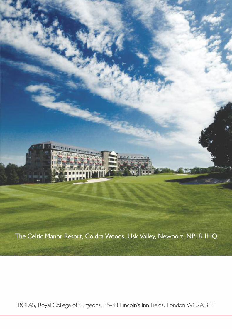
BOFAS, Royal College of Surgeons, 35-43 Lincoln’s Inn Fields. London WC2A 3PE
The Celtic Manor Resort, Coldra Woods, Usk Valley, Newport, NP18 1HQ