Embed Size (px)
Citation preview

Brit. J. Ophthal. (1953) 37, 1 1.
EXPERIMENTAL THIAMIN DEFICIENCY AS A CAUSEOF DEGENERATION IN THE VISUAL PATHWAY
OF THE RAT*BY
F. C. RODGERDepartment ofPhysiology, Medical School, University of Durham
THAT the fundamental alteration in the visual pathway in thiamin deficiencymight be biochemical and not cellular is an opinion that has been freelyexpressed; yet, as there is little doubt that such a deficiency leads to a wide-spread degeneration of the central vestibular neurones (Swank and Prados,1942), it seems reasonable enough to expect that the visual neurones mightbe similarly affected.
It is surprising, in view of the possible relationship between thiamin andnutritional amblyopia, to discover how little experimental work on thesubject has been performed. Most of the pioneers in this field were, ofcourse, under the impression that thiamin filled the role earlier assigned tothe hypothetical anti-beriberi vitamin. They believed wrongly that theywere inducing only a thiamin deficiency by feeding birds with polished rice.Optic nerve degeneration in experiments of this nature is described byBarletta (1932) and Marchesini and Papagno (1935).
Involvement of the visual pathway in animals on a diet deficient only inthe heat labile member of the B complex was noticed by Prickett (1934) inrats, and by Peters (1934) in pigeons.The blindness which they describe, however, was a subjective finding in
the terminal stages of an acute experiment. There might, then, be anexplanation for it other than a cellular degeneration of the visual neurones.When animals are severely deficient, they become no longer interested intheir surroundings, and need a deal of arousing, fatigue and lethargy beingdominant signs. For this reason, subjective investigation of an animal'sresponse to visual stimuli in the terminal stages would appear to be ofslight value. The rest of Peters' work, however, is of such a high order thatperhaps more stress has been laid upon this observation than was intended.The series of painstaking experiments on thiamin deficiency undertaken
by Swank and his various collaborators constitute the main argument infavour of thiamin being an antineuritic vitamin. In addition, Swank andPrados (1942), describe degeneration of the optic fibres within the centralnervous system in an avian thiamin deficiency, induced by administeringautoclaved yeast and a low protein diet. This seems an excellent paper,and most of the criticism it has aroused appears not to be justified.
* Received for publication July 14, 1952.
11
on 12 May 2018 by guest. P
rotected by copyright.http://bjo.bm
j.com/
Br J O
phthalmol: first published as 10.1136/bjo.37.1.11 on 1 January 1953. D
ownloaded from

Although primarily interested in the vestibular system, Swank and Pradosnoted that the central terminations of the optic fibres in the deficient birdswere either grossly thickened or fragmented; in the severe cases this wasassociated with sclerosis of the related cells within the optic lobes. Wecannot of course consider the optic lobe ofthe pigeon as being the homologueof the lateral geniculate body in man. Primitive geniculate bodies arealready present in teleosts, for example, which possess optic lobes in addition.One criticism levelled against Swank by Shaw and Phillips (1945) was that
food placed in the pigeon's crop is not always ultimately swallowed anddigested. More serious is the criticism that the diet lacked many aminoacids not required by mammals but essential to birds. Could absence ofthese be the causative factor in producing degeneration? Prados and Swank(1942) had the last word: they produced the same type of degeneration inmammals (kittens), but unfortunately their claims and conclusions are thereless clearly expressed.
Such, then, is the meagre positive evidence that exists.A somewhat larger number of workers interested in this problem, who
describe experiments in which they could find no significant degenerativechanges within the visual pathway in thiamin deficiency, includes Engel andPhillips (1938), McDermott and others (1943), and Leinfelder and Robbie(1947). One must mention, too, the positive opinions of Zimmerman (1943)whose brilliant work on canine nutritional neuropathies is well known toworkers in this field; there is no specific experiment of his own devoted toa study of the visual path, but he stated that he had never been convincedthat optic atrophy occurred experimentally in dogs, pigeons, or rats as aresult of thiamin deficiency.
This brief account of the relevant literature shows that the evidence isscanty and extremely conflicting; but the various discrepancies and dis-agreements proved helpful in planninSg the present experiments.
MethodsIn all our experiments, rats between 50-100 g. in weight were preferred.
The individual weights are given in detail below.The animals were weighed on the first day of the institution of the experimental
diet, at weekly intervals, and on the last day. On each occasion, the weighingtook place before feeding. At the same time, the opportunity was taken toexamine the eyes, skin, fur, gait, and posture. This was done in daylight on araised examination table. Towards the termination of each experiment a generalexamination was undertaken daily.The animals were housed iii individual cages fitted with wide wire mesh floors.
The cages were shaded, draught-free, and kept in a room of constant temperature(65-750 F.).The principle of paired feeding was adopted, the control animal in nearly all
cases being from the same litter.Force-feeding was not used. In the chronic deficiencies, we learned by trial
experiments how to prevent anorexia. In those few cases in which loss of appetite
12 F. C. RODGER
on 12 May 2018 by guest. P
rotected by copyright.http://bjo.bm
j.com/
Br J O
phthalmol: first published as 10.1136/bjo.37.1.11 on 1 January 1953. D
ownloaded from

EXPERIMENTAL THIAMIN DEFICIENCY
persisted, we feel paired feeding adequately covered the probable effects ofinanition. In our experience, anorexia is not such a striking symptom in chronicthiamin deficiency as it appears to be in a deficiency of the entire B complex(Drummond and Marrian, 1926). The appetite could be brought back by theadministration of as little as 10,ug. of thiamin hydrochloride. It rarely reacheda severe degree (i.e., less than 2/3 eaten) until the weight had fallen to its originallevel. Even then, by careful nursing, the animals could be made to eat as muchas 7.5-10.0 g./day per 100 g. body weight, which is far from starvation level in arat exhibiting a marked disinclination to exercise.
In the acutely deficient rats, so rapid was the onset of severe symptoms thatforce-feeding-a matter of great difficulty in rats-led to sudden death, and wastherefore abandoned. In such cases, having previously carefully investigated themorphological lesions resulting from inanition, it was a simple matter to take theminto account in the final assessment.The criterion taken as being most characteristic (in rats) of a chronic thiamin
deficiency was a reduction in heart rate. When this fell to the neighbourhood of300 beats/min. it was felt that the animals were deficient whether gross clinicalmanifestations were evident or not.The bradycardia of experimental thiamin deficiency is generally accepted as
being pathognomonic. It cannot be caused by inanition unless this is veryprolonged, which was never the case in our experiments. It was, moreover, neverfound in any of the control animals. Without altering the diet, the bradycardiacan be eliminated by administering 1 mg. intraperitoneal thiamin hydrochloride.If the rate fell below 200 beats/min., we were less successful in obtaining recovery.The cause of this bradycardia is not known. Drury, Harris, and Maudsley
(1930), though they produced in some rats a slight increase in heart rate by vagalsection, failed to restore it to the normal values. One must presume, therefore,that it is of sinus origin. Our method of recording the heart rates was essentiallythat described by Harris and his colleagues, and the procedure they described wasfollowed as carefully as possible. It appears that when the recordings are made understandard conditions, conclusive evidence may be obtained by comparing the figureswith those for the control rats, whose heart rates are recorded at the same time.
In acute thiamin deficiency, however, bradycardia could not be taken as anindex of the success of the experimental diet, for this appears in our experience todevelop only in the terminal stages, when the animals are moribund. The nervousdisorders characteristic of chronic deficiency become manifest sometimes before,usually after, the appearance of bradycardia. In acute deficiency, they alwayscoincided, and so, in the latter, it was the nervous symptoms which determinedthe termination of the experiment. In short, the acutely deficient rats were killedwhen they were seen to be dying, and the chronically deficient when bradycardiawas in the neighbourhood of 300 beats/min.On termination of the experiments, the rats were killed by decapitation under
urethane. The brains, optic nerves, and eyeballs were then removed and fixedwithin 10 minutes in 10 per cent. neutral formalin. Such speedy fixation isessential if autolytic distortions are to be prevented. Some twigs of sciatic nervetaken at random from each group were similarly treated. As the primary interestwas in the visual path, it was not possible to investigate each sciatic nerve inevery case in its entirety.
13
on 12 May 2018 by guest. P
rotected by copyright.http://bjo.bm
j.com/
Br J O
phthalmol: first published as 10.1136/bjo.37.1.11 on 1 January 1953. D
ownloaded from

The pathological alterations in the peripheral nerves in thiamin deficient animalsdo not appear to be as clear-cut as the clinical manifestations described above.(It is this finding which suggests that beriberi is not due solely to an absence ofthiamin). According to numerous authors (Prickett, 1934; Davison and Stone,1937; Engel and Phillips, 1938; Prickett, Salmon, and Schrader, 1939) examinationof the peripheral nerves of rats fed- on diets containing autoclaved yeast hasrevealed no significant morphological differences from those of control animals.The slight changes which occurred are ascribed to inanition. Berry, Neumann,and Hinsey (1945) placed cats on a thiamin-free diet for as long as 116 days butfound no trace of degeneration in the peripheral nerves, and the same conclusionwith regard to swine was reached by Follis and others (1943) and by Wintrobeand others (1944).
This fact is not, we feel, generally appreciated, but the evidence is too strong toignore. To have used peripheral nerve preparations as a means of controllingthe experiment, therefore, would appear somewhat pointless.
Constitution of Diets(1) Daily Caloric Requirements.-A rat of 75 g. will thrive on approximately
10 g. food daily; a rat of 150 g. requires 15 g., and rats above 200 g. vary in theirdemands. All rats in the experiment were less than 150 g. in weight, and 15 g.food was fed to each irrespective of weight. Thus, no rat, control or otherwise,was fed at too low a level. It was considered quite normal if less than 5 g. ofthe daily ration was left uneaten by rats of 75 g. or less.
(2) Composition of Diets.-The " stock " diet was made up as follows:(i) Casein ... ... ... ... 15(ii) Salt mixture ... ... ... 4(iii) Glucose ... ... ... ... 70 100(iv) Water soluble vitamins (W.S.V.) 10|(v) Fat soluble vitamins (F.S.V.) ... l
(3) Details of Dietary Components(i) Casein (lactic casein, unextracted)
Moisture ... ... ... ... 10Fat ... . ... ... .... 2.1Nitrogen ... ... ... ... 13.5 15Calcium ... . ... ... 0.04
It should be noted that the composition of the diet is based on dry weights, an allowancebeing made for the 10% of water present in casein.The quantity of protein is low, of course, as the proportion of carbohydrate is required
to be high. This precipitates thiamin deficiency (Yudkin, 1951). The protein is quitesufficient, however, for growth. It is vitamin-free.
(ii) Salt Mixture (g.)* Trace MixtureSodium chloride ... ... 22 Potassium iodide ... ... 12Calcium phosphate ... 130 Sodium fluoride ... ... 10Potassium citrate ... ... 125 Manganese sulphate ... 2Magnesium sulphate ... 30 Cuprous iodide ... ... 1Iron citrate ... ... 5 Potash alum ... ... 1Trace mixture ... ... 0.7 Zinc sulphate ... ... 1
312.7 27* This corresponds to DL6, a salt complex prepared by Glaxo Laboratories, Greenford, Middlesex, and was
purchased from them. In the initial planning of the diets, the helpful advice of Dr. W. P. J. Cuthbertson of thatsame laboratory is gratefully acknowledged.
14 F. C. RODGER
on 12 May 2018 by guest. P
rotected by copyright.http://bjo.bm
j.com/
Br J O
phthalmol: first published as 10.1136/bjo.37.1.11 on 1 January 1953. D
ownloaded from

EXPERIMENTAL THIAMIN DEFICIENCY .15
The manganese and magnesium proportions are higher than most workers prescribeto ensure good growth, and the normal excitability of the nervous system. There isnothing unusual in the quantities of the other salts present.
(iii) Glucose.-That incorporated in the diet was of the pure medicinal variety.
(iv) Water Soluble VitaminsCholine chloride ... ... ... ... 10.0 g.Inositol .. . .......... ... ...1.1 g.Nicotinic acid ... ... ... 0.5 g.Calcium-d-pantothenate ... ... ... 0.5 g.Pyridoxine hydrochloride ... ... ... 40.0 mg.d-Biotin .. .. ... ... ... 1.0 mg.Folic acid ... ... ... ... - 1.0 mg.Para-aminobenzoicacid ... ... ... 0.375 g.Distilled water to ... ... 250 ml.
Riboflavine, 150 mg., was then dissolved in the least amount of boiling 5 per cent.acetic acid, shaken in 50 ml. non-methylated 95 per cent. alcohol and added. Finally,distilled water brought the total volume up to 500 ml.The daily dose of W.S.V. was 1 ml. per rat.The absence of ascorbic acid from this complex is not an oversight. Rats need not
be given vitamin C, as they synthesize it readily in the intestine. On a diet deficient inthis factor considerable quantities are found in the livers, even after a long period(Coward, 1947).
If the relationship of riboflavine to thiamin is to be investigated, the former vitamincan be made up separately, the composition of W.S.V. being adjusted accordingly.Riboflavine itself is administered at the same level (150 mg. in 500 ml.). The daily doseper rat is 1 ml. as in the original W.S.V.The full maintenance dose of thiamin required by rats above 50 g. in weight is in the
region of 10 Ftg. a day. Woolley and White (1943) claim that mice require 2 ,ug.; Arnoldand Elvehjem (1938), that chicks require 10 ptg.; and Swank and Bessey (1941) thatpigeons require 20 Ftg.
Nevertheless, we administered thiamin at the extremely high level of 500 Lg. daily tothe control animals, giving 5 ,ug. only to the deficient, as follows:
(a) Full maintenance dose (thiamin full) (b) Minimal maintenance dose (thiamin minimal)Thiamin hydrochloride 250 mg. Thiamin hydrochloride ... 5 mg.Distilled water to 500 ml. Distilled water to ... 500 ml.Daily dose per rat 1 ml. (0.5 mg.) Dose, when directed, 0.5 ml. (5 ,ug.).
The detailed administration is given later with each experiment.
(v) Fat Soluble Vitaminsp-carotene (1.2 x 10⁶ i.U.) ... ... 5 mg.Calciferol (0.2 x 10⁶ i.u.) ... ... 5 mg.a-tocopherol acetate ... ... ... 2 mg.Arachis oil to ... ... ... 100 g.Daily dose per rat 0.1 ml. (about 3 drops).
Vitamin K (2 methyl-I, 4-naphtha-quinone) was not included, for no changes havebeen detected in the tissues of animals deficient in vitamin K, apart from the physiologicaldefects associated with haemorrhage.
Ferraro and Roizin (1943), in an interesting paper on vitamin K deficiency in rats,describe the presence of multiple haemorrhages in the brain. Best and Taylor (1950),however, maintain that it is not possible to induce a haemorrhagic tendency in ratsowing to the high degree of synthesis going on in the intestine.As the absence of vitamin K does not affect the central nervous system, it was omitted,
a practice in keeping with that of other workers.
on 12 May 2018 by guest. P
rotected by copyright.http://bjo.bm
j.com/
Br J O
phthalmol: first published as 10.1136/bjo.37.1.11 on 1 January 1953. D
ownloaded from

Histological TechnfquesThe eight different histological techniques employed are shown in Table I. A
full description with their inherent fallacies has been given elsewhere (Rodger,1951). It should be stressed that before the experiments here described wereperformed, a year was spent in becoming familiar with the anatomy of the ratvisual path, the histological methods of choice, and the appearance of the visualneurones in various known diseases.
TABLE I
HISTOLOGICAL TECHNIQUES
Part of Visual Path (1) (2) (3) (4) (5) (6) (7) (8)Nissi Giemsa Triple Polarized Marchi Weigert- Loye7 SilverLight Pal Nitrate
Retina ... ... X X X
{Axons ... X xOptic Nerve MyenX X X X
Optic Tract $Axons.. occasionally XPMyelin occasionally X X X
Lateral Cells... X XGeniculate Processes XBody Myelin X
The rat visual pathway is easily dissected. The cerebral hemispheres areremoved by hand. Then, splitting the chiasma, and laying the brain on its lateralside, one may excise the optic nerve, half-chiasma, tract, and lateral geniculatebody in one piece. Portions of the thalamus and cerebral peduncle are included.The visual fibres leave the tract to synapse in the dorsal nucleus of the lateralgeniculate body. By this dissection, an opportunity is afforded of studying thenerve, tract, and dorsal nucleus in the same section. The section may be describedas representing the anteroposterior and dorsoventral diameters, and as it ispossible to examine the entire length of the nucleus by serial sections, observationsmay be also made in its medio-lateral diameter.
Plan of ExperimentsIn several trial experiments, it was found that the onset of nervous
manifestations of acute deficiency began very suddenly-within 24 hrs or soof the appearance of anorexia and bradycardia. We wondered, therefore,if these manifestations might not have resulted from inanition. Further-more, if anatomical damage had caused the nervous disorders, it might bethat this, too, depended on caloric inadequacy.
These possibilities, unlikely though they seemed, and the further desire todiscover whether the nervous symptoms were due to lack of calories orlack of vitamins, led to the framing of the first two of the six followingexperiments:
(1) Caloric inadequacy without exogenous vitamins.(2) Caloric inadequacy associated with full vitamin intake.
16. F. C. RODGER
on 12 May 2018 by guest. P
rotected by copyright.http://bjo.bm
j.com/
Br J O
phthalmol: first published as 10.1136/bjo.37.1.11 on 1 January 1953. D
ownloaded from

EXPERIMENTAL THIAMIN DEFICIENCY
(3) Acute deficiency in thiamin due to administration of the analogue.(4) The same, recovered.(5) Reduced thiamin intake associated with a high carbohydrate diet, inducing a
chronic deficiency.(6) The same, recovered.
Results(1) Caloric Inadequacy without Exogenous Vitamins
Diet.-Potato starch ... ... ... 70Wheat straw pulp ... ... ... 20 100Agar-sucrose ... ... ... 10
Clinical Behaviour.-On this diet, fed ad libitum, six rats (average weight200 g.) died at the end of 11 days. They were perfectly healthy until themorning of the 10th day when they were seen to be very inactive, and to haveassumed a crouching position. They could be roused only with difficulty,their movements were inco-ordinated,and their postural sense appeareddiminished. Next morning they hadviolent convulsions while on the exam-ination table and within half an hourwere dead.Pathological Observations.-Vacuo-
lation of the ganglion cells of theretina and geniculate body was theonly cytological change found. Asvacuolation of the retinal cells is avery common artefact, more commonthan swelling, we are inclined to con-sider that this is the explanation of thefinding.The myelin sheaths viewed by polar-
ized light were hazy in outline and theirsubstance somewhat granular (Fig. 1), myelin sheaths in calotrocpismdof optic-tractand this was also true of the sciatic out exogenous vitamins. Polarized lighttwigs, but Loyez and Marchi stain x 250.revealed no abnormality. No sign of any degeneration involving the nervefibres was found.
(2) Caloric Inadequacy associated with Full Vitamin IntakeDiet.-
Potato starch ... ... ... 70Wheat straw pulp ... ... ... 20 100Agar-sucrose ... ... ... 10W.S.V. (less thiamin) ... ... 6 ml.
Clinical Behaviour.-Six rats of average weight 160 g. and members ofthe same litter were fed the usual 15 g. daily for 24 hrs. They were thendivided into two groups:
17
on 12 May 2018 by guest. P
rotected by copyright.http://bjo.bm
j.com/
Br J O
phthalmol: first published as 10.1136/bjo.37.1.11 on 1 January 1953. D
ownloaded from

(A) remained on the same diet(B) received 2.0 mg. thiamin (i.p.) daily in addition.
On the 18th day, the three rats in Group A and one of the rats in Group Bdied. Two days later the remaining two rats in Group B, now moribund,were killed. The manner of death in those permitted to die was exactly asdescribed in Experiment 1.
Pathological Observations.-There were no essential differences betweenthe two groups, A and B.
In the retina, the outline of many of the cells was indistinct, the cytoplasmstaining homogeneously, and the nucleus containing dark-stained granules.The geniculate cells exhibited signs of severe somatic disorder. The
Nissl bodies in many cells were clotted and darkly stained. The cell outlineswere frequently badly distorted. Some of the astrocytes were swollen, andthere were many hypertrophied cerebral histiocytes (rod cells) with dark-staining nuclei. Long oligodendrocytic chains lay in the fibre pathways.The nerve fibres themselves were tortuous, but not otherwise distorted.
The endings were unaffected.The myelin sheaths revealed more positive changes than in the previous
series, but, in view of the advancedacellular damage present, these changeswere much less than we expected. There was a similar slight degree ofdemyelinization in the sciatic nerves, revealing itself in polarized light in bothinstances as a granular isotropism.(3) Acute Deficiency in Thiamin due to Administration of the Analogue
Diet.-Stock (Glucose + W.S.V.) ... 15 g.
*Pyrithiamin ... ... ... ... 100 ,tg.Thiamin minimal ... ... ... 5,ug.
Clinical Behaviour.-The weights and heart rates are shown in Table II.TABLE II
EXPERIMENT 3 (DURATION 17 DAYS)
Weights (g.) Heart Rates (beats/min.)Rat No. Disposal
Initial Final Difference Initial Final Difference
1 90 80 -10 500 250 --250 Killed2 80 87 +7 500 240 -260 Killed3 75 95 +20 510 310 -200 Killed4 75 75 0 500 - Killed5 75 75 0 500 - - Killed6 85 90 +5 500 300 -200 Killed7 80 77 -3 500 Killed8 117 100 -17 400 200 -240 Died during recording9 100 102 +2 480 300 -180 Killed10 85 88 + 3 500 - Killed11 92 96 +4 490 Killed12 90 89 -1 460 - Killed
* As this was not available in Great Britain at the time, this was generously supplied by the Research Department,Merck and Company, Rahway, New Jers y. It was prepared in their laboratories, and renamed Neopynithiamine,and we found it most effective.
18 F. C. RODGER
on 12 May 2018 by guest. P
rotected by copyright.http://bjo.bm
j.com/
Br J O
phthalmol: first published as 10.1136/bjo.37.1.11 on 1 January 1953. D
ownloaded from

EXPERIMENTAL THIAMIN DEFICIENCY
On the 17th day all the animals were found to exhibit the same clinicalpicture. When they were picked up by the tail a convulsion appeared, and,on recovery, spasticity of the hind legs was marked, and occasionally theanimal fell over sideways in spasm. Both sides seemed equally affected.Some of the animals were ultimately (minutes) unable to move, assumingwhat later proved to be a characteristic pose, lying flat on their abdomenswith their limbs extended outwards. Spontaneous convulsions appeared intwo of them. Anorexia was first seen about 36 hrs before death.
Pathological Observations.-There was cloudy swelling of the retinalbipolar and ganglion cells (Fig. 2). This is of doubtful significance.The optic nerve fibres were slightly more tortuous than normal.There were no other signs of degeneration in retinae, nerve fibres, myelin
sheaths, or geniculate bodies.A few of the twigs of the sciatic nerve revealed marked degeneration of
myelin (Fig. 3), except in Rats 3 and 5, where no abnormality was discoveredin the twigs removed for examination.
FIG. 3. -Gross destruction of myelinFIG. 2.-Cloudy swelling of the retinal bipolar in main branch of sciatic nerve incells in acute thiamin deficiency. Triple x 1600. acute thiamin deficiency. A portion
of an unaffected twig can be seen onthe right. Marchi x 140.
(4) As in Experiment 3, RecoveredDiet.-As in Experiment 3.Clinical Behaviour.-Weights and heart rates are shown in Table III.
TABLE IIIEXPERIMENT 4 (DURATION 60 DAYS)
Weights (g.) Heart Rates (beats/min.)Rat NO. -DiSPOSal
Initial At 15 days Final Initial At 15 days Final
1 130 120 165 440 250 440 Killed2 110 111 148 480 300 460 Killed3 165 185 235 440 360 420 Killed4 110 107 500 200 Died 13th day5 120 115 155 480 240 480 Killed6 135 121 460 - - Died
19
on 12 May 2018 by guest. P
rotected by copyright.http://bjo.bm
j.com/
Br J O
phthalmol: first published as 10.1136/bjo.37.1.11 on 1 January 1953. D
ownloaded from
F. C. RODGER
Rat 4 died on the 13th day. Rat 6 died suddenly during the recordings.The others were all spastic and inco-ordinated on the 15th day when theirdiet was restored. Rats 1 and 5 were given intraperitoneal thiamin as theirheart rates were so low, and this was repeated for 3 days; otherwise theymight not have lived. The four survivors (Rats 1, 2, 3, and 5) were killedon the 60th day.
Pathological Observations.-No signs of degeneration or repair in the visualpathway nor sciatic nerves were seen in the recovered rats.
(5) Reduced Thiamin Intake associated with High Carbohydrate Diet,inducing Chronic Deficiency
Diet.-Stock (Glucose + W.S.V.)Thiamin minimal ... ... as required
The adoption of a preconceived plan for the administration of thiamin tothe group was rejected. We decided each animal required individualattention. A daily dose of 2 ug. was, therefore, administered individuallyuntil the first signs of anorexia. The dose was then increased in that ratto 5 ,Ag. until the appetite returned, when 2 ,1g. was once again given,ad infinitum.
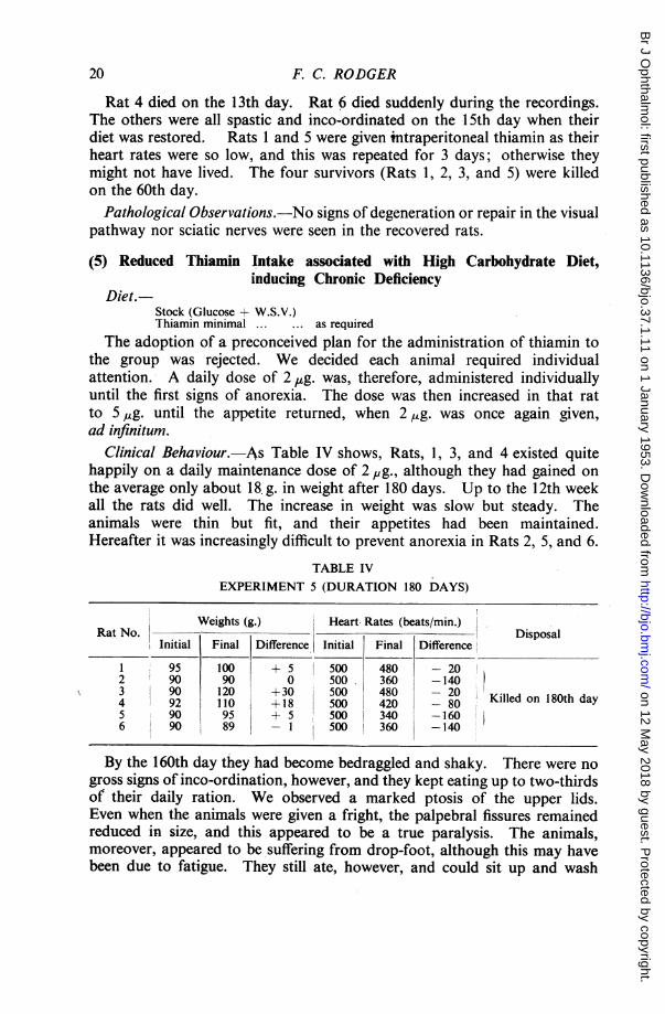
Clinical Behaviour.-As Table IV shows, Rats, 1, 3, and 4 existed quitehappily on a daily maintenance dose of 2 p,g., although they had gained onthe average only about 18. g. in weight after 180 days. Up to the 12th weekall the rats did well. The increase in weight was slow but steady. Theanimals were thin but fit, and their appetites had been maintained.Hereafter it was increasingly difficult to prevent anorexia in Rats 2, 5, and 6.
TABLE IVEXPERIMENT 5 (DURATION 180 DAYS)
Weights (g.) Heart Rates (beats/min.)Rat No. - Disposal
Initial Final Difference. Initial Final Difference
1 95 100 + 55 500 480 - 202 90 90 0 500. 360 -1403 90 120 +30 500 480 204 92 110 +18 500 420 - 80 Killed on 180th day5 90 95 + 5 500 340 -1606 90 89 1 500 360 -140
By the 160th day they had become bedraggled and shaky. There were nogross signs of inco-ordination, however, and they kept eating up to two-thirdsof their daily ration. We observed a marked ptosis of the upper lids.Even when the animals were given a fright, the palpebral fissures remainedreduced in size, and this appeared to be a true paralysis. The animals,moreover, appeared to be suffering from drop-foot, although this may havebeen due to fatigue. They still ate, however, and could sit up and wash
20
on 12 May 2018 by guest. P
rotected by copyright.http://bjo.bm
j.com/
Br J O
phthalmol: first published as 10.1136/bjo.37.1.11 on 1 January 1953. D
ownloaded from

EXPERIMENTAL THIAMIN DEFICIENCY
themselves. We observed one of them regurgitate while feeding on twooccasions but vomitus was never seen. When the heart rates fell below400 beats/min. (restrained), it was decided to end the experiment.The continued good health of the other three rats was. considered to be
due to intestinal resynthesis.
Pathological Observations.-Rats, 1, 3,
FIG. 4.-Fragmentation and tortuosity of optic-nerveterminals within dorsal nucleus in chronic thiamindeficiency. The larger fibres in the section are notfragmented. Author's silver x 670.
FIG. 5.-Enlargement of terminal nerve endings inchronic thiamin deficiency. Some argyrophil ringshapes are also present. Author's silver x 1000.
and 4 revealed no abnormality;Rats 2, 5, and 6 each exhibitedidentical changes.The retinal ganglion cells were
hyperchromatic. That was theonly change.The optic nerve and tract re-
vealed widespread signs of de-generation increasing in inten-sity as the fibres ascended: thenerve fibres were thickened,distorted, and very tortuous(corkscrew fibres). The terminalbranches within the lateralgeniculate body were similarlyafTected; and in addition thelarger ones had fragmented(Fig. 4). There were many smallblack patches, which rpight have
FIG. 6.-Final visual neurone within dorsalnucleus in chronic thiamin deficiency. Axongreatly swollen. Author's silver x 1000.
been distorted ring shapes. We found a few endings enlarged (Fig. 5).The fibres of the third neurone as far as we could see were varicose andthickened (Fig. 6).
21
on 12 May 2018 by guest. P
rotected by copyright.http://bjo.bm
j.com/
Br J O
phthalmol: first published as 10.1136/bjo.37.1.11 on 1 January 1953. D
ownloaded from

22 F. C. RODGER
if,A..
* A.
:. , 4s ... t +
..v#<#*
f; ifS Eb zF* 3Ir.
e. .24*~ ~
FIG. 7.-Sclerosis and hyperchromatismof visual neurones in chronic thiamindeficiency. Cf. Fig. 8. Giemsa x 175.
N.
FIG. 8.-Unaffected visual neurones ofdorsal nucleus in paired control animal.Giemsa x 175.
*.
*-!W f ..:~:
4:
FIG. 9.-Aggregation of oligodendro-cytes in chronic thiamin deficiency.Giemsa x 300.
A.
..W.
:.:. .:;. °.. -....... 2,4
a,....
A
FiG. 10.-Sclerosis and hyperchromatism of visualneurones in chronic thiamin deficiency. Notepresence of rod cells and cloudy swelling of astro-cytes. Giemsa x 750.
~~~'C 'i '^...:e ...R FIG. 11.-Fragmentation, globula-tion, and ballooning of persistentmyelin sheaths in chronic thiamindeficiency. Weigert-Pal x 350.
::
*. : x,.w.' :i .........l. p
i. .ep::
:.v
11l ....... on 12 May 2018 by guest. P
rotected by copyright.http://bjo.bm
j.com/
Br J O
phthalmol: first published as 10.1136/bjo.37.1.11 on 1 January 1953. D
ownloaded from
EXPERIMENTAL THIAMIN DEFICIENCY
Within the dorsal nucleus, the visual cells were nearly all hyperchromaticand sclerosed. The degree of sclerosis was not gross, but it was definite.Both nuclear and cellular outlines were distorted (Figs 7 and 8).Within the optic nerve and tract, the oligodendrocytes had proliferated
considerably into long chains (Fig. 9). These cells were of normal size,but the small amount of cytoplasm present was hyperchromatic. We didnot find any evidence of satellitosis, but many rod cells were seen withinthe dorsal nucleus (Fig. 10).These changes were widespread.The myelin changes, although variable, were diffuse. In places the
myelin had completely disappeared, many gitter cells being present. Wherethe myelin sheaths persisted, irregularity and ballooning was common(Fig. 11), and this was also true of some of the twigs of the sciatic nerves.
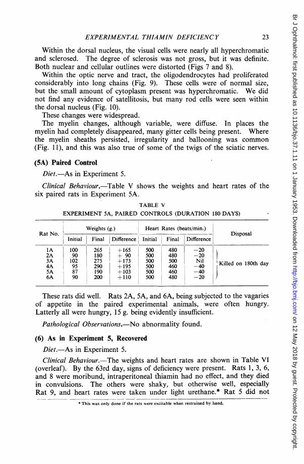
(5A) Paired ControlDiet.-As in Experiment 5.
Clinical Behaviour.-Table V shows the weights and heart rates of thesix paired rats in Experiment 5A.
TABLE VEXPERIMENT 5A, PAIRED CONTROLS (DURATION 180 DAYS)
Weights (g.) Heart Rates (beats/min.)Rat No. _ Disposal
Initial Final Difference Initial Final Difference
1A 100 265 +165 500 480 -202A 90 180 + 90 500 480 -203A 102 275 +173 500 460 Nil Killed on 180th day
5A 87 190 +103 500 460 -406A 90 200 +110 500 480 -20
These rats did well. Rats 2A, 5A, and 6A, being subjected to the vagariesof appetite in the paired experimental animals, were often hungry.Latterly all were hungry, 15 g. being evidently insufficient.
Pathological Observations.-No abnormality found.
(6) As in Experiment 5, RecoveredDiet.-As in Experiment 5.Clinical Behaviour.-The weights and heart rates are shown in Table VI
(overleaf). By the 63rd day, signs of deficiency were present. Rats 1, 3, 6,and 8 were moribund, intraperitoneal thiamin had no effect, and they diedin convulsions. The others were shaky, but otherwise well, especiallyRat 9, and heart rates were taken under light urethane.* Rat 5 did not
* This was only done if the rats were excitable when restrained by hand.
23
on 12 May 2018 by guest. P
rotected by copyright.http://bjo.bm
j.com/
Br J O
phthalmol: first published as 10.1136/bjo.37.1.11 on 1 January 1953. D
ownloaded from

TABLE VI
EXPERIMENT 6 (DURATION 140 DAYS)
Weights (g.) Heart Rates (beats/min.)Rat No. Disposal
Initial At 14th week Final Initial At 14th week
1 50 80 550 Died 63rd day2 115 110 160 480 340 Killed 140th day3 50 60 - 550 - Died 63rd day4 55 85 100 500 320 Killed 90th day5 55 92 500 360 Died during recording6 55 75 500 Died 63rd day7 70 60 235 500 380 Killed 140th day8 55 70 550 Died 63rd day9 72 125 255 480 390 Killed 140th day
regain consciousness. Rat 2 was spastic next day, and all the survivors(including Rat 9, which was still very active) were given intraperitonealthiamin, and a full diet was instituted. They made an uninterruptedrecovery, gaining weight rapidly. Anorexia in this experiment had beenremarkably well controlled so that the paired rats (Exp. 6A) seldom had tohave their intake reduced. Rat 4 was killed on the 90th day, and the reston the 140th. There was no residual palsy.
Pathological Observations.-Changes were found in Rats 2, 4, and 7.The visual path and eyeballs of Rat 9 were normal.The changes in the retinae, which were not striking, consisted ofthe presence
of what appeared to be " gemastete cells ". These cells approached in sizesclerosed ganglion cells, and might have been mistaken for them. Thenumber of them present in 7,. sections outside the macular region was,nevertheless, suggestive. The ganglion cells themselves were on the wholenormal, although satellitosis around them was common. The other retinalelements were unaffected.The visual cells in the dorsal geniculate nuclei were almost entirely normal;
here and there a few were sclerosed, and there was widespread hyper-chromatism of the glia, and some evidence of satellitosis (Figs 12 and 13).Many long oligodendrocyte chains were seen in the optic tract of Rat 2.Changes in the nerve fibres were of an equally slight nature. A moderate
degree of distortion appeared in the tract of Rat 2.The nerves and tracts, however, revealed a high degree of patchy
demyelinization, which was also present in the nerve head of Rat 7 (Fig. 14).None was found in Rat 4. Several hard grey plaques were found (Fig. 15).The sciatic nerves showed circumferential degeneration involving some of
the nerve trunks (Fig. 16). In others, there was distortion of the myelinsheaths when viewed transversely. Some sheaths exhibited ballooning.
24 F. C. RODGER
on 12 May 2018 by guest. P
rotected by copyright.http://bjo.bm
j.com/
Br J O
phthalmol: first published as 10.1136/bjo.37.1.11 on 1 January 1953. D
ownloaded from

EXPERIMENTAL THIAMIN DEFICIENC Y9 iw :.t.,
b
FIG. 12.-Hyperchromatic visual cell-bodies, hyperchromatism of glia, andslight evidence of satellitosis in chronicthiamin deficiency 57 days after recov-
ery. Giemsa x 220.
FIG. 13.-Satellitosis in recovered thiamindeficiency. Giemsa x 450.
iA
FIG. 14.-Central demyelinization of optic nerve in chronicthiamin deficiency. Loyez x 200.
FIG. 15. Hard grey plaque in optictract in recovered chronic thiamindeficiency. Note clear delimitationof healthy sheaths above this region.Loye7 x 150
FIG. 16.-Persistent demyelinization ofperipherally placed fibres in a large sciaticnerve twig in recovered chronic thiamindeficiency. Weigert-Pal x 140.
r>....e.
S, *
D
0
c.. 4
o:*1
....
ak
1.011k. ..
*.. 0a....^..
Eh. ...
.Ma 4
'U
IL
25
0
m
on 12 May 2018 by guest. P
rotected by copyright.http://bjo.bm
j.com/
Br J O
phthalmol: first published as 10.1136/bjo.37.1.11 on 1 January 1953. D
ownloaded from

(6A) Paired Control
Diet.-As in Experiment 6.
Clinical Behaviour.-Weights and heart rates are shown in Table VII.As anorexia in the experimental rats had been well controlled, these rats
flourished, seldom appearing hungry.TABLE VII
EXPERIMENT 6A, PAIRED CONTROLS (DURATION 140 DAYS)
Weights (g.) Heart Rates (beats/min.)Rat No. Disposal
Initial Final Initial Final
2A 110 220 480 4404A 55 190 500 480 Killed on 140th day7A 60 188 500 4809A 68 200 500 460
Pathological Observations.-No abnormality found.
DiscussionThe results of these experiments show that optic and sciatic nerve atrophy
can be produced in rats fed a diet high in carbohydrate at a level of thiamininadequate for health, but sufficient to keep the animals alive over a periodof about 180 days.The degeneration found in the visual pathways of rats fed under such
circumstances consisted of a slight degree of hyperchromatism and sclerosisof the retinal ganglion cells, and in more marked degree of the cells in thedorsal nucleus of the lateral geniculate body. In addition there was astriking proliferation within the optic nerve and tract of oligodendrocyteswhich formed long interfascicular chains; rod cells were present in greatabundance in the dorsal nucleus; and here and there in the same regionneuroglial hyperchromatism. The most important sign of all was thetortuosity, varicosity, and fragmentation of the visual fibres, especially oftheir central terminations. Enlargement of synaptic boutons and an increasein argyrophil ring shapes were also seen. The myelin sheaths in their turnexhibited a patchy demyelinization, several soft plaques were observed,, and,in many of the persistent sheaths, irregularity of outline and ballooning.
These changes, indicative of severe neuronal degeneration, were found tobe reversible with the following exceptions. The proliferation of the gliapersisted 76 days after re-institution of a full diet, as did the glial hyper-chromatism; the nerve fibres remained more tortuous than in the pairedanimals; the myelin lesions were for the greater part unchanged. A newfeature in the recovered animals was the appearance of several hard greyplaques.The paired control rats in the chronic thiamin deficiency experiment,
26 F. C. RODGER
on 12 May 2018 by guest. P
rotected by copyright.http://bjo.bm
j.com/
Br J O
phthalmol: first published as 10.1136/bjo.37.1.11 on 1 January 1953. D
ownloaded from
EXPERIMENTAL THIAMIN DEFICIENCY
having a high intake of thiamin, never presented this picture. They wereunder close observation throughout; their behaviour remained perfectlynormal, and their appetites were excellent, although at times they wenthungry. Subsequently, as has been said, the visual pathways revealed noabnormality whatsoever. It seems certain, then, that the histological changesin the central and peripheral nervous systems described above arose as aresult of thiamin deficiency, and for no other reason. At any rate,anorexia had never been gross or prolonged in the deficient animals. Thecell bodies within the central nervous system were distorted in those ratswhich were made to die of inanition at the end of 18 days, but theperipheral processes were never affected. This, it is believed, is a highlysignificant observation, yet, in the paired controls, not even this changewas found.The changes found in the sciatic nerves in chronic thiamin deficiency were
striking, but should be stressed again that they were not examined in everycase, nor was every twig sectioned in those nerves chosen. It is certain,however, that here as in the optic nerve demyelinization of a gross natureresulted. The interesting feature is that the degenerative process appearedto affect some twigs and not others in the same nerve, and that it was theperipheral fibres in a twig which were affected first. This perhaps explainsthe discrepancies expressed in the literature. These myelin changes werefor the greater part irreversible. For the same reasons as are given above,inanition could not b- responsible.
In acute thiamin deficiency, inanition was gross in the last day or two oflife, yet the visual pathway was not affected other than by a slight cloudyswelling of the retinal bipolars. There was no fragmentation nordemyelinization of the visual fibres, nor was there sclerosis of the geniculateneurones. The marked demyelinization found in the sciatic nerves mayhave been due to this terminal inanition, but the evidence does not altogethersupport this view. In the two 'inanition' experiments, the sciatic nervechanges were not marked. Under the polarizing microscope, there was somegranular isotropism of the myelin, but no more than that. Nevertheless,since the rats used were older, one hesitates to be dogmatic in claiming thatacute thiamin deficiency leads to sciatic nerve atrophy, though it certainlyappears to accentuate it, in the presence of inanition.
It was interesting to find that in acute deficiency, the gross myelin changesfound in the sciatic nerves were recoverable, whereas in chronic deficiency,the less gross changes were not recoverable. It is possible that the degreeof myelin-recovery depends -upon the integrity of the nerve cells and theirprocesses, for in acute deficiency (by analogy with the state of the visualneurones) the latter were intact, whereas in the chronic condition there werealways residual signs of cell-body disease. On these grounds it seemsprobable that as a general rule the integrity of myelin depends upon theintegrity of the cell-body, although the reverse is obviously not true. Thisbelief has been expressed by several workers in the past.
27
on 12 May 2018 by guest. P
rotected by copyright.http://bjo.bm
j.com/
Br J O
phthalmol: first published as 10.1136/bjo.37.1.11 on 1 January 1953. D
ownloaded from

To return to the visual neurones, just as different rats appear to be moresusceptible, so some fibres seem to be more readily affected than others.As we do not know the pathway taken by the macular fibres in the rat,however, we could not say, even if only the central fibres had degenerated,that the papillo-macular bundle was more susceptible than any other.
It is important to note that the lesions produced in myelin by a thiamindeficiency of long duration were disseminated throughout the visual pathway,and increased in severity as they ascended. This fact lends support to ourbelief that the retinal ganglion cells are not greatly affected, a somewhatparadoxical finding in view of their extremely low resistance to chemicalpoisons, and the generally held opinion that the causative agent in thiamindeficiency is an endogenous toxin, specifically affecting the macula.The appearance of the retina in the chronic experiments, however, suggests
that, if the thiamin deficiency had been even more prolonged, the retinalganglion cells would have become just as much affected in the end as werethe visual cells of the diencephalon. Perhaps the explanation of the longretinal immunity is to be found in the extremely active drainage afforded bythe choriocapillaris.Changes affecting the retinal pigment or the blood vessels were not found
in any specimen. Furthermore, there was a total lack within the visual pathof any sign of a haemorrhagic crisis, although the Giemsa technique isparticularly suited for such observations. Although the floor of the IVthventricle was not investigated and this is the preferential site for suchhaemorrhages in the rat, we seem to have been justified in leaving the rat tomanufacture its own vitamin K.The endogenous supply of vitamin C was also adequate for there were
no signs of scurvy.
Summary(1) When thiamin deficiency in rats is of moderate degree and prolonged
for about 180 days or over, degeneration of the visual pathway develops incertain susceptible animals, its intensity being greatest centrally. Thisoccurs even when the caloric intake is adequate.
(2) A chronic degeneration of this type comprises three distinctive features:(a) The visual cells become sclerosed and hyperchromatic, the retinal
much later than the geniculate.(b) The axis cylinders become thickened, tortuous, and varicose, the
terminal branches fragmented, and the nerve endings enlarged. Ring shapesincrease.
(c) Multiple foci of demyelinization occur, to be replaced in time byclearly delimited glial plaques.
(d) Long chains of oligodendrocytes form, and rod cells appear in thelateral geniculate body.
28 F. C. RODGER
on 12 May 2018 by guest. P
rotected by copyright.http://bjo.bm
j.com/
Br J O
phthalmol: first published as 10.1136/bjo.37.1.11 on 1 January 1953. D
ownloaded from

EXPERIMENTAL THIAMIN DEFICIENCY
(3) The chronic deficiency is also associated with demyelinization of someof the twigs of the sciatic nerves, affecting particularly the peripheral fibres.
(4) These changes are reversible only when the cell-body and its processhas not been unduly damaged. Myelin-sheath recovery is probablydependent upon complete recovery of the soma.
(5) In acute thiamin deficiency, the visual pathway is not affected-, but thesciatic nerves become grossly demyelinated. This demyelinization iscompletely reversible. It is probably associated with inanition.
(6) Demyelinization of the sciatic nerves also occurs in rats fed a diettotally inadequate in calories with or without vitamins, but in this case ismuch less gross than when combined with acute thiamin deficiency.My thanks are due to the Research Committee of the Medical School, King's College,
Newcastle, for a grant which enabled me to prosecute this work.
REFERENCESARNOLD, A., and ELVEHJEM, C. A. (1938). J. Nutr., 15, 403.BARLETTA, V. (1932). Rass. ital. Ottal., 1, 210.BERRY, C., NEUMANN, C., and HINSEY, J. C. (1945). J. Neurophysiol., 8, 315.BEST, C. H., and TAYLOR, N. B. (1950). " The Physiological Basis of Medical Practice ', 5th ed.
Bailli6re, Tindall and Cox, London.COWARD, K. H. (1947). " The Biological Standardisation of the vitamins ", 2nd ed. Bailliere,
Tindall and Cox, London.DAVISON, C., and STONE, L. (1937). Arch. Path., Chicago, 23, 207.DRUMMOND, J. C., and MARRIAN, G. F. (1926). Biochem. J., 20, 1229.DRURY, A. N., HARRIS, L. J., and MAUDSLEY, C. (1930). Ibid., 24, 1632.ENGEL, R. W., and PHILLIPS, P. H. (1938). J. Nutr., 16, 585.FERRARO, A., and ROIZIN, L. (1943). J. Neuropath., 2, 392.FOLLIS, R. H., MILLER, M. H., WINTROBE, M. M., and STEIN, H. J. (1943). Amer. J. Path., 19, 341.LEINFELDER, P. J., and ROBBIE, W. A. (1947). Amer. J. Ophthal., 30, 1135.McDERMOTT, W., WEBSTER, B., BAKER, R., LOCKHART, J., and ToMPSETr, R. (1943). J.
Pharmacol., 77, 24.MARCHESINI, E., and PAPAGNO, M. (1935). Ann. Ottal., 63, 81.PETERS, R. A. (1934). Proc. roy. Soc. Med., 27, 478.PRADOS, M., and SWANK, R. L. (1942). Arch. Neurol. Psychiat., 47, 626.PRICKETr, C. O. (1934). Amer. J. Physiol., 107, 459.
SALMON, W. D., and SCHRADER, G. A. (1939). Amer. J. Path., 15, 251.RODGER, F. C. (1951). " The Relationship of Thiamin to the Visual Pathway . Thesis.
Glasgow University.SHAW, J. H., and PHILLIPS, P. H. (1945). J. Nutr., 29, 113.SWANK, R. L., and BESSEY, 0. A. (1941). Ibid., 22, 77.
and PRADOS, M. (1942). Arch. Neurol. Psychiat., Chicago, 47, 97.WINTROBE, M. M., FOLLIS, R. H., HUMPHREYS, S., STEIN, H., and LAURITSEN, M. (1944).
J. Nutr., 28, 283.WOOLLEY, D. W., and WHITE, A. G. C. (1943). J. biol. Chem., 149, 285.YUDKIN, J. (1951). Biochem. J., 48, 608.ZIMMERMAN, H. M. (1943). Res. Publ. Ass. nerv. ment. Dis., 22, 51 and 75.
29
on 12 May 2018 by guest. P
rotected by copyright.http://bjo.bm
j.com/
Br J O
phthalmol: first published as 10.1136/bjo.37.1.11 on 1 January 1953. D
ownloaded from