Embed Size (px)
Citation preview
Topics in Catalysis Vol. 15, No. 2–4, 2001 201
Bridging the pressure and materials gaps between catalysis andsurface science: clean and modified oxide surfaces
H.-J. Freund, H. Kuhlenbeck, J. Libuda, G. Rupprechter, M. Bäumer and H. Hamann
Fritz-Haber-Institut der Max-Planck-Gesellschaft, Faradayweg 4-6, D-14195 Berlin, Germany
The preparation of model systems based on thin epitaxial oxide films and oxide single crystals is discussed. A variety of surfacesensitive techniques has been applied to study the geometric and electronic properties of these systems. The findings are correlated withadsorption and reaction of probe molecules on the surfaces. Metal vapor deposition under controlled conditions leads to the formationof metal aggregates with narrow size distributions. Their properties have been characterized, establishing that we can begin to bridge thematerials gap between catalysis and surface science. While mainly performed under UHV conditions, adsorption measurements can bepushed to ambient conditions using non-linear optical techniques such as sum frequency generation. Results for systems with depositedmetal aggregates will be discussed.
KEY WORDS: model catalysts; oxide surfaces; metal clusters on oxide surfaces; pressure gap; materials gap
1. Introduction
Catalysts are rather complex materials. Their surfacestructure is very difficult to study, in particular under work-ing conditions, i.e., ambient or elevated gas pressure. Un-derstanding catalysis at the atomic level is a formidable task.Whether it is possible at all has still to be shown. Surface sci-ence has to a large extent been driven by catalysis. With thedevelopment of a whole arsenal of surface analytical tools,the past 35 years of surface science have seen significantprogress, although the final goal of understanding catalysisat the atomic level has, of course, not been attained [1]. Sev-eral gaps between catalysis and traditional surface sciencehave been identified:
(1) the materials gap,
(2) the pressure gap,
(3) the complexity gap.
Surface science has reached a degree of maturity that al-lows us to bridge these gaps in part [2,3]. Using model sys-tems with increasing degree of complexity [4] or even modelcatalysts is one strategy to try to bridge the materials gaps.Closing the pressure gap can be achieved by virtue of surfacesensitive techniques that work in the presence of a gas phase,such as TDS, IRAS, ESR, SFG, STM, X-ray scattering andX-ray absorption [5]. Closing the complexity gap has to in-volve further methods to study gas and mass transport.
In order to start bridging the materials gap, several groupshave turned towards the study of oxide surfaces. In the fol-lowing we will address several examples, looking at the re-activity of molecules, in particular oxygen, on oxide surfacesas well as at structure–reactivity relations in the systems. Wethen move one step further to modify oxide surfaces with de-posited metal aggregates and study adsorption and reactionon such systems under UHV conditions. Then techniques
are applied for the study of such complex systems that al-low us to look at adsorption under higher gas pressures in anattempt to bridge the pressure gap [6–8].
2. Oxide surfaces
The preparation of a clean oxide surface in ultrahigh vac-uum is a rather difficult task. Strictly speaking, a certainoxygen activity is necessary in the gas phase to establishtrue equilibrium and then the stoichiometry is defined ac-cording to the chosen conditions [9]. In this respect, the ox-ide stoichiometry is not well-defined under dynamical UHVconditions, and the system is only kinetically stabilized. Itis therefore believed that defects determine the physical andchemical properties of oxide surfaces. Particularly interest-ing are vanadium oxides and vanadylpyrophosphate com-pounds [10,11]. Activation of hydrocarbons is thought totake place through abstraction of hydrogen atoms and theformation of surface hydroxyl groups involving defects andisolated transition metal oxide cluster sites [12]. Conceptualstudies in this area have been pioneered by Grasselli and hiscollaborators [10–12].
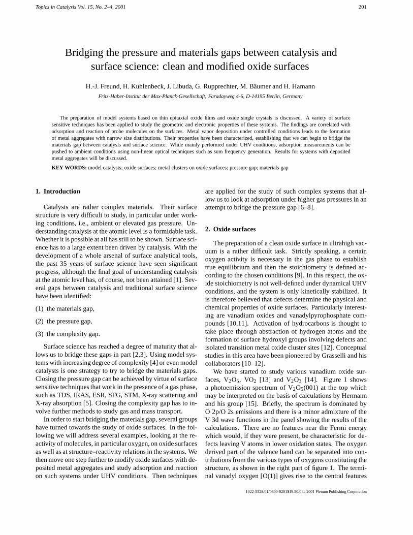
We have started to study various vanadium oxide sur-faces, V2O5, VO2 [13] and V2O3 [14]. Figure 1 showsa photoemission spectrum of V2O5(001) at the top whichmay be interpreted on the basis of calculations by Hermannand his group [15]. Briefly, the spectrum is dominated byO 2p/O 2s emissions and there is a minor admixture of theV 3d wave functions in the panel showing the results of thecalculations. There are no features near the Fermi energywhich would, if they were present, be characteristic for de-fects leaving V atoms in lower oxidation states. The oxygenderived part of the valence band can be separated into con-tributions from the various types of oxygens constituting thestructure, as shown in the right part of figure 1. The termi-nal vanadyl oxygen [O(1)] gives rise to the central features
1022-5528/01/0600-0201$19.50/0 2001 Plenum Publishing Corporation
202 H.-J. Freund et al. / Bridging the pressure and materials gaps between catalysis and surface science
Figure 1. Photoelectron spectra and schematic representations of structures of various vanadium oxides [13,14]. For comparison a computed density ofstates [15] is shown.
whereas the bridging oxygens [O(2) and O(3)] connectingthe vanadyl groups are contributing to the wings of the va-lence band. We will use this fact to identify oxygen spe-cific reactivity. The spectrum in the panel below V2O5 isthat of VO2. VO2(110) has been grown as a thin film onthe isostructural rutile(110) surface. The oxygen derived va-lence band is slightly different from that of V2O5. A charac-teristic difference is the appearance of a feature close to theFermi edge in VO2 indicating the presence of vanadium 3delectrons. Its intensity represents to some extent the popula-tion of the V 3d orbitals. When we turn from VO2 to V2O3an even stronger increase in the V 3d intensity is observed.In figure 1, in the lower panel, the valence band photoemis-sion spectrum of V2O3(0001)/Au(111) is shown. The V2O3film shows a sharp hexagonal LEED pattern representing thecorundum type structure similar to Al2O3, Cr2O3 and Fe2O3(see below). Again, the V2O3 oxygen valence band emissionis not very characteristic, as compared with VO2 and V2O5.It is only the change in the near Fermi edge structures thatshow a characteristic variation from V5+ to V3+.
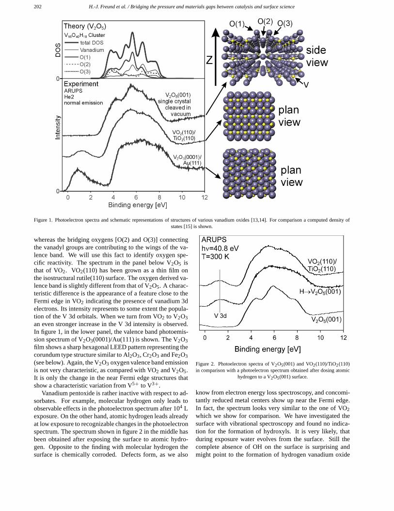
Vanadium pentoxide is rather inactive with respect to ad-sorbates. For example, molecular hydrogen only leads toobservable effects in the photoelectron spectrum after 104 Lexposure. On the other hand, atomic hydrogen leads alreadyat low exposure to recognizable changes in the photoelectronspectrum. The spectrum shown in figure 2 in the middle hasbeen obtained after exposing the surface to atomic hydro-gen. Opposite to the finding with molecular hydrogen thesurface is chemically corroded. Defects form, as we also
Figure 2. Photoelectron spectra of V2O5(001) and VO2(110)/TiO2(110)in comparison with a photoelectron spectrum obtained after dosing atomic
hydrogen to a V2O5(001) surface.
know from electron energy loss spectroscopy, and concomi-tantly reduced metal centers show up near the Fermi edge.In fact, the spectrum looks very similar to the one of VO2which we show for comparison. We have investigated thesurface with vibrational spectroscopy and found no indica-tion for the formation of hydroxyls. It is very likely, thatduring exposure water evolves from the surface. Still thecomplete absence of OH on the surface is surprising andmight point to the formation of hydrogen vanadium oxide
H.-J. Freund et al. / Bridging the pressure and materials gaps between catalysis and surface science 203
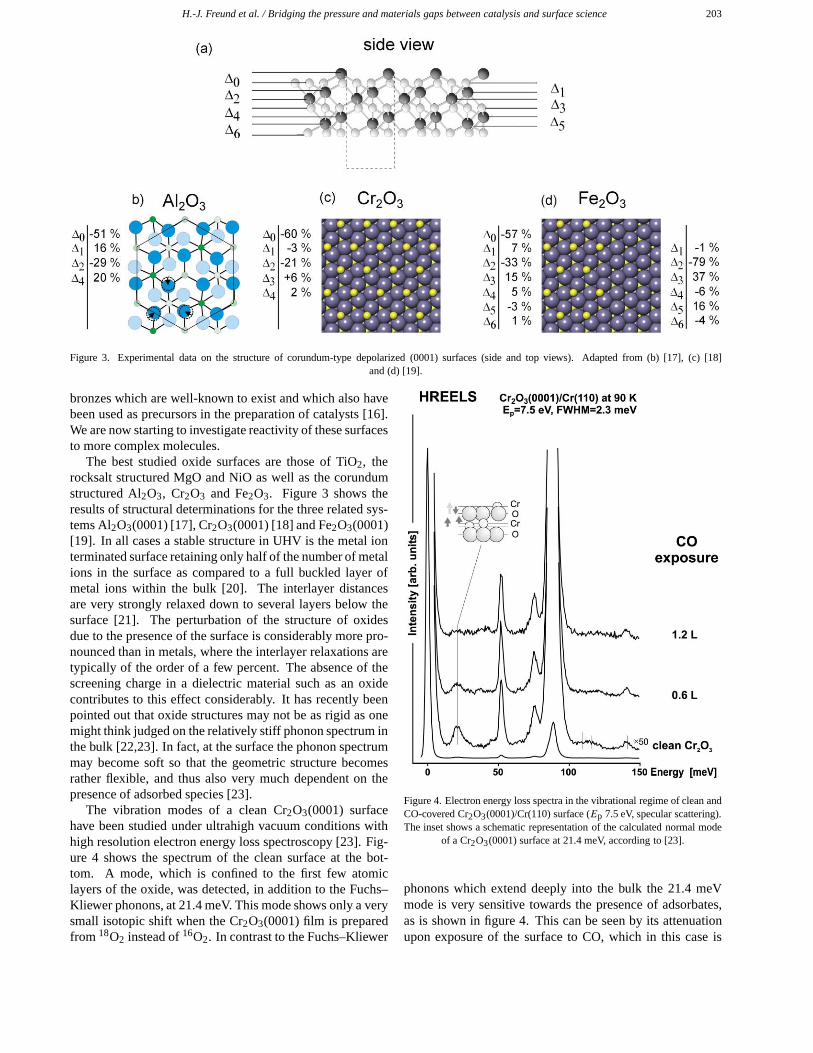
Figure 3. Experimental data on the structure of corundum-type depolarized (0001) surfaces (side and top views). Adapted from (b) [17], (c) [18]and (d) [19].
bronzes which are well-known to exist and which also havebeen used as precursors in the preparation of catalysts [16].We are now starting to investigate reactivity of these surfacesto more complex molecules.
The best studied oxide surfaces are those of TiO2, therocksalt structured MgO and NiO as well as the corundumstructured Al2O3, Cr2O3 and Fe2O3. Figure 3 shows theresults of structural determinations for the three related sys-tems Al2O3(0001) [17], Cr2O3(0001) [18] and Fe2O3(0001)[19]. In all cases a stable structure in UHV is the metal ionterminated surface retaining only half of the number of metalions in the surface as compared to a full buckled layer ofmetal ions within the bulk [20]. The interlayer distancesare very strongly relaxed down to several layers below thesurface [21]. The perturbation of the structure of oxidesdue to the presence of the surface is considerably more pro-nounced than in metals, where the interlayer relaxations aretypically of the order of a few percent. The absence of thescreening charge in a dielectric material such as an oxidecontributes to this effect considerably. It has recently beenpointed out that oxide structures may not be as rigid as onemight think judged on the relatively stiff phonon spectrum inthe bulk [22,23]. In fact, at the surface the phonon spectrummay become soft so that the geometric structure becomesrather flexible, and thus also very much dependent on thepresence of adsorbed species [23].
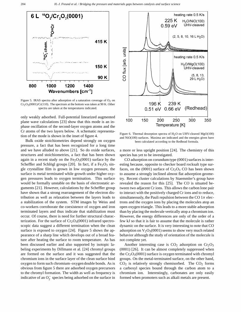
The vibration modes of a clean Cr2O3(0001) surfacehave been studied under ultrahigh vacuum conditions withhigh resolution electron energy loss spectroscopy [23]. Fig-ure 4 shows the spectrum of the clean surface at the bot-tom. A mode, which is confined to the first few atomiclayers of the oxide, was detected, in addition to the Fuchs–Kliewer phonons, at 21.4 meV. This mode shows only a verysmall isotopic shift when the Cr2O3(0001) film is preparedfrom 18O2 instead of 16O2. In contrast to the Fuchs–Kliewer
Figure 4. Electron energy loss spectra in the vibrational regime of clean andCO-covered Cr2O3(0001)/Cr(110) surface (Ep 7.5 eV, specular scattering).The inset shows a schematic representation of the calculated normal mode
of a Cr2O3(0001) surface at 21.4 meV, according to [23].
phonons which extend deeply into the bulk the 21.4 meVmode is very sensitive towards the presence of adsorbates,as is shown in figure 4. This can be seen by its attenuationupon exposure of the surface to CO, which in this case is
204 H.-J. Freund et al. / Bridging the pressure and materials gaps between catalysis and surface science
Figure 5. IRAS spectra after adsorption of a saturation coverage of O2 onCr2O3(0001)/Cr(110). The spectrum at the bottom was taken at 90 K. Other
spectra are taken at the temperatures indicated.
only weakly adsorbed. Full-potential linearized augmentedplane wave calculations [23] show that this mode is an in-phase oscillation of the second-layer oxygen atoms and theCr atoms of the two layers below. A schematic representa-tion of the mode is shown in the inset of figure 4.
Bulk oxide stoichiometries depend strongly on oxygenpressure, a fact that has been recognized for a long timeand we have alluded to above [21]. So do oxide surfaces,structures and stoichiometries, a fact that has been shownagain in a recent study on the Fe2O3(0001) surface by theScheffler and Schlögl groups [20]. In fact, if a Fe2O3 sin-gle crystalline film is grown in low oxygen pressure, thesurface is metal terminated while growth under higher oxy-gen pressures leads to oxygen termination. This surfacewould be formally unstable on the basis of electrostatic ar-guments [21]. However, calculations by the Scheffler grouphave shown that a strong rearrangement of the electron dis-tribution as well as relaxation between the layers leads toa stabilization of the system. STM images by Weiss andco-workers corroborate the coexistence of oxygen and ironterminated layers and thus indicate that stabilization mustoccur. Of course, there is need for further structural charac-terization. For the surface of Cr2O3(0001) infrared spectro-scopic data suggest a different termination when the cleansurface is exposed to oxygen [24]. Figure 5 shows the ap-pearance of a sharp line which develops out of a broad fea-ture after heating the surface to room temperature. As hasbeen discussed earlier and also supported by isotopic la-beling experiments by Dillmann et al. [24] chromyl groupsare formed on the surface and it was suggested that thechromium ions in the surface layer of the clean surface bindoxygen to form such chromium–oxygen double bonds. As isobvious from figure 5 there are adsorbed oxygen precursorsto the chromyl formation. The width as well as frequency isindicative of an O−2 species being adsorbed on the surface in
Figure 6. Thermal desorption spectra of H2O on UHV-cleaved MgO(100)and NiO(100) surfaces. Maxima are indicated and the energies given have
been calculated according to the Redhead formula.
a more or less upright position [24]. The chemistry of thisspecies has yet to be investigated.
CO adsorption on corundum type (0001) surfaces is inter-esting because, opposite to checker board rocksalt type sur-faces, on the (0001) surface of Cr2O3 CO has been shownto assume a strongly inclined almost flat adsorption geome-try. Recent cluster calculations by Staemmler’s group haverevealed the reason for this [25]. The CO is situated be-tween two adjacent Cr ions. This allows the carbon lone pairto interact with the positively charged Cr ions and to reduce,simultaneously, the Pauli repulsion between the CO 1π elec-trons and the oxygen ions by placing the molecules atop anopen oxygen triangle. This leads to a more stable adsorptionthan by placing the molecule vertically atop a chromium ion.However, the energy differences are only of the order of afew kJ so that it is fair to assume that the molecule is ratherdynamic on the surface. It is very interesting to note that COadsorption on V2O3(0001) seems to show very much relatedbehavior although the study of orientation of the molecule isnot complete yet.
Another interesting case is CO2 adsorption on Cr2O3(0001) [26]. It can be almost completely suppressed whenthe Cr2O3(0001) surface is oxygen terminated with chromylgroups. On the metal-terminated surface, on the other hand,CO2 is relatively strongly chemisorbed. The CO2 formsa carboxyl species bound through the carbon atom to achromium ion. Interestingly, carbonates are only easilyformed when promoters such as alkali metals are present.
H.-J. Freund et al. / Bridging the pressure and materials gaps between catalysis and surface science 205
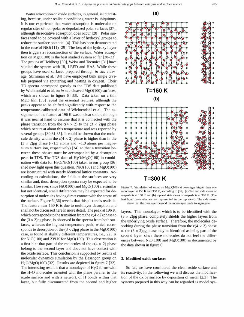
Water adsorption on oxide surfaces, in general, is interest-ing, because, under realistic conditions, water is ubiquitous.It is our experience that water adsorption is molecular onregular sites of non-polar or depolarized polar surfaces [27],although dissociative adsorption does occur [28]. Polar sur-faces tend to be covered with a layer of hydroxyl groups toreduce the surface potential [4]. This has been demonstratedin the case of NiO(111) [29]. The loss of the hydroxyl layerthen triggers a reconstruction of the surface. Water adsorp-tion on MgO(100) is the best studied system so far [30–33].The groups of Heidberg [30], Weiss and Toennies [31] havestudied the system with IR, LEED and HAS. While thesegroups have used surfaces prepared through in situ cleav-age, Stirniman et al. [34] have employed bulk single crys-tals prepared via sputtering and heating in oxygen. TheirTD spectra correspond grossly to the TDS data publishedby Wichtendahl et al. on in situ cleaved MgO(100) surfaces,which are shown in figure 6 [33]. Data taken on a thinMgO film [35] reveal the essential features, although thepeaks appear to be shifted significantly with respect to thetemperature-calibrated data of Wichtendahl et al. The as-signment of the feature at 196 K was unclear so far, althoughit was near at hand to assume that it is connected with thephase transition from the c(4 × 2) to the (3 × 2)pg phasewhich occurs at about this temperature and was reported byseveral groups [30,31,35]. It could be shown that the mole-cule density within the c(4 × 2) phase is higher than in the(3 × 2)pg phase (∼1.3 atoms and ∼1.0 atoms per magne-sium surface ion, respectively) [34] so that a transition be-tween these phases must be accompanied by a desorptionpeak in TDS. The TDS data of H2O/MgO(100) in combi-nation with data for H2O/NiO(100) taken in our group [36]shed new light upon this question. NiO(100) and MgO(100)are isostructural with nearly identical lattice constants. Ac-cording to calculations, the fields at the surfaces are verysimilar and, thus, desorption spectra may be expected to besimilar. However, since NiO(100) and MgO(100) are similarbut not identical, small differences may be expected for de-sorption of molecules having direct contact with the atoms ofthe surface. Figure 6 [36] reveals that this picture is realistic.The feature near 150 K is due to multilayer desorption andshall not be discussed here in more detail. The peak at 196 K,which corresponds to the transition from the c(4×2) phase tothe (3×2)pg phase, is observed in the spectra from both sur-faces, whereas the highest temperature peak, which corre-sponds to desorption of the (3×2)pg phase in the MgO(100)case, is found at slightly different temperatures, i.e., 225 Kfor NiO(100) and 239 K for MgO(100). This observation isa first hint that part of the molecules of the c(4 × 2) phasebelong to the second layer and does not have contact withthe oxide surface. This conclusion is supported by results ofmolecular dynamics simulation by the Besançon group onH2O/MgO(100) [32]. Results are depicted in figure 7 [32].The interesting result is that a monolayer of H2O forms withthe H2O molecules oriented with the plane parallel to theoxide surface and with a network of H bonds within thatlayer, but fully disconnected from the second and higher
Figure 7. Simulation of water on MgO(100) at coverages higher than onemonolayer at 150 K and 300 K, according to [32]. (a) Top and side views ofmap-shots at 150 K and (b) top and side views of map-shots at 300 K. (Thefirst layer molecules are not represented in the top view.) The side views
show that the overlayer beyond the monolayer tends to aggregate.
layers. This monolayer, which is to be identified with the(3 × 2)pg phase, completely shields the higher layers fromthe underlying oxide surface. Therefore, the molecules de-sorbing during the phase transition from the c(4× 2) phaseto the (3× 2)pg phase may be identified as being part of thesecond layer, since these molecules do not feel the differ-ences between NiO(100) and MgO(100) as documented bythe data shown in figure 6.
3. Modified oxide surfaces
So far, we have considered the clean oxide surface andits reactivity. In the following we will discuss the modifica-tion of the oxide surface by deposition of metal [2,3]. Thesystems prepared in this way can be regarded as model sys-
206 H.-J. Freund et al. / Bridging the pressure and materials gaps between catalysis and surface science
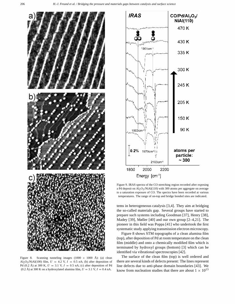
Figure 8. Scanning tunneling images (1000 × 1000 Å): (a) cleanAl2O3/NiAl(100) film, U = 4.2 V, I = 0.5 nA; (b) after deposition ofPd (0.2 Å) at 300 K, U = 3.1 V, I = 0.5 nA; (c) after deposition of Pd(0.2 Å) at 300 K on a hydroxylated alumina film, U = 3.1 V, I = 0.4 nA.
Figure 9. IRAS spectra of the CO stretching region recorded after exposinga Pd deposit on Al2O3/NiAl(110) with 300 atoms per aggregate on averageto a saturation exposure of CO. The spectra have been recorded at varioustemperatures. The range of on-top and bridge bonded sites are indicated.
tems in heterogeneous catalysis [3,4]. They aim at bridgingthe so-called materials gap. Several groups have started toprepare such systems including Goodman [37], Henry [38],Madey [39], Møller [40] and our own group [2–4,21]. Thepioneer in this field was Poppa [41] who undertook the firstsystematic study applying transmission electron microscopy.
Figure 8 shows STM topographs of a clean alumina film(top), after deposition of Pd at room temperature on the cleanfilm (middle) and onto a chemically modified film which isterminated by hydroxyl groups (bottom) [3] which can beidentified via vibrational spectroscopies [42].
The surface of the clean film (top) is well ordered andthere are several kinds of defects present: The lines representline defects due to anti-phase domain boundaries [43]. Weknow from nucleation studies that there are about 1 × 10¹³
H.-J. Freund et al. / Bridging the pressure and materials gaps between catalysis and surface science 207
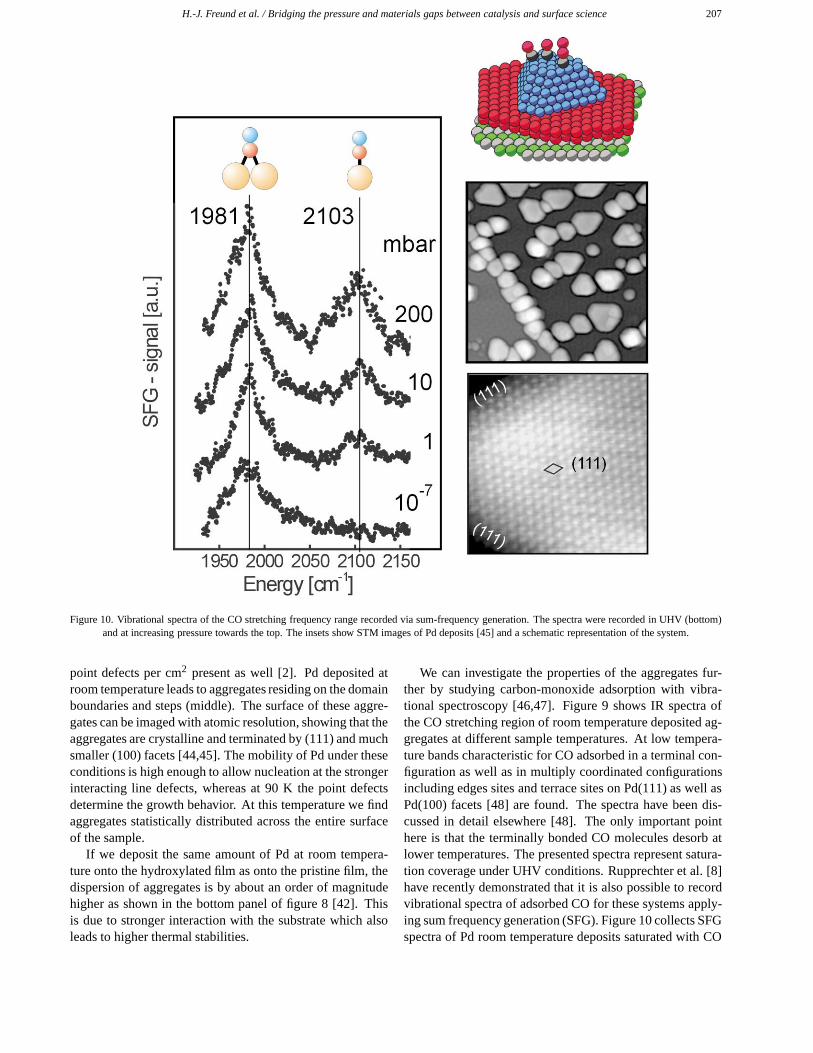
Figure 10. Vibrational spectra of the CO stretching frequency range recorded via sum-frequency generation. The spectra were recorded in UHV (bottom)and at increasing pressure towards the top. The insets show STM images of Pd deposits [45] and a schematic representation of the system.
point defects per cm2 present as well [2]. Pd deposited atroom temperature leads to aggregates residing on the domainboundaries and steps (middle). The surface of these aggre-gates can be imaged with atomic resolution, showing that theaggregates are crystalline and terminated by (111) and muchsmaller (100) facets [44,45]. The mobility of Pd under theseconditions is high enough to allow nucleation at the strongerinteracting line defects, whereas at 90 K the point defectsdetermine the growth behavior. At this temperature we findaggregates statistically distributed across the entire surfaceof the sample.
If we deposit the same amount of Pd at room tempera-ture onto the hydroxylated film as onto the pristine film, thedispersion of aggregates is by about an order of magnitudehigher as shown in the bottom panel of figure 8 [42]. Thisis due to stronger interaction with the substrate which alsoleads to higher thermal stabilities.
We can investigate the properties of the aggregates fur-ther by studying carbon-monoxide adsorption with vibra-tional spectroscopy [46,47]. Figure 9 shows IR spectra ofthe CO stretching region of room temperature deposited ag-gregates at different sample temperatures. At low tempera-ture bands characteristic for CO adsorbed in a terminal con-figuration as well as in multiply coordinated configurationsincluding edges sites and terrace sites on Pd(111) as well asPd(100) facets [48] are found. The spectra have been dis-cussed in detail elsewhere [48]. The only important pointhere is that the terminally bonded CO molecules desorb atlower temperatures. The presented spectra represent satura-tion coverage under UHV conditions. Rupprechter et al. [8]have recently demonstrated that it is also possible to recordvibrational spectra of adsorbed CO for these systems apply-ing sum frequency generation (SFG). Figure 10 collects SFGspectra of Pd room temperature deposits saturated with CO
208 H.-J. Freund et al. / Bridging the pressure and materials gaps between catalysis and surface science
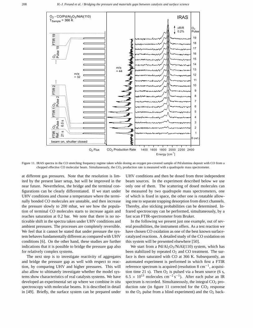
Figure 11. IRAS spectra in the CO stretching frequency regime taken while dosing an oxygen pre-covered sample of Pd/alumina deposit with CO from achopped effective CO molecular beam. Simultaneously, the CO2 production rate is measured with a quadrupole mass spectrometer.
at different gas pressures. Note that the resolution is lim-ited by the present laser setup, but will be improved in thenear future. Nevertheless, the bridge and the terminal con-figurations can be clearly differentiated. If we start underUHV conditions and choose a temperature where the termi-nally bonded CO molecules are unstable, and then increasethe pressure slowly to 200 mbar, we see how the popula-tion of terminal CO molecules starts to increase again andreaches saturation at 0.2 bar. We note that there is no no-ticeable shift in the spectra taken under UHV conditions andambient pressures. The processes are completely reversible.We feel that it cannot be stated that under pressure the sys-tem behaves fundamentally different as compared with UHVconditions [6]. On the other hand, these studies are furtherindications that it is possible to bridge the pressure gap alsofor relatively complex systems.
The next step is to investigate reactivity of aggregatesand bridge the pressure gap as well with respect to reac-tion, by comparing UHV and higher pressures. This willalso allow to ultimately investigate whether the model sys-tems show characteristics of real catalysts systems. We havedeveloped an experimental set up where we combine in situspectroscopy with molecular beams. It is described in detailin [49]. Briefly, the surface system can be prepared under
UHV conditions and then be dosed from three independentbeam sources. In the experiment described below we useonly one of them. The scattering of dosed molecules canbe measured by two quadrupole mass spectrometers, oneof which is fixed in space, the other one is rotatable allow-ing one to separate trapping desorption from direct channels.Thereby, also sticking probabilities can be determined. In-frared spectroscopy can be performed, simultaneously, by afast scan FTIR-spectrometer from Bruker.
In the following we present just one example, out of sev-eral possibilities, the instrument offers. As a test reaction wehave chosen CO oxidation as one of the best known surface-catalyzed reactions. A detailed study of the CO oxidation onthis system will be presented elsewhere [50].
We start from a Pd/Al2O3/NiAl(110) system, which hasbeen stabilized by repeated O2 and CO treatment. The sur-face is then saturated with CO at 366 K. Subsequently, anautomated experiment is performed in which first a FTIRreference spectrum is acquired (resolution 8 cm−1, acquisi-tion time 21 s). Then O2 is pulsed via a beam source (6 s,6.5 × 1013 molecules cm−2 s−1). After each pulse an IRspectrum is recorded. Simultaneously, the integral CO2 pro-duction rate (in figure 11 corrected for the CO2 responseto the O2 pulse from a blind experiment) and the O2 back-
H.-J. Freund et al. / Bridging the pressure and materials gaps between catalysis and surface science 209
ground pressure is recorded with a quadrupole mass spec-trometer fixed in space.
As a detailed analysis of the reaction kinetics is beyondthe scope of this paper, we will only qualitatively pointout which type of information is available from this experi-ment: Every CO2 response pulse shows a rise and decay timeslower than the originating O2 pulse being rectangular on thetime scale of the experiment. This is due to the limited rateof the surface reaction, the kinetics of which can be extractedfrom the waveform. The envelope of the waveforms repre-sent the overall CO2 reactive sticking coefficient of O2 as afunction of CO coverage. Due to inhibition of O2 sticking byCO we find a rising reactive sticking coefficient at high COcoverage, before CO depletion and O2 coadsorption leadsto a decaying probability for CO2 production. From the si-multaneously acquired IR spectra information on the occu-pation of different sites for the consumed CO is available.Additionally, quantitative information on the surface cover-age can be obtained via a coverage–absorption calibration,which can be easily performed by a simultaneous stickingcoefficient/IR absorption measurement.
4. Summary
In summary, we have tried to demonstrate in which waysurface science can contribute to study fundamental prob-lems in catalysis. The emphasis is on the attempts to bridgeboth the materials as well as the pressure gaps.
We have investigated the structure and reactivity of cleanoxide surfaces as well as metal modified oxide surfaces. Inparticular, the reactivity and adsorption abilities of depositedaggregates have been studied. The pressure gap has beenbridged applying non-linear optical techniques and mecha-nistic insight has been gained with a newly designed molec-ular beam apparatus.
References
[1] C.B. Duke, ed., Surface Science: The First Thirty Years (Elsevier,Amsterdam, 1994).
[2] M. Bäumer and H.-J. Freund, Prog. Surf. Sci. 61 (1999) 127.[3] H.-J. Freund, H. Kuhlenbeck and M. Bäumer, Adv. Catal. 45 (2000)
333.[4] H.-J. Freund, Angew. Chem. Int. Ed. Engl. 36 (1997) 452.[5] Ber. Bunsenges. Phys. Chem. 97 (1993).[6] X. Su, P.S. Cremer, Y.R. Shen and G.A. Somorjai, J. Am. Chem. Soc.
119 (1997) 3994.[7] G. Rupprechter, T. Dellwig, H. Unterhalt and H.-J. Freund, Topics
Catal. 15 (2001)19.[8] T. Dellwig, G. Rupprechter, H. Unterhalt and H.-J. Freund, Phys. Rev.
Lett., in press.[9] H. Schmalzried, Chemical Kinetics of Solids (VCH, Weinheim,
1995).[10] R.K. Grasselli and J.D. Burrington, Adv. Catal. 30 (1981) 133.[11] F. Cavani, G. Centi, F. Trifirò and R.K. Grasselli, Catal. Today 3
(1988) 185.[12] P.A. Agaskar, L. DeCaul and R.V. Grasselli, Catal. Lett. 23 (1994)
339.
[13] B. Tepper, Ph.D. thesis, in preparation.[14] A.-C. Dupuis, Ph.D. thesis, in preparation.[15] K. Hermann, M. Witko, R. Druzinic, A. Chakrabarti, B. Tepper, M.
Elsner, A. Gorschlüter, H. Kuhlenbeck and H.-J. Freund, J. ElectronSpectrosc. Relat. Phenom. 98–99 (1999) 245.
[16] M. Figlarz, Progr. Sol. State Chem. 19 (1989) 1.[17] G. Renaud, Surf. Sci. Rep. 32 (1998) 1.[18] F. Rohr, M. Bäumer, H.-J. Freund, J.A. Mejias, V. Staemmler, S.
Müller, L. Hammer and K. Heinz, Surf. Sci. 372 (1997) L291;F. Rohr, M. Bäumer, H.-J. Freund, J.A. Mejias, V. Staemmler, S.Müller, L. Hammer and K. Heinz, Surf. Sci. 389 (1997) 391.
[19] S.K. Shaikhoutdinov and W. Weiss, Surf. Sci. 432 (1999) L627.[20] X.-G. Wang, W. Weiss, S.K. Shaikhoutdinov, M. Ritter, M. Petersen,
F. Wagner, R. Schlögl and M. Scheffler, Phys. Rev. Lett. 81 (1998)1038.
[21] H.-J. Freund, Faraday Discuss. 114 (1999) 1.[22] N.M. Harrison, X.-G. Wang, M. Muscat and M. Scheffler, Faraday
Discussions 114 (1999) 305.[23] K. Wolter, D. Scarano, J. Fritsch, H. Kuhlenbeck, A. Zecchina and
H.-J. Freund, Chem. Phys. Lett. 105 (2000) 295.[24] B. Dillmann, F. Rohr, O. Seiferth, G. Klivenyi, M. Bender, K.
Homann, I.N. Yakovkin, D. Ehrlich, M. Bäumer, H. Kuhlenbeck andH.-J. Freund, Faraday Discuss. 105 (1996) 295.
[25] M. Pykavy, V. Staemmler, O. Seiferth and H.-J. Freund, Surf. Sci.,submitted.
[26] O. Seiferth, K. Wolter, B. Dillmann, G. Klivenyi, H.-J. Freund, D.Scarano and A. Zecchina, Surf. Sci. 421 (1999) 176.
[27] D. Cappus, C. Xu, D. Ehrlich, B. Dillmann, C.A. Ventrice, Jr., K. Al-Shamery, H. Kuhlenbeck and H.-J. Freund, Chem. Phys. 177 (1993)533.
[28] M.A. Henderson and S.A. Chambers, Surf. Sci. 449 (2000) 135.[29] F. Rohr, K. Wirth, J. Libuda, D. Cappus, M. Bäumer and H.-J. Freund,
Surf. Sci. 315 (1994) L977.[30] J. Heidberg, B. Redlich and D. Wetter, Ber. Bunsenges. Phys. Chem.
99 (1995) 1333.[31] D. Ferry, A. Glebov, V. Senz, J. Suzanne, J.P. Toennies and H. Weiss,
J. Chem. Phys. 105 (1996) 1697.[32] A. Marmier, P.N.M. Hoang, S. Picaud, C. Girardet and R.M. Lynden-
Bell, J. Chem. Phys. 109 (1988) 3245.[33] R. Wichtendahl, M. Rodriguez-Rodrigo, U. Härtel, H. Kuhlenbeck
and H.-J. Freund, Phys. Stat. Sol. 173 (1999) 93.[34] M.J. Stirniman, C. Huang. R.C. Smith, J.A. Joyce and B.D. Kay,
J. Chem. Phys. 105 (1996) 1295.[35] C. Xu and D.W. Goodman, Chem. Phys. Lett. L65 (1997) 341.[36] R. Wichtendahl, Ph.D. thesis, Freie Universität Berlin (1999).[37] D.W. Goodman, Surf. Rev. Lett. 2 (1995) 9.[38] C.R. Henry, Surf. Sci. Rep. 31 (1998) 231.[39] U. Diebold, J.-M. Pan and T.E. Madey, Surf. Sci. 331 (1995) 845.[40] M.C. Wu and P.J. Møller, Surf. Sci. 221 (1989) 250.[41] H. Poppa, Catal. Rev. Sci. Eng. 35 (1993) 359.[42] M. Heemeier, M. Frank, J. Libuda, K. Wolter, H. Kuhlenbeck, M.
Bäumer and H.-J. Freund, Catal. Lett. 68 (2000) 19.[43] J. Libuda, F. Winkelmann, M. Bäumer, H.-J. Freund, T. Bertrams, H.
Neddermeyer and K. Müller, Surf. Sci. 318 (1994) 61.[44] N. Ernst, B. Duncombe, G. Bozdech, M. Naschitzki and H.-J. Freund,
Ultramicroscopy 79 (1999) 231.[45] K. Højrup Hansen, T. Worren, S. Stempel, E. Laegsgaard, M. Bäumer,
H.-J. Freund, F. Besenbacher and I. Stensgaard, Phys. Rev. Lett. 83(1999) 4120.
[46] M. Frank, R. Kühnemuth, M. Bäumer and H.-J. Freund, Surf. Sci. 427(1999) 288.
[47] M. Frank and M. Bäumer, Phys. Chem. Chem. Phys., to be published.[48] K. Wolter, O. Seiferth, H. Kuhlenbeck, M. Bäumer and H.-J. Freund,
Surf. Sci. 399 (1998) 190.[49] J. Libuda, I. Meusel, J. Hartmann and H.-J. Freund, Rev. Sci. Instr.,
submitted.[50] I. Meusel, J. Libuda, J. Hartmann and H.-J. Freund, to be published.