Embed Size (px)
Citation preview
Brian de Souza, J; Hafalla, JC; Riley, EM; Couper, KN (2009) Cere-bral malaria: why experimental murine models are required to under-stand the pathogenesis of disease. Parasitology, 137 (5). pp. 755-72.ISSN 0031-1820 DOI: https://doi.org/10.1017/S0031182009991715
Downloaded from: http://researchonline.lshtm.ac.uk/4375/
DOI: 10.1017/S0031182009991715
Usage Guidelines
Please refer to usage guidelines at http://researchonline.lshtm.ac.uk/policies.html or alterna-tively contact [email protected].
Available under license: http://creativecommons.org/licenses/by-nc-nd/2.5/

REVIEW ARTICLE
Cerebral malaria: why experimental murine models are
required to understand the pathogenesis of disease
J. BRIAN DE SOUZA1,2, JULIUS C. R. HAFALLA1, ELEANOR M. RILEY1
and KEVIN N. COUPER1*
1 Immunology Unit, Department of Infectious and Tropical Diseases, London School of Hygiene and Tropical Medicine,Keppel Street, London WC1E 7HT, UK2Department of Immunology and Molecular Pathology, University College London Medical School, 46 Cleveland Street,London W1T 4JF, UK
(Received 14 September 2009; revised 20 October 2009; accepted 26 October 2009; first published online 23 December 2009)
SUMMARY
Cerebral malaria is a life-threatening complication of malaria infection. The pathogenesis of cerebral malaria is poorly
defined and progress in understanding the condition is severely hampered by the inability to study in detail, ante-mortem,
the parasitological and immunological events within the brain that lead to the onset of clinical symptoms. Experimental
murine models have been used to investigate the sequence of events that lead to cerebral malaria, but there is significant
debate on the merits of these models and whether their study is relevant to human disease. Here we review the current
understanding of the parasitological and immunological events leading to human and experimental cerebral malaria,
and explain why we believe that studies with experimental models of CM are crucial to define the pathogenesis of the
condition.
Key words: cerebral malaria, murine model, pathogenesis.
INTRODUCTION
Malaria remains a major public health problem in
many tropical countries. The World Health Organ-
ization (WHO) estimates that 40% of the world’s
population lives in areas affected by malaria, result-
ing in approximately 200–300 million clinical cases
each year, leading to the deaths of more than
2 million young children every year, mainly in sub-
Saharan Africa. The vast majority of cases of severe
malaria are caused by infection with the Plasmodium
falciparum species of the parasite. Clinical presen-
tations of severe malaria vary but include altered
consciousness, respiratory distress, severe anaemia
(haemoglobin level of <5 g/dl), multi-organ failure
and cerebral malaria. The WHO definition of CM
is unrousable coma (graded according to either
Blantyre or Glasgow coma scale) not attributable to
other causes (Teasdale and Jennet, 1974; Molyneux
et al. 1989). In areas of high malaria transmission,
susceptibility to severe malaria varies with age and
exposure to the parasite; adults are, in general,
resistant to severe malaria whilst infants and very
young children are at significantly increased risk of
developing severe malarial anaemia. Older children,
who have had at least one previous malaria infection,
are disproportionately at risk of developing cerebral
malaria (CM) (Marsh and Snow, 1999). The epi-
demiology of severe malaria is highly suggestive of a
role for the immune system in both initiation of
(in children) and protection from (in adults) cerebral
malaria, either indirectly – by selecting for infection
by parasites of differing virulence – or directly – by
contributing to the pathogenesis of the syndrome.
CEREBRAL MALARIA
The incidence of cerebral malaria is difficult to as-
sess, but hospital admission records indicate that
in the region of 1% of all P. falciparum infections
progress to CM, which is fatal in 10–20% of all cases
(300 000–500000 deaths each year). Moreover, at
least 10–20% of individuals who survive and recover
from CM display long-term physical or cognitive
dysfunction (Carter et al. 2005a, b ; Idro et al. 2005;
Boivin et al. 2007). Since the discovery by
Marchiafava and Bignami in 1894 of malaria para-
sites within the brain of humans during infection,
attention has focussed on understanding the patho-
physiological processes that predispose towards
* Corresponding author: Immunology Unit, Departmentof Infectious and Tropical Diseases, London School ofHygiene and Tropical Medicine, Keppel Street, LondonWC1E 7HT, UK. Tel: +44 207 927 2690. Fax:+44 207 927 2807. E-mail : [email protected]
755
Parasitology (2010), 137, 755–772. f Cambridge University Press 2009
doi:10.1017/S0031182009991715

CM, with a view to the development of preventative
measures or targeted therapies for the condition.
Contrasting theories on the roles of parasite seques-
tration within the brain and the host immune re-
sponse to the parasite in the pathogenesis of CM
were initially proposed (reviewed by Van der Heyde
et al. 2006), with current understanding suggesting
that neuropathology is the result of a combination of
both processes, as discussed below.
Cerebral malaria can develop rapidly after initial
bouts of fever lasting 2–3 days. Coma is the standard
definition of CM, but other symptoms associated
with the condition include general malaise, head-
ache, fits, vomiting and diarrhoea. The clinical
symptoms associated with early-stage CM are not
pathognomonic for the condition and are difficult
to differentiate from encephalitis, meningitis and
febrile convulsions. This has implications for the
rapid and early diagnosis of the condition, which
often significantly delays the initiation of treatment.
The early symptoms of CM can progress rapidly to
increased intracranial pressure, hemiparesis, ataxia
and coma if immediate medical treatment is not
provided. The diverse set of neurological complica-
tions associated with CM indicates that multiple
areas of the brain are affected by the condition.
Anti-malarial drug-based therapies are the first-
line treatment for patients with cerebral malaria ;
however, the incidence of neurological deficiencies
and mortality remain unacceptably high with fatal-
ity rates of around 15% following treatment with
Artemisinin compared with 20% for traditionally
used quinine-based treatments (Dondorp et al.
2005a). This is unsurprising as anti-malarial therapy
can only be implemented when CM is first suspected
or diagnosed at health care centres ; CM is at an
advanced state in the majority of these individuals
and anti-malarial drugs by themselves are often in-
sufficient to reverse and alleviate the symptoms of
CM. Therefore, there is an urgent need to develop
adjunctive therapies, such as immuno-modulators or
neuro-protective agents that may be administered
with anti-malarials. At present the lack of under-
standing of the pathogenesis of CM means that the
potentially most efficacious targets for therapeutic
intervention remain to be identified.
THE PATHOLOGY AND ASSOCIATED CLINICAL
FEATURES OF CM
Post-mortem examinations of brains from indivi-
duals that succumbed to CM have helped to uncover
the type and distribution of brain pathology that
occurs during the condition. Some of the most
commonly reported findings include swelling and
haemorrhaging in the white matter of the subcortical
rim and corpus callosum as well as petechial and
ring haemorrhages in both cerebral and cerebellar
cortices (reviewed by Haldar et al. 2007). In the
majority of cases, histopathological examinations
reveal cerebral capillaries plugged with parasitized
erythrocytes (reviewed by Haldar et al. 2007).
Margination of monocytes and macrophages within
cerebral vessels and the presence of pigmented
macrophages sequestered with pRBC are also well
described features of CM (Patnaik et al. 1994). Due
to the lack of detailed comparative histopathological
studies of pediatric and adult CM cases it is difficult
to conclude whether the pathology of CM varies
between children and non-immune adults, but as
there are a number of differences in the symptoms of
pediatric and adult CM, it is possible there may be
some age-related differences in cerebral pathology
(Mishra and Wiese, 2009).
Although parasite sequestration, haemorrhages
and inflammation are found in the majority of CM
brains, it is clear that CM is not a homogenous syn-
drome. For example, 3 different patterns of histo-
pathological changes have been described in African
children: in addition to the ‘classical ’ pattern of
CM of parasite sequestration, perivascular haemor-
rhages and immune cell infiltration within brain
micro-vessels, parasite sequestration may be obser-
ved within the brain in the absence of any other ab-
normalities and there are cases where individuals
with high peripheral parasitaemia develop a syn-
drome that is clinically defined as CM but where
there is no evidence of parasite sequestration within
the brain (Clark et al. 2003; Taylor et al. 2004). The
reasons for the variations in pathology of CM are
unclear but may be due to genetic variation in hosts
or parasites, environmental factors or the host im-
mune response to the parasite.
THE LIMITATIONS OF STUDIES OF HUMAN
CEREBRAL MALARIA IN DEFINING THE
PATHOGENESIS OF THE CONDITION
Cerebral malaria is likely the result of a complex
sequence of inter-related events, most probably
beginning either with sequestration of trophozoite-
infected red blood cells (pRBC) in the small blood
vessels (reviewed by Chakrovorty et al. 2008) and/or
with the rupture of infected red blood cells and
the release of parasite-derived toxins (Bate and
Kwiatkowski, 1994; Schofield et al. 1996). The re-
lative importance of systemic versus brain-localized
events – including pRBC sequestration and rupture,
lymphocyte, monocyte, endothelial and glial cell
activation and release of inflammatory mediators –
their sequence and timing in the pathogenesis of
CM are very much unknown. For obvious reasons,
histopathological examination of CM brains is
limited to post-mortem analysis of fatal cases and it
is thus not possible to describe the sequence of events
leading to the onset of CM symptoms nor to com-
pare fatal cases with those that resolve in response to
treatment. Such investigations and comparisons are
J. Brian De Souza and others 756

essential to delineate truly pathogenic systemic and
intra-cerebral processes from neutral and/or pro-
tective responses. Increased utility of non-invasive
in vivo imaging techniques, such as magnetic reson-
ance imaging (MRI) and spectroscopy (MRS) and
computational topography (CT), should hopefully
help to address these issues (Kampfl et al. 1993;
Crawley et al. 1996; Patanker et al. 2002; Penet et al.
2005, 2007), but these studies are severely restricted
by ethical constraints and the availability of the
expensive specialized equipment in malaria-endemic
areas. It is therefore extremely difficult to move be-
yond purely descriptive and correlative studies in
humans: defining the immunological pathways and
parasite-driven processes that underlie the patho-
genesis of the syndrome, and demonstrating caus-
ality, is difficult without direct intervention studies.
Moreover, examination of peripheral blood (which is
possible in non-fatal as well as fatal cases) may pro-
vide limited information on the immunological and
parasitological environment in the brain and, again,
patients usually present to hospital only once the
syndrome is well-established. For example, periph-
eral blood parasitaemia does not always accurately
predict total parasite biomass (Silamut et al. 1999)
and total parasite biomass is a stronger correlate of
severe malarial disease than is peripheral parasita-
emia (Dondorp et al. 2005b). It is clear that other
approaches – in combination with human studies –
are required to fully understand the pathogenesis
of CM.
EXPERIMENTAL MODELS OF CEREBRAL MALARIA
Much of our understanding of mammalian physi-
ology has come from studies of animals and the
extent of the conservation of basic immunological
and neuropathological processes between laboratory
rodents and humans is becoming ever more apparent
(Hau and Van Hoosier Jr, 2005). Experimental
models have proven invaluable for understanding
the pathogenesis of numerous autoimmune and in-
fectious diseases of humans and many vaccines and
immune-therapies currently in use were initially
developed and tested in experimental models (Hau
and Van Hoosier Jr, 2005). It is likely, therefore,
that the use of relevant experimental animal models
can significantly aid in the study of cerebral malaria.
Primate models of CM, including P. knowlesi and
P. coatneyi infections in Rhesus monkeys (Aikawa
et al. 1992; Ibiwoye et al. 1993) and P. falciparum
infection in squirrel monkeys (Gysin et al. 1992),
have allowed the investigation of some aspects of
CM, but these models are prohibitively expensive
and are restricted to low numbers for ethical reasons.
Consequently, other experimental models are re-
quired. Neuropathological syndromes have been
shown to develop in certain strains of inbred mice
infected with various strains of Plasmodium berghei
(Pb) (Rest, 1982; Curfs et al. 1993a) or the
lethal (XL) variant of P. yoelii (PyXL) (Yoeli and
Hargreaves, 1974) ; however, there has been – and
continues to be – significant disagreement within the
malaria research community as to whether the mu-
rine models share sufficient similarities with human
cerebral malaria to make them relevant or useful.
In the remainder of this review we will evaluate
the currently available models of ECM and we will
attempt to resolve the relevance of experimental
models of cerebral malaria to human infection.
Plasmodium yoelii XL and Plasmodium
berghei K173
Although more extensively studied as a model of
hyperparasitaemia and failure of parasite control
(Couper et al. 2007, 2008), PyXL has been shown to
sequester within the brain microvasculature and
produce a cerebral syndrome comparable with hu-
man cerebral malaria (Yoeli and Hargreaves, 1974;
Kaul et al. 1994); however, the hyper-parasitaemia
associated with this infection (rapidly ascending
peripheral parasitaemia that can reach 80–100%) is
not typical of human CM cases (Silamut et al. 1999)
and this model is not widely used to study CM. In a
few studies, P. berghei K173 has been found to
induce CM-like signs (Curfs et al. 1993a ; Mitchell
et al. 2005), but the dose-dependent onset of ECM
in this model (inducing cerebral pathology after low
dose but not high dose infection) (Mitchell et al.
2005) also limits its utility as a model of human CM:
indeed P. berghei K173 is frequently used as a non-
ECM-infection to compare with the most widely
used model of ECM, P. berghei ANKA infection
(Mitchell et al. 2005).
Plasmodium berghei ANKA
The Plasmodium berghei ANKA (PbA) model re-
plicates many events seen during human CM and
is accepted as the best available experimental model
of cerebral malaria. Infection of susceptible strains
of mice, including C57BL/6 and CBA, leads to the
development of fatal cerebral pathology, with clini-
cal signs including ataxia, fitting, respiratory distress
and coma (de Souza and Riley, 2002). The time to
onset of clinical signs varies depending on the in-
fection dose, the genetic background of the host and
the specific clone of infecting parasites, but is typi-
cally between 5 and10 days post-infection (de Souza
and Riley, 2002). As in humans, there is a rapid de-
terioration in the condition of infected animals once
clinical signs become apparent, with death often oc-
curring within 4 or 5 h after the onset of neurological
signs. Multiple areas of blood-brain barrier disrup-
tion with vascular leakage involving the cortex, cer-
ebellum and olfactory bulb are observed in brains
of PbA-infected mice displaying signs of ECM
The pathogenesis of CM and ECM 757

(Penet et al. 2005; Lackner et al. 2006; de Souza
and Couper, unpublished observations), with loss of
specific neuronal populations within the cortex and
striatum (Clark et al. 2005), accumulation of pRBC
within blood vessels (Rest, 1982; Hearn et al. 2000)
and focal perivascular inflammation (Engwerda et al.
2002). ECM is also associated with the significant
accumulation of platelets within the brain vascu-
lature (Wassmer et al. 2003; von Zur Muhlen et al.
2008): platelets have been shown to directly promote
endothelial cell damage during infection (Wassmer
et al. 2006). Cognitive dysfunction during P. berghei
ANKA infection, as shown by impaired visual
memory, is directly correlated with haemorrhage
and inflammation, including microglial activation
(Desuisseaux et al. 2008). Indeed, accumulation of
monocytes and macrophages, and activation of brain
resident mononuclear cells, including astrocytes and
microglial cells is believed to be a key feature of
ECM (Grau et al. 1987; Medana et al. 1997a, b ; Pais
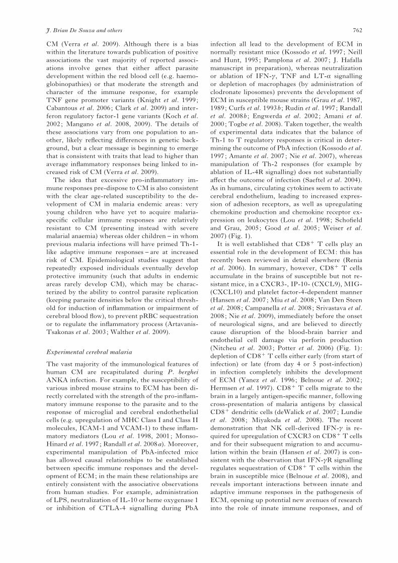
and Chatterjee, 2005) (Fig. 1).
As in humans, genetic and environmental factors
determine the susceptibility of mice to ECM. For
example, the resistance of F1 intercrossed BALB/c
(resistant) and C57BL/6 (susceptible) mice to the
development of ECM is determined by age, and
environmental exposure, with young mice (8–10
weeks) susceptible to ECM and older mice (16–20
weeks) resistant to the development of cerebral signs
(Hearn et al. 2000). Genetic resistance to ECM
and P. berghei ANKA infection has been mapped
using intercrossed resistant and susceptible strains
of mice to loci on chromosomes 1, 9, 11, 17 and 18
(Bagot et al. 2002; Nagayasu et al. 2002; Ohno and
Nishimura, 2004; Campino et al. 2005). However,
the genes encoded within each of these regions that
control resistance to ECM and parasite levels remain
to be identified.More recently, micro-array profiling
Fig. 1. A hypothetical schema of events leading to the development of experimental cerebral malaria. Rupture of
pRBCs releases molecules that activate brain microvascular endothelial cells leading to upregulation of receptors for
pRBC. Phagocytosis of parasite moieties in the spleen and liver, priming lymphocytes within the spleen, promotes
systemic inflammation which further amplifies endothelial cell activation in the brain and activates brain-resident
perivascular macrophages, microglia and astrocytes. pRBCs bind to endothelial receptors ; platelets binding to
endothelial receptors may provide additional ligands for pRBC adherence. Activated endothelial and glial cells provide
chemotactic signals for lymphocytes and myeloid cells. Sequestered pRBCs and leukocytes interfere with cerebral blood
flow and, together with cytotoxic molecules, damage the blood-brain barrier leading to oedema and haemorrhage.
J. Brian De Souza and others 758

of susceptible and resistant strains of mice have
identified distinct expression profiles in the brain
of genes involved in metabolic energy pathways,
immune-activation, apoptosis and neuroprotection/
neurotoxicity (Delahaye et al. 2007; Lovegrove et al.
2007; Oakley et al. 2008). Differences in the immune
response of ECM-susceptible and ECM-resistant
strains of mice to infection are discussed in more
detail below.
THE ROLE OF PARASITE SEQUESTRATION
DURING HUMAN AND EXPERIMENTAL CEREBRAL
MALARIA
The sequestration of mature parasites within peri-
pheral tissues via adherence of pRBC to vascular
endothelium is a common feature of malaria infec-
tions. It is believed that this prevents the clearance
of mature-stage parasites by the spleen, allowing
the development of sufficient numbers of infectious
parasites (gametocytes) to ensure transmission to
mosquitoes (Beeson et al. 2001; Engwerda et al.
2005). Although sequestration is initially beneficial
to the parasite, it is widely believed to have signifi-
cant deleterious consequences for the host. For
many years it was assumed that the symptoms of
CM were due solely to occlusion of brain micro-
vessels by sequestered pRBC (reviewed by Berendt
et al. 1994; Van der Heyde et al. 2006). In this
scenario, parasite adherence to brain endothelial
cells, combined with rosetting of uninfected and
infected red blood cells, impairs blood flow leading
to hypoxia, hypoglycaemia and the buildup of toxic
waste products, including lactic acid (Van der Heyde
et al. 2006), which rapidly leads to irreversible tissue
damage. However, the typically quite subtle neuro-
logical consequences experienced by CM survivors
are not consistent with this simple aetiology and
other causes of disrupted neuronal signalling are also
likely to play a part (Rae et al. 2004; Penet et al.
2005; Hunt et al. 2006).
Although parasite sequestration is usually seen in
CM brains, the association is not absolute and, de-
spite a plethora of associative data, there is very little
empirical evidence that parasite sequestration in
the brain is either necessary or sufficient to cause
CM. Deaths attributable to CM, as defined byWHO
guidelines, have been observed in the absence of
parasite sequestration within the brain (Clark et al.
2003; Taylor et al. 2004; Haldar et al. 2007).
Furthermore, parasite sequestration has been ob-
served in individuals that did not develop severe
cerebral malaria (Silamut et al. 1999; Seydel et al.
2006). This heterogeneous association between cer-
ebral parasite sequestration and clinical outcome
raises important questions regarding the precise
aetiology of CM (Clark et al. 2006). Specifically, we
need to consider the possibility that transient inter-
actions between sequestering pRBC and cerebral
tissues might be sufficient to trigger downstream
immunological and biochemical processes that lead
to the development of CM.
The widespread assumption that sustained para-
site sequestration in the brain is essential for devel-
opment of CM has led some researchers to question
whether cerebral parasite sequestration occurs dur-
ing P. berghei ANKA infection and thus whether
the pathogenesis of ECM is comparable to human
CM (Berendt et al. 1994; Franke-Fayard et al.
2005). Accumulation of PbA pRBCs in cerebral and
cerebellar capillaries of mice displaying signs of
ECM has been observed at both light and electron
microscopic levels (Rest, 1982; Jennings et al. 1998;
Hearn et al. 2000; Beghdadi et al. 2008). Detailed
investigations on the nature of parasite sequestration
during P. berghei ANKA infection have, however,
yet to be performed and as such it is unknown
whether PbA parasites adhere through strong, tight
junctions, or via weak easily disrupted interactions.
The comparison of parasite sequestration in the
brain during ECM and CM is also severely compli-
cated by the method of tissue preparation; mouse
brains are routinely perfused prior to histological
examination during ECM, but perfusion is seldom
performed prior to the examination of brains from
individuals with fatal CM. As such, parasite seques-
tration may be frequently under-estimated (in ECM)
or over-estimated (in CM). Nevertheless, most
blocked vessels during ECM contain a mixture of
parasitized RBC and leukocytes (Hearn et al. 2000;
Jennings et al. 1998). Consequently, parasite ac-
cumulation alone may not be sufficient to cause
blockage of brain-micro-vessels during P. berghei
ANKA infection.
Recently, Franke-Fayard et al. (2005) reported
CD36 (Scavenger type B receptor)-mediated seq-
uestration of luciferase-expressing P. berghei ANKA
pRBC, visualized by bioluminescent imaging, in
lung and adipose tissue but not in the brains of in-
fected mice. This is consistent with the requirement
for CD36-mediated PbA pRBC sequestration for
initiation of acute lung injury (Lovegrove et al.
2008), and with the observation that CD36x/x mice
are fully susceptible to ECM. These findings have
been interpreted as evidence that pRBC seques-
tration does not occur in the brain during PbA in-
fection and is not required for initiation of ECM,
and thus that ECM has a significantly different
aetiology to CM (Franke-Fayard et al. 2005). How-
ever, whole body imaging and multi-organ com-
parisons may under-estimate cerebral sequestration
since it is likely that the density of sequestered
pRBC is much lower in brain than in much more
heavily vascularized organs such as lung or spleen,
and higher resolution analysis of the brain is re-
quired to rule out sequestration; nevertheless, and
despite the authors claims, focal parasite seques-
tration was evident in one of the two examples of day
The pathogenesis of CM and ECM 759

7 p.i. brains shown by Franke-Fayard et al. (2005).
Secondly, the lack of a role for CD36 in ECM does
not rule out that (as in humans) there are other re-
ceptors mediating pRBC sequestration in the brain
(as discussed below). Importantly, other studies
using the same bioluminescent parasite system
have not only shown significant accumulation of P.
berghei ANKA pRBC in the brains of mice showing
signs of ECM, but have also demonstrated that
parasite biomass in the brain is directly correlated
with risk of ECM (Amante et al. 2007; Randall et al.
2008a ; Nie et al. 2009).
HOST CELL RECEPTORS MEDIATING
CYTO-ADHERENCE DURING CM AND ECM
Although CD36 appears to be the main receptor for
P. falciparum pRBC sequestration in peripheral or-
gans, CD36-mediated adhesion is not believed to be
involved in sequestration in the brain (Newbold et al.
1999); CD36 is expressed only at very low levels in
healthy brain tissue and it is not upregulated during
malaria infection (Turner et al. 1994; Newbold et al.
1999). Nonetheless, it has recently been postulated
that platelets and platelet and endothelial cell de-
rived microparticles – submicron particles generated
by vesiculation of cellular membranes (reviewed by
Coltel et al. 2006) – may bind to brain endothelial
cells, providing a source of CD36 that allows in-
direct CD36-mediated pRBC binding to brain en-
dothelial cells (Wassmer et al. 2004; Faille et al.
2009); however, this hypothesis remains to be vali-
dated in vivo.
At present, intercellular adhesion molecule 1
(ICAM-1) is the most studied putative endothelial
receptor for the sequestration of P. falciparum pRBC
within the brain (Newbold et al. 1999; Chakravorty
and Craig, 2005). The expression of ICAM-1 is
significantly upregulated on cerebral vasculature
endothelium during malaria infection (Turner et al.
1994; Newbold et al. 1999), and P. falciparum pRBC
bind to ICAM-1 in vitro under flow conditions
(Ockenhouse et al. 1991, Udomsangpetch et al. 1996;
Adams et al. 2000). Strains of P. falciparum differ in
their ability to bind to ICAM-1 and CD36 (Johnson
et al. 1993; Gardner et al. 1996; Udomsangpetch
et al. 1996) and although there is some evidence that
the degree of binding of pRBC to ICAM-1 is associ-
ated with risk of development of CM (Newbold et al.
1999), this correlation is not absolute (Rogerson et al.
1999; Heddini et al. 2001). Moreover, there are
conflicting data on links between risk of CM and
allelic variation in the ICAM-1 gene. Specifically, a
non-synonomous single nucleotide polymorphism in
ICAM-1 (ICAM-1 kilifi) has been shown to be either
associated (Fernandez-Ryes et al. 1997), or not as-
sociated (Bellamy et al. 1998; Fry et al. 2008) with
the risk of severe malaria. Consequently, it has
been proposed that other receptors may facilitate
pRBC sequestration within the brain (Ockenhouse
et al. 1992; Chakravorty and Craig 2005). Up-
regulated expression of VCAM-1, E-Selectin and
ELAM-1 by brain microvascular endothelium is
also observed duringCM;however, aswith ICAM-1,
there is significant debate on the role of these re-
ceptors (Ockenhouse et al. 1992; Silamut et al. 1999;
Udomsangpetch et al. 1996), which may reflect dif-
ficulties in comparing in vitro studies using plate-
bound receptors or (non-cerebral) endothelial cells
with what may occur in vivo during infection. The
failure to identify a single critical receptor mediating
pRBC sequestration in the brain during CM may
indicate promiscuous or redundant receptor binding
by the parasite. This fits the current model where
P. falciparum cyto-adherence is a multi-step process
involving multiple (possibly partially redundant)
receptor interactions mediating primary contact,
rolling and finally firm adhesion (Udomsangpetch
et al. 1997; McCormick et al. 1997; Yipp et al. 2000;
Gray et al. 2003; Ho et al. 1998).
As in human CM, ICAM-1, VCAM-1 and P-
selectin are all upregulated on brain vascular endo-
thelium in ECM-susceptible strains of mice during
P. berghei ANKA infection (reviewed by Schofield
and Grau, 2005; Good et al. 2005). Moreover,
ICAM-1 deficient mice (backcrossed onto the sus-
ceptible C57BL/6 background) do not develop
ECM, suggesting that ICAM-1 expression is an es-
sential step in the pathway of development of ECM
(Favre et al. 1999; Li et al. 2003). Leukocyte rolling
in these mice was unimpaired – indicating that re-
sistance to ECM was not due to decreased leukocyte
sequestration in the brain – but pRBC sequestration
within the brain was not specifically examined.
Similarly, mice with specific defects in endothelial
cell expression of P-selectin are also resistant to
ECM but, again, pRBC sequestration was not re-
ported (Combes et al. 2004). Clearly, more detailed
examinations of these mouse strains are required to
determine whether ECM resistance is due to re-
duced pRBC sequestration, attenuation of immune
responses (including suboptimal T cell activation)
or both.
PARASITE LIGANDS MEDIATING pRBC
SEQUESTRATION DURING CEREBRAL MALARIA
Clonally variant surface antigens that are expressed
on the surface of P. falciparum-infected erythrocytes
are known to facilitate binding of pRBC to endo-
thelial receptors (Newbold et al. 1999). The most
studied of these is the P. falciparum erythrocyte
membrane protein-1 (PfEMP-1) family of proteins.
Encoded by var genes, PfEMP-1 is a polymorphic,
high molecular weight (200–500 kDa) protein com-
prised of variable numbers and sequences of duffy
binding like (DBL) and cysteine-rich interdomain
region (CIDR) domains that mediate binding to
J. Brian De Souza and others 760

various host molecules (reviewed by Scherf, 2008).
Infected erythrocytes from most P. falciparum iso-
lates bind to CD36 through its interaction with
CIDR-1 (Baruch et al. 1995, 1997). DBL-1a, with
its clusters of glycosaminoglycan (GAG)-binding
motifs, is believed to mediate the formation of ro-
settes (i.e. binding of infected erythrocytes to un-
infected erythrocytes) (Chen et al. 1998), which have
been linked to the pathogenesis of CM (Newbold
et al. 1999), whereas DBL-1b binds to ICAM-1
(Smith et al. 2000; Oleinikov et al. 2009) and DBLcbinds to chondroitin suphate A (CSA) (Reeder et al.
1999; Buffet et al. 1999; Gamain et al. 2004), the
latter interaction mediating tissue-specific seques-
tration of pRBC in the placenta. Disease association
studies have suggested that differences in CIDRs
and DBLs between commonly expressed var genes
may contribute to variations in parasite virulence
(Jensen et al. 2004; Normark et al. 2007), such that
some parasite isolates are more likely than others to
sequester in particular tissues and therefore cause
differing clinical presentations, but – with the ex-
ception of particular PfEMP-1 molecules that favour
placental sequestration (Fried and Duffy, 2002;
Salanti et al. 2004) – direct evidence to support this
hypothesis is lacking. Moreover, the potential roles
in pRBC sequestration of other parasite-encoded
erythrocyte surface antigens such as stevors (subtelo-
meric variant open reading frame), rifins (repetitive
interspersed family of genes), Pfmc-2TM, surfs
(surface-associated interspersed genes), reticulocyte-
homologue binding proteins, EBA (erythro-
cyte binding antigen) and RhopH1/clag (high
molecular mass rhoptry complex/ cytoadherence
linked asexual gene) also need clarification (Scherf,
2008).
There are no known homologues of var genes in
other malaria species. However, a large multi-gene
Plasmodium interspersed repeat (pir) family has been
identified in P. vivax (del Portillo et al. 2001) and
is believed to be involved in antigenic variation.
Homologues members of the pir family have been
discovered in the rodent malaria parasites P. cha-
baudi (cir), P. yoelii (yir) and P. berghei (bir) (Carlton
et al. 2002; Janssen et al. 2002; Cunningham et al.
2005). Whilst clonal antigenic variation has been
described in P. chabaudi (McLean et al. 1986), the
role of the cir family remains to be determined.
Furthermore, whether the bir family plays a role in
the development of P. berghei-induced ECMmalaria
remains to be elucidated.
THE ROLE OF THE IMMUNE RESPONSE IN THE
PATHOGENESIS OF CEREBRAL MALARIA
Human cerebral malaria
The highly characteristic cytokine profiles that
are associated with acute severe malaria provide
associative evidence for involvement of the host im-
mune response in the aetiology of CM. High plasma
TNF, IFN-c, IL-6 concentrations and elevated
ratios of pro-to anti-inflammatory cytokines (in-
cluding IL-10) are consistently observed in indi-
viduals with cerebral malaria when compared with
individuals with uncomplicated malaria (reviewed
Schofield and Grau, 2005; Good et al. 2005) and
high concentrations of inflammatory cytokines in the
cerebrospinal fluid are associated with a high risk of
developing neurological sequelae (John et al. 2008).
Very recently it has been shown that the binding
of pRBCs to brain endothelial cells, causes the
activation of the NF-kB pathway, leading to the
production of CCL20, CXCL1, CXCL2, IL-6 and
IL-8 (Tripathi et al. 2009). Despite this, the diffi-
culty of carrying out mechanistic studies in humans
means that it is not at all clear whether (and if so,
how) these inflammatory responses lead to the onset
of CM; however, direct effects, such as upregulation
of endothelial ICAM-1 and VCAM-1 expression
(Esslinger et al. 1994), and indirect effects, such as
induction of fever leading to enhanced expression of
PfEMP1 on pRBCs (Udomsangpetch et al. 2002),
either of which might potentiate pRBC seques-
tration, have been suggested. Inflammatory cyto-
kines may also be responsible for the presence of
activated microglial cells (Schluesener et al. 1998),
the main phagocytic macrophage-like cell popu-
lation of the brain, and sequestered monocytes
(Patnaik et al. 1994) in CM. It is possible that acti-
vated myeloid cells amplify the local intra-cerebral
inflammatory response – by presenting antigen to T
cells and/or producing inflammatory cytokines – but
definitive exploration of this pathway in human CM
is not feasible. The constraints imposed by gaining
access to crucial tissues at key time-points in the
onset of CM also explains the relative lack of data on
the role of T cells in human CM. The only data that
are available compare peripheral blood T cell popu-
lations in CM and non-CM cases and since it is clear
that there is major re-alloacation of T cell subsets
between the tissues and peripheral blood during
acute malaria infection (Elhassan et al. 1994), these
data are extremely difficult to interpret. Neverthe-
less, reductions in numbers of circulating CD4+
T cells (reflecting either sequestration in tissues or
activation-induced cell death) (Elhassan et al. 1994;
Hviid et al. 1997) and increased frequencies of
CD4+ T cells expressing TCR Vb21.3 have been
correlated with disease severity (Loizon et al. 2007).
The potential for CD8+ T cells to play a role in the
aetiology of human CM has not been systematically
evaluated but there is no evidence as yet to implicate
this population in the pathogenesis of CM.
Further evidence that the immune response plays
a role in the pathogenesis of severe malaria comes
from a series of studies designed to identify genetic
polymorphisms that influence the risk of developing
The pathogenesis of CM and ECM 761
CM (Verra et al. 2009). Although there is a bias
within the literature towards publication of positive
associations the vast majority of reported associ-
ations involve genes that either affect parasite
development within the red blood cell (e.g. haemo-
globinopathies) or that moderate the strength and
character of the immune response, for example
TNF gene promoter variants (Knight et al. 1999;
Cabantous et al. 2006; Clark et al. 2009) and inter-
feron regulatory factor-1 gene variants (Koch et al.
2002; Mangano et al. 2008, 2009). The details of
these associations vary from one population to an-
other, likely reflecting differences in genetic back-
ground, but a clear message is beginning to emerge
that is consistent with traits that lead to higher than
average inflammatory responses being linked to in-
creased risk of CM (Verra et al. 2009).
The idea that excessive pro-inflammatory im-
mune responses pre-dispose to CM is also consistent
with the clear age-related susceptibility to the de-
velopment of CM in malaria endemic areas: very
young children who have yet to acquire malaria-
specific cellular immune responses are relatively
resistant to CM (presenting instead with severe
malarial anaemia) whereas older children – in whom
previous malaria infections will have primed Th-1-
like adaptive immune responses – are at increased
risk of CM. Epidemiological studies suggest that
repeatedly exposed individuals eventually develop
protective immunity (such that adults in endemic
areas rarely develop CM), which may be charac-
terized by the ability to control parasite replication
(keeping parasite densities below the critical thresh-
old for induction of inflammation or impairment of
cerebral blood flow), to prevent pRBC sequestration
or to regulate the inflammatory process (Artavanis-
Tsakonas et al. 2003; Walther et al. 2009).
Experimental cerebral malaria
The vast majority of the immunological features of
human CM are recapitulated during P. berghei
ANKA infection. For example, the susceptibility of
various inbred mouse strains to ECM has been di-
rectly correlated with the strength of the pro-inflam-
matory immune response to the parasite and to the
response of microglial and cerebral endothethelial
cells (e.g. upregulation of MHC Class I and Class II
molecules, ICAM-1 and VCAM-1) to these inflam-
matory mediators (Lou et al. 1998, 2001; Monso-
Hinard et al. 1997; Randall et al. 2008a). Moreover,
experimental manipulation of PbA-infected mice
has allowed causal relationships to be established
between specific immune responses and the devel-
opment of ECM; in the main these relationships are
entirely consistent with the associative observations
from human studies. For example, administration
of LPS, neutralization of IL-10 or heme oxygenase 1
or inhibition of CTLA-4 signalling during PbA
infection all lead to the development of ECM in
normally resistant mice (Kossodo et al. 1997; Neill
and Hunt, 1995; Pamplona et al. 2007; J. Hafalla
manuscript in preparation), whereas neutralization
or ablation of IFN-c, TNF and LT-a signalling
or depletion of macrophages (by administration of
clodronate liposomes) prevents the development of
ECM in susceptible mouse strains (Grau et al. 1987,
1989; Curfs et al. 1993b ; Rudin et al. 1997; Randall
et al. 2008b ; Engwerda et al. 2002; Amani et al.
2000; Togbe et al. 2008). Taken together, the wealth
of experimental data indicates that the balance of
Th-1 to T regulatory responses is critical in deter-
mining the outcome of PbA infection (Kossodo et al.
1997; Amante et al. 2007; Nie et al. 2007), whereas
manipulation of Th-2 responses (for example by
ablation of IL-4R signalling) does not substantially
affect the outcome of infection (Saeftel et al. 2004).
As in humans, circulating cytokines seem to activate
cerebral endothelium, leading to increased expres-
sion of adhesion receptors, as well as upregulating
chemokine production and chemokine receptor ex-
pression on leukocytes (Lou et al. 1998; Schofield
and Grau, 2005; Good et al. 2005; Weiser et al.
2007) (Fig. 1).
It is well established that CD8+ T cells play an
essential role in the development of ECM: this has
recently been reviewed in detail elsewhere (Renia
et al. 2006). In summary, however, CD8+ T cells
accumulate in the brains of susceptible but not re-
sistant mice, in a CXCR3-, IP-10- (CXCL9), MIG-
(CXCL10) and platelet factor-4-dependent manner
(Hansen et al. 2007; Miu et al. 2008; Van Den Steen
et al. 2008; Campanella et al. 2008; Srivastava et al.
2008; Nie et al. 2009), immediately before the onset
of neurological signs, and are believed to directly
cause disruption of the blood-brain barrier and
endothelial cell damage via perforin production
(Nitcheu et al. 2003; Potter et al. 2006) (Fig. 1) :
depletion of CD8+ T cells either early (from start of
infection) or late (from day 4 or 5 post-infection)
in infection completely inhibits the development
of ECM (Yanez et al. 1996; Belnoue et al. 2002;
Hermsen et al. 1997). CD8+ T cells migrate to the
brain in a largely antigen-specific manner, following
cross-presentation of malaria antigens by classical
CD8+ dendritic cells (deWalick et al. 2007; Lundie
et al. 2008; Miyakoda et al. 2008). The recent
demonstration that NK cell-derived IFN-c is re-
quired for upregulation of CXCR3 on CD8+ T cells
and for their subsequent migration to and accumu-
lation within the brain (Hansen et al. 2007) is con-
sistent with the observation that IFN-cR signalling
regulates sequestration of CD8+ T cells within the
brain in susceptible mice (Belnoue et al. 2008), and
reveals important interactions between innate and
adaptive immune responses in the pathogenesis of
ECM, opening up potential new avenues of research
into the role of innate immune responses, and of
J. Brian De Souza and others 762

genetic variation in innate response genes, in the
pathogenesis of human CM.
It is clear that effector CD4+ T cells also con-
tribute to the development ECM, potentially by
providing help to CD8+ T cells (Good et al. 2005) ;
thus, it has been shown that depletion of CD4+
T cells during the early (but not later) stages of
PbA infection prevents the development of ECM
(Belnoue et al. 2002). Nevertheless, in separate
studies, depletion of CD4+ T cells during the later
stages of infection also prevented the development of
ECM (Hermsen et al. 1997; Belnoue et al. 2008),
implying that although far fewer CD4+ than CD8+
T cells accumulate in ECM brains (Belnoue et al.
2002), CD4+ T cells may also be involved in the
effector phase of ECM. On the other hand, adoptive
transfer of PbA-specific CD4+ T cells reduces par-
asite burdens and prevents ECM in semi-susceptible
mice (Finley et al. 1983). Whether the protective and
pathogenic functions of CD4+ T cells are mediated
by distinct subpopulations of Th cells, or is a
consequence of the cellular location and/or the
number of cells – all of which may potentially vary
within different strains of inbred mice – requires
further investigation.
The above section clearly describes the associated
role of the pro-inflammatory immune response in
the pathogenesis of CM and ECM. Leukocyte ac-
cumulation within the brain is a significant feature of
CM and ECM, but, intriguingly, transmigration of
leukocytes into the brain parenchyma does not ap-
pear to occur in either condition, indicating that
the immunopathogenesis of CM is different from
other cerebral pathologies, including Experimental
Autoimmune Encephalitis and Multiple Sclerosis.
Significantly more is understood regarding the im-
munological basis of ECM compared with human
CM, where the relatively few studies performed are
by necessity purely correlative. Consequently, it is
impossible at present to definitively state whether
the pathogenesis of CM is more or less immune-
mediated than ECM, and whether cells, such as
CD8+ T cells, play comparable roles in the devel-
opment of pathology in the two conditions. The
ECM model provides valuable clues to processes
that can lead to the development of pathology during
malaria infection (Fig. 1), and should help to direct
focused research to define the immunopathogenesis
of CM.
IF ECM IS SUCH A GOOD MODEL FOR HUMAN
CM WHY DO PREVENTIVE INTERVENTIONS
IDENTIFIED IN ECM FAIL TO REDUCE THE
MORBIDITY AND MORTALITY OF HUMAN CM?
The most important reason for developing a
good model of CM is to identify and test novel
therapies for prevention, attenuation or reversal of
cerebral pathology. It is therefore disappointing that
interventions, such as anti-TNF therapy (Grau et al.
1987) and dexamethasone (Neill and Hunt, 1995)
that prevent the development of ECM have proven
ineffective in humans (Hoffman et al. 1988; van
Hensbroek et al. 1996). However, with hindsight, it
is perhaps not surprising that treatments that pre-
vent ECM when given prior to the development of
neurological signs may not be able to reverse estab-
lished CM pathologies, which is when they must
be effective in clinical practice. Indeed, ablation of
cytokine signalling, depletion of leukocyte popu-
lations and administration of blocking antibodies are
all able to prevent, but not reverse, ECM (reviewed
by Schofield and Grau 2005; Good et al. 2005). This
does not necessarily mean, however, that findings
from experimental models of CM are not relevant to
treatment of CM in humans. Indeed, data showing
that low bioavailability of NO contributes to the
development of ECM in mice are analogous to re-
sults obtained in humans during P. falciparum in-
fection (Gramglia et al. 2006; Yeo et al. 2007, 2008)
and reversal of low NO bioavailability by adminis-
tration of L-arginine or exogenous NO is protective
in mice and humans (Gramglia et al. 2006; Yeo et al.
2007). Combined, these data have led to the current
consideration of L-arginine therapy for phase II
clinical trials in humans with CM. Similarly, the
observation that erythropoietin protects susceptible
mice from ECM (Kaiser et al. 2006) prompted
comparison of erythropoietin levels in the plasma
of uncomplicated and severe malaria patients CM
(Casals-Pascual et al. 2008), leading to erythro-
poietin being considered as a potential adjunct
therapy for CM (Casals-Pascual et al. 2009).
CONCLUSIONS
New adjunct therapies to improve the outcomes of
cerebral malaria are urgently needed. Studies in
humans are severely limited by lack of access to
tissues, the impossibility of carrying out time-course
studies and our inability to infer causality from as-
sociative clinical and epidemiological studies. Whilst
not perfect, the neurological syndrome that develops
in mice infected with P. berghei ANKA recapitulates
most of the physiological, parasitological and im-
munological features of human CM. The ECM
model has allowed the molecular and cellular basis of
CM to be experimentally investigated and explained
and has provided clues that have led to clinical trials
of several potential new therapies. In the future,
the application of increasingly sophisticated exper-
imental techniques, including live imaging of para-
site-host interactions (Wilson et al. 2009; Schaeffer
et al. 2009; Ortolano et al. 2009), will allow us to
develop an even greater understanding of the se-
quence of events leading to ECM. We will, for
example, be able to determine whether cerebral in-
flammation precedes or follows pRBC sequestration,
The pathogenesis of CM and ECM 763

and whether brain–resident or brain-homing leuko-
cytes are more important for the development
of cerebral pathology, which will inform future
decisions about appropriate immune-modulatory
therapy. The now routine use of ophthalmoscopic
examination of retinal pathology as a diagnostic tool
for CM (White et al. 2009; Beare et al. 2006), which
was first described in the experimental P. berghei
ANKA model (Chang-Ling et al. 1992), demon-
strates the importance of translational science in the
understanding of cerebral malaria.
REFERENCES
Adams, S., Turner, G. D., Nash, G. B., Micklem, K.,
Newbold, C. I. and Craig, A. G. (2000). Differential
binding of clonal variants of Plasmodium falciparum to
allelic forms of intracellular adhesion molecule 1
determined by flow adhesion assay. Infection and
Immunity 68, 264–269.
Aikawa, M., Brown, A., Smith, C. D., Tegoshi, T.,
Howard, R. J., Hasler, T. H., Ito, Y., Perry, G.,
Collins, W. E. and Webster, K. (1992). A
primate model for human cerebral malaria :
Plasmodium coatneyi-infected rhesus monkeys.
American Journal of Tropical Medicine and Hygiene
46, 391–397.
Amani, V., Vigario, A. M., Belnoue, E., Marussig, M.,
Fonseca, L., Mazier, D. and Renia, L. (2000).
Involvement of IFN-gamma receptor-medicated
signaling in pathology and anti-malarial immunity
induced by Plasmodium berghei infection. European
Journal of Immunology 30, 1646–1655.
Amante, F. H., Stanley, A. C., Randall, L. M.,
Zhou, Y., Haque, A., Mcsweeney, K., Waters, A. P.,
Janse, C. J., Good, M. F., Hill, G. R. and
Engwerda, C. R. (2007). A role for natural
regulatory T cells in the pathogenesis of experimental
cerebral malaria. American Journal of Pathology 171,
548–559.
Artavanis-Tsakonas, K., Tongren, J. E. and
Riley, E. M. (2003). The war between the malaria
parasite and the immune system: immunity,
immunoregulation and immunopathology. Clinical
and Experimental Immunology 133, 145–152.
Bagot, S., Campino, S., Penha-Goncalves, C., Pied, S.,
Cazenave, P. A. and Holmberg, D. (2002).
Identification of two cerebral malaria resistance loci
using an inbred wild-derived mouse strain. Proceedings
of the National Academy of Sciences, USA 99,
9919–9923.
Baruch, D. I., Ma, X. C., Singh, H. B., Bi, X.,
Pasloske, B. L. and Howard, R. J. (1997).
Identification of a region of PfEMP1 that mediates
adherence of Plasmodium falciparum infected
erythrocytes to CD36: conserved function with
variant sequence. Blood 90, 3766–3775.
Baruch, D. I., Pasloske, B. L., Singh, H. B., Bi, X.,
Ma, X. C., Feldman, M., Taraschi, T. F. and
Howard, R. J. (1995). Cloning the P. falciparum gene
encoding PfEMP1, a malarial variant antigen and
adherence receptor on the surface of parasitized human
erythrocytes. Cell 82, 77–87.
Bate, C. A. and Kwiatkowski, D. P. (1994). Stimulators
of tumour necrosis factor production released by
damaged erythrocytes. Immunology 83, 256–261.
Beare, N. A., Taylor, T. E., Harding, S. P., Lewallen,
S. and Molyneux, M. E. (2006). Malarial retinopathy:
a newly established diagnostic sign in severe malaria.
American Journal of Tropical Medicine and Hygiene 75,
790–797.
Beeson, J. G., Reeder, J. C., Rogerson, S. J. and
Brown, G. V. (2001). Parasite adhesion and immune
evasion in placental malaria. Trends in Parasitology 17,
331–337.
Beghdadi, W., Porcherie, A., Schneider, B. S.,
Dubayle, D., Peronet, R., Huerre, M., Watanabe,
T., Ohtsu, H., Louis, J. and Mecheri, S. (2008).
Inhibition of histamine-mediated signaling confers
significant protection against severe malaria in mouse
models of disease. Journal of Experimental Medicine 205,
395–408.
Bellamy, R., Kwiatkowski, D. and Hill, A. V. (1998).
Absence of an association between intercellular adhesion
molecule 1, complement receptor 1 and interleukin 1
receptor antagonist gene polymorphisms and severe
malaria in a West African population. Transactions of
the Royal Society of Tropical Medicine and Hygiene 92,
312–316.
Belnoue, E., Kayibanda, M., Vigario, A. M.,
Deschemin, J. C., Van Rooijen, N., Viguier, M.,
Snounou, G. and Renia, L. (2002). On the pathogenic
role of brain-sequestered alphabeta CD8+ T cells in
experimental cerebral malaria. Journal of Immunology
169, 6369–6375.
Belnoue, E., Potter, S. M., Rosa, D. S., Mauduit, M.,
Gruner, A. C., Kayibanda, M., Mitchell, A. J.,
Hunt, N. H. and Renia, L. (2008). Control of
pathogenic CD8+ T cell migration to the brain by
IFN-gamma during experimental cerebral malaria.
Parasite Immunology 30, 544–553.
Berendt, A. R., Tumer, G. D. and Newbold, C. I.
(1994). Cerebral malaria: the sequestration hypothesis.
Parasitology Today 10, 412–414.
Boivin, M. J., Bangirana, P., Byarugaba, J.,
Opoka, R. O., Idro, R., Jurek, A. M. and John, C. C.
(2007). Cognitive impairment after cerebral malaria
in children: a prospective study. Pediatrics 119,
e360–366.
Buffet, P. A., Gamain, B., Scheidig, C., Baruch, D.,
Smith, J. D., Hernandez-Rivas, R., Pouvelle, B.,
Oishi, S., Fujii, N., Fusai, T., Parzy, D., Miller,
L. H., Gysin, J. and Scherf, A. (1999). Plasmodium
falciparum domain mediating adhesion to chondroitin
sulfate A: a receptor for human placental infection.
Proceedings of the National Academy of Sciences, USA
96, 12743–12748.
Cabantous, S., Doumbo, O., Ranque, S., Poudiougou,
B., Traore, A., Hou, X., Keita, M. M., Cisse, M. B.,
Dessein, A. J. and Marquet, S. (2006). Alleles 308A
and 238A in the tumor necrosis factor alpha gene
promoter do not increase the risk of severe malaria in
children with Plasmodium falciparum infection in Mali.
Infection and Immunity 74, 7040–7042.
Campanella, G. S., Tager, A. M., El Khoury, J. K.,
Thomas, S. Y., Abrazinski, T. A., Manice, L. A.,
Colvin, R. A. and Luster, A. D. (2008). Chemokine
J. Brian De Souza and others 764

receptor CXCR3 and its ligands CXCL9 and CXCL10
are required for the development of murine cerebral
malaria. Proceedings of the National Academy of Sciences,
USA 105, 4814–4819.
Campino, S., Bagot, S., Bergman, M. L., Almeida, P.,
Sepulveda, N., Pied, S., Penha-Goncalves, C.,
Holmberg, D. and Cazenave, P. A. (2005). Genetic
control of parasite clearance leads to resistance to
Plasmodium berghei ANKA infection and confers
immunity. Genes & Immunity 6, 416–421.
Carlton, J. M., Angiuoli, S. V., Suh, B. B., Kooij, T. W.,
Pertea, M., Silva, J. C., Ermolaeva, M. D., Allen,
J. E., Selengut, J. D., Koo, H. L., Peterson, J. D.,
Pop, M., Kosack, D. S., Shumway, M. F., Bidwell,
S. L., Shallom, S. J., Van Aken, S. E., Riedmuller,
S. B., Feldblyum, T. V., Cho, J. K., Quackenbush, J.,
Sedegah, M., Shoaibi, A., Cummings, L. M.,
Florens, L., Yates, J. R., Raine, J. D., Sinden, R. E.,
Harris, M. A., Cunningham, D. A., Preiser, P. R.,
Bergman, L. W., Vaidya, A. B., Van Lin, L. H.,
Janse, C. J., Waters, A. P., Smith, H. O., White,
O. R., Salzberg, S. L., Venter, J. C., Fraser, C. M.,
Hoffman, S. L., Gardner, M. J. and Carucci, D. J.
(2002). Genome sequence and comparative analysis of
the model rodent malaria parasite Plasmodium yoelii
yoelii. Nature, London 419, 512–519.
Carter, J. A., Mung’ala-Odera, V., Neville, B. G.,
Murira, G., Mturi, N., Musumba, C. and Newton,
C. R. (2005a). Persistent neurocognitive impairments
associated with severe falciparum malaria in Kenyan
children. Journal of Neurology, Neurosurgery &
Psychiatry 76, 476–481.
Carter, J. A., Ross, A. J., Neville, B. G., Obiero, E.,
Katana, K., Mung’ala-Odera, V., Lees, J. A. and
Newton, C. R. (2005b). Developmental impairments
following severe falciparum malaria in children.
Tropical Medicine & International Health 10, 3–10.
Casals-Pascual, C., Idro, R., Gicheru, N., Gwer, S.,
Kitsao, B., Gitau, E., Mwakesi, R., Roberts, D. J.
and Newton, C. R. (2008). High levels of
erythropoietin are associated with protection against
neurological sequelae in African children with cerebral
malaria. Proceedings of the National Academy of Sciences,
USA 105, 2634–2639.
Casals-Pascual, C., Idro, R., Picot, S., Roberts, D. J.
and Newton, C. R. (2009). Can erythropoietin be used
to prevent brain damage in cerebral malaria? Trends in
Parasitology 25, 30–36.
Chakravorty, S. J. and Craig, A. (2005). The role of
ICAM-1 in Plasmodium falciparum cytoadherence.
European Journal of Cell Biology 84, 15–27.
Chakravorty, S. J., Hughes, K. R. and Craig, A. G.
(2008). Host response to cytoadherence in Plasmodium
falciparum. Biochemical Society Transactions 36,
221–228.
Chang-Ling, T., Neill, A. L. and Hunt, N. H. (1992).
Early microvascular changes in murine cerebral malaria
detected in retinal wholemounts. American Journal of
Pathology 140, 1121–1130.
Chen, Q., Barragan, A., Fernandez, V., Sundstrom,
A., Schlichtherle, M., Sahlen, A., Carlson, J.,
Datta, S. and Wahlgren, M. (1998). Identification of
Plasmodium falciparum erythrocyte membrane protein
1 (PfEMP1) as the rosetting ligand of the malaria
parasite P. falciparum. Journal of Experimental Medicine
187, 15–23.
Clark, C. J., Phillips, R. S., McMillan, R. B.,
Montgomery, I. O. and Stone, T. W. (2005).
Differences in the neurochemical characteristics of the
cortex and striatum of mice with cerebral malaria.
Parasitology 130, 23–29.
Clark, I. A., Awburn, M. M., Whitten, R. O.,
Harper, C. G., Liomba, N. G., Molyneux, M. E.
and Taylor, T. E. (2003). Tissue distribution of
migration inhibitory factor and inducible nitric oxide
synthase in falciparum malaria and sepsis in African
children. Malaria Journal 2, 6.
Clark, I. A., Budd, A. C., Alleva, L. M. and Cowden,
W. B. (2006). Human malarial disease: a consequence
of inflammatory cytokine release.Malaria Journal 5, 85.
Clark, T. G., Diakite, M., Auburn, S., Campino, S.,
Fry, A. E., Green, A., Richardson, A., Small, K.,
Teo, Y. Y., Wilson, J., Jallow, M., Sisay-Joof, F.,
Pinder, M., Griffiths, M. J., Peshu, N., Williams,
T. N., Marsh, K., Molyneux, M. E., Taylor, T. E.,
Rockett, K. A. and Kwiatkowski, D. P. (2009).
Tumor necrosis factor and lymphotoxin-alpha
polymorphisms and severe malaria in African
populations. Journal of Infectious Diseases 199, 569–575.
Coltel, N., Combes, V., Wassmer, S. C., Chimini, G.
and Grau, G. E. (2006). Cell vesiculation and
immunopathology: implications in cerebral malaria.
Microbes and Infection 8, 2305–2316.
Combes, V., Rosenkranz, A. R., Redard, M.,
Pizzolato, G., Lepidi, H., Vestweber, D.,
Mayadas, T. N. and Grau, G. E. (2004). Pathogenic
role of P-selectin in experimental cerebral malaria:
importance of the endothelial compartment. American
Journal of Pathology 164, 781–786.
Couper, K. N., Blount, D. G., Hafalla, J. C., Van
Rooijen, N., De Souza, J. B. and Riley, E. M. (2007).
Macrophage-mediated but gamma interferon-
independent innate immune responses control the
primary wave of Plasmodium yoelii parasitemia.
Infection and Immunity 75, 5806–5818.
Couper, K. N., Blount, D. G., Wilson, M. S., Hafalla,
J. C., Belkaid, Y., Kamanaka, M., Flavell, R. A.,
De Souza, J. B. and Riley, E. M. (2008). IL-10 from
CD4CD25Foxp3CD127 adaptive regulatory T cells
modulates parasite clearance and pathology during
malaria infection. PLoS Pathogens 4, e1000004.
Crawley, J., Smith, S., Kirkham, F., Muthinji, P.,
Waruiru, C. and Marsh, K. (1996). Seizures and
status epilepticus in childhood cerebral malaria.
Quarterly Journal of Medicine 89, 591–597.
Cunningham, D. A., Jarra, W., Koernig, S.,
Fonager, J., Fernandez-Reyes, D., Blythe, J. E.,
Waller, C., Preiser, P. R. and Langhorne, J. (2005).
Host immunity modulates transcriptional changes in a
multigene family (yir) of rodent malaria. Molecular
Microbiology 58, 636–647.
Curfs, J. H., Hermsen, C. C., Kremsner, P., Neifer, S.,
Meuwissen, J. H., Van Rooyen, N. and Eling,W. M.
(1993b). Tumour necrosis factor-alpha and
macrophages in Plasmodium berghei-induced cerebral
malaria. Parasitology 107, 125–134.
Curfs, J. H., Van Der Meide, P. H., Billiau, A.,
Meuwissen, J. H. and Eling, W. M. (1993a).
The pathogenesis of CM and ECM 765

Plasmodium berghei : recombinant interferon-gamma
and the development of parasitemia and cerebral lesions
in malaria-infected mice. Experimental Parasitology 77,
212–223.
De Souza, J. B. and Riley, E. M. (2002). Cerebral
malaria: the contribution of studies in animal models to
our understanding of immunopathogenesis. Microbes
and Infection 4, 291–300.
Del Portillo, H. A., Fernandez-Becerra, C.,
Bowman, S., Oliver, K., Preuss, M., Sanchez, C. P.,
Schneider, N. K., Villalobos, J. M., Rajandream,
M. A., Harris, D., Pereira Da Silva, L. H., Barrell,
B. and Lanzer, M. (2001). A superfamily of variant
genes encoded in the subtelomeric region of Plasmodium
vivax. Nature, London 410, 839–842.
Delahaye, N. F., Coltel, N., Puthier, D., Barbier, M.,
Benech,P., Joly,F., Iraqi,F. A.,Grau,G. E.,Nguyen,
C. and Rihet, P. (2007). Gene expression analysis
reveals early changes in several molecular pathways in
cerebral malaria-susceptible mice versus cerebral
malaria-resistant mice. BMC Genomics, 8, 452–467.
Desruisseaux, M. S., Gulinello, M., Smith, D. N.,
Lee, S. C., Tsuji, M., Weiss, L. M., Spray, D. C.
and Tanowitz, H. B. (2008). Cognitive dysfunction in
mice infected with Plasmodium berghei strain ANKA.
Journal of Infectious Diseases 197, 1621–1627.
Dewalick, S., Amante, F. H., Mcsweeney, K. A.,
Randall, L. M., Stanley, A. C., Haque, A., Kuns,
R. D., Macdonald, K. P., Hill, G. R. and Engwerda,
C. R. (2007). Cutting edge: conventional dendritic cells
are the critical APC required for the induction of
experimental cerebral malaria. Journal of Immunology
178, 6033–6037.
Dondorp, A., Nosten, F., Stepniewska, K., Day, N.
and White, N. (2005a). Artesunate versus quinine for
treatment of severe falciparum malaria: a randomised
trial. Lancet 366, 717–725.
Dondorp, A. M., Desakorn, V., Pongtavornpinyo, W.,
Sahassananda, D., Silamut, K., Chotivanich, K.,
Newton, P. N., Pitisuttithum, P., Smithyman,
A. M., White, N. J. and Day, N. P. (2005b).
Estimation of the total parasite biomass in acute
falciparum malaria from plasma PfHRP2. PLoS
Medicine 2, e204.
Elhassan, I. M., Hviid, L., Satti, G., Akerstrom, B.,
Jakobsen, P. H., Jensen, J. B. and Theander, T. G.
(1994). Evidence of endothelial inflammation, T cell
activation, and T cell reallocation in uncomplicated
Plasmodium falciparum malaria. American Journal of
Tropical Medicine and Hygiene 51, 372–379.
Engwerda, C. R., Beattie, L. and Amante, F. H.
(2005). The importance of the spleen in malaria.
Trends in Parasitology 21, 75–80.
Engwerda, C. R., Mynott, T. L., Sawhney, S., De
Souza, J. B., Bickle, Q. D. and Kaye, P. M. (2002).
Locally up-regulated lymphotoxin alpha, not systemic
tumor necrosis factor alpha, is the principle mediator
of murine cerebral malaria. Journal of Experimental
Medicine 195, 1371–1377.
Esslinger, C. W., Picot, S. and Ambroise-Thomas, P.
(1994). Intra-erythrocytic Plasmodium falciparum
induces up-regulation of inter-cellular adhesion
molecule-1 on human endothelial cells in vitro.
Scandinavian Journal of Immunology 39, 229–232.
Faille, D., Combes, V., Mitchell, A. J., Fontaine, A.,
Juhan-Vague, I., Alessi, M. C., Chimini, G., Fusai,
T. and Grau, G. E. (2009). Platelet microparticles : a
new player in malaria parasite cytoadherence to human
brain endothelium. FASEB Journal 23, 3449–3458.
Favre, N., Da Laperousaz, C., Ryffel, B., Weiss, N. A.,
Imhof, B. A., Rudin, W., Lucas, R. and Piguet, P. F.
(1999). Role of ICAM-1 (CD54) in the development of
murine cerebral malaria. Microbes and Infection 1,
961–968.
Fernandez-Reyes, D., Craig, A. G., Kyes, S. A.,
Peshu, N., Snow, R. W., Berendt, A. R., Marsh, K.
and Newbold, C. I. (1997). A high frequency African
coding polymorphism in the N-terminal domain of
ICAM-1 predisposing to cerebral malaria in Kenya.
Human Molecular Genetics 6, 1357–1360.
Finley, R., Weintraub, J., Louis, J. A., Engers, H. D.,
Zubler, R. and Lambert, P. H. (1983). Prevention of
cerebral malaria by adoptive transfer of malaria-specific
cultured T cells into mice infected with Plasmodium
berghei. Journal of Immunology 131, 1522–1526.
Franke-Fayard, B., Janse, C. J., Cunha-Rodrigues,
M., Ramesar, J., Buscher, P., Que, I., Lowik, C.,
Voshol, P. J., Den Boer, M. A., Van Duinen, S. G.,
Febbraio, M., Mota, M. M. andWaters, A. P. (2005).
Murine malaria parasite sequestration: CD36 is the
major receptor, but cerebral pathology is unlinked to
sequestration. Proceedings of the National Academy of
Sciences, USA 102, 11468–11473.
Fried, M. and Duffy, P. E. (2002). Two DBLgamma
subtypes are commonly expressed by placental isolates
of Plasmodium falciparum. Molecular and Biochemical
Parasitology 122, 201–210.
Fry, A. E., Auburn, S., Diakite, M., Green, A.,
Richardson, A., Wilson, J., Jallow, M., Sisay-Joof,
F., Pinder, M., Griffiths, M. J., Peshu, N.,
Williams, T. N., Marsh, K., Molyneux, M. E.,
Taylor, T. E., Rockett, K. A. and Kwiatkowski, D. P.
(2008). Variation in the ICAM1 gene is not associated
with severe malaria phenotypes. Genes & Immunity 9,
462–469.
Gamain, B., Smith, J. D., Avril, M., Baruch, D. I.,
Scherf, A., Gysin, J. and Miller, L. H. (2004).
Identification of a 67-amino-acid region of the
Plasmodium falciparum variant surface antigen that
binds chondroitin sulphate A and elicits antibodies
reactive with the surface of placental isolates.
Molecular Microbiology 53, 445–455.
Gardner, J. P., Pinches, R. A., Roberts, D. J. and
Newbold, C. I. (1996). Variant antigens and
endothelial receptor adhesion in Plasmodium falciparum.
Proceedings of the National Academy of Sciences, USA
93, 3503–3508.
Good, M. F., Xu, H., Wykes, M. and Engwerda, C. R.
(2005). Development and regulation of cell-mediated
immune responses to the blood stages of malaria :
implications for vaccine research. Annual Review of
Immunology 23, 69–99.
Gramaglia, I., Sobolewski, P., Meays, D.,
Contreras, R., Nolan, J. P., Frangos, J. A.,
Intaglietta, M. and Van Der Heyde, H. C. (2006).
Low nitric oxide bioavailability contributes to the
genesis of experimental cerebral malaria. Nature
Medicine 12, 1417–1422.
J. Brian De Souza and others 766

Grau, G. E., Fajardo, L. F., Piguet, P. F., Allet, B.,
Lambert, P. H. and Vassalli, P. (1987). Tumor
necrosis factor (cachectin) as an essential mediator in
murine cerebral malaria. Science 237, 1210–1212.
Grau, G. E., Heremans, H., Piguet, P. F., Pointaire,
P., Lambert, P. H., Billiau, A. and Vassalli, P.
(1989). Monoclonal antibody against interferon gamma
can prevent experimental cerebral malaria and its
associated overproduction of tumor necrosis factor.
Proceedings of the National Academy of Sciences, USA
86, 5572–5574.
Gray, C., McCormick, C., Turner, G. and Craig, A.
(2003). ICAM-1 can play a major role in mediating
P. falciparum adhesion to endothelium under flow.
Molecular and Biochemical Parasitology 128, 187–193.
Gysin, J., Aikawa, M., Tourneur, N. and Tegoshi, T.
(1992). Experimental Plasmodium falciparum cerebral
malaria in the squirrel monkey Saimiri sciureus.
Experimental Parasitology 75, 390–398.
Haldar, K., Murphy, S. C., Milner, D. A. and
Taylor, T. E. (2007). Malaria: mechanisms of
erythrocytic infection and pathological correlates of
severe disease. Annual Review of Pathology 2, 217–249.
Hansen, D. S., Bernard, N. J., Nie, C. Q. and
Schofield, L. (2007). NK cells stimulate recruitment
of CXCR3+ T cells to the brain during Plasmodium
berghei-mediated cerebral malaria. Journal of
Immunology 178, 5779–5788.
Hau, J. and Van Hoosier, G. L. Jr. (2005). Handbook of
Laboratory Animal Science. 2nd Edn. CRC Press,
Boca Raton, FL, USA.
Hearn, J., Rayment, N., Landon, D. N., Katz, D. R.
and De Souza, J. B. (2000). Immunopathology of
cerebral malaria: morphological evidence of parasite
sequestration in murine brain microvasculature.
Infection and Immunity 68, 5364–5376.
Heddini, A., Pettersson, F., Kai, O., Shafi, J., Obiero,
J., Chen, Q., Barragan, A., Wahlgren, M. and
Marsh, K. (2001). Fresh isolates from children with
severe Plasmodium falciparum malaria bind to multiple
receptors. Infection and Immunity 69, 5849–5856.
Hermsen, C., Van De Wiel, T., Mommers, E.,
Sauerwein, R. and Eling, W. (1997). Depletion of
CD4+ or CD8+ T-cells prevents Plasmodium berghei
induced cerebral malaria in end-stage disease.
Parasitology 114, 7–12.
Ho, M., Schollaardt, T., Niu, X., Looareesuwan, S.,
Patel, K. D. and Kubes, P. (1998). Characterization
of Plasmodium falciparum-infected erythrocyte and
P-selectin interaction under flow conditions. Blood 91,
4803–4809.
Hoffman, S. L., Rustama, D., Punjabi, N. H.,
Surampaet, B., Sanjaya, B., Dimpudus, A. J.,
Mckee, K. T. Jr., Paleologo, F. P., Campbell, J. R.,
Marwoto, H. and Laughlin, L. (1988). High-dose
dexamethasone in quinine-treated patients with cerebral
malaria : a double-blind, placebo-controlled trial.
Journal of Infectious Diseases 158, 325–331.
Hunt, N. H., Golenser, J., Chan-Ling, T., Parekh, S.,
Rae, C., Potter, S., Medana, I. M., Miu, J. and Ball,
H. J. (2006). Immunopathogenesis of cerebral malaria.
International Journal for Parasitology 36, 569–582.
Hviid, L., Kurtzhals, J. A., Goka, B. Q.,
Oliver-Commey, J. O., Nkrumah, F. K. and
Theander, T. G. (1997). Rapid reemergence of T cells
into peripheral circulation following treatment of severe
and uncomplicated Plasmodium falciparum malaria.
Infection and Immunity 65, 4090–4093.
Ibiwoye, M. O., Howard, C. V., Sibbons, P., Hasan,
M. and Van Velzen, D. (1993). Cerebral malaria in
the rhesus monkey (Macaca mulatta) : observations on
host pathology. Journal of Comparative Pathology 108,
303–310.
Idro, R., Jenkins, N. E. and Newton, C. R. (2005).
Pathogenesis, clinical features, and neurological
outcome of cerebral malaria. Lancet Neurology 4,
827–840.
Janssen, C. S., Barrett, M. P., Turner, C. M. and
Phillips, R. S. (2002). A large gene family for putative
variant antigens shared by human and rodent malaria
parasites. Proceedings of the Royal Society of London, B
269, 431–436.
Jennings, V. M., Lal, A. A. and Hunter, R. L. (1998).
Evidence for multiple pathologic and protective
mechanisms of murine cerebral malaria. Infection and
Immunity 66, 5972–5979.
Jensen, A. T., Magistrado, P., Sharp, S., Joergensen,
L., Lavstsen, T., Chiucchiuini, A., Salanti, A.,
Vestergaard, L. S., Lusingu, J. P., Hermsen, R.,
Sauerwein, R., Christensen, J., Nielsen, M. A.,
Hviid, L., Sutherland, C., Staalsoe, T. and
Theander, T. G. (2004). Plasmodium falciparum
associated with severe childhood malaria
preferentially expresses PfEMP1 encoded by group A
var genes. Journal of Experimental Medicine 199,
1179–1190.
John, C. C., Panoskaltsis-Mortari, A., Opoka, R. O.,
Park, G. S., Orchard, P. J., Jurek, A. M., Idro, R.,
Byarugaba, J. and Boivin, M. J. (2008). Cerebrospinal
fluid cytokine levels and cognitive impairment in
cerebral malaria. American Journal of Tropical Medicine
and Hygiene 78, 198–205.
Johnson, J. K., Swerlick, R. A., Grady, K. K., Millet,
P. and Wick, T. M. (1993). Cytoadherence of
Plasmodium falciparum-infected erythrocytes to
microvascular endothelium is regulatable by cytokines
and phorbol ester. Journal of Infectious Diseases 167,
698–703.
Kaiser, K., Texier, A., Ferrandiz, J., Buguet, A.,
Meiller, A., Latour, C., Peyron, F., Cespuglio, R.
and Picot, S. (2006). Recombinant human
erythropoietin prevents the death of mice during
cerebral malaria. Journal of Infectious Diseases 193,
987–995.
Kampfl, A. W., Birbamer, G. G., Pfausler, B. E.,
Haring, H. P. and Schmutzhard, E. (1993). Isolated
pontine lesion in algid cerebral malaria : clinical features,
management, and magnetic resonance imaging findings.
American Journal of Tropical Medicine and Hygiene 48,
818–822.
Kaul, D. K., Nagel, R. L., Llena, J. F. and Shear, H. L.
(1994). Cerebral malaria in mice: demonstration of
cytoadherence of infected red blood cells and
microrheologic correlates. American Journal of Tropical
Medicine and Hygiene 50, 512–521.
Knight, J. C., Udalova, I., Hill, A. V., Greenwood,
B. M., Peshu, N., Marsh, K. and Kwiatkowski, D.
(1999). A polymorphism that affects OCT-1 binding to
The pathogenesis of CM and ECM 767

the TNF promoter region is associated with severe
malaria. Nature Genetics 22, 145–150.
Koch, O., Awomoyi, A., Usen, S., Jallow, M.,
Richardson, A., Hull, J., Pinder, M., Newport, M.
and Kwiatkowski, D. (2002). IFNGR1 gene promoter
polymorphisms and susceptibility to cerebral malaria.
Journal of Infectious Diseases 185, 1684–1687.
Kossodo, S., Monso, C., Juillard, P., Velu, T.,
Goldman, M. and Grau, G. E. (1997). Interleukin-10
modulates susceptibility in experimental cerebral
malaria. Immunology 91, 536–540.
Kremsner, P. G., Grundmann, H., Neifer, S.,
Sliwa, K., Sahlmuller, G., Hegenscheid, B. and
Bienzle, U. (1991). Pentoxifylline prevents murine
cerebral malaria. Journal of Infectious Diseases 164,
605–608.
Lackner, P., Beer, R., Helbok, R., Broessner, G.,
Engelhardt, K., Brenneis, C., Schmutzhard, E.
and Pfaller, K. (2006). Scanning electron microscopy
of the neuropathology of murine cerebral malaria.
Malaria Journal 5, 116.
Li, J., Chang, W. L., Sun, G., Chen, H. L., Specian,
R. D., Berney, S. M., Kimpel, D., Granger, D. N.
and Van Der Heyde, H. C. (2003). Intercellular
adhesion molecule 1 is important for the development
of severe experimental malaria but is not required for
leukocyte adhesion in the brain. Journal of Investigative
Medicine 51, 128–140.
Loizon, S., Boeuf, P., Tetteh, J. K., Goka, B.,
Obeng-Adjei, G., Kurtzhals, J. A., Rogier, C.,
Akanmori, B. D., Mercereau-Puijalon, O., Hviid,
L. and Behr, C. (2007). V beta profiles in African
children with acute cerebral or uncomplicated malaria :
very focused changes among a remarkable global
stability. Microbes and Infection 9, 1252–1259.
Lou, J., Gasche, Y., Zheng, L., Critico, B., Monso-
Hinard, C., Juillard, P., Morel, P., Buurman, W. A.
and Grau, G. E. (1998). Differential reactivity of brain
microvascular endothelial cells to TNF reflects the
genetic susceptibility to cerebral malaria. European
Journal of Immunology 28, 3989–4000.
Lou, J., Lucas, R. and Grau, G. E. (2001). Pathogenesis
of cerebral malaria: recent experimental data and
possible applications for humans. Clinical Microbiology
Reviews 14, 810–820.
Lovegrove, F. E., Gharib, S. A., Patel, S. N.,
Hawkes, C. A., Kain, K. C. and Liles, W. C. (2007).
Expression microarray analysis implicates apoptosis
and interferon-responsive mechanisms in susceptibility
to experimental cerebral malaria. American Journal of
Pathology 171, 1894–1903.
Lovegrove, F. E., Gharib, S. A., Pena-Castillo, L.,
Patel, S. N., Ruzinski, J. T., Hughes, T. R., Liles,
W. C. and Kain, K. C. (2008). Parasite burden and
CD36-mediated sequestration are determinants of
acute lung injury in an experimental malaria model.
PLoS Pathogens 4, e1000068.
Lundie, R. J., De Koning-Ward, T. F., Davey, G. M.,
Nie, C. Q., Hansen, D. S., Lau, L. S., Mintern, J. D.,
Belz, G. T., Schofield, L., Carbone, F. R.,
Villadangos, J. A., Crabb, B. S. and Heath, W. R.
(2008). Blood-stage Plasmodium infection induces
CD8+ T lymphocytes to parasite-expressed antigens,
largely regulated by CD8alpha+ dendritic cells.
Proceedings of the National Academy of Sciences, USA
105, 14509–14514.
Mangano, V. D., Clark, T. G., Auburn, S., Campino,
S., Diakite, M., Fry, A. E., Green, A., Richardson,
A., Jallow, M., Sisay-Joof, F., Pinder, M., Griffiths,
M. J., Newton, C., Peshu, N., Williams, T. N.,
Marsh, K., Molyneux, M. E., Taylor, T. E.,
Modiano, D., Kwiatkowski, D. P. and Rockett,
K. A. (2009). Lack of association of interferon
regulatory factor 1 with severe malaria in affected
child-parental trio studies across three African
populations. PLoS One 4, e4206.
Mangano, V. D., Luoni, G., Rockett, K. A., Sirima,
B. S., Konate, A., Forton, J., Clark, T. G., Bancone,
G., Sadighi Akha, E., Kwiatkowski, D. P. and
Modiano, D. (2008). Interferon regulatory factor-1
polymorphisms are associated with the control of
Plasmodium falciparum infection. Genes & Immunity 9,
122–129.
Marsh, K. and Snow, R. W. (1999). Malaria
transmission and morbidity. Parassitologia 41, 241–246.
McCormick, C. J., Craig, A., Roberts, D., Newbold,
C. I. and Berendt, A. R. (1997). Intercellular adhesion
molecule-1 and CD36 synergize to mediate adherence of
Plasmodium falciparum-infected erythrocytes to
cultured human microvascular endothelial cells.
The Journal of Clinical Investigation 100, 2521–2529.
McLean, S. A., Pearson, C. D. and Phillips, R. S.
(1986). Antigenic variation in Plasmodium chabaudi :
analysis of parent and variant populations by cloning.
Parasite Immunology 8, 415–424.
Medana, I. M., Hunt, N. H. and Chan-Ling, T.
(1997a). Early activation of microglia in the
pathogenesis of fatal murine cerebral malaria. Glia 19,
91–103.
Medana, I. M., Hunt, N. H. and Chaudhri, G. (1997b).
Tumor necrosis factor-alpha expression in the brain
during fatal murine cerebral malaria: evidence for
production by microglia and astrocytes. American
Journal of Pathology 150, 1473–1486.
Mishra, S. K. and Wiese, L. (2009). Advances in the
management of cerebral malaria in adults. Current
Opinion in Neurology 22, 302–307.
Mitchell, A. J., Hansen, A. M., Hee, L., Ball, H. J.,
Potter, S. M., Walker, J. C. and Hunt, N. H. (2005).
Early cytokine production is associated with protection
from murine cerebral malaria. Infection and Immunity
73, 5645–5653.
Miu, J., Mitchell, A. J., Muller, M., Carter, S. L.,
Manders, P. M., McQuillan, J. A., Saunders, B. M.,
Ball, H. J., Lu, B., Campbell, I. L. and Hunt, N. H.
(2008). Chemokine gene expression during fatal murine
cerebral malaria and protection due to CXCR3
deficiency. Journal of Immunology 180, 1217–1230.
Miyakoda, M., Kimura, D., Yuda, M., Chinzei, Y.,
Shibata, Y., Honma, K. and Yui, K. (2008).
Malaria-specific and nonspecific activation of CD8+T cells during blood stage of Plasmodium berghei
infection. Journal of Immunology 181, 1420–1428.
Molyneux, M. E., Taylor, T. E., Wirima, J. J. and
Borgstein, A. (1989). Clinical features and prognostic
indicators in paediatric cerebral malaria: a study of 131
comatose Malawian children. Quarterly Journal of
Medicine 71, 441–459.
J. Brian De Souza and others 768
Monso-Hinard, C., Lou, J. N., Behr, C., Juillard, P.
and Grau, G. E. (1997). Expression of major
histocompatibility complex antigens on mouse brain
microvascular endothelial cells in relation to
susceptibility to cerebral malaria. Immunology 92,
53–59.
Nagayasu, E., Nagakura, K., Akaki, M., Tamiya, G.,
Makino, S., Nakano, Y., Kimura, M. and Aikawa,
M. (2002). Association of a determinant on mouse
chromosome 18 with experimental severe Plasmodium
berghei malaria. Infection and Immunity 70, 512–516.
Neill, A. L. and Hunt, N. H. (1995). Effects of endotoxin
and dexamethasone on cerebral malaria in mice.
Parasitology 111, 443–454.
Newbold, C., Craig, A., Kyes, S., Rowe, A.,
Fernandez-Reyes, D. and Fagan, T. (1999).
Cytoadherence, pathogenesis and the infected red cell
surface in Plasmodium falciparum. International Journal
for Parasitology 29, 927–937.
Nie, C. Q., Bernard, N. J., Norman, M. U., Amante,
F. H., Lundie, R. J., Crabb, B. S., Heath, W. R.,
Engwerda, C. R., Hickey, M. J., Schofield, L. and
Hansen, D. S. (2009). IP-10-mediated T cell homing
promotes cerebral inflammation over splenic immunity
to malaria infection. PLoS Pathogens 5, e1000369.
Nie, C. Q., Bernard, N. J., Schofield, L. and Hansen,
D. S. (2007). CD4+ CD25+ regulatory T cells
suppress CD4+ T-cell function and inhibit the
development of Plasmodium berghei-specific TH1
responses involved in cerebral malaria pathogenesis.
Infection and Immunity 75, 2275–2282.
Nitcheu, J., Bonduelle, O., Combadiere, C., Tefit, M.,
Seilhean, D., Mazier, D. and Combadiere, B.
(2003). Perforin-dependent brain-infiltrating cytotoxic
CD8+ T lymphocytes mediate experimental cerebral
malaria pathogenesis. Journal of Immunology 170,
2221–2228.
Normark, J., Nilsson, D., Ribacke, U., Winter, G.,
Moll, K., Wheelock, C. E., Bayarugaba, J.,
Kironde, F., Egwang, T. G., Chen, Q., Andersson,
B. and Wahlgren, M. (2007). PfEMP1-DBL1alpha
amino acid motifs in severe disease states of Plasmodium
falciparum malaria. Proceedings of the National Academy
of Sciences, USA 104, 15835–15840.
Oakley, M. S., McCutchan, T. F., Anantharaman, V.,
Ward, J. M., Faucette, L., Erexson, C., Mahajan, B.,
Zheng, H., Majam, V., Aravind, L. and Kumar, S.
(2008). Host biomarkers and biological pathways that
are associated with the expression of experimental
cerebral malaria in mice. Infection and Immunity 76,
4518–4529.
Ockenhouse, C. F., Ho, M., Tandon, N. N., Van
Seventer, G. A., Shaw, S., White, N. J., Jamieson,
G. A., Chulay, J. D. and Webster, H. K. (1991).
Molecular basis of sequestration in severe and
uncomplicated Plasmodium falciparum malaria:
differential adhesion of infected erythrocytes to CD36
and ICAM-1. Journal of Infectious Diseases 164,
163–169.
Ockenhouse, C. F., Tandon, N. N., Magowan, C.,
Jamieson, G. A. and Chulay, J. D. (1989).
Identification of a platelet membrane glycoprotein as a
falciparum malaria sequestration receptor. Science 243,
1469–1471.
Ockenhouse, C. F., Tegoshi, T., Maeno, Y.,
Benjamin, C., Ho, M., Kan, K. E., Thway, Y.,
Win, K., Aikawa, M. and Lobb, R. R. (1992).
Human vascular endothelial cell adhesion receptors for
Plasmodium falciparum-infected erythrocytes : roles for
endothelial leukocyte adhesion molecule 1 and vascular
cell adhesion molecule 1. Journal of Experimental
Medicine 176, 1183–1189.
Ohno, T. and Nishimura, M. (2004). Detection of a new
cerebral malaria susceptibility locus, using CBA mice.
Immunogenetics 56, 675–678.
Oleinikov, A. V., Amos, E., Frye, I. T., Rossnagle, E.,
Mutabingwa, T. K., Fried, M. and Duffy, P. E.
(2009). High throughput functional assays of the
variant antigen PfEMP1 reveal a single domain in
the 3D7 Plasmodium falciparum genome that binds
ICAM1 with high affinity and is targeted by naturally
acquired neutralizing antibodies. PLoS Pathogens 5,
e1000386.
Ortolano, F., Maffia, P., Dever, G., Hutchison, S.,
Benson, R., Millington, O. R., De Simoni, M. G.,
Bushell, T. J., Garside, P., Carswell, H. V. and
Brewer, J. M. (2009). Imaging T-cell movement in the
brain during experimental cerebral malaria. Parasite
Immunology 31, 147–150.
Pais, T. F. and Chatterjee, S. (2005). Brain macrophage
activation in murine cerebral malaria precedes
accumulation of leukocytes and CD8+ T cell
proliferation. Journal of Neuroimmunology 163, 73–83.
Pamplona, A., Ferreira, A., Balla, J., Jeney, V.,
Balla, G., Epiphanio, S., Chora, A., Rodrigues,
C. D., Gregoire, I. P., Cunha-Rodrigues, M.,
Portugal, S., Soares, M. P. and Mota, M. M. (2007).
Heme oxygenase-1 and carbon monoxide suppress the
pathogenesis of experimental cerebral malaria. Nature
Medicine 13, 703–710.
Patankar, T. F., Karnad, D. R., Shetty, P. G.,
Desai, A. P. and Prasad, S. R. (2002). Adult cerebral
malaria : prognostic importance of imaging findings
and correlation with postmortem findings. Radiology
224, 811–816.
Patnaik, J. K., Das, B. S., Mishra, S. K., Mohanty, S.,
Satpathy, S. K. and Mohanty, D. (1994). Vascular
clogging, mononuclear cell margination, and enhanced
vascular permeability in the pathogenesis of human
cerebral malaria. American Journal of Tropical Medicine
and Hygiene 51, 642–647.
Penet, M. F., Kober, F., Confort-Gouny, S., Le Fur, Y.,
Dalmasso, C., Coltel, N., Liprandi, A., Gulian,
J. M., Grau, G. E., Cozzone, P. J. and Viola, A.
(2007). Magnetic resonance spectroscopy reveals an
impaired brain metabolic profile in mice resistant to
cerebral malaria infected with Plasmodium berghei
ANKA. Journal of Biological Chemistry 282,
14505–14514.
Penet, M. F., Viola, A., Confort-Gouny, S., Le Fur, Y.,
Duhamel, G., Kober, F., Ibarrola, D., Izquierdo,
M., Coltel, N., Gharib, B., Grau, G. E. and
Cozzone, P. J. (2005). Imaging experimental cerebral
malaria in vivo: significant role of ischemic brain edema.
Journal of Neuroscience 25, 7352–7358.
Potter, S., Chan-Ling, T., Ball, H. J., Mansour, H.,
Mitchell, A., Maluish, L. and Hunt, N. H. (2006).
Perforin mediated apoptosis of cerebral microvascular
The pathogenesis of CM and ECM 769

endothelial cells during experimental cerebral malaria.
International Journal for Parasitology 36, 485–496.
Rae, C., Mcquillan, J. A., Parekh, S. B., Bubb, W. A.,
Weiser, S., Balcar, V. J., Hansen, A. M., Ball, H. J.
and Hunt, N. H. (2004). Brain gene expression,
metabolism, and bioenergetics : interrelationships in
murine models of cerebral and noncerebral malaria.
FASEB Journal 18, 499–510.
Randall, L. M., Amante, F. H., Mcsweeney, K. A.,
Zhou, Y., Stanley, A. C., Haque, A., Jones, M. K.,
Hill, G. R., Boyle, G. M. and Engwerda, C. R.
(2008a). Common strategies to prevent and modulate
experimental cerebral malaria in mouse strains with
different susceptibilities. Infection and Immunity 76,
3312–3320.
Randall, L. M., Amante, F. H., Zhou, Y., Stanley,
A. C., Haque, A., Rivera, F., Pfeffer, K., Scheu, S.,
Hill, G. R., Tamada, K. and Engwerda, C. R.
(2008b). Cutting edge: selective blockade of
LIGHT-lymphotoxin beta receptor signaling protects
mice from experimental cerebral malaria caused by
Plasmodium berghei ANKA. Journal of Immunology 181,
7458–7462.
Reader, J. C., Cowman, A. F., Davern, K. M.,
Beeson, J. G., Thompson, J. K., Rogerson, S. J.
and Brown, G. V. (1999). The adhesion of Plasmodium
falciparum-infected erythrocytes to chondroitin sulfate
A is mediated by P. falciparum erythrocyte membrane
protein 1. Proceedings of the National Academy of
Sciences, USA 90, 5198–5202.
Renia, L., Potter, S. M., Mauduit, M., Rosa, D. S.,
Kayibanda, M., Deschemin, J. C., Snounou, G.
and Gruner, A. C. (2006). Pathogenic T cells in
cerebral malaria. International Journal for Parasitology
36, 547–554.
Rest, J. R. (1982). Cerebral malaria in inbred mice.
I. A new model and its pathology. Transactions of the
Royal Society of Tropical Medicine and Hygiene 76,
410–415.
Rogerson, S. J., Tembenu, R., Dobano, C., Plitt, S.,
Taylor, T. E. and Molyneux, M. E. (1999).
Cytoadherence characteristics of Plasmodium
falciparum-infected erythrocytes from Malawian
children with severe and uncomplicated malaria.
American Journal of Tropical Medicine and Hygiene 61,
467–472.
Rudin, W., Eugster, H. P., Bordmann, G., Bonato, J.,
Muller, M., Yamage, M. and Ryffel, B. (1997).
Resistance to cerebral malaria in tumor necrosis
factor-alpha/beta-deficient mice is associated with a
reduction of intercellular adhesion molecule-1
up-regulation and T helper type 1 response. American
Journal of Pathology 150, 257–266.
Saeftel, M., Krueger, A., Arriens, S., Heussler, V.,
Racz, P., Fleischer, B., Brombacher, F. and
Hoerauf, A. (2004). Mice deficient in interleukin-4
(IL-4) or IL-4 receptor alpha have higher resistance to
sporozoite infection with Plasmodium berghei (ANKA)
than do naive wild-type mice. Infection and Immunity
72, 322–331.
Salanti, A., Dahlback, M., Turner, L., Nielsen, M. A.,
Barfod, L., Magistrado, P., Jensen, A. T.,
Lavstsen, T., Ofori, M. F., Marsh, K., Hviid, L.
and Theander, T. G. (2004). Evidence for the
involvement of VAR2CSA in pregnancy-associated
malaria. Journal of Experimental Medicine 200,
1197–1203.
Schaeffer, M., Han, S. J., Chtanova, T., Van Dooren,
G. G., Herzmark, P., Chen, Y., Roysam, B.,
Striepen, B. and Robey, E. A. (2009). Dynamic
imaging of T cell-parasite interactions in the brains of
mice chronically infected with Toxoplasma gondii.
Journal of Immunology 182, 6379–6393.
Scherf, A., Lopez-Rubio, J. J. and Riviere, L. (2008).
Antigenic variation in Plasmodium falciparum. Annual
Review of Microbiology 62, 445–470.
Schluesener, H. J., Kremsner, P. G. and
Meyermann, R. (1998). Widespread expression of
MRP8 and MRP14 in human cerebral malaria by
microglial cells. Acta Neuropathologica 96, 575–580.
Schofield, L. and Grau, G. E. (2005). Immunological
processes in malaria pathogenesis. Nature Reviews
Immunology 5, 722–735.
Schofield, L., Novakovic, S., Gerold, P., Schwarz,
R. T., McConville, M. J. A and Tachado, S. D.
(1996). Glycosylphosphatidylinositol toxin of
Plasmodium up-regulates intercellular adhesion
molecule-1, vascular cell adhesion molecule-1, and
E-selectin expression in vascular endothelial cells and
increases leukocyte and parasite cytoadherence via
tyrosine kinase-dependent signal transduction. Journal
of Immunology 156, 1886–1896.
Seydel, K. B., Milner, D. A. Jr., Kamiza, S. B.,
Molyneux, M. E. and Taylor, T. E. (2006). The
distribution and intensity of parasite sequestration in
Comatose Malawian Children. Journal of Infectious
Diseases 194, 205–208.
Silamut, K., Phu, N. H., Whitty, C., Turner, G. D.,
Louwrier, K., Mai, N. T., Simpson, J. A., Hien, T. T.
and White, N. J. (1999). A quantitative analysis of the
microvascular sequestration of malaria parasites in the
human brain. American Journal of Pathology 155,
395–410.
Smith, J. D., Craig, A. G., Kriek, N., Hudson-Taylor,
D., Kyes, S., Fagan, T., Pinches, R., Baruch, D. I.,
Newbold, C. I. and Miller, L. H. (2000).
Identification of a Plasmodium falciparum intercellular
adhesion molecule-1 binding domain: a parasite
adhesion trait implicated in cerebral malaria.
Proceedings of the National Academy of Sciences, USA
97, 1766–1771.
Srivastava, K., Cockburn, I. A., Swaim, A.,
Thompson, L. E., Tripathi, A., Fletcher, C. A.,
Shirk, E. M., Sun, H., Kowalska, M. A., Fox-Talbot,
K., Sullivan, D., Zavala, F. and Morrell, C. N.
(2008). Platelet factor 4 mediates inflammation in
experimental cerebral malaria. Cell Host & Microbe 4,
179–187.
Taylor, T. E., Fu, W. J., Carr, R. A., Whitten, R. O.,
Mueller, J. S., Fosiko, N. G., Lewallen, S., Liomba,
N. G. andMolyneux, M. E. (2004). Differentiating the
pathologies of cerebral malaria by postmortem parasite
counts. Nature Medicine 10, 143–145.
Teasdale, G. and Jennett, B. (1974). Assessment of
coma and impaired consciousness. A practical scale.
Lancet 2, 81–84.
Togbe, D., De Sousa, P. L., Fauconnier, M., Boissay,
V., Fick, L., Scheu, S., Pfeffer, K., Menard, R.,
J. Brian De Souza and others 770

Grau, G. E., Doan, B. T., Beloeil, J. C., Renia, L.,
Hansen, A. M., Ball, H. J., Hunt, N. H., Ryffel, B.
and Quesniaux, V. F. (2008). Both functional LTbeta
receptor and TNF receptor 2 are required for the
development of experimental cerebral malaria. PLoS
One 3, e2608.
Tripathi, A. K., Sha, W., Shulaev, V., Stins, M. F. and
Sullivan, D. J., Jr. (2009). Plasmodium falciparum
infected erythrocytes induce NF-{kappa}B regulated
inflammatory pathways in human cerebral endothelium.
Blood 114, 4234–4252.
Turner, G. D., Morrison, H., Jones, M., Davis, T. M.,
Looareesuwan, S., Buley, I. D., Gatter, K. C.,
Newbold, C. I., Pukritayakamee, S., Nagachinta, B.
and et al. (1994). An immunohistochemical study of
the pathology of fatal malaria. Evidence for widespread
endothelial activation and a potential role for
intercellular adhesion molecule-1 in cerebral
sequestration. American Journal of Pathology 145,
1057–1069.
Udomsangpetch, R., Pipitaporn, B., Silamut, K.,
Pinches, R., Kyes, S., Looareesuwan, S., Newbold,
C. and White, N. J. (2002). Febrile temperatures
induce cytoadherence of ring-stage Plasmodium
falciparum-infected erythrocytes. Proceedings of the
National Academy of Sciences, USA 99, 11825–11829.
Udomsangpetch, R., Reinhardt, P. H., Schollaardt,
T., Elliott, J. F., Kubes, P. and Ho, M. (1997).
Promiscuity of clinical Plasmodium falciparum isolates
for multiple adhesion molecules under flow conditions.
Journal of Immunology 158, 4358–4364.
Udomsangpetch, R., Taylor, B. J., Looareesuwan, S.,
White, N. J., Elliott, J. F. andHo,M. (1996). Receptor
specificity of clinical Plasmodium falciparum isolates:
nonadherence to cell-bound E-selectin and vascular cell
adhesion molecule-1. Blood 88, 2754–2760.
Van Den Steen, P. E., Deroost, K., Van Aelst, I.,
Geurts, N., Martens, E., Struyf, S., Nie, C. Q.,
Hansen, D. S., Matthys, P., Van Damme, J. and
Opdenakker, G. (2008). CXCR3 determines strain
susceptibility to murine cerebral malaria by mediating
T lymphocyte migration toward IFN-gamma-induced
chemokines. European Journal of Immunology 38,
1082–1095.
Van Der Heyde, H. C., Nolan, J., Combes, V.,
Gramaglia, I. and Grau, G. E. (2006). A unified
hypothesis for the genesis of cerebral malaria :
sequestration, inflammation and hemostasis leading to
microcirculatory dysfunction. Trends in Parasitology 22,
503–508.
Van Hensbroek, M. B., Palmer, A., Onyiorah, E.,
Schneider, G., Jaffar, S., Dolan, G., Memming, H.,
Frenkel, J., Enwere, G., Bennett, S., Kwiatkowski,
D. and Greenwood, B. (1996). The effect of a
monoclonal antibody to tumor necrosis factor on
survival from childhood cerebral malaria. Journal of
Infectious Diseases 174, 1091–1097.
Verra, F., Mangano, V. D. and Modiano, D. (2009).
Genetics of susceptibility to Plasmodium falciparum :
from classical malaria resistance genes towards
genome-wide association studies. Parasite Immunology
31, 234–253.
Von Zur Muhlen, C., Sibson, N. R., Peter, K.,
Campbell, S. J., Wilainam, P., Grau, G. E., Bode, C.,
Choudhury, R. P. and Anthony, D. C. (2008). A
contrast agent recognizing activated platelets reveals
murine cerebral malaria pathology undetectable by
conventional MRI. The Journal of Clinical Investigation
118, 1198–1207.
Walther, M., Jeffries, D., Finney, O. C., Njie, M.,
Ebonyi, A., Deininger, S., Lawrence, E., Ngwa-
Amambua, A., Jayasooriya, S., Cheeseman, I. H.,
Gomez-Escobar, N., Okebe, J., Conway, D. J. and
Riley, E. M. (2009). Distinct roles for FOXP3 and
FOXP3 CD4 T cells in regulating cellular immunity
to uncomplicated and severe Plasmodium falciparum
malaria. PLoS Pathogens 5, e1000364.
Wassmer, S. C., Combes, V. and Grau, G. E. (2003).
Pathophysiology of cerebral malaria : role of host cells in
the modulation of cytoadhesion.Annals of the New York
Academy of Science 992, 30–38.
Wassmer, S. C., De Souza, J. B., Frere, C., Candal,
F. J., Juhan-Vague, I. and Grau, G. E. (2006).
TGF-beta1 released from activated platelets can
induce TNF-stimulated human brain endothelium
apoptosis : a new mechanism for microvascular lesion
during cerebral malaria. Journal of Immunology 176,
1180–1184.
Wassmer, S. C., Lepolard, C., Traore, B., Pouvelle, B.,
Gysin, J. and Grau, G. E. (2004). Platelets reorient
Plasmodium falciparum-infected erythrocyte
cytoadhesion to activated endothelial cells. Journal of
Infectious Diseases 189, 180–189.
Weiser, S., Miu, J., Ball, H. J. and Hunt, N. H. (2007).
Interferon-gamma synergises with tumour necrosis
factor and lymphotoxin-alpha to enhance the mRNA
and protein expression of adhesion molecules in mouse
brain endothelial cells. Cytokine 37, 84–91.
White, V. A., Lewallen, S., Beare, N. A., Molyneux,
M. E. and Taylor, T. E. (2009). Retinal pathology of
pediatric cerebral malaria in Malawi. PLoS One 4,
e4317.
Wilson, E. H., Harris, T. H., Mrass, P., John, B.,
Tait, E. D., Wu, G. F., Pepper, M., Wherry, E. J.,
Dzierzinski, F., Roos, D., Haydon, P. G., Laufer,
T. M., Weninger, W. and Hunter, C. A. (2009).
Behavior of parasite-specific effector CD8+ T cells in
the brain and visualization of a kinesis-associated system
of reticular fibers. Immunity 30, 300–311.
Yanez, D. M., Manning, D. D., Cooley, A. J.,
Weidanz, W. P. and Van Der Heyde, H. C. (1996).
Participation of lymphocyte subpopulations in the
pathogenesis of experimental murine cerebral malaria.
Journal of Immunology 157, 1620–1624.
Yeo, T. W., Lampah, D. A., Gitawati, R., Tjitra, E.,
Kenangalem, E., McNeil, Y. R., Darcy, C. J.,
Granger, D. L., Weinberg, J. B., Lopansri, B. K.,
Price, R. N., Duffull, S. B., Celermajer, D. S.
and Anstey, N. M. (2007). Impaired nitric oxide
bioavailability and L-arginine reversible endothelial
dysfunction in adults with falciparum malaria. Journal
of Experimental Medicine 204, 2693–2704.
Yeo, T. W., Lampah, D. A., Gitawati, R., Tjitra, E.,
Kenangalem, E., McNeil, Y. R., Darcy, C. J.,
Granger, D. L., Weinberg, J. B., Lopansri, B. K.,
Price, R. N., Duffull, S. B., Celermajer, D. S. and
Anstey, N. M. (2008). Recovery of endothelial
function in severe falciparum malaria: relationship with
The pathogenesis of CM and ECM 771

improvement in plasma L-arginine and blood lactate
concentrations. Journal of Infectious Diseases 198,
602–608.
Yipp, B. G., Anand, S., Schollaardt, T., Patel, K. D.,
Looareesuwan, S. and Ho, M. (2000). Synergism of
multiple adhesion molecules in mediating
cytoadherence of Plasmodium falciparum-infected
erythrocytes to microvascular endothelial cells under
flow. Blood 96, 2292–2298.
Yoeli, M. and Hargreaves, B. J. (1974). Brain capillary
blockage produced by a virulent strain of rodent
malaria. Science 184, 572–573.
J. Brian De Souza and others 772


![[Heather Couper, Nigel Henbest] Encyclopedia of Sp](https://img.pdfslide.us/doc/110x75/577c7f1c1a28abe054a34785/heather-couper-nigel-henbest-encyclopedia-of-sp.jpg)