Embed Size (px)
Citation preview
PROCEEDINGS
28th
annual meeting of the
INTERNATIONAL ELBOW WORKING GROUP
September 17th 2014 Cape Town International Conference Centre
Cape Town, SA
28th annual meeting IEWG, Cape Town SA, September 17th 2014, p 2
The International Elbow Working Group acknowledges the financial support by
HILL’S PET NUTRITION
28th annual meeting IEWG, Cape Town SA, September 17th 2014, p 3
WELCOME ADDRESS
Dear IEWG-symposium participant, It is a great honor for the board of the International Elbow Working Group (IEWG), that the congress committee of the World Small Animal Veterinary Assiociation (WSAVA), , the National Veterinary Clinician Group (NVGG) and the Suid-Afrikaanse Veterinère Vereniging (SAVV) have positively reacted on the request of the IEWG-board to include the IEWG symposium in the main program. This allows veterinarians originating from countries all over the world to hear the latest in the field of elbow dysplasia (ED). There is a growing interest among veterinarians and breeders to erase this disabling disease, all realizing that it is the responsibility of the breeders who ‘produce’ the puppies of breeds at risk, also to take care of health status of these puppies especially concerning hereditary diseases. The IEWG has been founded by a group of veterinarians and dog breeders in Davis, CA, U.S.A. in 1989 with the aim to increase the knowledge on and awareness of elbow disease in dogs, and to support all stakeholders in disseminating new knowledge in this field.
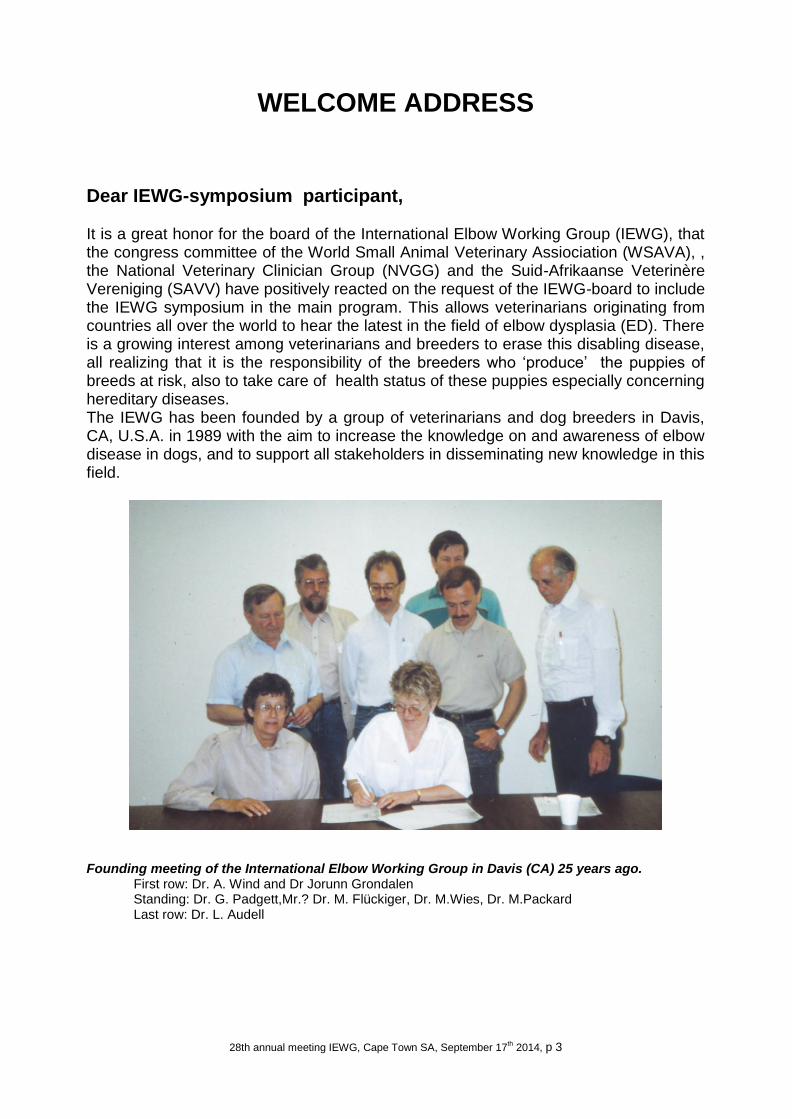
Founding meeting of the International Elbow Working Group in Davis (CA) 25 years ago.
First row: Dr. A. Wind and Dr Jorunn Grondalen Standing: Dr. G. Padgett,Mr.? Dr. M. Flückiger, Dr. M.Wies, Dr. M.Packard Last row: Dr. L. Audell
28th annual meeting IEWG, Cape Town SA, September 17th 2014, p 4
Certainly the awareness among breeders and veterinarians has increased over the past 25 years. From a bibliographic survey in PubMed, although possibly incomplete, it indicates the following trends:
Years Surgery/ arthroscopy
Clinics/diagnostic Imaging
Etiology Genetics
1980-1989 4 8 2 1
1990-1999 5 12 1 2
2000-2010 7 16 13 3
2011-2014 6 23 9 2
From this it is obvious that there is constant new information about surgical treatment and refinement, and an ever increasing amount of reports on diagnostics including a growing variety of imaging techniques and modalities. Scientists are struggling to find a proper explanation for the etiology of elbow dysplasias, especially for the fragmentation of the coronoid process. In recent years, a growing amount of hypothesis and studies focused on the anatomy, biomechanics, and abnormal development of antebrachium and elbow configurations. Although it was hypothesized at an early stage by Tirgari, Olsson, and Grondalen in their publications that the fragmented coronoid process was seen a hereditary entity in a limited amount of breeds, but only a few publications report on the molecular genetic aspects of these diseases. More knowledge of the molecular genetic background could eventually elucidate the etiology and could be a big help for the breeders to screen the dogs before mating and to develop a breeding program accordingly. Rather than via molecular genetics, screening of elbow joints with modern techniques and consequently implementing breeding measures are still of importance in our efforts to free the future generations dogs from elbow dysplasias. It is the aim of the IEWG to persuade veterinarians and breeders to perform the best possible methods to screen the breeding stock and the related animals, and to grade the findings in a transparent way allowing veterinarians and breeders to get insight in the elbow status when new dogs are introduced from other countries. For that, the IEWG makes available a screening form as printed in this proceeding. The board of the IEWG is proud that a scale of international speakers was willing to participate in the 28th meeting of the IEWG with a program of interest for the practicing veterinarian. After defining the different entities that play a major role in elbow dysplasia, presentations from different hemispheres on imaging techniques and modalities will be presented by experienced radiologists. After presentation on the clinical aspects (diagnostics, pathology and treatment), the IEWG-grading system will be explained and interactively exercises will be performed. Also on behalf of the other board members of the IEWG, Dr. B. Telhelm and Dr. K.L. How, I thank the speakers who were so kind spending their precious time to prepare and present a lecture at the IEWG symposium at the 17th of September 2014 in Kaapstad, South Africa. In addition we acknowledge the sponsorship of the IEWG by Hill’s, and the hospitality of the organizing committee of the congress committees from the 39th WSAVA-congress, the NVGG and the SAVV to host the 28th IEWG symposium.
Prof. dr. H.A.W. Hazewinkel President IEWG
The IEWG will keep in contact with interested veterinarians via its website (http://www.vet-iewg.org/joomla).
28th annual meeting IEWG, Cape Town SA, September 17th 2014, p 5
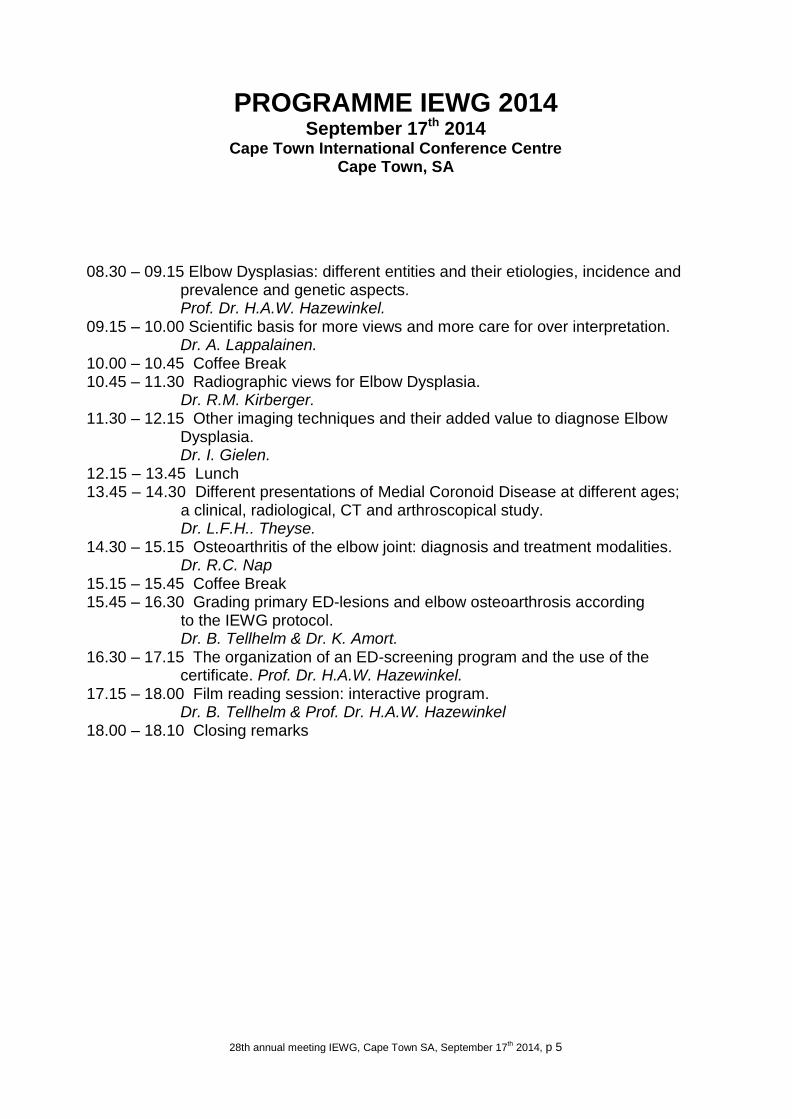
PROGRAMME IEWG 2014 September 17th 2014
Cape Town International Conference Centre Cape Town, SA
08.30 – 09.15 Elbow Dysplasias: different entities and their etiologies, incidence and prevalence and genetic aspects. Prof. Dr. H.A.W. Hazewinkel.
09.15 – 10.00 Scientific basis for more views and more care for over interpretation. Dr. A. Lappalainen.
10.00 – 10.45 Coffee Break 10.45 – 11.30 Radiographic views for Elbow Dysplasia.
Dr. R.M. Kirberger. 11.30 – 12.15 Other imaging techniques and their added value to diagnose Elbow
Dysplasia. Dr. I. Gielen.
12.15 – 13.45 Lunch 13.45 – 14.30 Different presentations of Medial Coronoid Disease at different ages;
a clinical, radiological, CT and arthroscopical study. Dr. L.F.H.. Theyse.
14.30 – 15.15 Osteoarthritis of the elbow joint: diagnosis and treatment modalities. Dr. R.C. Nap
15.15 – 15.45 Coffee Break 15.45 – 16.30 Grading primary ED-lesions and elbow osteoarthrosis according
to the IEWG protocol. Dr. B. Tellhelm & Dr. K. Amort.
16.30 – 17.15 The organization of an ED-screening program and the use of the certificate. Prof. Dr. H.A.W. Hazewinkel.
17.15 – 18.00 Film reading session: interactive program. Dr. B. Tellhelm & Prof. Dr. H.A.W. Hazewinkel
18.00 – 18.10 Closing remarks
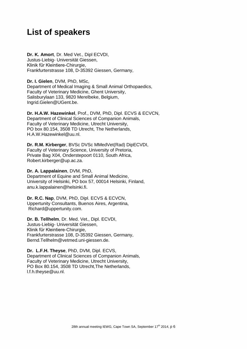
28th annual meeting IEWG, Cape Town SA, September 17th 2014, p 6
List of speakers Dr. K. Amort, Dr. Med Vet., Dipl ECVDI, Justus-Liebig- Universität Giessen, Klinik fűr Kleintiere-Chirurgie, Frankfurterstrasse 108, D-35392 Giessen, Germany,
Dr. I. Gielen, DVM, PhD, MSc, Department of Medical Imaging & Small Animal Orthopaedics, Faculty of Veterinary Medicine, Ghent University, Salisburylaan 133, 9820 Merelbeke, Belgium, [email protected].
Dr. H.A.W. Hazewinkel, Prof., DVM, PhD, Dipl. ECVS & ECVCN, Department of Clinical Sciences of Companion Animals, Faculty of Veterinary Medicine, Utrecht University, PO box 80.154, 3508 TD Utrecht, The Netherlands, [email protected]. Dr. R.M. Kirberger, BVSc DVSc MMedVet(Rad) DipECVDI, Faculty of Veterinary Science, University of Pretoria, Private Bag X04, Onderstepoort 0110, South Africa, [email protected]. Dr. A. Lappalainen, DVM, PhD, Department of Equine and Small Animal Medicine, University of Helsinki, PO box 57, 00014 Helsinki, Finland,
Dr. R.C. Nap, DVM, PhD, Dipl. ECVS & ECVCN, Uppertunity Consultants, Buenos Aires, Argentina, [email protected]. Dr. B. Tellhelm, Dr. Med. Vet., Dipl. ECVDI, Justus-Liebig- Universität Giessen, Klinik fűr Kleintiere-Chirurgie, Frankfurterstrasse 108, D-35392 Giessen, Germany, [email protected]. Dr. L.F.H. Theyse, PhD, DVM, Dipl. ECVS, Department of Clinical Sciences of Companion Animals, Faculty of Veterinary Medicine, Utrecht University, PO Box 80.154, 3508 TD Utrecht,The Netherlands, [email protected].
28th annual meeting IEWG, Cape Town SA, September 17th 2014, p 7
Elbow Dysplasias: different entities and their etiologies, incidence and prevalence and genetic aspects. Dr. H.A.W. Hazewinkel, Prof., DVM, PhD, Dipl. ECVS & ECVCN,
Introduction Elbow dysplasias (EDs) have in common that they all cause degenerative joint disease (DJD) eventually. There are at least 4 groups of entities known which can be grouped under EDs, each of them may be covering a variety of causes. -Medial coronoid disease (MCD) (Moores et al, 2008; Fitzpatrick et al 2009) formerly known as fragmented coronoid process (FCP) (Henry 1984) or ununited medial coronoid process (Tirgari 1974), but since the pathology can extend beyond the strict borders of the medial coronoid process MCD has been proposed. -Osteochondosis (OC) - osteochondritis dissecans (OCD) - OCD-like disease is referring to a radiological indentation at the medial side of the humeral condyle representing either a (temporal) thickening of the joint cartilage, a loose cartilage flap, or erased joint cartilage due to friction of a fragmented coronoid process against the young, dividing cartilage layer covering the humeral condyles. -Ununited anconeal process (UAP) this entity previously known as ED and only seen in those breeds which have a anconeal process as secondary ossification center. -Incongruity of the elbow joint, being the radio-humeral, the ulnar-humeral and the radio-ulnar joint surfaces each with their own causes and consequences. Clinically all these entities can be presented as cause of permanent lameness and eventually as cause of DJD with its typical lameness pattern with lameness at the beginning of the exercise and worsening after rest following a too heavy exercise. At clinical investigation, differentiation is not very discriminating especially since the entities are also seen in combinations (Kirberger et al 1998; Hazewinkel et al, 1998; Meyer-Lindenberg et al. 2006; Samoy et al, 2011) (Table 1). Plain radiology has always been the first-line diagnostic tool, but also the first choice as screening tool, since it is easy to perform, widely available in veterinary practices and not too expensive. A variety of preferred views are described for radiographs allowing to visualize the entities listed above. OC-OCD-OCD-like lesions can be seen best at anterioposterior views and/or mediolateral oblique views, whereas UAP can best be seen on mediolateral flexed view anticipating the superposition of the humeral condyles in case of an extended elbow joint. Incongruity due to relatively too long radius or too long ulna can best be seen in the non-twisted mediolateral (extended) view, where also the AP view can be supportive. Incongruity of the radioulnar joint cannot be visualized on plain radiographs. The sensitivity of radiographs to diagnose MCD ranges from 10-60% (Wasar et al, 1999; Haudiquet et al, 2002), and can even be false negative (Carpenter et al, 1993; Punke et al 2009, Lau et al, 2013). Knowledge about the etiologies of the entities listed above will help to understand the experience of breeders and veterinarians alike, that screening will not always predict the elbow status of the dog at an older age nor the elbow status of the offspring.
Etiology of FCP In Labradors, Golden retrievers and German Shepherd dogs (GSD), but not in Bernese Mountain dogs there is a preference for male dogs (Padgett et al 1995, Sjostrom 1998; Ryssen and Van Bree 1997, Lavrijsen et al 2014). In Labradors, but not in Bernese Mountain dogs, family clusters with MCD were identified with a higher incidence of MCD (45-60%) than other family groups (Ubbink et al 1998, 1999.). Recent research demonstrated that in Labrador puppies born out two MCD-positive parent dogs, radiographs did not detect abnormal medial coronoid development, whereas on CT in 50% of the dogs abnormal development was detected being 100% of the dogs with pathological medial coronoid process development, starting at the age of 15 weeks (Seng Fong Lau, 2013). Histologically the abnormal area of the medial coronoid process could be characterized as disturbed endochondral ossification with delayed cartilage mineralization and in some cases, a fracture line in the subchondral area could be noticed. Not in all cases with a fractured coronoid process, a fissure line in the articular cartilage was present (Seng Fong Lau, 2013). This study leads to the assumption that primary there is a delay in endochondral ossification with retained cartilage, which makes the medial coronoid vulnerable for mechanical stress. When a fragmentation in the coronoid process occurs in the subchondral area (with unmineralized cartilage and retained cartilage cores in the subchondral cancellous bone) eventually the fissure line can extend into the overlaying articular cartilage. When the latter occurs, the fragment can freely move and irritate the bordering radius and ulna, and thus leads to DJD; this can occur at young (4-8 months) but
28th annual meeting IEWG, Cape Town SA, September 17th 2014, p 8
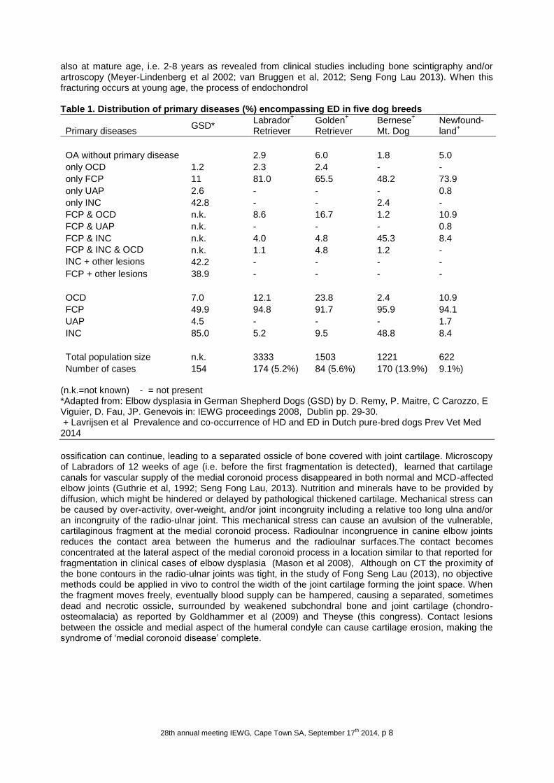
also at mature age, i.e. 2-8 years as revealed from clinical studies including bone scintigraphy and/or artroscopy (Meyer-Lindenberg et al 2002; van Bruggen et al, 2012; Seng Fong Lau 2013). When this fracturing occurs at young age, the process of endochondrol Table 1. Distribution of primary diseases (%) encompassing ED in five dog breeds
Primary diseases GSD*
Labrador+
Retriever Golden
+
Retriever Bernese
+
Mt. Dog Newfound- land
+
OA without primary disease 2.9 6.0 1.8 5.0
only OCD 1.2 2.3 2.4 - -
only FCP 11 81.0 65.5 48.2 73.9
only UAP 2.6 - - - 0.8
only INC 42.8 - - 2.4 -
FCP & OCD n.k. 8.6 16.7 1.2 10.9
FCP & UAP n.k. - - - 0.8
FCP & INC n.k. 4.0 4.8 45.3 8.4
FCP & INC & OCD n.k. 1.1 4.8 1.2 -
INC + other lesions 42.2 - - - -
FCP + other lesions 38.9 - - - -
OCD 7.0 12.1 23.8 2.4 10.9
FCP 49.9 94.8 91.7 95.9 94.1
UAP 4.5 - - - 1.7
INC 85.0 5.2 9.5 48.8 8.4
Total population size n.k. 3333 1503 1221 622
Number of cases 154 174 (5.2%) 84 (5.6%) 170 (13.9%) 9.1%) (n.k.=not known) - = not present *Adapted from: Elbow dysplasia in German Shepherd Dogs (GSD) by D. Remy, P. Maitre, C Carozzo, E Viguier, D. Fau, JP. Genevois in: IEWG proceedings 2008, Dublin pp. 29-30. + Lavrijsen et al Prevalence and co-occurrence of HD and ED in Dutch pure-bred dogs Prev Vet Med 2014
ossification can continue, leading to a separated ossicle of bone covered with joint cartilage. Microscopy of Labradors of 12 weeks of age (i.e. before the first fragmentation is detected), learned that cartilage canals for vascular supply of the medial coronoid process disappeared in both normal and MCD-affected elbow joints (Guthrie et al, 1992; Seng Fong Lau, 2013). Nutrition and minerals have to be provided by diffusion, which might be hindered or delayed by pathological thickened cartilage. Mechanical stress can be caused by over-activity, over-weight, and/or joint incongruity including a relative too long ulna and/or an incongruity of the radio-ulnar joint. This mechanical stress can cause an avulsion of the vulnerable, cartilaginous fragment at the medial coronoid process. Radioulnar incongruence in canine elbow joints reduces the contact area between the humerus and the radioulnar surfaces.The contact becomes concentrated at the lateral aspect of the medial coronoid process in a location similar to that reported for fragmentation in clinical cases of elbow dysplasia (Mason et al 2008), Although on CT the proximity of the bone contours in the radio-ulnar joints was tight, in the study of Fong Seng Lau (2013), no objective methods could be applied in vivo to control the width of the joint cartilage forming the joint space. When the fragment moves freely, eventually blood supply can be hampered, causing a separated, sometimes dead and necrotic ossicle, surrounded by weakened subchondral bone and joint cartilage (chondro-osteomalacia) as reported by Goldhammer et al (2009) and Theyse (this congress). Contact lesions between the ossicle and medial aspect of the humeral condyle can cause cartilage erosion, making the syndrome of ‘medial coronoid disease’ complete.
28th annual meeting IEWG, Cape Town SA, September 17th 2014, p 9
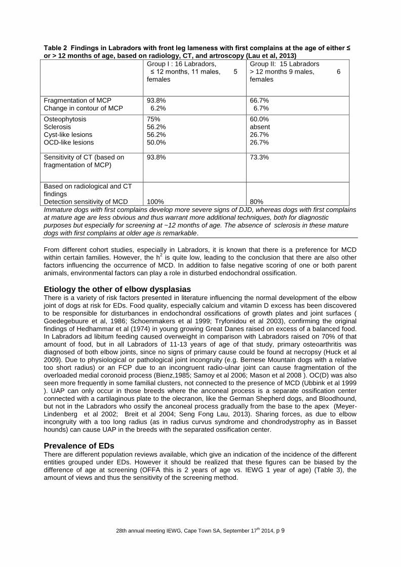
Table 2 Findings in Labradors with front leg lameness with first complains at the age of either ≤ or > 12 months of age, based on radiology, CT, and artroscopy (Lau et al, 2013)
Group I : 16 Labradors, ≤ 12 months, 11 males, 5 females
Group II: 15 Labradors > 12 months 9 males, 6 females
Fragmentation of MCP Change in contour of MCP
93.8% 6.2%
66.7% 6.7%
Osteophytosis Sclerosis Cyst-like lesions OCD-like lesions
75% 56.2% 56.2% 50.0%
60.0% absent 26.7% 26.7%
Sensitivity of CT (based on fragmentation of MCP)
93.8% 73.3%
Based on radiological and CT findings Detection sensitivity of MCD
100%
80%
Immature dogs with first complains develop more severe signs of DJD, whereas dogs with first complains at mature age are less obvious and thus warrant more additional techniques, both for diagnostic purposes but especially for screening at ~12 months of age. The absence of sclerosis in these mature dogs with first complains at older age is remarkable.
From different cohort studies, especially in Labradors, it is known that there is a preference for MCD within certain families. However, the h
2 is quite low, leading to the conclusion that there are also other
factors influencing the occurrence of MCD. In addition to false negative scoring of one or both parent animals, environmental factors can play a role in disturbed endochondral ossification.
Etiology the other of elbow dysplasias There is a variety of risk factors presented in literature influencing the normal development of the elbow joint of dogs at risk for EDs. Food quality, especially calcium and vitamin D excess has been discovered to be responsible for disturbances in endochondral ossifications of growth plates and joint surfaces ( Goedegebuure et al, 1986; Schoenmakers et al 1999; Tryfonidou et al 2003), confirming the original findings of Hedhammar et al (1974) in young growing Great Danes raised on excess of a balanced food. In Labradors ad libitum feeding caused overweight in comparison with Labradors raised on 70% of that amount of food, but in all Labradors of 11-13 years of age of that study, primary osteoarthritis was diagnosed of both elbow joints, since no signs of primary cause could be found at necropsy (Huck et al 2009). Due to physiological or pathological joint incongruity (e.g. Bernese Mountain dogs with a relative too short radius) or an FCP due to an incongruent radio-ulnar joint can cause fragmentation of the overloaded medial coronoid process (Bienz,1985; Samoy et al 2006; Mason et al 2008 ). OC(D) was also seen more frequently in some familial clusters, not connected to the presence of MCD (Ubbink et al 1999 ). UAP can only occur in those breeds where the anconeal process is a separate ossification center connected with a cartilaginous plate to the olecranon, like the German Shepherd dogs, and Bloodhound, but not in the Labradors who ossify the anconeal process gradually from the base to the apex (Meyer-Lindenberg et al 2002; Breit et al 2004; Seng Fong Lau, 2013). Sharing forces, as due to elbow incongruity with a too long radius (as in radius curvus syndrome and chondrodystrophy as in Basset hounds) can cause UAP in the breeds with the separated ossification center.
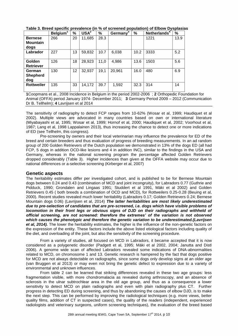
Prevalence of EDs There are different population reviews available, which give an indication of the incidence of the different entities grouped under EDs. However it should be realized that these figures can be biased by the difference of age at screening (OFFA this is 2 years of age vs. IEWG 1 year of age) (Table 3), the amount of views and thus the sensitivity of the screening method.
28th annual meeting IEWG, Cape Town SA, September 17th 2014, p 10
Table 3. Breed specific prevalence (in % of screened population) of Elbow Dysplasias
Belgium1 % USA
2 % Germany
3 % Netherlands
4 %
Bernese Mountain dogs
266 20 11,685 28.3 1221 13.9
Labrador 227 13 59,832 10.7 6,038 10.2
3333 5.2
Golden Retriever
126 18 28,923 11,0 4,986 13.6
1503 5.6
German Shepherd dog
130 12 32,937 19,1 20,961 16.0
480 6.9
Rottweiler
135 33 14,172 39.7 1,592
32.3 314 14
1Coopmans et al., 2008 Incidence in Belgium in the period 2002-2006 ; 2 Orthopedic Foundation for Animal (OFFA) period January 1974- December 2011; 3 Germany Period 2009 – 2012 (Communication Dr B. Tellhelm); 4 Lavrijsen et al 2014
The sensitivity of radiography to detect FCP ranges from 10-62% (Wosar et al, 1999; Haudiquet et al 2002). Multiple views are advocated in many countries based on own or international literature (Miyabayashi et al, 1995; Wosar et al, 1999; Hornof et al, 2000; Haudiquet et al, 2002; Voorhout et al, 1987; Lang et al, 1998 Lappalainen 2013), thus increasing the chance to detect one or more indications of ED (see Tellhelm, this congress).
Pre-screening by owners and their local veterinarian may influence the prevalence for ED of the breed and certain breeders and thus evaluation of progress of breeding measurements. In an ad random group of 200 Golden Retrievers of the Dutch population we demonstrated in 13% of the dogs ED (all had FCP, 5 dogs in addition OCD-like lesions and 4 in addition INC), similar to the findings in the USA and Germany, whereas in the national screening program the percentage affected Golden Retrievers dropped considerably (Table 3). Higher incidences than given at the OFFA website may occur due to national differences or a-selective screening (Kirberger et al, 2007)
Genetic aspects The heritability estimates differ per investigated cohort, and is published to be for Bernese Mountain dogs between 0.24 and 0.43 (combination of MCD and joint incongruity), for Labradors 0.77 (Guthrie and Pidduck, 1990; Grondalen and Lingaas 1991; Studdert et al 1991, Mäki et al 2002) and Golden Retrievers 0.45 ( both breeds a combination of OCD and MCD), for Rottweilers 0.25-0.28 (Beuing et al, 2000). Recent studies revealed much lower heritability (Labradors 0.17; Golden Retrievers 0.24; Bernese Mountain dogs 0.06) (Lavrijsen et al, 2014) The latter heritabilities are most likely underestimated due to pre-selection of candidates that are pre-screened, i.e. dogs which have visible problems of locomotion in their front legs or obvious signs of DJD on their radiographs and withheld of official screening, are not screened: therefore the extremes’ of the variation is not observed which causes the phenotypic and therefore the genetic variation to be underestimated.(Lavrijsen et al, 2014). The lower the heritability estimates, the higher is the influence of the non-genetic factors on the expression of the entity. These factors include the above listed etiological factors including quality of the diet, and overloading of the joint, but also the sensitivity of the screening procedure.
From a variety of studies, all focused on MCD in Labradors, it became accepted that it is now considered as a polygenetic disorder (Padgett et al, 1995; Mäki et al 2002, 2004; Janutta and Distl 2006). A genome wide scan of affected Labradors revealed some indications of DNA-abnormalities related to MCD, on chromosome 1 and 13. Genetic research is hampered by the fact that dogs positive for MCD are not always detectable on radiographs, since some dogs only develop signs at an older age (van Bruggen et al 2013) or may even not bring the genetic defect to expression due to a variety of environmental and unknown influences.
From table 2 can be learned that striking differences revealed in these two age groups: less fragmentation visible, with more chondromalacia as revealed during arthroscopy, and an absence of sclerosis in the ulnar subtrochlear area in the old age group, and thus as a consequence a lower sensitivity to detect MCD on plain radiographs and even with plain radiography plus CT. Further progress in detecting ED during screening, and thus by abandoning the causes of elbow DJD, is to make the next step. This can be performed by improving the radiological techniques (e.g. more views, better quality films, addition of CT in suspected cases), the quality of the readers (independent, experienced radiologists and veterinary surgeons, uniform screening techniques), the evaluation of the breed based
28th annual meeting IEWG, Cape Town SA, September 17th 2014, p 11
on the screening results (analysis of estimated breeding value to include results of relatives), the awareness of the consequences not cooping with the screening procedure. Also a radical change in detection method, e.g. development of DNA-screening technique should be considered by the breeders world. This allows for detecting carriers of the responsible gene(s) irrespective of the occurrence of the actual disease state (i.e. ED). It will thus be irrespective of radiological findings, will facilitate the availability of sensitive screening for the breeders and owners by shipping only a blood sample. It warrants however a serious investment to develop these molecular genetic techniques by well-equipped laboratories on request of the breeders clubs. Since each of the entities of ED has a polygenic inheritance, each genetic abnormality explains a relatively small part of the total genetic variation occurring in case of ED. Therefore a simple gene-test cannot be developed for these diseases. It might even be impossible to identify all chromosomal regions affecting a single trait. However, it does not imply that DNA data cannot be used for genetic improvement of poly-genetically inherited traits such as ED (Visscher et al, 2010; Lavrijsen et al, 2014). According to Meuwissen et al (2001) is the knowledge of chromosomal regions affecting a trait ( so-called Quantitative Trait Loci = QTLs), not required as long as there are a sufficient number of markers, e.g. SNPs, genotyped in the candidates. Estimated Breeding Values (EBVs) are traditionally calculated using health/disease recordings combined with information of relatives. EBVs based on DNA-data are called genomic EBVs (gEBVs): chromosomal regions that are identified to have an effect on a selection of ED should receive more weight when DNA information is used to calculate EBVs (Lavrijsen et al, 2014). However, first molecular genetic investigations should be performed, initiated by the breeders world. Till that time radiological screening with improved techniques and e.g expanded with CT-scanning will be the best way to overcome this deficit.
References Beuing, R., Mues, C., Tellhelm, B., Erhardt, G., 2000. Prevalence and inheritance of canine elbow dysplasia in German Rottweiler. Journal of Animal Breeding and Genetics 117, 375-383. Bienz HA. Klinische und radiologische Untersuchungen über den fragmentierten Processus Coronoideus Medialis im Ellenbogengelenk des Berner Sennenhundes und der anderen Sennenhunde-rassen. Inaugural-Dissertation, Zürich (S) 1985. Breit, S., Künzel, W., Seiler, S., 2004. Variation in the ossification process of the anconeal and medial coronoid processes of the canine ulna. Research in Veterinary Science 77, 9-16. Bruggen, L.W.van , Hazewinkel, H.A., Wolschrijn, C.F., Voorhout, G., Pollak, Y.W., Barthez, P.Y., 2010. Bone scintigraphy for the diagnosis of an abnormal medial coronoid process in dogs. Veterinary Radiology & Ultrasound 51, 344-348. Carpenter, L.G., Schwarz, P.D., Lowry, J.E., Park, R.D., Steyn, P.F., 1993. Comparison of radiologic imaging techniques for diagnosis of fragmented medial coronoid process of the cubital joint in dogs. Journal of the American Veterinary Medical Association 203, 78-83. Coopman, F., Verhoeven, G., Saunders, J., Duchateau, L., van Bree, H., 2008. Prevalence of hip dysplasia, elbow dysplasia and humeral head osteochondrosis in dog breeds in Belgium. Veterinary Record 163, 654-658. Fitzpatrick, N., Smith, T.J., Evans, R.B., Yeadon, R., 2009. Radiographic and arthroscopic findings in the elbow joints of 263 dogs with medial coronoid disease. Veterinary Surgery 38, 213-223. Goedegebuure SA, Hazewinkel HAW. 1986 Morphological findings in young dogs chronically fed a diet containing excess calcium. Veterinary Pathology, 23: 594-605. Goldhammer, M.A., Smith, S.H., Fitzpatrick, N., Clements, D.N., 2009. A comparison of radiographic, arthroscopic and histological measures of articular pathology in the canine elbow joint. The Veterinary Journal 186, 96-103. Grøndalen J, and Lindgaas F. 1991. Arthrosis in the elbow joint of young rapidly growing dogs: a genetic investigation. Journal of Small Animal Practice 32:460-464. Guthrie S, and Pidduck HG. 1990. Heritability of elbow osteochondrosis within a closed population of dogs. Journal of Small Animal Practice 31:93-96. Guthrie, S., Plummer, J.M., Vaughan, L.C., 1992. Post natal development of the canine elbow joint: a light and electron microscopical study. Research in Veterinary Science 52, 67-71. Haudiquet, P.R., Marcellin-Little, D.J., Stebbins, M.E., 2002. Use of the distomedial-proximolateral oblique radiographic view of the elbow joint for examination of the medial coronoid process in dogs. American Journal of Veterinary Research 63, 1000-1005. Hazewinkel, H.A.W., Meij, B. P., Theyse, L.F.H., 1998. Surgical treatment of elbow dysplasia. Veterinary Quartery 20 Supplement 1, 29-31. Hazewinkel HAW, Kantor A, Meij BP, Voorhout G 1988 Fragmented coronoid process and osteochondritis dissecans of the medial humeral condyle. Tijdschrift Diergeneesk 113, 41S-47S.
28th annual meeting IEWG, Cape Town SA, September 17th 2014, p 12
Hedhammar A, Wu FM, Krook L, Schryver HF, de LaHunta A, Wahlen JP, Kallfelz FA, Nunez EA, Hintz F, Sheffy BE, Ryan GD.1974 Overnutrition and skeletal disease. An experimental study in growing Great Dane dogs. Cornell Vet 64 (Suppl. 5): 1-160. Henry, W.B.,Jr, 1984. Radiographic diagnosis and surgical management of fragmented medial coronoid process in dogs. Journal of the American Veterinary Medical Association 184, 799-805. Hornof, W.J., Wind, A.P., Wallack, S.T., Schulz, K.S., 2000. Canine elbow dysplasia. The early radiographic detection of fragmentation of the coronoid process. Veterinary Clinics of North America: Small Animal Practice 30, 257-266. Huck JL, Biery DN, Lawler DF, Gregor TP, Runge JJ, Evans RH, Kealy RD, and Smith GK. 2009. A longitudinal study of the influence of lifetime food restriction on development of osteoarthritis in the canine elbow. Vet Surg. 38:192-198. Janutta V and Distl O. 2006. Inheritance of canine hip dysplasia: Review of estimation methods and of heritability estimates and prospects on further developments. Dtsch Tierarztl Wochenschr 113(1):6-12. Kirberger RM, Stander N.2007 Incidence of canine elbow dysplasia in South Africa. J S Afr Vet Assoc. 78:59-62 Kirberger RM
1, Fourie SL1998 Elbow dysplasia in the dog: pathophysiology, diagnosis and control. J S
Afr Vet Assoc. 69 :43-54. Lang, J., Busato, A., Baumgartner, D., Flückiger, M., Weber, U.T., 1998. Comparison of two classification protocols in the evaluation of elbow dysplasia in the dog. Journal of Small Animal Practice 39, 169-174. Lappalainen, A.K., Molsa, S., Liman, A., Laitinen-Vapaavuori, O., Snellman, M., 2009. Radiographic and computed tomography findings in Belgian shepherd dogs with mild elbow dysplasia. Veterinary Radiology & Ultrasound 50, 364-369. Lavrijsen, I.C., Heuven, H.C., Voorhout, G., Meij, B.P., Theyse, L.F., Leegwater, P.A., Hazewinkel, H.A., 2012. Phenotypic and genetic evaluation of elbow dysplasia in Dutch Labrador retrievers, Golden retrievers, and Bernese Mountain Dogs. The Veterinary Journal 193, 486-492. Lavrijsen IC, Heuven HC, Meij BP, Theyse LF, Nap RC, Leegwater PA, Hazewinkel HA. 2014 Prevalence and co-occurrence of hip dysplasia and elbow dysplasia in Dutch pure-bred dogs.Prev Vet Med.1;114(2):114-122 Mäki, K., Groen, A.F., Liinamo, A.E., Ojala, M., 2002. Genetic variances, trends and mode of inheritance for hip and elbow dysplasia in Finnish dog populations. Animal Science 75, 197-207. Mason DR, Schulz KS, Fujita Y, Kass PH, Stover SM 2008 Measurement of humeroradial and humeroulnar transarticular joint forces in the canine elbow joint after humeral wedge and humeral slide osteotomies. Vet Surg. ;37:63-70 Meuwissen TH, Goddard ME., 2001. Prediction of identity by descent probabilities from marker-haplotypes. Genet Sel Evol. 33(6):605-634. Meyer-Lindenberg, A., Langhann, A., Fehr, M., Nolte, I., 2002. Prevalence of fragmented medial coronoid process of the ulna in lame adult dogs. The Veterinary Record 151, 230-234. Miyabayashi, T., Takiguchi, M., Schrader, S.C., Biller, D.S., 1995. Radiographic anatomy of the medial coronoid process of dogs. Journal of the American Animal Hospital Association 31, 125-132. Moores, A.P., Benigni, L., Lamb, C.R., 2008. Computed tomography versus arthroscopy for detection of canine elbow dysplasia lesions. Veterinary Surgery 37, 390-398. Orthopedic Foundation for Animals http://www.offa.org/stats_ed.html Padgett, G.A., Mostosky, U.V., Probst, C.W., Thomas, M.W., Krecke, C.F., 1995. The inheritance of osteochondritis dissecans and fragmented coronoid process of the elbow joint in Labrador retrievers. Journal of the American Animal Hospital Association 31, 327-330. Punke, J.P., Hulse, D.A., Kerwin, S.C., Peycke, L.E., Budsberg, S.C., 2009. Arthroscopic documentation of elbow cartilage pathology in dogs with clinical lameness without changes on standard radiographic projections. Veterinary Surgery 38, 209-212. Ryssen B van, van Bree H. Arthroscopic findings in 100 dogs with elbow lameness. 1997 Vet Rec. 140 ,:360-362 Samoy, Y., Van Ryssen, B., Gielen, I., Walschot, N., van Bree, H., 2006. Review of the literature: elbow incongruity in the dog. Veterinary and Comparative Orthopaedics and Traumatology 19, 1-8. Schoenmakers I, Hazewinkel HAW, Voorhout G, Carlson CS, Richardson D. 2000 Effect of diets with different calcium and phosphorius contents on the skeletal development and blood chemistry of growing great danes. Veterinary Record 147, 652-660. Seng Fong Lau 2013 Development of medial coronoid disease in Labrador retrievers: diagnostic and pathogenic studies, Thesis Utrecht University October 2013 ISBN 978-90-393-5962-5964 Seng Fong Lau, Wolschrijn CF, Hazewinkel HA, Siebelt M, Voorhout G. 2013 The early development of medial coronoid disease in growing Labrador retrievers: radiographic, computed tomographic, necropsy and micro-computed tomographic findings. Vet J 197:724-730 Sjöström, L., 1998. Ununited anconeal process in the dog. Veterinary Clinics of North America: Small Animal Practice 28, 75-86.
28th annual meeting IEWG, Cape Town SA, September 17th 2014, p 13
Studdert, V.P., Lavelle, R.B., Beilharz, R.G., Mason, T.A., 1991. Clinical features and heritability of osteochondrosis of the elbow in Labradors Retrievers. Journal of Small Animal Practice 32, 557-563. Swenson L, Audell L, Hedhammar A. 1997a. Prevalence and inheritance of and selection for elbow arthrosis in bernese mountain dogs and rottweilers in sweden and benefit: Cost analysis of a screening and control program. J Am Vet Med Assoc 210(2):215-221. Tellhelm, B., 2011. Grading primary ED-lesions and elbow osteoarthrosis according to the IEWG protocol. Proceedings of the 26th Annual Meeting of International Elbow Working Group. Amsterdam, Holland. pp. 14-15. Tirgari, M., 1974. Clinical radiographical and pathological aspects of arthritis of the elbow joint in dogs. Journal of Small Animal Practice 15, 671-679. Tryfonidou MA, Holl MS, Stevenhagen JJ, Buurman CJ, Deluca HF, Oosterlaken-Dijksterhuis MA, van den Brom WE, van Leeuwen JPTM, Hazewinkel HAW. 2003 Dietary 135-fold vitamin D3 supplementation severely disturbs the endochondral ossification in growing dogs. Dom Anim Endocrinol 24: 265-285 Ubbink, G.J., van de Broek, J., Hazewinkel, H.A., Rothuizen, J., 1998. Cluster analysis of the genetic heterogeneity and disease distributions in purebred dog populations. Veterinary Record 142, 209-213. Ubbink, G.J., H.A.W. Hazewinkel, J. van de Broek, J. Rothuizen 1999. Familial clustering and risk analysis for fragmented coronoid process and elbow joint incongruity in Bernese Mountain Dogs in The Netherlands - Am J Vet Res 60, 1082-1087 Visscher PM, McEvoy B, Yang J., 2010. From Galton to GWAS: quantitative genetics of human height.Genet Res (Camb) 92, 371-379. Voorhout, G., Hazewinkel, H.A.W., 1987. Radiographic evaluation of the canine elbow joint with special reference to the medial humeral condyle and the medial coronoid process. Veterinary Radiology 28, 158-165. Wosar, M.A., Lewis, D.D., Neuwirth, L., Parker, R.B., Spencer, C.P., Kubilis, P.S., Stubbs, W.P., Murphy, S.T., Shiroma, J.T., Stallings, J.T., Bertrand, S.G., 1999. Radiographic evaluation of elbow joints before and after surgery in dogs with possible fragmented medial coronoid process. Journal of the American Veterinary Medical Association 214, 52-58.
28th annual meeting IEWG, Cape Town SA, September 17th 2014, p 14
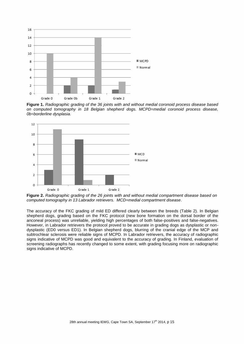
Scientific basis for more views and more care for over interpretation. Dr. A. Lappalainen, DVM, PhD. Elbow dysplasia (ED) refers to four hereditary developmental disorders of the elbow joint: the medial coronoid process disease (MCPD), also known as FCP (fragmented coronoid process), osteochondrosis (OC) of the medial part of the humeral condyle, ununited anconeal process, and incongruity of the elbow joint. Medial compartment disease (MCD) refers to all pathologies (MCPD, OC and the contact or “kissing” lesion caused by friction of the diseased MCP) of the medial side of the elbow joint. Each type can present alone or in any combination. Incongruity is currently proposed to be a common aetiology for the other three forms of ED. Some elbow conditions are not considered to be included in ED, e.g. calcified bodies seen near or at the medial epicondyle of the humerus. Calcified bodies have been referred to as ununited medial epicondyle, medial epicondylar spur, or calcified body in the joint capsule. In recent literature, these conditions are grouped together, as they are all new bone formation of tendons, either at the insertion or further away of the joint [1], and the term flexor enthesopathy has been suggested. In Labrador retrievers, a hereditary background has been suggested [2]. MCPD can be a diagnostic challenge radiographically since the radiological findings are often sparse or non-existent [3]. Therefore, an approach using secondary osteophytes as an indicator of ED was advocated in screening protocols [4]. However, abnormal contour and indistinctive cranial border of the MCP and subtrochlear sclerosis are radiological signs frequently seen in mediolateral projections (ML) in MCPD. Subtrochlear sclerosis, manifesting as increased radiopacity at the base of the coronoid process visible in ML radiographs, has proved to be a reliable indicator of MCPD. OC is radiographically best diagnosed from craniocaudal (CrCd) oblique radiographs as a radiolucent defect at the articular surface of the medial humeral condyle. A differential diagnosis for OC on radiographs is an abrasive contact (“kissing”) lesion caused by MCPD. Computed tomography (CT) is considered an accurate method for imaging the canine elbow joint, although some lesions might not be visible. CT can in most cases clearly show MCPD. Comparison of radiographic and CT findings of Belgian shepherd dogs and Labrador retrievers with grade ED1 elbows [5,6] Belgian shepherd dogs and Labrador retrievers have fairly similar screening statistics according to the Finnish Kennel Club’s database (12% and 17%, respectively), but Belgian shepherd dogs seldom have clinical elbow disease compared to Labrador retrievers. In Finland, ED 1 refers to mild OA with osteophyte formation of < 2 mm detected usually on the dorsal surface of the anconeal process (Table 1). In the studied population, according to the CT, MCPD was evident in 14% of the joints in 19% of the Belgian shepherd dogs (Figure 1). In contrast, MCD was found in 54% of the joints in 77% of the Labrador retrievers (Figure 2).
Table 1. Elbow dysplasia grades and their definitions used in the Finnish screening protocol
Grade 0 (free) No signs of OA
Grade 1 (mild) Mild OA with osteophyte formation of < 2 mm detected usually on the dorsal surface of the anconeal process
Grade 2 (moderate) Osteophytes on the dorsal surface of the anconeal process 2-5 mm high, changes in the MCP or the joint is mildly deformed
Grade 3 (severe)
Marked degenerative changes visible, or osteophyte formation on the anconeal process is over 5 mm high, UAP
OA = osteoarthritis, MCP = medial coronoid process, UAP = ununited anconeal process
28th annual meeting IEWG, Cape Town SA, September 17th 2014, p 15
Figure 1. Radiographic grading of the 36 joints with and without medial coronoid process disease based on computed tomography in 18 Belgian shepherd dogs. MCPD=medial coronoid process disease, 0b=borderline dysplasia.
Figure 2. Radiographic grading of the 26 joints with and without medial compartment disease based on computed tomography in 13 Labrador retrievers. MCD=medial compartment disease.
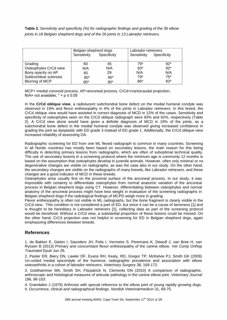
The accuracy of the FKC grading of mild ED differed clearly between the breeds (Table 2). In Belgian shepherd dogs, grading based on the FKC protocol (new bone formation on the dorsal border of the anconeal process) was unreliable, yielding high percentages of both false-positives and false-negatives. However, in Labrador retrievers the protocol proved to be accurate in grading dogs as dysplastic or non-dysplastic (ED0 versus ED1). In Belgian shepherd dogs, blurring of the cranial edge of the MCP and subtrochlear sclerosis were reliable signs of MCPD. In Labrador retrievers, the accuracy of radiographic signs indicative of MCPD was good and equivalent to the accuracy of grading. In Finland, evaluation of screening radiographs has recently changed to some extent, with grading focusing more on radiographic signs indicative of MCPD.
28th annual meeting IEWG, Cape Town SA, September 17th 2014, p 16
Table 2. Sensitivity and specificity (%) for radiographic findings and grading of the 36 elbow
joints in 18 Belgian shepherd dogs and of the 26 joints in 13 Labrador retrievers.
MCP= medial coronoid process, AP=anconeal process, CrCd=craniocaudal projection, N/A= not available, * = p ≤ 0.05 In the CrCd oblique view, a radiolucent subchondral bone defect on the medial humeral condyle was observed in 19% and flexor enthesopathy in 4% of the joints in Labrador retrievers. In this breed, the CrCd oblique view would have assisted in correct diagnosis of MCD in 12% of the cases. Sensitivity and specificity of osteophytes seen on the CrCd oblique radiograph were 93% and 92%, respectively (Table 2). A CrCd view alone would have given a definite diagnosis of MCD in 19% of the joints, as a subchondral bone defect in the medial humeral condyle was observed giving increased confidence in grading the joint as dysplastic with ED grade 3 instead of ED grade 1. Additionally, the CrCd oblique view increased reliability of assessing OA.
Radiographic screening for ED from one ML flexed radiograph is common in many countries. Screening in all Nordic countries has mostly been based on secondary lesions, the main reason for this being difficulty in detecting primary lesions from radiographs, which are often of suboptimal technical quality. The use of secondary lesions in a screening protocol where the minimum age is commonly 12 months is based on the assumption that osteophytes develop in juvenile animals. However, often only minimal or no degenerative changes are visible on radiographs, as was the case also in our study. On the other hand, the secondary changes are visible on the radiographs of many breeds, like Labrador retrievers, and these changes are a good indicator of MCD in these breeds. Osteophytes arise usually first on the proximal surface of the anconeal process. In our study, it was impossible with certainty to differentiate osteophytes from normal anatomic variation of the anconeal process in Belgian shepherd dogs using CT. However, differentiating between osteophytes and normal anatomy of the anconeal process might have less weight in evaluation of the screening radiographs in Belgian shepherd dogs if the radiological findings of MCPD weigh more in grading. Flexor enthesopathy is often not visible in ML radiographs, but the bone fragment is clearly visible in the CrCd view. This condition is not considered a part of ED, but since it can be a cause of lameness [1] and is thought to be hereditary in Labrador retrievers [2], collecting data as part of the screening protocol would be beneficial. Without a CrCd view, a substantial proportion of these lesions could be missed. On the other hand, CrCd projection was not helpful in screening for ED in Belgian shepherd dogs, again emphasizing differences between breeds.
References
1. de Bakker E, Gielen I, Saunders JH, Polis I, Vermeire S, Peremans K, Dewulf J, van Bree H, van Ryssen B (2013) Primary and concomitant flexor enthesiopathy of the canine elbow. Vet Comp Orthop Traumatol Epub Jun 26.
2. Paster ER, Biery DN, Lawler DF, Evans RH, Kealy, RD, Gregor TP, McKelvie PJ, Smith GK (2009) Un-united medial epicondyle of the humerus: radiographic prevalence and association with elbow osteoarthritis in a cohort of labrador retrievers. Veterinary Surgery 38, 169-172.
3. Goldhammer MA, Smith SH, Fitzpatrick N, Clements DN (2010) A comparison of radiographic, arthroscopic and histological measures of articular pathology in the canine elbow joint. Veterinary Journal 186, 96-103.
4. Grøndalen J (1979) Arthrosis with special reference to the elbow joint of young rapidly growing dogs. II. Occurrence, clinical and radiographical findings. Nordisk Veterinärmedicin 31, 69-75.
Belgian shepherd dogs Sensitivity Specificity
Labrador retrievers Sensitivity Specificity
Grading Osteophytes CrCd view Bony opacity on AP Subtrochlear sclerosis Blurring of MCP
60 45 N/A 29
79* 93* N/A 79* 86*
92* 92* N/A 75* 83*
N/A
40
80* 90*
80* 90*
28th annual meeting IEWG, Cape Town SA, September 17th 2014, p 17
5. Lappalainen AK, Mölsä S, Liman A, Laitinen-Vapaavuori O, Snellman M (2009) Radiographic and computed tomography findings in Belgian shepherd dogs with mild elbow dysplasia. Veterinary Radiology & Ultrasound 50, 364-369.
6. Lappalainen AK, Mölsä S, Liman A, Snellman M, Laitinen-Vapaavuori, O (2013) Evaluation of accuracy of the Finnish elbow dysplasia screening protocol in Labrador retrievers. Journal of Small Animal Practice 54, 195-200.
28th annual meeting IEWG, Cape Town SA, September 17th 2014, p 18
Radiographic views for Elbow Dysplasia. Dr. R.M. Kirberger, BVSc DVSc MMedVet(Rad) DipECVDI.
Introduction Radiographs are the routine imaging modality practitioners use to diagnose elbow dysplasia. As early
osteophytic changes and pathology associated with medial coronoid disease may be subtle, optimal
imaging techniques are essential to improver diagnostic accuracy. Standard film screen techniques
should use slow (detail) screens and short scale contrast techniques. No grid is required. Digital imaging
is more forgiving regarding image quality assuming the correct look up tables are used and standard
exposure principles are applied. Remember to collimate to the joint and not to over collimate on digital
systems.
Radiographs are usually taken in lateral or sternal recumbency. They may also be made in dorsal recumbency or with horizontal beam radiography but these are not described here.
Standard views Mediolateral extended For a mediolateral extended (ML extended) view the patient is positioned in lateral recumbency lying on the affected limb. The upper limb is retracted caudally and the head and neck are slightly extended. The angle between the humerus and radius and ulna is 120 degrees. The beam is centred on the medial epicondyle. This view optimizes the following:
• Evaluation of elbow incongruity • Osteophytes on the cranial aspect of the joint and lateral epicondylar crest • Medial coronoid process which is superimposed on the radial head.
Craniocaudal For a craniocaudal (CrCd) view the patient is positioned in sternal recumbency ensuring the humerus, radius and ulna are in a straight line. The head is elevated and retracted away from the affected limb. A thin foam pad under the elbow may prevent rotation. The beam is centred on the joint space just distal to the prominent medial epicondyle. This view optimizes the following:
• Medial humeral condyle osteochondral defects • Osteophytes on the medial humeral epicondyle • Distinguishing the supinator long tendon sesamoid from a fragmented medial coronoid process.
Mediolateral maximally flexed For a mediolateral maximally flexed (ML flexed) view the patient is positioned in lateral recumbency lying on the affected limb. The upper limb is retracted. The distal antebrachium is pulled towards the neck so that the angle between the humerus and radius and ulna is <45 degrees. The carpus should not be elevated to maintain the elbow in a true lateral position. The beam is centred on the medial epicondyle. This view optimizes the following:
• Osteophytes on the anconeal process • Ununited anconeal process • Flexor enthesopathy.
Extended supinated mediolateral For an extended supinated mediolateral (Cd75°MCrLO) view the patient is positioned in lateral recumbency lying on the affected limb. The upper limb is retracted. The joint is maximally extended and the limb supinated about 15 degrees. The beam is centred on the medial epicondyle. This view optimizes the cranial border of the medial coronoid process and increases the possibilities of detecting a fragmented medial coronoid process as the primary beam is more likely to be in line with the fragment edge Craniolateral-caudomedial oblique (pronated view) For a craniolateral-caudomedial oblique (Cr15°LCdMO) view the patient is positioned in sternal recumbency ensuring the humerus, radius and ulna are in a straight line and the limb is pronated 15 degrees (15–50 degrees is the range in the literature). The beam is centred on the joint. This view optimizes the following:
• Medial humeral condyle osteochondral defects • Elbow incongruity but extended ML view is more reliable • The medial coronoid process as it is isolated from other structures, improving visibility of
fragments.
28th annual meeting IEWG, Cape Town SA, September 17th 2014, p 19
Craniomedial-caudolateral oblique (supinated view) For a craniomedial-caudolateral oblique (Cr45°MCdLO) view the patient is positioned in sternal recumbency ensuring the humerus, radius and ulna are in a straight line and the limb is supinated 45–50 degrees. The beam is centred on the joint. This is not a standard elbow dysplasia view but is useful to optimize the following:
• Visibility of the lateral humeral condyle • Visibility of the supinatir longus tendon sesamoid which could be confused with a medial
coronoid fragment on ML views • Incomplete ossification of the humeral condyle; best seen on 15 degree supination.
Distomedial-proximolateral oblique Distomedial-proximolateral oblique (Di35°MPrLO) view is also known as the medlap view. The patient is positioned in lateral recumbency lying on the affected limb. The upper limb is retracted. The joint is flexed to 90 degrees, the antebrachium elevated 35 degrees and the extremity supinated 40 degrees. A foam wedge may be used for this. The beam is centred on the medial epicondyle. This view optimizes the medial coronoid process, which is now seen proximal to or superimposed on the humero-radial joint.
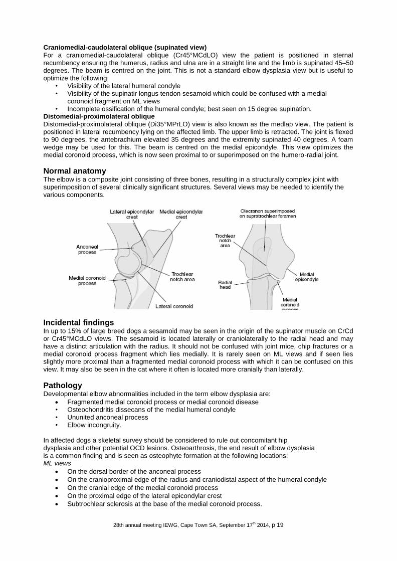
Normal anatomy The elbow is a composite joint consisting of three bones, resulting in a structurally complex joint with superimposition of several clinically significant structures. Several views may be needed to identify the various components.
Incidental findings In up to 15% of large breed dogs a sesamoid may be seen in the origin of the supinator muscle on CrCd or Cr45°MCdLO views. The sesamoid is located laterally or craniolaterally to the radial head and may have a distinct articulation with the radius. It should not be confused with joint mice, chip fractures or a medial coronoid process fragment which lies medially. It is rarely seen on ML views and if seen lies slightly more proximal than a fragmented medial coronoid process with which it can be confused on this view. It may also be seen in the cat where it often is located more cranially than laterally.
Pathology Developmental elbow abnormalities included in the term elbow dysplasia are:
Fragmented medial coronoid process or medial coronoid disease • Osteochondritis dissecans of the medial humeral condyle • Ununited anconeal process • Elbow incongruity.
In affected dogs a skeletal survey should be considered to rule out concomitant hip dysplasia and other potential OCD lesions. Osteoarthrosis, the end result of elbow dysplasia is a common finding and is seen as osteophyte formation at the following locations: ML views
On the dorsal border of the anconeal process
On the cranioproximal edge of the radius and craniodistal aspect of the humeral condyle
On the cranial edge of the medial coronoid process
On the proximal edge of the lateral epicondylar crest
Subtrochlear sclerosis at the base of the medial coronoid process.
28th annual meeting IEWG, Cape Town SA, September 17th 2014, p 20
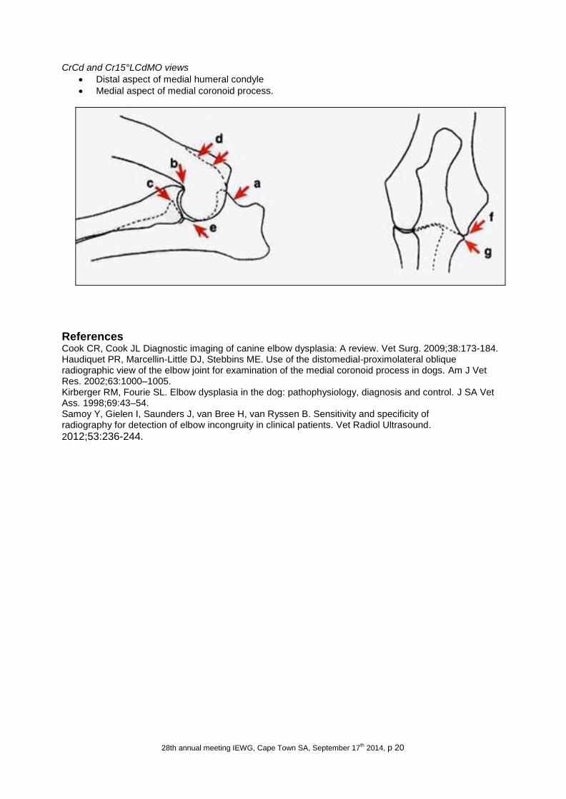
CrCd and Cr15°LCdMO views
Distal aspect of medial humeral condyle
Medial aspect of medial coronoid process.
References Cook CR, Cook JL Diagnostic imaging of canine elbow dysplasia: A review. Vet Surg. 2009;38:173-184. Haudiquet PR, Marcellin-Little DJ, Stebbins ME. Use of the distomedial-proximolateral oblique radiographic view of the elbow joint for examination of the medial coronoid process in dogs. Am J Vet Res. 2002;63:1000–1005. Kirberger RM, Fourie SL. Elbow dysplasia in the dog: pathophysiology, diagnosis and control. J SA Vet Ass. 1998;69:43–54. Samoy Y, Gielen I, Saunders J, van Bree H, van Ryssen B. Sensitivity and specificity of radiography for detection of elbow incongruity in clinical patients. Vet Radiol Ultrasound.
2012;53:236-244.
28th annual meeting IEWG, Cape Town SA, September 17th 2014, p 21
Other imaging techniques and their added value to diagnose Elbow Dysplasia.
Dr. I. Gielen, DVM, PhD, MSc, Dr. H. van Bree, Prof., DVM, PHD, Dipl. ECVS & ECVDI.
The diagnosis of elbow dysplasia (ED) in lame dogs is made from a combination of clinical signs, palpation of the joints, and medical imaging. A wide range of imaging options is now available but the “perfect” imaging protocol does not exist because each modality has his strengths and limitations. Although radiography is still the standard technique for diagnosing elbow disorders in the dog, other imaging techniques like scintigraphy, ultrasound, computed tomography (CT) and magnetic resonance imaging (MRI) can be useful. In diagnosing ED there a two different issues: there is the need for selecting ED free breeding stock and there is the diagnosis of the condition in the individual patient presented for forelimb lameness. For selection purposes, most of the time the secondary degenerative joint (DJD) changes are scrutinised by means of radiographs and mostly the individuals are not suffering lameness. For the individual patient the early diagnosis of the primary lesion is very important because an early treatment guaranties a better prognosis. Although the most important cause of elbow lameness in dogs is medial coronoid disease (MCD), recently flexor enthesopathy (FE) has been recognized as an elbow disorder in medium and large breed dogs and is characterized by lesions of the medial epicondyle and the attaching flexor muscles. The differential diagnosis between both elbow disorders is not obvious and a combination of these two elbow diseases is possible. The challenge in these cases is to define the cause of the elbow pain in order to make the correct treatment decision. In both, MCD and FE, the radiographic features may be minimal and indistinct. In case where the clinical examination is not providing a clear localisation or in case of uncertain radiographic findings, scintigraphy is a useful technique to localise the cause of lameness. Although it is very sensitive, it is not very specific and the spatial resolution offered, is not well enough to specify anatomic structures. Recently a micro-single photon emission tomography (μ-SPECT) technique has been described. HiSPECT has a much higher resolution and allows better differentiation of the anatomical areas in the elbow joint. A major drawback to joint imaging by scintigraphy is the normal uptake at the end of long bones, especially in immature animals. In some instances it is difficult to determine whether a difference in counts between two joints represents a meaningful finding. Comparison of bilateral images, acquired over the same time, and quantitative analysis of joint images by computer can provide diagnostic guidelines. In cases of flexor enthesopathy (FE), HiSPECT, reveals focal increased bone tracer uptake in the region of the medial humeral epicondyle. Ultrasound (US) is a potential valuable imaging technique of the musculoskeletal system in small animals. High frequency linear transducers are used because of their flat application surface and high resolution power. Accurate examination of joints requires substantial ultrasonographic experience and a standardised examination procedure. In most of the joints even small amounts of fluid accumulation (hypo- to anechoic) can be easily demonstrated in the area of the joint pouches. Although a thorough US study of the normal elbow joint has been conducted, US is only of limited use in the diagnosis of a fragmented
28th annual meeting IEWG, Cape Town SA, September 17th 2014, p 22
coronoid process. Only large displaced fragments can be diagnosed with certainty. Also US is helpful in diagnosing flexor tendon pathology. The main ultrasonographic findings of flexor enthesopathy are pre-insertional hypoechoic swelling, outward bowing and thickening of the common tendon of the flexor muscles. The tendon appears to be heterogenous with decreased echogenicity and focal or diffuse areas of irregular fibrillar appearance and ill- defined margins with partial or complete tears. Additionally cortical irregularities at the medial epicondyle (spur formation) and intratendinous calcifications can be detected. Computed Tomography (CT) can help significantly in establishing a definite diagnosis. The positioning of the patient is very important and CT of both elbow joints extended with the head pulled back outside the gantry results is better quality images and less artefacts. The scan parameters kV and mA should be high and thin slices eventually with an overlap are preferred. Images should be obtained in bone algorithm and proper windowing during the evaluation of a study is a necessity. The modality of multiplanar reconstructions in different planes is useful in order to evaluate the complete joint surface. Abnormalities in the area of the medial coronoid process include: fragmentation (displaced or nondisplaced), fissure, abnormal shape, sclerosis, osteophytes, and lucencies. A recent study attempts to objectivise the measurement of sclerosis. In the area of the medial humeral condyle sclerosis, lucency, and/or flattening can be evaluated and a differential diagnosis between kissing lesions and real OCD lesions can be made All these abnormalities can be diagnosed on the transverse and reconstructed images. In several cases CT findings, like fissures at medial coronoid process and subchondral luscencies at medial humeral condyle, were useful for decision making in the arthroscopic treatment of these lesions. A recent study shows that CT is a very reliable technique to evaluate fragmented coronoid process and its results are comparable with arthroscopy, still considered to be the “gold standard”. Ununited anconeal process with or without humeroulnar incongruity can be appreciated and the incidence of incongruities of the humeroradial, humeroulnar, and/or radioulnar joints can be accurately appreciated. On transverse CT slices, at the level of the trochlear notch of the ulna and the humerus, the fitting of the joint space can be noticed. On the reconstructions in the sagittal and dorsal plane, at the level of the trochlea humeri and the lateral ompartment the incidence of a step between the ulna and radial head, the shape of the trochlear notch and the fitting of the humeral condyle in the trochlear notch can be evaluated. In cases of FE, the medial epicondyle appears sclerotic and shows a clear periosteal reaction in all cases. Mineralized opacities can be present within the flexor tendons. CT also shows concomitant lesions like coronoid disease whenever present. The soft tissue studies presents a thickening of the involved tendons in and IV administration of contrast shows enhancement in the affected tendons. Arthro-CT can be used to evaluate loss of cartilage in cases of medial compartment syndrome. Magnetic Resonance Imaging (MRI) has limitations for imaging the canine elbow based on the relatively small size of the joint and complex articulations in conjunction with the thin articular cartilage surfaces of the humerus, radius, and ulna. These limitations depend also of the field strength of the MR device. All MRI planes, dorsal, sagittal, and axial/transverse, are potentially useful for diagnosis of elbow disorders. The incidence of subchondral bone pathology and oedema can be diagnosed. This technique offers a great visualisation of the soft tissues around the elbow joint and in cases of pathology within the flexor tendons its application can be very useful. On Magnetic Resonance Imaging (MRI), the sagittal T2- weighted sequence reveals a hyperintense signal around the proximal aspect of the flexor muscles extending in the muscle bellies. This signal can be confirmed as being a fluid signal
28th annual meeting IEWG, Cape Town SA, September 17th 2014, p 23
on the fat suppressed STIR sequence. The T1 and T2 studies showed a thickening and irregular delineation of the involved tendons. There is obvious enhancement on T1 contrast studies. As well as providing valuable diagnostic information about the elbow, arthroscopy also allows minimally-invasive treatment of coronoid disease. It allows us to obtain a magnified panoramic view of the inside of a joint. The drawback of arthroscopy is that it only allows the inspection of the articular surface. The combination of CT and arthroscopy allows a more complete diagnosis of ED. In cases of FE, arthroscopy shows the presence of loose fibres, degenerated tendinous tissue, cartilage loss and/or local synovitis at the attachment of the flexor muscles to the medial humeral epicondyle.
Suggested reading: Y. Baeumlin, L. De Rycke, A. Van Caelenberg, H. Van Bree, I Gielen. Magnetic Resonance Imaging of the Canine Elbow: An Anatomic Study. Vet Surg. 2010, 39(5): 566-573. De Rycke LM, Gielen IM, van Bree H, et al. Computed tomography of the elbow joint in clinically normal dogs. American Journal of Veterinary Research 2002, 63: 1400-1407. De Bakker, Evelien, Gielen Ingrid, Kromhout Kaatje, van Bree Henri, and Van Ryssen Bernadette. Magnetic Resonance Imaging of Primary and Concomitant Flexor Enthesopathy in the Canine Elbow. Veterinary Radiology & Ultrasound 2014. 55 (1): 56–62. De Bakker, Evelien, Gielen Ingrid, Van Caelenberg Annemie, van Bree Henri, and Van Ryssen Bernadette. Computed Tomographic Findings of Canine Elbow Joints Affected by Primary and Concomitant Flexor Enthesopathy. Veterinary Radiology & Ultrasound 2014.55 (1): 45–55. De Bakker, Evelien · Saunders, Jimmy H · van Bree, Henri · Gielen, Ingrid · Van Ryssen, Bernadette. Radiographic features of primary and concomitant flexor enthesopathy in the canine elbow. Vet Radiol Ultrasound. 2013;54(2):107-13.
De Bakker E, Samoy Y, Gielen I, et al. Medial humeral epicondylar lesions in the canine elbow. A review of the literature. Vet Comp Orthop Traumatol 2011;24:9–17. Gielen I, Van Ryssen B, Buijtels J, et al. Canine elbow incongruity evaluated with computerised tomography (CT), radiography and arthroscopy. Abstracts 8TH Annual EAVDI Conference, July, 18-21st, 2001, P22. Lamb CR, Wong K. Ultrasonographic anatomy of the canine elbow. Vet Radiol Ultrasound 46:319-25, 2005. K. Peremans, S. Vermeire, A. Dobbeleir, I. Gielen, Y. Samoy, K. Piron, E. Vandermeulen, G. Slegers, H. van Bree, B. De Spiegeleer, K. Dik. Recognition of anatomical predilection sites in canine elbow pathology on bone scans using micro-single photon emission tomography. The Veterinary Journal 2011,188(1): 64-72. Samoy Y, Van Ryssen B, Gielen I, et al. Review of the literature: Elbow incongruity in the dog. Vet Comp Orthop Traumatol 2006; 19: 1-8.
Seyrek-Intas D, Michele U, Tacke S, et al. Accuracy of ultrasonography in detecting fragmentation of the medial coronoid process in dogs. J Am Vet Med Assoc 2009;234:480–5. T.C. Tromblee, J.C. Jones, A.M. Bahr, et al. Effect of computed tomography display window and image plane on diagnostic certainty for characteristics of dysplastic elbow joints in dogs. American Journal of Veterinary Research 2007, 68: 858-871. van Bree H, Van Ryssen B. Diagnostic imaging of the canine elbow including radiology,arthroscopy and computed tomography (CT). Oral Abstracts, 10th IRVA Meeting. Vet Radiol Ultrasound. 1994, 35: p 248, nr 069.
van Bree H, Gielen I, Van Ryssen B, et al. Comparative joint imaging in small animals. The European Journal of Companion Animal Practice 2002, 12: 25-36. van Bree H, Gielen I, Van Ryssen B, De Rooster H. Early diagnosis of fragmented coronoid process in the dog: elbow arthroscopy compared to radiographic signs of degenerative joint disease. The European Journal of Companion Animal Practice 2012. 22(4). p.6-14.
28th annual meeting IEWG, Cape Town SA, September 17th 2014, p 24
Different presentations of medial coronoid disease at different ages: a clinical, radiological, CT and arthroscopic study. Dr. L.F.H. Theyse, PhD, DVM, Dipl. ECVS, Medial coronoid disease is the most common cause of fore limb lameness in young and adult dogs. The term medial coronoid disease includes all the pathological changes which can be attributed to fragmentation of the medial coronoid process. Although fragmentation is an important feature, coronoid disease can be present without any clear fragmentation of bony and cartilaginous structures. In view of this the typical pathology of the coronoid can me described with the term coronoid dysplasia while using the term medial coronoid disease for the combination of successive secondary pathologies including osteoarthritis. Coronoid dysplasia is a common cause of lameness in the Labrador retriever with a prevalence of MCD of 6% in a screened cohort of Dutch Labrador Retrievers(1). The diagnosis MCD is based on a combination of clinical evaluation, radiography, and CT imaging. For final diagnosis and treatment arthroscopy is considered the gold standard(2,3). Plain radiography following the guidelines of the International Elbow Working Group is used for elbow dysplasia screening programs(1). The radiographic diagnosis MCD is based on the detection of secondary degenerative joint and bone changes, including periarticular osteophytosis at specific joint locations, ulnar subtrochlear sclerosis (STS), and loss of delineation of the cranial edge of the medial coronoid process, rather than the detection of the primary lesion of the coronoid. In general, coronoid dysplasia can be present in combination with other types of elbow dysplasia, including osteochondritis dissecans (OCD)-like lesions, incongruity of the elbow joint and ununited anconeal process. Computed tomography (CT) is superior to plain radiography as it provides assessment of the elbow joint on transverse slices and multiplanar reconstructed images(3-5). However, neither CT nor radiography are able to assess of the integrity of the subchondral bone and articular cartilage with a high spatial resolution. Arthroscopy allows for precise visual and tactile evaluation of cartilage and subchondral bone. In addition, arthroscopy can be used for surgical intervention in treating the diseased bone and cartilage. In our study we evaluated the radiographic, CT, and arthroscopic findings of the elbow joints of Labradors Retrievers diagnosed with MCD. In addition we compared the data of dogs younger than 12 months of age with dogs older than 12 months of age. A third objective was to assess the correlation of radiographic ulnar STS with the CT ratio between the mean attenuation of the ulnar subtrochlear bone and the mean attenuation of the cortical bone(6). The prospective clinical study included 31 Labrador retrievers with MCD. Six healthy Labrador retrievers from a nonrelated study underwent an identical complete radiographic and CT evaluation and served as a control population. Their elbow joints were diagnosed healthy after histological examination. Ulnar STS (88%) was the most common radiographic findings in age group ≤12 months and loss of delineation of the cranial edge of the medial coronoid process (67%) was the most common radiographic findings in age group >12 months. Fragmentation of the MCP was the most common findings on CT in both age groups with 94% and 67%, respectively . A displaced fragment (69%) was the most common arthroscopic finding in dogs ≤12 months, whereas osteonecrosis and chondromalacia (53%) was the most common pathology in dogs >12 months. Based on the combination of the primary and secondary lesions, the sensitivity of radiography for detecting MCD in our study was 94% (95% confidence interval, 71.7%-98.9%) in dogs ≤12 months and 74% (95% confidence interval, 48.1%-89.1%) in dogs >12 months. Based on the evidence of fragmentation of the medial coronoid process the sensitivity of CT in detecting MCD in our study was 94% (95% confidence interval, 71.7%-98.9%) in dogs ≤12 months and 67% (95% confidence interval, 41.7%-84.8%) in dogs >12 months. The sensitivity of the combination of both radiography and CT in detecting MCD was 100% (95% confidence interval, 80.7%-100%) in dogs ≤12 months and 80% (95% confidence interval, 54.8%-93.0%) in dogs >12 months. Nine dogs from the patient group (n=9 elbows) diagnosed with MCD and ulnar STS without periarticular osteophytosis were selected and compared with elbow joint data obtained from healthy control group (n=6 elbows) negative for STS. On reconstructed CT images, ulnar STS could be
28th annual meeting IEWG, Cape Town SA, September 17th 2014, p 25
seen in the intramedullary cavity distal to the ulnar trochlear notch. Radiographic assessed ulnar STS was strongly correlated with CT evaluated ulnar STS. Although coronoid dysplasia was originally attributed to disturbed endochondral ossification, more resent data also point in the direction of the subchondral bone and a possible relation to STS(5,7). In a previous study including several other breeds, we assessed dysplastic bone and cartilage of dogs that underwent arthroscopic subtotal coronoidectomy unilateral or bilateral for the treatment of MCD(3). Arthroscopic findings and histopathology of removed bone and cartilage of elbow joints with coronoid dysplasia were compared. The most common arthroscopic finding was fragmentation with softening of the subchondral bone of the central part of the medial coronoid process. In dogs without obvious fragmentation, coronoid dysplasia was characterized by bone softening and chondromalacia. During arthroscopic intervention dysplastic bone and cartilage was collected for histopathologic assessment. Forty-five slices of formalin-fixed, paraffin-embedded bone and cartilage samples were stained using hematoxylin and eosin (HE) and evaluated. Histopathologic findings primarily showed osteonecrosis of subchondral bone with necrosis within marrow spaces. The articular cartilage showed histopathologic changes characterized by fibrillation, chondrocyte clone formation, and focal cartilage necrosis. The main pathology was found in the subchondral bone and not in the articular cartilage. The osteonecrosis of the coronoid with extension into the bone marrow could be a factor in pathogenesis of STS as found during radiography and CT imaging. The osteonecrosis could also account for the decreased density and irregular structure of the coronoid during CT imaging. In conclusion, MCD shows a wide range of radiographic and CT abnormalities in the Labrador retriever. Nevertheless a direct translation of these findings to the arthroscopic evaluation of elbow joint with coronoid dysplasia remains challenging. Part of the radiographic and CT findings could be a explained as a result of osteonecrosis and secondary pathology .
References
(1) Lavrijsen IC, Heuven HC, Voorhout G, Meij BP, Theyse LF, Leegwater PA, et al. Phenotypic and genetic evaluation of elbow dysplasia in Dutch Labrador Retrievers, Golden Retrievers, and Bernese Mountain dogs. Vet J 2012 Aug;193(2):486-492.
(2) Fitzpatrick N, Smith TJ, Evans RB, Yeadon R. Radiographic and arthroscopic findings in the elbow joints of 263 dogs with medial coronoid disease. Vet Surg 2009 Feb;38(2):213-223.
(3) Mariee IC, Grone A, Theyse LF. The role of osteonecrosis in canine coronoid dysplasia: Arthroscopic and histopathological findings. Vet J 2014 Apr 18.
(4) Kunst CM, Pease AP, Nelson NC, Habing G, Ballegeer EA. Computed Tomographic Identification of Dysplasia and Progression of Osteoarthritis in Dog Elbows Previously Assigned Ofa Grades 0 and 1. Vet Radiol Ultrasound 2014 May 16.
(5) Lau SF, Wolschrijn CF, Hazewinkel HA, Siebelt M, Voorhout G. The early development of medial coronoid disease in growing Labrador retrievers: radiographic, computed tomographic, necropsy and micro-computed tomographic findings. Vet J 2013 Sep;197(3):724-730.
(6) Smith TJ, Fitzpatrick N, Evans RB, Pead MJ. Measurement of ulnar subtrochlear sclerosis using a percentage scale in labrador retrievers with minimal radiographic signs of periarticular osteophytosis. Vet Surg 2009 Feb;38(2):199-208.
(7) Lau SF, Hazewinkel HA, Grinwis GC, Wolschrijn CF, Siebelt M, Vernooij JC, et al. Delayed endochondral ossification in early medial coronoid disease (MCD): a morphological and unohistochemical evaluation in growing Labrador retrievers. Vet J 2013 Sep;197(3):731-738.
28th annual meeting IEWG, Cape Town SA, September 17th 2014, p 26
Osteoarthritis of the elbow joint: diagnosis and treatment modalities.
Dr. R.C. Nap, DVM, PhD, Dipl. ECVS & ECVCN.
Diagnosis of Osteo-Arthritis Osteo-arthritis (OA) is a painful disabling condition that can affect all joint. The onset can be induced by an acute traumatic incident but in most cases it is a slowly progressing condition. The focus of this abstract is on OA of the elbow joint in the dog from the perspective of the veterinary practitioner, veterinary surgeon or veterinary GP. Elbow OA in the dog in the majority of cases is the result of Elbow Dysplasia (ED) which can be one of 4 conditions: Ununited Anconeal Process (UAP), Osteo-Chondrosis Dissecans of the (in most cases medial) Humeral Condyl (OCD), Fragmented medial Coronoid Process (FCP) and or Elbow Inconcruency (EI). The EI is caused in most cases by the Radius being longer (higher) than the Ulna, or in some cases the semi-lunar joint of the Ulna not being congruent with the Humeral intercondylar joint surfaces. The anatomy and pathogenesis of Elbow Dysplasia is outside the scope of this abstract. The patients are typically presented with signs of lameness in one of the front legs. However, some dogs may be presented for an annual health check and especially when belonging to one of the breeds at risk, special attention to lameness and elbow function is indicated. Some owners will not notice signs of subtle lameness or bi-lateral balanced lameness. Dogs with bi-lateral lameness might not present any gait imbalance because both elbows are affected to the same extend. As a result the patient does not favor one leg over the other as is the case in one leg lameness or in the case the elbows are affected to different degrees. The bottom line is that young large- and giant breed dogs with front leg lameness always have to be carefully checked for both legs, even when one leg is favored over the other (one side lameness). In case of bi-lateral lameness the owner may report the dog to be less interested to make (longer walks) and have start up stiffness, or needs to warm up into exercise. This is especially the case when also the hips are affected. In many cases breeds at risk for elbow disease are also on the risk-list for Hip Dysplasia (HD). At presentation the signalment and history already offer valuable information regarding the diagnosis. From the author’s perspective dogs belonging to breeds at highest risk for ED are suspected to have elbow problems, unless they are proven not to be affected (to be clean). Examination starts with carefully observing the dog to sit, walk en trot (pace) for at least 10 meters on the leash in a straight line. It is not unusual for owners to report the lameness to be in the wrong leg. They easily misinterprete the gate anomaly and don’t know that “the head drops” while pacing when the dog puts weight on the (most) healthy leg. So the problem is in the other leg. If possible, also observe the dog from the side, in order to evaluate the range of motion of the elbow joints. Bilateral problems can be masked by a bilateral stiff gate and short strides with insufficient elbow action. It is advised to train the eye of the observing clinician by examining normal dogs on a regular basis until a good sense and perception of the “normal” for the different age and breeds has been established. The same is true for the rest of the clinical exam. The veterinarian cannot expect the skills of the clinical exam to be achieved without many hours of training to develop the full expert capacity to distinguish normal from abnormal. One has to train the eyes and the finger tips. This cannot be learned from the book. One can obtain valuable information for ED by just observing the dog while sitting. Many dogs that have FCP have the tendency to rotate the foot slightly outward (15 to 20 degrees). In case of OA the clinical exam of the front leg in standing position may reveal joint effusion at palpation over the lateral aspect of the elbow, just caudo-proximal of the Humeral lateral epicondyle, under the M. Anconeus. Some fluctuation might be noticed by skilled fingers. In lateral recumbency both legs are examined and problems in other areas are excluded while working from distal to proximal. Elbow problems might be confused with shoulder problems when passive motion and especially joint extension is done. One reason for this confusion is that the shoulder joint cannot be extended without also extending the elbow. OA can be characterized by joint effusion, broadening of the joint due to secondary Degenerative Joint Disease (DJD; the joint feels less dry and the structures can less clearly be identified compared to the normal situation in the healthy joint. In serious cases crepitation can be felt under the Anconeal muscle and sometimes even heard! However, these are the serious and obvious OA cases. Many times the changes are subtle
28th annual meeting IEWG, Cape Town SA, September 17th 2014, p 27
and the clinician has to be well trained and pay full attention to all details. In case of OA the Range of Motion (ROM) of the joint can be restricted and the full flection and especially extension of the elbow can be painful. Elbow extension while at the same time creating slight endo-rotation may result in the dogs giving sign of discomfort. The objective of the exam is not to cause the dog pain. The objective is to discover when discomfort starts and whether this is normal or abnormal. Prudence during all parts of the exam is an obvious condition. It is important to talk to the owner during the physical exam to explain what is done and why. Most owners and their pets perceive the physical orthopedic exam as stressful. When the lameness problem has been localized in the elbow joint, the next step is to confirm and objectivate the degree of changes and the presence and degree of OA by radiographic examination (Xray). The standard Xray exam includes the AP and ML view. AP extended and ML flexed at 90 degrees. At least 2 additional views are advised to support the diagnosis of the individual conditions behind ED. The extended AP with 15 degrees exo-rotation and the ML extended with 15 degrees endo-rotation. These techniques and logic behind these views has been described and published by G. Voorhout and HAW Hazewinkel and are the standard in the ED screening program for breeding by the Dutch Kennel Club. The development of OA starts subtle and in young animals at the age of 1 year, can be absent despite the presence of an ED condition. Especially in case of FCP the changes can be absent or minimal at 1 year of age. In case of FCP the location where changes can be first observed is on the medial aspect of the distal Humeral Condyl and the proximal part distal Ulna (IEWG location g and h). Typically in case of UAP, OCD and EI the OA changes are more pronounced. The first and most prominent presence is often over the dorsal ridge of the Anconeal Process (IEWG location a). These are also present at a younger age in general. It is not unusual that OA in case of FCP shows up well after the age of 1 year, while for the other three it might be diagnosed as early as at 6 to 7 months of age. However, exceptions to theses rules of thumb are common and sometimes can be the results of combinations of forms of ED that go undiagnosed. When the clinical signalment, history and clinical exam all point towards elbow problems and no diagnosis can be made, other non-invasive methods of diagnosing might be available in selected veterinary facilities. These include a bone scan (using radioactive Calcium), CT and MRI scanning. In addition invasive techniques like arthroscopy can show changes that were not visible without direct visualization. Detailed description of these techniques is outside the scope of this abstract.
Treatment of Osteo-Arthritis OA is by nature a progressive pathologic condition for which today there is no cure. The good news is that dogs have shown to be able to live a (according to their owners) happy life with significant OA changes (based on radiographic evaluation) present in their major articulations including the elbow joint. In other words, the clinical prognosis of dogs with OA is not directly or linearly correlated to the degree of radiographic changes. The other good news is that there are various ways in which to influence the course of disease. The surgical therapies for the various ED conditions are outside the scope of this abstract. Several studies have shown that most dogs after surgery will continue to develop OA changes and sometimes the degree of OA at longer term follow-up does not differ between surgical and non-surgical therapy. Non-surgical treatment options of OA, available to the veterinary practitioner today, that can be and should be practiced in combination include 1. Client education, 2. Nutritional support, 3. Physiotherapy and exercise and 4. Medication. Nutritional support includes a. energy intake, b. fatty-acids intake as well as c. chondro-protective nutraceuticals such glucosamine and chondroitin-sulphate. Physiotherapy includes a. home care as well as b. professional support with the aid of in and partly underwater exercise as well as training schedule advice and encouragement to the owner / caretaker. The medical support for dogs suffering OA can include the classic NSAIDs as well as the more recent drugs classified and Cox1 and Cox2 inhibitors. Nutrition: Classic long term studies in growing Labradors (Kealy ea) have shown that the development of OA changes in joints is linked to the relative overweight of the growing pups. Puppies with overweight while growing up while affected by ED will develop more and more severe OA changes resulting in more clinical problems. The first (preventive) action with regard to the OA development in dogs at risk for ED and consequent OA is to raise them lean while immature. This will help them to develop less ED and as a result less OA. Also when OA is present based on ED, it has been shown that reducing body weight significantly increases mobility, reduces clinical signs of lameness and increases owner satisfaction. So both in growing as well as adult dogs the total nutritional intake (read daily energy intake) has to be regulated in a way that the body condition
28th annual meeting IEWG, Cape Town SA, September 17th 2014, p 28
score does not exceed the 3 out of 5 (or 5 out of 9). It has to be remembered that owners (and veterinarians) have a tendency to underestimate overweight due to the high prevalence of the conditions. Society has gotten used to overweight people and their pets and may consider a score 4 on a 5 point scale as normal because dogs scoring 7 on a 5 point scale are no longer an exception. Restricting food intake is one of the most simple and most powerful ways to treat OA. It is available to the veterinary practitioner at no extra costs and it has the potential to significantly impact the well-being of the canine patient and the owner. The importance of the role of the veterinarian and other members of the health care team in managing overweight in the dog population cannot be underestimated. The second group of nutritional components with relevance to OA include the fatty-acids in the diet and more specifically the unsaturated fatty-acids. It has been proven that Omega-3 fatty acids positively influence the inflammatory conditions (reduce inflammation) including those in the joints. The amount of Omega-3 fatty acids and the relative amount compared to the Omega-6 fatty acids (the Omega-6 / Omega-3 balance) have been proven to be important. The two groups of fatty acids act in competition for metabolic pathways. The amount of Omega-3 fatty acids in the diet can be increased by adding it to the food as a supplement or by providing prepared (industrial complete and balanced) foods that have increase levels according to producer specifications. The effect of these fatty acids should not be overestimated or exaggerated. They should be used as one of the available therapeutic options to influence the OA condition, in combination with others. The same applies for the use of chondro-protectives like glucosamine and chondroitin-sulphate. Clinical response is reported to be individual. It has been shown in multiple species that providing these cartilage building blocks via different routes may have a positive effect on the development of OA, meaning a reduction of inflammatory and degenerative OA process and improved clinical status. However, significant controversy continues as to the impact that adding these ingredients to the daily diet may have on the condition of the patients and the levels needed in the diets to provoke an impact (if any). These kind of diets include foods for large breeds, overweight and senior dogs. A dose response curve for the neutraceuticals has yet to be established. May be the best way to state their clinical relevance today is to position them as factors that may be beneficial without offering anything like a guarantee. Dogs at risk for OA and dogs suffering (early stage) OA are both candidates for the use of these neutraceuticals. Physiotherapy and exercise: The second important factor to influence OA together with weight management is exercise. Owner education has to increase understanding of the nature of OA changes and the commitment to work on managing the condition. Excess and peak loading of joints has to be avoided. This means that dogs should be walked several times per day and for a shorter period to begin with. It is better to walk 6 times 5 to 10 minutes than to walk 1x for an hour and have 2 or 3 minimal P-breaks after that. The exercise should also be the same for all days of the week. Not an excessive long walk during the weekend while minimal exercise is practiced during the week. Every day of the week the same schedule. It has been shown that dogs with significant OA can run op to 5 or more kilometers (or miles) per day with their jogging owner, as long as the distance has been increase gradually and is practiced daily. Because when this daily exercise is interrupted for whatever reason (such as the owner being sick or traveling) it might show that after a period of 1 or 2 weeks inactivity the dogs are hardly able to move! Training has to start from zero. Together with the veterinarian and the hospital staff the professional physiotherapist plays an important role in the education of the owner and support of the owner during the training. The physiotherapist may also play an important in the initial phase of the treatment. Getting the dogs to start moving again. In principle movement is good for any joint and immobility negatively impacts joint health. Movement is one of the important factors to supply nutrients via the process of diffusion from the synovia to the avascular joint cartilage. In-clinic hydro-therapy and gentle passive movement of the joints by trained staff can help to get the dogs started and bring them back to a more active lifestyle. This can be done in combination with the use of medication to overcome initial painful reactions and limit the amount of inflammatory mediators generated in the reactivated joints. Excess mobility (more than the usual duration and intensity) increases the risk of increased release of inflammatory mediators with increased pain and clinical signs of lameness as a result. Movement is good but over flection and -extension and peak loading should be avoided in joints affected by OA. The medical component of the OA treatment plan should be aimed at temporary intervention with the objective to give support in the initial phase to get the dogs moving again preferably for no longer than 2 weeks or maximum one month supporting the exercise and nutritional components of
28th annual meeting IEWG, Cape Town SA, September 17th 2014, p 29
the treatment. The classical medical support for the treatment of OA included the Non Steroidal Anti Inflammatory Drugs (NSAIDs). The treatment with non-steroidal anti-inflammatory drugs (NSAIDs) is designed to reduce pain and inflammation, both hallmarks of the disease. The NSAIDs effects include anti-pyretic, anti-inflammatory and analgesic effects. Dosage should be kept at a minimum and the response might differ significantly between patients. The long-term use of NSAIDs, notably with complications of inappropriate use has been associated with adverse effects including gastrointestinal ulceration, hepatic toxicity, renal failure and, in some cases, negative effects on chondrocytes and cartilage. Older OA patients may suffer hepatic and renal compromise and are at higher risk for the NSAID side effects. The NSAIDs can be classified as COX1 and or COX2 inhibitors. Cox1 is mainly physiologic (gastric protection, renal blood flow, clotting activity). Some inflammatory activity has also been described in certain situations. Cox2 is mainly inflammatory. Some physiologic activity has been described (renal fluid balance, uterine implantation, wound healing including gastric ulcer healing). The older generation NSAIDs are characteristically less COX-2-selective (potent) and at a higher risk for Gastro-Intestinal complications, due to less COX-1-sparing effects. These include Acetyl Salicylic Acid (Aspirin), Phenylbutazon, Acetaminophen / Paracetamol [acetaminophen is not a true NSAID as it has analgesic features, but is not anti-inflammatory], Diclofenac as well as Flunixin and Naproxen. Contemporary NSAIDs are more COX-2-selective or specific and are inferred to be safer due to their COX-1-sparing feature. These include agents such as Carprofen (Rimadyl) [carprofen is more COX-2-selective but is not COX-1-sparing. The same is true for Ketoprofen, Etodolac, Mefenamic Acid, Tolfenamic Acid (Tolfedine) and Tepoxalin (Zubrin). Higher specificity for COX2 (more COX1 sparing) is obtained using Vedaprofen, Firocoxib, Mavacoxib, Meloxicam, Deracoxib and Robenacoxib. Tramadol is primarily a tricyclic (TCA) rather than opioid and is used in combination with NSAIDs. An estimated 60% of its activity is TCA, while 40% is opioid at the mu receptor. Lab animal studies suggest concurrent use with an NSAID can increase the risk of GI adverse events. The concurrent use of Tramadol with NSAIDs is only advocated when 'justified' but not as 'routine' practice in a cavalier manner (Dr. Steven Fox 2014, personal communication). Intra-articular administration of chondroprotectants agents that ‘protect’ the cartilage against destruction can include polysulfated glycosaminoglycan (PSGAG)(Adequan Canine®), as well as a pentosan polysulfate (PPS)(Cartrophen V®) and a hyaluronic acid product (Legend.™). Studies showed comfortable angle of extension and lameness scores were both improved following PSGAG (Adequan®) administration at both 4 and 8 weeks following anterior cruciate ligament transection, while the concentration of neutral metalloproteinase was reduced relative to controls. The licensed PSGAG (Adequan®) is most appropriately administered in the early stages of OA, since once hyaline cartilage is lost, it is lost forever! The strategy in administering this chondroprotective is to delay the time during progression of osteoarthritis at which medically-aggressive treatment is required (Dr. Steven Fox, 2012). The administration of corticosteroids both systemic and intra articular has to be used very conservatively because of the well-known side effects. The discussion of cytotoxic immune mediating agents for the treatment of artritis (such as Azathioprine, Cyclophosphamide, Mercaptopurine, Gold salts, Auranofin, Sodium Aurothiomate and Cicloprori) is outside the scope of this abstract. More recent developments for OA treatment include the use of Mesenchymal Stem Cells (MSC). These may offer interesting opportunities for the future but today are outside the therapeutic portfolio for the practicing veterinarian. Some owners and veterinarians report excellent results for OA treatment using acupuncture or homeopathy. The majority of these reports have to be classified as case reports or personal communications which do not qualify for a high ranking in terms of quality of evidence by the standards for Evidence Based Medicine. The golden standard to measure effects of an OA treatment protocol is a force plate gait analysis with a double-blind study design under controlled conditions. However, in the real world most owners have one dog with OA and they see and interpret the results of veterinary intervention through their own eyes turning their perception into reality.
>> references provided by author on request <<
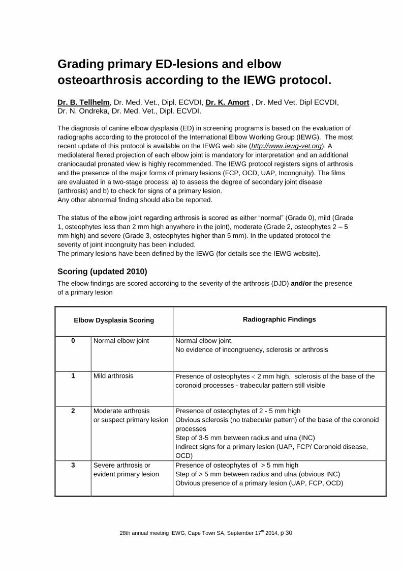
28th annual meeting IEWG, Cape Town SA, September 17th 2014, p 30
Grading primary ED-lesions and elbow
osteoarthrosis according to the IEWG protocol.
Dr. B. Tellhelm, Dr. Med. Vet., Dipl. ECVDI, Dr. K. Amort , Dr. Med Vet. Dipl ECVDI, Dr. N. Ondreka, Dr. Med. Vet., Dipl. ECVDI.
The diagnosis of canine elbow dysplasia (ED) in screening programs is based on the evaluation of
radiographs according to the protocol of the International Elbow Working Group (IEWG). The most
recent update of this protocol is available on the IEWG web site (http://www.iewg-vet.org). A
mediolateral flexed projection of each elbow joint is mandatory for interpretation and an additional
craniocaudal pronated view is highly recommended. The IEWG protocol registers signs of arthrosis
and the presence of the major forms of primary lesions (FCP, OCD, UAP, Incongruity). The films
are evaluated in a two-stage process: a) to assess the degree of secondary joint disease
(arthrosis) and b) to check for signs of a primary lesion.
Any other abnormal finding should also be reported.
The status of the elbow joint regarding arthrosis is scored as either “normal” (Grade 0), mild (Grade
1, osteophytes less than 2 mm high anywhere in the joint), moderate (Grade 2, osteophytes 2 – 5
mm high) and severe (Grade 3, osteophytes higher than 5 mm). In the updated protocol the
severity of joint incongruity has been included.
The primary lesions have been defined by the IEWG (for details see the IEWG website).
Scoring (updated 2010)
The elbow findings are scored according to the severity of the arthrosis (DJD) and/or the presence
of a primary lesion
Elbow Dysplasia Scoring Radiographic Findings
0 Normal elbow joint Normal elbow joint,
No evidence of incongruency, sclerosis or arthrosis
1 Mild arthrosis Presence of osteophytes 2 mm high, sclerosis of the base of the
coronoid processes - trabecular pattern still visible
2 Moderate arthrosis
or suspect primary lesion
Presence of osteophytes of 2 - 5 mm high
Obvious sclerosis (no trabecular pattern) of the base of the coronoid
processes
Step of 3-5 mm between radius and ulna (INC)
Indirect signs for a primary lesion (UAP, FCP/ Coronoid disease,
OCD)
3 Severe arthrosis or
evident primary lesion
Presence of osteophytes of > 5 mm high
Step of > 5 mm between radius and ulna (obvious INC)
Obvious presence of a primary lesion (UAP, FCP, OCD)
28th annual meeting IEWG, Cape Town SA, September 17th 2014, p 31
A Borderline (BL) score between ED 0 and ED 1 is allotted to dogs with minimal anconeal process
modelling of undetermined aetiology in some countries (i. g. Germany, France, Italy).
How many projections?
The minimal requirement is a true ML projection of each elbow. Excessive pronation or supination
should be avoided. In a maximally flexed position (as it is the standard view in many countries) the
elbow is often markedly supinated, making correct interpretation of shape and structure of MCP,
sclerosis caudal to MCP and spur formation cranially difficult.
An OC defect may easily be missed on the ML projection, but can usually be identified on a Cr Cd
15° pronated view. As scrutineers in many European countries (e.g. Scandinavia, UK) ask only for
a maximally flexed ML view or two ML views with different flexion of the elbows respectively, an
OC lesion may not be recognized.
For many years a Cr15L-CdMO pronated view was considered mandatory for the diagnosis of
FCP. However recent results of CT examinations and arthroscopy indicate that radiological findings
typical for the presence of FCP can be identified on the ML view quite consistently. The ML
projection may therefore be sufficient to diagnose or suspect the presence of a FCP reliably in a
screening program. As reported before two ML-projections - flexed (30°-40°)and neutral (100° -
120°) position give the best information concerning shape and structure of MCP and is also
diagnostic for incongruity and osteophytes. On radiographs of good quality even many OC lesions
are visible on the flexed ML.
The main problem from my point of view is not the number of radiographs but the intention of the
expert to register all findings which can be detected even on one ML view if it is of good quality and
the elbow positioned correctly.
How to score ED? ED scoring on the basis of a combination of the severity of arthrosis (DJD) and radiographic
findings indicative for a primary lesion or evidence of a primary lesion is not uniformly used in
Europe and overseas. The Scandinavian countries for example started scoring in the early 80ies
prior to the foundation of IEWG. Their classification is based on the degree of arthrosis, while of the
primary lesions only UAP is recorded. This scoring system is used in Scandinavia and also in the
UK and USA/Canada.
The most common primary elbow lesion is a FCP. Pertinent radiological findings on the ML
projection are a blurred and deformed cranial edge of the medial coronoid process (MCP), a
reduced opacity of its tip, an increased opacity of the ulnar notch at the level of the coronoid
processes and an increased and/or incongruent joint space between humerus and radius. It is
important to recognize that even minimal changes are usually pathognomonic for FCP qualifying an
elbow for at least an ED grade 2 (moderate ED, Coronoid disease/ FCP indicated ) according to
the current IEWG protocol regardless of the height of osteophytic new bone formation. The severity
of new bone formation is quite variable and some dogs may not show any new bone formation at
all. If grading is based on the size of the osteophytes only, many elbows with FCP will be
underscored and may even be considered free of ED.
Beware of conflicting data As mentioned above the IEWG scoring system is a two-step procedure, a) assessing the degree of
arthrosis and b) registering any signs indicative of a primary form of ED. Bear in mind that various
countries in Europe and overseas only rely on step a). Both concepts have proven to be useful in
reducing ED in a population. However problems arise when dogs are to be used for breeding in
28th annual meeting IEWG, Cape Town SA, September 17th 2014, p 32
countries with differing scoring system. In such a case it is advised to re-score the dog again
according to the local scoring mode. It will be the aim of IEWG to harmonize the scoring systems in
the future.
Slice imaging and appeal procedure Diagnosing FCP radiographically may be based on subtle findings which may be difficult to convey
to the dog owner. As a consequence an increasing number of appeals are filed and owners ask for
a CT study to be included in the re-evaluation process. No standardized protocol for CT
examination of the canine elbow have been proposed. IEWG plans to install a standardised
protocol for appeal procedures, the use of CT and/or MRI examinations and the technical
requirements of such studies.
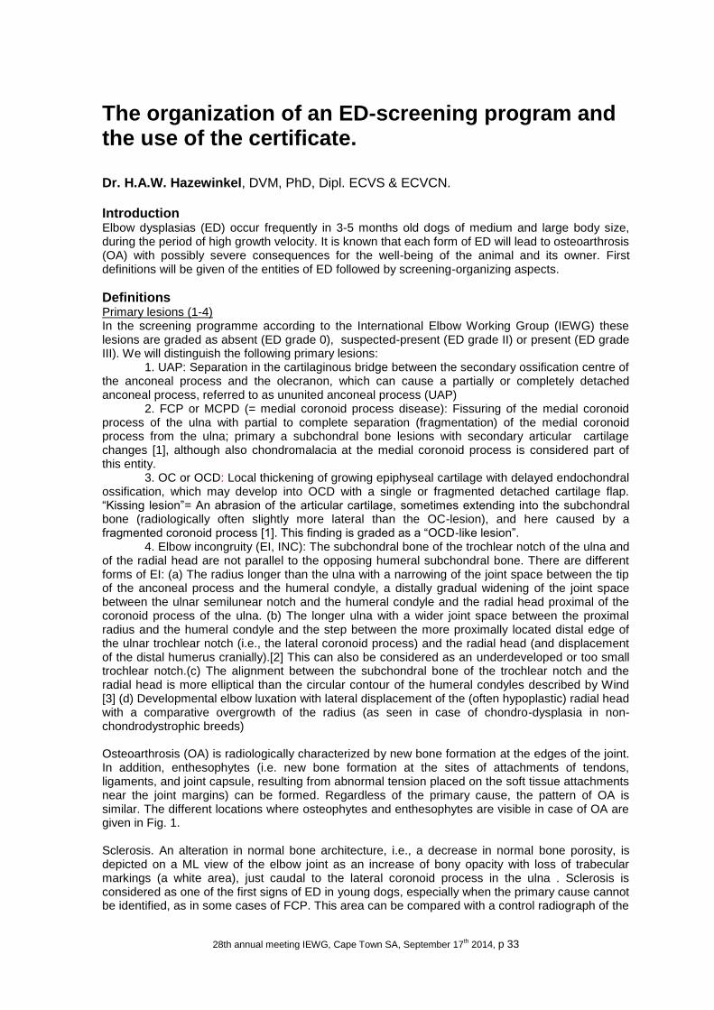
28th annual meeting IEWG, Cape Town SA, September 17th 2014, p 33
The organization of an ED-screening program and the use of the certificate.
Dr. H.A.W. Hazewinkel, DVM, PhD, Dipl. ECVS & ECVCN. Introduction Elbow dysplasias (ED) occur frequently in 3-5 months old dogs of medium and large body size, during the period of high growth velocity. It is known that each form of ED will lead to osteoarthrosis (OA) with possibly severe consequences for the well-being of the animal and its owner. First definitions will be given of the entities of ED followed by screening-organizing aspects.
Definitions Primary lesions (1-4) In the screening programme according to the International Elbow Working Group (IEWG) these lesions are graded as absent (ED grade 0), suspected-present (ED grade II) or present (ED grade III). We will distinguish the following primary lesions:
1. UAP: Separation in the cartilaginous bridge between the secondary ossification centre of the anconeal process and the olecranon, which can cause a partially or completely detached anconeal process, referred to as ununited anconeal process (UAP)
2. FCP or MCPD (= medial coronoid process disease): Fissuring of the medial coronoid process of the ulna with partial to complete separation (fragmentation) of the medial coronoid process from the ulna; primary a subchondral bone lesions with secondary articular cartilage changes [1], although also chondromalacia at the medial coronoid process is considered part of this entity.
3. OC or OCD: Local thickening of growing epiphyseal cartilage with delayed endochondral ossification, which may develop into OCD with a single or fragmented detached cartilage flap. “Kissing lesion”= An abrasion of the articular cartilage, sometimes extending into the subchondral bone (radiologically often slightly more lateral than the OC-lesion), and here caused by a fragmented coronoid process [1]. This finding is graded as a “OCD-like lesion”.
4. Elbow incongruity (EI, INC): The subchondral bone of the trochlear notch of the ulna and of the radial head are not parallel to the opposing humeral subchondral bone. There are different forms of EI: (a) The radius longer than the ulna with a narrowing of the joint space between the tip of the anconeal process and the humeral condyle, a distally gradual widening of the joint space between the ulnar semilunear notch and the humeral condyle and the radial head proximal of the coronoid process of the ulna. (b) The longer ulna with a wider joint space between the proximal radius and the humeral condyle and the step between the more proximally located distal edge of the ulnar trochlear notch (i.e., the lateral coronoid process) and the radial head (and displacement of the distal humerus cranially).[2] This can also be considered as an underdeveloped or too small trochlear notch.(c) The alignment between the subchondral bone of the trochlear notch and the radial head is more elliptical than the circular contour of the humeral condyles described by Wind [3] (d) Developmental elbow luxation with lateral displacement of the (often hypoplastic) radial head with a comparative overgrowth of the radius (as seen in case of chondro-dysplasia in non-chondrodystrophic breeds) Osteoarthrosis (OA) is radiologically characterized by new bone formation at the edges of the joint. In addition, enthesophytes (i.e. new bone formation at the sites of attachments of tendons, ligaments, and joint capsule, resulting from abnormal tension placed on the soft tissue attachments near the joint margins) can be formed. Regardless of the primary cause, the pattern of OA is similar. The different locations where osteophytes and enthesophytes are visible in case of OA are given in Fig. 1. Sclerosis. An alteration in normal bone architecture, i.e., a decrease in normal bone porosity, is depicted on a ML view of the elbow joint as an increase of bony opacity with loss of trabecular markings (a white area), just caudal to the lateral coronoid process in the ulna . Sclerosis is considered as one of the first signs of ED in young dogs, especially when the primary cause cannot be identified, as in some cases of FCP. This area can be compared with a control radiograph of the
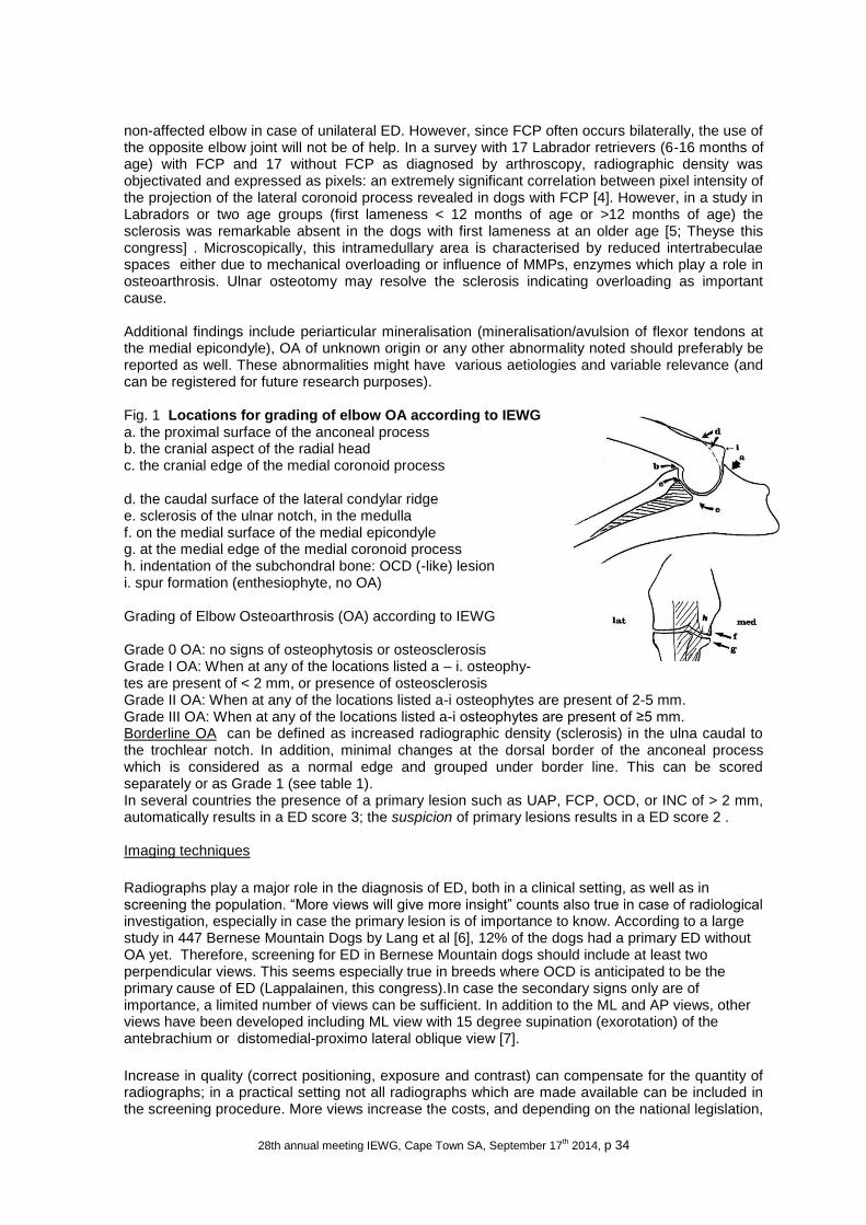
28th annual meeting IEWG, Cape Town SA, September 17th 2014, p 34
non-affected elbow in case of unilateral ED. However, since FCP often occurs bilaterally, the use of the opposite elbow joint will not be of help. In a survey with 17 Labrador retrievers (6-16 months of age) with FCP and 17 without FCP as diagnosed by arthroscopy, radiographic density was objectivated and expressed as pixels: an extremely significant correlation between pixel intensity of the projection of the lateral coronoid process revealed in dogs with FCP [4]. However, in a study in Labradors or two age groups (first lameness < 12 months of age or >12 months of age) the sclerosis was remarkable absent in the dogs with first lameness at an older age [5; Theyse this congress] . Microscopically, this intramedullary area is characterised by reduced intertrabeculae spaces either due to mechanical overloading or influence of MMPs, enzymes which play a role in osteoarthrosis. Ulnar osteotomy may resolve the sclerosis indicating overloading as important cause. Additional findings include periarticular mineralisation (mineralisation/avulsion of flexor tendons at the medial epicondyle), OA of unknown origin or any other abnormality noted should preferably be reported as well. These abnormalities might have various aetiologies and variable relevance (and can be registered for future research purposes). Fig. 1 Locations for grading of elbow OA according to IEWG a. the proximal surface of the anconeal process b. the cranial aspect of the radial head c. the cranial edge of the medial coronoid process d. the caudal surface of the lateral condylar ridge e. sclerosis of the ulnar notch, in the medulla f. on the medial surface of the medial epicondyle g. at the medial edge of the medial coronoid process h. indentation of the subchondral bone: OCD (-like) lesion i. spur formation (enthesiophyte, no OA) Grading of Elbow Osteoarthrosis (OA) according to IEWG Grade 0 OA: no signs of osteophytosis or osteosclerosis Grade I OA: When at any of the locations listed a – i. osteophy- tes are present of < 2 mm, or presence of osteosclerosis Grade II OA: When at any of the locations listed a-i osteophytes are present of 2-5 mm. Grade III OA: When at any of the locations listed a-i osteophytes are present of ≥5 mm. Borderline OA can be defined as increased radiographic density (sclerosis) in the ulna caudal to the trochlear notch. In addition, minimal changes at the dorsal border of the anconeal process which is considered as a normal edge and grouped under border line. This can be scored separately or as Grade 1 (see table 1). In several countries the presence of a primary lesion such as UAP, FCP, OCD, or INC of > 2 mm, automatically results in a ED score 3; the suspicion of primary lesions results in a ED score 2 . Imaging techniques
Radiographs play a major role in the diagnosis of ED, both in a clinical setting, as well as in screening the population. “More views will give more insight” counts also true in case of radiological investigation, especially in case the primary lesion is of importance to know. According to a large study in 447 Bernese Mountain Dogs by Lang et al [6], 12% of the dogs had a primary ED without OA yet. Therefore, screening for ED in Bernese Mountain dogs should include at least two perpendicular views. This seems especially true in breeds where OCD is anticipated to be the primary cause of ED (Lappalainen, this congress).In case the secondary signs only are of importance, a limited number of views can be sufficient. In addition to the ML and AP views, other views have been developed including ML view with 15 degree supination (exorotation) of the antebrachium or distomedial-proximo lateral oblique view [7].
Increase in quality (correct positioning, exposure and contrast) can compensate for the quantity of radiographs; in a practical setting not all radiographs which are made available can be included in the screening procedure. More views increase the costs, and depending on the national legislation,
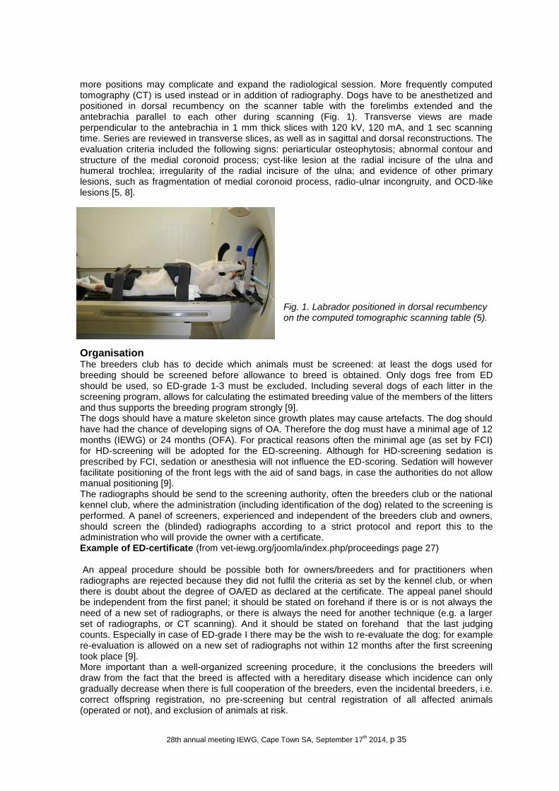
28th annual meeting IEWG, Cape Town SA, September 17th 2014, p 35
more positions may complicate and expand the radiological session. More frequently computed tomography (CT) is used instead or in addition of radiography. Dogs have to be anesthetized and positioned in dorsal recumbency on the scanner table with the forelimbs extended and the antebrachia parallel to each other during scanning (Fig. 1). Transverse views are made perpendicular to the antebrachia in 1 mm thick slices with 120 kV, 120 mA, and 1 sec scanning time. Series are reviewed in transverse slices, as well as in sagittal and dorsal reconstructions. The evaluation criteria included the following signs: periarticular osteophytosis; abnormal contour and structure of the medial coronoid process; cyst-like lesion at the radial incisure of the ulna and humeral trochlea; irregularity of the radial incisure of the ulna; and evidence of other primary lesions, such as fragmentation of medial coronoid process, radio-ulnar incongruity, and OCD-like lesions [5, 8].
Fig. 1. Labrador positioned in dorsal recumbency on the computed tomographic scanning table (5).
Organisation The breeders club has to decide which animals must be screened: at least the dogs used for breeding should be screened before allowance to breed is obtained. Only dogs free from ED should be used, so ED-grade 1-3 must be excluded. Including several dogs of each litter in the screening program, allows for calculating the estimated breeding value of the members of the litters and thus supports the breeding program strongly [9]. The dogs should have a mature skeleton since growth plates may cause artefacts. The dog should have had the chance of developing signs of OA. Therefore the dog must have a minimal age of 12 months (IEWG) or 24 months (OFA). For practical reasons often the minimal age (as set by FCI) for HD-screening will be adopted for the ED-screening. Although for HD-screening sedation is prescribed by FCI, sedation or anesthesia will not influence the ED-scoring. Sedation will however facilitate positioning of the front legs with the aid of sand bags, in case the authorities do not allow manual positioning [9]. The radiographs should be send to the screening authority, often the breeders club or the national kennel club, where the administration (including identification of the dog) related to the screening is performed. A panel of screeners, experienced and independent of the breeders club and owners, should screen the (blinded) radiographs according to a strict protocol and report this to the administration who will provide the owner with a certificate. Example of ED-certificate (from vet-iewg.org/joomla/index.php/proceedings page 27) An appeal procedure should be possible both for owners/breeders and for practitioners when radiographs are rejected because they did not fulfil the criteria as set by the kennel club, or when there is doubt about the degree of OA/ED as declared at the certificate. The appeal panel should be independent from the first panel; it should be stated on forehand if there is or is not always the need of a new set of radiographs, or there is always the need for another technique (e.g. a larger set of radiographs, or CT scanning). And it should be stated on forehand that the last judging counts. Especially in case of ED-grade I there may be the wish to re-evaluate the dog: for example re-evaluation is allowed on a new set of radiographs not within 12 months after the first screening took place [9]. More important than a well-organized screening procedure, it the conclusions the breeders will draw from the fact that the breed is affected with a hereditary disease which incidence can only gradually decrease when there is full cooperation of the breeders, even the incidental breeders, i.e. correct offspring registration, no pre-screening but central registration of all affected animals (operated or not), and exclusion of animals at risk.
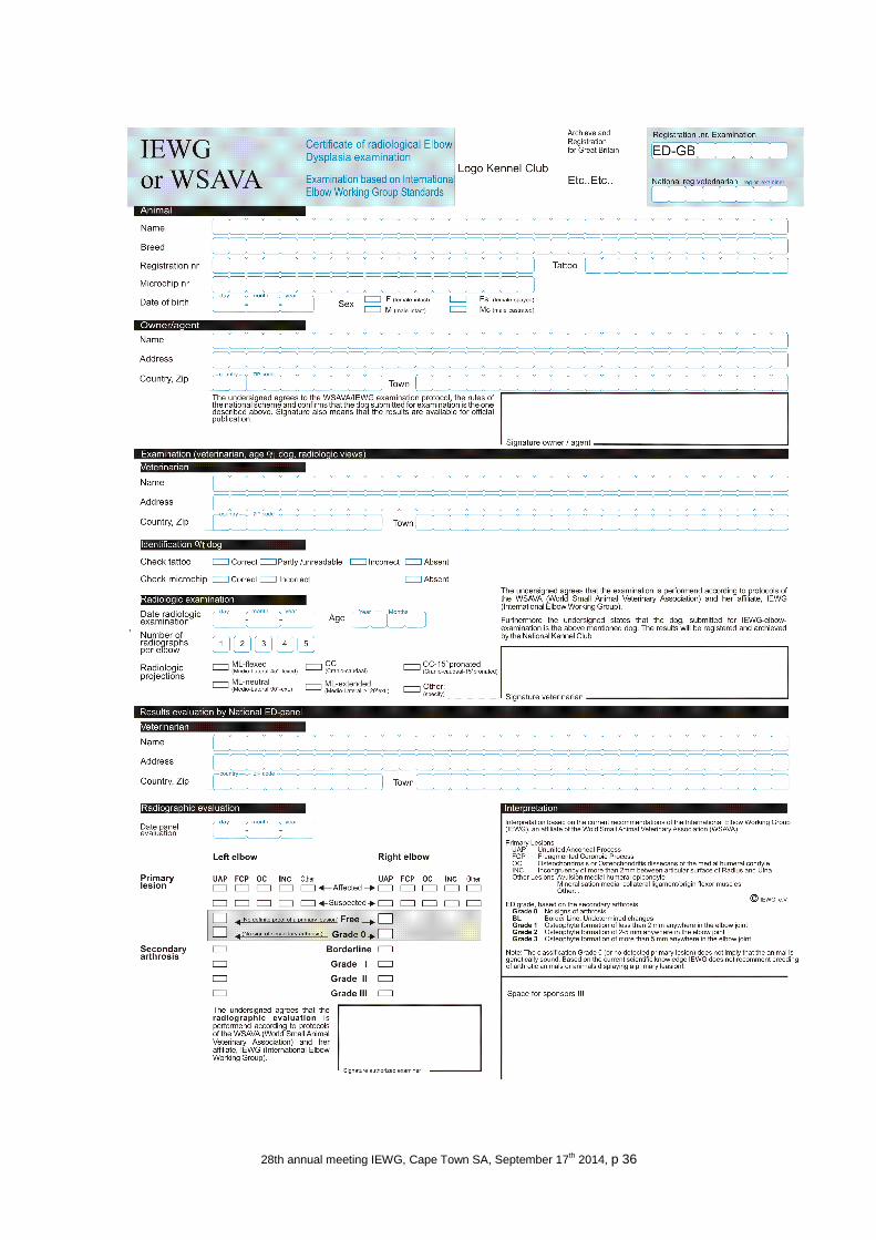
28th annual meeting IEWG, Cape Town SA, September 17th 2014, p 36
28th annual meeting IEWG, Cape Town SA, September 17th 2014, p 37
References 1.Morgan J.P. 2000 Hereditary bone and joint diseases in the dog. Morgan, Wind Davidson (eds.) Schlütersche, Hannover (G) 2. Samoy, Y., Van Ryssen, B., Gielen, I., Walschot, N., van Bree, H., 2006. Review of the literature; elbow incongruity in the dog. VCOT 19, 1-8. 3.Wind, A,1986 Elbow incongruity and developmental elbow diseases in the dog: JAVMA 22, 711-724. 4.Burton, N.J., Comerford, E.J., Bailey, M., Pead, M.J., Owen, M.R., 2007. Digital analysis of ulnar trochlear notch sclerosis in Labrador retrievers. JSAP 48, 220-224. 5.Seng Fong Lau 2013 Development of medial coronoid disease in Labrador retrievers: diagnostic and pathogenic studies, Thesis Utrecht University October 2013 ISBN 978-90-393-5962-5964 6. Lang, J., Busato, A., Baumgartner, D., Flückiger, M., Weber, U.T., 1998. Comparison of two classification protocols in the evaluation of elbow dysplasia in the dog. JSAP 39, 169-174. 7.Haudiquet, P.R., Marcellin-Little, D.J., Stebbins, M.E., 2002. Use of the distomedial-proximolateral oblique radiographic view of the elbow joint for examination of the medial coronoid process in dogs. AJVR 63, 1000-1005. 8.Reichle, J.K., Park, R.D., Bahr, A.M., 2000. Computed tomographic findings of dogs with cubital joint lameness. Veterinary Radiology & Ultrasound 41, 125-130. 9. IEWG 2012 proceedings (http://www.vet-iewg.org/joomla/index.php/proceedings )
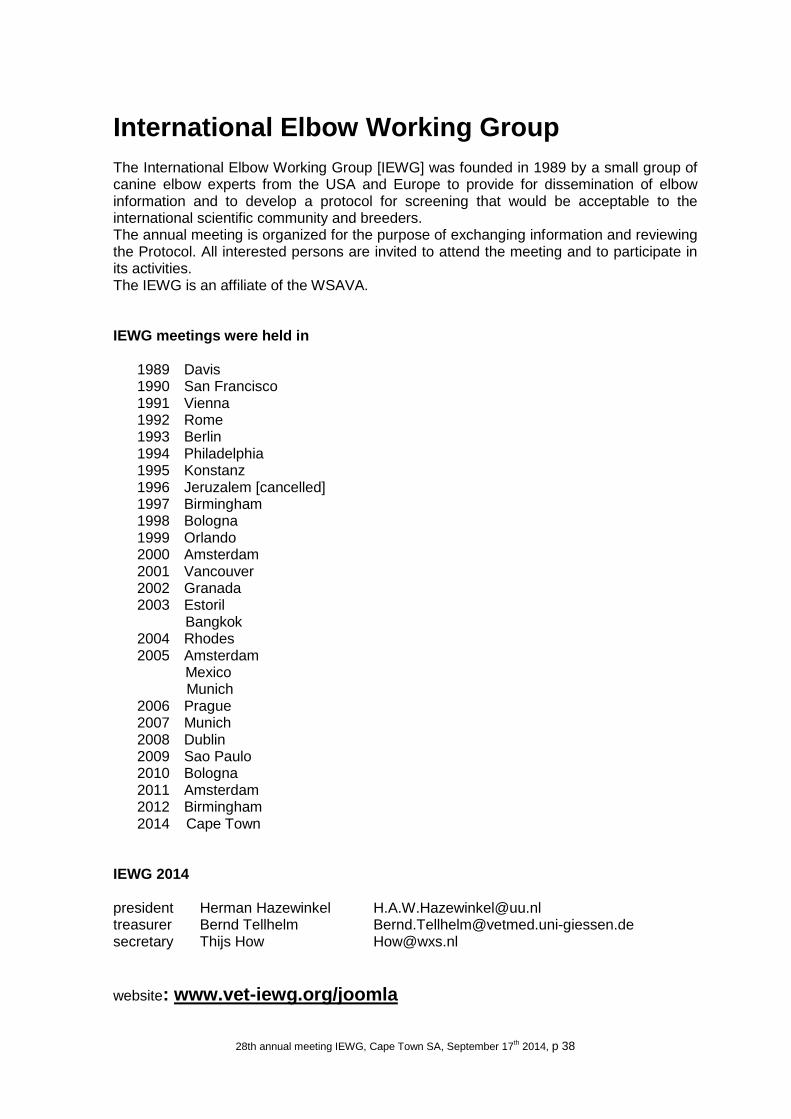
28th annual meeting IEWG, Cape Town SA, September 17th 2014, p 38
International Elbow Working Group
The International Elbow Working Group [IEWG] was founded in 1989 by a small group of canine elbow experts from the USA and Europe to provide for dissemination of elbow information and to develop a protocol for screening that would be acceptable to the international scientific community and breeders. The annual meeting is organized for the purpose of exchanging information and reviewing the Protocol. All interested persons are invited to attend the meeting and to participate in its activities. The IEWG is an affiliate of the WSAVA. IEWG meetings were held in
1989 Davis 1990 San Francisco 1991 Vienna 1992 Rome 1993 Berlin 1994 Philadelphia 1995 Konstanz 1996 Jeruzalem [cancelled] 1997 Birmingham 1998 Bologna 1999 Orlando 2000 Amsterdam 2001 Vancouver 2002 Granada 2003 Estoril
Bangkok 2004 Rhodes 2005 Amsterdam
Mexico Munich
2006 Prague 2007 Munich 2008 Dublin 2009 Sao Paulo 2010 Bologna 2011 Amsterdam 2012 Birmingham 2014 Cape Town
IEWG 2014 president Herman Hazewinkel [email protected] treasurer Bernd Tellhelm [email protected] secretary Thijs How [email protected]
website: www.vet-iewg.org/joomla