Embed Size (px)
Citation preview
Breast MRI: Imaging and Intervention
Jaroslaw Nicholas Tkacz, M. D.
Purpose• To examine the typical morphologic,
enhancement and kinetic features of breast lesions on MR Imaging and determine the role of each in interpretation
• Identify which lesions are amenable for MRI- guided intervention
• Briefly discuss how to successfully perform and trouble-shoot MR-guided biopsy
Brief History of Breast MRI• Mid 1980’s
– Research in Germany and US begins• Mid-late 1980’s
– Faster MRI sequences developed, whole breast dynamic scanning feasible
• Late 1980’s, early 1990’s– Kinetic data promising for evaluation of breast CA– Breast MRI used clinically to evaluate silicone
implant integrity
Brief History of Breast MRI• Early-mid 90’s
– clinical applications for evaluation of breast cancer with dynamic Gd-DTPA
• Kinetics, morphology or both• Spatial or temporal resolution in clinical applications
• Late 1990’s– Breast MRI utilized to evaluate cancer in selected
patients
Initial approaches• CE-MRI was first performed in the mid 1980’s
on patients with known Breast CA• Heywang and colleagues demonstrated rapid
uptake of contrast within the first five minutes in malignant lesions
• Other investigations show benign lesions can show a similar degree of enhancement
• Investigators turn to time-course kinetic analysis of breast lesions
Early Dynamic Approach• Concentrated on the initial, early enhancement
characteristic of breast lesions.• With faster scanning techniques this early phase
diminished from 5min post Gd-DTPA (T1W SE) to 11.5 secs (segmented low fast-angle shot sequence) after aortic enhancement.
• Gilles and co-workers classified any lesion with early vascular enhancement as malignant, with a sensitivity of 95% and specificity of 53%.
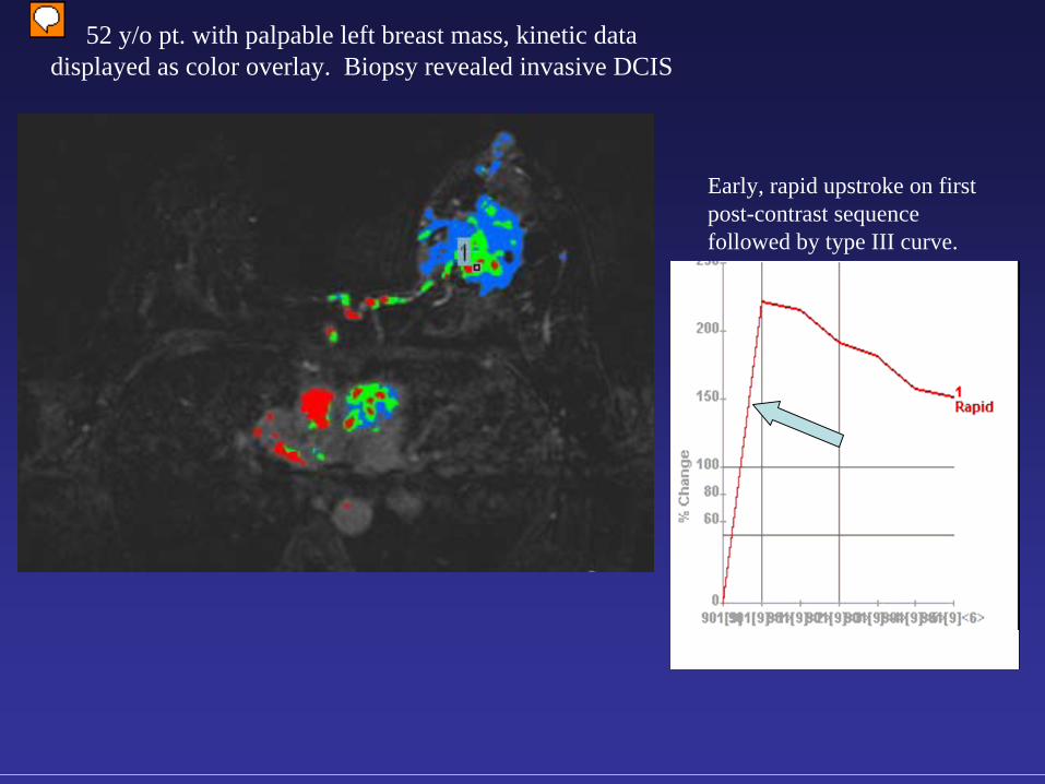
52 y/o pt. with palpable left breast mass, kinetic data displayed as color overlay. Biopsy revealed invasive DCIS
Early, rapid upstroke on first post-contrast sequence followed by type III curve.
Early Dynamic Approach• However, later investigators found similar
early enhancement characteristics with benign lesions, particularly lymph nodes and fibroadenomas.
• Although benign lesions tended to enhance more gradually, there was overlap between benign and malignant lesions with this approach.
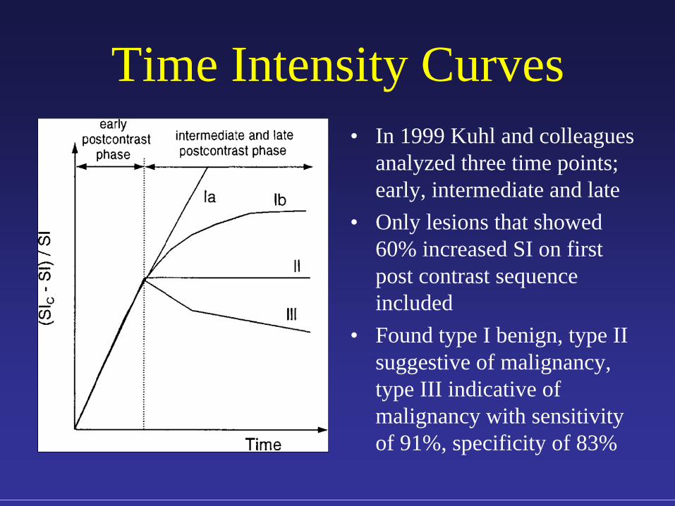
Time Intensity Curves• In 1999 Kuhl and colleagues
analyzed three time points; early, intermediate and late
• Only lesions that showed 60% increased SI on first post contrast sequence included
• Found type I benign, type II suggestive of malignancy, type III indicative of malignancy with sensitivity of 91%, specificity of 83%
Problems with Exclusive use of Dynamic Approach
• False positives– Fibroadenomas (especially “young”, vascular lesions)– Intramammary lymph nodes
• What about Non-mass lesions?• What about DCIS?
– Does not show rapid enhancement or present as a mass-like lesion
• Some investigators found no significant difference in enhancement characteristics between benign and malignant lesions
Advancing Technologies, Morphologic Approach
• Concurrently, investigators were analyzing morphology of breast lesions on MRI.
• In 1994, Orel and coworkers using high spatial resolution imaging techniques, sacrificing temporal resolution, found architectural features helpful in differentiating between benign and malignant lesions.
• In 1997, Nunes and colleagues exclusively used morphologic features to distinguish benign from malignant lesions and developed an interpretation model based on lesion architecture.
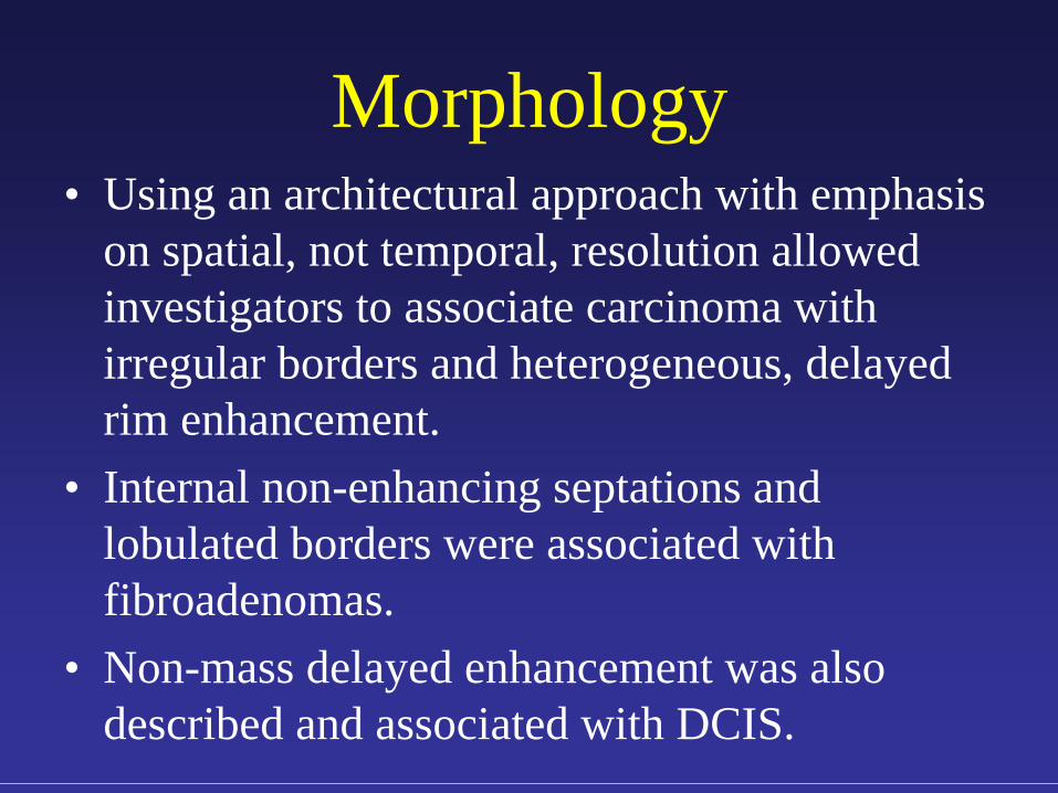
Morphology• Using an architectural approach with emphasis
on spatial, not temporal, resolution allowed investigators to associate carcinoma with irregular borders and heterogeneous, delayed rim enhancement.
• Internal non-enhancing septations and lobulated borders were associated with fibroadenomas.
• Non-mass delayed enhancement was also described and associated with DCIS.
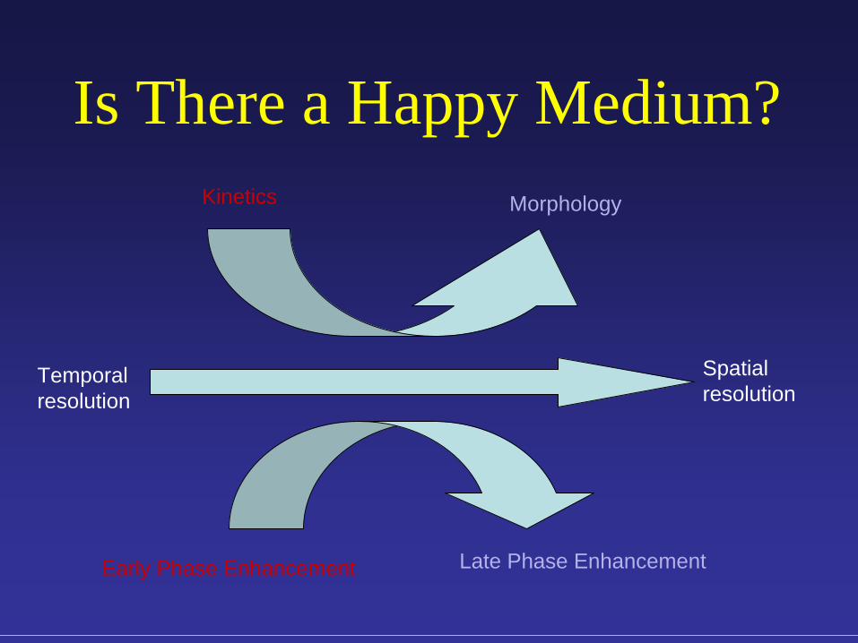
Temporal resolution
Spatial resolution
Kinetics Morphology
Early Phase Enhancement Late Phase Enhancement
Is There a Happy Medium?
Combination
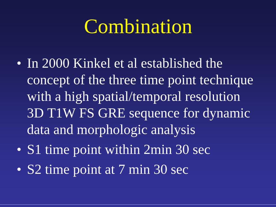
• In 2000 Kinkel et al established the concept of the three time point technique with a high spatial/temporal resolution 3D T1W FS GRE sequence for dynamic data and morphologic analysis
• S1 time point within 2min 30 sec• S2 time point at 7 min 30 sec
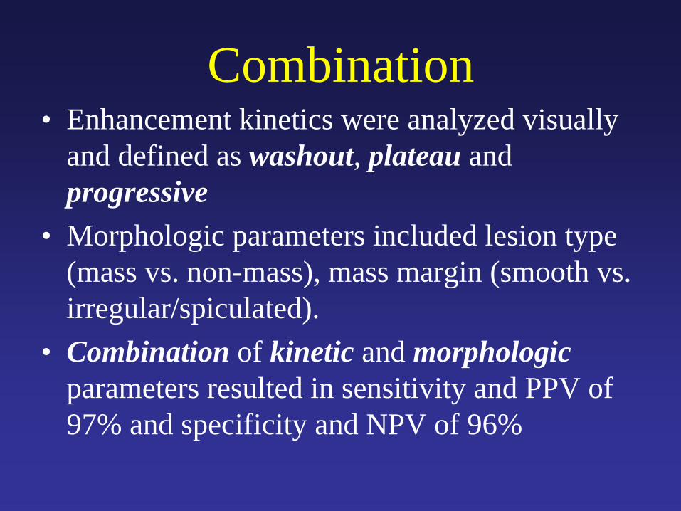
Combination• Enhancement kinetics were analyzed visually
and defined as washout, plateau and progressive
• Morphologic parameters included lesion type (mass vs. non-mass), mass margin (smooth vs. irregular/spiculated).
• Combination of kinetic and morphologic parameters resulted in sensitivity and PPV of 97% and specificity and NPV of 96%
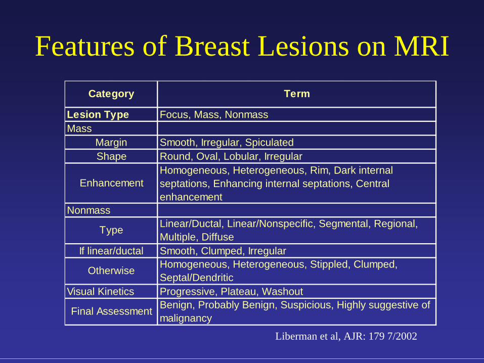
Features of Breast Lesions on MRI
Lesion Type
Visual Kinetics Progressive, Plateau, WashoutBenign, Probably Benign, Suspicious, Highly suggestive of malignancyFinal Assessment
If linear/ductal Smooth, Clumped, IrregularHomogeneous, Heterogeneous, Stippled, Clumped, Septal/Dendritic
Otherwise
NonmassLinear/Ductal, Linear/Nonspecific, Segmental, Regional, Multiple, Diffuse
Enhancement
Type
Homogeneous, Heterogeneous, Rim, Dark internal septations, Enhancing internal septations, Central enhancement
MarginShape
Smooth, Irregular, SpiculatedRound, Oval, Lobular, Irregular
Category Term
MassFocus, Mass, Nonmass
Liberman et al, AJR: 179 7/2002
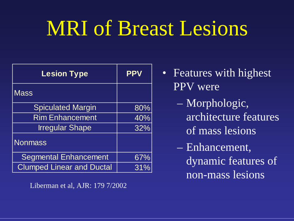
MRI of Breast Lesions
80%40%32%
67%31%Clumped Linear and Ductal
Rim EnhancementIrregular Shape
Nonmass
Segmental Enhancement
Lesion Type PPV
Mass
Spiculated Margin
• Features with highest PPV were– Morphologic,
architecture features of mass lesions
– Enhancement, dynamic features of non-mass lesions
Liberman et al, AJR: 179 7/2002
MRI of Breast Lesions
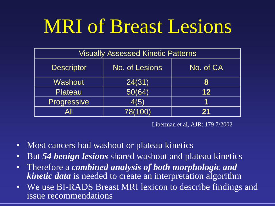
• Most cancers had washout or plateau kinetics• But 54 benign lesions shared washout and plateau kinetics• Therefore a combined analysis of both morphologic and
kinetic data is needed to create an interpretation algorithm• We use BI-RADS Breast MRI lexicon to describe findings and
issue recommendations
78(100)
Visually Assessed Kinetic Patterns
124(5) 1
24(31)
Descriptor No. of Lesions No. of CA
Washout 8
21
PlateauProgressive
All
50(64)
Liberman et al, AJR: 179 7/2002
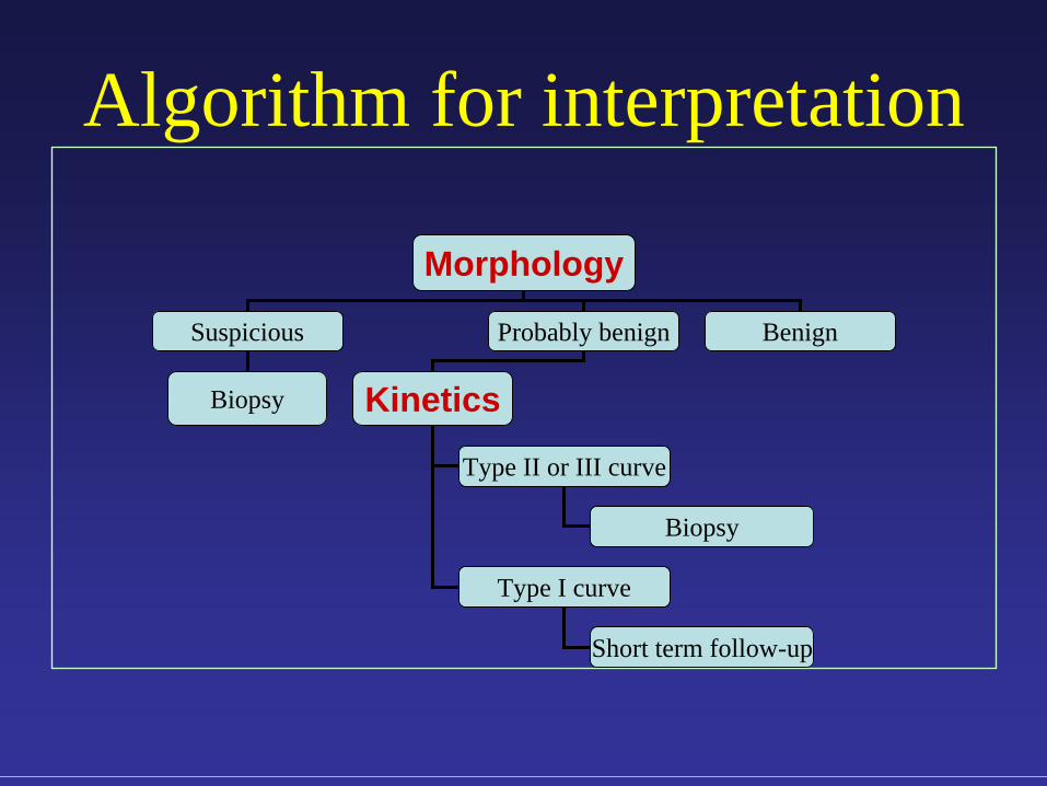
Algorithm for interpretation
Morphology
Suspicious Probably benign Benign
Biopsy Kinetics
Type II or III curve
Type I curve
Biopsy
Short term follow-up
MR Imaging of the Breast: Clinical Indications
• Assessing silicone implant integrity• Determining disease extent• Searching for the breast primary• Differentiating scar from recurrence• Evaluating suspicious mammographic finding• Screening high-risk women• Monitoring response to neoadjuvant chemotherapy
*
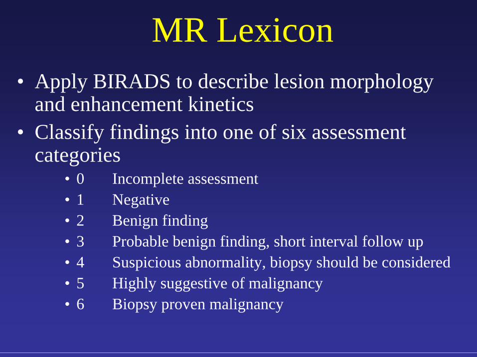
MR Lexicon• Apply BIRADS to describe lesion morphology
and enhancement kinetics• Classify findings into one of six assessment
categories• 0 Incomplete assessment• 1 Negative• 2 Benign finding• 3 Probable benign finding, short interval follow up• 4 Suspicious abnormality, biopsy should be considered• 5 Highly suggestive of malignancy• 6 Biopsy proven malignancy

Examples
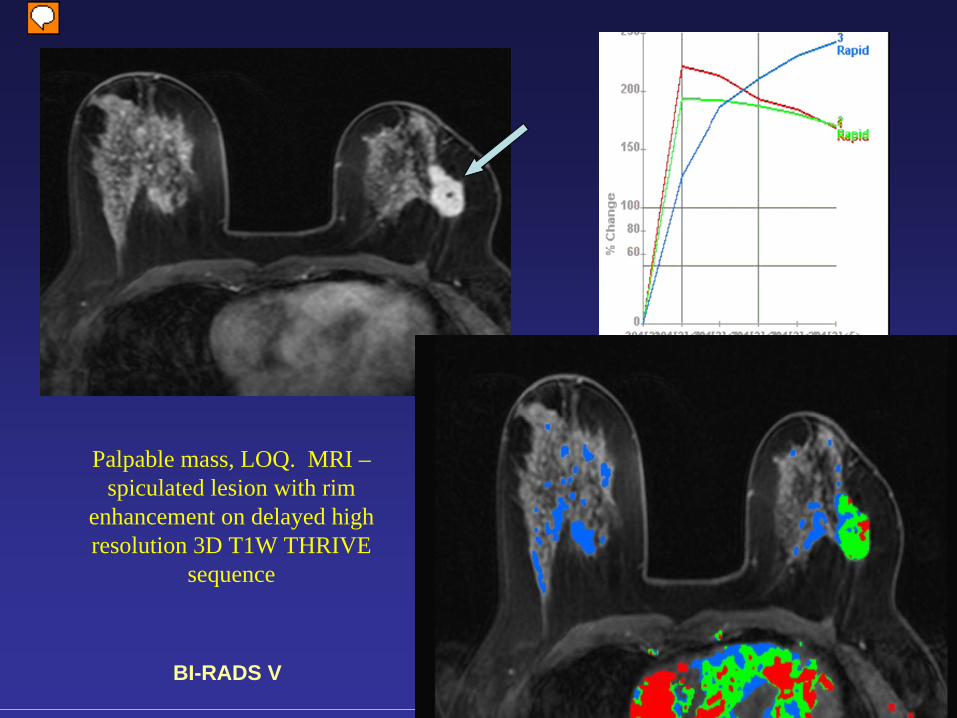
Palpable mass, LOQ. MRI – spiculated lesion with rim
enhancement on delayed high resolution 3D T1W THRIVE
sequence
Lesion also demonstrates early enhancement.
BI-RADS V
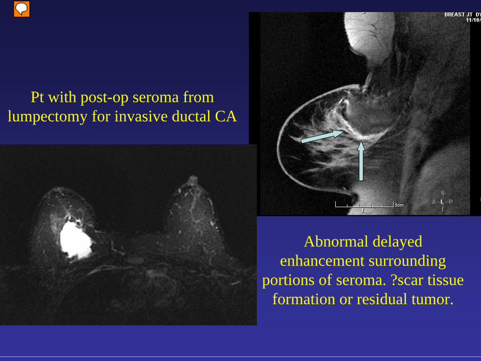
Pt with post-op seroma from lumpectomy for invasive ductal CA
Abnormal delayed enhancement surrounding
portions of seroma. ?scar tissue formation or residual tumor.
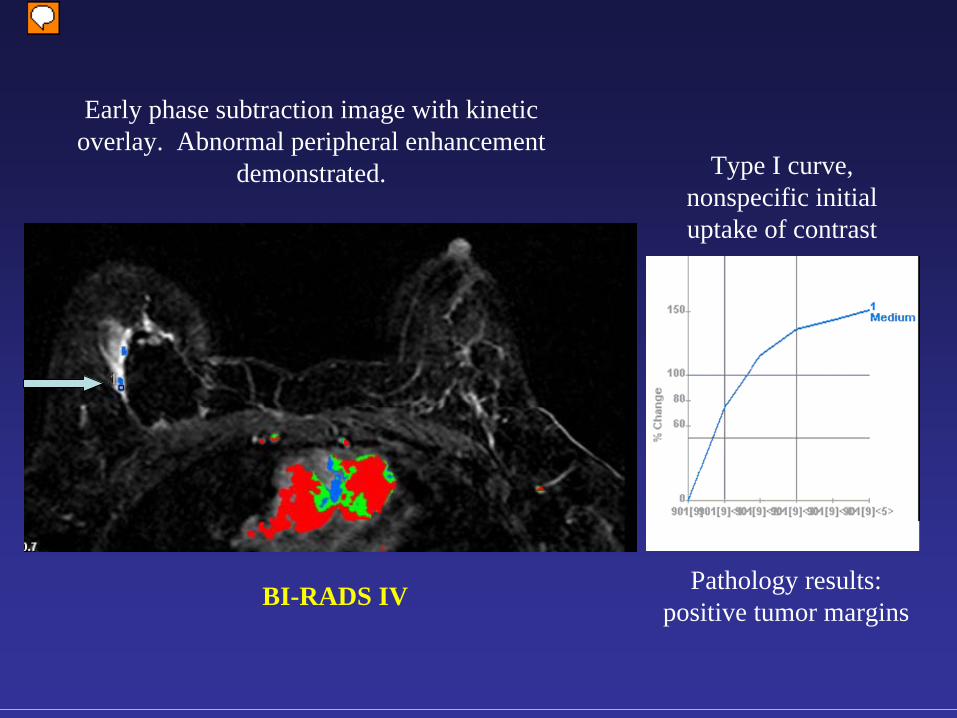
Early phase subtraction image with kinetic overlay. Abnormal peripheral enhancement
demonstrated. Type I curve, nonspecific initial uptake of contrast
Pathology results: positive tumor marginsBI-RADS IV
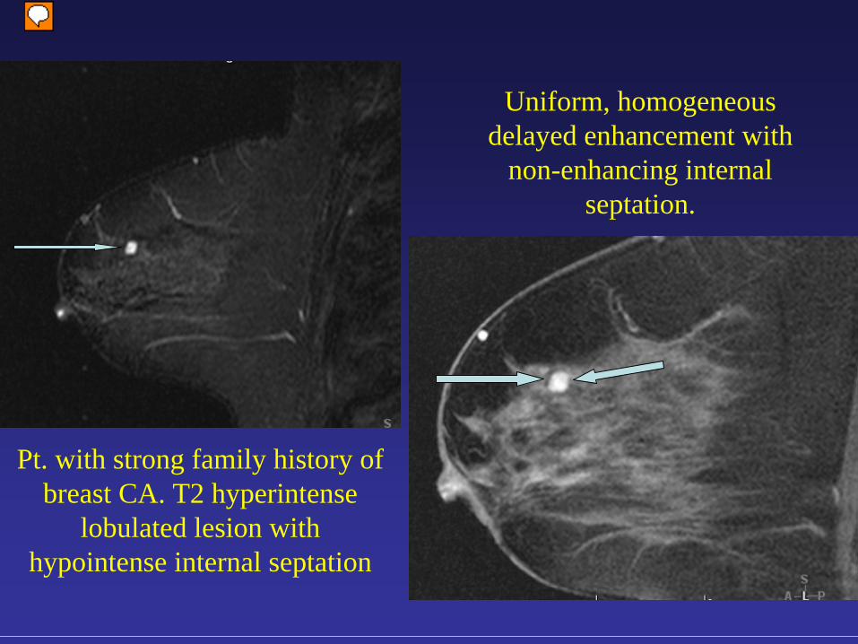
Pt. with strong family history of breast CA. T2 hyperintense
lobulated lesion with hypointense internal septation
Uniform, homogeneous delayed enhancement with
non-enhancing internal septation.
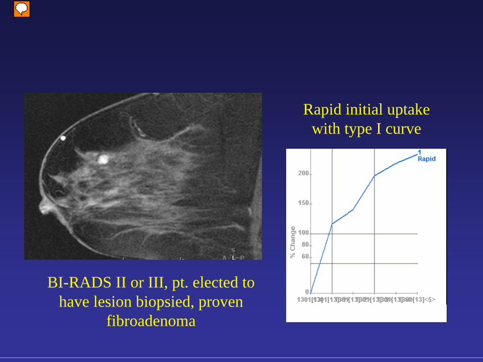
Rapid initial uptake with type I curve
BI-RADS II or III, pt. elected to have lesion biopsied, proven
fibroadenoma
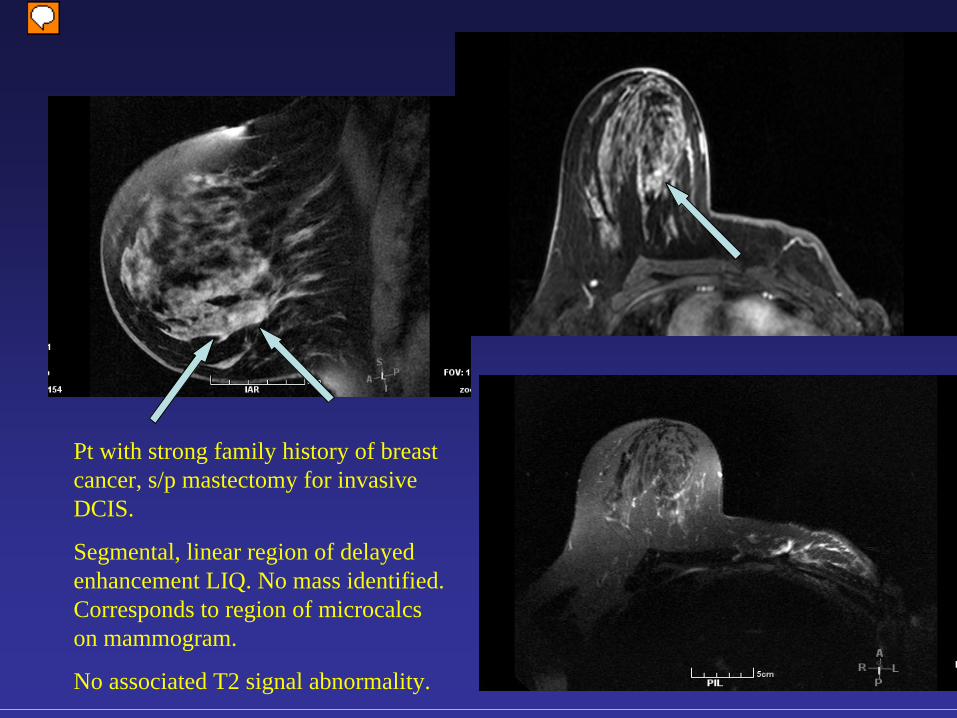
Pt with strong family history of breast cancer, s/p mastectomy for invasive DCIS.
Segmental, linear region of delayed enhancement LIQ. No mass identified. Corresponds to region of microcalcs on mammogram.
No associated T2 signal abnormality.
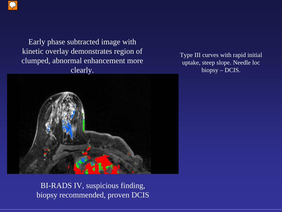
Early phase subtracted image with kinetic overlay demonstrates region of clumped, abnormal enhancement more
clearly.
Type III curves with rapid initial uptake, steep slope. Needle loc
biopsy – DCIS.
BI-RADS IV, suspicious finding, biopsy recommended, proven DCIS
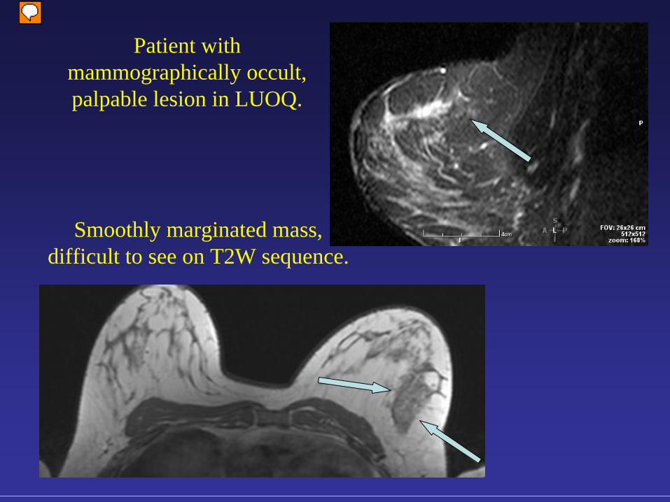
Patient with mammographically occult, palpable lesion in LUOQ.
Smoothly marginated mass, difficult to see on T2W sequence.
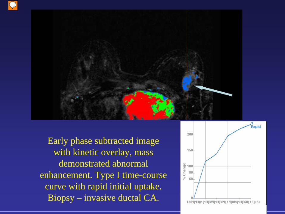
Early phase subtracted image with kinetic overlay, mass
demonstrated abnormal enhancement. Type I time-course
curve with rapid initial uptake. Biopsy – invasive ductal CA.
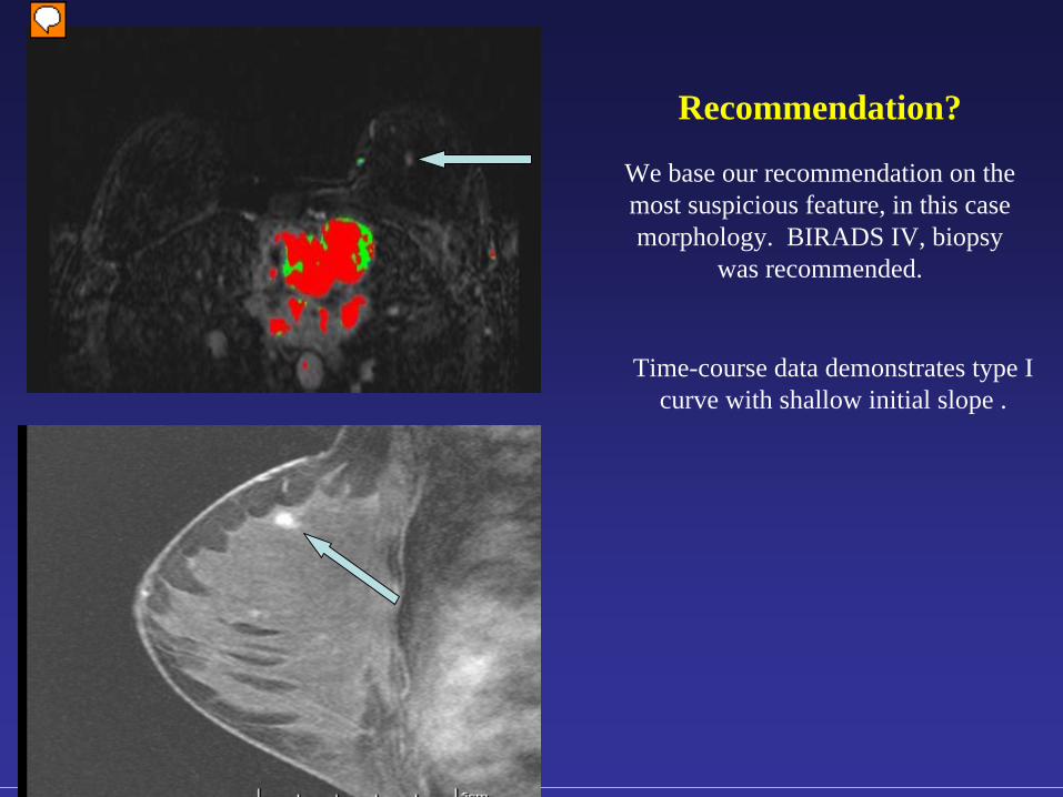
Time-course data demonstrates type I curve with shallow initial slope .
Recommendation?
We base our recommendation on the most suspicious feature, in this case morphology. BIRADS IV, biopsy
was recommended.
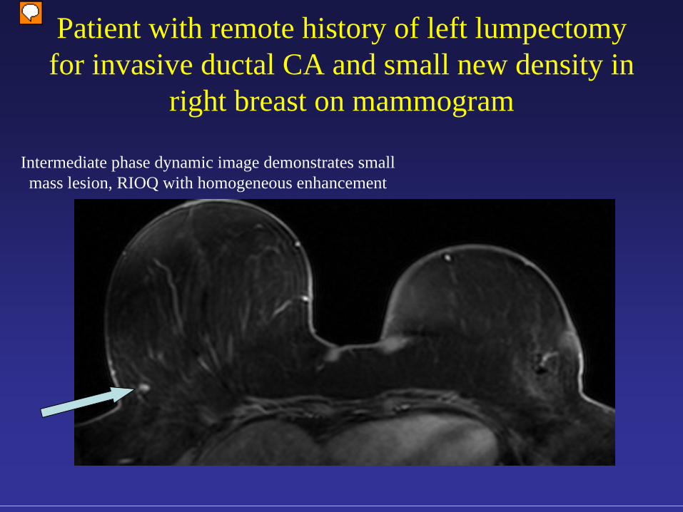
Patient with remote history of left lumpectomy for invasive ductal CA and small new density in
right breast on mammogram
Intermediate phase dynamic image demonstrates small mass lesion, RIOQ with homogeneous enhancement

Kinetic overlay demonstrates abnormal, uniform enhancement of lesion
Type III curve with rapid initial uptake of
contrast
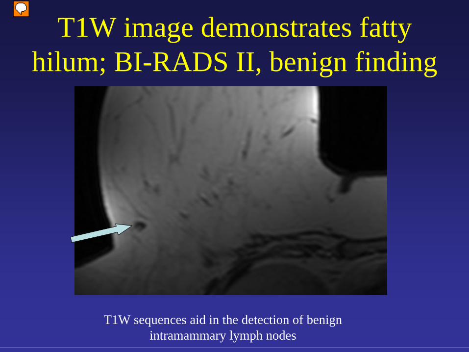
T1W image demonstrates fatty hilum; BI-RADS II, benign finding
T1W sequences aid in the detection of benign intramammary lymph nodes
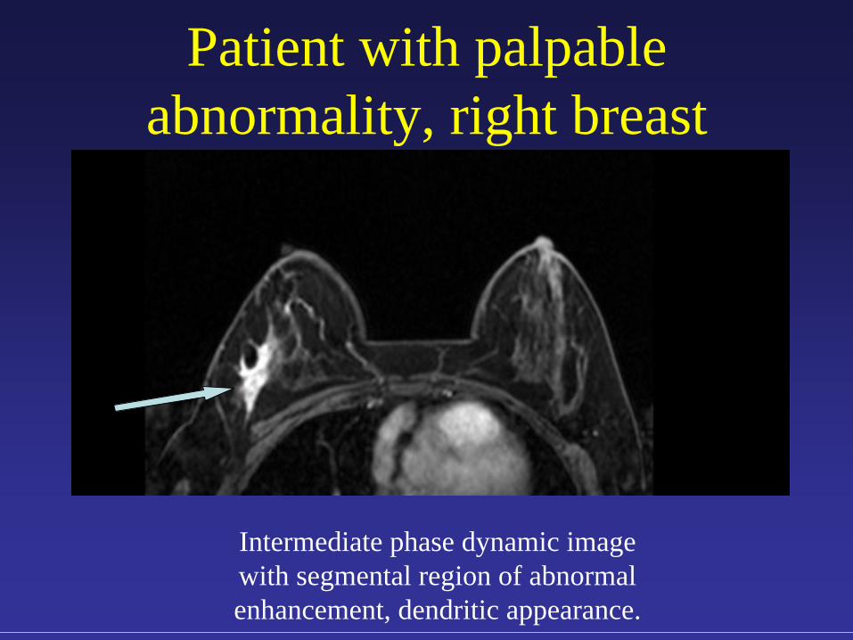
Patient with palpable abnormality, right breast
Intermediate phase dynamic image with segmental region of abnormal enhancement, dendritic appearance.
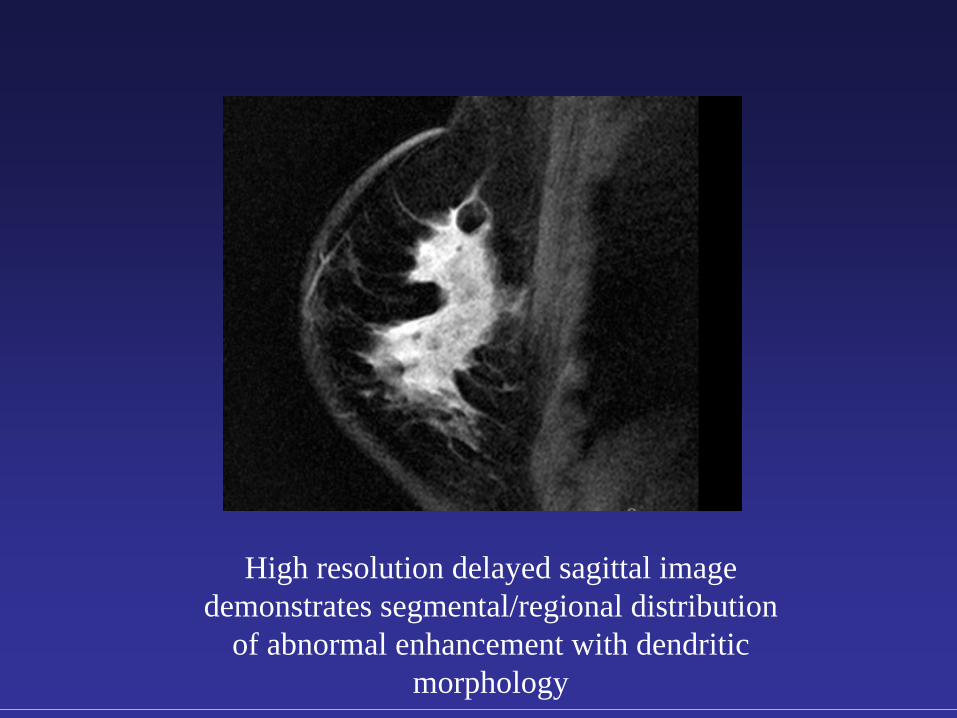
High resolution delayed sagittal image demonstrates segmental/regional distribution
of abnormal enhancement with dendritic morphology
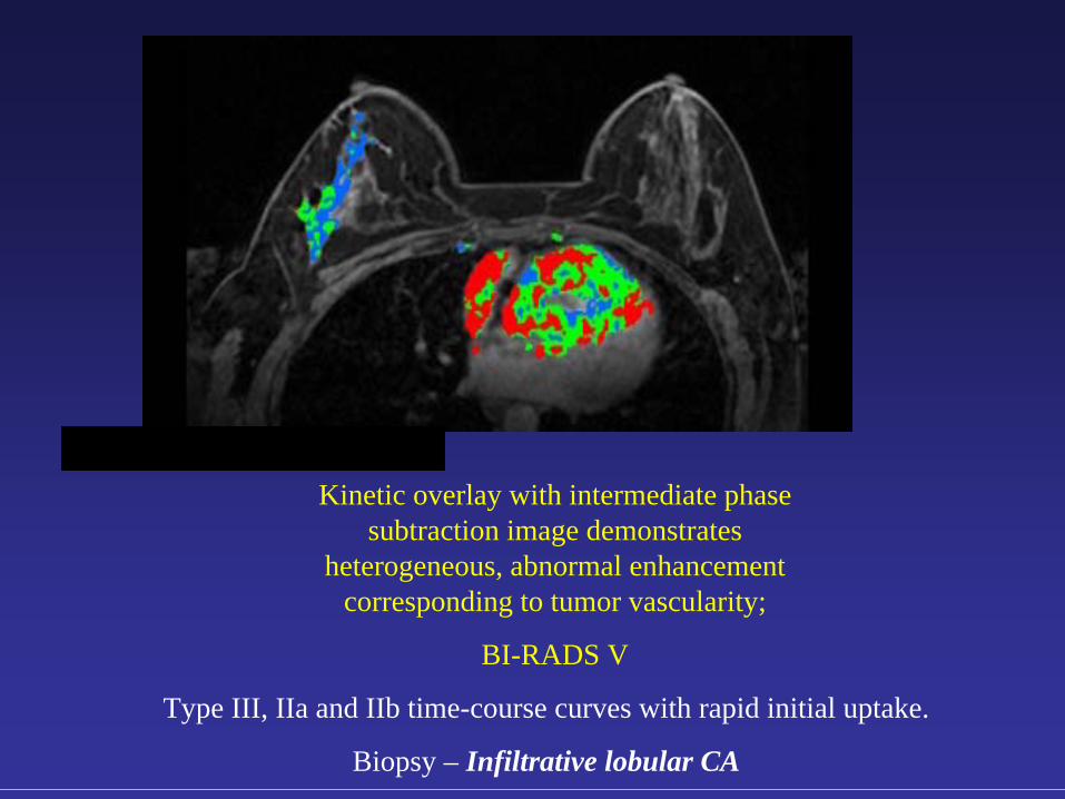
Kinetic overlay with intermediate phase subtraction image demonstrates
heterogeneous, abnormal enhancement corresponding to tumor vascularity;
BI-RADS V
Type III, IIa and IIb time-course curves with rapid initial uptake.
Biopsy – Infiltrative lobular CA
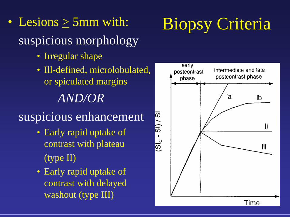
Biopsy Criteria• Lesions > 5mm with:suspicious morphology
• Irregular shape• Ill-defined, microlobulated,
or spiculated margins
AND/ORsuspicious enhancement
• Early rapid uptake of contrast with plateau (type II)
• Early rapid uptake of contrast with delayed washout (type III)
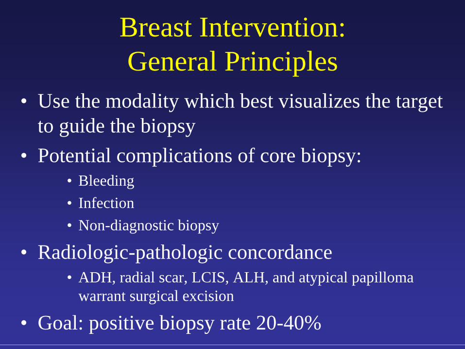
Breast Intervention: General Principles
• Use the modality which best visualizes the target to guide the biopsy
• Potential complications of core biopsy:• Bleeding • Infection• Non-diagnostic biopsy
• Radiologic-pathologic concordance• ADH, radial scar, LCIS, ALH, and atypical papilloma
warrant surgical excision
• Goal: positive biopsy rate 20-40%
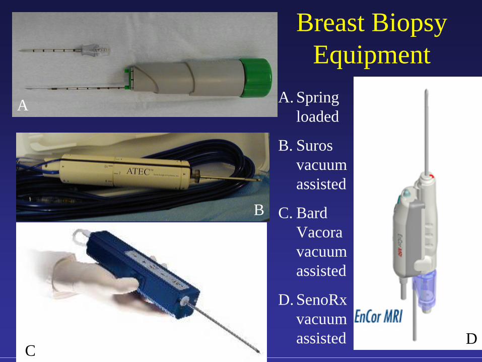
Breast Biopsy Equipment
A. Spring loaded
B. Suros vacuum assisted
C. Bard Vacora vacuum assisted
D. SenoRx vacuum assisted
A
B
CD
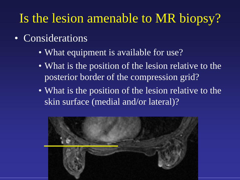
Is the lesion amenable to MR biopsy?• Considerations
• What equipment is available for use?• What is the position of the lesion relative to the
posterior border of the compression grid?• What is the position of the lesion relative to the
skin surface (medial and/or lateral)?
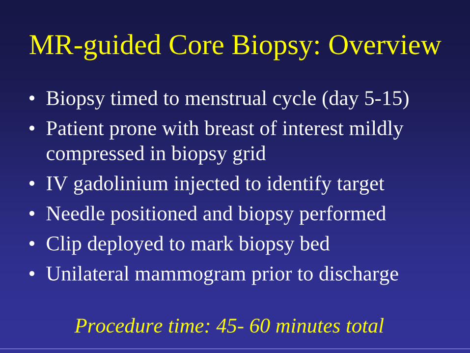
MR-guided Core Biopsy: Overview
• Biopsy timed to menstrual cycle (day 5-15)• Patient prone with breast of interest mildly
compressed in biopsy grid• IV gadolinium injected to identify target• Needle positioned and biopsy performed• Clip deployed to mark biopsy bed• Unilateral mammogram prior to discharge
Procedure time: 45- 60 minutes total
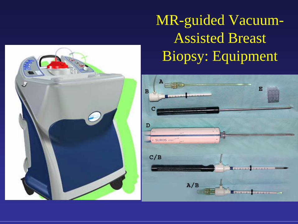
MR-guided Vacuum- Assisted Breast
Biopsy: Equipment
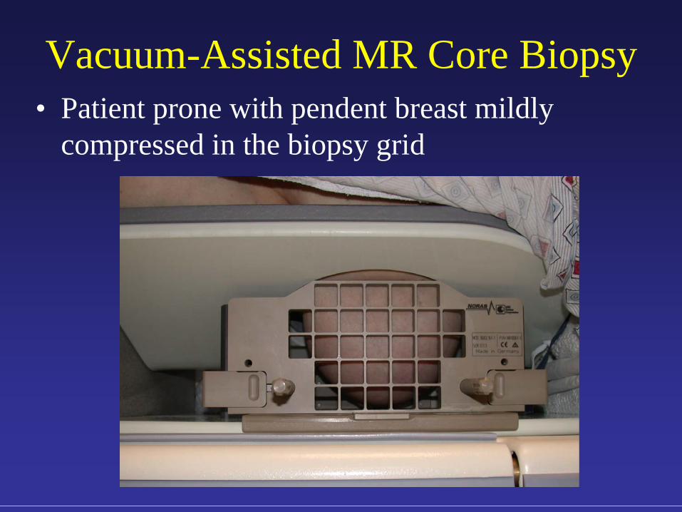
Vacuum-Assisted MR Core Biopsy• Patient prone with pendent breast mildly
compressed in the biopsy grid
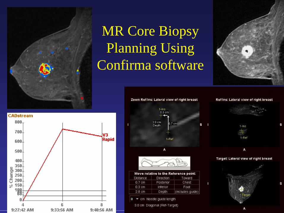
MR Core Biopsy Planning Using
Confirma software
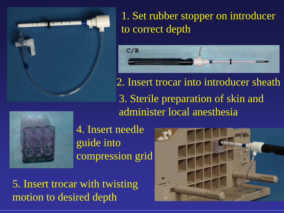
1. Set rubber stopper on introducer to correct depth
4. Insert needle guide into compression grid
2. Insert trocar into introducer sheath3. Sterile preparation of skin and administer local anesthesia
5. Insert trocar with twisting motion to desired depth
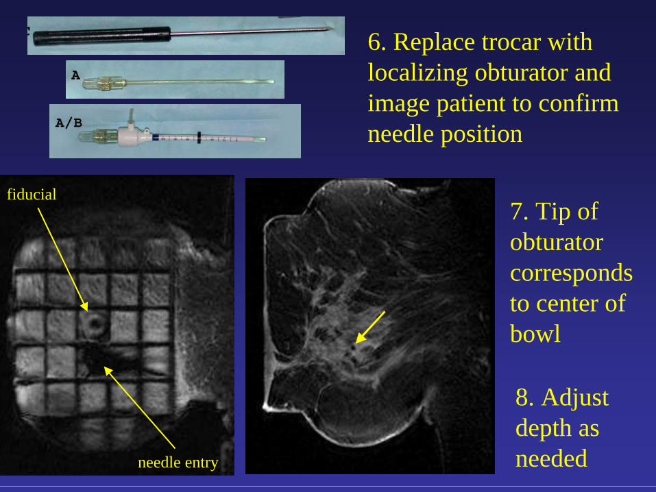
6. Replace trocar with localizing obturator and image patient to confirm needle position
7. Tip of obturator corresponds to center of bowl
fiducial
needle entry
8. Adjust depth as needed
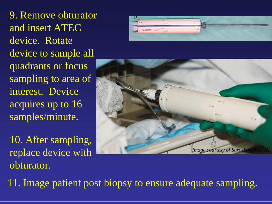
9. Remove obturator and insert ATEC device. Rotate device to sample all quadrants or focus sampling to area of interest. Device acquires up to 16 samples/minute.
Image courtesy of SurosSurgical, IN10. After sampling, replace device with obturator.
11. Image patient post biopsy to ensure adequate sampling.
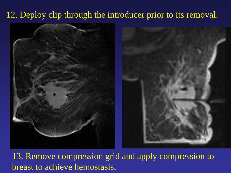
12. Deploy clip through the introducer prior to its removal.
13. Remove compression grid and apply compression to breast to achieve hemostasis.
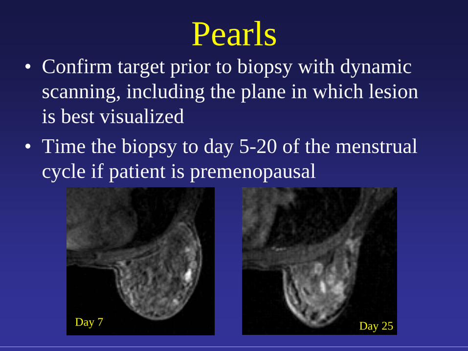
Pearls• Confirm target prior to biopsy with dynamic
scanning, including the plane in which lesion is best visualized
• Time the biopsy to day 5-20 of the menstrual cycle if patient is premenopausal
Day 7 Day 25
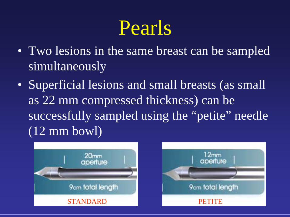
Pearls• Two lesions in the same breast can be sampled
simultaneously• Superficial lesions and small breasts (as small
as 22 mm compressed thickness) can be successfully sampled using the “petite” needle (12 mm bowl)
STANDARD PETITE
Post Biopsy Procedures
• Unilateral mammography to demonstrate clip position on day of biopsy
• Discharge patient to home with standard post biopsy instructions
• Correlate pathology to the MR finding to determine concordance or discordance
Conclusions
• Two different concepts evolved to improve the specificity of breast MRI – Morphology with high spatial resolution imaging– Kinetics with high temporal resolution imaging
• Implementation of high temporal and spatial resolution sequences allow integration of both kinetic and morphologic features for improved sensitivity and specificity
Conclusions• In combination, morphologic and dynamic
data increase the sensitivity and specificity of breast MRI.
• An algorithm can be established for interpretation.
• We always base recommendations on the most suspicious feature - architecture, enhancement or kinetics.
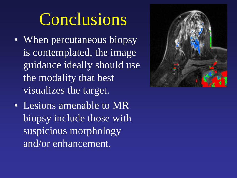
Conclusions• When percutaneous biopsy
is contemplated, the image guidance ideally should use the modality that best visualizes the target.
• Lesions amenable to MR biopsy include those with suspicious morphology and/or enhancement.
The End