Embed Size (px)
Citation preview

Breast CancerA Family Medicine Perspective
By Robert R. Zaid, DO
PrimeCare of Novi

Overview
• Epidemiology• Etiology• Risk Factors• Screening• Presentation• Workup• Staging• Treatment


Breast CancerEpidemiology
• Epidemiology• Etiology• Risk Factors• Screening• Presentation• Workup• Staging• Treatment
• Incidence:– Invasive breast cancer 1
• 1.4 million new cases in 2008
– Incidence rates for 2002 varied internationally • 3.9 cases per 100,000 in Mozambique • 101.1 cases per 100,000 in the United States
– Past 25 years• Breast cancer incidence rates have risen globally• Highest rates occurring in the westernized countries
– Change in reproductive patterns– Increased screening– Dietary changes– Decreased activity
• Mortality– Mortality has been decreasing– Especially in industrialized countries.
1 American Cancer Society

Breast CancerEpidemiology
• Epidemiology• Etiology• Risk Factors• Screening• Presentation• Workup• Staging• Treatment
• Projection (2009)– United States– Estimated 192,370 new cases in women– 1,910 cases in men
• Incidence rates– 70’s to 90’s had increasing incidence– 1999-2005
• Decreased by 2.2% per year• Why?
– Reduced use of hormone replacement therapy (HRT)
– Women’s Health Initiative in 2002
Swart, R; Downey, L, www.emedicine.com, Breast Cancer

Breast CancerEpidemiology
• Epidemiology• Etiology• Risk Factors• Screening• Presentation• Workup• Staging• Treatment
• Lifetime Risk of Breast Cancer– All Women
• 12.7%
– Non-Hispanic Whites• 13.3%
– African American Women• 9.98% • More likely to be diagnosed with larger,
advanced stage tumors (>5 cm)
Swart, R; Downey, L, www.emedicine.com, Breast Cancer

Breast CancerEpidemiology
• Epidemiology• Etiology• Risk Factors• Screening• Presentation• Workup• Staging• Treatment
• Death rates – Steadily decreased since 1990– Estimated 40,610 breast cancer deaths
for 2009– Women < 50 years
• Largest decrease in mortality • 3.3% per year• Thought to represent
– Earlier detection
– Improved treatment modalities
Swart, R; Downey, L, www.emedicine.com, Breast Cancer

Breast CancerEtiology
• Epidemiology• Etiology• Risk Factors• Screening• Presentation• Workup• Staging• Treatment
• Mechanism-– Current understanding of breast tumorigenesis
• Molecular alterations at the cellular level• Outgrowth and spread of breast epithelial cells
– Immortal features – Uncontrolled growth
• Genomic profiling– Demonstrated the presence of discrete breast tumor
subtypes » Luminal A» Luminal B» Basal» HER2+
– The exact number of disease subtypes and molecular alterations from which these subtypes derive remains to be fully elucidated
– Generally align closely with the presence or absence of hormone receptor and mammary epithelial cell type (luminal or basal).
Swart, R; Downey, L, www.emedicine.com, Breast Cancer

Breast CancerEtiology
• Epidemiology• Etiology• Risk Factors• Screening• Presentation• Workup• Staging• Treatment
Breast CancerRisk Factors
• Epidemiology• Etiology• Risk Factors• Screening• Presentation• Workup• Staging• Treatment
• Risk factors found by studies– Many of these factors form the basis for
breast cancer risk assessment tools. – Common denominator
• Level and duration of exposure to endogenous estrogen
• Increase lifetime exposure to estrogen– Premenopausal women
» Early menarche» Nulliparity» Late menopause
– Postmenopausal women» Obesity and hormone replacement therapy

Breast CancerRisk Factors
• Epidemiology• Etiology• Risk Factors• Screening• Presentation• Workup• Staging• Treatment
• Family History of breast cancer– 1st degree relative
• Risk 5 times greater in women with 2 or more first-degree relatives
• A family history of ovarian cancer in a first-degree relative
– Especially if the disease occurred at an early age (< 50 years old)
– Associated with a doubling of risk of breast cancer
Breast CancerRisk Factors
• Epidemiology• Etiology• Risk Factors• Screening• Presentation• Workup• Staging• Treatment
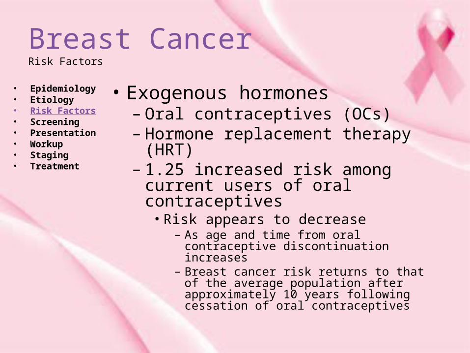
• Exogenous hormones– Oral contraceptives (OCs) – Hormone replacement therapy
(HRT)– 1.25 increased risk among current
users of oral contraceptives• Risk appears to decrease
– As age and time from oral contraceptive discontinuation increases
– Breast cancer risk returns to that of the average population after approximately 10 years following cessation of oral contraceptives

Breast CancerRisk Factors
• Epidemiology• Etiology• Risk Factors• Screening• Presentation• Workup• Staging• Treatment
• HRT– Consistent epidemiologic data support an
increased risk of breast cancer incidence and mortality (2003) with the use of postmenopausal HRT
– Directly associated with length of exposure
• Lobular (relative risk [RR]=2.25, 95% confidence interval [CI]= 2.00-2.52)
• Mixed ductal–lobular (RR=2.13, 95% CI= 1.68-2.70)
• Tubular cancers (RR=2.66, 95% CI= 2.16-3.28).
Breast CancerRisk Factors
• Epidemiology• Etiology• Risk Factors• Screening• Presentation• Workup• Staging• Treatment
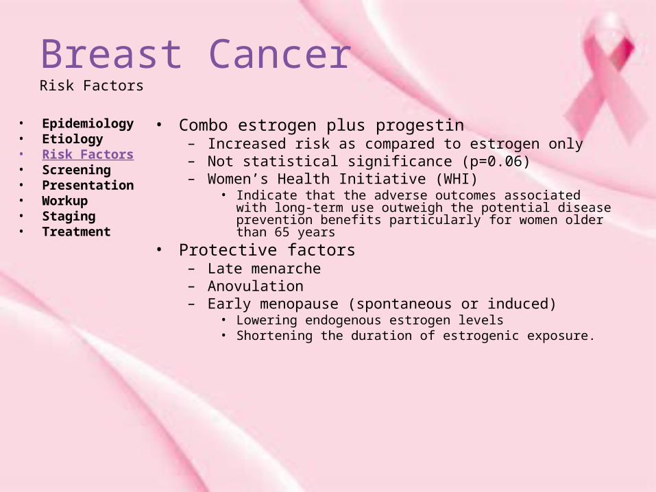
• Combo estrogen plus progestin – Increased risk as compared to estrogen only– Not statistical significance (p=0.06)– Women’s Health Initiative (WHI)
• Indicate that the adverse outcomes associated with long-term use outweigh the potential disease prevention benefits particularly for women older than 65 years
• Protective factors– Late menarche– Anovulation– Early menopause (spontaneous or induced)
• Lowering endogenous estrogen levels • Shortening the duration of estrogenic exposure.

Breast CancerRisk Factors
• Epidemiology• Etiology• Risk Factors• Screening• Presentation• Workup• Staging• Treatment
Advanced age
Family history
Two or more relatives (mother, sister)
One first-degree relativ
Family history of ovarian cancer in women <50y
Personal history Personal history Positive BRCA1/BRCA2 mutation Breast biopsy with atypical hyperplasia Breast biopsy with LCIS or DCIS
Reproductive history
Early age at menarche (<12 y)
Late age of menopause
Late age of first term pregnancy (>30 y)/nulliparity
Use of combined estrogen/progesterone
Current or recent use of oral contraceptives
Lifestyle factorsAdult weight gainSedentary lifestyleAlcohol consumption
>4
>5
>2
>2
3-4
>4
4-5
8-10
2
1.5-2
2
1.5-2
1.25
1.5-2
1.3-1.5
1.5

Breast CancerRisk Assessment Tools
• Epidemiology• Etiology• Risk Factors• Screening• Presentation• Workup• Staging• Treatment
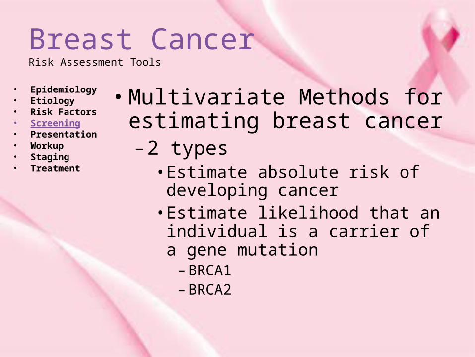
• Multivariate Methods for estimating breast cancer– 2 types
• Estimate absolute risk of developing cancer
• Estimate likelihood that an individual is a carrier of a gene mutation
– BRCA1– BRCA2
Breast CancerRisk Assessment Tools
• Epidemiology• Etiology• Risk Factors• Screening• Presentation• Workup• Staging• Treatment
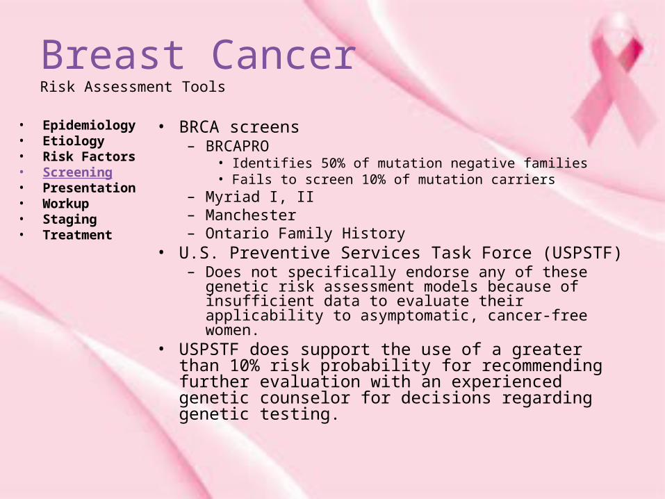
• BRCA screens– BRCAPRO
• Identifies 50% of mutation negative families• Fails to screen 10% of mutation carriers
– Myriad I, II– Manchester– Ontario Family History
• U.S. Preventive Services Task Force (USPSTF) – Does not specifically endorse any of these genetic risk
assessment models because of insufficient data to evaluate their applicability to asymptomatic, cancer-free women.
• USPSTF does support the use of a greater than 10% risk probability for recommending further evaluation with an experienced genetic counselor for decisions regarding genetic testing.

Breast CancerRisk Assessment Tools
• Epidemiology• Etiology• Risk Factors• Screening• Presentation• Workup• Staging• Treatment
• Risk Prediction Models– Gail Model (1989)
• Made from data from Breast Cancer Detection and Demonstration study
• Probability of developing breast cancer over a defined age interval
• Intended to improve screening guidelines– Gail Model 2
• Includes history of first-degree affected family members• Used extensively in clinical practice • Most accurate for non-Hispanic White women who receive
annual mammograms• Tends to overestimate risk in younger women who do not
receive annual mammograms• Reduced accuracy in populations with demographics (age,
race, screening habits) that differ from the population on which it was built
• http://www.cancer.gov/bcrisktool/

Breast CancerRisk Assessment Tools
• Epidemiology• Etiology• Risk Factors• Screening• Presentation• Workup• Staging• Treatment
• Care – Address concerns regarding
applicability of the Gail Model to African American women
– Data from a large case control study of African American
– CARE Model demonstrated high concordance between the numbers of breast cancer predicted and the number of breast cancers observed among African American women when validated in the WHI cohort.

Breast CancerGenetic Factors
• Epidemiology• Etiology• Risk Factors• Screening• Presentation• Workup• Staging• Treatment
• Heredity– 5-10% of women have an identifiable familial
predisposition– 20-30% of women with breast cancer have a
relative with history
• BRCA1 and BRCA2 mutations – Responsible for 3-8% of all cases of breast cancer – 15-20% of familial cases– Gene mutation on Chromosome 17 and 18
• Account for majority of inherited disease• Believed to be tumor suppressor genes
• Rare mutations are seen in the PTEN, TP53, MLH1, MLH2, and STK11 genes.

Breast CancerGenetic Factors
• Epidemiology• Etiology• Risk Factors• Screening• Presentation• Workup• Staging• Treatment
• Mutation rates may vary by ethnic and racial groups. – BRCA1 mutations
• Highest rates occur among Ashkenazi Jewish women (8.3%)
• Hispanic women (3.5%)• Non-Hispanic white women (2.2%)• African American women (1.3%)• Asian American women (0.5%)• Women with BRCA1 or BRCA2 gene
– Estimated 50-80% lifetime risk of developing breast cancer.
Breast CancerBreast Cancer Screening
• Epidemiology• Etiology• Risk Factors• Screening• Presentation• Workup• Staging• Treatment
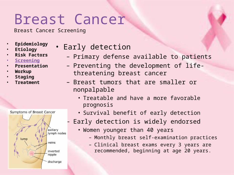
• Early detection – Primary defense available to patients – Preventing the development of life-threatening
breast cancer– Breast tumors that are smaller or nonpalpable
• Treatable and have a more favorable prognosis
• Survival benefit of early detection
– Early detection is widely endorsed • Women younger than 40 years
– Monthly breast self-examination practices – Clinical breast exams every 3 years are
recommended, beginning at age 20 years.
Breast CancerBreast Cancer Screening
• Epidemiology• Etiology• Risk Factors• Screening• Presentation• Workup• Staging• Treatment
• Mammography– Annual screening mammography beginning at age 40 years
• Widely recommended approach in the United States
– U.S. Preventive Services Task Force (USPSTF) Nov 2009• Updated breast cancer screening guidelines• Recommend against routine mammography before age 50 years• 40 to 49 years of age
– USPSTF suggests that the decision to start regular screening mammography be individualized and should include the patient's values regarding specific benefits and harms
– American College of Obstetricians and Gynecologists (ACOG)• Continues to recommend adherence to current ACOG guidelines• Screening mammography every 1-2 years for women aged 40-49 • Screening mammography every year for women age 50 or older• ACOG notes, however, that because of the USPSTF downgrading,
some insurers may no longer cover some of these studies.

Breast CancerBreast Self Examination
• Epidemiology• Etiology• Risk Factors• Screening• Presentation• Workup• Staging• Treatment
• Breast self-examination – Inexpensive and
noninvasive procedure– Evidence supporting
effectiveness• Controversial and
largely inferred– Not been found to
reduce mortality– Improvements in
treatment for early, localized disease
• Breast self-examination and clinical breast exam, continues to be recommended
• Clinical trials support combining clinical breast exam with mammography

Breast CancerBreast Self Examination
• Epidemiology• Etiology• Risk Factors• Screening• Presentation• Workup• Staging• Treatment
• Recommendations– USPSTF
• Inadequate evidence to make a recommendation for teaching or performing BSE
• 2009 USPSTF guidelines recommend against teaching women how to perform BSE
• Resulted in additional imaging procedures and biopsies– ACOG
• Continues to recommend counseling • BSE has potential to detect palpable breast cancer
Breast CancerMammography
• Epidemiology• Etiology• Risk Factors• Screening• Presentation• Workup• Staging• Treatment
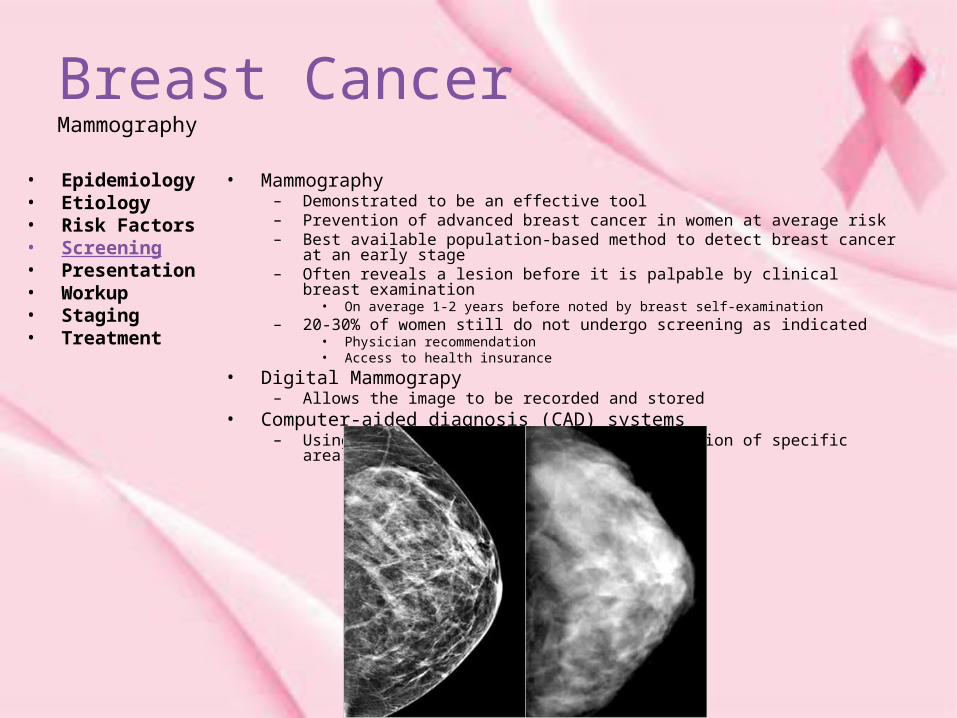
• Mammography– Demonstrated to be an effective tool– Prevention of advanced breast cancer in women at average risk– Best available population-based method to detect breast cancer at an early stage – Often reveals a lesion before it is palpable by clinical breast examination
• On average 1-2 years before noted by breast self-examination – 20-30% of women still do not undergo screening as indicated
• Physician recommendation • Access to health insurance
• Digital Mammograpy – Allows the image to be recorded and stored
• Computer-aided diagnosis (CAD) systems– Using an image modified to improve evaluation of specific areas in question.
Breast CancerMammography
• Epidemiology• Etiology• Risk Factors• Screening• Presentation• Workup• Staging• Treatment
• Recommendations:– USPSTF
• Estimates benefit of mammography in women – 50-74 years to be a 30% reduction risk of death – 40-49 years, the risk of death is decreased by 17%
• Non-white women and those of lower socioeconomic status remain less likely to obtain mammography services and more likely to present with life-threatening, advanced-stage disease

Breast CancerMammography
• Epidemiology• Etiology• Risk Factors• Screening• Presentation• Workup• Staging• Treatment
• Ultrasound– Widely available and useful adjunct to
mammography
• MRI– Combination of T-1, T-2, and 3-D contrast-
enhanced MRI techniques has been found to be highly sensitive
• Approximating 99%
– Limitations• 10-fold higher cost than mammography • Poor specificity (26%)• Significantly more false-positive reads
– Significant additional diagnostic costs and procedures.

Breast CancerMammography
• Epidemiology• Etiology• Risk Factors• Screening• Presentation• Workup• Staging• Treatment
• Below are the criteria for using breast MRI screening per the American Cancer Society (ACS).6
• Annual breast MRI – Evidence based
• BRCA mutation • First-degree relative of BRCA carrier, but untested • Lifetime risk approximately 20-25% or greater as
defined by BRCAPRO or other risk models
– Lifetime risk of breast cancer• Radiation to chest when aged 10-30 years • Li-Fraumeni syndrome and first-degree relatives • Cowden and Bannayan-Riley-Ruvalcaba syndromes
and first-degree relatives

Breast CancerMammography
• Epidemiology• Etiology• Risk Factors• Screening• Presentation• Workup• Staging• Treatment
• Insufficient evidence to recommend for or against MRI screening – Lifetime risk 15-20%, as defined by BRCAPRO or other
risk models
– Lobular carcinoma in situ or atypical lobular hyperplasia (ALH)
– Atypical ductal hyperplasia (ADH)
– Heterogeneously or extremely dense breast on mammography
– Women with a personal history of breast cancer, including ductal carcinoma in situ
• American Cancer Society does not recommend the use of breast MRI in women who have less than 15% lifetime risk

Breast CancerPresentation
• Epidemiology• Etiology• Risk Factors• Screening• Presentation• Workup• Staging• Treatment
• Mammogram-– Often irst detected as an abnormality on a mammogram – Mammographic features
• Asymmetry• Microcalcifications• A mass• Architectural distortion
• Larger tumors – May present as a painless mass
• Pain– 5% of patients with a malignant mass present with breast pain
• Other symptoms – Immobility– Skin changes (ie, thickening, swelling, redness) – Nipple abnormalities (ie, ulceration, retraction, spontaneous
bloody discharge)

Breast CancerWorkup
• Epidemiology• Etiology• Risk Factors• Screening• Presentation• Workup• Staging• Treatment
• Core biopsy– Percutaneous vacuum-assisted – Image guided breast biopsy
• Recommended diagnostic approach • Performed with • Ultrasound• Stereotactic, or MRI guidance
– Core biopsies spare the need for operative intervention
• Provides pathological results quicker than surgical excisions
• Excisional biopsy– As the initial operative approach
» Shown to increase the rate of positive margins

Breast CancerWorkup
• Epidemiology• Etiology• Risk Factors• Screening• Presentation• Workup• Staging• Treatment
• Palpation directed core biopsy– If a breast mass may be palpable but not
correlate with imaging
• Complications of a diagnostic core or excisional biopsy– Hematoma– Infection– Scarring– Re-operation– Sampling error resulting in inaccurate
diagnosis.

Breast CancerHistological Findings
• Epidemiology• Etiology• Risk Factors• Screening• Presentation• Workup• Staging• Treatment
• Ductal Carcinoma in situ (DCIS)• Lobular Carcinoma in situ (LCIS)• Medullary Carcinoma• Mucinous Carcinoma• Tubular Carcinoma• Papillary Carcinoma• Metaplastic Carcinoma• Mammary Paget’s Disease

Breast CancerHistological Findings
• Epidemiology• Etiology• Risk Factors• Screening• Presentation• Workup• Staging• Treatment
• Ductal Carcinoma in situ (DCIS)– Identified in ducts (non-invasive)– Identified on mammography
• Suspicious calcifications,
• Distribution– Linear– Clustered– Segmental– Focal– Mixed– DCIS is divided into comedo (ie, cribriform,
micropapillary, solid) and noncomedo subtypes, which provides additional prognostic information regarding likelihood of progression or local recurrence
Breast CancerHistological Findings
• Epidemiology• Etiology• Risk Factors• Screening• Presentation• Workup• Staging• Treatment
• Ductal Carcinoma in situ (DCIS)– Standard treatment of DCIS is surgical resection
with or without radiation– Adjuvant radiation and hormonal therapies
• Reserved for • Younger women• Patients undergoing lumpectomy• Comedo subtype
– Mastectomy• 30% of women with DCIS in the United States
– Conservative Surgery• 30% with conservative surgery alone
– Conservative surgery with whole breast radiation• 40% with conservative surgery followed by whole-
breast radiation therapy

Breast CancerHistological Findings
• Epidemiology• Etiology• Risk Factors• Screening• Presentation• Workup• Staging• Treatment
• Ductal Carcinoma in situ (DCIS)– Axillary or sentinel lymph node dissection is not
routinely recommended for patients with DCIS– Metastatic disease
• Disease to the axillary node in 10% of patients
– Whole-breast radiotherapy• Delivered 5-6 weeks following
– Tamoxifen • Adjuvant therapy for breast conserving surgery
• Only hormonal therapy currently approved
– Aromatase inhibitor (anastrozole)
• Currently in clinical trials

Breast CancerHistological Findings
• Epidemiology• Etiology• Risk Factors• Screening• Presentation• Workup• Staging• Treatment
• Lobular Carcinoma in situ (LCIS)– Found in the lobules (or glands)– Non-palpable mass
• Diffuse distribution throughout the breast• Incidence
– Doubled over last 25 years– 2.8% per 100,000 women– Peak incidence is in women aged 40-50 years
– No consistent features on breast imaging • Often an incidental finding• 10-20% of women with LCIS develop invasive breast cancer
– Within 15 years from diagnosis.
– LCIS is considered a biomarker of increased breast cancer risk• Treatment options
– Chemoprevention with a SERM– Bilateral mastectomy – Close observation.

Breast CancerHistological Findings
• Epidemiology• Etiology• Risk Factors• Screening• Presentation• Workup• Staging• Treatment
• Medullary Carcinoma– Relatively uncommon (5%)– Invasive– Occurs in younger women– Presentation
• Bulky palpable mass with axillary lymphadenopathy– Diagnosis
• Sheets of anaplastic tumor cells with scant stroma• Moderate or marked stromal lymphoid infiltrate• Histologic circumscription or a pushing border
– Other findings• DCIS may be observed in the surrounding normal tissues• ER, PR, and HER2/neu are typically negative, and TP53 is
commonly mutated.• Roughly 30% of patients have lymph node metastasis.
– Prognosis• Good

Breast CancerHistological Findings
• Epidemiology• Etiology• Risk Factors• Screening• Presentation• Workup• Staging• Treatment
• Mucinous Carcinoma– Rare histologic type
• Fewer than 5% of invasive breast cancer• Produces Mucin• Usually presents during the seventh decade• Excellent prognosis (>80% 10-year survival).

Breast CancerHistological Findings
• Epidemiology• Etiology• Risk Factors• Screening• Presentation• Workup• Staging• Treatment
• Tubular Carcinoma– Uncommon histologic type
• 1-2% of all breast cancers• Single layer of epithelial cells • Low incidence of lymph node involvement • Very high overall survival rate
Breast CancerHistological Findings
• Epidemiology• Etiology• Risk Factors• Screening• Presentation• Workup• Staging• Treatment
• Papillary Carcinoma– 1-2% of all carcinomas– Usually seen in women older than 60– Types
• Cystic (non-invasive)– Good prognosis
• Micropapillary ductal carcinoma (invasive)– Poor prognosis
– Lymph node metastasis

Breast CancerHistological Findings
• Epidemiology• Etiology• Risk Factors• Screening• Presentation• Workup• Staging• Treatment
• Metaplastic Carcinoma– 1% of breast cancers– Combination of adenocarcinoma plus mesenchymal and
epithelial components– Wide variety of histological patterns
• Spindle-cell carcinoma• Carcinosarcoma• Squamous cell carcinoma of ductal origin• Adenosquamous carcinoma• Carcinoma with pseudosarcomatous metaplasia• Matrix-producing carcinoma
– Metaplastic breast cancer tumors• Larger• More rapidly growing• Commonly node negative• Typically ER, PR, and HER-2 negative• Average age of onset in the sixth decade• Higher incidence in African Americans.

Breast CancerHistological Findings
• Epidemiology• Etiology• Risk Factors• Screening• Presentation• Workup• Staging• Treatment
• Metaplastic Carcinoma– Demonstrated a worse prognosis for
metaplastic breast cancer as compared to infiltrating ductal carcinoma
– 3-year overall survival rate of 48-71% – 3-year disease-free survival rate of 15-60%– Prognosis / predictors of poor overall survival
• Large tumor size
• Advanced stage
• Nodal status does not appear to impact survival in metaplastic breast cancer

Breast CancerHistological Findings
• Epidemiology• Etiology• Risk Factors• Screening• Presentation• Workup• Staging• Treatment
• Mammary Paget’s Disease– 1-4% of all breast cancers– Peak incidence is seen in the sixth decade of life (mean age 57 y)– Adenocarcinoma
• Localized within the epidermis of the nipple-areola complex • Paget cells
– Large– Pale epithelial cells
– Presentation• Lesions
– Unilateral developing insidiously – Scaly– Fissured– Oozing– Erythematous nipple-areola complex– Retraction or ulceration of the nipple is often noted– Itching, tingling, burning, or pain.
– Mammary Paget disease is associated with an underlying breast cancer in 75% of cases.
– Overall 5-year and 10-year survival rates are 59% and 44%, respectively.
Breast CancerPrognosis
• Epidemiology• Etiology• Risk Factors• Screening• Presentation• Workup• Staging• Treatment
– Predictors / prognostic factors of BC• Axillary lymph node status • Tumor size • Lymphatic/vascular invasion • Patient age • Histologic grade • Histologic subtypes (eg, tubular, colloid [mucinous],
papillary) • Response to neoadjuvant therapy • Estrogen receptor/progesterone receptor status • Her2/neu gene amplification and/or overexpression • Breast cancer predictive factors include the following: • Estrogen receptor/progesterone receptor status • Her2/neu gene amplification and/or overexpression • Lymph node status
Breast CancerStaging
• Epidemiology• Etiology• Risk Factors• Screening• Presentation• Workup• Staging• Treatment
• T- tumor size• N- Lymph node status• M- Metastasis
• Separated into stages 0- IV
• Survival Rates 5 year• Stages
– 0• 99-100%
– I• 95-100%
– II• 86%
– III• 57%
– IV• 20%

Breast CancerStaging
• Epidemiology• Etiology• Risk Factors• Screening• Presentation• Workup• Staging• Treatment
• National Cancer Center Network (NCCN) guideline – Stage I or II
• Recommends a history and physical examination
• Laboratory studies (CBC with differential, liver and renal function tests, and calcium levels)
– Stage III• Chest x-ray or CT scan of the chest• CT scan of the abdomen and pelvis• Bone scan for evaluation of distant metastasis• Tumor markers (CEA and CA15.3 or CA27.29)
may also be obtained in these patients

Breast CancerTreatment
• Epidemiology• Etiology• Risk Factors• Screening• Presentation• Workup• Staging• Treatment
• Lumpectomy• Mastectomy• Breast Reconstruction• Management of Contralateral breast• Sentinel Node Dissection• Axillary Lymph node dissection• Breast Conserving radiation therapy• Adjuvant Chemotherapy• Adjuvant Hormonal Therapy
• Behavioral therapy--- Very Important

Breast CancerTreatment
• Epidemiology• Etiology• Risk Factors• Screening• Presentation• Workup• Staging• Treatment
• Lumpectomy
– Defined as complete surgical resection of a primary tumor
– Goal of achieving widely negative margins (ideally a 1 cm margin around the lesion)
– Synonyms for lumpectomy • Partial mastectomy• Segmental mastectomy• Tylectomy• A quadrantectomy is a type of lumpectomy • Complete removal of the entire affected breast
quadrant• Performed with palpation guidance or with image
guidance

Breast CancerTreatment
• Epidemiology• Etiology• Risk Factors• Screening• Presentation• Workup• Staging• Treatment
• Mastectomy– Total mastectomy
• Complete removal of all breast tissue– Clavicle superiorly– Sternum medially– Inframammary crease inferiorly– Anterior axillary line laterally with en bloc resection of
the fascia of the pectoralis major– The nipple-areolar complex (NAC) is resected along
with a skin paddle to achieve a flat chest wall closure when performing a total mastectomy.
– No removal of any axillary nodes
• Modified radical mastectomy – Total mastectomy with axillary lymph node dissection

Breast CancerTreatment
• Epidemiology• Etiology• Risk Factors• Screening• Presentation• Workup• Staging• Treatment
• Postmastectomy Radiation Therapy– Positive postmastectomy margins – Primary tumors larger than 5 cm – Involvement of 4 or more lymph
nodes
• Breast Reconstruction– SSM– NSM
Breast CancerTreatment
• Epidemiology• Etiology• Risk Factors• Screening• Presentation• Workup• Staging• Treatment
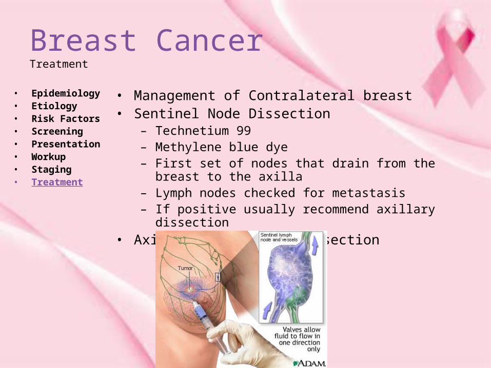
• Management of Contralateral breast• Sentinel Node Dissection
– Technetium 99– Methylene blue dye– First set of nodes that drain from the breast to the axilla– Lymph nodes checked for metastasis– If positive usually recommend axillary dissection
• Axillary Lymph node dissection

Breast CancerTreatment
• Epidemiology• Etiology• Risk Factors• Screening• Presentation• Workup• Staging• Treatment
• Breast Conserving radiation therapy– Used to eliminate residual subclinical disease
– Side effects • Fatigue• Breast pain• Swelling• Skin desquamation• Late toxicity (lasting 6 mo or longer following
treatment) – Persistent breast edema– Pain– Fibrosis– Skin hyperpigmentation

Breast CancerTreatment
• Epidemiology• Etiology• Risk Factors• Screening• Presentation• Workup• Staging• Treatment
• Adjuvant Chemotherapy• Adjuvant Hormonal Therapy
– Estrogen-receptor positive early stage breast cancer
• Hormonal therapy plays a main role• May be used with chemotherapy• Function to decrease estrogen's ability
to stimulate existing micro-metastases or dormant cancer cells
• Can reduce the relative risk of distant, ipsilateral, and contralateral breast cancer recurrence by up to 50%

Breast Cancer
• Any questions?• Powerpoint can be found at www.drzaid.com/presentations


![BANGLAKITAB · — 90 to. I Binoria Fatwa Against Zaid Hamid [Fatwa] Zaid Hamid Exposition I avoid Dr](https://img.pdfslide.us/doc/110x75/5e8663c5ff94e709d06f8158/a-90-to-i-binoria-fatwa-against-zaid-hamid-fatwa-zaid-hamid-exposition-i-avoid.jpg)