Embed Size (px)
Citation preview

8/14/2019 Branchial Osmoregulatory Response to Salinityin the çipura
http://slidepdf.com/reader/full/branchial-osmoregulatory-response-to-salinityin-the-cipura 1/14
Branchial Osmoregulatory Response to Salinity
in the Gilthead Sea Bream, Sparus auratusRAU L LAIZ-CARRIO N 1 , PEDRO M. GUERREIRO 2 , JUAN FUENTES 2 , ADELINO V.M. CANARIO 2 , MARI A P. MARTI´N DEL RI O 1 , and JUAN M. MANCERA 1 n
1 Departamento de Biologı ´a, Facultad de Ciencias del Mar y Ambientales,Universidad de Ca ´diz, 11510 Puerto Real, Ca ´diz, Spain 2 Centro de Cie ˆncias do MAR (CCMAR), Universidade do Algarve,Campus de Gambelas, 8005-139 Faro, Portugal
ABSTRACT The branchial osmoregulatory response of gilthead sea bream ( Sparus auratus L.)to short-term (2–192 hr) and long-term (2 weeks) exposure to different environmental salinities (5 % ,15% , 25% , 38% and 60 % ) was investigated. A ‘‘U-shaped’’ relationship was observed betweenenvironmental salinity and gill Na þ ,K þ -ATPase activity in both long- and short-term exposure toaltered salinity, with the increase in activity occurring between 24 and 96 hr after the onset of exposure. Plasma osmolality and plasma ions (sodium, chloride, calcium and potassium) showed a tendency to increase in parallel with salinity. These variables only differed signicantly ( P o 0.05) insh adapted to 60 % salinity with respect to sh adapted to full-strength sea-water (SW). Plasma glucose remained unchanged whereas plasma lactate was elevated at 5 % and 60 % . Muscle watercontent (MWC) was signicantly lower in sh adapted to 60 % . Chloride cells (CC) were only presenton the surface of the gill laments and absent from the secondary lamellae. CC distribution was notaltered by external salinity. However, the number and size of CC were signicantly increased atsalinity extremes (5 % and 60 % ), whereas sh exposed to intermediate salinities (15 % and 25 % ) hadfewer and smaller cells. Furthermore, the CC of sh exposed to diluted SW became rounder whereasthey were more elongated in sh in full-strength and hypersaline SW. This is consistent withprevious reports indicating the existence of two CC types in euryhaline sh. At likely environmentalsalinities, gilthead sea bream show minor changes in plasma variables and the effective regulation of
gill Na þ
,K þ
-ATPase. However, at very low salinities both haemodilution and up-regulation of gillNa þ ,K þ -ATPase predict a poor adaptation most likely related to deciency or absence of speciccomponents of the CC important for ion uptake. J. Exp. Zool. 303A:563–576 , 2005. r 2005 Wiley-Liss, Inc.
INTRODUCTION
Euryhaline teleosts have the ability to adapt todifferent environmental salinities while maintain-ing essentially constant their internal milieu bythe activation of several osmoregulatory mechan-isms, namely in the branchial and renal epithelia.In this adaptive process, two consecutive phasesoccur: an initial period characterized by changing osmotic variables, followed by a chronic regulatoryperiod, when these variables reach a new home-ostasis (Holmes and Donaldson, ’69; Maetz, ’74).
The modulation of Na þ ,K þ -ATPase pump activ-ity of chloride cells (CC) in the branchial epithelia is essential for acclimation to a new environmentalsalinity (Epstein et al., ’80; Zadunaisky, ’84;McCormick, ’95; Marshall, 2002). The alterationsin gill Na þ ,K þ -ATPase activity in relation to
environmental salinity are diverse, but two typicalsituations seem to prevail: (i) a direct relationship,characteristic of anadromous species, in whichhigher salinities induce higher values of gill Na þ ,K þ -ATPase activity (McCormick, ’95) and (ii) a
Grant sponsors: This work was supported by grants BOS2001-4031-C02-01 (Ministerio de Ciencia y Tecnologı ´a, Spain) and PTR95-0431-OP (DGES, Ministerio de Educacio ´ n y Ciencia, Madrid, Spain) to J.M.M. Travelling between labs was partially funded by grants DGESHP1999-0098 and HP2001-0061 (Ministerio de Ciencia y Tecnologı ´a)from integrated actions to J.M.M. and CRUP, Portugal, to J.F. R.L.-C.was in receipt of a MIT-F2 pre-doctoral fellowship from Ministerio deCiencia y Tecnologı ´a, Spain. P.M.G. was in receipt of fellowshipPRAXIS/BD/9207/96 from Fundac ¸a ˜ o para a Cie ncia e Tecnologia (Portugal) and visited the University of Cadiz in the frame of the EUSocrates Exchange Programme.
*Correspondence to: Dr. Juan Miguel Mancera Romero Departa-mento de Biologı ´a, Facultad de Ciencias del Mar, Universidad de Ca ´ diz11510 Puerto Real, Ca ´ diz, Spain. E-mail: [email protected]
Received 7 October 2004; Accepted 21 February 2005Published online in Wiley InterScience (www.interscience.wiley.
com). DOI: 10.1002/jez.a.183.
2005 WILEY-LISS, INC.
JOURNAL OF EXPERIMENTAL ZOOLOGY 303A:563–576 (2005)

8/14/2019 Branchial Osmoregulatory Response to Salinityin the çipura
http://slidepdf.com/reader/full/branchial-osmoregulatory-response-to-salinityin-the-cipura 2/14
U-shaped relationship, described for some euryha-line teleosts (Towle et al., ’77; Gallis et al., ’79; Jensen et al., ’98), in which lower values of gillNa þ ,K þ -ATPase activity occur at intermediatesalinities and higher values at low and high
salinities. Changes in gill Na þ
,K þ
-ATPase activityare observed 2–3 days after transfer from a hypoosmotic to hyperosmotic environment ineuryhaline teleosts ( Anguilla rostrata : Forrestet al., ’73; Dormitator maculatus : Evans andMallery, ’75; Fundulus heteroclitus : Jacob andTaylor, ’83; Dicentrarchus labrax : Jensen et al.,’98). In anadromous species ( Oncorhynchus kisutch : Boeuf et al., ’78; Salvelinus fontinalis :McCormick and Naiman, ’85; Salmo gairdneri :Madsen and Naamansen, ’89; Salmo salar : Bergeet al., ’95), activation of gill Na þ ,K þ -ATPase takesplace 3–7 days after transfer to seawater (SW).The delay in gill Na þ ,K þ -ATPase activation inresponse to osmotic challenge is proposed to reectchanging gene expression. Thus modications inenvironmental salinity alter not only the activityof Na þ ,K þ -ATPase but also transcript expressionand protein synthesis (Lee et al., 2000; Seidelinet al., 2000; Tipsmark et al., 2002). However, inaddition to this slow activation, a rapid, non-genomic activation of gill Na þ ,K þ -ATPase pumphas been reported, involving phosphorylation and/ or membrane insertion of the protein (Hwang et al., ’89; Uchida and Kaneko, ’96; Mancera and
McCormick, 2000; Tipsmark and Madsen, 2001).Changes in Na þ ,K þ -ATPase pump activity dur-ing salinity adaptation in most sh are paralleledby alteration in the number and size of branchialCC, the site at which most of the branchial ionicregulation takes place (Perry, ’97; Marshall andBryson, ’98; Marshall, 2002). Slow salinity adapta-tion usually involves the biogenesis or reshaping of existing CC, which undergo important changesin sh exposed to salinity variations (Pisam andRambourg, ’91; Sakamoto et al., 2001; Varsamoset al., 2002; Wilson and Laurent, 2002).
The gilthead sea bream ( Sparus auratus L.) is a marine teleost living in coastal waters, capable of adapting to considerable changes in environmen-tal salinity (Chervinski, ’84; Mancera et al., ’93a).Previous studies with this species showed that a decrease in environmental salinity (from 38 % to7% ) activates the prolactin, growth hormone andcorticotrophic cells in the adenohypophysis (Man-cera et al., ’93b, ’95) and that the transfer fromseawater (SW) to brackish water (BW) leads totransitory blood hypomineralization (Mancera et al., ’93a). Additionally, it was observed that
prolactin and cortisol increase the Na þ ,K þ - ATPase activity and blood osmolality in BW exposed gilthead sea bream, thus improving itshypoosmoregulatory capacity (Mancera et al., ’94,2002; Laiz-Carrio ´ n et al., 2003). The distribution,
density and morphology of CC in response to thesalinity challenge remain unknown in the marinegilthead sea bream. Most studies have focused onSW adaptation of sh previously kept in fresh-water (FW) (Marshall et al., ’99; Wong and Chan,’99; Lee et al., 2000; Tipsmark et al., 2002),and the response to acclimation to hypo- andhyperosmotic environments has received littleattention.
Therefore, the aim of the present study was todescribe and analyse the alterations and thecompensatory mechanisms occurring in the bran-chial osmoregulatory system of juvenile giltheadsea bream exposed and acclimated to a wide rangeof environmental salinities, and to determinewhether the adaptation in this species is animmediate phenomenon or a slow, long-lasting gradual process.
MATERIALS AND METHODS
Experimental protocol
Immature gilthead sea bream (40–60 g bodyweight) were provided by a commercial sh farm(CUPIMAR SA, San Fernando, Ca ´ diz, Spain). Fishwere transferred to the wet laboratories at theFaculty of Marine Sciences (Puerto Real, Ca ´ diz),where they were acclimated for 30 days to full SW (38% , 1162mOsmkg 1 H 2 O) in 300-l tanks in anopen system. After this period, sh were used forexperiments to assess how they adapted to alteredsalinities. The experimental salinities wereachieved either by mixing full-strength SW withdechlorinated tap water or by mixing full-strengthSW with natural marine salt (Instant Ocean,
TABLE1. Osmolalityand ioniccompositonof the waterat di¡erentsalinities used in the experiments
Salinity
5% 15% 25% 38% 60%
Osmolality (mOsm) 130 366 613 1162 1494Na þ (mmol l 1) 55 185 304 468 734Cl (mmol l 1) 71 210 353 534 821Ca 2 þ (mmol l 1) 1.8 4.3 7.3 11.1 16.9K þ (mmol l 1) 1.6 4.3 6.5 10.8 16.6Mg 2 þ (mmol l 1) 7.7 24.3 38.9 61.6 93.4
R. LAIZ-CARRIO ´ N ET AL.564

8/14/2019 Branchial Osmoregulatory Response to Salinityin the çipura
http://slidepdf.com/reader/full/branchial-osmoregulatory-response-to-salinityin-the-cipura 3/14
Aquarium Systems, Sarrebourg, France). Theosmolality and ionic composition of the waterused for the different experimental groups areshown in Table 1. During the experiments the shwere maintained under natural photoperiod and
constant temperature (181
C). Fish were fed oncedaily with 1% body weight commercial dry pellets(Dibaq-Diprotg SA, Segovia, Spain) and werefasted for 24 hr before sampling.
Trial 1 F long-term exposure
Four different experimental salinities weretested and all experiments were conducted induplicate tanks (100-l capacity) (7–8 sh per tank). After an initial 7-day acclimation period inSW, sh were exposed to gradually changing
salinity over 2 hr until it reached 5 % (130 mOsmkg 1 H 2 O), 15 % (366mOsmkg 1 H 2 O), 25 %
(613 mOsmkg 1 H 2 O) or 60 % (1494 mOsm kg 1
H 2 O), while another group (SW control) was keptat 38 % (1162 mOsm kg 1 H 2 O). Fish were exposedto a specic environmental salinity for 2 weeks byrecirculating tank water. At the end of theexperiment, sh were anaesthetized in 2-phenox- yethanol (1 ml l 1 water, Sigma-Aldrich, Madrid,Spain) and samples were collected (see below).
Trial 2 F short-term exposure
Fish were transferred directly from SW (38 % )to tanks containing water at 5 % , 15% , 38% and60% (n ¼ 7–8 per tank). Five tanks were used persalinity and were sampled consecutively at 2, 4, 24,96 and 192 hr after transfer. At each time point allthe animals from one tank per salinity wereanaesthetized in 2-phenoxyethanol (1ml l 1
water), and a blood sample and a biopsy of gilltissue were collected. Blood samples collected fromsh previously withdrawn directly from the stock-ing tank were considered as representative fortime zero.
During both trials the water salinity waschecked daily and corrected when necessary bythe addition of small volumes of either FW or SW. At the beginning and during the experiments,water samples were collected for ion compositiondetermination. The experiments described complywith the Guidelines of the European UnionCouncil (86/609/EU) and of the University of Cadiz (Spain) for the use of laboratory animals.No mortality was observed during the experi-ments.
Sampling
Fish were anaesthetized with 2-phenoxyethanol(Sigma-Aldrich) (1 ml l 1 water), weighed andsampled. The blood was collected from the caudalpeduncle into 1-ml ammonia-heparinized syr-inges. Plasma was separated from cells by cen-trifugation of whole blood (5 min at 10,000 g) andwas immediately frozen in liquid nitrogen andstored at 80 1 C until analysis. A biopsy of gilltissue was placed in 100 ml of ice-cold sucrose-EDTA-imidazole (SEI) buffer (150 mM sucrose,10mM EDTA, 50mM imidazole, pH 7.3) andfrozen at 80 1 C. A piece of paraxial muscle wasdissected for gravimetrical determination of totalwater content.
Immunolocalization and morphology
of CC Gill arches were removed and placed in freshly
mixed Champy-Maillet’s xative (0.2% osmiumtetraoxide, 25 mg ml 1 iodine, and saturated me-tallic zinc) for 16 hr (Avella et al., ’87). The tissuewas rinsed with deionized water, dehydrated andembedded in parafn. Sections (6 mm thick) cutparallel to the long axis of the lament wereobtained. In addition, gill laments were placedfor 24 h in 4% paraformaldehyde in 0.1 M phos-phate buffer (pH 7.2) at 4 1 C. Tissues weredehydrated, embedded in parafn and sections
(6 mm thick) parallel to the long axis of thelament were obtained. The sections were stainedwith haematoxylin–eosin for histochemistry. Forimmunocytochemistry, tissue sections were im-munostained using the unlabelled enzyme methodof Sternberger (’86) with the monoclonal a5antiserum raised in mouse against the chickenNKA a1-subunit (Developmental Studies Hybri-doma Bank, Department of Biological Science,University of Iowa, Iowa, USA). This antiserum isspecic for a cytosolic epitope, and reacts with allisoforms of the a-subunit of distant species (seeTakeyasu et al., ’88). All sections were incubatedfor 18hr at 22 1 C in the primary antiserum at a dilution of 1:500. The second antiserum (anti-mouse IgG, raised in goat, Sigma M6898) was usedat a dilution of 1:40 for 60min at 22 1 C and thePAP complex (1:100) (peroxidase antiperoxidasesoluble complex mouse, Sigma P2416) was usedfor 45 min at 22 1 C. The chromogen was 3.3 0-diaminobenzidine tetrahydrochloride (DAB, Sigma). Antisera and the PAP complex were diluted inTris buffer, pH 7.8, containing 0.7% non-gelling seaweed gelatin, lambda carrageenan (Sigma),
BRANCHIAL OSMOREGULATION IN GILTHEAD SEA BREAM 565
8/14/2019 Branchial Osmoregulatory Response to Salinityin the çipura
http://slidepdf.com/reader/full/branchial-osmoregulatory-response-to-salinityin-the-cipura 4/14
0.5% Triton X-100 (Sigma) and 0.02% sodiumazide. Coplin jars were used for incubation of sections in the rst and second antisera, whereasPAP incubation was carried out in a moistchamber. To test the specicity of the immunor-
eaction, adjacent sections were processed asdescribed above, but incubation in the primera antisera was omitted.
Immunostained CC were analysed using a LeitzFluovert inverted microscope with a CCD Kapa CF15/2 video camera. The size of cell bodies wasmeasured using the image processor VIDS V program (AMD, Analitical Measuring Systems).The number of positively stained cells wasexpressed as cells per millimetre of lament(frequency; in both sides ventral and dorsal of a primary lament) measured in 5 transects perslide, 4 slides per sh and 6 sh per salinity. ForCC size and shape, results are the average 7 SEMof at least 50 cells per slide, 4 slides per sh and 6sh per salinity. Shape factor is dened as 4 p A / P 2
(where A is the area and P is the perimeter) suchthat values approaching 1 indicate a near-circularshape and lower values indicate a more elongatedshape. The sh from which slides containing stained CC were prepared was not disclosedduring the observations to avoid bias.
Gill Na þ ,K þ -ATPase activity
Gill Na þ ,K þ -ATPase activity was determinedusing the microassay method of McCormick (’93)adapted to gilthead sea bream (Mancera et al.,2002). Gill tissue was homogenized in 125 ml of SEIbuffer with 0.1% deoxycholic acid and thencentrifuged at 2,000 g for 30 sec. Duplicate 10 mlhomogenate samples were added to 200 ml assaymixture in the presence or absence of 0.5 mmoll 1
ouabain in 96-well microplates at 25 1 C and read at340nm for 10 min with intermittent stirring byvibration. Ouabain-sensitive ATPase activity wasdetected by enzymatic coupling of ATP depho-sphorylation to NADH oxidation and expressed as
mmolADP mg protein1
hr1
. The Pierce BCA Pro-tein kit (Pierce, Rockford, IL) was used withbovine albumin as standard. Both assays were runon a microplate reader (EL340i, Bio-Tek Instru-ments, Winooski, VT) using Delta Soft3 softwarefor Macintosh (BioMetallics Inc., Princeton, NJ).
Blood chemistry
Plasma and water osmolality was measuredwith a vapour pressure osmometer (Fiske One-Ten Osmometer, Fiske, VT) and expressed as
mOsm kg 1 . Plasma and water Na þ , K þ , Ca 2 þ andMg 2 þ levels were measured using atomic absorp-tion spectrophotometry (Philips PU7000) and Cllevels were measured with the Chloride Sigma- Aldrich kit (no. 461). Plasma glucose and lactate
were measured using commercial kits from Sigma- Aldrich (Glucose HK #16-20UV; Lactate #735-10)adapted to microplates (Stein, ’63; Iwama et al.,’89). Plasma protein concentration was deter-mined by diluting the plasma 1:40 and measuring protein concentration using the bicinchoninic acidmethod (Smith et al., ’85) with a BCA protein kit(Pierce) for microplates, with bovine albumin asstandard. The assays were read on a Bio KineticsEL-340i Automated Microplate Reader (Bio-TekInstruments) using DeltaSoft3 software forMacintosh (BioMetallics Inc.). Muscle water con-tent (MWC) was determined gravimetically aspercent weight loss after drying to a constantweight at 100 1 C over 2 days.
Cortisol assay
Plasma levels of cortisol were determined byradioimmunoassay (RIA) as described previously(Rotllant el al., 2005). Briey, plasma sampleswere diluted in phosphate buffer containing 0.5gl 1 gelatine, pH 7.6, and denatured at 70 1 Cfor 30 min. The antiserum used for the assay wasraised in rabbits against cortisol-3-(O-carboxy-methyl)oxime–bovine serum albumin conjugate(Sigma-Aldrich). This antiserum cross-reacts100% with cortisol, 54% with 11-desoxycortisol,10% with cortisone, 16% with 17,21-dihydroxy-5b-pregnan-3,11,20-trione, 5% with 11 b,17,21-trihydroxy-5 b-pregnan-3,20-dione, 0.05% with11-hydroxytestosterone and o 0.001% with testos-terone. Although cross-reactivity exists with 11-desoxycortisol and cortisone (as observed whenvalidating the assay), we have found that thesesubstances are virtually non-existent in sea breamplasma: binding of the antisera to thin layerchromatography fractioned sh plasma conrmed
the specicity of the assay by revealing a singlepeak in the cortisol-specic fraction and none inthe region of the other compounds.
Statistics
Signicant differences among groups weretested by one-way ANOVA, followed by theStudent–Newman–Keuls multiple comparisontest. For the study of gill density, size and shape,the effect of treatments was tested by a hierarch-ical (nested) ANOVA. The levels of variability
R. LAIZ-CARRIO ´ N ET AL.566

8/14/2019 Branchial Osmoregulatory Response to Salinityin the çipura
http://slidepdf.com/reader/full/branchial-osmoregulatory-response-to-salinityin-the-cipura 5/14
were the sh in each treatment ( n ¼ 6) and theslides from each sh ( n ¼ 4). Because no signicantdifferences were found in the experimental units(sh and slides within treatments), one-way ANOVA was applied, using the pooled data of sh
for each salinity (Zar, ’84; Underwood, ’97). Posthoc recommendation of Underwood (’97) beforethe use of pooled data was followed to reduce therisk of type I or II errors, increasing the power of the ANOVA test because of the large number of degrees of freedom associated with the pooledmean-square (Underwood ’97). Results were con-sidered signicantly different at P o 0.05.
RESULTS
Trial 1 F long-term exposure
Substantial changes in plasma osmolality wereobserved after 2 weeks of exposure to alteredexternal salinity (Fig. 1A). This parameter in-creased signicantly from sh adapted at 5 % tosh adapted at 60 % . Interestingly, the effect of environmental salinity on the activity of gillNa þ ,K þ -ATPase after 2 weeks of exposure (Fig.1B) was U-shaped and sh adapted to extremes of salinity (high and low) had signicantly higher( Po 0.05) gill Na þ ,K þ -ATPase activity than shexposed to other salinities. In fact, sh maintainedin 60 % had more than double the gill Na þ ,K þ - ATPase than sh maintained in 15 % , 25% and38% .
Plasma concentrations of ions (sodium, chloride,calcium and potassium) increased in parallel withsalinity, and were signicantly higher in 60 %
acclimated sh than in sh adapted to all othersalinities (Table 2). In contrast, MWC was sig-nicantly lower in 60 % adapted sh than in shadapted to lower salinities.
Table 3 shows the levels of plasma metabolitesmeasured in sh adapted to different salinities.
Plasma glucose levels were not signicantlydifferent among sh maintained at differentsalinities. However, plasma lactate levels weresignicantly higher ( P o 0.05) in sh adapted to
Fig. 1. Effect of different environmental salinities onosmolality ( A) and gill Na þ ,K þ -ATPase activity ( B ). Fish wereacclimated to different salinities for 2 weeks. Data areexpressed as mean 7 SEM ( n ¼ 7–8). The letters indicatesignicant differences among groups ( Po 0.05).
TABLE 2 Plasma ion levels and muscle water content (MWC) in ¢sh acclimated to di¡erent environmental salinities for 2 weeksSalinity
5% 15% 25% 38% 60%
Na þ (mmol l 1) 1707 3a 1787 2a,b 1807 2b,c 1857 3b,c 1967 4c
Cl (mmol l 1) 1457 3a 148 7 2a 1497 1a 1527 3a 1587 2b
Ca 2 þ (mmol l 1) 2.227 0.21 a 2.35 7 0.20 a 2.37 7 0.19 a 2.51 7 0.18 a 2.73 7 0.23 b
K þ (mmol l 1) 4.67 0.4 a 4.87 0.3a 5.07 0.2a 5.17 0.3a 5.37 0.3b
MWC% 78.2 7 0.6a 77.4 7 1.1a 77.5 7 0.5a 76.9 7 1.0 a 75.6 7 1.1b
a Data are shown as mean 7 SEM ( n ¼ 7^8).b Groups that are signi¢cantly di¡erent ( Po 0.05) are indicated by a di¡erent letter.c The same letter indicates that no signi¢cant di¡erences exist.
BRANCHIAL OSMOREGULATION IN GILTHEAD SEA BREAM 567

8/14/2019 Branchial Osmoregulatory Response to Salinityin the çipura
http://slidepdf.com/reader/full/branchial-osmoregulatory-response-to-salinityin-the-cipura 6/14
5% and 60 % than in those maintained at 15 % ,25% and 38 % . The highest concentrations of plasma lactate were measured in sh maintainedat the lowest salinity (5 % ). Plasma protein levelsincreased in parallel with ambient salinity, andsignicant differences ( P o 0.05) were only ob-served between sh adapted to 60 % and all othergroups of sh adapted to lower salinities.
Trial 2 F short-term exposure
At 2 hr after transfer, the plasma osmolality wasslightly higher in sh transferred to 60 % than inthose maintained at 38 % (although not statisti-cally different from the values from undisturbedsh, 312 7 3.2 mOsmkg 1 ), whereas those trans-ferred to lower salinities (5 % and 15 % ) had si-
gnicantly lower plasma osmolality levels (Fig. 2A). Values of plasma osmolality between the 5 % andthe 60 % groups were signicantly different 2 hrafter transfer and thereafter. There was a clearrelationship between plasma osmolality and ex-ternal salinity after 8 days of exposure, and in shmaintained at 5 % , 15% and 60 % , plasma osmol-ality averaged 83 % , 89% and 109% of SW sh,respectively. In all groups, 4 hr after transferplasma osmolality increased slightly over thatmeasured 2 hr after transfer and had returned topre-transfer levels within 24hr. Interestingly, therange of plasma osmolality values of the shassayed throughout trial 2 was greater than thatmeasured at the end of trial 1.
Gill Na þ ,K þ -ATPase activity of gilthead sea bream increased signicantly between 24 and96 hr after transfer from 38 % to 5 % or 60 % andremained elevated at 192 hr (Fig. 2B). At this timepoint, the activity in the extreme salinity twogroups was almost 2.5-fold higher than in thecontrol group. Transfer to 15 % did not causesignicant modications in gill Na þ ,K þ -ATPaseactivity at any time point.
TABLE 3. Plasma levels of glucose, lactate and proteins in ¢sh acclimated to di¡erent environmental salinities for 2 weeks
Salinity
5% 15% 25% 38% 60%
Glucose (mmol l 1) 3.767 0.29 a 3.18 7 0.21 a 3.31 7 0.19 a 3.66 7 0.24 a 4.06 7 0.30 a
Lactate (mmol l1
) 0.967 0.10a
0.58 7 0.04b
0.58 7 0.03b
0.70 7 0.04b,c
0.81 7 0.05a,c
Protein (mmol l 1) 32.2 7 4.3a 35.4 7 4.1a 37.5 7 3.5a 37.5 7 4.3a 40.3 7 3.3b
a Data are shown as mean 7 SEM ( n ¼ 7^8).b Groups that are signi¢cantly di¡erent ( Po 0.05) are indicated by a di¡erent letter.c The same letter indicates that no signi¢cant di¡erences exist.
Fig. 2. Time-course effect of different environmentalsalinities in osmolality ( A) and gill Na þ ,K þ -ATPase activity(B ) at 2, 4, 24, 96 and 192hr after transfer. The grey dotbefore time zero indicates the osmolality value for undis-turbed sh in the stocking tank. Na þ ,K þ -ATPase activity data are shown as % of control (control ¼ 100%). Each value is themean 7 SEM of n¼ 7–8 sh per group in each sampling time.Different letters indicate signicant differences ( Po 0.05)between groups within the same time point. n indicatessignicant differences between the current and preceding timepoint. # indicates signicant differences from undisturbedsh.
R. LAIZ-CARRIO ´ N ET AL.568
8/14/2019 Branchial Osmoregulatory Response to Salinityin the çipura
http://slidepdf.com/reader/full/branchial-osmoregulatory-response-to-salinityin-the-cipura 7/14
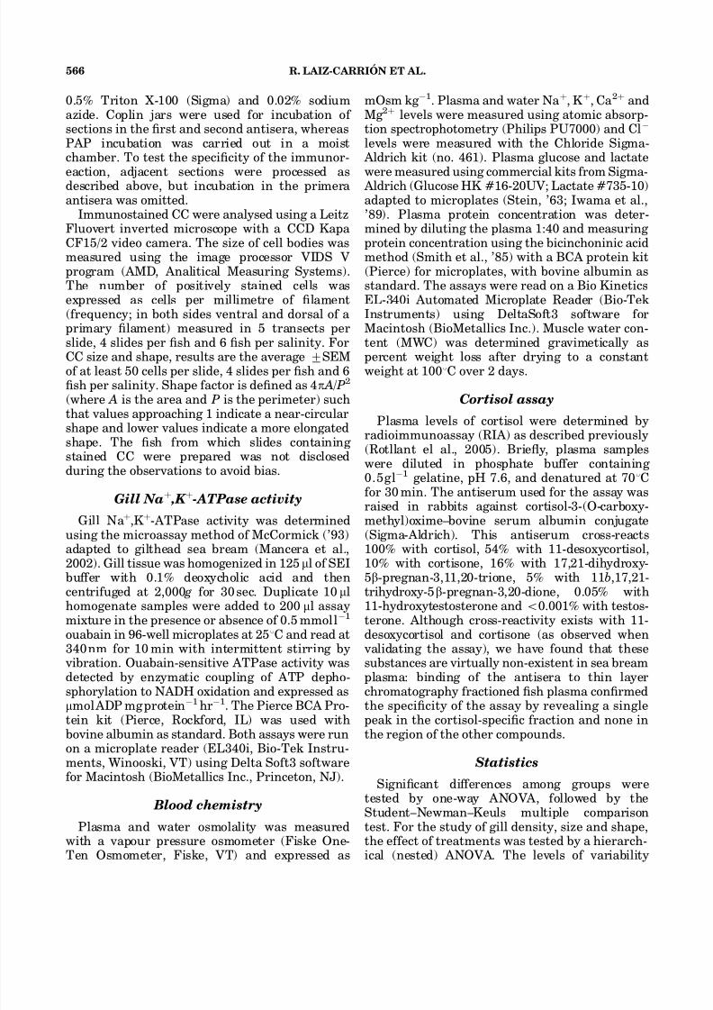
Plasma cortisol levels were elevated at 2hr aftertransfer in all groups (Fig. 3), well above thevalues measured for undisturbed sea bream beforethis experiment (3.3 7 0.33ngml 1 ) and in similarstocking conditions (Rotllant et al., 2001; Tortet al., 2001). At this time point, in the extremesalinity two groups, sh had higher cortisol levels(21.7 7 1.99 and 21.3 7 4.02 ngml 1 , respectively)than sh at 38 % salinity (13.1 7 2.84 ngml 1 ),whereas sh at 15 % had intermediate values closeto the extreme groups (21.2 7 1.78ngml 1 ) but notstatistically different from the control. At 4 hrafter transfer cortisol levels had decreased sig-nicantly, and from 24 hr onwards these wereconstant in the intermediate salinities and similar
to those of undisturbed animals. The restorationof normal cortisol levels was slower in sh at5% and 60 % , in which values were only identicalto those sh in the control after 192hours post-transfer.
Branchial CC density and morphology
There was complete overlap in the resultsobtained with the two CC identication methodsutilized, namely, immunolocalization with an anti-serum against Na þ ,K þ -ATPase and the osmium
tetraoxide technique. CC were present only on thesurface of the gill primary lament but never inthe secondary lamellae, and this distribution wasnot affected by salinity challenge (Fig. 4).
Fish acclimated to SW (38 % ) showed an average
of 106.4 7 1.73CCmm1
and adaptation to hyper-saline SW (60 % ) signicantly increased the num-ber of CC present in sh gills (Figs 4 and 5A). Insh exposed to intermediate salinities (15 % and25% ), a signicant reduction in CC numberrelative to the SW group was observed. However,the CC frequency in sh acclimated to 5 % wassimilar to that observed in the sh at 38 % .Nevertheless, at 5 % , CC cell size increasedmarkedly and they were 1.3 times larger thanthose in control sh. CC in sh maintained at 60 %
were also larger than in control sh, whereas insh exposed to 15 % and 25 % the area of the CCdecreased signicantly (Fig. 5B). In sh main-tained in full-strength and hypersaline SW, CChad an oblate ellipsoid and contrasted with thatof sh in diluted SW, which were more closelyspheroidal (ratio of area to perimeter closer to 1;Fig. 5C).
DISCUSSION
This study shows that gilthead sea bream canadapt to a wide range of salinities with adjustmentin body uids osmolytes, plasma lactate and
cortisol, which are more evident at the extremerange of salinities experienced in nature (5–60 % ).The gill, a major osmoregulatory organ in sh,undergoes large morphological changes, even atintermediate salinities, but Na þ ,K þ -ATPase onlyshowed increased activity at the low and highextremes of salinity.
Time course of salinity adaptation
After an abrupt salinity transfer, an adaptiveperiod involving changes in osmotic variables isexpected (Holmes and Donaldson, ’69; Maetz, ’74;Goswami et al., ’83). In our experiments (trial 2),the transfer from 38 % to the different experi-mental salinities induced immediate (2 hr aftertransfer) changes in plasma osmolality that weresignicantly different from control in the extremesalinities throughout the duration of the experi-ment. Almost instant changes in plasma osmol-ality and/or electrolyte concentration after salinitytransfer have also been described by Woodand Marshall (’94) and Marshall et al. (’99) in F. heteroclitus , by Kelly and Woo (’99) in Sparus
Fig. 3. Effects of different environmental salinities onplasma cortisol levels at 2, 4, 24, 96 and 192hr after transfer todifferent salinities. The grey dot before time zero indicates thecortisol value for undisturbed sh in the stocking tank. Eachvalue is the mean 7 SEM of n¼ 7–8 sh per group in eachsampling time. Different letters indicate statistical differences( Po 0.05) among groups within the same time point. n
indicates signicant differences between the current andpreceding time point. # indicates signicant differences fromundisturbed sh.
BRANCHIAL OSMOREGULATION IN GILTHEAD SEA BREAM 569

8/14/2019 Branchial Osmoregulatory Response to Salinityin the çipura
http://slidepdf.com/reader/full/branchial-osmoregulatory-response-to-salinityin-the-cipura 8/14
sarba and by Lin et al. (2004) in Oreochromismossambicus .
Sustained elevation of plasma cortisol is knownto induce CC proliferation and the increase of Na þ ,K þ -ATPase activity ( O. mossambicus opercu-lar membrane, McCormick, ’90, ’95; Dang et al.,2000). In addition, cortisol injections signicantlyelevated gill Na þ ,K þ -ATPase activity in giltheadsea bream in full-strength SW and BW (Mancera et al., 2002; Laiz-Carrio ´ n et al., 2003). However,the mechanisms underlying this effect are notuniversal, and in the marine S. sarba hypercorti-solaemia did not affect the branchial osmoregula-tory response (Deane et al., 2000).
The dynamics of plasma cortisol in our experi-ments were similar to those caused by handling stress in this species (Arends et al., ’99; Rotllantet al., 2001). Cortisol levels peaked just aftertransfer and rapidly recovered to basal levels,suggesting that the transfer itself and not salinitywas the main cause for the prole observed. Itwould be tempting to associate the short-term
alteration in plasma osmolality (within 4 hr aftertransfer) with the increase in plasma cortisol,which may have increased the leakage of the gillepithelia as part of its SW-adapting effects(McCormick, ’95). However, in sh adapted to38% , plasma osmolality remains unchanged de-spite the increase in plasma cortisol, as a goodindication that in addition to handling stress othermechanisms are involved in short-term modica-tions of epithelial permeability. The reestablish-ment of normal circulating levels of cortisol wasslower in sh exposed to 5 % and 60 % than inthose at 15 % and 38 % , indicating an effect of salinity, the relevance of which cannot be effec-tively determined.
In the present study a direct relationshipbetween cortisol and either Na þ ,K þ -ATPase activ-ity or CC distribution could not be establishedbecause, after following an initial generalizedincrease in plasma cortisol subsequent to salinitytransfer, all groups recovered rapidly to basallevels. This result was similar to that observed in
Fig. 4. Section through the gill showing the immunolocalization of chloride cells (CC) in sea bream juveniles adapted to( A) 5% , (B ) 15% , (C ) 38% and ( D ) 60% environmental salinities. 350 .
R. LAIZ-CARRIO ´ N ET AL.570

8/14/2019 Branchial Osmoregulatory Response to Salinityin the çipura
http://slidepdf.com/reader/full/branchial-osmoregulatory-response-to-salinityin-the-cipura 9/14
S. sarba transferred to different environmentalsalinities (Kelly and Woo, ’99), in which changes inCC distribution occurred only after the reestab-lishment of normal plasma cortisol. However, thepossibility that the initial increase in cortisol can
trigger indirect stimulation of Na þ
,K þ
-ATPaseactivity and proliferation of CC cannot be ruledout.
Pattern of gill Na þ ,K þ -ATPase activityin salinity adapted sh
In gilthead sea bream the relationship betweengill Na þ ,K þ -ATPase activity and environmentalsalinity was ‘‘U-shaped’’, with the highest activ-ities observed in hyper- and hyposaline water. Inmost anadromous species and other migratorysh, this relationship has been observed to belinear (McCormick, ’95), but in other euryhalinespecies such as F. heteroclitus (Towle et al., ’77),Chelon labrosus (Gallis et al., ’79) and D. labrax(Jensen et al., ’98) this relationship is also ‘‘U-shaped’’. This has been suggested to be a generalcharacteristic of marine euryhaline species oreuryhaline FW species living in intertidal waterswith rapid and frequent changes in environmentalsalinity (Jensen et al., ’98). However, it should benoted that gilthead sea bream never enter full-strength FW and the transfer to environmentswith very low salinity ( o 2–3 ppt) results in
mortality within a few hours (Chervinski, ’84;author’s personal observation).The typical ‘‘U-shape’’ salinity dependence of
the gill Na þ ,K þ -ATPase activity described after 14days of acclimation was rst observed at 96 hrafter transfer, but the initial changes in gillNa þ ,K þ -ATPase activity occurred between 24and 96 hr after exposure to the new environmentalconditions. This is in agreement with the time-course studies for most euryhaline sh, in whichresponses are delayed 2–3 days after the onset of the alterations in environmental salinity (Forrestet al., ’73; Evans and Mallery, ’75; Jacob andTaylor, ’83; Jensen et al., ’98). Quantitativeexpression of Na þ ,K þ -ATPase mRNA or proteinwas not studied, but the interval between transferand enzyme activity response is in good agree-ment with the processes of reshufing of existing protein and/or the synthesis of novelprotein described for other species ( D. labrax : Jensen et al., ’98; F. heteroclitus : Mancera andMcCormick, 2000; Salmo truta : Seidelin et al.,2000; Chanos chanos : Lin et al., 2003). Indeed,expression of alpha and beta subunits of the
Fig. 5. Chloride cell (CC) distribution ( A), size ( B ) andshape (4 p A / P2) (C ) in the gills of sea bream adapted to thedifferent salinities for 2 weeks. Distribution was measured in120 gill transects per treatment. For cell size and shape,results are the average 7 SEM of at least 50 cells per slide, 4slides per sh and 6 sh per salinity. Cell shape index ¼ 1corresponds to a perfect circumference. Different lettersindicate statistical differences ( Po 0.05) among salinitygroups.
BRANCHIAL OSMOREGULATION IN GILTHEAD SEA BREAM 571

8/14/2019 Branchial Osmoregulatory Response to Salinityin the çipura
http://slidepdf.com/reader/full/branchial-osmoregulatory-response-to-salinityin-the-cipura 10/14
Na þ ,K þ -ATPase mRNA in the gill (Deane and Woo, 2004) follow the same pattern in S. sarbathat our measurements of enzyme activity inresponse to similar salinity challenge in S.auratus .
In some euryhaline teleosts a rapid activation of gill Na þ ,K þ -ATPase activity has been reportedduring the adaptive period after transfer toenvironments of different salinity (Mancera andMcCormick, 2000; Tipsmark and Madsen, 2001;Lin et al., 2004). This rapid increase in activity isnot likely due to an increase in the number and/orsize of CC, but could involve modications of pump catalytic subunits, changes in the subcellu-lar distribution of pump units, or increase intranslational or post-translational kinetics (seeMancera and McCormick, 2000; Tipsmark andMadsen, 2001). Our results with gilthead sea bream (trial 2) showed a slight increase inNa þ ,K þ -ATPase activity after transfer to hyper-osmotic or hypoosmotic environments visible asearly as 4 hr after transfer, although no signicantchanges were observed before 96hr. In theeuryhaline F. heteroclitus , the increase in gillNa þ ,K þ -ATPase activity observed during the rstadaptive period after transfer from FW to SW isalso transitory (Mancera and McCormick, 2000). Ithas not been determined whether a similar short-term increase in gill Na þ ,K þ -ATPase activitytakes place in the gilthead sea bream. Further
detailed in vivo and in vitro studies during theinitial adaptation period are being carried out toclarify this hypothesis.
FW species have a tight gill epithelium thatprevents water entry and ion loss (McCormick,’95; Marshall, 2002), and it is generally acceptedthat an apical V-type H þ -ATPase generates thedriving force for a channel mediated apical Na þ
uptake (Lin and Randall, ’95; Marshall, 2002;Kirschner, 2004). Katoh et al. (2002) indicatedthat the V-type H þ -ATPase is present in thebasolateral membrane of killish adapted to a low ion environment, supposedly joining forceswith the Na þ ,K þ -ATPase to promote a gradientfor Na þ uptake. Marine sh living in FW or diluteSW maintain higher Na þ uxes via paracellularroutes than demanded by the environmentalconditions, and this has led to the suggestion thatthey are unable to adapt totally in these conditions(McCormick, ’95), probably by lacking elements inthe osmoregulatory machinery. The increase ingill Na þ ,K þ -ATPase and reduction in plasma Na þ
and osmolality observed in the present study withgilthead sea bream maintained at low salinity
(5% ) suggests that this sh is unable to maintainhomeostasis at such low salinities. Similar ndingshave been observed in other euryhaline sh suchas the sheepshead minnow Cyprinodon variegatesand ounder Paralichthys orbignyanus (Nordlie,
’85; Sampaio and Bianchini, 2002), and it is notclear whether this could reect an ill-adaptation of the H þ -ATPase in these sh. In this context, theincrease in gill Na þ ,K þ -ATPase activity observedin gilthead sea bream adapted to 5 % salinity maybe a compensatory mechanism to balance theincreased branchial ion loss setting the conditionsfor an inward Na þ ux, at an increased energycost.
The energetic cost of NaCl transport across thegill in FW and SW represents a relatively smallproportion (4%) of the animal’s total energybudget in cut-throat trout, Oncorhynchus clarki clarki (Morgan and Iwama, ’99). However, gilt-head sea bream juveniles cultured in 6 % salinitygrow substantially less than sh maintained atsalinities of 12 % or 38% (Laiz-Carrio ´ n et al.,2005). This observation and those reported inother studies (reviewed by Boeuf and Payan, 2001)indicate that a rather large proportion of the totalenergy budget is allocated to osmoregulation inthe sea bream maintained at low salinity.
Plasma lactate levels were signicantly higherin sh exposed to the extreme salinities (5 % and60% ) and correlate well with the higher gill
Na þ
,K þ
-ATPase activity. The increase in lactatecould indicate, as suggested for other teleosts(Mommsen, ’84), that this metabolite is importantin fuelling the osmoregulatory mechanisms. Thedata of Sangiao-Alvarellos et al. (2003) describing high plasma lactate, high gill Na þ ,K þ -ATPaseactivity and high plasma glucose in gilthead sea bream acclimated to 55 % are in good agreementwith the present data. Hyperglycaemia at 55 %
and hypoglycaemia at 12 % suggest that glucoseis mobilized to satisfy the increased energetic of the higher gill Na þ ,K þ -ATPase activity demandobserved in the gills at extreme salinities. A comparable situation could explain the highervalues of plasma glucose observed in sh accli-mated to hypoosmotic conditions (this study),which also showed elevated values of gill Na þ ,K þ -ATPase activity.
CC distribution and morphology
Long-term changes in gill Na þ ,K þ -ATPaseactivity usually reect differences in the synthesis
R. LAIZ-CARRIO ´ N ET AL.572

8/14/2019 Branchial Osmoregulatory Response to Salinityin the çipura
http://slidepdf.com/reader/full/branchial-osmoregulatory-response-to-salinityin-the-cipura 11/14
of new Na þ ,K þ -ATPase units as well as anincrease in the biogenesis or apoptosis of CC (seeMcCormick, ’95; Jensen et al., ’98, Marshall,2002). CC in the sea bream are localized solely inthe primary lament, which is in keeping with the
assumption that this area of the branchial epithe-lia is involved in ion exchange while the secondarylamellae are mainly responsible for gas transfer(Perry, ’97; Marshall, 2002). In contrast withother species such as Oncorhynchus keta (Uchida and Kaneko, ’96), Lateolabrax japonicus (Hiraiet al., ’99), D. labrax (Varsamos et al., 2002) and C. chanos (Lin et al., 2003), adaptation to alteredsalinities does not change CC location but pro-motes changes in the number, size and shapeof these cells (Figs. 4 and 5). Adaptation to a hyperosmotic environment induces the prolifera-tion of CC, an effect described in most teleostspecies studied during the transition from FW toSW (see Sakamoto et al. (2001) for a review). Thisresponse has been linked with the need for anenlarged ion transport capacity as presumablyreects an increase in ion pumps and transporters.Concomitantly, acclimation of gilthead sea breamto a more diluted environment (15 % and 25 % )was followed by a reduction in CC number.However, the transfer of gilthead sea bream to5% did not signicantly change the number of CCin the branchial epithelia, unlike what has beenobserved in S. sarba (Kelly and Woo, ’99) and D.
labrax (Varsamos et al., 2002).Cell size was lower at the intermediate salinitiesand signicantly higher at the extreme salinitieswith some degree of parallelism to gill Na þ ,K þ - ATPase activity, although the largest cells weredetected not in the 60 % but in the 5 % acclimatedsh. The size of the CC in sh adapted to 5 % and60% was almost twice that of sh in a near-iso-osmotic situation (15 % ). A similar situation wasdescribed for sea bass adapted to FW, SW anddoubly concentrated SW (Varsamos et al., 2002). Itis tempting to speculate that larger cells canaccommodate a higher number of pumps andtherefore to relate cell size to increased enzymeactivity. In fact, a rough estimate of the total gillarea occupied by CC at each salinity, obtained bymultiplying the number of CC by their surfacearea, results in a pattern almost identical to thatof the gill Na þ ,K þ -ATPase activity.
After salinity acclimation, the CC of shadapted to full-strength SW (38 % ) and those of sh in hypersaline water (60 % ) showed anangular and elongated shape whereas those of shexposed to reduced salinities had a more circular
prole. It is well established that CC in gills andopercular epithelium of FW- and SW-adapted shhave different morphofunctional characteristics(Pisam and Rambourg, ’91; Sakamoto et al.,2001), and that polymorphism is believed to play
crucial roles in the uptake of diverse ions as well asin acid–base regulation (Perry, ’97). RecentlyChang et al., (2003) have demonstrated that thenumber and apical size of a specic type of mitochondria-rich cell in tilapia gills are positivelyassociated with the level of Cl inux, but not toNa þ or Ca 2 þ . Further studies should aid inclarifying whether that is the case in the sea bream.
The time course of CC dynamics was notevaluated in this study, but previous studies(Marshall et al., ’99; Sakamoto et al., 2001;Kaneko et al., 2002; Katoh and Kaneko, 2003;Lin et al., 2004) indicate that transformations incell structures can occur within hours afterthe osmotic challenge and that replacement of FW- by SW-type cells (or vice versa) as well asthe onset of cell proliferation usually take a fewdays.
In conclusion, the sea bream S. auratus is ableto withstand the range of their normal environ-mental salinities with minor changes in plasma osmoregulatory variables and the effective regula-tion of the gill Na þ ,K þ -ATPase. At extreme lowsalinities, only a moderate adaptive capacity is
observed, possibly related to insufcient or absentexpression of specic components of the CCtransport machinery (e.g., H þ -ATPase). There-fore, the U-shape response observed in gillNa þ ,K þ -ATPase in relation to salinity in S.auratus and the parallel activation of alpha andbeta subunit mRNA expression of Na þ ,K þ -ATPasedescribed in S. sarba (Deane and Woo, 2004)reect an alternative for the inadequate capacityof the H þ -ATPase to generate enough Na þ
uptake in the gill as occurs in stenohaline FW species.
ACKNOWLEDGMENTS
The authors are grateful to CUPIMAR SA (SanFernando, Ca ´ diz, Spain) for providing the sh.The authors are indebted to Dr. C. Garcı ´a Jimenezfor his help in using the apparatus for imageanalysis, Dr. C. Valero and Dr. F. Brun (Uni-versidad de Ca diz) for the statistical assistance,and Dr. D. M. Power for a critical reading of themanuscript.
BRANCHIAL OSMOREGULATION IN GILTHEAD SEA BREAM 573

8/14/2019 Branchial Osmoregulatory Response to Salinityin the çipura
http://slidepdf.com/reader/full/branchial-osmoregulatory-response-to-salinityin-the-cipura 12/14
REFERENCES
Arends RJ, Mancera JM, Munoz JL, Wendelaar Bonga SE,Flik G. 1999. The stress response of the gilthead sea bream(Sparus auratus L.) to air exposure and connement. J Endocrinol 163:149–157.
Avella A, Masoni A, Bornancin M Mayer-Gostan N. 1987. Gillmorphology and sodium inux in the rainbow trout ( Salmo gairdneri ) acclimated to articial freshwater environments. J Exp Zool 241:159–169.
Berge A I, Berg A, Fyhn HJ, Barnung T, Hansen T, StefanssonSO. 1995. Development of salinity tolerance in under- yearling smolts of Atlantic salmon ( Salmo salar ) rearedunder different photoperiods. Can J Fish Aquat Sci 52:243–251.
Boeuf G, Payan P. 2001. How should salinity inuence shgrowth? Comp Biochem Physiol C 130:411–423.
Boeuf G, Laserre P, Harache Y. 1978. Osmotic adaptation of Oncorhynchus kisutch Walbaum, II: plasma osmotic andionic variations and gill Na þ ,K þ -ATPase of yerling Cohosalmon transferred to sea water. Aquaculture 15:35–52.
Chang I-C, Wei Y-Y, Chou F-I, Hwang P-P. 2003. Stimulationof Cl uptake and morphological changes in gill mitochon-dria-rich cells in freshwater tilapia ( Oreochromis mossam-bicus ). Physiol Biochem Zool 76:544–552.
Chervinski J. 1984. Salinity tolerance of young gilthead sea bream Sparus auratus . Bamidgeh 36:121–124.
Dang Z, Balm PH, Flik G, Wendelaar Bonga SE, Lock, RA.2000. Cortisol increases Na( þ )/K( þ )-ATPase density inplasma membranes of gill chloride cells in the freshwatertilapia Oreochromis mossambicus . J Exp Biol 203:2349–2355.
Deane EE, Woo NYS. 2004. Differential gene expressionassociated with euryhalinity in sea bream ( Sparus sarba ). Am J Physiol Regul Integr Comp Physiol 287(5):R1054-1063.
Deane EE, Kelly SP, Woo NYS. 2000. Hypercortisolemia doesnot affect the branchial osmoregulatory responses of themarine teleost Sparus sarba . Life Sci 66:1435–1444.
Epstein, FH, Silva P, Kormanik G. 1980. Role of Na-K-ATPasein chloride cell function. Am J Physiol 238:R246–R250.
Evans DH, Mallery CH 1975. Time course of seawateracclimatation by the euryhaline teleost, Dormitator macu-latus : correlation between potassium stimulation of sodiumefux and Na/K activated activity. J Comp Physiol 96:117–122.
Forrest JN, Cohen AD, Schon DA, Epstein FH. 1973. Na transport and Na/K-ATPase in gills during adaptation toseawater: effects of cortisol. Am J Physiol 224:709–713.
Gallis JL, Laserre, P, Belloc F. 1979. Freshwater adaptation inthe euryhaline teleost, Chelon labrosus . I. Effects of adaptation, prolactin, cortisol and actinomicyn D on plasma osmotic balance and (Na þ -K þ )ATPase in gill and kidney.Gen Comp Endocrinol 38:1–10.
Goswami SV, Parwez I, Sundararaj BI. 1983. Some aspects of osmoregulation in a stenohaline freshwater catsh, Hetero- pneustes fossilis (Bloch), in different salinities. J Fish Biol23:475–487.
Hirai N, Tagawa M, Kaneko T, Seikai T, Tanaka M. 1999.Distributional changes in branchial chloride cells during freshwater adaptation in Japonese sea bass, Lateolabrax japonicus . Zool Sci 16:43–49.
Holmes WN, Donaldson EM. 1969. The body compartmentsand the distribution of electrolytes. In: Hoar WS, Randall
DJ, editors. Fish physiology. Vol. 1. San Diego, CA: Academic Press. p 1–89.
Hwang PP, Sun CM, Wu SM. 1989. Changes of plasma osmolarity, chloride concentration and gill Na-K-ATPaseactivity in tilapia Oreochromis mossambicus during sea water acclimation. Mar Biol 100:295–299.
Iwama GK, McGeer JC, Pawluk MP. 1989. The effects of vesh anesthetics on acid–base balance, hematocrit, bloodgases, cortisol and adrenaline in rainbow trout. Can J Fish Aquat Sci 67:2065–2073.
Jacob WF, Taylor MH. 1983. The time course of seawateracclimation in Fundulus heteroclitus L. J Exp Zool 228:33–39.
Jensen MK, Madsen SS, Kristiansen K. 1998. Osmoregulationand salinity effects on the expression and activity of Na þ ,K þ -ATPase in the gills of European sea bass, Dicen-trarchus labrax (L.). J Exp Zool 282:290–300.
Kaneko T, Shiraishi K, Katoh F, Hasegawa S, Hiroi J. 2002.Chloride cells during early stages of sh and their functionaldifferentiation. Fish Sci 68:1–9.
Katoh F, Hyodo S, Kaneko T. 2002. Vacuolar-type proton
pump in the basolateral plasma membrane energizes ionuptake in branchial mitochondria-rich cells of killish Fundulus heteroclitus , adapted to a low ion environment. J Exp Biol 206:793–803.
Katoh F, Kaneko T. 2003. Short-term transformation andlong-term replacement of branchial chloride cells in killishtransferred from seawater to freshwater, revealed bymorphofunctional observations and a newly established‘time-differential double uorescent staining’ technique. J Exp Biol 206:4113–4123.
Kelly SP, Woo NYS. 1999. Cellular and biochemical char-acterization of hyposmotic adaptation in a marine teleost,Sparus sarba . Zool Sci 16:505–514.
Kirschner LB. 2004. The mechanism of sodium chlorideuptake in hyperregulating aquatic animals. J Exp Biol207:1439–52.
Laiz-Carrio ´ n R, Martı ´n del Rı o MP, Miguez, JM, Mancera, JM,Soengas JL. 2003. Inuence of cortisol on osmoregulatoinand energy metabolism in gilhead sea beam Sparus auratus . J Exp Zool 298:105–118.
Laiz-Carrio ´ n R, Sangiao-Alvarellos S, Guzman JM, Martı ´n delRıo MP, Soengas JL, Mancera, JM. 2005. Growth perfor-mance of gilthead sea bream Sparus auratus in differentosmotic conditions: implications for osmoregulation andenergy metabolism. Aquaculture in press.
Lee TH, Hwang PP, Shieh YE, Lin CH. 2000. The relationshipbetween ‘‘deep-hole’’ mitochondria-rich cells and salinityadaptation in the euryhaline teleost, Oreochromis mossam-bicus . Fish Physiol Biochem 23:133–140.
Lin H, Randall D. 1995. Proton pumps in sh gills. In: WoodCM, Shuttlewoth TJ, editors. Fish physiology. Vol. 14. SanDiego, CA: Academic Press. p 222–256.
Lin YM, Chen CN, Lee, TH. 2003. The expression of gill Na,K-ATPase in milksh, Chanos chanos , acclimated to sea-water, brackish water and fresh water. Comp BiochemPhysiol A 135:489–497.
Lin CH, Huang CL, Yang CH, Lee TH, Hwang PP. 2004.Time-course changes in the expression of Na, K-ATPase andthe morphometry of mitochondrion-rich cells in gillsof euryhaline tilapia ( Oreochromis mossambicus ) during freshwater acclimation. J Exp Zool 301:85–96.
Madsen SS, Naamansen ET. 1989. Plasma ionic regulationand gill Na þ ,K þ -ATPase changes during rapid transfer to
R. LAIZ-CARRIO ´ N ET AL.574

8/14/2019 Branchial Osmoregulatory Response to Salinityin the çipura
http://slidepdf.com/reader/full/branchial-osmoregulatory-response-to-salinityin-the-cipura 13/14
sea water of yearling rainbow trout, Salmo gairdneri : timecourse and seasonal variation. J Fish Biol 34:829–840.
Maetz J. 1974. Aspects of adaptation to hypo-osmotic andhyper-osmotic environments. In: Malins DC, Sargent JR,editors. Biochemical and biophysical perspectives in marinebiology. New York: Academic Press. p 1–167.
Mancera JM, McCormick SD. 2000. Rapid activation of gillNa þ ,K þ -ATPase in the euryhaline teleost Fundulus hetero- clitus . J Exp Zool 287:263–274.
Mancera JM, Pe ´ rez-Fı´gares JM, Ferna ´ ndez-Llebrez P. 1993a.Osmoregulatory responses to abrupt salinity changes in theeuryhaline gilthead sea bream ( Sparus auratus ). CompBiochem Physiol A 106:245–250.
Mancera JM, Ferna ´ ndez-Llebrez P, Grondona JM,Pe rez-Fı´gares, JM. 1993b. Inuence of environmentalsalinity on prolactin and corticotropic cells in the euryhalinegilthead sea bream ( Sparus auratus L.). Gen CompEndocrinol 90:220–231.
Mancera JM, Pe ´ rez-Fı´gares JM, Ferna ´ ndez-Llebrez P. 1994.Effect of cortisol on brackish water adaptation in theeuryhaline gilthead sea bream ( Sparus auratus L.). Comp
Biochem Physiol A 107:397–402.Mancera JM, Pe ´ rez-Fı´gares, JM, Ferna ´ ndez-Llebrez P. 1995.Effect of decreased environmental salinity on growthhormone cells in the euryhaline gilthead sea bream ( Sparusauratus L.). J Fish Biol 46:494–500.
Mancera JM, Laiz-Carrio ´ n R, Martı ´n del Rı o MP. 2002.Osmoregulatory action of PRL, GH, and cortisol in thegilthead seabream ( Sparus auratus L.). Gen Comp Endocri-nol 129:95–103.
Marshall WS. 2002. Na þ , Cl , Ca 2 þ and Zn 2 þ transport by shgills: retrospective review and prospective synthesis. J ExpZool 293:264–283.
Marshall WS, Bryson SE. 1998. Transport mechanisms of seawater teleost chloride cells: an inclusive model of a multifunctional cell. Comp Biochem Physiol A 119:97–106.
Marshall WS, Emberley TR, Singer TD, Bryson SE, McCor-mick SD. 1999. Time course of salinity adaptation in a strongly euryhaline estuarine teleost, Fundulus heterocli-tus : a multivariable approach. J Exp Biol 202:1535–1544.
McCormick SD. 1990. Cortisol directly stimulates differentia-tion of chloride cells in tilapia opercular membrane. Am JPhysiol 259:R857–R863.
McCormick SD. 1993. Methods for nonlethal gill biopsy andmeasurement of Na þ ,K þ -ATPase activity. Can J Fish AquatSci 50:656–658.
McCormick SD. 1995. Hormonal control of gill Na þ ,K þ - ATPase and chloride cell function. In: Wood CM, Shut-tlewoth TJ, editors. Fish physiology. Vol. 14. San Diego, CA: Academic Press. p 285–315.
McCormick SD, Naiman RJ. 1985. Hypoosmoregulation in ananadromous teleost: inuence of sex and maturation. J ExpZool 234:193–198.
Mommsen TP. 1984. Metabolism of the sh gill. In Hoar WS,Randall DJ, editors. Fish physiology. Vol. XB. San Diego,CA: Academic Press. p 203–238.
Morgan JD, Iwama, GK. 1999. Energy cost of NaCl transportin isolated gills of cutthroat trout. Am J Physiol 277:R631–R639.
Nordlie FG. 1985. Osmotic regulation in the sheepsheadminnow Cyprinodon variegates Lace pede. J Fish Biol26:161–170.
Perry SF. 1997. The chloride cell: structure and function inthe gills of freshwater shes. Annu Rev Physiol 59:325–347.
Pisam M, Rambourg A. 1991. Mitochondria-rich cells in thegill epithelium of teleost shes: an ultrastructural approach.Int Rev Cytol 130:191–232.
Rotllant J, Balm PHM, Perez-Sanchez J, Wendelaar-Bonga SE, Tort L. 2001. Pituitary and interrenal function ingilthead sea bream ( Sparus aurata L., Teleostei) after
handling and connement stress. Gen Comp Endocrinol121:333–342.Rotllant J, Guerreiro PM, Anjos L, Redruello B, Canario AVM,
Power DM. 2005. Stimulation of cortisol release by the Nterminus of teleost parathyroid hormone-related protein ininterregnal cells in vitro. Endocrinology. 146:71–76.
Sakamoto T, Uchida K, Yolota S. 2001. Regulation of theion-transporting mitochondrion-rich cell during adaptationof teleost shes to different salinities. Zool Sci 18:1163–1174.
Sampaio LA, Bianchini A. 2002. Salinity effects on osmor-egulation and growth of the euryhaline ounder Para-lichthys orbignyanus . J Exp Mar Biol Ecol 269:187–196.
Sangiao-Alvarellos S, Laiz-Carrion R, Guzman JM, Martin delRio MP, Miguez JM, Mancera JM, Soengas JL. 2003. Acclimation of S. auratus to various salinities alters energy
metabolism of osmoregulatory and nonosmoregulatoryorgans. Am J Physiol 285:R897–R907.Seidelin M, Madsen SS, Blenstrup H, Tipsmark CK. 2000.
Time-course changes in the expression of Na þ , K þ -ATPasein gills and pyloric caeca of brown trout ( Salmo trutta )during acclimation to seawater. Physiol Biochem Zool73:446–453.
Smith OK, Krohon RI, Hermanson GT, Mallia AK, GartnerFH, Provenzano MD, Fujimoto EK, Goeke NM, Olson BJ,Klenk DC. 1985. Measurement of protein using bicincho-ninic acid. Anal Biochem 150:76–85.
Stein MW. 1963. D-Glucose, determination with hexokinaseand glucose-6-phosphate dehydrogenase. In: BergmeyerHU, editor. Methods of enzymatic analysis. New York: Academic Press. p 117.
Sternberger LA. 1986. Immunocytochemistry. New York: Wiley-Liss.
Takeyasu K, Tamkun MM, Renaud KJ, Fambrough DM. 1988.Ouabain-sensitive (Na þ
þ K þ )-ATPase activity expressed inmouse L cells by transfection with DNA encoding the a-subunit of the avian sodium pump. J Biol Chem 263:4247–4354.
Tipsmark CK, Madsen SS. 2001. Rapid modulation of Na þ /K þ - ATPase activity in osmoregulatory tissues of a salmonidsh. J Exp Biol 204:701–709.
Tipsmark CK, Madsen SS, Seidelin M, Christensen AS.,Cutler CP, Cramb G. 2002. Dynamics of Na( þ ),K( þ ),2Cl( )cotransporter and Na( þ ),K( þ )-ATPase expression in thebranchial epithelium of brown trout ( Salmo trutta ) and Atlantic salmon ( Salmo salar ). J Exp Zool 293:106–118.
Tort L, Montero D, Robaina L, Fernandez-Palacios H,Izquierdo MS. 2001. Consistency of stress response torepeated handling in the gilthead sea bream Sparus aurataLinnaeus, 1758. Aquat Res 32:593–598.
Towle DW, Gilman ME, Hempel JD. 1977. Rapid modulationof gill Na þ , K þ -ATPase activity during acclimation of thekillsh Fundulus heteroclitus to salinity change. J Exp Zool202:179–186.
Uchida K, Kaneko T. 1996. Enhanced chloride cell turnover inthe gill of chum salmon fry in seawater. Zool Sci 13:655–660.
Underwood AJ 1997. Experiments in ecology: their logicaldesign and interpretation using analysis of variance. Cam-bridge, UK: Cambridge University Press.
BRANCHIAL OSMOREGULATION IN GILTHEAD SEA BREAM 575
8/14/2019 Branchial Osmoregulatory Response to Salinityin the çipura
http://slidepdf.com/reader/full/branchial-osmoregulatory-response-to-salinityin-the-cipura 14/14
Varsamos S, Diaz JP, Charmantier G, Flik G, Blasco C,Connes R. 2002. Branchial chloride cells in sea bass( Dicentrarchus labrax ) adapted to fresh water, seawater,and doubly concentrated seawater. J Exp Zool 293:12–26.
Wilson JM, Laurent P. 2002. Fish gill morphology: inside out.
J Exp Zool 293:192–213. Wong CK, Chan DK. 1999. Chloride cell subtypes in the gillepithelium of Japanese eel Anguilla japonica . Am J Physiol277:R517–R522.
Wood CM, Marshall WS. 1994. Ion balance, acid–baseregulation, and chloride cell function in the commonkillish, Fundulus heteroclitus F a euryhaline estuarineteleost. Estuaries 17:34–52.
Zadunaisky JS. 1984. The chloride cells: the active transportof chloride and the paracellular pathway. In: Hoar WS,
Randall DJ, editors. Fish physiology. Vol. XIB. San Diego,CA: Academic Press. p 275–343.Zar JH. 1984. Biostatistical analysis. Englewood Cliffs, NJ:
Prentice-Hall.
R. LAIZ-CARRIO ´ N ET AL.576