Embed Size (px)
Citation preview

BioMEDIA ASSOCIATESLearning Programs for Biology Education
Branches on the Tree of Life: FlatwormsStudy Guide
Written and Photographed by Bruce J. Russell
Supplement to Video ProgramAll Text and Images Copyright 2007 BioMEDIA ASSOCIATES
The earliest motile animals were probably small squishy creatures drifting about, feedingon the single cells that had, until that time, been the dominant forms of life on planetEarth. Their bodies, made from two cell-layers were radially symmetrical, like today'sjellyfish. This configuration works quite well for drifters, but radial symmetry has severelimitations for any form of directional swimming.
Some time around 600 million years ago multicellular animal having three cell layers andbilateral symmetry appeared. In other words, they had a right and left side and a headend. These “bilateral animals” coexisted in a world of complex single cells – competingwith them, and eating them just as their descendents do today.
Stenostomum (right) and Stentor, a single cell
From such a bilateral ancestor evolved the major branches of animal life, molluscs,annelids, arthropods, echinoderms, chordates and a large phylum of animals that havemaintained the characteristics of that bilateral ancestor – the flatworms, phylumplatyhelminthes – “platy” means flat, like a plate – “helminth” means worm.Platyhelminths are three-cell layer, bilateral animals.Locomotion: As flatworms glide over surfaces using their coat of cilia, their course is setby information processed by a brain and communicated through nerves that extend both

2
forward from the brain and back, through the worm's body. This hard-wiring makespossible the worm’s behavior.
Mesostoma (fresh water) showing eye spots, brain, and two nerve trunks.
Microscopic and primitive, these flatworms may mimic the bilateral ancestor in theirreproduction. They are without sex, but effectively increase their numbers bydeveloping chains of individuals that will separate and continue the process.
Feeding on single cells is fine for microscopic worms, but larger worms need moresubstantial meals. This one has captured a copepod. Closing in, the worm smears thecrustacean with slime, binding its appendages, preventing escape. Now the flatwormmaneuvers the prey until its mouth is firmly in contact—and begins sucking out thecopepod's organs.

3
This flatworm uses slime in a different way. It lays out a slime trap-line, and in a few minutes traps a daphnia.
The feeding process involves a sequence of behaviors. First the worm inserts its feedingtube between the waterflea’s shells, pumping in digestive enzymes. After a few minutes,the enzymes begin breaking down the prey’s tissues and its heart stops. Now the wormchanges its position, preparing for the actual feast.
With suction applied, Daphnia’s tissues are quickly transferred into the worms stomach.
These high-protein meals make possible the production of eggs. Flatworms living inponds that freeze or dry produce two kinds of eggs depending on conditions: Eggs thatproduce new baby flatworms, a strategy that allows the population to build up whenliving conditions are favorable—

4
– and eggs that develop thick outer walls. These thick walled eggs result from sexualreproduction, a process often stimulated by deteriorating conditions. Most flatworms areparthenogenetic – both sexes in the same individual, so one mating produces two sets ofeggs.
When ponds dry, the adult worms perish, but they leave behind their resistant eggs, readyto start a new flatworm population when water returns.
Continued…

5
The green color of this worm is due to a garden of symbiotic algae growing in its tissues.The algae contribute products made by photosynthesis to their host. In turn the flatwormsunbathes, giving its helpers light needed for photosynthesis.
Green worms with embryos. Embryos prior to infection with symbiotic algae.
The babies are born without green guests and must “infect themselves” by ingesting algaecells.
6
This unforgettable face belongs to a planarian, a type of flatworm often studied inbiology classes.
The "eyes" are light receptors that help the worms avoid bright sunlight, lesseningexposure to harmful ultra violet radiation.
The ear-like flaps are loaded with chemical receptors, capable of picking up the weakchemical signals that mean food.
Turning its head from side to side, a planarian can home in on the smells diffusing outfrom food, in this case, a bit of earthworm placed in their aquarium.
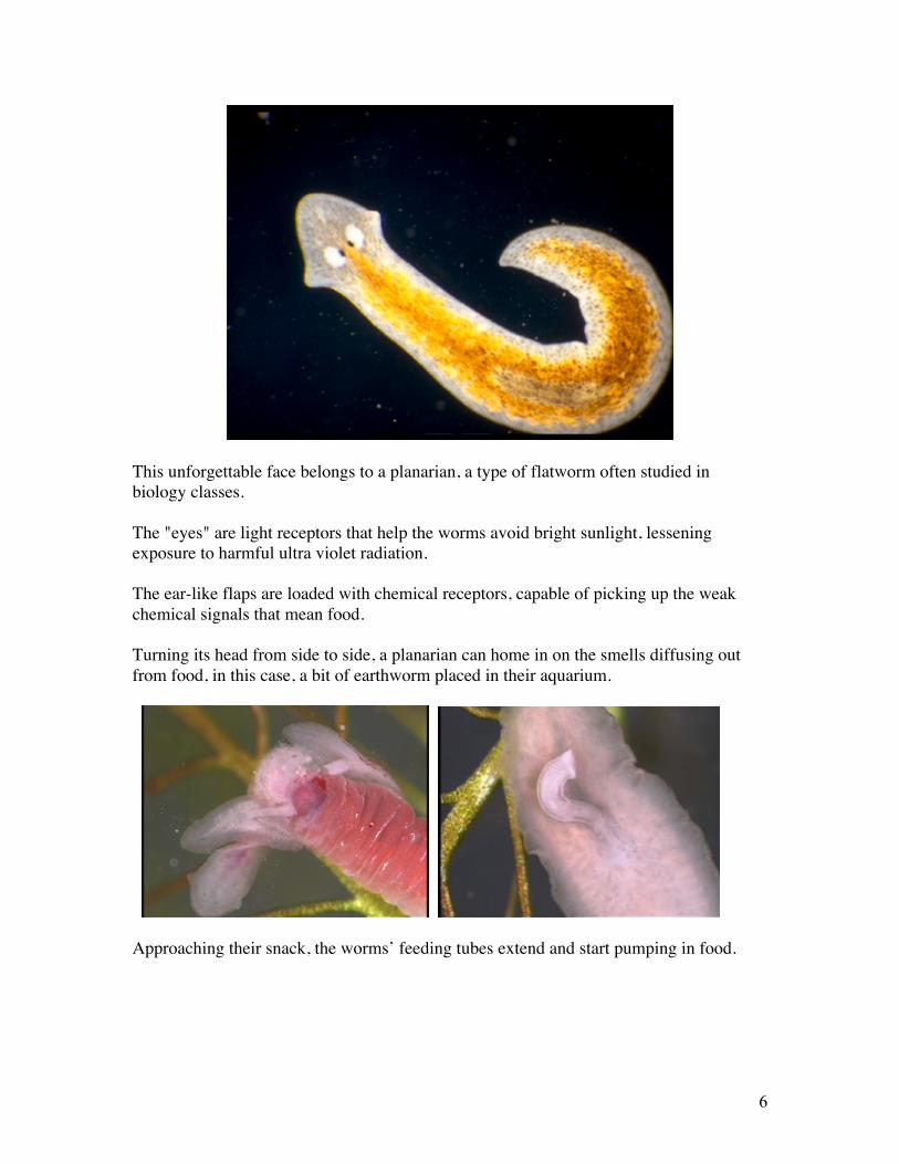
Approaching their snack, the worms’ feeding tubes extend and start pumping in food.

7
The worm's branched digestive system extends throughout its body, assuring that all ofits tissues are in close contact with digesting food.
Although they are sexual organisms, planarians build up their numbers by simply pullingin two. In a few days each half will regenerate its missing parts. The ability toregenerate new body parts is so highly developed in planarians that if one’s head is split,each half will regenerate a complete new head.
Planarians and most other free-living flatworms belong to Class Turbellaria – named forthe turbulence their cilia create as they move through the water. Turbellarian-likeancestral flatworms probably preceded many of the other lines of animal life. Then, asanimal life diversified, the worms took advantage of parasitic opportunities, evolving intoectoparasites of fish, flukes, and tapeworms.

8
Turbellarian flatworms from a vernal pool, one with drought-resistant eggs.
Class Monogenea
Flatworms were around for a long time before the appearance of the first primitive fish.However, once fish evolved, they opened up a whole new food source for flatworms –"fish scum."
Today these ectoparasites make up a distinct, but little known branch of flatworms livingon fish and amphibians – the flatworm class monogenea.
Continued…

9
The next time you go fishing, look closely at the fins of your catch. You may see amonogenetic flatworm hanging on with its powerful tail hooks.
Flukes (Class Trematoda)In earlier times, when brain-destroyed frogs were dissected in biology classes, studentswere often surprised to discover flukes living in their subject's organs.
Here, a large bladder fluke can be seen holding on to the frog's bladder wall.Its oversized sucker prevents loss when the frog empties its bladder.

10
Lung flukes feed upon the linings of the lungs, breaking down the lung tissue and gorgingon blood. The mature lung flukes are little more than sacks of reproductive organsmanufacturing thousands of eggs.
The eggs are coughed up by the frog, swallowed and shed into the pond with the frog'sfeces.
Within each egg develops a ciliated larva that goes off in search of the first host in thelung fluke's life cycle – a pond snail.
Burrowing into the snail, the larva initiates an amazing reproductive multiplication.Living on the snail tissue, the larva develops into a sac of germinal cells. Each of thesehundreds of germinal cells produces a new sac. Inside of these secondary sacs developmasses of larvae called cercaria, a staggering multiplication of parasites that leaves thesnail in rather poor health.
A forked-tail cercarium
Vast numbers of Cercaria burrow out of the snail and swim away to find their next host.In the case of the frog lung fluke, the next host is an aquatic insect larva.
When they reach the insect the tail will be discarded and the worm will burrow in andform a cyst. Now it waits for the insect to emerge and be eaten by a frog.
Breaking out in the frog’s stomach, the flukes crawl up the esophagus and enter the airtubes leading to the lungs where they eventually mate and start egg-production all overagain.

11
Chinese liver fluke Unidentified fluke from gull intestine
Frogs are not alone in having fluke problems. Every kind of animal has them, and all ofthese parasites have complex lifecycles involving snails or clams, suggesting that theflukes first evolved as parasites of molluscs.
The sexually mature flukes come in all sizes, each adapted by its shape and behavior forlife in a particular organ of its host.
Sheep live fluke
This species, reaching two centimeters in length, occupies the livers of sheep and deer,often jamming their host’s bile ducts. Sheep get the parasite by eating water plants onwhich the cercaria have formed cysts

12
A schistosome blood fluke male holds the female in his copulatory fold. These flukeslive in blood vessels of warm-blooded animals (including humans), impeding circulation.
Title: Tape worms (Class Cestoda)
Monogenetic flatworms live mostly on outer surfaces of fish. Fluke species inhabit theorgans of every kind of vertebrate animals. A third branch of flatworm parasites hasbecome adapted to living in a concentrated source of predigested nutrients—thevertebrate intestine.
Continued…

13
Tapeworms are distinguished by having bodies made up of chains of reproductiveindividuals called proglottids. At times, you may have seen these egg-loaded proglottidsemerging from a pet. The chain of proglottids that make up a tapeworm are kept withinthe intestine by one specialized individual at the head of the chain – the scolex
Scolex of Tanea solium, the pork tapeworm.
In this human infesting species, the scolex is equipped with both hooks and suckers –allowing it to hang on to the intestinal lining, where the proglottids can absorb the soupof digested nutrients.

14
Tapeworm matings must occur in the unromantic surroundings of the their host's smallintestine, where each hermaphroditic proglottid mates with a proglottid from aneighboring worm. Following mating, the fertilized proglottids become sacs of eggs,ready to escape and be eaten by an intermediate host.
Tapeworm eggs
In Tanea solium, a tapeworm of humans, the intermediate host is a pig that has ingestedhuman feces containing the tapeworm eggs, still a possibility in many areas of the world.
The tapeworm embryos hook their way through the pig’s intestinal wall, where they arecarried by the blood stream to the muscles. Here they form small, encapsulatedtapeworm embryos.

15
Preparing a pig pit-roast.
When the pork is eaten without sufficient cooking the embryos digests out and grow intoa mature intestinal parasites.
A large tapeworm is an unwelcome guest, but the situation becomes much worse if thehuman, instead of the pig, ingests the tapeworm eggs. In this case the larvae burrowthrough the human's intestinal wall and form cysts in muscles and organs.
This old autopsy photo shows a human brain riddled with tapeworm cysts – the cause ofdeath.

16
Conclusion
Flatworms are an ancient line of animal life with four surviving branches: ClassTurbellaria, the free-living flatworms, Class Monogenea- ectoparasites of fish andamphibians, Class Trematoda, the flukes, and Class Cestoda, the tapeworms.
Even before these lines began their evolutionary divergence, flatworm-like ancestors withthree-cell layers and bilateral symmetry opened the door for the great evolutionaryexperiments that produced the diversity of animals living today.
Primitive flatworms from a pond water sample.
Research Tips
To find planarians in ponds, rinse water plants in a white pan where the brown worms areeasily seen crawling over the white surface. In streams, carefully lift rocks and look forplanarians on the under side. Planarians can be kept in aquariums, or individually, inplastic cups where you can investigate their feeding behavior and their regenerativeabilities. In regeneration studies, worms are cut using an ice cube as an operating table.The ice slows the worm, and presumably acts as an anesthetic.

17
Microscopic flatworms are often found in cultures of decaying plant vegetation wherethey are easily spotted using a stereo dissecting microscope. However, some are in thesame size range as larger protozoans, such as Spirostomum. How to tell worms fromcells? Look for the eyes, suckers and mouths. Observing the behavior of these tinyworms may be as close as we can come to visualizing life on Earth 600 million years ago,a time when animal life was just getting started.
Flukes and fish go together like lichen on rock. If a fish is caught for the table, removeits intestine, carefully tease it open and look for flukes. For purposes of identification,flukes can be killed in alcohol (starting with 30% and going through a series of increasingconcentrations up to 90%). Following dehydration in alcohol, they can then be stainedusing a variety of stains that differentiate the fluke’s organs. Then you may be able toidentify the fluke using a parasitology reference book, or possibly from a search of theinternet. Also, if flukes are found, return to the fishing hole and collect water snails.Keep a dozen or so snails in a glass jar changing the water daily. Watch for rapidlywiggling cercaria in the water. Give the cercaria access to possible host organisms inorder to determine more about the fluke’s lifecycle. A word of warning: If you findforked tail cercaria, be careful about giving them access to your skin as they are likely tobe blood flukes. In North America, blood flukes are hosted by aquatic birds andmammals, but usually not by humans. As they burrow in your arm they will soondiscover they have entered a very unfriendly host. Although your immune response willkill the cercaria, their dead bodies in combination with a strong immune response willleave a big red welt. People who wade or swim where animal-infesting blood flukes live,often come out of the water covered with red welts, a condition known as “swimmer’sitch.” In some tropical regions there are related blood flukes (Schistosomes) that get byhuman resistance and make their homes in blood vessels, creating a serious health risk fortheir human host.
Also examine the fins of fresh caught fish with a magnifying glass to locate the curiousmonogenetic flatworms, once thought to be a kind of fluke. When opening the fish’sintestine look for slowly undulating white ribbons – tapeworms. Many fish tapewormsrely on their eggs being eaten by copepods and other small water animals, so finding atapeworm brings up the possibility of running an experimental lifecycle in order to learnhow the particular tapeworm gets into the fish. Recreating lifecycles in the laboratoryhas given biologists the understanding needed to stop parasitic diseases in animals, wild,domestic, or human.
www.eBioMEDIA.comemail: [email protected]