Embed Size (px)
Citation preview

BRAINSEGNET: BRAIN TRACTOGRAPHY SEGMENTATION NETWORK 1
BrainSegNet : A Segmentation Network forHuman Brain Fiber Tractography Data intoAnatomically Meaningful Clusters
Tushar Gupta1
Shreyas Malakarjun Patil2
Mukkaram Tailor1
Daksh Thapar1
Aditya Nigam1
1 School of Computing and ElectricalEngineering,Indian Institute of Technology, MandiMandi, HP, India
2 Department of Electrical Engineering,Indian Institute of Technology, JodhpurJodhpur, Rajasthan, India
Abstract
The segregation of brain fiber tractography data into distinct and anatomically mean-ingful clusters can help to comprehend the complex brain structure and early investiga-tion and management of various neural disorders. We propose a novel stacked bidirec-tional long short-term memory(LSTM) based segmentation network, (BrainSegNet) forhuman brain fiber tractography data classification. We perform a two-level hierarchicalclassification a) White vs Grey matter (Macro) and b) White matter clusters (Micro).BrainSegNet is trained over three brain tractography data having over 250,000 fiberseach. Our experimental evaluation shows that our model achieves state-of-the-art results.We have performed inter as well as intra class testing over three patient’s brain tractog-raphy data and achieved a high classification accuracy for both macro and micro levelsboth under intra as well as inter brain testing scenario.
1 IntroductionThe brain is the central processing unit of our body which maintains order and controls theactions of all the organs. Naturally, brain also communicates between its own subdivisions(active regions) for which it uses neuronal connections (termed as neuronal fibers), whichconsist of dendrites and axons. The dendrites serve as the receivers in a neuron and ax-ons in any neuron are responsible for transmission of signals to other neurons. Typically abrain consist of several billions of such neurons constantly receiving and transmitting signalsbetween themselves, forming a very complex network that commands our day to day activi-ties. The brain fibers are broadly divided into two classes, namely the grey and white fibersas shown in Fig. 1(a). The white fibers mainly consist of axons that connect various parts
c© 2017. The copyright of this document resides with its authors.It may be distributed unchanged freely in print or electronic forms.
arX
iv:1
710.
0515
8v1
[cs
.CV
] 1
4 O
ct 2
017

2 BRAINSEGNET: BRAIN TRACTOGRAPHY SEGMENTATION NETWORK
(a) Brain Slice (b) Diffusion (c) Directions (d) Fiber Tracking (e) Tractography (f) Segmentation
Figure 1: Images are taken from [2], [3]
of grey matter while the gray matter contains cell bodies, dendrites and axons as shown inFig. 1(b). The white matter can be further classified into eight clusters - Arcute, Cingulum,Corticospinal, Forceps Major, Fornix, Inferior Occipitofrontal Fasciculus, Superior Longitu-dinal Fasciculus and Uncinate. These are basically bundles or clusters of fibers called neuraltracts that form the pathways between different hemispheres and brain regions as shown inFig. 1(f).
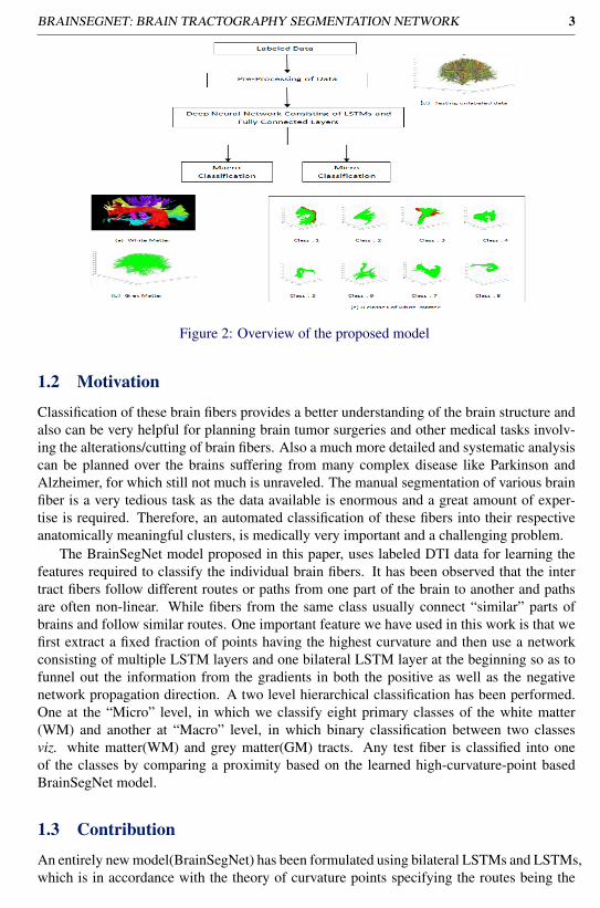
The brain fiber tractography data can be extracted from 3T Magnetic resonance imag-ing(MRI) data using diffusion tensor imaging which is a non-invasive MRI technique. Inthis technique diffusion has been considered as the molecular fluid spread and its extent de-pends on the diffusive property of the medium as shown in Fig. 1(c). In brain white matter,a tissue consists of bundles of myelination axons. Crucially, it is more hindered across thanalong such bundles. Hence, by measuring diffusion along many directions and observingthat it is faster in one direction than in others, one can deduce the direction of fiber bun-dles [3]. The process known as tractography, helps in visualizing the fiber structure in threedimensions using Diffusion Tensor Imaging(DTI) as shown in Fig. 1(e). One such examplehas been shown in Fig. 2, where on the top-right each fiber is coloured randomly, for bettervisualization. The eight major fiber clusters are shown in bottom right, while the grey matterfibers are shown in bottom-left. Any fiber can be represented as a set of points in 3D space(typically 20 to 100 points per fiber). Tractography produces thousands of fiber trajectoriesper subject (around 250K). Finally, obtained data can be seen as a 3D point cloud that doesnot convey anything useful. In order to extract any useful information the fibers must have tobe organized into anatomically meaningful structure. DTI may improve preservation of elo-quent regions during surgery by providing access to direct connectivity information betweenfunctional regions of the brain, and it has progressively been incorporated into strategic plan-ning for resection of complex brain lesions [2].
1.1 Problem Statement
Designing brain tractography segmentation network (BrainSegNet) : Given tractogra-phy data of human brain, we have designed a moderately deep recurrent neural network thatcan automatically segment brain fibers into tracts having “similar” fibers which are anatom-ically meaningful. Medically it is also believed that at coarser level there are two classesgrey and white matter (Macro Level) and further in white matter their are eight cluster/tracts(Micro Level) viz. Arcute, Cingulum, Corticospinal, Forceps Major, Fornix, Inferior Occip-itofrontal Fasciculus, Superior Longitudinal Fasciculus and Uncinate.
BRAINSEGNET: BRAIN TRACTOGRAPHY SEGMENTATION NETWORK 3
Figure 2: Overview of the proposed model
1.2 Motivation
Classification of these brain fibers provides a better understanding of the brain structure andalso can be very helpful for planning brain tumor surgeries and other medical tasks involv-ing the alterations/cutting of brain fibers. Also a much more detailed and systematic analysiscan be planned over the brains suffering from many complex disease like Parkinson andAlzheimer, for which still not much is unraveled. The manual segmentation of various brainfiber is a very tedious task as the data available is enormous and a great amount of exper-tise is required. Therefore, an automated classification of these fibers into their respectiveanatomically meaningful clusters, is medically very important and a challenging problem.
The BrainSegNet model proposed in this paper, uses labeled DTI data for learning thefeatures required to classify the individual brain fibers. It has been observed that the intertract fibers follow different routes or paths from one part of the brain to another and pathsare often non-linear. While fibers from the same class usually connect “similar” parts ofbrains and follow similar routes. One important feature we have used in this work is that wefirst extract a fixed fraction of points having the highest curvature and then use a networkconsisting of multiple LSTM layers and one bilateral LSTM layer at the beginning so as tofunnel out the information from the gradients in both the positive as well as the negativenetwork propagation direction. A two level hierarchical classification has been performed.One at the “Micro” level, in which we classify eight primary classes of the white matter(WM) and another at “Macro” level, in which binary classification between two classesviz. white matter(WM) and grey matter(GM) tracts. Any test fiber is classified into oneof the classes by comparing a proximity based on the learned high-curvature-point basedBrainSegNet model.
1.3 Contribution
An entirely new model(BrainSegNet) has been formulated using bilateral LSTMs and LSTMs,which is in accordance with the theory of curvature points specifying the routes being the

4 BRAINSEGNET: BRAIN TRACTOGRAPHY SEGMENTATION NETWORK
distinctive factors. The trained model when tested, surpassed the previous state of the artresults.
1.4 Previous Work
Although the problem of brain fiber classification is quite a recent development, still severalunsupervised techniques have already been suggested [1], [5] along with few supervisedapproaches. Majorly the unsupervised approaches manually select the Region of Interest(ROI) and then group the fibers through these ROIs. In [6], authors used spectral clusteringto generate a white matter atlas automatically. The similarity is calculated between fibersusing Hausdorff distance, and the clustering is employed in embedded space, that is formedusing the Eigen-vectors of the distance matrix. In [9], hierarchical Dirichlet process has beenused to determine the number of clusters. Another supervised approach presented in [7],selects few major points having maximum curvature and fed them to a clustering algorithmwhich takes into account the position and the curvature values while classifying.
2 Proposed Model : BrainSegNet
In this section we discuss the various components of our model, the overall structure, dataformat, data pre-processing (25% of data pruning) and the detailed architecture of the pro-posed network consisting of LSTMs and bilateral LSTMs layers. The overview of our pro-posed model has been shown in Fig. 2.
2.1 Tractography Data and its Format
The full data of three patients brain and their respective labels has been taken from Universityof Pittsburg as a part of contest. The ground truth had been manually annotated by expertneurologists and surgeons using their own interactive tools. The data has been provided as.trk or track file format. A track file is a single binary file, with the first 1000 bytes areheader information and the rest constitute the required fiber information. Each patient dataconsists of about 250,000 fibers. The average number of sample points over a fiber acrossdifferent classes varies between 36 to 120 highlighting that our approach is insensitive tothe fiber length. Each of the fiber is labelled an integer in the range 0 to 8 with respect to9 classes, where there 1 is used for grey matter and remaining 8 different types of whitematter classes/tracts. The number of fibers per class in the training set considered for all the3 patients has been depicted in Table 1.
Patient ID Gray Matter White MatterC-0 C-1 C-2 C-3 C-4 C-5 C-6 C-7 C-8
B1 74,486 900 247 3,428 570 358 120 274 111B2 75,384 462 868 1,937 374 246 497 107 125B3 74,834 525 782 2,225 788 116 618 46 66
Table 1: Number of fibres of different classes in 3 brains considered in this work

BRAINSEGNET: BRAIN TRACTOGRAPHY SEGMENTATION NETWORK 5
2.2 Curvature based Fiber Data Pruning using Partial DerivativesFiber pruning has been done in two steps : a) Extracting meaningful data points - highcurvature points, and b) Conversion into fixed input using masking. The labelled data of 3brains is available to us in the form of .trk (Track) file format [Ref.], Track file is one singlebinary file, with the first 1000 bytes as the header and the rest as the body.
1. Fiber Pruning : Extraction of meaningful data points has been done under the as-sumption that similar fibers follow similar paths from one part brain region to anotherand hence would have almost “similar” curvature points. Only those sample pointsinvolved in high curvature are selected by taking projections of these vectors on eachof the planes (XY,Y Z and ZX). Then accumulate their gradients in both directionswith respect to the sample points just before and after them and as well as with respectto points, four steps ahead and behind from the current point. This scheme is chosenin order to handle the multi-scale curvature. Finally gradients are sorted and bottom25% points are pruned.
2. Fixed Length Conversion : The brain fibers are represented by a variable lengthsequence of points in a three dimensional space (3D vectors). Since our network re-quires fixed length input, the length of sequences was restricted to 100 points. Longersequences has been truncated while shorter ones are padded with zeros. This strat-egy lead to poor results as the fiber structure severely got affected. Later, maskinglayer was used instead of padding to preserve the structure of fibers. Masking skips atime-step where all features are equal to the mask value.
Justification : To evaluate pre-processing, we have tested our model over both prunedand original fiber and have observed that with longer sizes the training time increases whileaccuracy does not improve.
2.3 Model ArchitectureDeep Neural Networks (DNNs) are powerful models that have achieved excellent perfor-mance on very complex learning tasks. We have used a deep stacked Long Short TermMemory (LSTM) network as shown in Fig. 3. Histotically RNNs [8] are built to utilize pastinformation to predict the future using hidden layers. But RNN’s failed to train well due tovanishing gradient problem and later LSTM Networks has been proposed.
2.3.1 RNN (Recurrent Neural Networks)
RNNs [8] are built in order to utilize past information to predict the future by using a loopinto the hidden layers that pass information to their respective successors. But RNN’s withlong dependencies failed to train well due to vanishing gradient problem. In order to learnlong and short both types of dependencies LSTM Networks has been proposed.
2.3.2 Long Short Term Memory Networks (LSTM)
The LSTMs [4] are special RNNs capable of learning long term dependencies. At eachiteration information passed to their respective successors, that learns which informationappeared in the past is most relevant discard remaining. The core concept behind LSTMs is
6 BRAINSEGNET: BRAIN TRACTOGRAPHY SEGMENTATION NETWORK
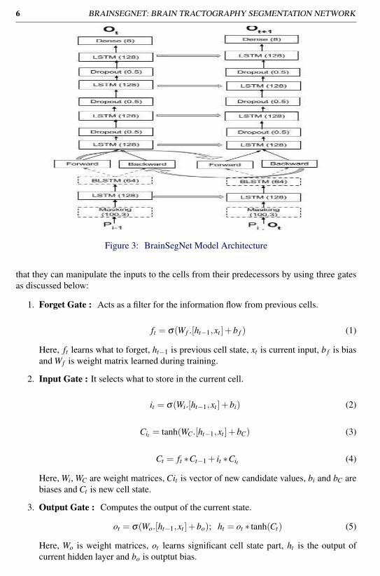
Figure 3: BrainSegNet Model Architecture
that they can manipulate the inputs to the cells from their predecessors by using three gatesas discussed below:
1. Forget Gate : Acts as a filter for the information flow from previous cells.
ft = σ(Wf .[ht−1,xt ]+b f ) (1)
Here, ft learns what to forget, ht−1 is previous cell state, xt is current input, b f is biasand Wf is weight matrix learned during training.
2. Input Gate : It selects what to store in the current cell.
it = σ(Wi.[ht−1,xt ]+bi) (2)
Cit = tanh(WC.[ht−1,xt ]+bC) (3)
Ct = ft ∗Ct−1 + it ∗Cit (4)
Here, Wi, WC are weight matrices, Cit is vector of new candidate values, bi and bC arebiases and Ct is new cell state.
3. Output Gate : Computes the output of the current state.
ot = σ(Wo.[ht−1,xt ]+bo); ht = ot ∗ tanh(Ct) (5)
Here, Wo is weight matrices, ot learns significant cell state part, ht is the output ofcurrent hidden layer and bo is outptut bias.

BRAINSEGNET: BRAIN TRACTOGRAPHY SEGMENTATION NETWORK 7
2.3.3 Justification for the selected model
We have observed that RNNs need to be deep enough to capture subtle differences in struc-ture of fibres from different classes. Initial few layers consist of one Bi-directional LSTM tocapture structural understanding at each fiber point and its curvature characteristics. The next3 are LSTM layers with reducing memory units as shown in Fig. 3. Finally, fully connectedlayers with a sigmoid activation function has been used to make predictions.
2.3.4 Training
Deep stacked LSTMs often give better accuracy over shallower models. However, simplystacking more layers of LSTM works only to a certain number of layers, beyond which thenetwork becomes too slow and difficult to train due to exploding and vanishing gradientproblem [4]. We have observed that stacked LSTM layers work well up to 4 layers, barelywith 6 layers, and very poorly beyond 8 layers. The training set was split for validating andtraining at a ratio of 1 : 5, so as to validate the training after each epoch.
Network Hyper-Parameters : Hyper parameter used in the model are as follows :
• Number of epochs : 15
• Batch Size : 64
• Activation Function : Sigmoid
• Optimizer Function : Adam
• Fraction of Data used for training : 0.4
• Loss functions : Binary Cross-entropy and Categorical Cross-entropy for macro andmicro respectively
• Validation set of size 0.2 of training set size has been used.
3 Experimental Analysis
In this section we talk about the methods applied for validation of our proposed model.The major challenge that we have faced is to get more and more “labeled tractography”data. We some how got hold of three brain data from a contest organized by Pittsburg.Hence, we have done the validation over a dataset of three patients brain data containingvarious fibers which are represented by a variable length of sequence and each of the fiber islabeled to one of the 9 classes. Each brain data contains 250,000 fibers and their respectiveclass. We have formulated this problem as two level hierarchical classification problem inwhich experiments are carried first at, a) Macro Level - In which the fibers undergo binaryclassification with respect to two classes viz. grey and the white matter (data imbalance is abig challenge), secondly at b) Micro Level - In which the fibers are classified into one of the8 sub-classes of white fibers (intra-class variation is a big challenge).
8 BRAINSEGNET: BRAIN TRACTOGRAPHY SEGMENTATION NETWORK
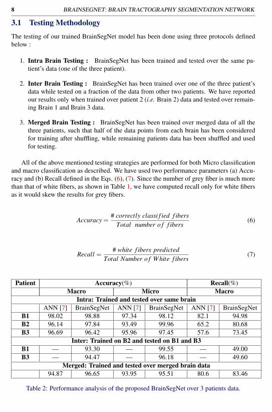
3.1 Testing Methodology
The testing of our trained BrainSegNet model has been done using three protocols definedbelow :
1. Intra Brain Testing : BrainSegNet has been trained and tested over the same pa-tient’s data (one of the three patient).
2. Inter Brain Testing : BrainSegNet has been trained over one of the three patient’sdata while tested on a fraction of the data from other two patients. We have reportedour results only when trained over patient 2 (i.e. Brain 2) data and tested over remain-ing Brain 1 and Brain 3 data.
3. Merged Brain Testing : BrainSegNet has been trained over merged data of all thethree patients, such that half of the data points from each brain has been consideredfor training after shuffling, while remaining patients data has been shuffled and usedfor testing.
All of the above mentioned testing strategies are performed for both Micro classificationand macro classification as described. We have used two performance parameters (a) Accu-racy and (b) Recall defined in the Eqs. (6), (7). Since the number of grey fiber is much morethan that of white fibers, as shown in Table 1, we have computed recall only for white fibersas it would skew the results for grey fibers.
Accuracy =# correctly classi f ied f ibers
Total number o f f ibers(6)
Recall =# white f ibers predicted
Total Number o f White f ibers(7)
Patient Accuracy(%) Recall(%)Macro Micro Macro
Intra: Trained and tested over same brainANN [7] BrainSegNet ANN [7] BrainSegNet ANN [7] BrainSegNet
B1 98.02 98.88 97.34 98.12 82.1 94.98B2 96.14 97.84 93.49 99.96 65.2 80.68B3 96.69 96.42 95.96 97.45 57.6 73.45
Inter: Trained on B2 and tested on B1 and B3B1 — 93.30 — 99.55 — 49.00B3 — 94.47 — 96.18 — 49.60
Merged: Trained and tested over merged brain data94.87 96.65 93.95 95.51 80.6 83.46
Table 2: Performance analysis of the proposed BrainSegNet over 3 patients data.
BRAINSEGNET: BRAIN TRACTOGRAPHY SEGMENTATION NETWORK 9
3.2 Experimental ResultsThe results with respect to the accuracy values and the recall values according to the testingstrategy has been depicted in Table 2. As we can see that we have achieved state of the artresults in almost all the testing strategies adopted in this paper. As we see when we movefrom intra to inter testing strategy the accuracy falls, this is due to the fact that the model hasbeen trained on an entirely other brain and the brain may differ in size or shape and hencemay differ slightly in the paths taken from one part to another. One can also observe that themerged training and testing strategy also gives less accurate results than intra as it is more ofa generalized model for accommodating fiber from any one of the brains. For the Table 2,one can infer that the proposed BrainSegNet achieves an accuracy more than 93% and 95%for macro and micro level respectively. Over such a huge and diverse dataset with smalltraining samples we have achieved quit high performance.
3.3 Comparative AnalysisThe recall values in Table 2, signify the superiority of the proposed BrainSegNet in classi-fying white fibers (which are fewer in number due to sever data imbalance). The proposedBrainSegNet gives far better results than the model proposed in [7], in terms of recall val-ues with better accuracy’s in most of the experiments. In [7], inter brain analysis has notbeen performed at all, which is very challenging and important to be reported. Even aftersuch a huge data imbalance and intra-class variations the proposed network has achievedstate-of-the-art performance and significantly outperforms the existing work [7].
4 ConclusionIn this paper we propose a novel stacked bidirectional LSTM based segmentation network,(BrainSegNet) for human brain fiber tractography data classification. We perform a two-level hierarchical classification a) White vs Grey matter (Macro) and b) White matter clusters(Micro). Our experimental evaluations show that our model achieves state-of-the-art results.We performed classification at both macro and micro levels that can eliminate the need formanually segmenting the brain fiber tracts which is presently a big issue. We are in theprocess to get more labeled data, so that we can train another better and deep generalizablenetwork.
References[1] Marco Catani, Robert J Howard, Sinisa Pajevic, and Derek K Jones. Virtual in vivo
interactive dissection of white matter fasciculi in the human brain. Neuroimage, 17(1):77–94, 2002.
[2] Juan C Fernandez-Miranda, Sudhir Pathak, Johnathan Engh, Kevin Jarbo, Timothy Ver-stynen, Fang-Cheng Yeh, Yibao Wang, Arlan Mintz, Fernando Boada, Walter Schneider,et al. High-definition fiber tractography of the human brain: neuroanatomical validationand neurosurgical applications. Neurosurgery, 71(2):430–453, 2012.
[3] Catarina Guise, Margarida M Fernandes, JoaÌCo M NoÌAbrega, Sudhir Pathak, WalterSchneider, and Raul Fangueiro. Hollow polypropylene yarns as a biomimetic brain
10 BRAINSEGNET: BRAIN TRACTOGRAPHY SEGMENTATION NETWORK
phantom for the validation of high-definition fiber tractography imaging. ACS AppliedMaterials & Interfaces, 8(44):29960–29967, 2016.
[4] Sepp Hochreiter and Jürgen Schmidhuber. Long short-term memory. Neural Comput.,9(8):1735–1780, November 1997. ISSN 0899-7667. doi: 10.1162/neco.1997.9.8.1735.URL http://dx.doi.org/10.1162/neco.1997.9.8.1735.
[5] Mahnaz Maddah, Andrea Mewes, Steven Haker, W Grimson, and Simon Warfield. Au-tomated atlas-based clustering of white matter fiber tracts from dtmri. Medical imagecomputing and computer-assisted intervention–MICCAI 2005, pages 188–195, 2005.
[6] Lauren J O’Donnell and Carl-Fredrik Westin. Automatic tractography segmentationusing a high-dimensional white matter atlas. IEEE transactions on medical imaging, 26(11):1562–1575, 2007.
[7] Vedang Patel, Anand Parmar, Arnav Bhavsar, and Aditya Nigam. Automated braintractography segmentation using curvature points. In Proceedings of the Tenth In-dian Conference on Computer Vision, Graphics and Image Processing, ICVGIP ’16,pages 18:1–18:6, New York, NY, USA, 2016. ACM. ISBN 978-1-4503-4753-2. doi:10.1145/3009977.3010013. URL http://doi.acm.org/10.1145/3009977.3010013.
[8] Ilya Sutskever. Training recurrent neural networks. PhD thesis, University of Toronto,2013.
[9] Xiaogang Wang, W Eric L Grimson, and Carl-Fredrik Westin. Tractography segmenta-tion using a hierarchical dirichlet processes mixture model. NeuroImage, 54(1):290–302,2011.