Embed Size (px)
Citation preview
Brain
Send comments to paddy.jagan(at)gmail.com
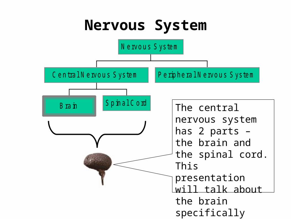
Nervous System
B ra in S p in a l C o rd
C e n tra l N e rvo u s S ys tem P e rip he ra l N ervo u s S ys tem
N e rvo u s S ys tem
The central nervous system has 2 parts – the brain and the spinal cord. This presentation will talk about the brain specifically
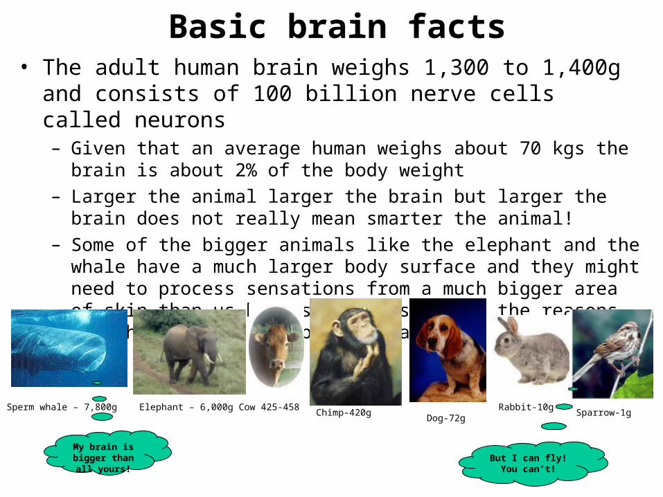
Basic brain facts• The adult human brain weighs 1,300 to 1,400g and consists
of 100 billion nerve cells called neurons– Given that an average human weighs about 70 kgs the brain is about
2% of the body weight– Larger the animal larger the brain but larger the brain does not really
mean smarter the animal!– Some of the bigger animals like the elephant and the whale have a
much larger body surface and they might need to process sensations from a much bigger area of skin than us humans. This is one of the reasons why their brains are bigger than us
Sperm whale – 7,800g Elephant – 6,000g Cow 425-458Chimp-420g
Dog-72gRabbit-10g
Sparrow-1g
My brain is bigger than all
yours!But I can fly! You
can’t!
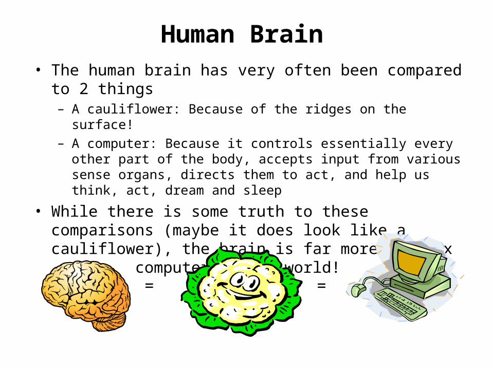
Human Brain• The human brain has very often been compared to 2
things– A cauliflower: Because of the ridges on the surface!– A computer: Because it controls essentially every other part of
the body, accepts input from various sense organs, directs them to act, and help us think, act, dream and sleep
• While there is some truth to these comparisons (maybe it does look like a cauliflower), the brain is far more complex than any computer in the world!
= =
The Neuron
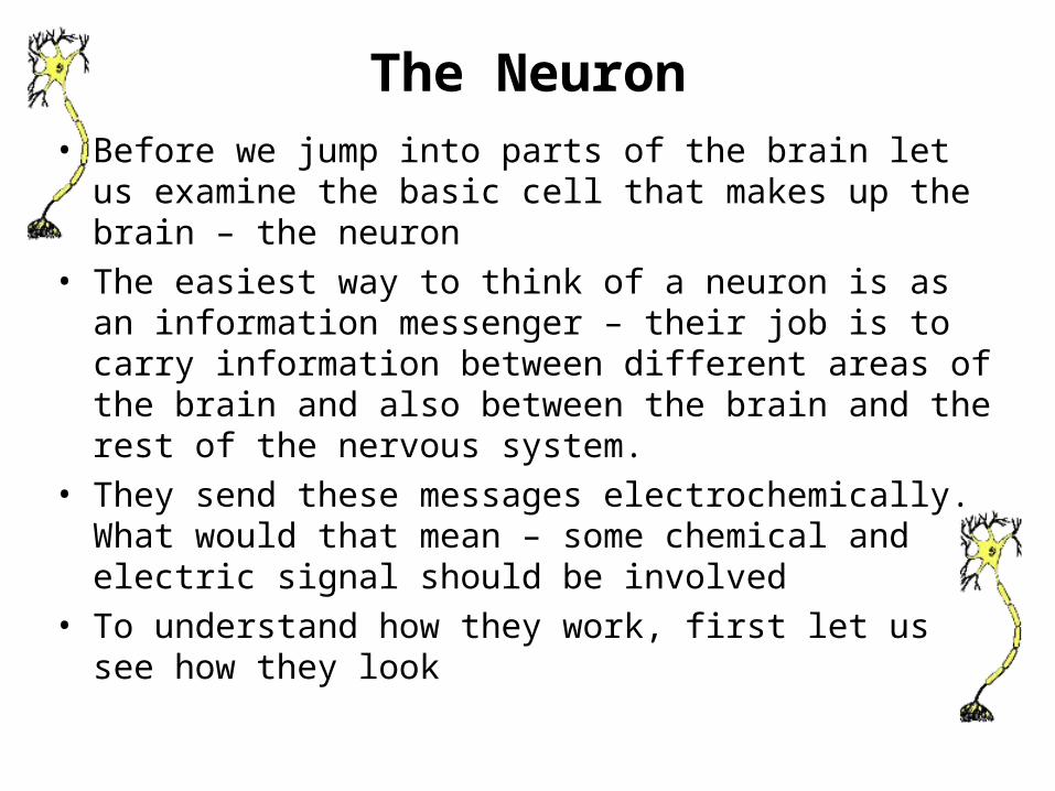
The Neuron• Before we jump into parts of the brain let us examine the
basic cell that makes up the brain – the neuron• The easiest way to think of a neuron is as an information
messenger – their job is to carry information between different areas of the brain and also between the brain and the rest of the nervous system.
• They send these messages electrochemically. What would that mean – some chemical and electric signal should be involved
• To understand how they work, first let us see how they look
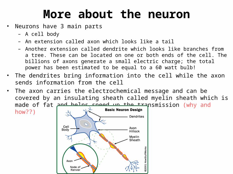
More about the neuron• Neurons have 3 main parts
– A cell body– An extension called axon which looks like a tail– Another extension called dendrite which looks like branches from a tree. These can be
located on one or both ends of the cell. The billions of axons generate a small electric charge; the total power has been estimated to be equal to a 60 watt bulb!
• The dendrites bring information into the cell while the axon sends information from the cell
• The axon carries the electrochemical message and can be covered by an insulating sheath called myelin sheath which is made of fat and helps speed up the transmission (why and how??)

S e n so ry In te rn e uro ns M o tor
T yp e s o f N eu ro ns
Carry information FROM sense organs (eyes, ears, skin etc)
TO the brain
Carry information FROM brain to the
Muscles and glands of the body
Neurons with short axons that communicate b/w
sensory and motor neurons
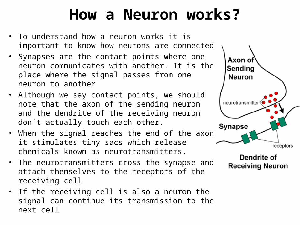
How a Neuron works?• To understand how a neuron works it is important to
know how neurons are connected• Synapses are the contact points where one neuron
communicates with another. It is the place where the signal passes from one neuron to another
• Although we say contact points, we should note that the axon of the sending neuron and the dendrite of the receiving neuron don’t actually touch each other.
• When the signal reaches the end of the axon it stimulates tiny sacs which release chemicals known as neurotransmitters.
• The neurotransmitters cross the synapse and attach themselves to the receptors of the receiving cell
• If the receiving cell is also a neuron the signal can continue its transmission to the next cell
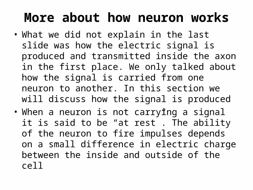
More about how neuron works• What we did not explain in the last slide was how
the electric signal is produced and transmitted inside the axon in the first place. We only talked about how the signal is carried from one neuron to another. In this section we will discuss how the signal is produced
• When a neuron is not carrying a signal it is said to be “at rest”. The ability of the neuron to fire impulses depends on a small difference in electric charge between the inside and outside of the cell
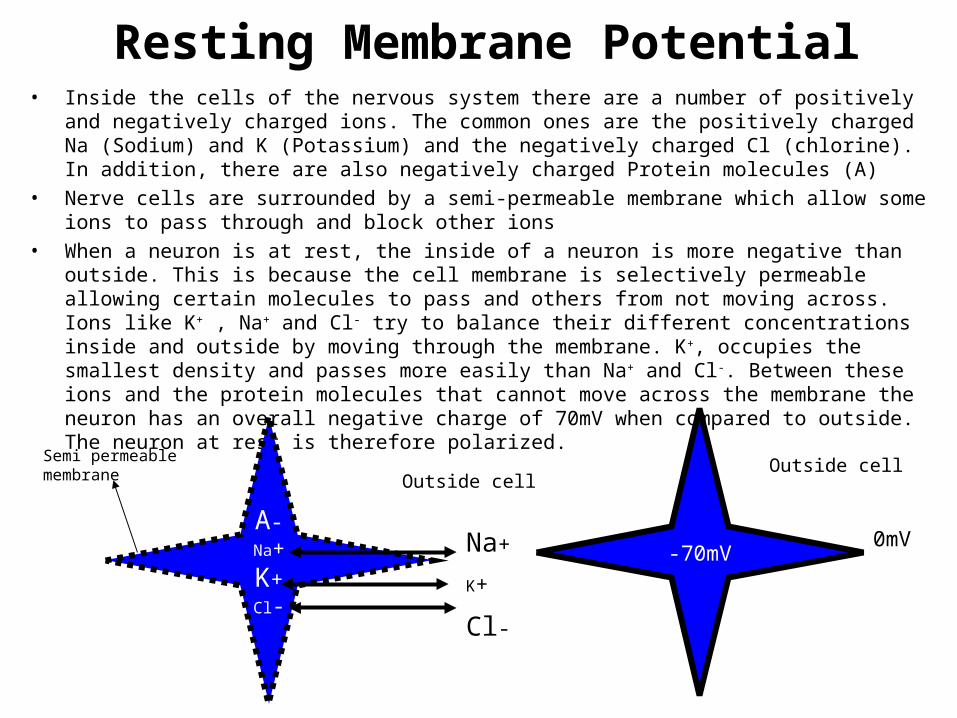
Resting Membrane Potential• Inside the cells of the nervous system there are a number of positively and negatively charged
ions. The common ones are the positively charged Na (Sodium) and K (Potassium) and the negatively charged Cl (chlorine). In addition, there are also negatively charged Protein molecules (A)
• Nerve cells are surrounded by a semi-permeable membrane which allow some ions to pass through and block other ions
• When a neuron is at rest, the inside of a neuron is more negative than outside. This is because the cell membrane is selectively permeable allowing certain molecules to pass and others from not moving across. Ions like K+ , Na+ and Cl- try to balance their different concentrations inside and outside by moving through the membrane. K+, occupies the smallest density and passes more easily than Na+ and Cl-. Between these ions and the protein molecules that cannot move across the membrane the neuron has an overall negative charge of 70mV when compared to outside. The neuron at rest is therefore polarized.
A-Na+K+Cl-
Na+
K+
Cl-
-70mV0mV
Outside cellOutside cellSemi permeable
membrane
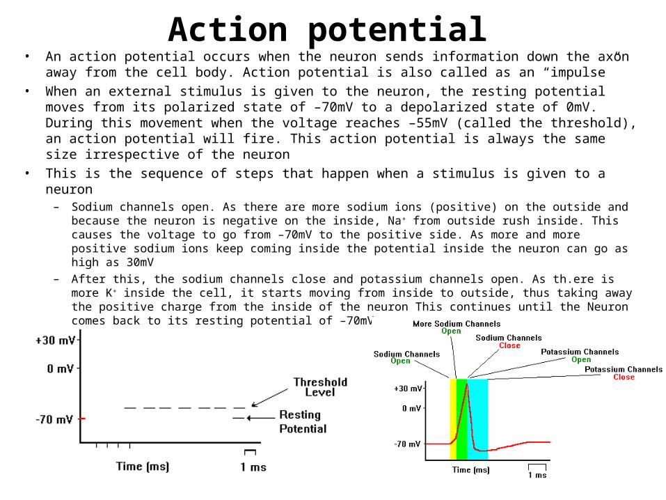
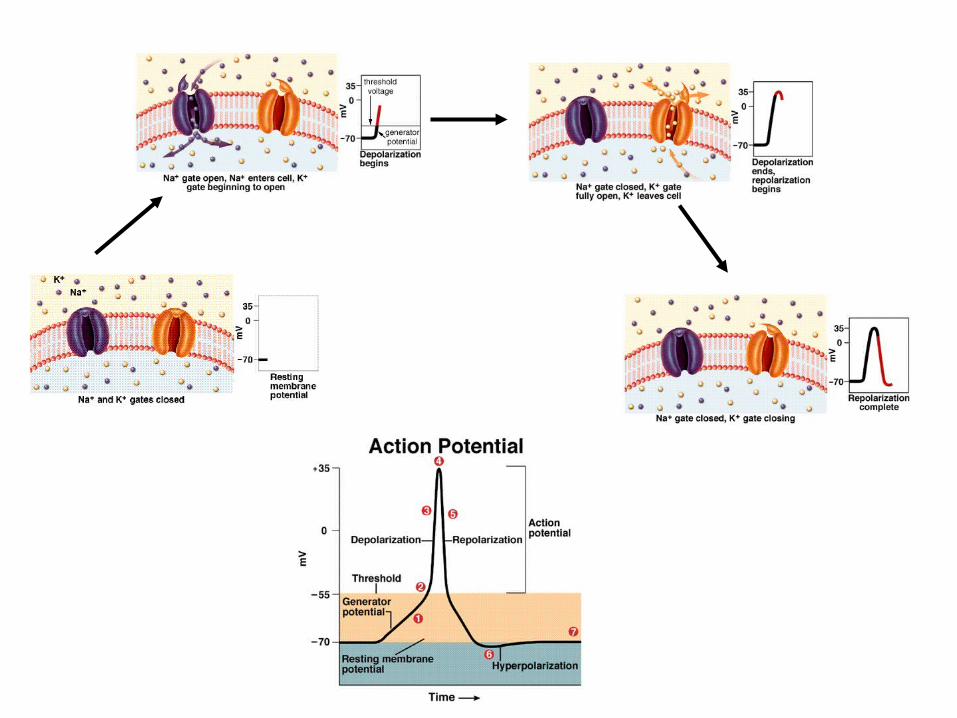
Action potential• An action potential occurs when the neuron sends information down the axon away from
the cell body. Action potential is also called as an “impulse”• When an external stimulus is given to the neuron, the resting potential moves from its
polarized state of –70mV to a depolarized state of 0mV. During this movement when the voltage reaches –55mV (called the threshold), an action potential will fire. This action potential is always the same size irrespective of the neuron
• This is the sequence of steps that happen when a stimulus is given to a neuron– Sodium channels open. As there are more sodium ions (positive) on the outside and because the
neuron is negative on the inside, Na+ from outside rush inside. This causes the voltage to go from –70mV to the positive side. As more and more positive sodium ions keep coming inside the potential inside the neuron can go as high as 30mV
– After this, the sodium channels close and potassium channels open. As th.ere is more K+ inside the cell, it starts moving from inside to outside, thus taking away the positive charge from the inside of the neuron This continues until the Neuron comes back to its resting potential of –70mV
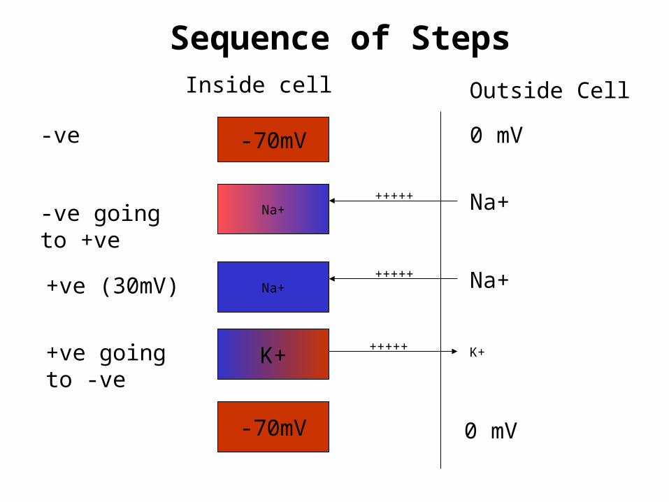
Sequence of Steps
-70mV 0 mV
Na+ Na+
Na+ Na+
K+ K+
+++++
+++++
+++++
-ve
-ve going to +ve
+ve (30mV)
+ve going to -ve
-70mV 0 mV
Inside cell Outside Cell
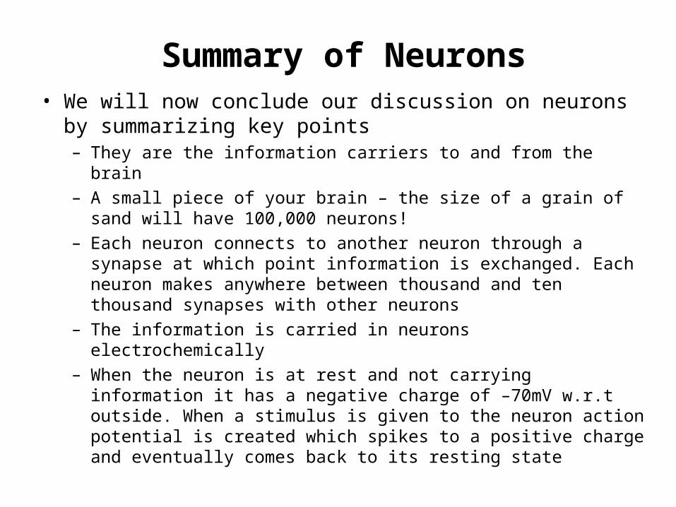
Summary of Neurons• We will now conclude our discussion on neurons by
summarizing key points– They are the information carriers to and from the brain– A small piece of your brain – the size of a grain of sand will have
100,000 neurons!– Each neuron connects to another neuron through a synapse at
which point information is exchanged. Each neuron makes anywhere between thousand and ten thousand synapses with other neurons
– The information is carried in neurons electrochemically– When the neuron is at rest and not carrying information it has a
negative charge of –70mV w.r.t outside. When a stimulus is given to the neuron action potential is created which spikes to a positive charge and eventually comes back to its resting state
Brain - Structure
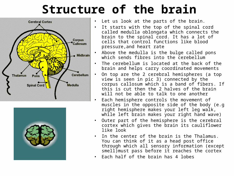
Structure of the brain• Let us look at the parts of the brain.• It starts with the top of the spinal cord called medulla
oblongata which connects the brain to the spinal cord. It has a lot of cells that control functions like blood pressure,and heart rate
• Above the medulla is the bulge called pons which sends fibres into the cerebellum
• The cerebellum is located at the back of the brain and helps carry coordinated movements
• On top are the 2 cerebral hemispheres (a top view is seen in pic 3) connected by the corpus callosum which is a band of fibers. If this is cut then the 2 halves of the brain will not be able to talk to one another
• Each hemisphere controls the movement of muscles in the opposite side of the body (e.g right hemisphere makes your left leg walk, while left brain makes your right hand wave)
• Outer part of the hemisphere is the cerebral cortex which gives the brain its cauliflower like look
• In the center of the brain is the Thalamus. You can think of it as a head post office through which all sensory information (except smell)must pass before it reaches the cortex
• Each half of the brain has 4 lobes
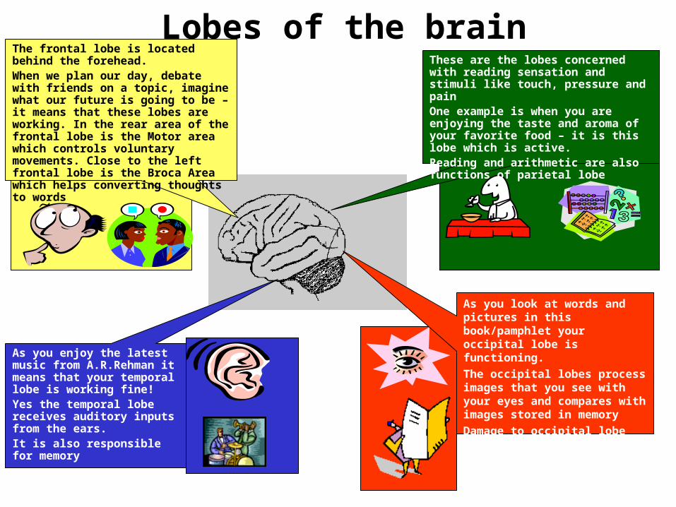
Lobes of the brainThe frontal lobe is located behind the forehead.When we plan our day, debate with friends on a topic, imagine what our future is going to be – it means that these lobes are working. In the rear area of the frontal lobe is the Motor area which controls voluntary movements. Close to the left frontal lobe is the Broca Area which helps converting thoughts to words
These are the lobes concerned with reading sensation and stimuli like touch, pressure and painOne example is when you are enjoying the taste and aroma of your favorite food – it is this lobe which is active.Reading and arithmetic are also functions of parietal lobe
As you look at words and pictures in this book/pamphlet your occipital lobe is functioning.
The occipital lobes process images that you see with your eyes and compares with images stored in memory
Damage to occipital lobe causes blindness
As you enjoy the latest music from A.R.Rehman it means that your temporal lobe is working fine!Yes the temporal lobe receives auditory inputs from the ears.It is also responsible for memory
Functioning of the Brain
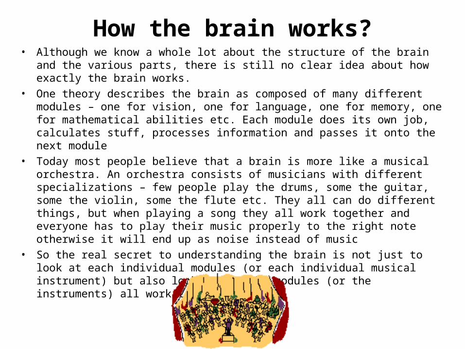
How the brain works?• Although we know a whole lot about the structure of the brain and the various
parts, there is still no clear idea about how exactly the brain works.
• One theory describes the brain as composed of many different modules – one for vision, one for language, one for memory, one for mathematical abilities etc. Each module does its own job, calculates stuff, processes information and passes it onto the next module
• Today most people believe that a brain is more like a musical orchestra. An orchestra consists of musicians with different specializations – few people play the drums, some the guitar, some the violin, some the flute etc. They all can do different things, but when playing a song they all work together and everyone has to play their music properly to the right note otherwise it will end up as noise instead of music
• So the real secret to understanding the brain is not just to look at each individual modules (or each individual musical instrument) but also look at how the modules (or the instruments) all work together.
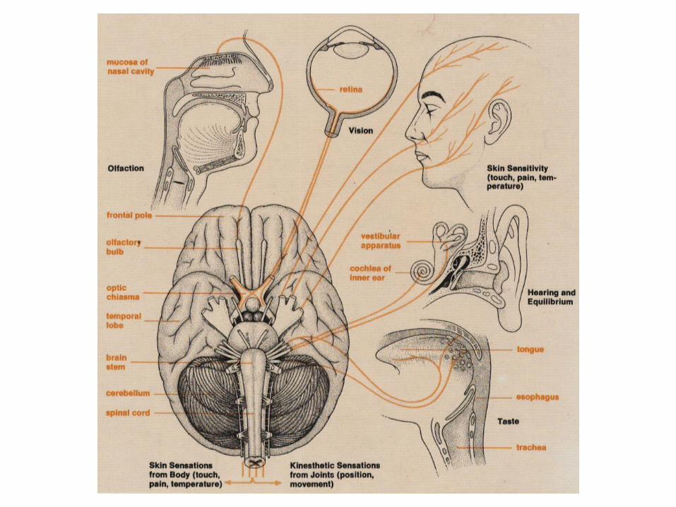
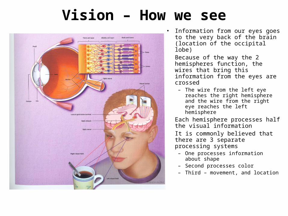
Vision – How we see• Information from our eyes goes to
the very back of the brain (location of the occipital lobe)
• Because of the way the 2 hemispheres function, the wires that bring this information from the eyes are crossed
– The wire from the left eye reaches the right hemisphere and the wire from the right eye reaches the left hemisphere
• Each hemisphere processes half the visual information
• It is commonly believed that there are 3 separate processing systems
– One processes information about shape
– Second processes color– Third – movement, and location
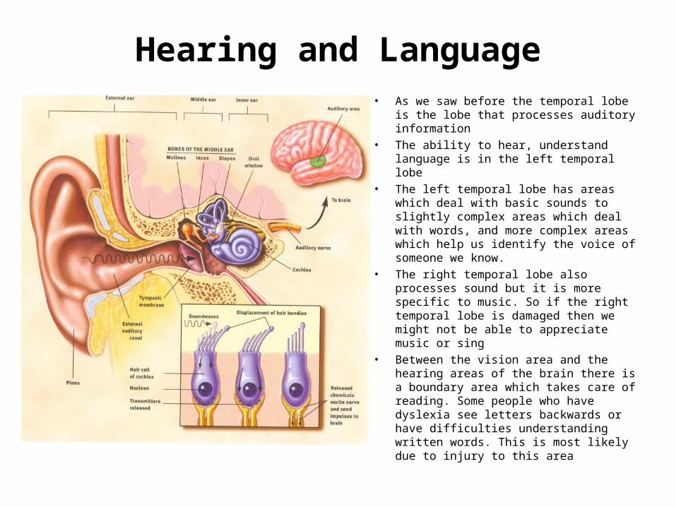
Hearing and Language• As we saw before the temporal lobe is
the lobe that processes auditory information
• The ability to hear, understand language is in the left temporal lobe
• The left temporal lobe has areas which deal with basic sounds to slightly complex areas which deal with words, and more complex areas which help us identify the voice of someone we know.
• The right temporal lobe also processes sound but it is more specific to music. So if the right temporal lobe is damaged then we might not be able to appreciate music or sing
• Between the vision area and the hearing areas of the brain there is a boundary area which takes care of reading. Some people who have dyslexia see letters backwards or have difficulties understanding written words. This is most likely due to injury to this area
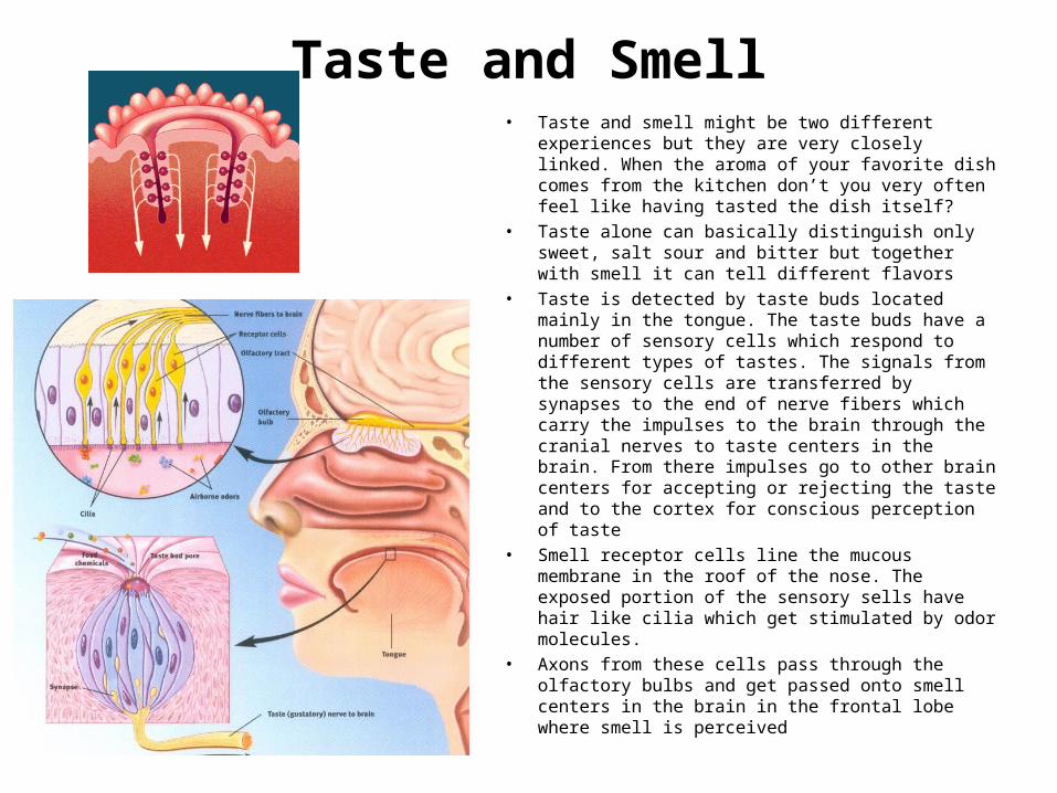
Taste and Smell• Taste and smell might be two different
experiences but they are very closely linked. When the aroma of your favorite dish comes from the kitchen don’t you very often feel like having tasted the dish itself?
• Taste alone can basically distinguish only sweet, salt sour and bitter but together with smell it can tell different flavors
• Taste is detected by taste buds located mainly in the tongue. The taste buds have a number of sensory cells which respond to different types of tastes. The signals from the sensory cells are transferred by synapses to the end of nerve fibers which carry the impulses to the brain through the cranial nerves to taste centers in the brain. From there impulses go to other brain centers for accepting or rejecting the taste and to the cortex for conscious perception of taste
• Smell receptor cells line the mucous membrane in the roof of the nose. The exposed portion of the sensory sells have hair like cilia which get stimulated by odor molecules.
• Axons from these cells pass through the olfactory bulbs and get passed onto smell centers in the brain in the frontal lobe where smell is perceived
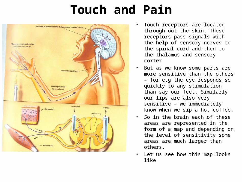
Touch and Pain• Touch receptors are located through
out the skin. These receptors pass signals with the help of sensory nerves to the spinal cord and then to the thalamus and sensory cortex
• But as we know some parts are more sensitive than the others – for e.g the eye responds so quickly to any stimulation than say our feet. Similarly our lips are also very sensitive – we immediately know when we sip a hot coffee.
• So in the brain each of these areas are represented in the form of a map and depending on the level of sensitivity some areas are much larger than others.
• Let us see how this map looks like
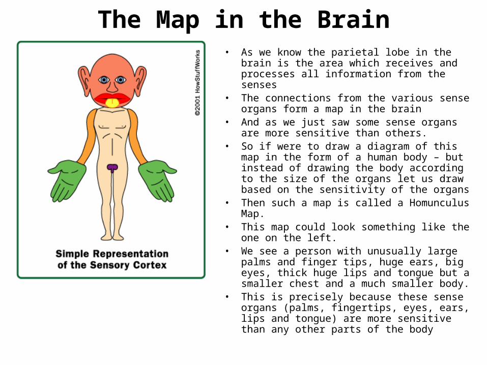
The Map in the Brain• As we know the parietal lobe in the brain is the
area which receives and processes all information from the senses
• The connections from the various sense organs form a map in the brain
• And as we just saw some sense organs are more sensitive than others.
• So if were to draw a diagram of this map in the form of a human body – but instead of drawing the body according to the size of the organs let us draw based on the sensitivity of the organs
• Then such a map is called a Homunculus Map.• This map could look something like the one on
the left.• We see a person with unusually large palms
and finger tips, huge ears, big eyes, thick huge lips and tongue but a smaller chest and a much smaller body.
• This is precisely because these sense organs (palms, fingertips, eyes, ears, lips and tongue) are more sensitive than any other parts of the body
Conclusion – Key ideas learnt• Divisions of nervous system• Neuron
– Structure– How it functions
• Brain – Structure
• Lobes of the brain and what they are responsible for
– How the brain works• How we see, touch, hear, and smell
• Map in the brain


![The Nervous System. Divisions of the Nervous System Central Nervous System [CNS] = Spinal Cord Brain Peripheral Nervous System [PNS]= Spinal Nerves](https://img.pdfslide.us/doc/110x75/56649d6c5503460f94a4c71d/the-nervous-system-divisions-of-the-nervous-system-central-nervous-system.jpg)