Embed Size (px)
Citation preview
Brain Research, 225 (1981) 431-436 Elsevier/North-Hollaod Biomedical Press
Selectivity of neuronal PH]GABA accumulation in the visual cortex as revealed by Golgi staining of the labeled neurons
431
PETER SOMOGYI, TAMA.S F. FREUND\ NORBERT HALAsz and ZOLTA.N F. KISVA.RDAY**
I Si Department 0/ Anatomy, Semmelweis Medical School, 1450 Budapest, Tiizolt6 lI. 58 and ( N.H.; Institule o/Biophysics, Biological Research Centre , Hungarian Academy 0/ Sciences, Szeged (Hungary,;
(Accepted August 6th, ]981)
Key words: f3H]GABA accumulation - visual cortex - Golgi staining - labe1ed neurons
[3HIGABA was injected into the visual cortex of rats in vivo. The labeled amino acid was demonstrated by autoradiography using semithin sections o'f Golgi material. Selective accumulation was seen in the perikarya of Golgi-stained, gold-toned, aspinous stellate neurons. Spine-laden pyramidal-like cells did not show JabeJing. This method gives direct information about the denclritic arborization of a neuron, and its putative transmitter, and allows the identification of its synaptic connections.
Selective, high affinity uptake of exogenous, radiolabeled putative neurotransmitters has been a valuable tool for the characterization of certain neuron populations (see refs. 5 and 6) . In particular, it has been shown that [3H]GABA is selectively accumulated by certain types of neurons in the cerebellum4,1l, olfactory bulb3 and cerebral cortex1,4. Some of the types of neurons accumulating [3H]GABA have been shown in separate immunohistochemical studies to contain the GABA synthetizing enzyme glutamic acid decarboxylase (GAD)8- 10. This, together with other biochemical evidence3,6, indicates that the selective uptake of exogenous GABA can be used as evidence of the GABAergic nature of a neuron. However, autoradiographic studies can provide little information about the shape, dendritic and axonal arborization of a single neuron .
Axonal and dendritic arborization are best revealed by single cell staining procedures such as the Golgi method. The Golgi method has gained new impetus with the introduction of new electron microscopic combinations for tracing synaptic connections of identified neurons 2,14,16. However, Golgi staining gives no information about the transmitter or chemical nature of the stained neuron. Therefore most often indirect correlation is used to evaluate the transmitters of various neuron types.
* T F F' . . leund and Z. F. Kisvarday are undergrad uate students at L6nind Ebtvbs University, Budapest.
0006--8993181/0000 0000,/$0 . - 2.75 © Elsevier/North-HoJ1and Biomedical Press
_ I
--L I I.
Ill.
v
V I.
~ ' :' .. " .... ; .. i f ~:' .1-; ... :'- ," .:" '." , E ';" " ; .... ~.: , , .. ' . ' .
'. ,' • • J
',. , , ~
433
' :.: ," ,y . ,; .. , ,~
, .,,! ·t" , ..... ,.;. . : : a .
~,·~J~;,i~;~. , ~ J .; ,~~li'X ; j':~
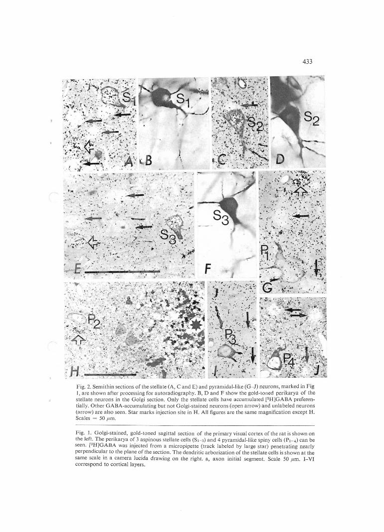
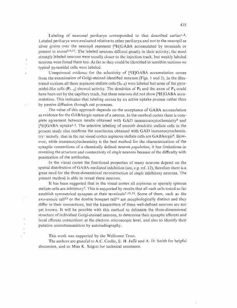
Fig. 2, Semithin sections of the steIlate (A, C and E) and pyramidal-like (G- J) neurons, marked in Fig 1, are shown after processing for autoradiography. B, D and F show the gold-toned perikarya of the steIlate neurons in the Golgi section . Only the steJJate cells have accumulated [3H]GABA preferentially. Other GABA-accumulating but not Golgi-stained neurons (open arrow) and unlabeled neurons (arrow) are also seen. Star marks injection site in H. AJJ figures are the same magnification except H, Scales = 50 llm,
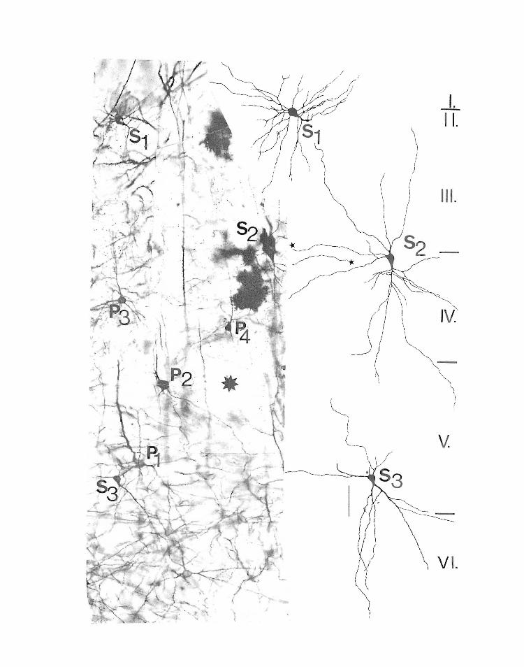
Fig. 1. Golgi-stained, gold-toned sagittal section of the primary visual cortex of the rat is shown on the left. The perikarya of 3 aspinous stellate cells (Sl-3) and 4 pyramidal-like spiny cells (Pl-4) can be seen, [3H1GABA was injected from a micropipette (track labeled by large star) penetrating nearly perpendicular to the plane of the section. The dendritic arborization of the stellate cells is shown at the same scale in a camera lucida drawing on the right. a, axon initial segment. Scale 50 llm. I-VI correspond to cortical Jayers.
434
In order to overcome the limitations of both we have combined the selective of a neuron with transmitter and the
0<CI,H""I", of the same neuron, its structure. The visual cortex of adult Wistar and CFY strain
chloral'·\lfirc>,he>
m
mM and 0.18% diameter 30--50 from vertical and perpE:ndlcll1ar
illustrated in this paper received two series of in the visual cortex, 2 mm apart In both sets of 0.1 of was ed at 4 sites; 1 total time of 5 min. Ten minutes after the last ft'LP('t,,,.,
the animal was in 0.1 M pll,osClllate
"'!,A"llA' cortex were dissected and pr()CeSSeCl as described After silver nitrate treatment, 90 ,am
sections were on slides with of illuminated from both sides for 20 min with a Zeiss embedded in DURCUPAN ACM on slides. After
removed and metallic are AS' a result in a semithin section most
of der)OSIIt,
in the section
flP''1ClT'\1 and different
!V-,V'!"\.! neuron appear as C;;:'lJlllll,,- from
of
showed accumulation of silver shown electron
distributed over the nucleus neurons, 1 n some cases we observed that in lCLUv!V~, ",-'Vl,-,'-C'Jl''-''"
over the nucleus than over the "'Itr.nl"",-n
sections not the nucleus fewer SUjggE:sts that some
label be removed from the ('"t .... ."."I/C1':''1'1
t .
435
Labeling of neuronal perikarya corresponded to that described earlier1,4.
Labeled perikarya were evaluated relative to other perikarya and not to the neuropil as silver grains over the neuropil represent [3H]GABA accumulated by terminals or present in axons3,6,1? The labeled neurons differed greatly in their activity; the most strongly labeled neurons were usually closer to the injection track, but weakly labeled neurons were found there too. As far as they could be identified in semithin sections no typical pyramidal cells were labeled.
Unequivocal evidence for the selectivity of [3H]GABA accumulation comes from the examination of Golgi-stained identified neurons (Figs. 1 and 2). In the illustrated sections all three aspinous stellate cells (Sl - 3) were labeled but none of the pyramidal-like cells (Pl-4) showed activity. The dendrites of P2 and the axon of P 4 could have been cut by the capillary track, but these neurons did not show [3H]GABA accumulation. This indicates that labeling occurs by an active uptake process rather than by passive diffusion through cut processes.
The value of this approach depends on the acceptance of GABA accumulation as evidence for the GABAergic nature of a neuron. In the cerebral cortex there is complete agreement between results obtained with GAD immmunocytochemistry8 and [3H]GABA uptake1,4. The selective labeling of smooth dendritic stellate cells in the present study also confirms the conclusion obtained with GAD immunocytochemistry: namely, that in the rat visual cortex aspinous stellate cells are GABAergic8. However, while immunocytochemistry is the best method for the characterization of the synaptic connections of a chemically defined neuron population, it has limitations in revealing the structure and connectivity of single neurons because of the difficulty with penetration of the antibodies.
In the visual cortex the functional properties of many neurons depend on the spatial distribution of GABA-mediated inhibition (see, e.g. ref. 12), therefore there is a great need for the three-dimensional reconstruction of single inhibitory neurons. The present method is able to reveal these neurons.
It has been suggested that in the visual cortex all aspinous or sparsely spinous stellate cells are inhibitory? This is supported by results that all such cells tested so far establish symmetrical synapses at their terminals? ,13,15. Some of them, such as the axo-axonic cell13 or the double bouquet cell15 are morphologically distinct and they differ in their connections, but the transmitters of these well-defined neurons are not yet known. It wjJJ be possibJe with this method to delineate the three-dimensional structure of individual Golgi-stained neurons, to determine their synaptic afferent and local efferent connectionE: at the electron microscopic level, and also to identify their putative neurotransmitters by autoradiography.
This work was supported by the Wellcome Trust. The authors are grateful to A.C. Cuello, E. H. J affe and A. D. Smith for helpful
discussion, and to Miss K. Szigeti for technical assistance.
436
ChronwaJl, B. and Wollf, J. R., Prenatal and development of GABA-accumulating cells in the occipita! neocortex of rat, J. comp. Neurol., 190 (1980) 187--208.
Peters, and Saldanha, .1., A new procedure for examining Golgi impregnated neurons by and electron microscopy, J. Neul'ocyfol., 6 (! 977) 31
3 Halasz, N., Ljungdahl, A. and H6kfelt, Transmitter histochemistry of rat olfactory bulb. 1lI. Autoradiographic localization of [3H1GABA, Bmin Research, 167 (1979) Hbkfelt, T. and Ljungdahl, Autoradiographic identification of cerebral and cerebellar cortical neurons accumulating labeled acid ([:lH]GABA), Bmin Res., 14 ([972)
5 Hokfelt, T. Ljungdahl, A., Uptake mechanisms a basis for the tl1stoc:llem!.ca identification Cuenod (Eds.), and tracing of transmitter neuron populations. In W. M.
The of Axonal Tl'(/lIspol't for Studies of Neuro/lal Connecril'ily,
6 L. Identification of transmitter-specific niques, In L. L Iversen, S. D. S. H. Vol. 9, Chemical Pathways in the Broill, Plenum
ill CNS by autoraeliographic tech(Eels.), Handbook oIPsycl!oplwrtlwc%gy. New York, 1978, pp.
Peters, and A., Smooth and a study using combined
cells in the visual cortex or the rat: mi(~roscc)pe technique, J. ('omp. Nelllo!., 181 (1978)
129-172. 8 Ribak, C. E., Aspinous and sparsely-spinous stellate visual cortex of rats contain
mic acid decarboxylase, J. Neurocyfol., 7 (1978) 461-479. 9 Ribak, C. E., Vaughn, J. E., Saito, K., Barber, R. and Roberts, E., Glutamate decarboxylase loca-
lization in neurons of the olfactory bulb, Brain Research, 1 (1977) 8. 10 Saito, K., Barber, R., Wu, J.-Y., Matsuda, T., Robens, Vaughn, J. E, Immunohisto-
chemical localization of glutamic acid decarboxylase in rat cerebellulll, Proc. /la!. A cad. Sci. U.S.A., 71 (1974) 269-273.
J I SChOIl, F. and Iversen, L. L., Selective accumulation of ['lHjGABA by stellate in rat bellar in vivo, Brain Research, 42 (1972) 503-507. SiJlito, A. M., Inhibitory mechanism influencing complex cell orientation selectivity their modification at high levels, J. PhyslOl. (Lond.), 289 (1979) Somogyi, P., A the rat, Brain Research, J 36 (1977) .J"tJ--.)JV.
14 Somogyi, P., The study Golgi stained cells of experimental degeneration under the electron microscope: a direct method for identification in the visual cortex of three links in a neuron chain, Neuroscience, 3 (1978) 167-180.
15 Somogyi, P. and Cowey, A., Combined Golgi and electron microscopic study on tile synapses formed by double bouquet cells in the visual cortex of the cat and monkey, J. COlI/jJ. NeMO!., 195 ([981) 547-566.
16 Somogyi, P., Hodgson, A. J. and Smith, A. D., An approach to tracing neuron networks in the cerebral cortex and basal ganglia. Combination of Golgi-staining, retrograde transport of horseradish peroxidase and anterograde degeneration of synaptic boutons in the same material, Neuroscience, (1979) 1805···1852. Sterling, P. and T. L. Neurons in lateral nucleus that concentrate eXI)g(;IlC)US
)-y-arlllllobutj(rlc acid (GABA), J. con;p. Neurol., 192 (1980)