Embed Size (px)
Citation preview
BRAIN REGIONCOMPILATION CHARTS
Functional Neurology Seminars LP © 2016 Dr. Datis Kharrazian and Dr. Brandon Brock
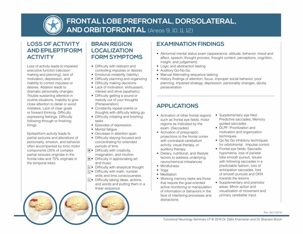
FRONTAL LOBE PREFRONTAL, DORSOLATERAL, AND ORBITOFRONTAL (Areas 9, 10, 11, 12)
BRAIN REGION LOCALIZATION FORM SYMPTOMS• Difficulty with restraint and
controlling impulses or desires• Emotional instability (lability)• Difficulty planning and organizing• Difficulty making decisions • Lack of motivation, enthusiasm,
interest and drive (apathetic)• Difficulty getting a sound or
melody out of your thoughts (Perseveration)
• Constantly repeat events or thoughts with difficulty letting go
• Difficulty initiating and finishing tasks
• Episodes of depression• Mental fatigue • Decrease in attention span• Difficulty staying focused and
concentrating for extended periods of time
• Difficulty with creativity, imagination, and intuition
• Difficulty in appreciating art and music
• Difficulty with analytical thought• Difficulty with math, number
skills and time consciousness• Difficulty taking ideas, actions,
and words and putting them in a linear sequence
EXAMINATION FINDINGS • Abnormal mental status exam (appearance, attitude, behavior, mood and
affect, speech, thought process, thought content, perceptions, cognition, insight, and judgement)
• Logic and abstraction testing• Auditory Go-No-Go• Manual Alternating sequence tasking • History findings of attention, focus, improper social behavior, poor
planning, impaired strategy, depression, personality changes, abulia, perseveration
LOSS OF ACTIVITY AND EPILEPTIFORM ACTIVITYLoss of activity leads to impaired executive function (decision making and planning), lack of motivation, depression, and inability to control impulses or desires. Ablation leads todramatic personality changes.Trouble sustaining attention in routine situations. Inability to give close attention to detail or avoid mistakes. Lack of clear goals or forward thinking. Difficulty expressing feelings. Difficulty following through or finishing things.
Epileptiform activity leads topartial seizures and alterations of personality, emotion, and behavior often accompanied by tonic motor components (25% of complex partial seizures originate in the frontal lobe and 75% originate in the temporal lobe)
APPLICATIONS• Activation of other frontal regions
such as frontal eye fields, motor regions as indicated by the exam. (Saccades)
• Activation of presynaptic projections to the frontal cortex with contralaral cerebellum activity, visual therapy, or auditory therapy
• Dietary, nutritional, and lifestyle factors to address underlying neurochemical imbalances.
• Mindfulness• Yoga• Meditation• Working memory tasks are those
that require the goal-oriented active monitoring or manipulation of information or behaviors in the face of interfering processes and distractions.
• Supplementary eye filed: Predictive saccades, Memory guided saccades
• DLPF: Prioritization and motivation and organization techniques.
• Go No Go inhibitory techniques for orbitofrontal. Impulse control.
• Frontal eye fields: Saccadic hypometria, loss of parietal lobe smooth pursuit, Issues with following saccades in a predictable fashion, loss of anticipation saccades, loss of smooth pursuits and OKN towards the lesions.
• Supplementary and premotor areas: Mirror action and visualization of movement and primary cerebellar input
Rev. 05|11|2016
Functional Neurology Seminars LP © 2016 Dr. Datis Kharrazian and Dr. Brandon Brock
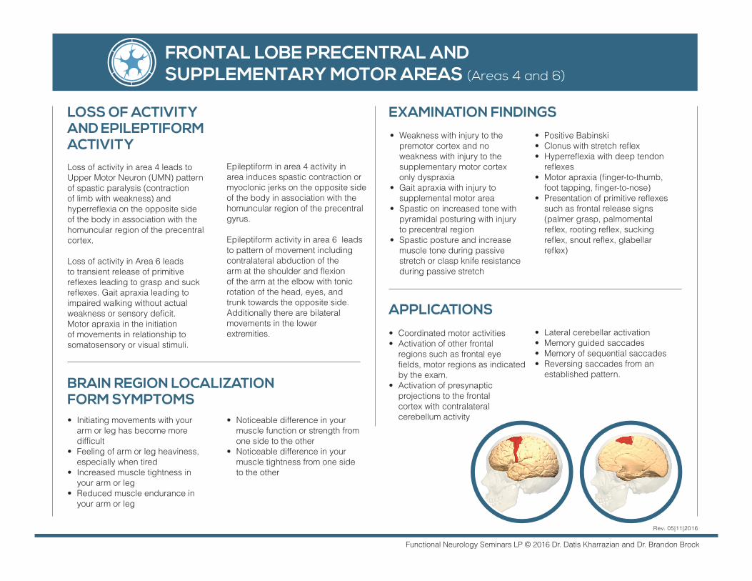
FRONTAL LOBE PRECENTRAL AND SUPPLEMENTARY MOTOR AREAS (Areas 4 and 6)
Epileptiform in area 4 activity in area induces spastic contraction or myoclonic jerks on the opposite side of the body in association with the homuncular region of the precentral gyrus.
Epileptiform activity in area 6 leads to pattern of movement includingcontralateral abduction of thearm at the shoulder and flexionof the arm at the elbow with tonicrotation of the head, eyes, andtrunk towards the opposite side.Additionally there are bilateralmovements in the lowerextremities.
LOSS OF ACTIVITY AND EPILEPTIFORM ACTIVITYLoss of activity in area 4 leads to Upper Motor Neuron (UMN) pattern of spastic paralysis (contraction of limb with weakness) and hyperreflexia on the opposite side of the body in association with the homuncular region of the precentral cortex.
Loss of activity in Area 6 leads to transient release of primitive reflexes leading to grasp and suck reflexes. Gait apraxia leading to impaired walking without actual weakness or sensory deficit. Motor apraxia in the initiation of movements in relationship to somatosensory or visual stimuli.
APPLICATIONS• Coordinated motor activities • Activation of other frontal
regions such as frontal eye fields, motor regions as indicated by the exam.
• Activation of presynaptic projections to the frontal cortex with contralateral cerebellum activity
• Lateral cerebellar activation• Memory guided saccades• Memory of sequential saccades• Reversing saccades from an
established pattern.
• Initiating movements with your arm or leg has become more difficult
• Feeling of arm or leg heaviness, especially when tired
• Increased muscle tightness in your arm or leg
• Reduced muscle endurance in your arm or leg
EXAMINATION FINDINGS
• Noticeable difference in your muscle function or strength from one side to the other
• Noticeable difference in your muscle tightness from one side to the other
BRAIN REGION LOCALIZATION FORM SYMPTOMS
• Weakness with injury to the premotor cortex and no weakness with injury to the supplementary motor cortex only dyspraxia
• Gait apraxia with injury to supplemental motor area
• Spastic on increased tone with pyramidal posturing with injury to precentral region
• Spastic posture and increase muscle tone during passive stretch or clasp knife resistance during passive stretch
• Positive Babinski• Clonus with stretch reflex • Hyperreflexia with deep tendon
reflexes • Motor apraxia (finger-to-thumb,
foot tapping, finger-to-nose)• Presentation of primitive reflexes
such as frontal release signs (palmer grasp, palmomental reflex, rooting reflex, sucking reflex, snout reflex, glabellar reflex)
Rev. 05|11|2016
Functional Neurology Seminars LP © 2016 Dr. Datis Kharrazian and Dr. Brandon Brock
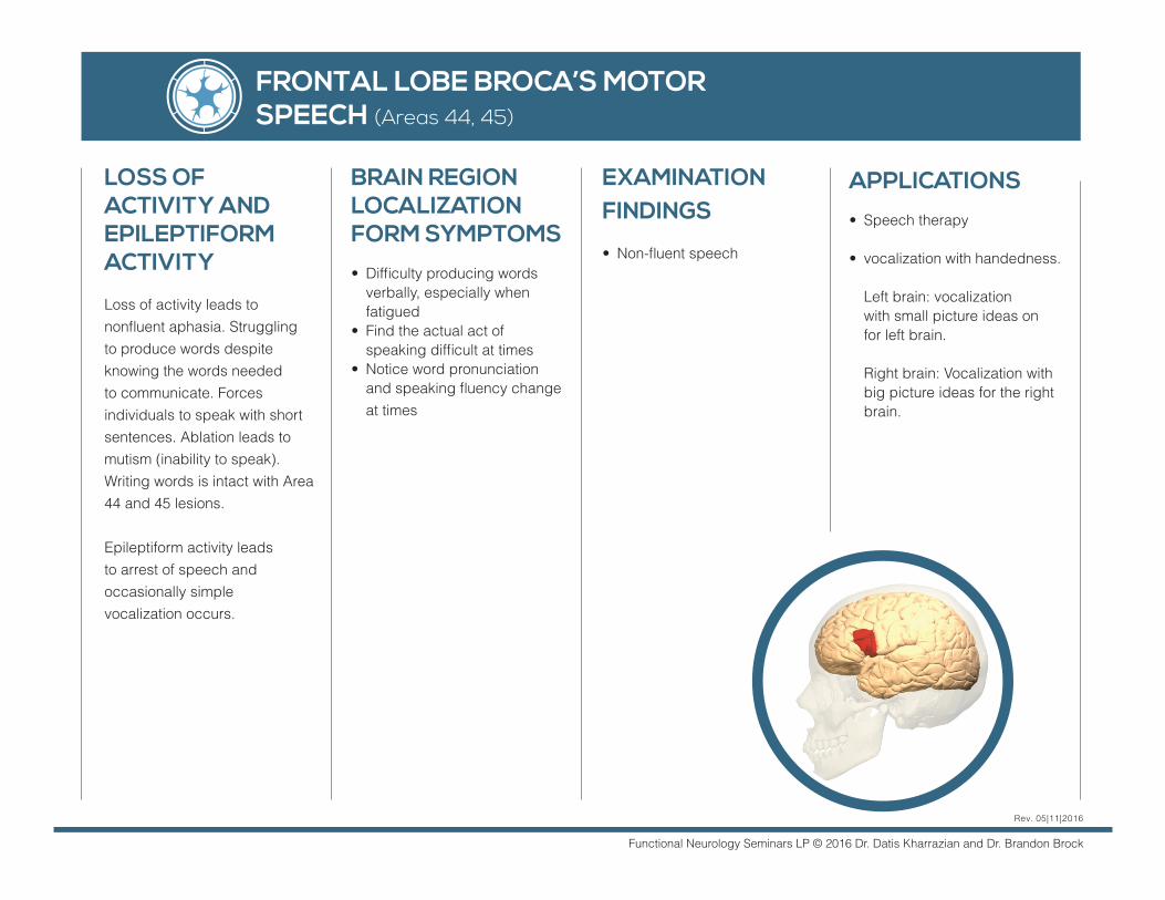
FRONTAL LOBE BROCA’S MOTOR SPEECH (Areas 44, 45)
BRAIN REGION LOCALIZATION FORM SYMPTOMS
• Difficulty producing words verbally, especially when fatigued
• Find the actual act of speaking difficult at times
• Notice word pronunciation and speaking fluency change at times
EXAMINATION FINDINGS • Non-fluent speech
APPLICATIONS
• Speech therapy
• vocalization with handedness. Left brain: vocalization with small picture ideas on for left brain. Right brain: Vocalization with big picture ideas for the right brain.
Rev. 05|11|2016
LOSS OF ACTIVITY AND EPILEPTIFORM ACTIVITY
Loss of activity leads to nonfluent aphasia. Struggling to produce words despite knowing the words needed to communicate. Forces individuals to speak with shortsentences. Ablation leads tomutism (inability to speak). Writing words is intact with Area 44 and 45 lesions.
Epileptiform activity leads to arrest of speech and occasionally simple vocalization occurs.
Functional Neurology Seminars LP © 2016 Dr. Datis Kharrazian and Dr. Brandon Brock
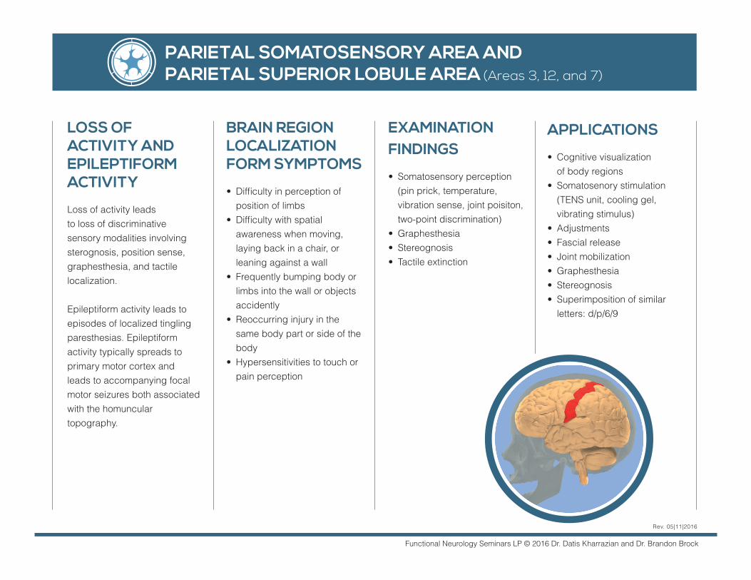
PARIETAL SOMATOSENSORY AREA AND PARIETAL SUPERIOR LOBULE AREA (Areas 3, 12, and 7)
LOSS OF ACTIVITY AND EPILEPTIFORM ACTIVITY
Loss of activity leads to loss of discriminative sensory modalities involving sterognosis, position sense, graphesthesia, and tactilelocalization.
Epileptiform activity leads toepisodes of localized tinglingparesthesias. Epileptiformactivity typically spreads toprimary motor cortex andleads to accompanying focalmotor seizures both associated with the homuncular topography.
BRAIN REGION LOCALIZATION FORM SYMPTOMS
• Difficulty in perception of position of limbs
• Difficulty with spatial awareness when moving, laying back in a chair, or leaning against a wall
• Frequently bumping body or limbs into the wall or objects accidently
• Reoccurring injury in the same body part or side of the body
• Hypersensitivities to touch or pain perception
EXAMINATION FINDINGS • Somatosensory perception
(pin prick, temperature, vibration sense, joint poisiton, two-point discrimination)
• Graphesthesia• Stereognosis• Tactile extinction
APPLICATIONS
• Cognitive visualization of body regions
• Somatosenory stimulation (TENS unit, cooling gel, vibrating stimulus)
• Adjustments• Fascial release• Joint mobilization• Graphesthesia• Stereognosis• Superimposition of similar
letters: d/p/6/9
Rev. 05|11|2016
Functional Neurology Seminars LP © 2016 Dr. Datis Kharrazian and Dr. Brandon Brock
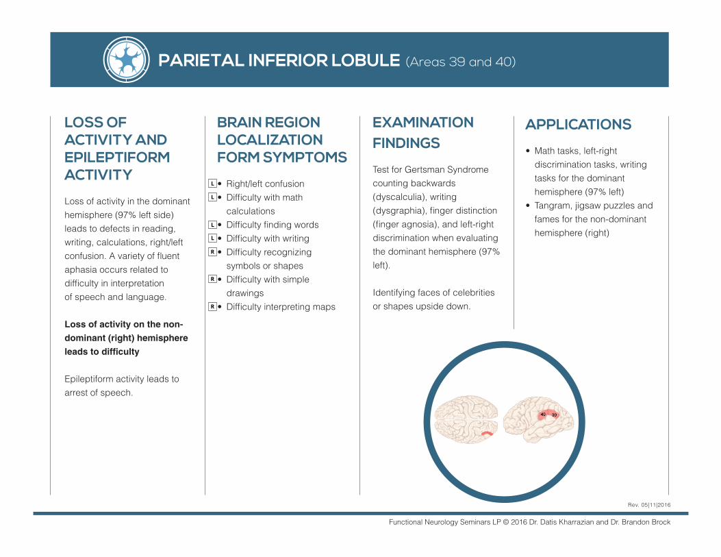
PARIETAL INFERIOR LOBULE (Areas 39 and 40)
LOSS OF ACTIVITY AND EPILEPTIFORM ACTIVITY
Loss of activity in the dominanthemisphere (97% left side) leads to defects in reading, writing, calculations, right/left confusion. A variety of fluent aphasia occurs related to difficulty in interpretationof speech and language.
Loss of activity on the non-
dominant (right) hemisphere
leads to difficulty
Epileptiform activity leads to arrest of speech.
BRAIN REGION LOCALIZATION FORM SYMPTOMS
• Right/left confusion• Difficulty with math
calculations• Difficulty finding words• Difficulty with writing• Difficulty recognizing
symbols or shapes• Difficulty with simple
drawings• Difficulty interpreting maps
EXAMINATION FINDINGS Test for Gertsman Syndrome counting backwards (dyscalculia), writing (dysgraphia), finger distinction (finger agnosia), and left-right discrimination when evaluating the dominant hemisphere (97% left).
Identifying faces of celebrities or shapes upside down.
APPLICATIONS
• Math tasks, left-right discrimination tasks, writing tasks for the dominant hemisphere (97% left)
• Tangram, jigsaw puzzles and fames for the non-dominant hemisphere (right)
Rev. 05|11|2016
Functional Neurology Seminars LP © 2016 Dr. Datis Kharrazian and Dr. Brandon Brock
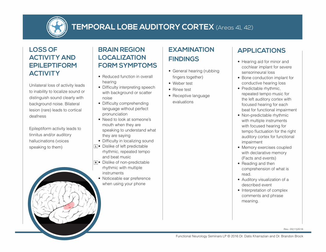
TEMPORAL LOBE AUDITORY CORTEX (Areas 41, 42)
LOSS OF ACTIVITY AND EPILEPTIFORM ACTIVITY
Unilateral loss of activity leads to inability to localize sound ordistinguish sound clearly withbackground noise. Bilaterallesion (rare) leads to corticaldeafness
Epileptiform activity leads totinnitus and/or auditoryhallucinations (voicesspeaking to them)
BRAIN REGION LOCALIZATION FORM SYMPTOMS
• Reduced function in overall hearing
• Difficulty interpreting speech with background or scatter noise
• Difficulty comprehending language without perfect pronunciation
• Need to look at someone’s mouth when they are speaking to understand what they are saying
• Difficulty in localizing sound• Dislike of left predictable
rhythmic, repeated tempo and beat music
• Dislike of non-predictable rhythmic with multiple instruments
• Noticeable ear preference when using your phone
EXAMINATION FINDINGS • General hearing (rubbing
fingers together)• Weber test• Rinee test• Receptive language
evaluations
APPLICATIONS
• Hearing aid for minor and cochlear implant for severe sensorineural loss
• Bone conduction implant for conductive hearing loss
• Predictable rhythmic, repeated tempo music for the left auditory cortex with focused hearing for each beat for functional impairment
• Non-predictable rhythmic with multiple instruments with focused hearing for tempo fluctuation for the right auditory cortex for functional impairment
• Memory exercises coupled with declarative memory (Facts and events)
• Reading and then comprehension of what is read.
• Auditory visualization of a described event
• Interpretation of complex comments and phrase meaning.
Rev. 05|11|2016
Functional Neurology Seminars LP © 2016 Dr. Datis Kharrazian and Dr. Brandon Brock
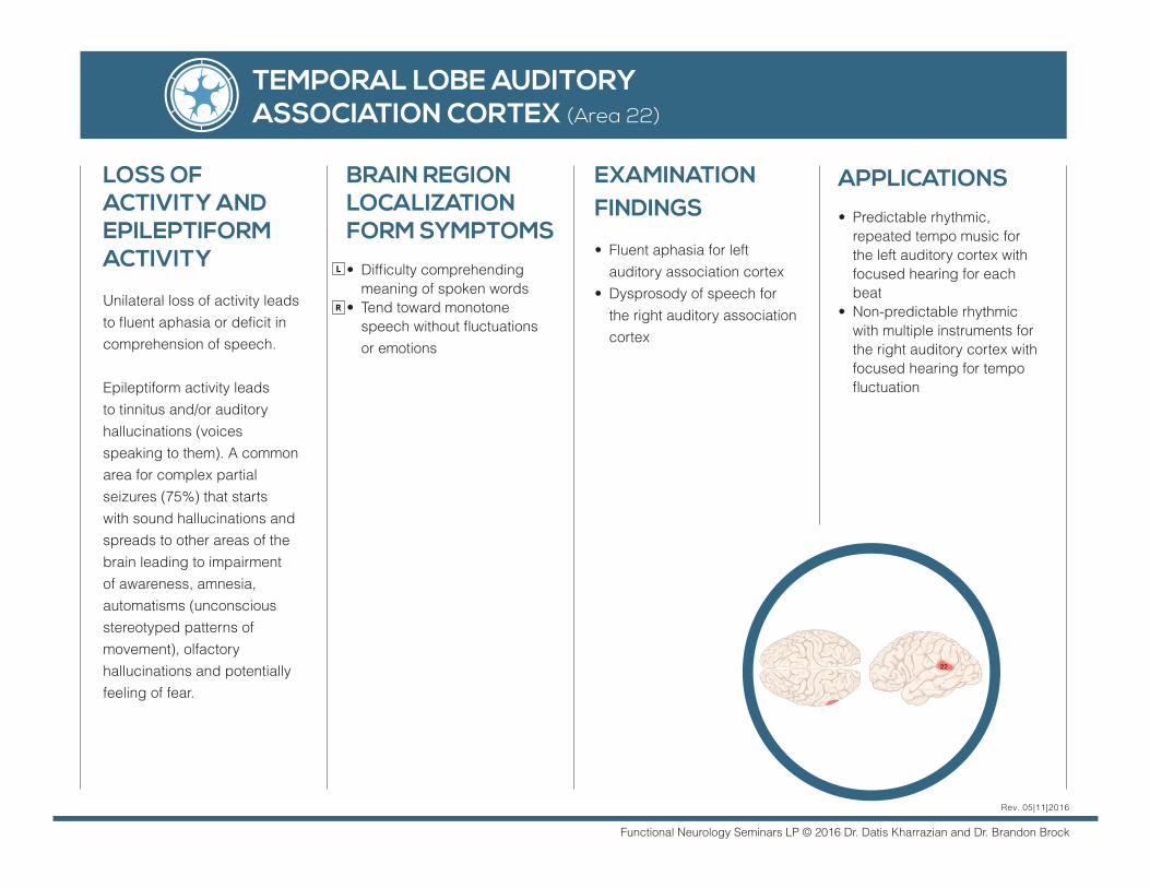
TEMPORAL LOBE AUDITORY ASSOCIATION CORTEX (Area 22)
BRAIN REGION LOCALIZATION FORM SYMPTOMS
• Difficulty comprehending meaning of spoken words
• Tend toward monotone speech without fluctuations or emotions
EXAMINATION FINDINGS • Fluent aphasia for left
auditory association cortex• Dysprosody of speech for
the right auditory association cortex
APPLICATIONS
• Predictable rhythmic, repeated tempo music for the left auditory cortex with focused hearing for each beat
• Non-predictable rhythmic with multiple instruments for the right auditory cortex with focused hearing for tempo fluctuation
Rev. 05|11|2016
LOSS OF ACTIVITY AND EPILEPTIFORM ACTIVITY
Unilateral loss of activity leadsto fluent aphasia or deficit incomprehension of speech.
Epileptiform activity leads to tinnitus and/or auditory hallucinations (voicesspeaking to them). A common area for complex partial seizures (75%) that starts with sound hallucinations and spreads to other areas of the brain leading to impairment of awareness, amnesia, automatisms (unconscious stereotyped patterns of movement), olfactory hallucinations and potentiallyfeeling of fear.
Functional Neurology Seminars LP © 2016 Dr. Datis Kharrazian and Dr. Brandon Brock
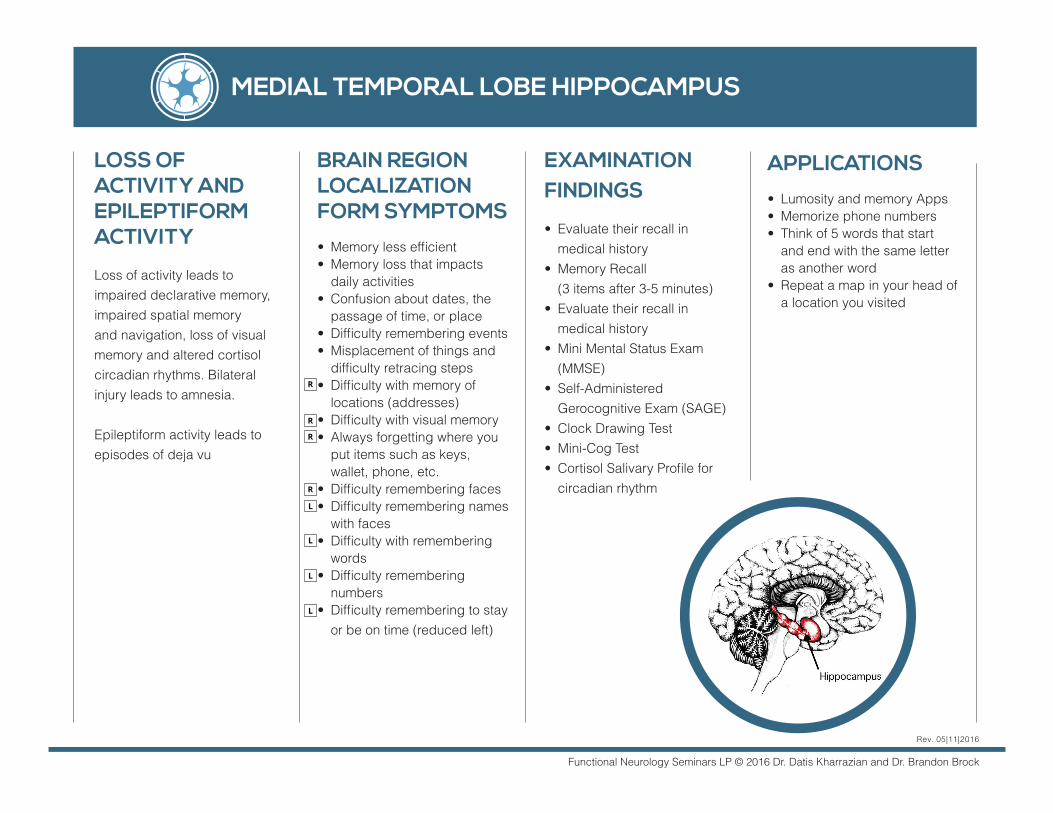
MEDIAL TEMPORAL LOBE HIPPOCAMPUS
BRAIN REGION LOCALIZATION FORM SYMPTOMS
• Memory less efficient • Memory loss that impacts
daily activities• Confusion about dates, the
passage of time, or place• Difficulty remembering events• Misplacement of things and
difficulty retracing steps • Difficulty with memory of
locations (addresses)• Difficulty with visual memory• Always forgetting where you
put items such as keys, wallet, phone, etc.
• Difficulty remembering faces• Difficulty remembering names
with faces• Difficulty with remembering
words• Difficulty remembering
numbers• Difficulty remembering to stay
or be on time (reduced left)
EXAMINATION FINDINGS • Evaluate their recall in
medical history • Memory Recall
(3 items after 3-5 minutes)• Evaluate their recall in
medical history • Mini Mental Status Exam
(MMSE)• Self-Administered
Gerocognitive Exam (SAGE)• Clock Drawing Test • Mini-Cog Test • Cortisol Salivary Profile for
circadian rhythm
APPLICATIONS
• Lumosity and memory Apps• Memorize phone numbers• Think of 5 words that start
and end with the same letter as another word
• Repeat a map in your head of a location you visited
Rev. 05|11|2016
LOSS OF ACTIVITY AND EPILEPTIFORM ACTIVITY
Loss of activity leads to impaired declarative memory, impaired spatial memory and navigation, loss of visual memory and altered cortisol circadian rhythms. Bilateral injury leads to amnesia.
Epileptiform activity leads to episodes of deja vu
Functional Neurology Seminars LP © 2016 Dr. Datis Kharrazian and Dr. Brandon Brock
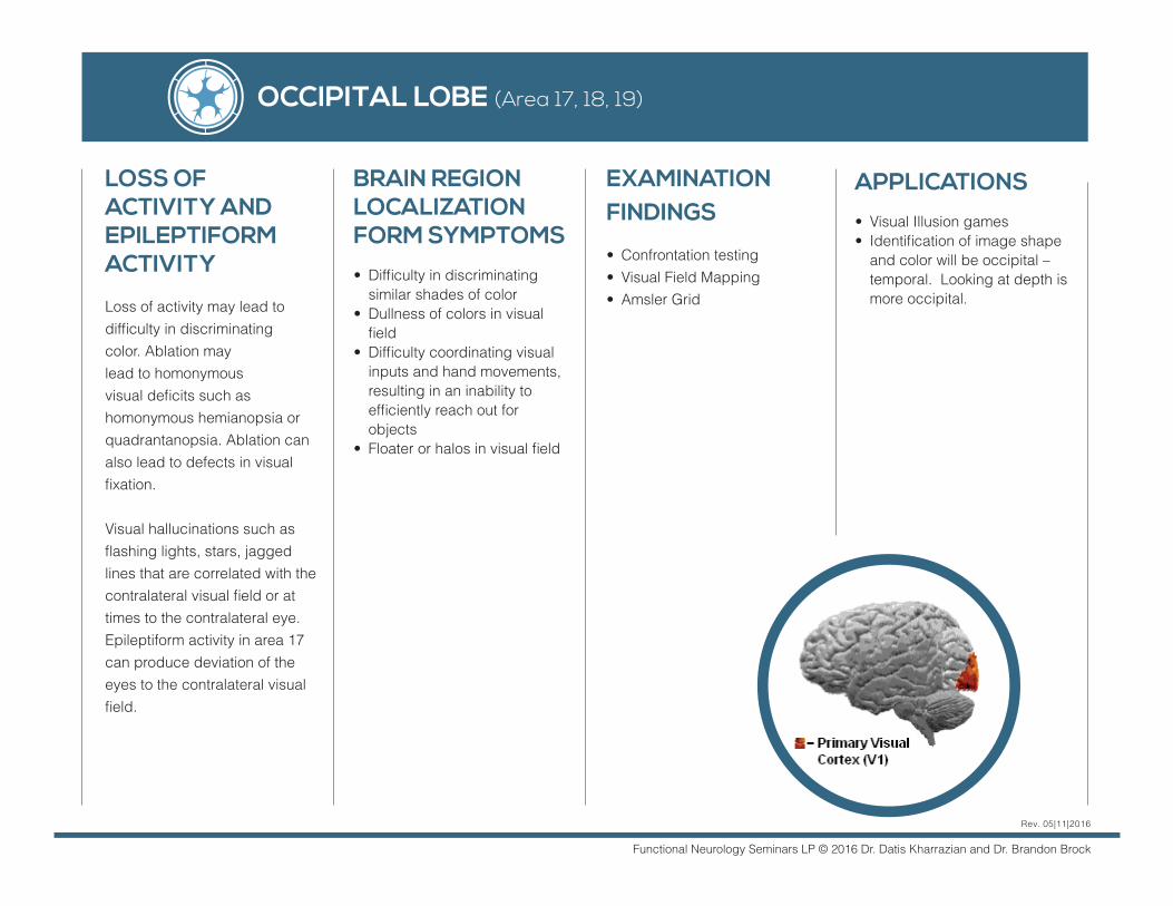
OCCIPITAL LOBE (Area 17, 18, 19)
BRAIN REGION LOCALIZATION FORM SYMPTOMS
• Difficulty in discriminating similar shades of color
• Dullness of colors in visual field
• Difficulty coordinating visual inputs and hand movements, resulting in an inability to efficiently reach out for objects
• Floater or halos in visual field
EXAMINATION FINDINGS • Confrontation testing• Visual Field Mapping• Amsler Grid
APPLICATIONS
• Visual Illusion games • Identification of image shape
and color will be occipital – temporal. Looking at depth is more occipital.
Rev. 05|11|2016
LOSS OF ACTIVITY AND EPILEPTIFORM ACTIVITY
Loss of activity may lead to difficulty in discriminating color. Ablation may lead to homonymous visual deficits such as homonymous hemianopsia or quadrantanopsia. Ablation can also lead to defects in visual fixation.
Visual hallucinations such as flashing lights, stars, jagged lines that are correlated with the contralateral visual field or at times to the contralateral eye.Epileptiform activity in area 17 can produce deviation of the eyes to the contralateral visual field.
Functional Neurology Seminars LP © 2016 Dr. Datis Kharrazian and Dr. Brandon Brock
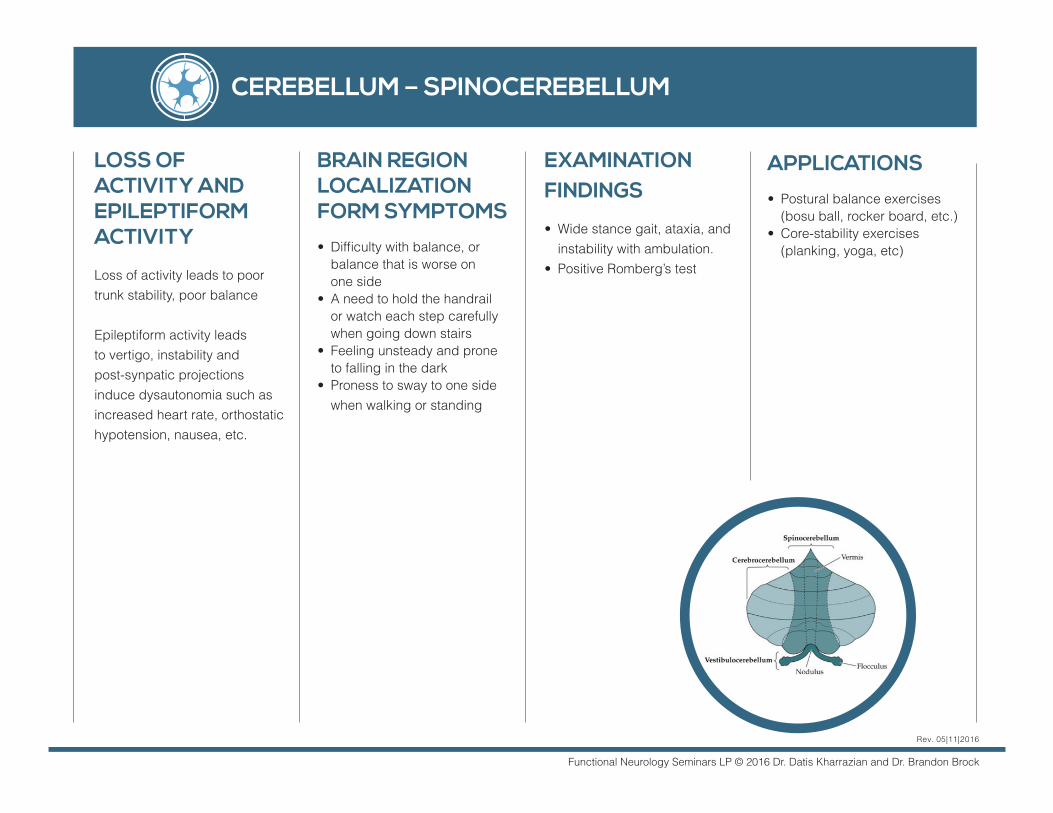
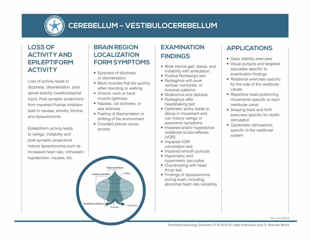
CEREBELLUM – SPINOCEREBELLUM
BRAIN REGION LOCALIZATION FORM SYMPTOMS
• Difficulty with balance, or balance that is worse on one side
• A need to hold the handrail or watch each step carefully when going down stairs
• Feeling unsteady and prone to falling in the dark
• Proness to sway to one side when walking or standing
EXAMINATION FINDINGS • Wide stance gait, ataxia, and
instability with ambulation. • Positive Romberg’s test
APPLICATIONS
• Postural balance exercises (bosu ball, rocker board, etc.)
• Core-stability exercises (planking, yoga, etc)
Rev. 05|11|2016
LOSS OF ACTIVITY AND EPILEPTIFORM ACTIVITY
Loss of activity leads to poor trunk stability, poor balance
Epileptiform activity leads to vertigo, instability and post-synpatic projections induce dysautonomia such as increased heart rate, orthostatic hypotension, nausea, etc.
Functional Neurology Seminars LP © 2016 Dr. Datis Kharrazian and Dr. Brandon Brock
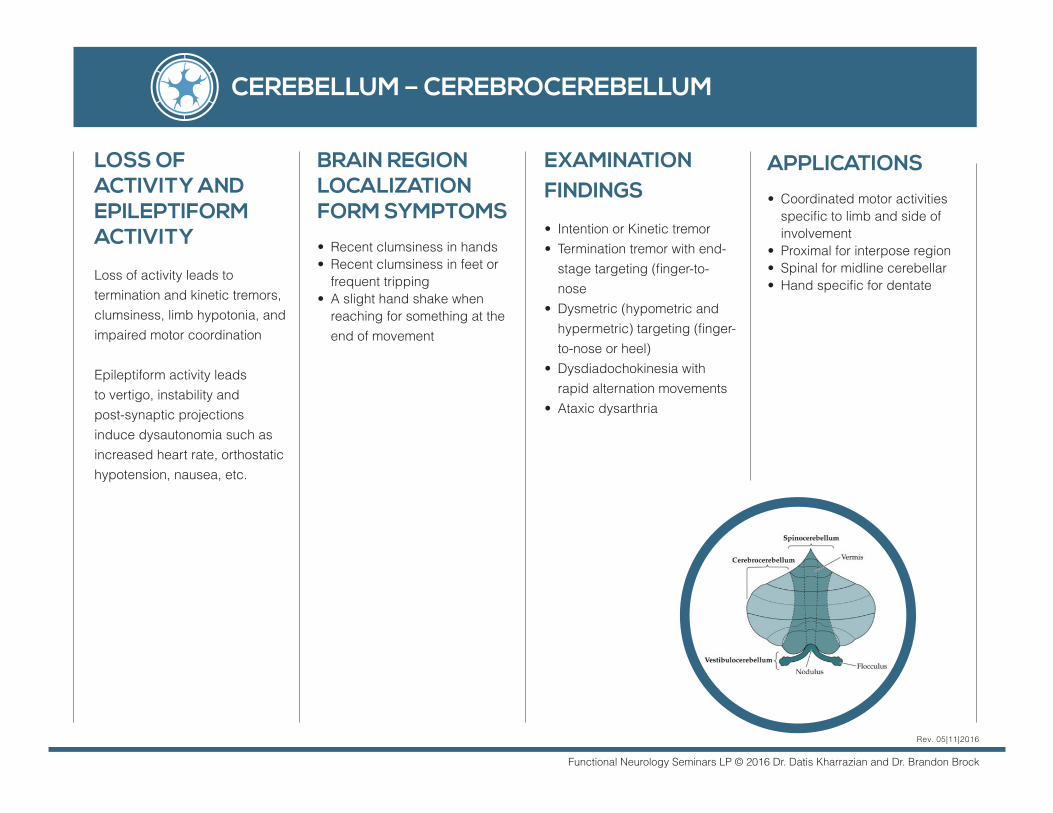
CEREBELLUM – CEREBROCEREBELLUM
BRAIN REGION LOCALIZATION FORM SYMPTOMS
• Recent clumsiness in hands • Recent clumsiness in feet or
frequent tripping • A slight hand shake when
reaching for something at the end of movement
EXAMINATION FINDINGS • Intention or Kinetic tremor • Termination tremor with end-
stage targeting (finger-to-nose
• Dysmetric (hypometric and hypermetric) targeting (finger-to-nose or heel)
• Dysdiadochokinesia with rapid alternation movements
• Ataxic dysarthria
APPLICATIONS
• Coordinated motor activities specific to limb and side of involvement
• Proximal for interpose region• Spinal for midline cerebellar• Hand specific for dentate
Rev. 05|11|2016
LOSS OF ACTIVITY AND EPILEPTIFORM ACTIVITY
Loss of activity leads to termination and kinetic tremors, clumsiness, limb hypotonia, and impaired motor coordination
Epileptiform activity leads to vertigo, instability and post-synaptic projections induce dysautonomia such as increased heart rate, orthostatic hypotension, nausea, etc.
Functional Neurology Seminars LP © 2016 Dr. Datis Kharrazian and Dr. Brandon Brock
CEREBELLUM – VESTIBULOCEREBELLUM
BRAIN REGION LOCALIZATION FORM SYMPTOMS
• Episodes of dizziness or disorientation
• Back muscles that tire quickly when standing or walking
• Chronic neck or back muscle tightness
• Nausea, car sickness, or sea sickness
• Feeling of disorientation or shifting of the environment
• Crowded places cause anxiety
APPLICATIONS
• Gaze stability exercises• Visual pursuits and targeted
saccades specific to examination findings
• Rotational exercises specific for the side of the vestibular canals
• Repetitive head positioning movements specific to each vestibular canal
• Swaying back and forth exercises specific for otolith stimulation
• Optokinetic stimulations specific to the vestibular system
Rev. 05|11|2016
LOSS OF ACTIVITY AND EPILEPTIFORM ACTIVITY
Loss of activity leads to dizziness, disorientation, poor spinal stability (vestibulospinal tract). Post-synaptic projections from impaired Pukinje inhibition lead to nausea, anxiety, tinnitus, and dysautonomia.
Epileptiform activity leads to vertigo, instability and post-synaptic projections induce dysautonomia such as increased heart rate, orthostatic hypotension, nausea, etc.
EXAMINATION FINDINGS • Wide stance gait, ataxia, and
instability with ambulation. • Positive Romberg’s test• Nystagmus with pure
vertical, horizontal, or torsional patterns
• Strabismus and diplopia• Nystagmus after
headshaking test• Optkinetic activy leads to
decay in movement and can induce vertigo or autonomic symptoms
• Impaired and/or hyperactive vestibular-ocular-reflexes (VOR)
• Impaired VOR- cancelation test
• Impaired smooth pursuits• Hypometric and
hypermetric saccades• Overshooting with head
thrust test• Findings of dysautonomia
during exam including, abnormal heart rate variability
Functional Neurology Seminars LP © 2016 Dr. Datis Kharrazian and Dr. Brandon Brock
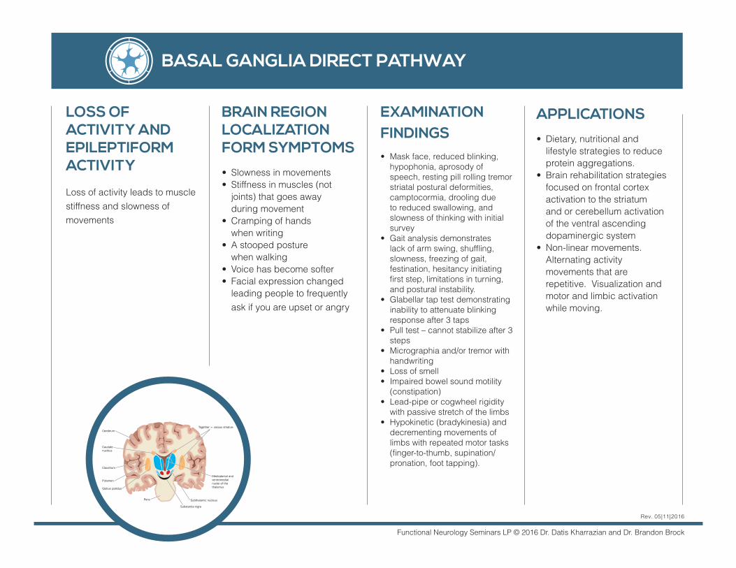
BASAL GANGLIA DIRECT PATHWAY
BRAIN REGION LOCALIZATION FORM SYMPTOMS
• Slowness in movements• Stiffness in muscles (not
joints) that goes away during movement
• Cramping of hands when writing
• A stooped posture when walking
• Voice has become softer • Facial expression changed
leading people to frequently ask if you are upset or angry
EXAMINATION FINDINGS • Mask face, reduced blinking,
hypophonia, aprosody of speech, resting pill rolling tremor striatal postural deformities, camptocormia, drooling due to reduced swallowing, and slowness of thinking with initial survey
• Gait analysis demonstrates lack of arm swing, shuffling, slowness, freezing of gait, festination, hesitancy initiating first step, limitations in turning, and postural instability.
• Glabellar tap test demonstrating inability to attenuate blinking response after 3 taps
• Pull test – cannot stabilize after 3 steps
• Micrographia and/or tremor with handwriting
• Loss of smell • Impaired bowel sound motility
(constipation)• Lead-pipe or cogwheel rigidity
with passive stretch of the limbs• Hypokinetic (bradykinesia) and
decrementing movements of limbs with repeated motor tasks (finger-to-thumb, supination/pronation, foot tapping).
APPLICATIONS
• Dietary, nutritional and lifestyle strategies to reduce protein aggregations.
• Brain rehabilitation strategies focused on frontal cortex activation to the striatum and or cerebellum activation of the ventral ascending dopaminergic system
• Non-linear movements. Alternating activity movements that are repetitive. Visualization and motor and limbic activation while moving.
Rev. 05|11|2016
LOSS OF ACTIVITY AND EPILEPTIFORM ACTIVITY
Loss of activity leads to muscle stiffness and slowness of movements
Functional Neurology Seminars LP © 2016 Dr. Datis Kharrazian and Dr. Brandon Brock
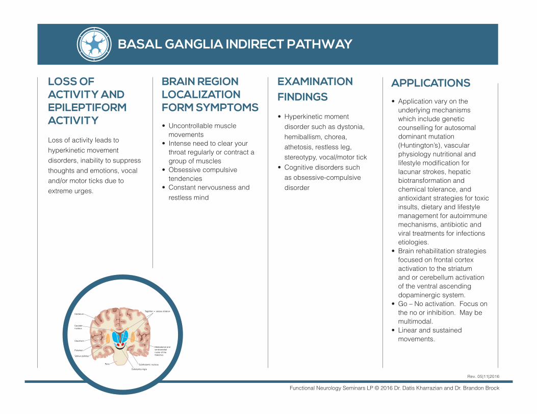
BASAL GANGLIA INDIRECT PATHWAY
BRAIN REGION LOCALIZATION FORM SYMPTOMS
• Uncontrollable muscle movements
• Intense need to clear your throat regularly or contract a group of muscles
• Obsessive compulsive tendencies
• Constant nervousness and restless mind
EXAMINATION FINDINGS • Hyperkinetic moment
disorder such as dystonia, hemiballism, chorea, athetosis, restless leg, stereotypy, vocal/motor tick
• Cognitive disorders such as obsessive-compulsive disorder
APPLICATIONS
• Application vary on the underlying mechanisms which include genetic counselling for autosomal dominant mutation (Huntington’s), vascular physiology nutritional and lifestyle modification for lacunar strokes, hepatic biotransformation and chemical tolerance, and antioxidant strategies for toxic insults, dietary and lifestyle management for autoimmune mechanisms, antibiotic and viral treatments for infections etiologies.
• Brain rehabilitation strategies focused on frontal cortex activation to the striatum and or cerebellum activation of the ventral ascending dopaminergic system.
• Go – No activation. Focus on the no or inhibition. May be multimodal.
• Linear and sustained movements.
Rev. 05|11|2016
LOSS OF ACTIVITY AND EPILEPTIFORM ACTIVITY
Loss of activity leads to hyperkinetic movement disorders, inability to suppress thoughts and emotions, vocal and/or motor ticks due to extreme urges.