Embed Size (px)
Citation preview

REVIEW
Brain organoids: advances, applications and challengesXuyu Qian1,2, Hongjun Song1,3,4,5 and Guo-li Ming1,3,4,6,*
ABSTRACTBrain organoids are self-assembled three-dimensional aggregatesgenerated from pluripotent stem cells with cell types andcytoarchitectures that resemble the embryonic human brain. Assuch, they have emerged as novel model systems that can be usedto investigate human brain development and disorders. Althoughbrain organoids mimic many key features of early human braindevelopment at molecular, cellular, structural and functionallevels, some aspects of brain development, such as the formationof distinct cortical neuronal layers, gyrification, and the establishmentof complex neuronal circuitry, are not fully recapitulated. Here, wesummarize recent advances in the development of brain organoidmethodologies and discuss their applications in disease modeling. Inaddition, we compare current organoid systems to the embryonichuman brain, highlighting features that currently can and cannot berecapitulated, and discuss perspectives for advancing current brainorganoid technologies to expand their applications.
KEY WORDS: Brain organoids, Neuroscience, Stem cell
IntroductionThe rapidly advancing field of stem cell biology continuallyprovides new insights into basic biology and human disorders, anddrives innovation for new treatments. Human pluripotent stem cells(hPSCs), including human embryonic stem cells (hESCs) andhuman induced pluripotent stem cells (hiPSCs), have emerged asinvaluable tools for modeling human disorders, especially thosewith complex genetic origins that are challenging to model inanimals (Takahashi et al., 2007; Takahashi and Yamanaka, 2006;Thomson et al., 1998). Under specific cues and favorableconditions, hPSCs have the potential to differentiate into any cellor tissue type found in the human body. They can also be usedto generate organoids – organ-like three-dimensional (3D) tissuecultures containing multiple cell types that represent accessiblesystems for modeling organogenesis and developmental disorders.Brain organoids are hPSC-derived organoids that self-assemble
to form an organized architecture, composed of progenitor, neuronaland glial cell types, resembling the fetal human brain (Jo et al.,2016; Kadoshima et al., 2013; Lancaster et al., 2013; Mariani et al.,2015; Pasca et al., 2015; Qian et al., 2016). Unlike conventionaltwo-dimensional (2D) cell cultures, brain organoids recapitulate the
human brain not only at the cellular level, but also in terms ofgeneral tissue structure and developmental trajectory, thereforeproviding a unique opportunity to model human brain developmentand function, which are often inaccessible to direct experimentation.Combined with recent technological advances, such as patient-derived hiPSCs, genetic engineering, genomic editing, and high-throughput single cell transcriptomics and epigenetics, brainorganoids have revolutionized our toolboxes to investigatedevelopment, diseases and evolution of the human brain.
In this Review, we first introduce recent advances in brainorganoid technologies, and their applications as model systemsand discovery tools. We then discuss the hurdles that currentmethodologies have yet to overcome, and offer feasible suggestionsfor future steps and objectives that would enable organoidtechnologies to progress further. Despite the tremendous promiseof brain organoids, it is important to understand the limitations oforganoid models. Therefore, this Review also aims to provide abalanced view between the advantages and disadvantages of brainorganoids. Only when interpreted complementarily with othermodels can the results obtained using brain organoids shed light onour understanding of human biology.
Current brain organoid methodologiesMethodologies to induce neural differentiation from PSCs in 3Dhave been pursued since the early 1990s, but not all 3D neuralculture systems should be considered brain organoids.Neurospheres, for instance, are 3D aggregations of several centralnervous system (CNS) cell types derived from neural progenitorcells (NPCs) (Reynolds et al., 1992). Neurospheres contain varioussubtypes of neurons and glial cells, but distinctive cytoarchitecturesare typically absent (Pamies et al., 2017). Neural cells can also beartificially assembled, using designed culture chambers andbiomaterial scaffolds, into ‘organ-on-a-chip’ structures, which areadvantageous for modeling cell-cell interactions in a highlycontrolled manner (Phan et al., 2017). Nevertheless, as far as thisReview is concerned, brain organoids are distinct from neurosphereand microchip approaches most notably because they harness theself-organization capability of PSCs to mimic the cytoarchitectureand developmental trajectories found in vivo.
In general, two different types of methodologies can be used togenerate brain organoids: unguided methods and guided methods(Fig. 1). Whereas unguided methods rely fully on spontaneousmorphogenesis and intrinsic differentiation capacities within hPSCaggregates, guided organoid methods require supplementation ofexternal patterning factors to induce hPSCs to differentiate towardsdesired lineages. The number and combination of external factorsused in differentiation protocols varies, and the choice betweenunguided and guided approaches is often seen as a trade-off betweendiversity and consistency.
Unguided brain organoid methodologiesThe unguided brain organoid, or the cerebral organoid, is inspiredby methodologies developed for generating gastrointestinal
1Department of Neuroscience and Mahoney Institute for Neurosciences, PerelmanSchool for Medicine, University of Pennsylvania, Philadelphia, PA 19104, USA.2Biomedical Engineering Graduate Program, Johns Hopkins University School ofMedicine, Baltimore, MD 21205, USA. 3Department of Cell and DevelopmentalBiology, Perelman School for Medicine, University of Pennsylvania, Philadelphia,PA 19104, USA. 4Institute for Regenerative Medicine, Perelman School forMedicine, University of Pennsylvania, Philadelphia, PA 19104, USA. 5TheEpigenetics Institute, Perelman School for Medicine, University of Pennsylvania,Philadelphia, PA 19104, USA. 6Department of Psychiatry, Perelman School forMedicine, University of Pennsylvania, Philadelphia, PA 19104, USA.
*Author for correspondence ([email protected])
G.M., 0000-0002-2517-6075
1
© 2019. Published by The Company of Biologists Ltd | Development (2019) 146, dev166074. doi:10.1242/dev.166074
DEVELO
PM
ENT

organoids (Table 1) (Lancaster et al., 2013; Sato et al., 2011, 2009).In the protocol developed by the Knoblich group, embryoid bodies(EBs) derived from hPSC aggregates are embedded into anextracellular matrix (ECM), such as Matrigel, and subsequentlycultured in spinning bioreactors to promote tissue expansion andneural differentiation (Lancaster and Knoblich, 2014). Withminimal external interference, cerebral organoids produced by thisapproach give hPSCs the most freedom for self-organization, andsometimes give rise to very elongated neuroepithelial structures.They exhibit a variety of cell lineage identities ranging fromforebrain, midbrain and hindbrain, to retina, choroid plexus andmesoderm (Camp et al., 2015; Lancaster et al., 2013). Large-scalesingle cell transcriptomic profiling has further revealed that cerebralorganoids contain neural progenitors, excitatory neurons, inhibitoryneurons, astrocytes and oligodendrocyte precursor cells (OPCs)found in the CNS, as well as photosensitive cells found in the retina(Quadrato et al., 2017). The stochastic nature of hPSC spontaneousdifferentiation, however, results in unpredictable proportions and aheterogeneous arrangement of each lineage and cell type acrossbatches of differentiated organoids and across hPSC lines. Althoughthis cell-type diversity in cerebral organoids offers a uniqueopportunity to model the interactions between different brain
regions, the high variability and heterogeneity present significantchallenges for systematic and quantitative studies.
Guided brain organoid methodologiesGuided methods for generating brain organoids were pioneered bythe Sasai group, who developed a series of 3D differentiationprotocols based on the serum-free culture of EB-like aggregates(Table 1) (Danjo et al., 2011; Eiraku et al., 2008; Kadoshima et al.,2013; Muguruma et al., 2015; Sakaguchi et al., 2015; Sasai, 2013).In these guided organoid, or ‘spheroid’ methods, small moleculesand growth factors are used throughout the differentiation process toinstruct hPSCs to form cells and tissues representative of certainbrain regions, such as the cerebral cortex, hippocampus andmidbrain (Jo et al., 2016; Mariani et al., 2015; Pasca et al.,2015; Sakaguchi et al., 2015; Yoon et al., 2019). These directedorganoid cultures are sometimes capable of generating mixturesof cell types with relatively consistent proportions, exhibitingless variation across batches and cell lines (Sloan et al., 2017).However, directed organoids typically contain relatively smallneuroepithelial structures and their cytoarchitecture is sometimesnot well-defined, possibly owing to the interference of hPSCself-organization and cell-cell interactions by excessive use ofexternal factors.
Guided organoid differentiation protocols can be carefullytailored to require the use of external patterning factors only at theearly differentiation stage, thereby allowing hPSCs to be specifiedas progenitor cells exhibiting certain brain region identities withminimal heterogeneity. For these brain region-specific organoids,external factors are then removed or minimized after successfulpatterning during the initial stage of differentiation, and subsequentdifferentiation follows intrinsic programs similar to those operatingin vivo after neural patterning. This approach has been usedsuccessfully to generate large ventricle-like structures with elaboratelaminar organization and architecture (Lancaster et al., 2017; Qianet al., 2016). In addition to using chemical factors, syntheticbiomaterials can be engineered to guide the formation of brainorganoids physically. This is exemplified by the microfilament-engineered cerebral organoids method, in which elongated EBsform around scaffolds made from polymer microfilaments, resultingin more-consistent formation of enlarged ventricular structures andneuro-epithelium (Lancaster et al., 2017).
The use of spinning bioreactors can also provide enhancednutrient and oxygen diffusion and sustained 3D suspension culture(Lancaster et al., 2013). However, commercial bioreactors are bulkyand consume large volumes of culture medium, limiting theefficiency and throughput of organoid culture. To reduce theconsumption of culture medium, multiple-well culture plates havebeen used together with orbital shakers placed in the incubator as analternative to a spinning bioreactor (Lancaster and Knoblich, 2014).More recently, custom-designed miniaturized multi-well spinningbioreactors have been developed to both reduce the cost ofmaintaining organoid cultures and remove the need for bulkymachines in the incubator; this approach allowed for protocoloptimization with improved efficiency, and enabled the generationof brain region-specific organoids mimicking the dorsal forebrain,midbrain and hypothalamus (Qian et al., 2016). Notably, forebrainorganoids generated via this approach consistently form corticalstructures with distinct layers that resemble the ventricular zone(VZ), the inner and outer subventricular zone (iSVZ and oSVZ) andthe cortical plate (CP) at molecular, cellular and structural levels.The recapitulation of primate/human-specific developmentalfeatures, such as an enlarged oSVZ, in forebrain organoids offers
+
Human pluripotentstem cells (hPSCs)
Aggregation
Embryoidbody (EB)
Unguideddifferentiation
Cerebral organoid
Guideddifferentiation
Spheroid Spheroid
Fusion
Assembloid
Brain region-specificorganoid
Fig. 1. Unguided and guided approaches for making brain organoids.Unguided approaches (top) harness the intrinsic signaling and self-organization capacities of hPSCs to differentiate spontaneously into tissuesmimicking the developing brain. The resulting cerebral organoids often containheterogeneous tissues resembling various brain regions. By contrast, guidedapproaches (bottom) use small molecules and growth factors to generatespheroids that are specifically representative of one tissue type. Brain region-specific organoid methods involve the use of patterning factors at an earlystage to specify progenitor fate; these factors are then removed in subsequentdifferentiation stages. Guided approaches can also be used to generate two ormore spheroids/organoids representative of different brain region identities thatcan then be fused to form ‘assembloids’, which model interactions betweendifferent brain regions.
2
REVIEW Development (2019) 146, dev166074. doi:10.1242/dev.166074
DEVELO
PM
ENT

unique advantages for understanding human cortical developmentand developmental disorders.
Fused organoid technologiesAlthough cerebral organoid methods can produce tissuesresembling various interacting brain regions, their proportion andspatial organization are highly heterogeneous and unpredictable. Toimprove modeling of inter-regional interactions, several groupsconcurrently developed new approaches, first differentiating hPSCsinto different brain region-specific organoids separately, and thenfusing them together to form organoids with multiple distinct regionidentities in a controlled manner (Bagley et al., 2017; Birey et al.,2017; Xiang et al., 2017). For example, fused dorsal and ventralforebrain organoids have been shown to form an ‘assembloid’(Fig. 1) with two distinctive but interfacing domains (Birey et al.,2017). In these structures, interneurons generated from the ventraldomain preferentially migrate towards the dorsal domain,resembling the tangential migration of interneurons from thesubpallium to the cerebral cortex in vivo (Anderson et al., 1997).Moreover, electrophysiological characterization of forebrainassembloids revealed that migrating interneurons connectsynaptically with local excitatory neurons to form microcircuits(Birey et al., 2017).Overall, these recent advances in brain organoid methodologies
have expanded our toolbox for modeling human development anddisorders. The choice between guided and unguided methodologieswill depend on the specific focus of investigation. For instance,unguided organoids are suitable for exploring cell-type diversityduring whole-brain development, brain region-specific organoids
better recapitulate brain cytoarchitecture with less heterogeneity,and assembloids allow for the investigation of interactions betweenspecific brain regions with more consistent molecular andfunctional characterization.
Modeling cerebral cortex development using organoids:in vitro versus in vivoOrganoids mimicking the cerebral cortex have so far been bettercharacterized and more frequently used than other brain organoids.Such cortical organoids have also attracted the most interest in thefield because the cerebral cortex is the most unique andevolutionarily expanded region of the human brain, in comparisonto that of other animals, and is often severely affected in manyneurological disorders (Bystron et al., 2008; Hansen et al., 2010; Luiet al., 2011; Rakic, 2009). The presence of profound speciesdifferences in the cortex justifies the use of human cell-derivedcortical organoids over animal models. However, like all in vitromodels, these organoids are not identical replicas of their in vivocounterparts. Below, we use cortical organoids as an example todiscuss the aspects of brain development that have been successfullyreproduced in cortical organoids, and those aspects that are stilllacking (Fig. 2).
Size and general architectureFirst of all, cortical organoids are much smaller in size comparedwith human cerebral cortex (Fig. 2). Whereas cortical organoids canat most expand to∼4 mm in diameter, the human neocortex is about15 cm in diameter, with the thickness of gray matter alone being2-4 mm (Lancaster et al., 2017; Qian et al., 2016; Rakic, 2009).
Table 1. Overview of brain organoid methods
Organoid type Brain region modeledUnguided/guided? Requirements for support of 3D culture References
Serum-free culture of EB-likeaggregates (SFEBq)
Cerebral cortex Guided Stationary floating culture, followed byre-plating of aggregates
Eiraku et al., 2008
Cerebral organoid Whole brain Unguided Spinning bioreactor Lancaster and Knoblich, 2014;Lancaster et al., 2013
Cortical neuroepithelium Cerebral cortex Guided Floating culture in 40% oxygen, permeablefilm-based culture plates (Lumox plates)
Kadoshima et al., 2013
Neural cyst Spinal cord Guided Stationary floating culture Meinhardt et al., 2014Cerebellar-plate-likeneuroepithelium
Cerebellum Guided Stationary floating culture Muguruma et al., 2015
Cortical spheroid Cerebral cortex Guided Stationary floating culture Pasca et al., 2015;Sloan et al., 2017
Forebrain rosettes Forebrain Guided Stationary floating culture Mariani et al., 2015Choroid plexus-like tissue Choroid plexus Guided Floating culture in 40% oxygen Sakaguchi et al., 2015Medial pallium-like tissue Hippocampus Guided Floating culture permeable film-based
culture plates, mechanical cuttingSakaguchi et al., 2015
Forebrain organoid Cerebral cortex Guided Miniaturized multi-well spinning bioreactor Qian et al., 2018, 2016Hypothalamus organoid Hypothalamus Guided Miniaturized multi-well spinning bioreactor Qian et al., 2018, 2016Midbrain organoid Midbrain Guided Miniaturized multi-well spinning bioreactor Qian et al., 2018, 2016Midbrain organoid Midbrain Guided Orbital shaker Jo et al., 2016Pituitary organoid Anterior pituitary Guided Stationary floating culture Ozone et al., 2016Microfilament-engineeredcerebral organoid
Cerebral cortex Guided Orbital shaker Lancaster et al., 2017
Brain assembloid Dorsal and ventralforebrain
Guided Stationary floating culture Birey et al., 2017
Fused cerebral organoids Dorsal and ventralforebrain
Guided Orbital shaker Bagley et al., 2017
Medial ganglionic eminenceorganoid
Medial ganglioniceminence
Guided Orbital shaker Xiang et al., 2017
Organoid grafts in mouse brain Cerebral cortex Unguided Intracerebral grafting of brain organoidsinto mouse brain
Mansour et al., 2018
Oligocortical spheroids Cerebral cortex andmyelination
Guided Stationary floating culture Madhavan et al., 2018
3
REVIEW Development (2019) 146, dev166074. doi:10.1242/dev.166074
DEVELO
PM
ENT

Moreover, owing to a lack of vascularization and circulation, anecrotic core inevitably builds up in the organoid’s interior and theactual viable thickness is further limited (Fig. 3). The dramaticallydifferent sizes and numbers of cells in cortical organoids suggestthat it is almost impossible for current organoid technologies torepresent fully the complexity of a bona fide cortex.Surprisingly, however, cortical organoids do recapitulate the
organization of neural progenitor zones with considerable accuracy,especially when analyzed in vertical columns from the apical(inner) surface to the basal (outer) surface (Fig. 2). Indeed, corticalorganoids contain a large VZ formed by SOX2+ ventricular radialglia cells (vRGs) organized into polarized radial structures with alumen and apical surfaces expressing adherens junction proteins(Kadoshima et al., 2013; Lancaster et al., 2017; Qian et al.,2016). Immediately adjacent to the VZ, a thin and dense layer ofTBR2 (EOMES)+ intermediate progenitor cells (IPCs) can be
found, resembling the iSVZ (Lui et al., 2011). A well-definedoSVZ, populated by IPCs, immature neurons and unipolarNPCs expressing human outer radial glia (oRG)-specific markers,such as HOPX and PTPRZ1, can also be observed in corticalorganoids (Bershteyn et al., 2017; Lancaster et al., 2017; Li et al.,2017a,b; Qian et al., 2016; Watanabe et al., 2017). Notably,in forebrain organoids developed by Qian et al., the oSVZexpands disproportionally as organoids age, and by 80 days ofdifferentiation, the oSVZ becomes several times larger than the VZ,similar to human corticogenesis where the oSVZ is the dominantsource of neurogenesis in the second and third trimester (Hansenet al., 2010; Qian et al., 2016). Moreover, the basal processes of RGsin cortical organoids are radially aligned, forming migrational tracksfor newborn neurons to reach the CP, as occurs in vivo (Qian et al.,2018). Live imaging of fluorescently labeled vRGs and oRGs incortical organoids has revealed their mitotic behavior and dynamics,
Cortical organoids Human embryonic cortex
4 mm 5 cmOrganoids are much smaller
A necrotic core builds up inside the organoid, limiting its size
MZ
CP
IZ
oSVZ
iSVZ
VZ
Ventricle lumen
Organoid progenitor zonesresemble embryonic cortex
Apical surface
Basal surface
Neuronal layers of organoidscontain deep layer andsuperficial layer neuronswith rudimentary separation,but not separated layers
Basement membrane can bereconstituted in organoids bysupplementing ECM proteinsin the culture medium
Gyrification starts aroundGW 23, but folding of theCP has not been observedin organoids
Key
vRG IPC oRG Newbornneuron
Deep layerneuron
Upper layerneuron
Layer Ineuron Interneuron Glial cell
Organoids produce mostneural lineage cell typesfound in the human cortex
Organoids usually containmultiple discontinuousneuroepithelial structures
Fig. 2. Structural comparison between cortical organoids and the human embryonic cortex. Cortical organoids resemble the cytoarchitecture of humandeveloping cerebral cortex in early and mid-gestation with remarkable fidelity, despite their small size. A cortical organoid usually contains multiple short andindependent neuroepithelial structures. Within each structure, well-defined layers resembling the VZ, iSVZ, oSVZ, CP and MZ can be observed. Major neurallineage cell types in the embryonic cortex can also be detected in cortical organoids, but vascular and immune cells are absent. Late-gestational features ofcorticogenesis, such as the formation of cortical folding and the six separated cortical layers are not observed in cortical organoids generated by currentlyavailable approaches. CP, cortical plate; IPC, intermediate progenitor cells; iSVZ, inner subventricular zone; IZ, intermediate zone;MZ,marginal zone; oRG, outerradial glia; oSVZ, outer subventricular zone; vRG, ventricular radial glia; VZ, ventricular zone.
4
REVIEW Development (2019) 146, dev166074. doi:10.1242/dev.166074
DEVELO
PM
ENT

again suggesting that they resemble RGs in vivo (Bershteyn et al.,2017; Subramanian et al., 2017).The cortical organoids developed by Kadoshima et al.
demonstrated a cell-sparse region filled by neuronal processesbetween the SVZ and the CP, resembling the intermediate zone(Kadoshima et al., 2013). In early stages of cortical organoiddevelopment, the neuronal layer made from TBR1+ and CTIP2(BCL11B)+ early-born neurons and reelin+ Cajal–Retzius cells ishighly reminiscent of the preplate (PP) seen in first trimestercorticogenesis (Lancaster et al., 2013; Qian et al., 2016). As thisneuronal layer expands in size and becomes more condensed, itexhibits a compact morphology and starts to resemble the CP of thefetal cortex, with the help of ECM proteins supplemented in culturemedium to reconstitute the basement membrane on the organoidsurface (Kadoshima et al., 2013; Lancaster et al., 2017; Qian et al.,2018). Moreover, the temporal order of neurogenesis in corticalorganoids follows the ‘inside-out’ rules that have been demonstratedin vivo: neurons expressing deep layer markers are born early,whereas upper layer neurons, expressing markers such as SATB2and CUX1, are found later. However, extensive mixing andcolocalization of upper and deep layer neurons are often observed,indicating that the specification of cortical lamination is incompletein these organoids (Lancaster et al., 2017; Qian et al., 2016).Interestingly, such co-expression and intermingling of upper anddeep layer fate are similarly observed in the CP of mid-gestationhuman embryonic cortex (Ip et al., 2011; Nowakowski et al., 2017;Ozair et al., 2018; Zhong et al., 2018). However, at the perinatalperiod, the separation of upper and deep cortical layers becomesevident, suggesting that the establishment of distinct cortical layerstakes place near the end of the second trimester (Saito et al., 2011;Zeng et al., 2012). Furthermore, unlike in the perinatal neocortex,the cortical layers in organoids cannot be discriminated based ontheir neuron distribution and density, partially owing to themexhibiting drastically fewer layers of CP neurons (Molyneaux et al.,2007). Organoid neurons also lack layer-specific dendriticmorphology and axonal projection patterns (Zhong et al., 2018).It is also difficult to divide the neuronal layer of cortical organoidsunambiguously into distinctive subplate (SP) and CP regions, likelyowing to the fact that the SP-like region is interspersed between theCP and the SVZ (Ozair et al., 2018).
Neuronal function and activityExcitatory and inhibitory neurons in cortical organoids arefunctional, but unsurprisingly less mature, compared to adultneurons (Birey et al., 2017; Pasca et al., 2015; Qian et al., 2016).Because cortical organoids are a model for the early-mid stages of
embryonic development, during which time newborn neurons arecontinuously produced, electrophysiological characterization oftenreveals highly variable functionality and maturation parameterswhen cells are randomly chosen. The neurons in more matureorganoids, for example, fire trains of action potentials upon currentinjection and show robust spontaneous excitatory/inhibitorypostsynaptic currents (Pasca et al., 2015; Qian et al., 2016), andextracellular recording using a multi-electrode probe has revealedspontaneous burst-like firing activities in these neurons (Quadratoet al., 2017). Sparse labeling sometimes reveals neurons withpyramidal-like morphology and dendritic spines (Lancaster et al.,2017; Qian et al., 2016). Synaptogenesis also appears to beabundant when examined using immunostaining or electronmicroscopy (Birey et al., 2017; Pasca et al., 2015; Quadrato et al.,2017; Sloan et al., 2017). However, neuron subtype-specificfiring patterns and plasticity are yet to be observed. Nonetheless,despite their large variability, the basic electrophysiological propertiesof neurons, such as resting membrane potential, resistance,capacitance, amplitude and frequency of spontaneous post-synapticcurrents, show statistically significant trends ofmaturation over time asorganoids age (Qian et al., 2016). This is in line with a recent study,using patch-clamping on organotypic slices of human fetal cortex,which reported that most neurons at the end of the second trimester areimmature, and only early-born SP and deep layer neurons are able tofire action potentials and exhibit spontaneous synaptic transmission(Zhong et al., 2018). Thus, neurons generated in organoids mightreflect the development and maturation of neuronal functionalitiesduring embryonic stages in vivo.
Cell-type diversityIn addition to generating various types of RGs and neurons,organoids recapitulate the cellular diversity of the cerebral cortex byproducing interneurons, astrocytes and OPCs (Birey et al., 2017;Qian et al., 2016; Quadrato et al., 2017; Sloan et al., 2017). Similarto rodents, the majority of human cortical interneurons originatefrom the ganglionic eminence of the ventral forebrain andtangentially migrate to the cerebral cortex (Hansen et al., 2013;Ma et al., 2013), but several studies have provided evidence fordorsal origins of some cortical interneurons, which is an observationunique to primates (Jakovcevski et al., 2011; Letinic et al., 2002;Petanjek et al., 2009; Radonjic et al., 2014; Yu and Zecevic, 2011).Although NKX2.1-expressing ventral progenitors are absent indorsally patterned forebrain organoids at early stages, a smallpopulation of interneurons is detected later, suggesting thatdorsal forebrain progenitors are also potentially capable ofproducing interneurons in vitro (Qian et al., 2016). In addition, a
Oxygen
Dept
h in
to o
rgan
oid
surf
ace � Cells near the organoid surface receive
sufficient oxygen and nutrients
� Acidification and released inflammatory contents induce apoptosis of neighboring cells; adherens junctions at the apical surface are disrupted
� Oxygen becomes limited deeper into the surface
� Cell necrosis occurs due to lack of oxygen and nutrients
NPCs are ‘buried’ inside ascortical structure expands
A necrotic core builds upinside the organoid owingto lack of oxygen andnutrient diffusion
Fig. 3. The diffusion limit depletes progenitors and prohibits organoid expansion. Brain organoids grown as a sphere in 3D suspension cultures canexpand to 3-4 mm in diameter, but only those cells within a limited distance from the surface can receive sufficient oxygen and nutrients via diffusion. The build-upof a necrotic core is therefore common in brain organoids. As the outer layers expand, the more metabolically demanding progenitors in the interior willeventually deplete, resulting in a loss of proliferation and structural disorganization over long-term cultures.
5
REVIEW Development (2019) 146, dev166074. doi:10.1242/dev.166074
DEVELO
PM
ENT
recent study described a protocol to specifically enrich OPCsand oligodendrocytes to generate oligo-cortical spheroids withmyelination (Madhavan et al., 2018). Together, these findingshighlight that cortical organoids appear to generate all cell types fromthe neuroectoderm lineage. By contrast, cortical organoids lack cellsfound in the brain with non-neural origins, including vascular cellsand immune cells. Endothelial cells (ECs) can be cultured separatelyand added to organoids during the EB formation stage, and can self-organize to form network-like structures (Nzou et al., 2018; Phamet al., 2018). Such primitive endothelial networks, however, areincapable of functionally facilitating nutrient delivery into organoidsand alleviating the build-up of a necrotic core, although they doinstead provide a potential platform for modeling the neurovascularniche and blood brain barrier (Nzou et al., 2018; Pham et al., 2018).Human iPSC-derived microglia can also be co-cultured with brainorganoids; they adhere to the organoid surface and migrate to theinterior, suggesting chemotaxis of microglia towards CNS cues(Abud et al., 2017). Remarkably, when brain organoids aremechanically injured, the microglia cluster near the injury site andadopt a round morphology, in contrast to their normal ramifiedmorphology, resembling the activated microglia morphologyobserved upon CNS injury in vivo (Abud et al., 2017). It remains achallenge, however, to develop protocols to generate non-neural celltypes within brain organoids intrinsically without introducing furtherheterogeneity, and artificially combining separately cultured celltypes is thus likely to be the most promising approach.
Gene expressionTranscriptomic profiling of entire organoids through bulk or singlecell RNA sequencing (scRNA-seq) has revealed encouragingsimilarities, at the gene expression level, between corticalorganoids and human fetal brain tissues (Camp et al., 2015; JeongandTiwari, 2018;Quadrato et al., 2017; Sloan et al., 2017). Similarly,brain organoids appear to share some epigenetic signatures with invivo cortical development (Luo et al., 2016). These studies haverevealed that the developmental trajectory of the organoidtranscriptome and epigenome dynamically parallels the trajectoryof human cortical development: for instance, after 100 days inculture, the expression signature of forebrain organoids resemblesthat of the gestational week (GW) 22 prefrontal cortex (Qian et al.,2016). However, growing forebrain organoids beyond 100 days doesnot necessarily mimic the third trimester or neonatal stages, becauseexpansion of the organoid is stalled owing to depletion ofproliferative NPCs (caused by the aforementioned diffusionlimitation and necrosis observed in organoids). We should becautious, however, when making conclusions based solely on geneexpression analysis, as bulk gene expression patterns alone are notenough to specify function.
Mimicking arealizationOur discussion so far has regarded the cerebral cortex as one entity,but the cerebral cortex is specified, in a process termed arealization,into various anatomically and functionally distinct areas along theanterior-posterior (A-P) and medial-lateral (M-L) axes (Chambersand Fishell, 2006; O’Leary et al., 2007; Sur and Rubenstein, 2005).Studies in rodents have revealed that morphogens and signalingmolecules, such as WNT and BMP, expressed in early patterningcenters are responsible for establishing areal identities, but thespatiotemporal mechanisms regulating arealization of the humanneocortex remain unclear (Caronia-Brown et al., 2014; De Clercqet al., 2018; Ozair et al., 2018; Zembrzycki et al., 2015). Anotherchallenge in modeling cortical arealization in organoids is the lack
of distinguishable features of cortical areas during early- and mid-gestational stages. Indeed, cortical progenitors fated to give rise todifferent areas express very similar markers, and the morphologicaldifferences between cortical areas are not distinct until postnatalstages (Elsen et al., 2013; Rakic et al., 2009). Recent studies onhuman fetal cortical tissues have identified differences in theexpression and segregation patterns of upper and deep layer neuronmarkers in the second trimester along the A-P axis, anddemonstrated the involvement of WNT signals in controlling arealidentities post-mitotically (Nowakowski et al., 2017; Ozair et al.,2018). Based on this knowledge, cortical organoids resembling aspecific cortical area or areas along the A-P axis could theoreticallybe engineered by tailoring differentiation protocols further.
Cortical foldingFinally, cortical folding remains an unachieved ‘holy grail’ forcortical organoids. The dramatic increase in cortical neurogenesisfrom the oSVZ has been considered the source of cortical surfacearea expansion and gyrification in gyrencephalic mammals, butoSVZ-containing cortical organoids have not shown folding of thepial surface and CP layers underneath (Borrell, 2018; Lewitus et al.,2013; Zilles et al., 2013). This may be because current corticalorganoids do not reach the developmental stage at whichgyrification occurs; indeed, it is known that the demarcation ofprimary gyri does not begin until around GW 23 in humans, withsecondary gyri and sulci emerging later in the third trimester(Lewitus et al., 2013). Attempts have been made to induceneuroepithelial ‘crinkling’ or ‘pseudo-folding’ in early organoiddifferentiation, either by induction of NPC overgrowth at the cost ofneurogenesis via knockout of PTEN, or by mechanical spatialconfinement in microchips (Chenn and Walsh, 2002; Karzbrunet al., 2018; Li et al., 2017b). However, this induced neuroepithelialcrinkling does not lead to the formation of gyrus- and sulcus-likestructures in the CP (Chenn and Walsh, 2002). A variety of factors,including uneven neurogenesis from the neuroepithelium, cell-celladhesion, and transformation of migration scaffolds and mechanicalforces, have been found to influence the complex but highlycontrolled process of gyrification during late human corticogenesis(Bae et al., 2014; Bayly et al., 2014; Del Toro et al., 2017; Florioet al., 2015; Nowakowski et al., 2016; Stahl et al., 2013; Zilles et al.,2013). A better understanding of these mechanisms may shed lighton how we can engineer cortical organoids with early features ofgyrification. However, it is unlikely that current organoid structurescan fully recapitulate the folding of the human neocortex, asstatistical analyses have revealed that the degree of folding acrossmammalian species scales with the surface area and the thickness ofthe CP, and organoids – at least those generated using currentlyavailable approaches – may just be too small to achieve this(Lewitus et al., 2016; Mota and Herculano-Houzel, 2015).
Applications of brain organoidsDisease modelingBrain organoids derived from hPSCs, especially patient-derivediPSCs, have been extensively explored for the potential to modelneurodevelopmental brain disorders (Fig. 4) (Chen et al., 2018).They have been particularly successful for recapitulating disease-related phenotypes of conditions in which structural malformationsare apparent at early embryonic stages. The mechanisms ofsuch disorders are frequently attributed to disrupted progenitorcell regulation, including premature differentiation, reducedproliferation, and cell cycle disruption, all of which can beanalyzed reliably in brain organoids.
6
REVIEW Development (2019) 146, dev166074. doi:10.1242/dev.166074
DEVELO
PM
ENT
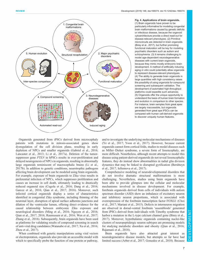
Organoids generated from iPSCs derived from microcephalypatients with mutations in mitosis-associated genes showdysregulation of the cell division plane, resulting in earlydepletion of NPCs and smaller organoids (Gabriel et al., 2016;Lancaster et al., 2013; Li et al., 2017a). Deletion of the tumor-suppressor gene PTEN in hPSCs results in over-proliferation anddelayed neurogenesis of NPCs in organoids, resulting in abnormallylarge organoids reminiscent of macrocephalic brains (Li et al.,2017b). In addition to genetic conditions, neurotrophic pathogensaffecting brain development can be modeled using brain organoids.For example, exposure of brain organoids to Zika virus results inpreferential infection of NPCs, which suppresses proliferation andcauses an increase in cell death, ultimately leading to drasticallyreduced organoid size (Cugola et al., 2016; Dang et al., 2016;Garcez et al., 2016; Qian et al., 2017, 2016). Moreover, suchinfected cortical organoids display a series of characteristicsidentified in congenital Zika syndrome, including thinning of theneuronal layer, disruption of apical surface adherens junctions anddilation of the ventricular lumen, offering direct evidence for thecausal relationship between embryonic Zika exposure andneurological disorders (Ming et al., 2016; Nguyen et al., 2016;Qian et al., 2017, 2016; Rasmussen et al., 2016; Wen et al., 2017;Zhang et al., 2016). Subsequently, brain organoids have been usedas platforms for validating results of compound screening in searchof anti-viral drug candidates (Watanabe et al., 2017; Xu et al., 2016;Zhou et al., 2017).When combined with genetic manipulations using viral vectors
or electroporation, organoids also provide an accessible model withwhich to specifically probe the function of one protein or pathway,
and to investigate the underlying molecular mechanisms of diseases(Ye et al., 2017; Yoon et al., 2017). However, because currentorganoids cannot form cortical folds, studies to model diseases suchas Miller–Dieker syndrome, a severe form of lissencephaly, aremore difficult. Nonetheless, although recent attempts to model thisdisease using patient-derived organoids do not reveal lissencephalicfeatures, they do instead show abnormalities in radial glia divisiondynamics that may be linked to disrupted gyrification (Bershteynet al., 2017; Iefremova et al., 2017).
Comprehensive modeling of neurodevelopmental disorders thatdo not involve dramatic structural malformations is morechallenging. Nevertheless, studies using brain organoids havebeen able to provide glimpses into the cellular and molecularmechanisms involved in disease development. For example,forebrain organoids derived from cells of individuals with autismspectrum disorder (ASD) show an imbalance of excitatory neuronand inhibitory neuron proportions, which is associated withoverexpression of the forebrain transcription factor FOXG1 (Choiet al., 2017; Mariani et al., 2015). Defects in interneuron migrationare observed in dorsal-ventral forebrain ‘assembloids’ generatedfrom iPSCs derived from individuals with Timothy syndrome thatharbor a mutation in the L-type calcium channel gene (Birey et al.,2017). Moreover, hypothalamic organoids containing nuclei-likeclusters of neuropeptidergic neuron subtypes are promising modelsfor studying metabolic disorders and obesity (Qian et al., 2016;Rajamani et al., 2018).
Brain organoids have also attracted great interest asneurodegenerative disease models, but attempts so far have hadlimited success (Arber et al., 2017; Gonzalez et al., 2018). Because
Automated read-out
4. Compound screening
Functional maturation
2. Major psychiatric disorders
1. Congenital brain malformations
Structural recapitulation
3. Neurodegenerative diseases
5. Human evolution
Induced aging
Inter-species comparison
Fig. 4. Applications of brain organoids.(1) Brain organoids have proven to beparticularly informative for modeling congenitalbrain malformations caused by genetic deficitsor infectious disease, because the organoidcytoarchitecture provide a direct read-out fordisease-relevant phenotypes. (2) Primitivemicrocircuits are detected in brain organoids(Birey et al., 2017), but further promotingfunctional maturation will be key for modelingpsychiatric disorders such as autism andschizophrenia. (3) It remains challenging tomodel age-dependent neurodegenerativediseases with current brain organoids,because they mimic mostly embryonic braindevelopment. A method of artificially inducingaging in vitro could potentially allow organoidsto represent disease-relevant phenotypes.(4) The ability to generate brain organoids inlarge quantities with high consistency raisesthe possibility of using organoids for compoundscreening and subsequent validation. Thedevelopment of automated high-throughputplatforms could expedite such advances.(5) Organoids offer the unique opportunity tounderstand the basis of human brain formationand evolution in comparison to other species.For instance, brain samples from great apesare largely inaccessible, but organoidsgenerated from great ape iPSCs can becompared with human cell-derived organoidsto discover uniquely human features.
7
REVIEW Development (2019) 146, dev166074. doi:10.1242/dev.166074
DEVELO
PM
ENT

most neurodegenerative diseases are late onset and age related, brainorganoids mimicking embryonic brain development may notrobustly reproduce disease-relevant endophenotypes. Although notstrictly classified as organoids, human neural cultures in ECM gel orneurospheres derived from individuals with Alzheimer’s disease(AD)have been shown to recapitulateAD-like pathologies, includingamyloid aggregation, hyperphosphorylated tau protein, andendosome abnormalities (Choi et al., 2014; Jorfi et al., 2018; Rajaet al., 2016). Midbrain organoids containing tyrosine hydroxylase(TH)-positive dopaminergic neurons, when combined withpharmacological treatment to induce neurodegeneration, maypotentially serve as a relevant model for Parkinson’s disease and acellular source for cell-replacement therapies (Monzel et al., 2017;Qian et al., 2016; Xiao et al., 2016).In summary, although brain organoids clearly provide a unique
platform for understanding complicated neurological diseases, it isimportant to keep in mind that the current organoids are verysimplistic and immature, and the knowledge obtained from themmay be biased owing to their in vitro nature. Further development oftechnologies, such as accelerating functional maturation andincorporating other cell and tissue types, will push the fieldtowards more comprehensive and faithful models.
Understanding human evolutionThe human brain, especially the neocortex, has evolved to bedisproportionally larger compared with that of other species (Rakic,2009; Sousa et al., 2017). A better understanding of this species-dependent difference will help us to understand better themechanisms that make humans unique, and may aid thetranslation of findings made in animal models into therapeuticstrategies (Fig. 3). Brain organoids open up new possibilities forcomparative studies of the brain across species. Moreover, brainsamples from human’s close relatives, great apes, are almostimpossible to obtain, and iPSC-derived organoids may be the onlyfeasible method to gain access to great ape brain development.Indeed, a comparison between human, chimpanzee and macaqueiPSC-derived organoids has revealed differences in the proliferativedynamics of NPCs that lead to different neurogenesis outputs,which may explain the neocortical size differences among primates(Otani et al., 2016). Consistent with this finding, scRNA-seq studiesdetected upregulation of genes involved in proliferation of radialglia in human organoids compared with chimpanzee organoids(Kronenberg et al., 2018; Mora-Bermudez et al., 2016). Onelimitation of this approach is that the differentiation protocolfollowed for both human and non-human primate organoids hasbeen developed and optimized exclusively for hPSCs, and theobserved differences in non-human organoids might simply be aresult of suboptimal differentiation conditions, rather than species-dependent distinctions. Moreover, as human pregnancy is longerthan that in many species, and neurodevelopment continues untiladulthood, the difference in the temporal control of braindevelopment is difficult to gauge when performing inter-speciescomparison. Establishing hallmarks for developmental stages thatare shared across primate species could provide a more solid basisfor such comparisons.With the help of genome-editing technologies, it is now possible
to investigate the function of human-specific genes or geneticvariants in organoid models (Ran et al., 2013). Two recent studiesidentified high expression of human-specific paralogs of theNOTCH2 receptor, NOTCH2NL genes in human radial glia,suggesting important functions for these genes in neurogenesis inhumans (Fiddes et al., 2018; Suzuki et al., 2018). Indeed, deletion of
NOTCH2NL results in premature neurogenesis and microcephaly-like deficits in hESC-derived organoids; on the other hand, ectopicexpression of NOTCH2NL in mouse ESCs gives rise to organoidswith delayed neural differentiation (Fiddes et al., 2018; Suzuki et al.,2018). Future studies could further exploit genome engineering toknock-in human genes into iPSCs of other species and generate‘humanized’ organoids, in order to understand the evolutionarysignificance of human-specific genetic variants.
Conclusions and perspectivesTremendous breakthroughs have been made in brain organoidtechnologies in the past few years. Although these brain organoidsfaithfully recapitulate a number of key features of the human brain,they are not perfect replica, and overcoming current limitations toengineer ‘better’ organoids will greatly expand our ability toinvestigate human brain development and disorders. The definitionof ‘better’ organoids may vary based on specific applications, butthe benchmark is to make organoids that more faithfully recapitulatefeatures of the human brain. Therefore, continued systematiccharacterization of human fetal and postnatal brain tissue isfundamental. Despite the difficulty in collecting human brainsamples with consistent quality, analyses of these samples havecontinued to expand our knowledge of normal and diseased humanbrains (Calvet et al., 2016; Hansen et al., 2010; Moore et al., 2017;Nowakowski et al., 2016; Ozair et al., 2018; Pollen et al., 2015;Stoner et al., 2014). Remarkably, large-scale single celltranscriptome profiling has resulted in unprecedented resolution,revealing the extent of cellular diversity and molecular identitiesof the embryonic human brain (Nowakowski et al., 2017;Zhong et al., 2018). Pioneered by The Allen Institute for BrainScience, the eventual establishment of a comprehensive humanbrain atlas containing immunohistology, in situ hybridization andtranscriptomics data will provide an invaluable resource fororganoid engineers (Ding et al., 2017).
The small size of current organoids remains the fundamentallimiting factor that prohibits us from using organoids to fullyrecapitulate late stages of human brain development. The viablethickness of organoids is restricted by the physical distance overwhich oxygen and nutrients can diffuse from the surface, which istypically less than400 µm (Rambani et al., 2009).BecauseNPCswithhigh metabolic demands are often located in the most interior part ofthe cortical structures, they are the first to succumb to the diffusionlimit, and neurogenesis cannot be sustained in long-term organoidcultures (Fig. 3). The formation of a CP with six distinct layers andcortical folding is therefore still out of reach.Methods to create a morepermissive environment to alleviate this condition include the use ofspinning bioreactors or orbital shakers to enhance diffusion, and toprovide a higher oxygen concentration in the incubator (Kadoshimaet al., 2013; Lancaster et al., 2017;Qian et al., 2016). Current organoidpreparations also lack vascular cells and, although endothelial cellscan be exogenously incorporated into brain organoids, this alone isinsufficient to improve long-term culture as the primitive endothelialnetwork that forms is not functional (Pham et al., 2018). Future workto incorporate biomaterials and microfluidic systems could thereforebe used to engineer vascular-like networks with perfusion to supplythe organoid interior with adequate oxygen and nutrients. Thetransplantation of organoids into animals, allowing the hostvasculature to grow into the organoid graft, has also proven to be apromising approach to promote organoid viability and maturation(Mansour et al., 2018).
The fact that brain organoids dynamically mimic the temporalprogression of human brain development is both an advantage and a
8
REVIEW Development (2019) 146, dev166074. doi:10.1242/dev.166074
DEVELO
PM
ENT

disadvantage for researchers. On the one hand, brain organoids ofdifferent ages recapitulate their corresponding in vivo counterparts,offering researchers a versatile platform to probe differentdevelopmental stages. On the other hand, from a practical point ofview, brain organoids take a long time to grow and mature, raisingthe cost and hindering the efficiency of experiments. Methods forspeeding up the maturation process thus need to be explored. Forinstance, the use of NOTCH inhibitors is very effective inaccelerating neuronal differentiation in vitro and could be appliedto brain organoids. But this poses a dilemma because such amanipulation could interfere with the intrinsic program of neuraldifferentiation, making the resulting organoids no longerrepresentative of their in vivo counterparts (Borghese et al., 2010).Furthermore, to faithfully model age-dependent neurodegenerativediseases requires inducing aging in organoids by pharmacologicalor genetic methods (Studer et al., 2015). One such strategy forinducing aging-related features in human iPSC-derived organoids,used to model PD, involves the expression of progerin, a truncatedform of lamin A associated with premature aging (Miller et al.,2013). Telomere shortening induced via downregulation oftelomerase has also been shown to result in age-related anddisease-relevant phenotypes in human iPSC-derived neurons (Veraet al., 2016). However, whether these ‘induced’ aging eventsaccurately reflect the aging process that occurs naturally in vivoremains to be determined.The introduction of fused organoids or ‘assembloid’ systems
provides a path to a modular design approach to investigate inter-brain-region and inter-organ crosstalk. Assembly of corticalorganoids and organoids with subcortical identities, such as thethalamus, may offer insights into the development of corticofugalprojections of deep layer cortical neurons (Pasca, 2018). Morecomplex assembloid systems composed of three or more brainregions are feasible, and the ultimate goal is the assembly of awhole-brain organoid for comprehensive modeling of braindevelopment and function. The combination of organoids fromdifferent tissue types could also capture the interface between thenervous system and other organs. Such is the case for the recentlyreported hPSC-derived intestinal organoids containing neural crestcells, which self-organize to resemble the enteric nervous systemand produce rhythmic waves to regulate contraction of the organoids(Workman et al., 2017). Moreover, oncogene manipulation usingCRISPR/Cas9 can be applied to initiate tumorigenesis in humanbrain organoids as an innovative approach to model brain tumors(Bian et al., 2018; Ogawa et al., 2018). Alternatively, brain tumororganoids can be generated from primary glioblastoma specimensthat are dissected into smaller pieces and cultured in a 3Denvironment (Hubert et al., 2016). The subsequent fusion of thesetumor organoids with normal brain organoids may create a scalablein vitro model for cancer metastasis, providing the means to screenfor therapies that specifically block cancer cells’ invasion intosurrounding tissue.Lastly, it should be noted that organoid differentiation protocols
that rely on the self-organization of hPSCs, and stochasticity in theirspontaneous differentiation, lead to inherently variable outcomes.Therefore, unlike organs that arise from the precisely controlledprocess of in vivo organogenesis, no two organoids are identical. Toimprove quality control, variables in the system should beeliminated wherever possible (Jabaudon and Lancaster, 2018).Feeder-dependent hPSC cultures are more technique dependent,and properties of each hPSC line are sometimes inconsistent. A shiftto using feeder-free hPSC culture is likely to significantly improvereproducibility across laboratories and cell lines (Lancaster et al.,
2017; Nakagawa et al., 2014; Yoon et al., 2019). The use of variableingredients, such as animal-derived ECM (Matrigel), in cultureprotocols should also be minimized, and recombinant growthfactors should be replaced by small molecules whenever applicable(Cruz-Acuña et al., 2017; Gjorevski et al., 2016). Furthermore,newly developed organoid generation methods need to bequantitatively characterized to show consistent results withmultiple hPSC lines and independent batches before they areready for publication. As mentioned above, the development of aminiaturized multi-well spinning bioreactor has enabled efficientoptimization of organoid protocols and scalable organoidproduction (Qian et al., 2016), but current methods for organoidcharacterization are labor intensive and prevent scaling-up oforganoid experiments. Moving forward, the development ofsystems with automated read-outs for high-throughput analyseswill be instrumental for transformation of organoid models intohigh-throughput platforms suitable for compound screening anddrug discovery.
Together, the rapid advances in brain organoid technologies haveopened up new avenues for gaining a better understanding of humanbrain development, function, evolution and disorders. The brainorganoid field has made exciting steps to empower researchers withnew tools to address questions that are difficult to address in othermodel systems, but there remains a long way to go towardsobtaining a more faithful in vitro representation of the developinghuman brain. It is important to keep in mind that no model is perfect.Only through synergy across different model systems can we trulygain knowledge that will light the path to overcoming neurologicaldiseases.
AcknowledgementsWe would like to thank members of the Ming and Song laboratories for commentsand suggestions.
Competing interestsThe authors declare no competing or financial interests.
FundingThe research in the authors’ laboratories was supported by grants from the NationalInstitutes of Health (R37NS047344, P01NS097206, U19MH106434 to H.S. andR01MH105128, R35NS097370, U19AI131130 to G.M.), The Simons Foundation(575050 to H.S.) and The Dr Miriam and Sheldon G. Adelson Medical ResearchFoundation (to G.M.). Deposited in PMC for release after 12 months.
ReferencesAbud, E. M., Ramirez, R. N., Martinez, E. S., Healy, L. M., Nguyen, C. H. H.,
Newman, S. A., Yeromin, A. V., Scarfone, V. M., Marsh, S. E., Fimbres, C. et al.(2017). iPSC-Derived human microglia-like cells to study neurological diseases.Neuron 94, 278-293.e279. doi:10.1016/j.neuron.2017.03.042
Anderson, S. A., Eisenstat, D. D., Shi, L. and Rubenstein, J. L. (1997).Interneuron migration from basal forebrain to neocortex: dependence on Dlxgenes. Science 278, 474-476. doi:10.1126/science.278.5337.474
Arber, C., Lovejoy, C. and Wray, S. (2017). Stem cell models of Alzheimer’sdisease: progress and challenges. Alzheimers Res. Ther. 9, 42. doi:10.1186/s13195-017-0268-4
Bae, B.-I., Tietjen, I., Atabay, K. D., Evrony, G. D., Johnson, M. B., Asare, E.,Wang, P. P., Murayama, A. Y., Im, K., Lisgo, S. N. et al. (2014). Evolutionarilydynamic alternative splicing of GPR56 regulates regional cerebral corticalpatterning. Science 343, 764-768. doi:10.1126/science.1244392
Bagley, J. A., Reumann, D., Bian, S., Levi-Strauss, J. andKnoblich, J. A. (2017).Fused cerebral organoids model interactions between brain regions. Nat.Methods 14, 743-751. doi:10.1038/nmeth.4304
Bayly, P. V., Taber, L. A. and Kroenke, C. D. (2014). Mechanical forces in cerebralcortical folding: a review of measurements and models. J. Mech. Behav. Biomed.Mater 29, 568-581. doi:10.1016/j.jmbbm.2013.02.018
Bershteyn, M., Nowakowski, T. J., Pollen, A. A., Di Lullo, E., Nene, A.,Wynshaw-Boris, A. and Kriegstein, A. R. (2017). Human iPSC-derived cerebralorganoidsmodel cellular features of lissencephaly and reveal prolongedmitosis ofouter radial glia. Cell Stem Cell 20, 435-449. doi:10.1016/j.stem.2016.12.007
9
REVIEW Development (2019) 146, dev166074. doi:10.1242/dev.166074
DEVELO
PM
ENT

Bian, S., Repic, M., Guo, Z. M., Kavirayani, A., Burkard, T., Bagley, J. A.,Krauditsch, C. and Knoblich, J. A. (2018). Genetically engineered cerebralorganoids model brain tumor formation. Nat. Methods 15, 631. doi:10.1038/s41592-018-0070-7
Birey, F., Andersen, J., Makinson, C. D., Islam, S., Wei, W., Huber, N., Fan, H. C.,Metzler, K. R. C., Panagiotakos, G., Thom, N. et al. (2017). Assembly offunctionally integrated human forebrain spheroids. Nature 15, 631-639. doi:10.1038/nature22330
Borghese, L., Dolezalova, D., Opitz, T., Haupt, S., Leinhaas, A., Steinfarz, B.,Koch, P., Edenhofer, F., Hampl, A. and Brustle, O. (2010). Inhibition of notchsignaling in human embryonic stem cell-derived neural stem cells delays G1/Sphase transition and accelerates neuronal differentiation in vitro and in vivo. StemCells 28, 955-964. doi:10.1002/stem.408
Borrell, V. (2018). How cells fold the cerebral cortex. J. Neurosci. 38, 776-783.doi:10.1523/JNEUROSCI.1106-17.2017
Bystron, I., Blakemore, C. and Rakic, P. (2008). Development of the humancerebral cortex: boulder committee revisited. Nat. Rev. Neurosci. 9, 110-122.doi:10.1038/nrn2252
Calvet, G., Aguiar, R. S., Melo, A. S. O., Sampaio, S. A., de Filippis, I., Fabri, A.,Araujo, E. S., de Sequeira, P. C., de Mendonca, M. C., de Oliveira, L. et al.(2016). Detection and sequencing of Zika virus from amniotic fluid of fetuses withmicrocephaly in Brazil: a case study. Lancet Infect. Dis. 16, 653-660. doi:10.1016/S1473-3099(16)00095-5
Camp, J. G., Badsha, F., Florio, M., Kanton, S., Gerber, T., Wilsch-Brauninger,M., Lewitus, E., Sykes, A., Hevers, W., Lancaster, M. et al. (2015). Humancerebral organoids recapitulate gene expression programs of fetal neocortexdevelopment. Proc. Natl. Acad. Sci. USA 112, 15672-15677. doi:10.1073/pnas.1520760112
Caronia-Brown, G., Yoshida, M., Gulden, F., Assimacopoulos, S. and Grove,E. A. (2014). The cortical hem regulates the size and patterning of neocortex.Development 141, 2855-2865. doi:10.1242/dev.106914
Chambers, D. and Fishell, G. (2006). Functional genomics of early cortexpatterning. Genome Biol. 7, 202. doi:10.1186/gb-2006-7-1-202
Chen, H. I., Song, H. andMing, G. L. (2018). Applications of human brain organoidsto clinical problems. Dev. Dyn. 248, 53-64. doi:10.1002/dvdy.24662
Chenn, A. andWalsh, C. A. (2002). Regulation of cerebral cortical size by control ofcell cycle exit in neural precursors. Science 297, 365-369. doi:10.1126/science.1074192
Choi, S. H., Kim, Y. H., Hebisch, M., Sliwinski, C., Lee, S., D’Avanzo, C., Chen,H., Hooli, B., Asselin, C., Muffat, J. et al. (2014). A three-dimensional humanneural cell culture model of Alzheimer’s disease. Nature 515, 274-278. doi:10.1038/nature13800
Choi, H., Song, J., Park, G. and Kim, J. (2017). Modeling of autism using organoidtechnology. Mol. Neurobiol. 54, 7789-7795. doi:10.1007/s12035-016-0274-8
Cruz-Acun a, R., Quiros, M., Farkas, A. E., Dedhia, P. H., Huang, S., Siuda, D.,Garcıa-Hernandez, V., Miller, A. J., Spence, J. R., Nusrat, A. et al. (2017).Synthetic hydrogels for human intestinal organoid generation and colonic woundrepair. Nat. Cell Biol. 19, 1326-1335. doi:10.1038/ncb3632
Cugola, F. R., Fernandes, I. R., Russo, F. B., Freitas, B. C., Dias, J. L.,Guimaraes, K. P., Benazzato, C., Almeida, N., Pignatari, G. C., Romero, S.et al. (2016). The Brazilian Zika virus strain causes birth defects in experimentalmodels. Nature 534, 267-271. doi:10.1038/nature18296
Dang, J., Tiwari, S. K., Lichinchi, G., Qin, Y., Patil, V. S., Eroshkin, A. M. andRana, T. M. (2016). Zika virus depletes neural progenitors in human cerebralorganoids through activation of the innate immune receptor TLR3. Cell Stem Cell19, 258-265. doi:10.1016/j.stem.2016.04.014
Danjo, T., Eiraku, M., Muguruma, K., Watanabe, K., Kawada, M., Yanagawa, Y.,Rubenstein, J. L. and Sasai, Y. (2011). Subregional specification of embryonicstem cell-derived ventral telencephalic tissues by timed and combinatorytreatment with extrinsic signals. J. Neurosci. 31, 1919-1933. doi:10.1523/JNEUROSCI.5128-10.2011
De Clercq, S., Keruzore, M., Desmaris, E., Pollart, C., Assimacopoulos, S.,Preillon, J., Ascenzo, S., Matson, C. K., Lee, M., Nan, X. et al. (2018). DMRT5together with DMRT3 directly controls hippocampus development and neocorticalarea map formation. Cereb. Cortex 28, 493-509. doi:10.1093/cercor/bhw384
Del Toro, D., Ruff, T., Cederfjall, E., Villalba, A., Seyit-Bremer, G., Borrell, V. andKlein, R. (2017). Regulation of cerebral cortex folding by controlling neuronalmigration via FLRT adhesion molecules. Cell 169, 621-635.e616. doi:0.1016/j.cell.2017.04.012
Ding, S.-L., Royall, J. J., Sunkin, S. M., Ng, L., Facer, B. A. C., Lesnar, P.,Guillozet-Bongaarts, A., McMurray, B., Szafer, A., Dolbeare, T. A. et al. (2017).Comprehensive cellular-resolution atlas of the adult human brain. J. Comp.Neurol. 525, 407. doi:10.1002/cne.24130
Eiraku, M., Watanabe, K., Matsuo-Takasaki, M., Kawada, M., Yonemura, S.,Matsumura, M., Wataya, T., Nishiyama, A., Muguruma, K. and Sasai, Y.(2008). Self-organized formation of polarized cortical tissues from ESCs and itsactive manipulation by extrinsic signals. Cell Stem Cell 3, 519-532. doi:10.1016/j.stem.2008.09.002
Elsen, G. E., Hodge, R. D., Bedogni, F., Daza, R. A. M., Nelson, B. R., Shiba, N.,Reiner, S. L. and Hevner, R. F. (2013). The protomap is propagated to cortical
plate neurons through an Eomes-dependent intermediate map. Proc. Natl. Acad.Sci. USA 110, 4081-4086. doi:10.1073/pnas.1209076110
Fiddes, I. T., Lodewijk, G. A., Mooring, M., Bosworth, C. M., Ewing, A. D.,Mantalas, G. L., Novak, A. M., van denBout, A., Bishara, A., Rosenkrantz, J. L.et al. (2018). Human-specific NOTCH2NLgenes affect notch signaling and corticalneurogenesis. Cell 173, 1356-1369.e1322. doi:10.1016/j.cell.2018.03.051
Florio, M., Albert, M., Taverna, E., Namba, T., Brandl, H., Lewitus, E., Haffner, C.,Sykes, A., Wong, F. K., Peters, J. et al. (2015). Human-specific geneARHGAP11B promotes basal progenitor amplification and neocortexexpansion. Science 347, 1465-1470. doi:10.1126/science.aaa1975
Gabriel, E., Wason, A., Ramani, A., Gooi, L. M., Keller, P., Pozniakovsky, A.,Poser, I., Noack, F., Telugu, N. S., Calegari, F. et al. (2016). CPAP promotestimely cilium disassembly to maintain neural progenitor pool. EMBO J. 35,803-819. doi:10.15252/embj.201593679
Garcez, P. P., Loiola, E. C., Madeiro da Costa, R., Higa, L. M., Trindade, P.,Delvecchio, R., Nascimento, J. M., Brindeiro, R., Tanuri, A. and Rehen, S. K.(2016). Zika virus impairs growth in human neurospheres and brain organoids.Science 352, 816-818. doi:10.1126/science.aaf6116
Gjorevski, N., Sachs, N., Manfrin, A., Giger, S., Bragina, M. E., Ordon ez-Moran,P., Clevers, H. and Lutolf, M. P. (2016). Designer matrices for intestinal stem celland organoid culture. Nature 539, 560-564. doi:10.1038/nature20168
Gonzalez, C., Armijo, E., Bravo-Alegria, J., Becerra-Calixto, A., Mays, C. E. andSoto, C. (2018). Modeling amyloid beta and tau pathology in human cerebralorganoids. Mol. Psychiatry 23, 2363-2374. doi:10.1038/s41380-018-0229-8
Hansen, D. V., Lui, J. H., Parker, P. R. L. and Kriegstein, A. R. (2010). Neurogenicradial glia in the outer subventricular zone of human neocortex. Nature 464,554-561. doi:10.1038/nature08845
Hansen, D. V., Lui, J. H., Flandin, P., Yoshikawa, K., Rubenstein, J. L., Alvarez-Buylla, A. and Kriegstein, A. R. (2013). Non-epithelial stem cells and corticalinterneuron production in the human ganglionic eminences. Nat. Neurosci. 16,1576-1587. doi:10.1038/nn.3541
Hubert, C. G., Rivera, M., Spangler, L. C., Wu, Q. L., Mack, S. C., Prager, B. C.,Couce, M., McLendon, R. E., Sloan, A. E. and Rich, J. N. (2016). A three-dimensional organoid culture system derived from human glioblastomasrecapitulates the hypoxic gradients and cancer stem cell heterogeneity oftumors found in vivo. Cancer Res. 76, 2465-2477. doi:10.1158/0008-5472.CAN-15-2402
Iefremova, V., Manikakis, G., Krefft, O., Jabali, A., Weynans, K., Wilkens, R.,Marsoner, F., Brandl, B., Muller, F. J., Koch, P. et al. (2017). An Organoid-based model of cortical development identifies non-cell-autonomous defects inWnt signaling contributing to miller-dieker syndrome. Cell Rep. 19, 50-59. doi:10.1016/j.celrep.2017.03.047
Ip, B. K., Bayatti, N., Howard, N. J., Lindsay, S. and Clowry, G. J. (2011). Thecorticofugal neuron-associated genes ROBO1, SRGAP1, and CTIP2 exhibit ananterior to posterior gradient of expression in early fetal human neocortexdevelopment. Cereb. Cortex 21, 1395-1407. doi:10.1093/cercor/bhq219
Jabaudon, D. and Lancaster, M. (2018). Exploring landscapes of brainmorphogenesis with organoids. Development 145, dev172049. doi:10.1242/dev.172049
Jakovcevski, I., Mayer, N. and Zecevic, N. (2011). Multiple origins of humanneocortical interneurons are supported by distinct expression of transcriptionfactors. Cereb. Cortex 21, 1771-1782. doi:10.1093/cercor/bhq245
Jeong, H. and Tiwari, V. K. (2018). Exploring the complexity of corticaldevelopment using single-cell transcriptomics. Front. Neurosci. 12, 31. doi:10.3389/fnins.2018.00031
Jo, J., Xiao, Y., Sun, A. X., Cukuroglu, E., Tran, H.-D., Goke, J., Tan, Z. Y., Saw,T. Y., Tan, C.-P., Lokman, H. et al. (2016). Midbrain-like organoids from humanpluripotent stem cells contain functional dopaminergic and neuromelanin-producing neurons. Cell Stem Cell 19, 248-257. doi:10.1016/j.stem.2016.07.005
Jorfi, M., D’Avanzo, C., Tanzi, R. E., Kim, D. Y. and Irimia, D. (2018). Humanneurospheroid arrays for in vitro studies of Alzheimer’s disease. Sci. Rep. 8, 2450.doi:10.1038/s41598-018-20436-8
Kadoshima, T., Sakaguchi, H., Nakano, T., Soen, M., Ando, S., Eiraku, M. andSasai, Y. (2013). Self-organization of axial polarity, inside-out layer pattern, andspecies-specific progenitor dynamics in human ES cell-derived neocortex. Proc.Natl. Acad. Sci. USA 110, 20284-20289. doi:10.1073/pnas.1315710110
Karzbrun, E., Kshirsagar, A., Cohen, S. R., Hanna, J. H. and Reiner, O. (2018).Human brain organoids on a chip reveal the physics of folding. Nat. Phys. 14,515-522. doi:10.1038/s41567-018-0046-7
Kronenberg, Z. N., Fiddes, I. T., Gordon, D., Murali, S., Cantsilieris, S.,Meyerson, O. S., Underwood, J. G., Nelson, B. J., Chaisson, M. J. P.,Dougherty, M. L. et al. (2018). High-resolution comparative analysis of great apegenomes. Science 360, eaar6343. doi:10.1126/science.aar6343
Lancaster, M. A. and Knoblich, J. A. (2014). Generation of cerebral organoids fromhumanpluripotent stemcells.Nat.Protoc.9, 2329-2340. doi:10.1038/nprot.2014.158
Lancaster, M. A., Renner, M., Martin, C.-A., Wenzel, D., Bicknell, L. S., Hurles,M. E., Homfray, T., Penninger, J. M., Jackson, A. P. and Knoblich, J. A. (2013).Cerebral organoids model human brain development and microcephaly. Nature501, 373-379. doi:10.1038/nature12517
10
REVIEW Development (2019) 146, dev166074. doi:10.1242/dev.166074
DEVELO
PM
ENT

Lancaster, M. A., Corsini, N. S., Wolfinger, S., Gustafson, E. H., Phillips, A. W.,Burkard, T. R., Otani, T., Livesey, F. J. and Knoblich, J. A. (2017). Guided self-organization and cortical plate formation in human brain organoids. Nat.Biotechnol. 35, 659-666. doi:10.1038/nbt.3906
Letinic, K., Zoncu, R. and Rakic, P. (2002). Origin of GABAergic neurons in thehuman neocortex. Nature 417, 645-649. doi:10.1038/nature00779
Lewitus, E., Kelava, I. and Huttner, W. B. (2013). Conical expansion of the outersubventricular zone and the role of neocortical folding in evolution anddevelopment. Front. Hum. Neurosci. 7, 424. doi:10.3389/fnhum.2013.00424
Lewitus, E., Kelava, I., Kalinka, A. T., Tomancak, P. and Huttner, W. B. (2016).Comment on “Cortical folding scales universally with surface area and thickness,not number of neurons”. Science 351, 825. doi:10.1126/science.aad2029
Li, R., Sun, L., Fang, A., Li, P., Wu, Q. and Wang, X. (2017a). Recapitulatingcortical development with organoid culture in vitro and modeling abnormalspindle-like (ASPM related primary) microcephaly disease. Protein Cell 8,823-833. doi:10.1007/s13238-017-0479-2
Li, Y., Muffat, J., Omer, A., Bosch, I., Lancaster, M. A., Sur, M., Gehrke, L.,Knoblich, J. A. and Jaenisch, R. (2017b). Induction of expansion and folding inhuman cerebral organoids. Cell Stem Cell 20, 385-396.e383. doi:10.1016/j.stem.2016.11.017
Lui, J. H., Hansen, D. V. and Kriegstein, A. R. (2011). Development and evolutionof the human neocortex. Cell 146, 18-36. doi:10.1016/j.cell.2011.06.030
Luo, C., Lancaster, M. A., Castanon, R., Nery, J. R., Knoblich, J. A. and Ecker,J. R. (2016). Cerebral organoids recapitulate epigenomic signatures of the humanfetal brain. Cell Rep. 17, 3369-3384. doi:10.1016/j.celrep.2016.12.001
Ma, T., Wang, C., Wang, L., Zhou, X., Tian, M., Zhang, Q., Zhang, Y., Li, J., Liu, Z.,Cai, Y. et al. (2013). Subcortical origins of human and monkey neocorticalinterneurons. Nat. Neurosci. 16, 1588-1597. doi:10.1038/nn.3536
Madhavan, M., Nevin, Z. S., Shick, H. E., Garrison, E., Clarkson-Paredes, C.,Karl, M., Clayton, B. L. L., Factor, D. C., Allan, K. C., Barbar, L. et al. (2018).Induction of myelinating oligodendrocytes in human cortical spheroids. Nat.Methods 15, 700-706. doi:10.1038/s41592-018-0081-4
Mansour, A. A. F., Gonçalves, J. T., Bloyd, C. W., Li, H., Fernandes, S., Quang,D., Johnston, S., Parylak, S. L., Jin, X. and Gage, F. H. (2018). An in vivo modelof functional and vascularized human brain organoids. Nat. Biotechnol. 36,432-441. doi:10.1038/nbt.4127
Mariani, J., Coppola, G., Zhang, P., Abyzov, A., Provini, L., Tomasini, L.,Amenduni, M., Szekely, A., Palejev, D., Wilson, M. et al. (2015). FOXG1-dependent dysregulation of GABA/glutamate neuron differentiation in autismspectrum disorders. Cell 162, 375-390. doi:10.1016/j.cell.2015.06.034
Meinhardt, A., Eberle, D., Tazaki, A., Ranga, A., Niesche, M., Wilsch-Brauninger, M., Stec, A., Schackert, G., Lutolf, M. and Tanaka, E. M. (2014).3D reconstitution of the patterned neural tube from embryonic stem cells. StemCell Rep. 3, 987-999. doi:10.1016/j.stemcr.2014.09.020
Miller, J. D., Ganat, Y. M., Kishinevsky, S., Bowman, R. L., Liu, B., Tu, E. Y.,Mandal, P. K., Vera, E., Shim, J.-W., Kriks, S. et al. (2013). Human iPSC-basedmodeling of late-onset disease via progerin-induced aging. Cell Stem Cell 13,691-705. doi:10.1016/j.stem.2013.11.006
Ming, G.-L., Tang, H. and Song, H. (2016). Advances in Zika virus research: stemcell models, challenges, and opportunities. Cell Stem Cell 19, 690-702. doi:10.1016/j.stem.2016.11.014
Molyneaux, B. J., Arlotta, P., Menezes, J. R. L. and Macklis, J. D. (2007).Neuronal subtype specification in the cerebral cortex. Nat. Rev. Neurosci. 8,427-437. doi:10.1038/nrn2151
Monzel, A. S., Smits, L. M., Hemmer, K., Hachi, S., Moreno, E. L., van Wuellen,T., Jarazo, J., Walter, J., Bruggemann, I., Boussaad, I. et al. (2017). Derivationof human midbrain-specific organoids from neuroepithelial stem cells. Stem CellRep. 8, 1144-1154. doi:10.1016/j.stemcr.2017.03.010
Moore, C. A., Staples, J. E., Dobyns, W. B., Pessoa, A., Ventura, C. V., Fonseca,E. B., Ribeiro, E. M., Ventura, L. O., Neto, N. N., Arena, J. F. et al. (2017).Characterizing the pattern of anomalies in congenital Zika syndrome for pediatricclinicians. JAMA Pediatr 171, 288-295. doi:10.1001/jamapediatrics.2016.3982
Mora-Bermudez, F., Badsha, F., Kanton, S., Camp, J. G., Vernot, B., Kohler, K.,Voigt, B., Okita, K., Maricic, T., He, Z. et al. (2016). Differences and similaritiesbetween human and chimpanzee neural progenitors during cerebral cortexdevelopment. Elife 5, e18683. doi:10.7554/eLife.18683
Mota, B. and Herculano-Houzel, S. (2015). Brain structure. Cortical folding scalesuniversally with surface area and thickness, not number of neurons. Science 349,74-77. doi:10.1126/science.aaa9101
Muguruma, K., Nishiyama, A., Kawakami, H., Hashimoto, K. and Sasai, Y.(2015). Self-organization of polarized cerebellar tissue in 3D culture of humanpluripotent stem cells. Cell Rep. 10, 537-550. doi:10.1016/j.celrep.2014.12.051
Nakagawa, M., Taniguchi, Y., Senda, S., Takizawa, N., Ichisaka, T., Asano, K.,Morizane, A., Doi, D., Takahashi, J., Nishizawa, M. et al. (2014). A novelefficient feeder-free culture system for the derivation of human induced pluripotentstem cells. Sci. Rep. 4, 3594. doi:10.1038/srep03594
Nguyen, H. N., Qian, X., Song, H. and Ming, G.-L. (2016). Neural stem cellsattacked by Zika virus. Cell Res. 26, 753-754. doi:10.1038/cr.2016.68
Nowakowski, T. J., Pollen, A. A., Sandoval-Espinosa, C. and Kriegstein, A. R.(2016). Transformation of the radial glia scaffold demarcates two stages of human
cerebral cortex development. Neuron 91, 1219-1227. doi:10.1016/j.neuron.2016.09.005
Nowakowski, T. J., Bhaduri, A., Pollen, A. A., Alvarado, B., Mostajo-Radji, M. A.,Di Lullo, E., Haeussler, M., Sandoval-Espinosa, C., Liu, S. J., Velmeshev, D.et al. (2017). Spatiotemporal gene expression trajectories reveal developmentalhierarchies of the human cortex. Science 358, 1318-1323. doi:10.1126/science.aap8809
Nzou, G., Wicks, R. T., Wicks, E. E., Seale, S. A., Sane, C. H., Chen, A., Murphy,S. V., Jackson, J. D. and Atala, A. J. (2018). Human cortex spheroid with afunctional blood brain barrier for high-throughput neurotoxicity screening anddisease modeling. Sci. Rep. 8, 7413. doi:10.1038/s41598-018-25603-5
Ogawa, J., Pao, G. M., Shokhirev, M. N. and Verma, I. M. (2018). Glioblastomamodel using human cerebral organoids. Cell Rep. 23, 1220-1229. doi:10.1016/j.celrep.2018.03.105
O’Leary, D. D., Chou, S. J. and Sahara, S. (2007). Area patterning of themammalian cortex. Neuron 56, 252-269. doi:10.1016/j.neuron.2007.10.010
Otani, T., Marchetto, M. C., Gage, F. H., Simons, B. D. and Livesey, F. J. (2016).2D and 3D stem cell models of primate cortical development identify species-specific differences in progenitor behavior contributing to brain size.Cell StemCell18, 467-480. doi:10.1016/j.stem.2016.03.003
Ozair, M. Z., Kirst, C., van den Berg, B. L., Ruzo, A., Rito, T. and Brivanlou, A. H.(2018). hPSC Modeling reveals that fate selection of cortical deep projectionneurons occurs in the subplate. Cell Stem Cell 23, 60-73.e66. doi:10.1016/j.stem.2018.05.024
Ozone, C., Suga, H., Eiraku, M., Kadoshima, T., Yonemura, S., Takata, N., Oiso,Y., Tsuji, T. and Sasai, Y. (2016). Functional anterior pituitary generated in self-organizing culture of human embryonic stem cells. Nat. Commun. 7, 10351.doi:10.1038/ncomms10351
Pamies, D., Barreras, P., Block, K., Makri, G., Kumar, A., Wiersma, D.,Smirnova, L., Zang, C., Bressler, J., Christian, K. M. et al. (2017). A humanbrain microphysiological system derived from induced pluripotent stem cells tostudy neurological diseases and toxicity. ALTEX 34, 362-376. doi:10.14573/altex.1609122
Pasca, S. P. (2018). The rise of three-dimensional human brain cultures. Nature553, 437-445. doi:10.1038/nature25032
Pasca, A. M., Sloan, S. A., Clarke, L. E., Tian, Y., Makinson, C. D., Huber, N.,Kim, C. H., Park, J.-Y., O’Rourke, N. A., Nguyen, K. D. et al. (2015). Functionalcortical neurons and astrocytes from human pluripotent stem cells in 3D culture.Nat. Methods 12, 671-678. doi:10.1038/nmeth.3415
Petanjek, Z., Berger, B. and Esclapez, M. (2009). Origins of cortical GABAergicneurons in the cynomolgus monkey. Cereb. Cortex 19, 249-262. doi:10.1093/cercor/bhn078
Pham, M. T., Pollock, K. M., Rose, M. D., Cary, W. A., Stewart, H. R., Zhou, P.,Nolta, J. A. and Waldau, B. (2018). Generation of human vascularized brainorganoids. Neuroreport 29, 588-593. doi:10.1097/WNR.0000000000001014
Phan, D. T. T., Bender, R. H. F., Andrejecsk, J. W., Sobrino, A., Hachey, S. J.,George, S. C. and Hughes, C. C. W. (2017). Blood-brain barrier-on-a-chip:Microphysiological systems that capture the complexity of the blood-centralnervous system interface. Exp. Biol. Med. (Maywood) 242, 1669-1678. doi:10.1177/1535370217694100
Pollen, A. A., Nowakowski, T. J., Chen, J., Retallack, H., Sandoval-Espinosa, C.,Nicholas, C. R., Shuga, J., Liu, S. J., Oldham, M. C., Diaz, A. et al. (2015).Molecular identity of human outer radial glia during cortical development.Cell 163,55-67. doi:10.1016/j.cell.2015.09.004
Qian, X., Nguyen, H. N., Song, M. M., Hadiono, C., Ogden, S. C., Hammack, C.,Yao, B., Hamersky, G. R., Jacob, F., Zhong, C. et al. (2016). Brain-region-specific organoids using mini-bioreactors for modeling ZIKV exposure. Cell 165,1238-1254. doi:10.1016/j.cell.2016.04.032
Qian, X., Nguyen, H. N., Jacob, F., Song, H. and Ming, G. L. (2017). Using brainorganoids to understand Zika virus-induced microcephaly. Development 144,952-957. doi:10.1242/dev.140707
Qian, X., Jacob, F., Song, M. M., Nguyen, H. N., Song, H. and Ming, G. L. (2018).Generation of human brain region-specific organoids using a miniaturizedspinning bioreactor. Nat. Protoc. 13, 565-580. doi:10.1038/nprot.2017.152
Quadrato, G., Nguyen, T., Macosko, E. Z., Sherwood, J. L., Min Yang, S., Berger,D. R., Maria, N., Scholvin, J., Goldman, M., Kinney, J. P. et al. (2017). Celldiversity and network dynamics in photosensitive human brain organoids. Nature545, 48-53. doi:10.1038/nature22047
Radonjic, N. V., Ayoub, A. E., Memi, F., Yu, X., Maroof, A., Jakovcevski, I.,Anderson, S. A., Rakic, P. and Zecevic, N. (2014). Diversity of corticalinterneurons in primates: the role of the dorsal proliferative niche. Cell Rep. 9,2139-2151. doi:10.1016/j.celrep.2014.11.026
Raja, W. K., Mungenast, A. E., Lin, Y. T., Ko, T., Abdurrob, F., Seo, J. and Tsai,L. H. (2016). Self-organizing 3D human neural tissue derived from inducedpluripotent stem cells recapitulate Alzheimer’s disease phenotypes. PLoS ONE11, e0161969. doi:10.1371/journal.pone.0161969
Rajamani, U., Gross, A. R., Hjelm, B. E., Sequeira, A., Vawter, M. P., Tang, J.,Gangalapudi, V., Wang, Y., Andres, A. M., Gottlieb, R. A. et al. (2018). Super-obese patient-derived iPSC hypothalamic neurons exhibit obesogenic signatures
11
REVIEW Development (2019) 146, dev166074. doi:10.1242/dev.166074
DEVELO
PM
ENT

and hormone responses. Cell Stem Cell 22, 698-712.e699. doi:10.1016/j.stem.2018.03.009
Rakic, P. (2009). Evolution of the neocortex: a perspective from developmentalbiology. Nat. Rev. Neurosci. 10, 724-735. doi:10.1038/nrn2719
Rakic, P., Ayoub, A. E., Breunig, J. J. and Dominguez, M. H. (2009). Decision bydivision: making cortical maps. Trends Neurosci. 32, 291-301. doi:10.1016/j.tins.2009.01.007
Rambani, K., Vukasinovic, J., Glezer, A. and Potter, S. M. (2009). Culturing thickbrain slices: an interstitial 3D microperfusion system for enhanced viability.J. Neurosci. Methods 180, 243-254. doi:10.1016/j.jneumeth.2009.03.016
Ran, F. A., Hsu, P. D., Wright, J., Agarwala, V., Scott, D. A. and Zhang, F. (2013).Genome engineering using the CRISPR-Cas9 system.Nat. Protoc. 8, 2281-2308.doi:10.1038/nprot.2013.143
Rasmussen, S. A., Jamieson, D. J., Honein, M. A. and Petersen, L. R. (2016).Zika virus and birth defects–reviewing the evidence for causality. N. Engl. J. Med.374, 1981-1987. doi:10.1056/NEJMsr1604338
Reynolds, B. A., Tetzlaff, W. and Weiss, S. (1992). A multipotent EGF-responsivestriatal embryonic progenitor cell produces neurons and astrocytes. J. Neurosci.12, 4565-4574. doi:10.1523/JNEUROSCI.12-11-04565.1992
Saito, T., Hanai, S., Takashima, S., Nakagawa, E., Okazaki, S., Inoue, T., Miyata,R., Hoshino, K., Akashi, T., Sasaki, M. et al. (2011). Neocortical layer formationof human developing brains and lissencephalies: consideration of layer-specificmarker expression. Cereb. Cortex 21, 588-596. doi:10.1093/cercor/bhq125
Sakaguchi, H., Kadoshima, T., Soen, M., Narii, N., Ishida, Y., Ohgushi, M.,Takahashi, J., Eiraku, M. and Sasai, Y. (2015). Generation of functionalhippocampal neurons from self-organizing human embryonic stem cell-deriveddorsomedial telencephalic tissue. Nat. Commun. 6, 8896. doi:10.1038/ncomms9896
Sasai, Y. (2013). Next-generation regenerative medicine: organogenesis from stemcells in 3D culture. Cell Stem Cell 12, 520-530. doi:10.1016/j.stem.2013.04.009
Sato, T., Vries, R. G., Snippert, H. J., van de Wetering, M., Barker, N., Stange,D. E., van Es, J. H., Abo, A., Kujala, P., Peters, P. J. et al. (2009). Single Lgr5stem cells build crypt-villus structures in vitro without a mesenchymal niche.Nature 459, 262-265. doi:10.1038/nature07935
Sato, T., Stange, D. E., Ferrante, M., Vries, R. G. J., Van Es, J. H., van den Brink,S., van Houdt, W. J., Pronk, A., van Gorp, J., Siersema, P. D. et al. (2011).Long-term expansion of epithelial organoids from human colon, adenoma,adenocarcinoma, and Barrett’s epithelium. Gastroenterology 141, 1762-1772.doi:10.1053/j.gastro.2011.07.050
Sloan, S. A., Darmanis, S., Huber, N., Khan, T. A., Birey, F., Caneda, C., Reimer,R., Quake, S. R., Barres, B. A. and Pasca, S. P. (2017). Human astrocytematuration captured in 3D cerebral cortical spheroids derived from pluripotentstem cells. Neuron 95, 779-790.e776. doi:10.1016/j.neuron.2017.07.035
Sousa, A. M. M., Meyer, K. A., Santpere, G., Gulden, F. O. and Sestan, N. (2017).Evolution of the human nervous system function, structure, and development.Cell170, 226-247. doi:10.1016/j.cell.2017.06.036
Stahl, R., Walcher, T., De Juan Romero, C., Pilz, G. A., Cappello, S., Irmler, M.,Sanz-Aquela, J. M., Beckers, J., Blum, R., Borrell, V. et al. (2013). Trnp1regulates expansion and folding of the mammalian cerebral cortex by control ofradial glial fate. Cell 153, 535-549. doi:10.1016/j.cell.2013.03.027
Stoner, R., Chow, M. L., Boyle, M. P., Sunkin, S. M., Mouton, P. R., Roy, S.,Wynshaw-Boris, A., Colamarino, S. A., Lein, E. S. and Courchesne, E. (2014).Patches of disorganization in the neocortex of children with autism.N. Engl. J. Med. 370, 1209-1219. doi:10.1056/NEJMoa1307491
Studer, L., Vera, E. and Cornacchia, D. (2015). Programming and reprogrammingcellular age in the era of induced pluripotency. Cell Stem Cell 16, 591-600. doi:10.1016/j.stem.2015.05.004
Subramanian, L., Bershteyn, M., Paredes, M. F. and Kriegstein, A. R. (2017).Dynamic behaviour of human neuroepithelial cells in the developing forebrain.Nat. Commun. 8, 14167. doi:10.1038/ncomms14167
Sur, M. and Rubenstein, J. L. (2005). Patterning and plasticity of the cerebralcortex. Science 310, 805-810. doi:10.1126/science.1112070
Suzuki, I. K., Gacquer, D., Van Heurck, R., Kumar, D., Wojno, M., Bilheu, A.,Herpoel, A., Lambert, N., Cheron, J., Polleux, F. et al. (2018). Human-specificNOTCH2NL genes expand cortical neurogenesis through Delta/Notch regulation.Cell 173, 1370-1384.e1316. doi:10.1016/j.cell.2018.03.067
Takahashi, K. and Yamanaka, S. (2006). Induction of pluripotent stem cells frommouse embryonic and adult fibroblast cultures by defined factors. Cell 126,663-676. doi:10.1016/j.cell.2006.07.024
Takahashi, K., Tanabe, K., Ohnuki, M., Narita, M., Ichisaka, T., Tomoda, K. andYamanaka, S. (2007). Induction of pluripotent stem cells from adult humanfibroblasts by defined factors. Cell 131, 861-872. doi:10.1016/j.cell.2007.11.019
Thomson, J. A., Itskovitz-Eldor, J., Shapiro, S. S., Waknitz, M. A., Swiergiel,J. J., Marshall, V. S. and Jones, J. M. (1998). Embryonic stem cell lines derivedfrom human blastocysts. Science 282, 1145-1147. doi:10.1126/science.282.5391.1145
Vera, E., Bosco, N. and Studer, L. (2016). Generating late-onset human iPSC-based disease models by inducing neuronal age-related phenotypes throughtelomerase manipulation. Cell Rep. 17, 1184-1192. doi:10.1016/j.celrep.2016.09.062
Watanabe, M., Buth, J. E., Vishlaghi, N., de la Torre-Ubieta, L., Taxidis, J.,Khakh, B. S., Coppola, G., Pearson, C. A., Yamauchi, K., Gong, D. et al.(2017). Self-Organized cerebral organoids with human-specific features predicteffective drugs to combat Zika virus infection. Cell Rep. 21, 517-532. doi:10.1016/j.celrep.2017.09.047
Wen, Z., Song, H. and Ming, G.-L. (2017). How does Zika virus causemicrocephaly? Genes Dev. 31, 849-861. doi:10.1101/gad.298216.117
Workman,M. J., Mahe, M. M., Trisno, S., Poling, H. M.,Watson, C. L., Sundaram,N., Chang, C. F., Schiesser, J., Aubert, P., Stanley, E. G. et al. (2017).Engineered human pluripotent-stem-cell-derived intestinal tissues with afunctional enteric nervous system. Nat. Med. 23, 49-59. doi:10.1038/nm.4233
Xiang, Y., Tanaka, Y., Patterson, B., Kang, Y. J., Govindaiah, G., Roselaar, N.,Cakir, B., Kim, K. Y., Lombroso, A. P., Hwang, S. M. et al. (2017). Fusion ofregionally specified hPSC-derived organoids models human brain developmentand interneuron migration. Cell Stem Cell 21, 383-398.e387. doi:10.1016/j.stem.2017.07.007
Xiao, B., Ng, H. H., Takahashi, R. and Tan, E. K. (2016). Induced pluripotent stemcells in Parkinson’s disease: scientific and clinical challenges. J. Neurol.Neurosurg. Psychiatry 87, 697-702. doi:10.1136/jnnp-2015-312036
Xu, M., Lee, E. M., Wen, Z., Cheng, Y., Huang, W.-K., Qian, X., Tcw, J.,Kouznetsova, J., Ogden, S. C., Hammack, C. et al. (2016). Identification ofsmall-molecule inhibitors of Zika virus infection and induced neural cell death via adrug repurposing screen. Nat. Med. 22, 1101-1107. doi:10.1038/nm.4184
Ye, F., Kang, E., Yu, C., Qian, X., Jacob, F., Yu, C., Mao, M., Poon, R. Y. C., Kim,J., Song, H. et al. (2017). DISC1 Regulates neurogenesis via modulatingkinetochore attachment of Ndel1/Nde1 during Mitosis. Neuron 96,1041-1054.e1045. doi:10.1016/j.neuron.2017.10.010
Yoon, K. J., Song, G., Qian, X., Pan, J., Xu, D., Rho, H. S., Kim, N. S., Habela, C.,Zheng, L., Jacob, F. et al. (2017). Zika-virus-encoded NS2A disrupts mammaliancortical neurogenesis by degrading adherens junction proteins.Cell Stem Cell 21,349-358.e346. doi:10.1016/j.stem.2017.07.014
Yoon, S.-J., Elahi, L. S., Pasca, A. M., Marton, R. M., Gordon, A., Revah, O.,Miura, Y., Walczak, E. M., Holdgate, G. M., Fan, H. C. et al. (2019). Reliability ofhuman cortical organoid generation. Nat. Methods 16, 75-78. doi:10.1038/s41592-018-0255-0
Yu, X. and Zecevic, N. (2011). Dorsal radial glial cells have the potential to generatecortical interneurons in human but not in mouse brain. J. Neurosci. 31, 2413-2420.doi:10.1523/JNEUROSCI.5249-10.2011
Zembrzycki, A., Perez-Garcia, C. G., Wang, C.-F., Chou, S.-J. and O’Leary,D. D. M. (2015). Postmitotic regulation of sensory area patterning in themammalian neocortex by Lhx2. Proc. Natl. Acad. Sci. USA 112, 6736-6741.doi:10.1073/pnas.1424440112
Zeng, H., Shen, E. H., Hohmann, J. G., Oh, S. W., Bernard, A., Royall, J. J.,Glattfelder, K. J., Sunkin, S. M., Morris, J. A., Guillozet-Bongaarts, A. L. et al.(2012). Large-scale cellular-resolution gene profiling in human neocortex revealsspecies-specific molecular signatures.Cell 149, 483-496. doi:10.1016/j.cell.2012.02.052
Zhang, F., Hammack, C., Ogden, S. C., Cheng, Y., Lee, E. M., Wen, Z., Qian, X.,Nguyen, H. N., Li, Y., Yao, B. et al. (2016). Molecular signatures associated withZIKV exposure in human cortical neural progenitors. Nucleic Acids Res. 44,8610-8620. doi:10.1093/nar/gkw765
Zhong, S., Zhang, S., Fan, X., Wu, Q., Yan, L., Dong, J., Zhang, H., Li, L., Sun, L.,Pan, N. et al. (2018). A single-cell RNA-seq survey of the developmentallandscape of the human prefrontal cortex. Nature 555, 524-528. doi:10.1038/nature25980
Zhou, T., Tan, L., Cederquist, G. Y., Fan, Y., Hartley, B. J., Mukherjee, S.,Tomishima, M., Brennand, K. J., Zhang, Q., Schwartz, R. E. et al. (2017).High-content screening in hPSC-neural progenitors identifies drug candidatesthat inhibit Zika virus infection in fetal-like organoids and adult brain.Cell StemCell21, 274-283.e275. doi:10.1016/j.stem.2017.06.017
Zilles, K., Palomero-Gallagher, N. and Amunts, K. (2013). Development ofcortical folding during evolution and ontogeny. Trends Neurosci. 36, 275-284.doi:10.1016/j.tins.2013.01.006
12
REVIEW Development (2019) 146, dev166074. doi:10.1242/dev.166074
DEVELO
PM
ENT