Embed Size (px)
Citation preview

Brain (1997),120,825–837
Muscle activity adapts to anti-gravity postureduring pedalling in persons with post-strokehemiplegiaD. A. Brown, S. A. Kautz and C. A. Dairaghi
Rehabilitation Research and Development Center, Correspondence to: David A. Brown, Rehabilitation R andV. A. Palo Alto Health Care System, Palo Alto, California, D Center (153), V. A. Palo Alto Health Care System,USA 3801 Miranda Avenue, Palo Alto, CA 94304, USA
SummaryWith hemiplegia following stroke, a person’s movement the work done by each leg and their net positive and negative
components. The EMG was recorded from four leg musclesresponse to anti-gravity posture often appears rigid andinflexible, exacerbating the motor dysfunction. A major (tibialis anterior, medial gastrocnemius, rectus femoris and
biceps femoris). The main result from this study was thatdeterminant of pathological movement in anti-gravitypostures is the failure to adapt muscle-activity patterns impaired plegic leg performance, as measured by net negative
work done by the plegic leg and abnormal early rectusautomatically to changes in posture. The aim of the presentstudy was to determine whether the impaired motor femoris activity, was exacerbated at the most vertical body
orientations. However, contrary to the belief that muscleperformance observed when persons with hemiplegia pedalin a horizontal position is exacerbated at more vertical anti- activity cannot adapt to anti-gravity postures, net positive
work increased appropriately and EMG activity in all musclesgravity body orientations. Twelve healthy elderly subjects and17 subjects with chronic (.6 months) post-stroke hemiplegia showed modulated levels of activity similar to those in elderly
control subjects. These results support the hypothesis thatparticipated in the study. Subjects pedalled a modifiedergometer at different body orientations (from horizontal to increased verticality exacerbates the already impaired
movement performance. Yet, much of the motor response tovertical), maintaining the same workload, cadence, and hipand knee kinematics. Pedal reaction forces, and crank and verticality was flexible and appropriate, given the mechanics
of the task.pedal kinematics, were measured and then used to calculate
Keywords: body orientation; hemiplegia; muscle activity; pedalling
Abbreviations: BF 5 biceps femoris; IEMG5 integrated electromyography; MG5 medial gastrocnemius; RF5 rectusfemoris; TA 5 tibialis anterior
IntroductionCo-ordinating muscular activity to produce a given movement control improves. A major determinant of pathological
movement in anti-gravity postures is the failure to adaptin a variety of anti-gravity postures (e.g. supine, sitting andstanding) is a complex motor control problem that requires muscle-activity patterns automatically to changes in posture
(Brunnstrom, 1970; Bobath, 1978). This failure to adapt mayappropriate integration of sensory information to adapt to theenvironment (Young, 1984). When a person with post-stroke be due to the pathological release of inappropriate postural
responses to anti-gravity body orientations (Bobath, 1978)hemiplegia moves in an anti-gravity posture the movementappears rigid and inflexible compared with movements in and/or to an inability to coordinate muscle activity with
changing sensory information so as to generate the movementgravity-assisted postures. For this reason, clinicians prefer tobegin exercise in gravity-assisted postures and progress task effectively under the altered environmental conditions
(Carr and Shephard, 1987).persons with hemiplegia to anti-gravity postures as movement
© Oxford University Press 1997

826 D. A. Brownet al.
Traditional reflex models have been used to explain normal limb, reciprocal, propulsive movements can be isolated.Because pedalling is functional, safe, and accessible toresponses to static tilting into anti-gravity body orientations
and to explain exaggerated postural responses in persons patients with a wide range of ambulatory function, the bicycleergometer has been used to study bilateral movement patternswith cerebral damage. By tilting neurologically normal human
subjects from horizontal to vertical and eliciting H-reflex in several patient populations, including stroke (Brown andDeBacher, 1987; Rosecrance and Giuliani, 1991; Boormanresponses in soleus motor neuron pools, Chan and Kearney
(1982) (during quiet standing) and Brookeet al. (1995) et al., 1992). For example, Beneckeet al. (1983) foundsignificant relationships between disordered leg muscle(during stepping movements) have demonstrated decreased
soleus motor neuron excitability at increased vertical activation patterns during pedalling and indices of spasticity.More recent work has established direct functionalpositions. They proposed (in light of classical animal
experiments (see Magnus, 1926) that tonic labyrinthine relationships between the degree of abnormal muscleactivation and poor pedalling performance (Brownet al.,reflexes, elicited by otolith receptors, inhibit lower limb
extensor motor neuron excitability during upright stance. 1996b).By studying pedalling and modelling the task mechanics,Although reflex responses to static anti-gravity postures are
demonstrable, they may not contribute to the functional our previous work with neurologically normal subjectsshowed systematically changed joint torque and muscleintegrity of a movement and may not be present in muscle
groups other than the triceps surae. Further, responses to activation patterns as body orientation was altered (Brownet al., 1996a). Changes in muscle activation patterns weretilting in persons with hemiplegia, especially during a
movement task, would demonstrate whether or not a consistent with a neural control strategy that integratessensory information and produces steady-state pedallingsupposedly exaggerated tonic labyrinthine reflex was
operating as a pathological impairment to movement in anti- trajectories consistent with some internal model of themovement.gravity postures.
Contemporary task-oriented models of control suggest that Mechanical measures of pedalling performance can beused to characterize impairment in persons with hemiplegia.responses to anti-gravity postures are the product of the
interaction of many components organized around a In order to pedal at a given cadence and workload thecombined mechanical work done by the two limbs must befundamental movement goal (Carr and Shephard, 1987).
Although neural mechanisms, such as reflex pathways, play sufficient to overcome the resistive load. The total mechanicalwork done by the plegic leg (total work) represents the neta major role in determining muscle-activity patterns,
environmental forces and the musculoskeletal mechanics contribution of the plegic limb and can be positive (the limbassists crank propulsion), negative (the limb resists crankalso contribute to the production of motor patterns to achieve
cyclical and repetitive bipedal and quadrapedal locomotion propulsion) or zero. Since the total work provides a netmeasure for a cyclic movement, and pedalling typically(Hoy and Zernicke, 1985; Capaday and Stein, 1987). For
example, gravity has the potential to influence the control of includes periods of assistance and resistance (Gregoret al.,1991), it is useful to assess the assistance and resistancelimb movements strongly, since it not only affects the sensory
input (Young, 1984), but also alters the task mechanics provided by the leg independently. The net positive workdone by the plegic leg represents the component of the(McMahon, 1984; Davis and Cavanagh, 1993). Therefore,
with hemiplegia, degradation of movement capability mechanical work that propels the crank against the loadand is typically dominated by muscular contributions inoccurring at anti-gravity postures may be partly the result of
an inability to adapt an already impaired muscle-activity neurologically normal subjects (Kautz and Hull, 1993). Thenet negative work done by the plegic leg represents thepattern to the altered task mechanics. Unless the task
mechanics are understood, identifying the impairments component of the mechanical work that resists crankpropulsion. Negative work typically occurs in the upstroke forassociated with the pathological movement is impossible.
Pedalling provides a controlled locomotor movement in neurologically normal subjects and represents the combinedeffect of muscular activity with gravity and inertial forceswhich the task mechanics are well understood (Gregoret al.,
1991). Therefore, the relative contribution of a post-stroke (Kautz and Hull, 1993). In the research reported here, wecalculated the net work, the positive work and the negativeneurological impairment to the movement deficit observed
during anti-gravity postures can be assessed by observing work done by the plegic limb in order to quantify andcompare changes in pedalling performance at different bodypedalling behaviour. In a pedalling protocol, the task
mechanics can be maintained while body orientation with orientations. In addition, EMG activity was measured inorder to understand how individual muscle groups respondrespect to gravity is altered because potentially confounding
postural requirements can be eliminated (Brookeet al., 1995) to altered body orientation, especially during inappropriateperiods of the crank cycle.and the anti-phased movement of the two limbs which are
coupled by a crank can minimize the net contribution of The aim of the present study was to determine whetherthe impaired motor performance observed when persons withgravity (Brown et al., 1996a). Since challenges to balance
can be minimized by stabilization of the trunk and upper hemiplegia pedal in a horizontal position is exacerbatedat more vertical anti-gravity body orientations. If furtherbody, the effects of altered orientation on the control of lower

Plegic muscle activity during tilting 827
Table 1 Subject population characteristicsimpaired, are the changes in performance consistent with therelease of tonic labyrinthine reflexes as a causative factor, or
Subject Age Sex Time since Plegiccan the results be explained as an appropriate, yet limited, (years) stroke sideeffort to deal with altered task mechanics? We compared the (months)motor performances of a control population consisting of
M.I. 68 M 38 Lhealthy, elderly subjects with those from persons withM.M. 61 F 18 Rhemiplegia. It is generally believed that performance isM.U. 60 F 10 L
degraded and muscle-activity patterns do not respondM.V. 65 F 56 Lappropriately to changes in anti-gravity body orientations.N.C. 65 F 29 R
N.H. 57 F 7 LThus, we hypothesized that, in persons with hemiplegia, totalN.M. 64 M 34 Rwork done by the plegic leg relative to the nonplegic legP.O. 60 M 19 Rwould decrease because net positive work by the plegicP.P. 55 M 14 R
leg would decrease and net negative work by the plegic legP.Q. 63 M 8 Rwould increase. Also, it was expected that inappropriateP.U. 74 M 23 L
P.V. 61 M 21 Lregions of EMG activity would show increased activity,Q.A. 73 M 15 Rcontributing to performance degradation.Q.G. 60 M 12 RK.G. 61 M 10 LK.V. 61 M 15 RK.O. 65 F 12 LHemiplegic group 63.16 5.0 11M, 6F 20.056 5.2 8L, 9RMethods
(n 5 17)Experiments were performed using 12 healthy elderly subjectsHealthy elderly 72.96 5.2 10M, 2F – –and 17 subjects with chronic (.6 months) post-stroke
group (n 5 12)hemiplegia. The healthy elderly subjects showed no signs orsymptoms of neurological disease or lower limb orthopaedicimpairment. Subjects with hemiplegia were recruited from
Therefore, we feel the subjects represented a typical sub-the surrounding community and were selected if they (i) hadpopulation of rehabilitation candidates.sustained a single, unilateral cerebral vascular accident with
residual lower limb plegia, (ii) could tolerate sitting on abicycle seat for ~1 h, and (iii) had no severe perceptual orcognitive deficits, no severe sensory deficits, no lower limbPedalling set upcontractures, and no cardiovascular impairments that couldA standard ergometer with a frictionally loaded flywheel wasprohibit pedalling activity. In addition, we selected individuals modified so that body orientation with respect to gravitywhose onset of stroke was.6 months before the study, since could be altered in a controlled manner (Fig. 1). Details ofwe wished to exclude subjects who were still undergoingthis set up are described elsewhere (Brownet al., 1996a, b).significant neural recovery. All subjects gave informedReaction forces oriented normal and fore-aft to the pedalconsent, and the study was approved by Stanford Universitysurfaces were measured using instrumented pedalsSchool of Medicine Ethics Committee. A summary of the (Newmiller et al., 1988). Feet were attached firmly on theclinical status of the subjects appears in Table 1. pedal surfaces. Angular rotation of the crank and pedals were
All subjects with hemiplegia underwent the lower limb measured using optical encoders. The angular rotation andportion of the Fugl-Meyer Assessment (Fugl-Meyeret al., pedal force measures were used to calculate net torque about1975) for assessment of global motor function. Thisthe crank axis (crank torque).assessment tool has been shown to be valid and reliable for Surface EMGs were recorded from the tibialis anteriorthis population (Duncanet al., 1983, 1992). Patient scores (TA), medial gastrocnemius (MG), rectus femoris (RF) andappear in Table 2 which also shows the scores obtained inbiceps femoris (BF) muscles of the right leg in healthyselected subsections of the test. The reflex activity scoresubjects, and of both legs in subjects with hemiplegia. EMGshows that all subjects had some reflex activity present inelectrodes (Therapeutics Unlimited, Iowa City, Ia., USA)the plegic limb and that more than half of the subjects (ninewere positioned over the distal half of the muscle belly suchout of 17) had hyperactive responses in at least one musclethat contact surfaces were aligned longitudinally to musclegroup. The synergy control sub-score shows that four subjectsfibres. Electrode sites were prepared by cleaning the skinwere unable to move out of abnormal synergy patterns,with isopropyl alcohol and, when necessary, shaving the hairanother four were able to combine abnormal flexor andto insure good contact. Ag–AgCl electrodes (diameter 8 mm,extensor synergy patterns, and the remaining nine subjectsinterelectrode distance 22 mm) were attached using adhesivedemonstrated some isolated movements out of synergy.pads and electrode gel. The first stage preamplifiers providedFinally, most subjects (12 out of 17) were unable to balancea gain of 335. A common reference electrode was placedon the plegic leg. Although four of the subjects attained aon the distal end of the right tibia. Amplifier gain was
selectable from3500 to 310 000 with a bandwidth of100% score, all subjects exhibited difficulty in walking.

828 D. A. Brownet al.
Table 2 Modified Fugl-Meyer scores broken down into selected subsections
Subject Reflex activity Synergy control Balance Total modified(max. 5 6) (max.5 22) (max.5 10) Fugl-Meyer Score (%)
M.I. 4 13 6 79M.M. 6 22 9 98M.U. 6 22 10 100M.V. 3 20 8 94N.C. 6 22 10 100N.H. 3 7 6 83N.M. 6 22 10 100P.O. 2 17 8 81P.P. 4 17 8 82P.Q. 5 22 7 95P.U. 4 18 6 88P.V. 6 22 10 100Q.A. 4 22 6 92Q.G. 4 12 6 70K.G. 6 17 6 89K.V. 5 22 8 96K.O. 4 10 5 59Distibutions of hemiplegics’ scores (n 5 17)
0–1,n 5 0 0–14,n 5 4 0–4,n 5 0 70–79,n 5 32–4,n 5 9 15–18,n 5 4 5–8,n 5 12 80–89,n 5 55–6,n 5 8 19–22,n 5 9 9–10,n 5 5 90–100,n 5 9
The reflex activity score reflects either the absence of activity (0–1), or hyperactivity (2–4) or normal activity (5–6) during reflexes; thesynergy control score reflects the ability to move within (0–14), to combine (15–18), or to move out of (19–22) primitive synergypatterns; the balance score reflects the ability to sit without support (0–4), or to stand with (5–8) or without (9–10) support. In general,higher scores reflect greater abilities.
20–4000 Hz. The common mode rejection ratio was 87 dB activity because the muscles are active during the switchfrom either limb flexion to extension or vice versa.at 60 HZ and the input impedance was.15 MΩ at 100 Hz.
The experimental protocol, conducted in one 2-h period, EMG patterns were quantified to determine both absoluteand relative activity during each phase. First, the signalsconsisted of measurement of pedal forces, pedal and crank
kinematics, and EMG during pedalling at nine randomly were rectified and then integrated (IEMG) over each of thefour phases. The absolute IEMG in each phase was used toordered body orientations. Each subject pedalled against an
applied friction of 15 N (workload of ~80 J) at each quantify the changes in the magnitude of activity during eachphase as the body orientation changed. Also, EMG wasorientation (random presentations of 0°, 10°, 20°, 30°, 40°,
50°, 60°, 70° and 80° of tilt with respect to horizontal). quantified for each muscle by expressing the IEMG of eachphase as a percent of the total IEMG from the entire cycle.Subjects were instructed to maintain a 50 r.p.m. cadence by
following a metronome. Once a steady cadence was achieved, This allowed periods of relative activity and inactivity to beidentified within the cycle and differences noted between the15 s of EMG, pedal force, and encoder data were collected
and stored at 1000 samples/s. hemiplegic and the control population.Since clear bursts of muscle activity are difficult to identify
in subjects with hemiplegia, muscle-activity patterns werecharacterized in terms of activity present during fourData analysis
Mechanical work measures were calculated from theequal phases (90° duration) in the pedalling cycle (Fig. 1).Since 0° of the crank cycle was referenced relative to the kinematic and kinetic data. Total work was calculated by
integrating both (left and right) crank torques over theseat tube, and hence the body axis, the four phases reflectthe relationship between the movement of the crank and pedalling cycle. Single leg work was calculated by integrating
only a single (left or right) crank torque over the cycle. Netthe body. Phase I is when the crank is moving away fromthe hip and anterior to the body axis; phase II is away and positive work and net negative work were calculated by
integrating all positive areas and negative areas of the singleposterior; phase III is towards the body and posterior, andphase IV is towards the body and anterior. Muscle activity leg crank torque curve, respectively.
Individual, non-averaged crank kinematics, kinetics andoccurring during phases I and II is considered ‘extensor’ innature (active during limb extension), while activity during EMG activity were examined for general trends. The kinetics
were then characterized (total work, single leg work, netphases III and IV is considered ‘flexor’ (active during limbflexion). Also, activity during phases IV and I or II and III positive and net negative work by a leg) by calculating all
measures for each revolution and averaging to get the meanis termed, respectively, ‘top transition’ and ‘bottom transition’

Plegic muscle activity during tilting 829
Fig. 1 Experimental set up showing the ergometer, subject and variables recorded. The tilt angle of thebackboard (θ) which is used to define body orientation with respect to gravity (horizontal correspondsto 0° and vertical to 90°). The strapping system stabilized the subject so that a consistent bodyconfiguration was maintained throughout the experiment. Inset shows orientation of the four phases ofthe pedalling cycle that were used to analyse EMG activity (seetext for further explanation).
values for each trial. A repeated-measures ANOVA model hemiplegia performed less total work with the plegic legwas used to identify overall differences at each of the ninethan control subjects did with the non-preferred leg (definedbody orientations while accounting for differences betweenas the leg generating,50% of the total work, namelysubjects. Regression analyses were performed to test for2.6 6 31.9% versus 45.56 4.1% of total work done by bothassociations between work done by the plegic leg and meanlegs,P , 0.0001) and produced a very large range of workvalues of crank forces and relative EMG phasic distributions.values (41.02 37.8% of the total work done by both legs,Correlation coefficients were used to express the linearn 5 17).strength of association between dependent variables and workFor both study populations, assistive and resistive forcedone by the plegic leg for the group means (Kleinbaumgeneration increased with more vertical body orientations. Inet al., 1987). control subjects, net negative work became more negative as
body orientation increased, becoming most negative at 50°of body orientation, and then leveling off at 60°, 70° and
Results 80° (Fig. 2). Similarly, net positive work systematicallyincreased until 50°, and then leveled off at 60°, 70° and 80°Mechanical work done by the leg(Fig. 3). However, in subjects with hemiplegia, neither netImpaired pedalling in subjects with hemiplegia wasnegative nor net positive work values peaked at 50° but,characterized by decreased assistive and increased resistiverather, they continued to increase up to 80° (from225.0 6force generation in the plegic limb. Compared with24.4% to –39.26 27.5% for net negative work; fromcontrol subjects, single leg crank torque trajectory was33.26 19.8% to 43.76 16.4% for net positive work) (Figscharacterized by more net negative work (Fig. 2) and less
net positive work (Fig. 3) in the plegic leg. Subjects with 2 and 3). As a consequence of the parallel increases in net

830 D. A. Brownet al.
Fig. 2 Mean net negative mechanical work done by a single legat each of the tested body orientations (0–80°) for all controlsubjects (n 5 12; open diamonds) and the plegic leg of subjectswith hemiplegia (n 5 17; open squares). Values are expressed as Fig. 4 Correlation plot between single leg work done by thepercentages of the total work done by both legs in each subject. plegic leg and synergy score from the Modified Fugl-MeyerBars represent the SE of the mean. Control subjects show less- assessment. Group I represents those subjects (n 5 4) who werenegative values that peak at 50° of body orientation and then only able to move within extensor/flexor synergy patterns. Grouplevel off, while subjects with hemiplegia show more-negative II represents those subjects (n 5 4) who were only able to movevalues that continue to decrease to 80° of body orientation. with combined patterns of mass flexion and extension. Group III
(n 5 9) represents those subjects who were able to move out ofsynergy patterns. Note that all Group I subjects generatednegative single leg work values and that all but one Group IIIsubject generated positive single leg work values.
to complete the pedalling task at the 70° and 80° bodyorientations. Further, an even greater correlation was foundfor the synergy scores of the Fugl-Meyer and plegic leg work(P 5 0.0001,r2 5 0.854) (Fig. 4), indicating that the abilityto move out of abnormal synergy patterns is a strong predictorof pedalling performance. As shown in Fig. 4, all subjects(n 5 4) unable to combine, or move out, of synergy patterns(score 0–14) generated negative values of work done by theplegic leg. Also, with the exception of Subject Q.A., allsubjects (n 5 9) able to move out of synergy (score 19–22)generated positive values of work done by the plegic leg.Fig. 3 Mean net positive mechanical work done by a single leg atSide of lesion did not appear to affect plegic leg performanceeach of the tested body orientations (0–80°) for all control
subjects (n 5 12; open diamonds) and the plegic leg of subjects (29.7 6 39.5% for left sided plegia versus 11.86 226.3%with hemiplegia (n 5 17; open squares). Values are expressed as for right-sided plegia,P 5 0.4739).percentages of the total work done by both legs from eachsubject. Bars represent the SEM. Control subjects show greatervalues that peak at 50° of body orientation and then level off,
EMG activitywhile subjects with hemiplegia show lesser values that continue toincrease to 80° of body orientation. Increased resistive force generation was consistent with the
EMG patterns of subjects with hemiplegia. On average, at0° of tilt, subjects with hemiplegia exhibited differences innegative and net positive work (and similar to control
subjects), subjects with hemiplegia pedalled with no net the percentage of total IEMG (%IEMG) that occurred in atleast one phase of the pedalling cycle in all four muscleschange in total single leg work done over the range of body
orientations (P 5 0.8619). investigated when compared with healthy, elderly controlsubjects (Fig. 5). The RF and BF muscles showed changesSubjects with the lowest levels of motor recovery generated
both the least assistive force and most resistive force. Work in %IEMG (when compared with those of control subjects)that suggest changes in the temporal aspect of the EMG. Asdone by all plegic legs was correlated with motor recovery
as measured by each subject’s Fugl-Meyer score for the a group, the RF muscle in subjects with hemiplegia showedno clear absence of activity in any of the phases (activity inlower extremities (P 5 0.0001, r2 5 0.775). In fact, the
subject with the lowest Fugl-Meyer score (K.O.) was unable all phases exceeded 19.3%). RF activity in phase III was

Plegic muscle activity during tilting 831
Fig. 5 IEMG activity in each of four phases of the pedalling cycle. Values are expressed as percentagesof the overall integrated EMG activity for each muscle throughout the cycle for the control subjects(n 5 12; open bars) and the plegic leg of the subjects with hemiplegia (n 5 17; filled bars) when thebody is at 0° tilt. Asterisks represent phases where the mean plegic leg activity is different (P , 0.05level) using Student’s two-tailedt test. Bars represent the SEM. Note the large increase in RF activityand large decrease in BF activity during phase III for the plegic leg.
strongly negatively correlated with work done by the plegic For the TA and MG muscles, similar levels of %IEMGoccurred in all phases for subjects with hemiplegia andleg (P 5 0.0001,r2 5 0.83) thus implying some relationship
between the two measures. In fact, RF activity in phase III control subjects. The TA was mainly active as a flexor muscle(phases III and IV), and the MG was mainly active as awas also strongly correlated with negative work done by the
plegic leg, so that the greater the activity in this phase, the bottom transition muscle (phases II and III). The minordifferences between plegic and control TA and MG activitygreater the negative work done by the plegic leg (Fig. 6).
further, the RF muscle activity, which was normally very were apparently the result of more diffuse plegic activity sothat ‘off’ periods were less well defined.low during phase III (i.e. the muscle is usually off), showed
greatly increased activity during this phase when compared In general, total muscle activity throughout the cyclechanged as a consequence of verticality indicating somewith control subjects (P 5 0.0001). With the BF muscle, the
relative percent of phase III activity was reduced so that degree of tonic influence over motor neuron excitability(Fig. 7). In control subjects, there was an increase in thephase II activity was dominant. Therefore, in general, the
plegic leg had a heightened amount of mistimed RF flexor total TA muscle activity and decrease in the total MG muscleactivity with verticality (P 5 0.001). Although there was noactivity, and when combined with abnormally low BF flexor
muscle activity in phase III, this perhaps contributed to the statistically significant increase in plegic leg TA activity inresponse to verticality, five subjects with hemiplegia showedexaggerated increase in negative work done during the
upstroke phase of the pedalling cycle. over-responsive increases, and four subjects showed unduly

832 D. A. Brownet al.
when compared with 0° (P 5 0.0001) (Fig. 9). Most subjects(12 out of 16) showed some level of increase in phase IIIactivity with the RF muscle at the most vertical bodyorientation, when compared with control subjects (Fig. 10).
Figure 11 shows the changes in absolute IEMG amplitudethat occurred in each muscle during its predominant twophases (as defined by control subject activity) of the pedallingcycle. With the notable exception of the BF muscle, allmuscles demonstrated changes in EMG activity as a resultof tilting that were similar to those in the control subjects.Whereas the average absolute IEMG of BF remainedrelatively unchanged with body orientation in control subjects,it increased during the bottom transition phase at the 70°and 80° body orientations in the plegic limb of subjects withhemiplegia. Figure 12 demonstrates, in one representativesubject (N.M.), how plegic leg activity tended to increase in
Fig. 6 Correlation between negative work done by the plegic leg the TA, RF and BF muscles, and decrease in the MG muscles,and percent of total RF activity occurring in phase III. Since
when tilted from a horizontal to a vertical orientation. Thephase III is normally a silent period in the cycle, greater activityresponses in the plegic subject can be seen to parallel thein this phase is strongly associated with increased resistive work
during the upstroke. responses, on average, in control subjects.
EMG activity in the nonplegic legAlthough, on average, the nonplegic leg demonstrated EMGpatterns and responses to tilting that were similar to the legof control subjects, the nonplegic RF muscle activity showedcompensatory alterations in its timing. Percentage of RFmuscle activity in the nonplegic leg was dominant in phasesI (50.16 10.2%) and II (21.36 8.5%), the extension phases,rather than phases IV (33.56 8.1%) and I (44.96 12.4%),the top transition phases, as in control subjects. This phasicshift in activity is probably the result of increased demandfor extensor activity required to counteract the increasednegative work produced by the plegic leg during the upstroke.Similar to control subjects, subjects with hemiplegia, onaverage, exhibited nonplegic TA and RF activity thatincreased, MG activity that decreased and BF activity thatFig. 7 Bar graph representation of the amount of total integrateddid not change at the most vertical body orientations.EMG activity over an average cycle in control subjects (openTherefore, the nonplegic muscles performed very much likebars) versus plegic muscles in subjects with hemiplegia (filled
bars) at the most vertical tilt angle (referenced to the most control muscles except that they were forced to generatehorizontal orientation). Stars represent activity levels that more positive work to compensate for the decreased worksignificantly exceed (P . 0.05) the level of activity at 0° of tilt done by the plegic leg.(horizontal). Note that, except for the decreased MG activity,plegic activity increased with verticality in the RF and BFmuscles while control leg activity increased with verticality in theTA muscle. Discussion
The main finding from this study was that plegic legperformance, as measured by net negative work done by thelarge decreases (compared with control subjects) in TA
activity during phases IV and I (the predominant phases of plegic leg and poorly timed RF activity, was exacerbated atthe most vertical body orientations. However, net positiveTA activity) (Fig. 8). Also in the plegic legs, MG muscle
activity decreased (P 5 0.001) while there was an overall work increased appropriately and EMG activity in mostmuscles showed modulated levels of activity, contrary toincrease in the RF and BF muscles (P 5 0.001) which was
not present in controls. the belief that muscle activity cannot adapt to anti-gravitypostures. Therefore, it appears that even though the patternsThe inappropriate activity in the RF muscle in the plegic
limb characterized above at 0° was exacerbated by increased of muscle activity are abnormal in hemiplegia, and eventhough these abnormal patterns tend to degrade withverticality. The increased RF IEMG activity in phase III, on
average, was different at 70° and 80° of body orientation verticality, the ability to modulate agonist and antagonist
Plegic muscle activity during tilting 833
Fig. 8 Bar graph representation of the relative increase in TA integrated EMG activity during itspredominantly active phases (IV and I) in subjects with hemiplegia as they were tilted from a horizontalto the most vertical body orientation. Included is the mean value for the control subject population andlines representing63 SD. Note that five subjects (K.O., P.V., Q.G., N.M. and P.U.) showed over-responsive increases to verticality, while four subjects showed significant decreases as a result ofverticality.
demonstrated a leveling off of the net negative work at 50°presumably because the negative contribution of gravitypeaks at that body orientation. Therefore, in the hemiplegicpopulation, the continued decrease in net negative work after60° of tilt suggests that impaired muscle activity, ratherthan gravity, has produced the negative functional effect.Furthermore, increased net extensor muscle activity must becontributing to the observed over-generation of net negativework since the majority of net negative work is generatedduring upstroke (Redfield and Hull, 1986), when the limbis flexing.
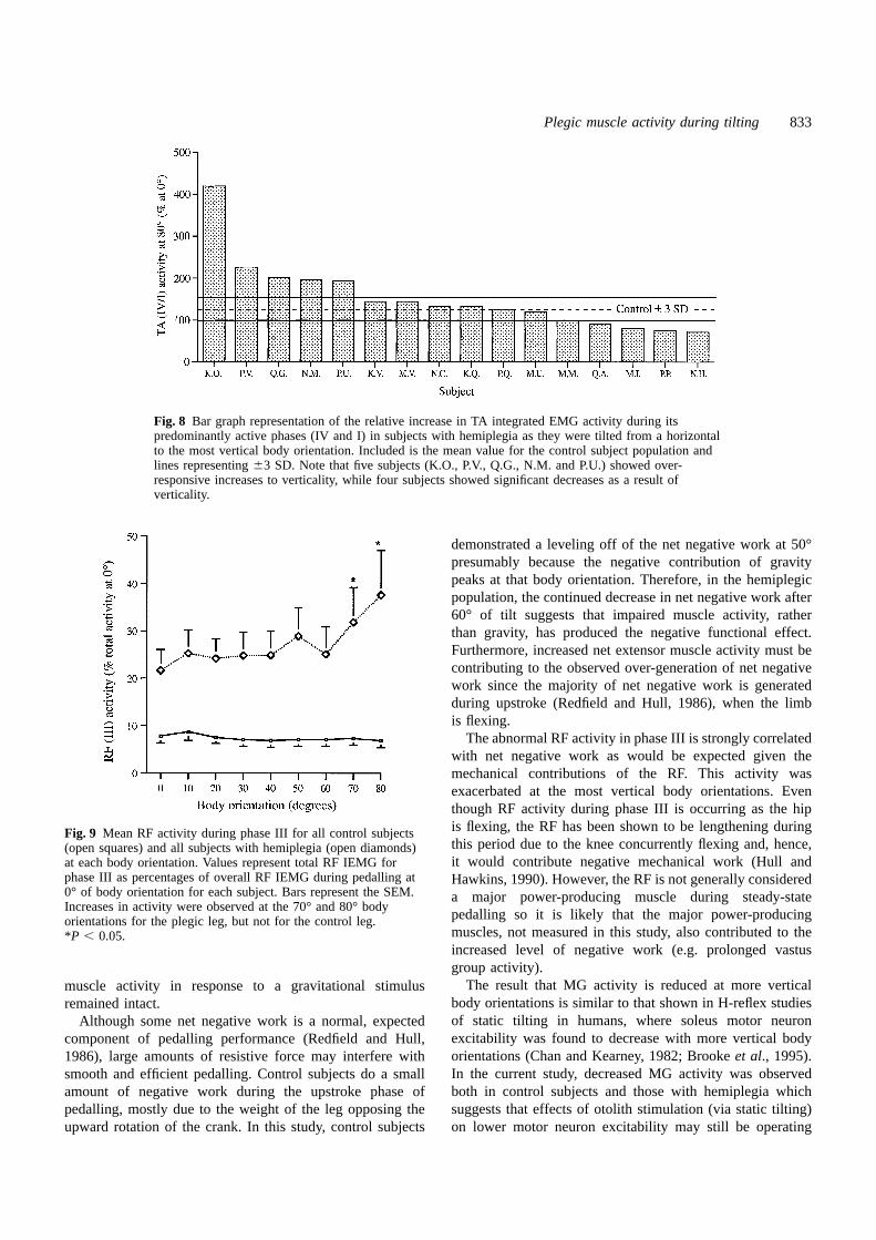
The abnormal RF activity in phase III is strongly correlatedwith net negative work as would be expected given themechanical contributions of the RF. This activity wasexacerbated at the most vertical body orientations. Eventhough RF activity during phase III is occurring as the hipis flexing, the RF has been shown to be lengthening duringFig. 9 Mean RF activity during phase III for all control subjectsthis period due to the knee concurrently flexing and, hence,(open squares) and all subjects with hemiplegia (open diamonds)it would contribute negative mechanical work (Hull andat each body orientation. Values represent total RF IEMG for
phase III as percentages of overall RF IEMG during pedalling at Hawkins, 1990). However, the RF is not generally considered0° of body orientation for each subject. Bars represent the SEM. a major power-producing muscle during steady-stateIncreases in activity were observed at the 70° and 80° body
pedalling so it is likely that the major power-producingorientations for the plegic leg, but not for the control leg.muscles, not measured in this study, also contributed to the*P , 0.05.increased level of negative work (e.g. prolonged vastusgroup activity).
The result that MG activity is reduced at more verticalmuscle activity in response to a gravitational stimulusbody orientations is similar to that shown in H-reflex studiesremained intact.of static tilting in humans, where soleus motor neuronAlthough some net negative work is a normal, expectedexcitability was found to decrease with more vertical bodycomponent of pedalling performance (Redfield and Hull,orientations (Chan and Kearney, 1982; Brookeet al., 1995).1986), large amounts of resistive force may interfere withIn the current study, decreased MG activity was observedsmooth and efficient pedalling. Control subjects do a smallboth in control subjects and those with hemiplegia whichamount of negative work during the upstroke phase ofsuggests that effects of otolith stimulation (via static tilting)pedalling, mostly due to the weight of the leg opposing the
upward rotation of the crank. In this study, control subjects on lower motor neuron excitability may still be operating

834 D. A. Brownet al.
Fig. 10 Mean change in RF activity during phase III for each individual subject with hemiplegia at themost vertical (either 70° or 80°) body orientation when compared with its value at horizontalorientation. Values represent total RF IEMG for phase III at 70o or 80o as percentages of phase III RFIEMG occurring at the 0° body orientation. Twelve out of 16 subjects demonstrated some increase inactivity at the most vertical body orientation when compared with mean63 SD in control subjects. Onesubject was unable to obtain either the 70° or 80° body orientation.
after cerebral damage, even during movement tasks. Although our experimental set up since bilateral pedalling allowsflexion movements to occur even though flexor muscleresponses to static tilting in TA muscles have not been studied
because of the inherent difficulties associated with eliciting activity may be low and overwhelmed by extensor activity.Since subjects with vestibular response deficits werean H-reflex in this muscle group (Teasdallet al., 1951), the
strong reciprocal activation pathways between the TA and excluded from this study, we may suppose that thevestibulospinal system remained intact, thus allowing tonicMG motor neuron pools (Tanaka, 1974) suggest that the
increase in TA activity at more vertical body orientations influences to remain operational. Otolith contributions topostural control in humans are difficult to demonstrate but,should be expected. Although the strong increase in TA
activity with verticality observed in control subjects is in one study, has been shown to have strong gravito-inertialinfluences on lower extremity motor-neuron pools (Frieset al.,expected and has been reported in an earlier paper (Brown
et al., 1996a), some subjects with hemiplegia (Fig. 8) showed 1993). These influences appear to increase the excitability ofanti-gravity muscles with changing posture in a systematicgreater than expected increases that are consistent with
observations of released inhibition, by higher centres, of TA manner. Also, the vestibulospinal tract has been shown toexert a strong facilitatory influence over flexor reflex afferentmotor-neuron excitability in this population (Yanagisawa
et al., 1976). Yet other subjects with hemiplegia showed a pathways (Bruggencateet al., 1969) that may be participatingin the reciprocal flexor/extensor muscle activation duringgreater than expected decrease in TA activity, possibly as a
result of an inability to recruit motor units in response to pedalling (Brown and Kukulka, 1993). Flexor reflex afferentpathways are known to be released from inhibition in personsa gravitational stimulus. Similar to the TA muscle, the RF
muscle activity, also present during limb flexion, is increased with spasticity (Meincket al., 1985). This release, in additionto an increased afferent flow that can accompany uprightwith verticality, although the increase occurs during a period
in the pedalling cycle which is considered pathological posture (e.g. increased seat and foot pressure) can contributeto some of the exaggerated responses in proximal musculature(contributing to negative work). However, the BF muscle,
shifting its timing of activity more toward the extension that occurred with increased verticality.Contrary to expectations, the hemiplegic population in thisphases of pedalling, indicates an increase in extensor bias
with increased verticality. study were able to increase net positive work output andsystematically change much of the abnormal muscle activityDuring vertical pedalling, with the exception of the MG
muscle, a general picture emerges of enhanced extensor in response to increased verticality. Control subjectsmaintained the percentage of work done by the dominant legactivity during the downstroke phase of pedalling, and
enhanced flexor activity during the upstroke phase of at all body orientations by systematically increasing thepositive work done as the negative contribution from gravitypedalling. Indeed, the results from this study show an overall
increase in muscle activity results from verticality in persons similarly increased. Subjects with hemiplegia also maintainedthe percent work done by the plegic limb. However, theywith hemiplegia (see Fig. 7). This non-biased increase in
activity is contrary to the extensor bias that is expected with also had an exaggerated concomitant increase in both positiveand negative work at the most vertical body orientations.lesions of the corticospinal tract (Lawrence and Kuypers,
1968a, b). The expression of flexor bias may be unique to The plegic leg was already performing a reduced level of

Plegic muscle activity during tilting 835
Fig. 11 Mean activity during the two contiguous phases with greatest total activity (based on controlsubjects), for all control subjects (open squares) and all subjects with hemiplegia (open diamonds) ateach body orientation. Values represent total IEMG for these phases as percentages of overall IEMGoccurring at 0° of body orientation for each muscle. Bars represent the SEM. Asterisks represent valuesthat were different when compared with 0° using apost hocTukey comparison with aP , 0.05corrected for multiple comparisons. In the cases of TA, MG, and RF activity, the changes in plegic legsparalleled the changes in control legs. However, BF muscle activity was altered at vertical bodyorientations for plegic legs, but not for control legs.
positive work, so the exaggerated increase in positive work overall excitability in flexors and extensors of persons withhemiplegia can be used therapeutically to grade the level ofrepresented additional force-generation capacity that was not
used at more horizontal orientations. activity during exercise, as well as to affect the relativebalance of activity between ankle flexors and ankle extensors.The biomechanics of tilted pedalling dictate that increased
extensor activity is necessary during the downstroke in more For example, if the goal of therapy is to minimize overallmuscle tone during movement, then pedalling can be practisedvertical postures to overcome the increased torque produced
by the weight of the opposite limb during its upstroke (Brown in less vertical body orientations. However, if the goal oftherapy is to increase muscle force generation, then moreet al., 1996a). With hemiplegia, extensor tone may already
dominate the control of the pedalling task and, if prolonged vertical body orientations would be indicated. Also, the morevertical orientations would enable preferential excitation ofinto the upstroke, may be responsible for the increased
negative work that is exacerbated by verticality. Therefore, TA muscle activity over MG muscle activity, thereby reducingthe frequently observed coupling of weak TA tone withincreased extensor tone with verticality could be a major
contributor to the parallel increase in both net positive and strong MG tone. Concurrent studies of the therapeutic effectsof gravitational stimuli on the recovery of motor function afternet negative work done.
The observation that verticality results in an increased stroke have yielded promising results (Brownet al., 1994).

836 D. A. Brownet al.
Fig. 12 The EMG, averaged, rectified and smoothed with a 9-ms moving average window, from TA, MG, RF and BF muscle groups atthe 0° (continuous line) and 80° (broken line) body orientations. Left panels represent average curves generated from all control subjects.Right panels represent average curves from N.M., a representative subject with hemiplegia. EMG profiles for all plegic musclesdemonstrate systematic changes in the timing and amplitude as a function of body orientation, with N.M. also showing pathological earlyRF muscle onset and early BF muscle onset.
mechanisms in health and disease. New York: Raven Press, 1983:Acknowledgements1035–46.The authors wish to acknowledge the assistance of Drs Felix
E. Zajac and Kevin McGill for their editorial contributions Bobath B. Adult hemiplegia: evaluation and treatment. London:to this paper. This work was funded by core funds from theHeinemann Medical Books, 1978.Department of Veterans Affairs, Rehabilitation Research and
Boorman G, Becker WJ, Morrice BL, Lee RG. Modulation of theDevelopment Division.soleus H-Reflex during pedalling in normal humans and in patientswith spinal spasticity. J Neurol Neurosurg Psychiatry 1992; 55:1150–6.
ReferencesBenecke R, Conrad B, Meinck HM, Hohne J. Electromyographic Brooke JD, Cheng J, Misiaszek JE, Lafferty K. Amplitude
modulation of the soleus H-reflex in the human during active andanalysis of bicycling on an ergometer for evaluation of spasticityof lower limbs in man. In: Desmedt JE, editor. Motor control passive stepping movements. J Neurophysiol 1995; 73: 102–11.

Plegic muscle activity during tilting 837
Brown DA, DeBacher GA. Bicycle ergometer and Hoy MG, Zernicke RF. Modulation of limb dynamics in the swingphase of locomotion. J Biomech 1985; 18: 49–60.electromyographic feedback for treatment of muscle imbalance in
patients with spastic hemiparesis. Phys Ther 1987; 67: 1715–9.Hull ML, Hawkins DA. Analysis of muscular work inmultisegmental movements: application to cycling. In: Winters JM,Brown DA, Kukulka CG. Human flexor reflex modulation duringWoo SL-Y, editors. Multiple muscle systems: Biomechanics andcycling. J Neurophysiol 1993; 69: 1212–24.movement organization. New York: Springer-Verlag, 1990: 621–38.
Brown DA, Burgar CG, Kautz SA, Dairaghi CA, Dunn-GabrielliKautz SA, Hull ML. A theoretical basis for interpreting the forceS. Improving lower extremity force symmetry in individuals withapplied to the pedal in cycling. J Biomech 1993; 26: 155–65.hemiplegia. In: Taguchi K, Igarashi M, Mori S, editors. Vestibular
and neural front. Amsterdam: Elsevier, 1994: 263–6. Kleinbaum DG, Kupper LL, Muller KE. Applied regression analysisand other multivariate methods. 2d ed. Boston (MA): PWS-Kent,Brown DA, Kautz SA, Dairaghi CA. Muscle activity patterns altered1987.during pedaling at different body orientations. J Biomech 1996a;
29: 1349–56. Lawrence DG, Kuypers HG. The functional organization of themotor system in the monkey. I. The effects of bilateral pyramidalBrown DA, Kautz SA, Dairaghi CA. Prolonged muscle activitylesions. Brain 1968a; 91: 1–14.versus premature onset during pedaling in persons with post-
stroke hemiplegia. Society for Neuroscience 26th Annual Meeting.Lawrence DG, Kuypers Hg. The functional organization of the1996b: 2041. motor system in the monkey. II. The effects of lesions of the
descending brainstem pathways. Brain 1968b; 91: 15–36.Bruggencate G ten, Burke R, Lundberg A, Udo M. Interactionbetween the vestibulospinal tract, contralateral flexor reflex afferentsMagnus R. Cameron prize lectures on some results of studies inand Ia afferents. Brain Res 1969; 14: 529–32. the physiology of posture. Lancet 1926; 2: 531–6, 585–8.
Brunnstrom S. Movement therapy in hemiplegia: a McMahon TA. Muscles, reflexes, and locomotion.Princeton (NJ):neurophysiological approach. Hagerstown: Harper and Row, 1970.Princeton University Press, 1984.
Capaday C, Stein RB. Difference in the amplitude of the humanMeinck HM, Benecke R, Conrad B. Spasticity and the flexor reflex.soleus H reflex during walking and running. J Physiol (Lond) 1987;In: Delwaide PJ, Young RR, editors. Clinical neurophysiology in392: 513–22. spasticity. Amsterdam: Elsevier, 1985: 41–54.
Newmiller J, Hull ML, Zajac FE. A mechanically decoupled twoCarr JH, Shepherd RB. A motor relearning programme for troke.force component bicycle pedal dynanometer. J Biomech 1988; 21:2nd ed. London: Heinemann, 1987.375–86.
Chan C, Kearney R. Influence of static tilt on soleus motoneuronRedfield R, Hull ML. Prediction of pedal forces in bicycling usingexcitability. Neurosci Lett 1982; 33: 333–8.optimization methods. J Biomech 1986; 19: 523–40.
Davis BL, Cavanagh PR. Simulating reduced gravity: a review ofRosecrance JC, Giuliani CA. Kinematic analysis of lower-limbbiomechanical issues pertaining to human locomotion. [Review].movement during ergometer pedaling in hemiplegic andAviat Space Environ Med 1993; 64: 557–66.nonhemiplegic subjects. Phys Ther 1991; 71: 334–43.
Duncan PW, Propst M, Nelson SG. Reliability of the Fugl-MeyerTanaka R. Reciprocal Ia inhibition during voluntary movements inassessment of sensorimotor recovery following cerebrovascularman. Exp Brain Res 1974; 21: 529–40.accident. Phys Ther 1983; 63: 1606–10.
Teasdall RD, Park AM, Porter WE, Magladery JW.Duncan PW, Goldstein LB, Matchar D, Divine GW, Feussner J.Electrophysiological studies of nerve and reflex activity in normalMeasurement of motor recovery after stroke. Outcome assessmentman. VI. Excitation and inhibition of 2-neuron reflexes by impulsesand sample size requirements. Stroke 1992; 23: 1084–9.in other nerves. Bull Johns Hopkins Hosp 1951; 88: 538–48.
Fries W, Dieterich M, Brandt T. Otolith contributions to posturalYanagisawa N, Tanaka R, Ito Z. Reciprocal Ia inhibition in spasticcontrol in man: short latency motor responses following soundhemiplegia of man. Brain 1976; 99: 555–74.stimulation in a case of otolithic Tullio phenomenon. Gait Posture
1993; 1: 145–53. Young LR. Perception of the body in space: mechanisms. In:Brookhart JM, Mountcastle VB, Darian-Smith, editors. HandbookFugl-Meyer AR, Jaasko L, Leyman I, Olsson S, Seglind S. Theof physiology, Sect 1, Vol 3, Pt 2. Bethesda (MD): Americanpost-stroke hemiplegic patient. 1. A method for evaluation ofPhysiological Society, 1984: 1023–66.physical performance. Scand J Rehabil Med 1975; 7: 13–31.
Gregor RJ, Broker JP, Ryan MM. The biomechanics of cycling.[Review]. Exerc Sports Sci Rev 1991; 19: 127–69. Received November 8, 1996. Accepted December 12, 1996